
Submitted:
27 August 2026
Posted:
27 August 2026
You are already at the latest version
Abstract
Background/Objective Krüppel-like factor 5 (KLF5) as a member of the zinc finger protein family has been reported in hepatocellular carcinoma (HCC). However, the KLF5 as a novel biomarker for patients with chronic liver diseases (CLD) remains to be identified. This study investigated the clinical values of KLF5 in CLD malignant transformation. Methods KLF5 transcripts from the TCGA database were analyzed with functions and related signaling pathways. Under the ethics committee consent, microarrays were constructed from HCC and noncancerous tissues. KLF5 distribution and expression were analyzed by multiplex immunofluorescence or Western blotting. Bloods were collected from a cohort of cases with CLD. Serum KLF5 levels were quantitatively detected by an enzyme-linked immune-sorbent assay. Results KLF5 at mRNA levels were signifi-cantly upregulating expressed (P<0.001) in HCC tissues more than these in normal livers from the TCGA database. Strong KLF5 expressions were verified in human HCC cell lines or tissues. Clini-copathological features of high KLF5 levels were remarked related (P<0.001) to tumor size, AFP level, HBV infection, tumor/node/metastasis stage and overall survival rate. Furthermore, the in-cidence of KLF5 in the HCC group were significantly higher (P<0.001) more than those in cases with liver cirrhosis or chronic hepatitis. Also, KLF5 was an independent prognostic factor for HCC. Interestingly, the co-expressions of KLF5 with Wnt3a promoted CLD malignant transfor-mation. Conclusion Upregulated KLF5 was associated with CLD malignancy and could be as a promising diagnostic or prognostic biomarker for HCC.
Keywords:
Krüppel-like transcription factor 5
; chronic liver diseases
; biomarker
; signaling pathway
; multi-plex immunofluorescence
; malignant transformation
1. Introduction
The prevention, early diagnosis and precise treatment of hepatocellular carcinoma (HCC) are still challenging problems worldwide [1]. HCC is associated with hepatitis B or C virus (HBV or HCV) persistent infection [2,3], chemical carcinogens, and metabolic dysfunction-associated fatty liver disease (MAFLD) [4] and other factors. Malignant transformation of hepatocytes is induced by the activation of oncogenes, inactivation of anti-oncogenes, and reactivation of certain oncogenes normally only expressed during the embryonic stage, which leads to a variety of specific marker expressions that are subsequently secreted into the blood, including Krüppel-like transcription factors (KLFs) [5]. Recently, KLFs were shown to control essential cellular processes and various cancer-relevant processes [6]. The zinc finger domain can bind to target DNA and has the ability to bind to deacetylases, which can activate or inhibit the target gene expressions and regulate various pathways related to development, metabolism and other cellular processes [7,8]. For example, intestinal-enriched KLF5 has been found to be involved in regulating the proliferation, differentiation, invasion and metastasis of cancer cells, with potential applications in diagnosis, prognosis and therapy [9].
As a transcriptional activator, KLF5 can regulate gene transcription, cell cycle, cell proliferation and differentiation and plays a pivotal role in the regulating cancer stem-like cells [10] and the promotion of cell growth and metastasis by activating the PI3K/AKT/Snail pathway in HCC [11]. Aberrant KLF5 expression is associated with the malignancy of chronic liver diseases (CLD) via direct or indirect effects at the transcriptional or posttranslational level [12]. However, the role of oncogenic KLF5 as a novel biomarker and its function in HCC progression remains to be identified [13]. In this study, the KLF5 expression in HCC tissues or sera from cases with CLD was further investigated to analyze clinicopathological characteristics and the values of KLF5 for HCC diagnosis and prognosis. Moreover, the biological function of KLF5 and related signaling pathways were verified using data from a bioinformatics database.
2. Materials and Methods
2.1. Bioinformatics Analysis
The Cancer Genome Atlas (TCGA, https://www.cancer.gov) [14] contains the data on crucial interactive and customizable functions, such as differential KLF5 transcripts in HCC and adjacent tissues, patient survival and correlation. Download the KLF5 mRNA transcript information from the TCGA-LIHC dataset (371 HCC and 50 adjacent non-cancerous tissue as normal samples), use the R software to normalize the data, and analyze the expressing differences of KLF mRNA between HCC and normal tissues. KLF5-related pathways based on Biocarta (https://cn.bing.com/dict/biocarta) [15] or Enrichment of KLF5-related protein acylation and regulation of RNA splicing by GO analysis (http://geneontology.org) [16].
2.2. Liver Tissues and Clinical Data
This study was approved by the Ethics Committee (TDFY2019-L006) of the Affiliated Hospital of Nantong University, China, from Mar 2019 to Dec 2025, and prior written informed consent was obtained from the HCC patients according to the Declaration of Helsinki of the World Medical Association. In all, 132 pairs of HCC and their para-noncancerous tissues (para-HCC, 2 cm away from cancer) were collected from cases after surgery, frozen in liquid nitrogen. HCC patients included 111 males and 21 females aged 25 ~ 67 years (average age, 48.1 ± 12.9 years), 120 cases positive for HBV surface antigen (HBsAg), 106 had a tumor size ≥ 3.0 cm, 104 had an AFP level ≥ 25 ng/mL, and 73 stage III-IV disease by International Union Against Cancer (IUAC) tumor-node-metastasis (TNM) classification. Livers were subjected to pathological examination, and diagnosis of HCC was based on the criteria established by the Chinese National Collaborative Cancer Research Group [17]. No case received radiation or chemotherapy prior to surgery and regularly followed-up until Dec 2025.
2.3. Blood
Blood was collected from hospitalized patients with CLD, including HCC (n = 132), chronic hepatitis (CH, n = 50) and liver cirrhosis (LC, n = 50), with complete follow-up data. Healthy individuals served as the normal control (NC, n = 50) group, with liver or kidney functions, blood glucose, lipid, and AFP level (less than 25 ng/mL) were within the normal reference range. Separated sera was stored at -85℃.
2.4. Human HCC Cell Lines
Human HCC cell lines (MHCC-97L, MHCC-97H, HCCLM3, HepG2, BEL-7404, SMMC-7721 and Hep3B) and LO2 cells were purchased from the Shanghai Institute of Cells, Chinese Academy of Sciences.
2.5. Tissue Microarrays (TMAs)
The TMAs were generated by the Department of Pathology, Affiliated Hospital of Nantong University, China, and contained formalin-fixed paraffin-embedded specimens from 85 HCC tissues and corresponding para-cancerous tissues. Tissue cores (1.5 mm) from representative areas were sectioned and placed on slides.
2.6. Multiplex Immunofluorescence (MIF)
MIF staining was performed with an OPAL IHC kit from PerkinElmer (ABSIN, Shanghai, CN). Steps of multiplex IHC were followed consecutively for each marker: blocking was performed with antibody diluent, followed by incubation with the first antibody for 1 h, detection using an OPAL Polymer HRP antibody (Waltham, MA, USA), and visualization using OPAL tyramide signal amplification plus agent, after which the section was placed in EDTA buffer (pH 8.0) and heated in a microwave. Primary antibodies included against KLF5, Wnt3a and TUFT1 (Abcam, UK, 1:1,000). In the first set of assays, KLF5, Wnt3a and TUFT1 were identified, with anti-KLF5 (red), anti-Wnt3a (green) and anti-TUFT1 (pink). After staining, the specimens were washed and then sealed with glycerin at 25 ℃ in the dark. Negative controls omitted primary antibodies. KLF5 expression was calculated with Image-Pro Plus 6.0 software (Media Cybernetics, Rockville, MD, USA). Blinded evaluated staining were simultaneously or independently observed by two pathologists, and scores were calculated based on staining intensities and positive cells.
2.7. Analysis of KLF5 Staining
Intensity of KLF5 expression was divided into the following 4 categories [18]: negative (-, score of 0); weak positive (+, 1); moderate (++, 2); and strong positive (+++, 3), with low expression (0 ~ 1 score) and high expression (2~3 scores). Images were acquired under a light microscope with a 40 × objective lens (Olympus, Japan). KLF5 expression in TMA sections was measured by Vectra 3.0 automatic quantitative pathology imaging system. Finally, INFORM software was used to score the stains [19]. Sections were observed and imaged under a microscope and were analyzed by ImagePro Plus v6.0 software, which also calculated the integral optical density (IOD) value [20].
2.8. Western Blotting
Total protein from liver tissues was extracted and quantified by a Bicinchoninic Acid Protein Assay Kit (Biyuntian Biotech. Co., Ltd., CN). The stained samples and markers were into separation gel, voltage at 120 V on electrophoresis. Then the proteins were transferred onto polyvinylidene fluoride membranes, with slowly blocked in 5% skim milk in Tris-Buffered Saline Tween 20 solution at 25 ℃ for 2 h, and was then incubated with KLF5 (Abcam, UK, 1: 1,000) and glyceraldehyde-3-phosphate dehydrogenase (GAPDH) antibodies (Abcam, UK) overnight at 4 ℃. Next, the membrane was incubated with their secondary antibodies for 2 h at 25 ℃. Solution A and solution B were then mixed at a 1:1 ratio, and the mixture was evenly added to the film in the dark, which was incubated for 2 min.
2.9. Enzyme-Linked Immunosorbent Assay (ELISA)
Serum KLF5 (ng/mL) and liver KLF5 specific concentrations (ng/mg wet tissue) were quantified according to the manual of the human KLF5 ELISA kit (Qiming Biotech. Co., Shanghai, CN). The average absorbance (A) at 450 nm (n = 3) was measured for the standards, controls and test samples. KLF5 levels were calculated based on the corresponding standard curve.
2.10. Statistical Analysis
Data were expressed as mean ± standard deviation (M ± SD) and analyzed by GraphPad Prism 8.0 software and SPSS statistical package (Version 25.0) using a two-tailed Student’s t test and χ2 test. Multiple comparisons were used by ANOVA followed by the q test. The curves of overall survival (OS) were calculated by the Kaplan-Meier (K-M) method with log-rank test. A P < 0.05 indicated statistical significance.
3. Results
3.1. KLF5 in Bioinformatics Database
The KLF5 mRNA expressions in the HCC group and the non-cancerous (normal) group from the TCGA database are shown in Figure 1. According to the TCGA data, the expressing level of KLF5mRNA in the LIHC group (n = 371) was significantly higher (P < 0.001) than that in the normal liver group (n = 50, Figure 1A). The average levels of KLF5mRNA in the HCC tissues were over-transcribed more than that in normal tissues (P < 0.001, Figure 1B). K-M survival curve of KLF5mRNA in IHC tissues was the remarked lower survival time (Figure 1C). Based on enrichment analysis, the high levels of KLF5mRNA transcripts in LIHC tissues were related to Wnt (Figure 1D), MAPK (Figure 1E), and FAS (Figure 1F) signaling pathways. Also, the GO analysis of these transcript data revealed that the high KLF5mRNA were related to the protein acylation (Figure 1G) and the regulation of RNA splicing (Figure 1H) in LIHC tissues. The data from online databases indicated that hepatic KLF5 should be over-expressing statues and promote HCC progress via multiple signaling pathways, protein acylation or RNA splicing and so on.
3.2. Upregulated KLF5 Expressions in HCC Tissues
Comparative analysis of KLF5 expressions between HCC and their para-HCC tissues are shown in Figure 2. Immunofluorescence analysis of KLF5 expression revealed that hepatic KLF5 (red granules) were mainly located in the cell membrane, cytoplasm and nucleus in HCC tissues (Figure 2A), with light staining observed in the para-HCC tissues (Figure 2B). Significantly upregulated KLF5 levels (P < 0.001, Figure 2C) were confirmed in the HCC group not in the para-HCC group by the analysis of the integral optic density (IOD) or by Western blotting (0.546 ± 0.308 vs. 0.231 ± 0.241, t = 11.234, P < 0.001, Figure 2D). Also, the immunohistochemical analysis of KLF5 expression in para-HCC (Figure 2E) and HCC tissues (Figure 2F) are summarized in Table 1. The positive percentage of KLF5 in the HCC group was 90.9% (120 of 132), which was significantly greater (χ2 = 72.310, P < 0.001) more than the corresponding percentage in the para-HCC group (38.6%, 51 of 132). The percentage of cases with high KLF5 expression (2 ~ 3 sores) was 72.0% (95 of 132) in the HCC group and low KLF5 expression (0 ~ sores) was 15.2% (20 of 132) in the para-HCC group (Z = 11.341, P < 0.001). Additionally, liver KLF5 specific concentrations (ng/mg wet tissue) was remarked high (P < 0.001) in HCC more than that in their para-HCC tissues (Figure 2G). High KLF5 expression in HCC tissues was the remarked lower survival time (Figure 2H) by the K-M survival curve.
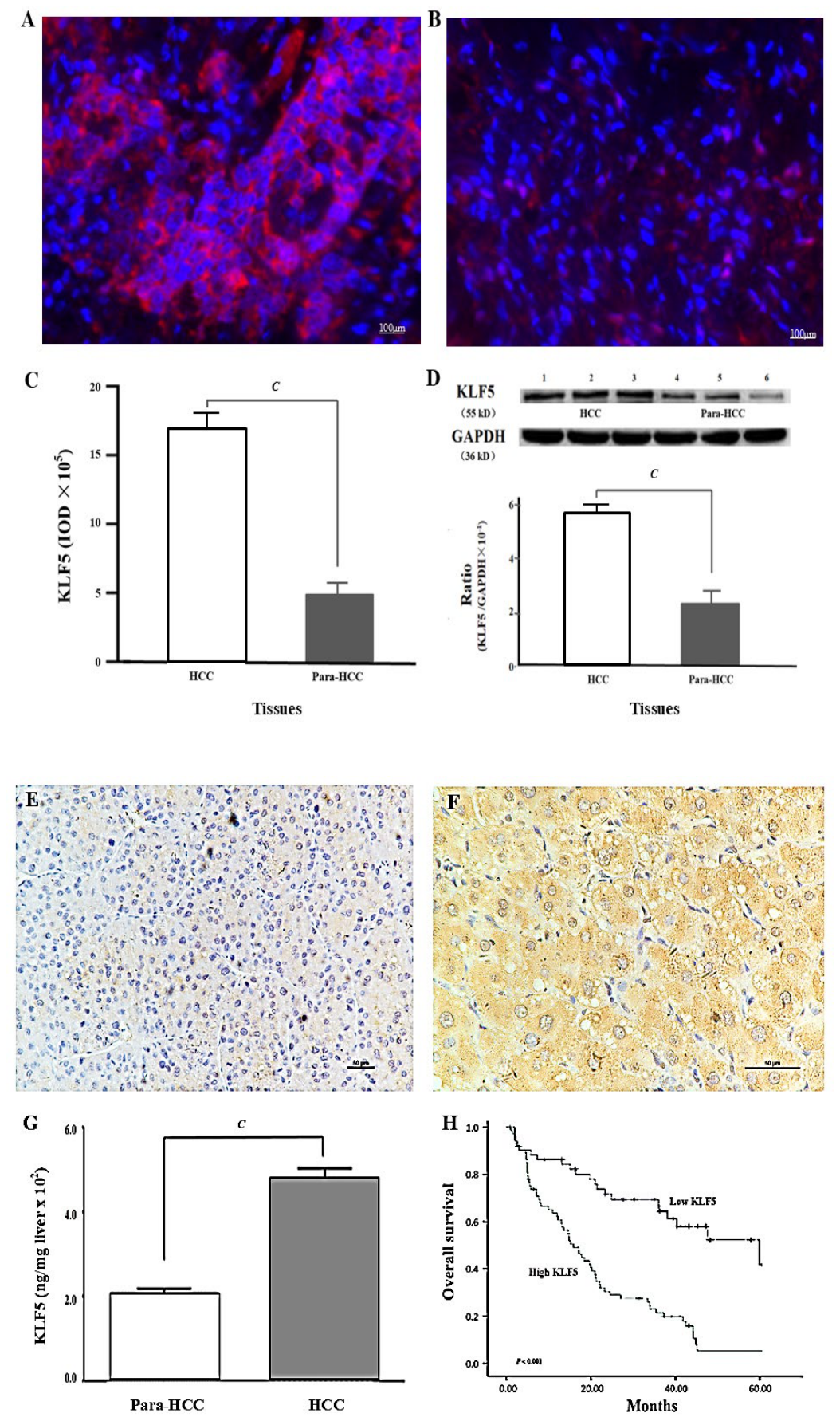
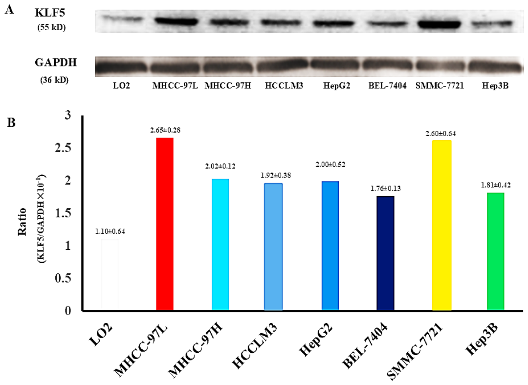
Figure 2.
Comparation analysis of KLF5 expressions in HCC tissues. (A) KLF5 immuno-fluorescence in HCC tissues (n = 132). (B) KLF5 immunofluorescence in para-HCC tissues (n = 132). (C) Integral optic density (IOD) of KLF5 immunofluorescence. (D) KLF5 expression by Western blotting (Up) and the relative ratio from KLF5 to GAPDH (Down). (E) Immunohisto-chemical analysis of KLF5 in para-HCC tissues (n = 132). (F) KLF5 immunohistochemistry in HCC tissues (n = 132). (G) KLF5 specific concentration (ng/mg wet liver) between HCC and para-HCC tissues. (H) The K-M survival curve in HCC cases with high or low KLF5 expression. HCC: hepatocellular carcinoma tissues. GAPDH, glyceraldehyde-3-phosphate dehydrogenase. Para-HCC: para-noncancerous tissues. KLF5, Krüppel-like factor-5. Yao/Sai, et al.
Figure 2.
Comparation analysis of KLF5 expressions in HCC tissues. (A) KLF5 immuno-fluorescence in HCC tissues (n = 132). (B) KLF5 immunofluorescence in para-HCC tissues (n = 132). (C) Integral optic density (IOD) of KLF5 immunofluorescence. (D) KLF5 expression by Western blotting (Up) and the relative ratio from KLF5 to GAPDH (Down). (E) Immunohisto-chemical analysis of KLF5 in para-HCC tissues (n = 132). (F) KLF5 immunohistochemistry in HCC tissues (n = 132). (G) KLF5 specific concentration (ng/mg wet liver) between HCC and para-HCC tissues. (H) The K-M survival curve in HCC cases with high or low KLF5 expression. HCC: hepatocellular carcinoma tissues. GAPDH, glyceraldehyde-3-phosphate dehydrogenase. Para-HCC: para-noncancerous tissues. KLF5, Krüppel-like factor-5. Yao/Sai, et al.
Figure 3.
KLF5 expressions among different cell lines. (A) KLF5 expression by Western blotting among LO2 cells and HCC cell lines (MHCC-97L, MHCC-97H, HCCLM3, HepG2, BEL-7404, SMMC-7721 and Hep3B). (B) The ratios from KLF5 to GAPDH among LO2 cells (n = 3) and HCC cell lines (MHCC-97L, MHCC-97H, HCCLM3, HepG2, BEL-7404, SMMC-7721 and Hep3B, n = 3/each). KLF5, Krüppel-like factor-5. Yao/Sai, et al.
Figure 3.
KLF5 expressions among different cell lines. (A) KLF5 expression by Western blotting among LO2 cells and HCC cell lines (MHCC-97L, MHCC-97H, HCCLM3, HepG2, BEL-7404, SMMC-7721 and Hep3B). (B) The ratios from KLF5 to GAPDH among LO2 cells (n = 3) and HCC cell lines (MHCC-97L, MHCC-97H, HCCLM3, HepG2, BEL-7404, SMMC-7721 and Hep3B, n = 3/each). KLF5, Krüppel-like factor-5. Yao/Sai, et al.
3.3. Clinicopathological Features of KLF5 Expression in HCC Tissues
Hepatic KLF5 overexpressed in HCC tissues correlated with major clinico-pathological features is shown in Table 2. Among the HCC patients, no significant statistical difference was observed between KLF5 expression and gender, age or differentiation degree. However, KLF5 expression was significantly correlated (P < 0.05) with AFP concentration, HCC diameter, extrahepatic metastasis, TNM stage, and HBV infection, especially HBV replication. According to the IUAC clinical staging criteria, the 132 HCC tissues were divided into Stage I (20 cases, 15.1%), Stage II (39 cases, 29.6%), Stage III (41 cases, 31.1%), and Stage IV (32 cases, 24.2%). The incidences of KLF5 expression were 35% for Stage I, 30.8% for Stage II, 73.2% for Stage III, and 53.1 for Stage IV. The high expression rate of KLF5 in Stages III-IV (76.7%) was significantly (χ2 = 5.270, P = 0001) higher than that in Stages I-II (42.4%), which suggested that high KLF5 expression should be associated with HCC progression.
3.4. Univariate/Multivariate Analysis of KLF5 in Prognosis of HCC
Univariate/multivariate analysis of the relationship between KLF5 expression and overall survival (OS) of HCC patients are shown in Table 3. Univariate analysis of upregulated KLF5 expression had significant correlation with tumor size, extrahepatic metastasis, AFP level, and advanced TNM stage. The OS of HCC cases with high KLF5 expression were significantly shorter than that of ones with low KLF5 level at stage I-II. The multivariate analysis of high KLF5 level was associated with HCC size or extrahepatic metastasis, and an independent prognostic factor for OS of HCC patients.
3.5. Circulating KLF5 Expression in CLD
The levels of serum KLF5 and AFP expression in patients with CLD are shown in Table 4. Compared with those in the NC group, the levels of serum KLF5 and AFP expression were significantly unregulated (P < 0.001) in the HCC, CH or LC groups. The cutoff value was set at 750 ng/mL (M ± 1.96 SD) for KLF5 and 25 ng/mL for AFP. The positive percentages of KLF5 and AFP were 0% (0 of 50) and 12.0% (6 of 50) for CH, 0.4% (2 of 50) and 16.0% (8 of 50) for LC, and 89.4% (118 of 132) and 61.4% (81 of 132) for HCC, respectively. The area under the receiver operating characteristic curve (ROC) was 0.876 for KLF5 and 0.760 for AFP, which suggests that KLF5 might be a useful biomarker for the diagnosis or differential diagnosis of HCC.
3.6. Potential Mechanism of KLF5 Promoting CLD Malignancy
The co-expression of KLF5 with other signals and its potential interaction in promoting CLD malignant progression are shown in Figure 4. First, KLF5 in HCC cases with high KLF5 expression is shown in red (Figure 4A), Wnt3a in green (Figure B), and the merged image of KLF5/Wnt3a (red/green, Figure 4C) overexpression. Second, more interestingly, in low KLF5 expression cases (red, Figure 4D), Wnt3a (green, Figure 4E), TUFT1 (pink, Figure 4F), and their merged image (red/green/pink, Figure 4G) showed clear co-expression in the same cell via their cellular localization, which also confirmed the above bioinformatics findings.
The above data clearly indicate that the upregulation of KLF5 expression can interact with other signaling molecules, thereby promoting the progression of CLD. Based on these, the possible mechanism by which KLF5 promotes the malignant transformation of CLD is hypothesized (Figure 4H). The transcription factors KLF5 and TUFT1 are activated HBV replication, and their interaction promotes the malignant progression of CLD by activating the Wnt3a signaling pathway or inhibiting HCC cell apoptosis.
4. Discussion
HCC is one of the most common malignant tumors in the inshore area of the Yangtze River [21]. Early diagnosis and effective treatment of HCC are crucial [22,23,24]. Hepatocarcinogenesis involves multiple genes and processes with complicated mechanisms. Recently, KLFs (total of 17 members), which comprise the largest zinc finger protein (ZNF) family in the human genome [25], were shown to be involved in the regulation of cell differentiation, embryonic development, and disease-related functional proteins in different cancers [7,9,26]. According to their biological functions, KLFs are divided into the following groups: promoting (KLF5, 7, 8 and 13), inhibiting (KLF3, 4, 9~12, 14 and 17), dual (KLF2, 6), and unknown (KLF1, 15, 16) functions. To understand the relationship between upregulated KLF5 and HCC [13,27], the intracellular localization of KLF5 in HCC tissues was determined and the serum level of KLF5 in patients with chronic liver diseases was quantitatively investigated to analyze its clinical value and explore the mechanism by which KLF5 promotes HCC progression.
Studies on the functions of KLFs in HCC have revealed additional details in this field. To date, more than 12 members of the KLF family have been reported to function in HCC pathogenesis in multiple ways [28]. Thus, knowledge of KLFs could deepen our understanding of HCC progression [29]. Aberrant KLF5 expression is involved in tumor biological activity, induces pluripotent stem cells and maintains an embryonic stem cell state. KLF5 contains a zinc finger domain that binds to target DNA and regulates not only physiological processes, such as cell proliferation, development, differentiation and embryonic development, but also the progression of multiple diseases and conditions, including HCC and inflammation [30,31]. In this study, significantly high KLF5 expression in the HCC group was associated with TNM stage, tumor size, AFP level, portal vein thrombosis, HBV infection and the 5-year survival rate of HCC patients. These findings suggest that high KLF5 expression not only promotes HCC progression but that KLF5 overexpression might also be a prognostic marker for HCC patients.
Recently, RNA sequencing confirmed that KLF5 was significantly upregulated in CD44 (high)/CD133 (high) HCC cells, and cells with high KLF5 levels have been shown to exhibit increased resistance to anticancer drugs and increased colony formation ability [10,32]. Abnormal KLF5, a soluble protein similar to AFP in tissues, can be directly secreted into circulating blood [33,34]. In this study, serum KLF5 levels were quantified in patients with chronic liver disease, and significantly high KLF5 levels were detected in HCC patients. However, the levels of both KLF5 and AFP in the serum of HCC patients were markedly higher than those in the serum of patients with chronic hepatitis or liver cirrhosis and normal controls. However, the use of KLF5 as a new tumor marker was superior to AFP in terms of specificity and sensitivity, with complementary diagnostic value for HCC, especially in patients with low AFP levels. These data indicated that KLF5 might be a novel biomarker for diagnosis or differential diagnosis of HCC and to distinguish benign and malignant liver diseases [13].
Oncogenic KLF5 is regulated by a variety of factors, and the exact mechanism of HCC progression is still being explored. KLF5 upregulation promotes HCC growth and metastasis via activation of the PI3K/AKT/Snail pathway [11] and via the induction of epithelial–mesenchymal transition [35]. Interfering with KLF5 transcription by small interfering RNA (siRNA) significantly inhibits the proliferation, apoptosis, migration and invasiveness of HCC cells [26,36]. KLF5 is negatively correlated with ncRNAs (miR-145-5p [12], miR-21 [37], miR-214-5p [38] and miR-217 [30]) that can regulate key transcription factors of CSCs. KLF5 acetylation plays the opposite role in HCC growth, as deacetylated KLF5 exhibits protumor activity, and blocking TGF-β signal attenuates the inhibitory activity of KLF5 [10,39] or activated Wnt [40], MAPK [41], and FAS [42] signaling pathways. Upregulated KLF5mRNA levels were related to the protein acylation [43] and the regulation of RNA splicing [44,45]. In this study, hepatic KLF5, Wnt3a and TUFT1 [46] were clearly expressed in the same cells, and because of their cellular localization, their co-expression or interaction might represent a novel mechanism by which KLF5 promotes CLD malignant progression [47,48].
In summary, high KLF5 transcript levels in HCC tissues were confirmed via the TCGA database, and KLF5 was found to be overexpressed by MIF or IHC analysis. Additionally, the clinicopathological features related to KLF5 overexpression were TNM stage, tumor size, AFP level, and HBV infection and were negatively correlated with the prognosis of HCC patients. Clinically, increased circulating KLF5 levels might be helpful in HCC diagnosis or in the differential diagnosis of benign and malignant liver diseases. Mechanistically, upregulated KLF5 could be co-expressed with Wnt3a and TUFT1 in the same HCC cells and might promote CLD progression. However, further basic and clinical studies are needed to determine whether KLF5 could serve as a novel diagnostic or prognostic biomarker for HCC with the exact mechanisms. Similarly, strategies to silence KLF5 transcription or Wnt3a with multitargeting strategies need investigate for effective HCC therapy.
5. Conclusions
Upregulated KLF5 expression promoted CLD malignancy and was associated with TNM stage, tumor size, AFP level, HBV infection, and negatively correlated with HCC prognosis, which might be helpful in diagnosis or prognosis of HCC.
Author Contributions
Yao Y, Sai WL and Zhang SC contributed equally to this work and wrote the first draft; Yao Y, Tang H, Zhang SC and Wu MN performed the experiments; Sai WL, Xie Q and Yao M analyzed the data; Yao M and Yao DF revised the manuscript. All authors approved the final version of the manuscript.
Funding
This study was supported by the National Natural Science Foundation of China, No. 81673241 and 32470985.
Institutional animal care and use committee statement
The study protocol was approved by the Ethics Committee of the Affiliated Hospital of Nantong University (TDFY2019-L006).
Data sharing statement
No additional unpublished data are available.
Conflict-of-interest statement
The authors declare no conflicts of interest.
Open-Access
This article is an open-access article that was selected by an in-house editor and fully peer-reviewed by external reviewers. It is distributed in accordance with the Creative Commons Attribution NonCommercial (CCBY-NC4.0) license, which permits others to distribute, remix, adapt, build upon the work noncommercially, and license their derivative works on different terms, provided the original work is properly cited and the use is noncommercial.
References
- Chen, Z.; Zhuang, H.; Zhang, J.; Zhang, Y.; Xu, S.; Zhou, Q. Chronic hepatitis B virus infection and the risk of extrahepatic malignancies: an updated review of epidemiology, mechanisms, and clinical implications. Virol. J. 2026, 23. 166. [Google Scholar] [CrossRef] [PubMed]
- Rizzo, G.E.M.; Cabibbo, G.; Craxì, A. Hepatitis B virus-associated hepatocellular carcinoma. Viruses 2022, 14. 986. [Google Scholar] [CrossRef] [PubMed]
- Wu, J.; He, J.; Xu, H. Global prevalence of occult HBV infection in children and adolescents: A systematic review and meta-analysis. Ann. Hepatol. 2024, 29, 101158. [Google Scholar] [CrossRef] [PubMed]
- Fouad, Y.; Lazarus, J.V.; Negro, F.; Peck-Radosavljevic, M.; Sarin, S.K.; Ferenci, P.; Esmat, G.; Ghazinian, H.; Nakajima, A.; Silva, M.; Lee, S.; Colombo, M. MAFLD considerations as a part of the global hepatitis C elimination effort: an international perspective. Aliment. Pharmacol. Ther. 2021, 53, 1080–1089. [Google Scholar] [CrossRef] [PubMed]
- Chen, R.; Zhao, M.; An, Y.; Liu, D.F.; Tang, Q.S.; Teng, G.J. A Prognostic Gene Signature for Hepatocellular Carcinoma. Front. Oncol. 2022, 12, 841530. [Google Scholar] [CrossRef] [PubMed]
- Jha, K.; Kumar, A.; Bhatnagar, K.; Patra, A.; Bhavesh, N. S.; Singh, B.; Chaudhary, S. Modulation of Krüppel-like factors (KLFs) interaction with their binding partners in cancers through acetylation and phosphorylation. Biochim. Biophys. Acta Gene. Regul. Mech. 2023. 1867. 1950. [Google Scholar] [CrossRef] [PubMed]
- Yuce, K.; Ozkan, A. I. The kruppel-like factor (KLF) family, diseases, and physiological events. Gene 2024, 895.148027. [Google Scholar] [CrossRef] [PubMed]
- Li, Z.Y.; Zhu, Y.X.; Chen, J.R.; Chang, X.; Xie, Z.Z. The role of KLF transcription factor in the regulation of cancer progression. Biomed. Pharmacother. 2023, 162, 114661. [Google Scholar] [CrossRef] [PubMed]
- Fang, R.; Sha, C.; Xie, Q.; Yao, D.; Yao, M. Alterations of Krüppel-like Factor Signaling and Potential Targeted Therapy for Hepatocellular Carcinoma. Anticancer Agents Med. Chem. 2025, 25, 75–85. [Google Scholar] [CrossRef] [PubMed]
- Maehara, O.; Sato, F.; Natsuizaka, M.; Asano, A.; Kubota, Y.; Itoh, J.; Tsunematsu, S.; Terashita, K.; Tsukuda, Y.; Nakai, M.; Sho, T.; Suda, G.; Morikawa, K.; Ogawa, K.; Chuma, M.; Nakagawa, K.; Ohnishi, S.; Komatsu, Y.; Whelan, K.A.; Nakagawa, H.; Takeda, H.; Sakamoto, N. A pivotal role of Krüppel-like factor 5 in regulation of cancer stem-like cells in hepatocellular carcinoma. Cancer Biol. Ther. 2015, 16, 1453–1461. [Google Scholar] [CrossRef] [PubMed]
- An, T.; Dong, T.; Zhou, H.; Chen, Y.; Zhang, J.; Zhang, Y.; Li, Z.; Yang, X. The transcription factor Krüppel-like factor 5 promotes cell growth and metastasis via activating PI3K/AKT/Snail signaling in hepatocellular carcinoma. Biochem. Biophys. Res. Commun. 2019, 508, 159–168. [Google Scholar] [CrossRef] [PubMed]
- Liang, H.; Sun, H.; Yang, J.; Yi, C. miR-145-5p reduces proliferation and migration of hepato-cellular carcinoma by targeting KLF5. Mol. Med. Rep. 2018, 17, 8332–8338. [Google Scholar] [CrossRef] [PubMed]
- Yuan, W.; Sun, Q.; Zhu, X.; Li, B.; Zou, Y.; Liu, Z. M2-polarized tumor-associated macrophage-secreted exosomal lncRNA NEAT1 upregulates galectin-3 by recruiting KLF5 and promotes HCC immune escape. J. Cell Commun. Signal. 2024, 19. e12060. [Google Scholar] [CrossRef] [PubMed]
- Sai, W.L.; Wang, L.; Sun, J.Y.; Yang, J.L.; Yao, M.; Yao, D.F. Value of abnormal expression of Krüppel-like zinc-finger protein transcription factor 5 in the diagnosis and prognosis of liver cancer. Zhonghua Gan Zang Bing. Za Zhi 2021, 29, 781–787. [Google Scholar] [CrossRef] [PubMed]
- Chang, J. T.; Lee, Y. M.; Huang, R.S. The impact of the Cancer Genome Atlas on lung cancer. Transl. Res. 2015, 166, 568–585. [Google Scholar] [CrossRef] [PubMed]
- Shen, C.; Cao, Y.; Qi, G.; Huang, J.; Liu, Z.P. Discovering pathway biomarkers of hepatocellular carcinoma occurrence and development by dynamic network entropy analysis. Gene 2023. [Google Scholar] [CrossRef] [PubMed]
- Shen, S.; Kong, J.; Qiu, Y.; Yang, X.; Wang, W.; Yan, L. Identification of core genes and outcomes in hepatocellular carcinoma by bioinformatics analysis. J. Cell Biochem. 2019, 120, 10069–10081. [Google Scholar] [CrossRef] [PubMed]
- Xie, D.Y.; Ren, Z.G.; Zhou, J.; Fan, J.; Gao, Q. 2019 Chinese clinical guidelines for the management of hepatocellular carcinoma: updates and insights. Hepatobiliary Surg. Nutr. 2020, 9, 452–463. [Google Scholar] [CrossRef] [PubMed]
- Dong, Z.; Yao, M.; Zhang, H.; Wang, L.; Huang, H.; Yan, M.; Wu, W.; Yao, D. Inhibition of Annexin A2 gene transcription is a promising molecular target for hepatoma cell proliferation and metastasis. Oncol. Lett. 2014, 7, 28–34. [Google Scholar] [CrossRef] [PubMed]
- Hernandez, S.; Rojas, F.; Laberiano, C.; Lazcano, R.; Wistuba, I.; Parra, E.R. Multiplex Immuno-fluorescence Tyramide Signal Amplification for Immune Cell Profiling of Paraffin-Embedded Tumor Tissues. Front. Mol. Biosci. 2021, 8, 667067. [Google Scholar] [CrossRef] [PubMed]
- Chen, K.J.; Jin, R.M.; Shi, C.C.; Ge, R.L.; Hu, L.; Zou, Q.F.; Cai, Q.Y.; Jin, G.Z.; Wang, K. The prognostic value of Niemann-Pick C1-like protein 1 and Niemann-Pick disease type C2 in hepatocellular carcinoma. J. Cancer 2018, 9, 556–563. [Google Scholar] [CrossRef] [PubMed]
- Chen, J.G.; Zhu, J.; Zhang, Y.H.; Chen, Y.S.; Ding, L.L.; Chen, H.Z.; Shen, A.G.; Wang, G.R. Liver Cancer Survival: A Real World Observation of 45 Years with 32,556 Cases. J. Hepatocell. Carcinoma 2021, 8, 1023–1034. [Google Scholar] [CrossRef] [PubMed]
- Ganesan, P.; Kulik, L.M. Hepatocellular Carcinoma: New Developments. Clin. Liver Dis. 2023, 27, 85–102. [Google Scholar] [CrossRef] [PubMed]
- Ghavimi, S.; Apfel, T.; Azimi, H.; Persaud, A.; Pyrsopoulos, N.T. Management and Treatment of Hepatocellular Carcinoma with Immunotherapy: A Review of Current and Future Options. J. Clin. Transl. Hepatol. 2020, 168–176. [Google Scholar] [CrossRef] [PubMed]
- Li, X.; Hu, Z.; Shi, Q.; Qiu, W.; Liu, Y.; Liu, Y.; Huang, S.; Liang, L.; Chen, Z.; He, X. Elevated choline drives KLF5-dominated transcriptional reprogramming to facilitate liver cancer progression. Oncogene 2024, 43, 3121–3136. [Google Scholar] [CrossRef] [PubMed]
- Kim, C.K.; Bialkowska, A.B.; Yang, V.W. SP and KLF transcription factors in digestive physiology and diseases. Gastroenterology 2017, 152, 1845–1875. [Google Scholar] [CrossRef] [PubMed]
- Zeng, L.; Zhu, Y.; Moreno, C.S.; Wan, Y. New insights into KLFs and SOXs in cancer pathogenesis, stemness, and therapy. Semin. Cancer Biol. 2023, 90, 29–44. [Google Scholar] [CrossRef] [PubMed]
- Li, Y.; Zhao, X.; Xu, M.; Chen, M. Krüppel-like factors in glycolipid metabolic diseases. Mol. Biol. Rep. 2022, 49, 8145–8152. [Google Scholar] [CrossRef] [PubMed]
- Jen, J.; Wang, Y.C. Zinc finger proteins in cancer progression. J. Biomed. Sci. 2016, 23. 53. [Google Scholar] [CrossRef] [PubMed]
- Ye, Q.; Liu, J.; Xie, K. Zinc finger proteins and regulation of the hallmarks of cancer. Histol. Histopathol. 2019, 34, 1097–1109. [Google Scholar] [CrossRef] [PubMed]
- Gao, W.; Lu, Y.X.; Wang, F.; Sun, J.; Bian, J.X.; Wu, H.Y. miRNA-217 inhibits proliferation of hepatocellular carcinoma cells by regulating KLF5. Eur. Rev. Med. Pharmacol. Sci. 2019, 23, 7874–7883. [Google Scholar] [CrossRef] [PubMed]
- Yerra, V.G.; Drosatos, K. Specificity Proteins (SP) and Krüppel-like Factors (KLF) in Liver Physiology and Pathology. Int. J. Mol. Sci. 2023, 24. 4682. [Google Scholar] [CrossRef] [PubMed]
- Luo, Y.; Chen, C. The roles and regulation of the KLF5 transcription factor in cancers. Cancer Sci. 2021, 112, 2097–2117. [Google Scholar] [CrossRef] [PubMed]
- Butaye, E.; Somers, N.; Grossar, L.; Pauwel, N.; Lefere, S.; Devisscher, L.; Raevens, S.; Geerts, A.; Meuris, L.; Callewaert, N.; Van; Vlierberghe, H.; Verhelst, X. Systematic review: Glycomics as diagnostic markers for hepatocellular carcinoma. Aliment Pharmacol. Ther. 2024, 59. 23–38. [Google Scholar] [CrossRef] [PubMed]
- Christou, C.; Stylianou, A.; Gkretsi, V. Midkine (MDK) in Hepatocellular Carcinoma: More than a Biomarker. Cells 2024, 13. 136. [Google Scholar] [CrossRef] [PubMed]
- Orzechowska-Licari, E.J.; LaComb, J.F.; Mojumdar, A.; Bialkowska, A.B. SP and KLF Transcription Factors in Cancer Metabolism. Int. J. Mol. Sci. 2022, 23. 9956. [Google Scholar] [CrossRef] [PubMed]
- Zhang, Y.; Yao, C.; Ju, Z.; Jiao, D.; Hu, D.; Qi, L.; Liu, S.; Wu, X.; Zhao, C. Krüppel-like factors in tumors: Key regulators and therapeutic avenues. Front. Oncol. 2023, 13, 1080720. [Google Scholar] [CrossRef] [PubMed]
- Wang, J.; Chu, Y.; Xu, M.; Zhang, X.; Zhou, Y.; Xu, M. miR-21 promotes cell migration and invasion of hepatocellular carcinoma by targeting KLF5. Oncol. Lett. 2019, 17, 2221–2227. [Google Scholar] [CrossRef] [PubMed]
- Pang, J.; Li, Z.; Wang, G.; Li, N.; Gao, Y.; Wang, S. miR-214-5p targets KLF5 and suppresses proliferation of human hepatocellular carcinoma cells. J. Cell Biochem. 2019, 120, 1850–1859. [Google Scholar] [CrossRef] [PubMed]
- Zhang, D.H.; Yin, H.D.; Li, J.J.; Wang, Y.; Yang, C.W.; Jiang, X.S.; Du, H.R.; Liu, Y.P. KLF5 regulates chicken skeletal muscle atrophy via the canonical Wnt/beta-catenin signaling pathway. Exp. Anim. 2020, 69, 430–440. [Google Scholar] [CrossRef] [PubMed]
- Kim, D.; Li, H. Y.; Lee, J. H.; Oh, Y. S.; Jun, H. S. Lysophosphatidic acid increases mesangial cell proliferation in models of diabetic nephropathy via Rac1/MAPK/KLF5 signaling. Exp. Mol. Med. 2019, 51, 1–10. [Google Scholar] [CrossRef] [PubMed]
- Ishimaru, Y.; Ijiri, D.; Shimamoto, S.; Ishitani, K.; Nojima, T.; Ohtsuka, A. Single injection of the β2-adrenergic receptor agonist, clenbuterol, into newly hatched chicks alters abdominal fat pad mass in growing birds. Gen. Comp. Endocrinol. 2015, 211. 9–13. [Google Scholar] [CrossRef] [PubMed]
- Wei, W.; Chen, W.; He, N. HDAC4 induces the development of asthma by increasing Slug-upregulated CXCL12 expression through KLF5 deacetylation. J. Transl. Med. 2021, 19. 258. [Google Scholar] [CrossRef] [PubMed]
- Wang, Z.; Yang, L.; Wu, P.; Li, X.; Tang, Y.; Ou, X.; Zhang, Y.; Xiao, X.; Wang, J.; Tang, H. The circROBO1/KLF5/FUS feedback loop regulates the liver metastasis of breast cancer by inhibiting the selective autophagy of afadin. Mol. Cancer 2022, 21. 29. [Google Scholar] [CrossRef] [PubMed]
- Liu, P.; Wang, Z.; Ou, X.; Wu, P.; Zhang, Y.; Wu, S.; Xiao, X.; Li, Y.; Ye, F.; Tang, H. The FUS/circEZH2/ KLF5/feedback loop contributes to CXCR4-induced liver metastasis of breast cancer by enhancing epithelial-mesenchymal transition. Mol. Cancer 2022, 21, 198. [Google Scholar] [CrossRef] [PubMed]
- Wu, M.N.; Zheng, W.J.; Ye, W.X.; Wang, L.; Chen, Y.; Yang, J.; Yao, D.F.; Yao, M. Oncogenic tuftelin 1 as a potential molecular-targeted for inhibiting hepatocellular carcinoma growth. World J. Gastroenterol. 2021, 27, 3327–3341. [Google Scholar] [CrossRef] [PubMed]
- Zaki, P.; Thonglert, K.; Apisarnthanarax, S.; Grassberger, C.; Bowen, S.R.; Tsai, J.; Sham, J.G.; Chiang, B.H.; Nyflot, M.J. Liver injury and recovery following radiation therapy for hepatocellular carcinoma: insights from functional liver imaging. Hepatoma Res. 2024, 10. 36. [Google Scholar] [CrossRef] [PubMed]
- Singh, A.; Anjum, B.; Naz, Q.; Raza, S.; Sinha, R.A.; Ahmad, M.K.; Mehdi, A.A.; Verma, N. Night shift-induced circadian disruption: links to initiation of non-alcoholic fatty liver disease/non-alcoholic steatohepatitis and risk of hepatic cancer. Hepatoma Res. 2024. 2024. 88. [CrossRef]
Figure 1.
Bioinformatics analysis of KLF5mRNA and related pathways in LIHC from database. (A) Comparative analysis of KLF5 mRNA between HCC (n = 371) and normal tissues (n = 50) from the TCGA database. (B) Average levels of KLF5 mRNA between HCC (n = 371) and normal tissues (n=50) from the TCGA database. (C) The Kaplan-Meier (K-M) survival curve of KLF5 mRNA in LIHC tissues. (D) Enrichment of KLF5-related Wnt pathway. (E) Enrichment of KLF5-related MAPK pathway. (F) Enrichment of KLF5-related FAS pathway. (G) Enrichment of KLF5-related protein acylation by GO analysis (http://geneontology.org). (H) Enrichment of KLF5-related regulation of RNA splicing by GO analysis. KLF5, Krüppel-like factor-5. TCGA, the cancer genome atlas. Yao/Sai, et al.
Figure 1.
Bioinformatics analysis of KLF5mRNA and related pathways in LIHC from database. (A) Comparative analysis of KLF5 mRNA between HCC (n = 371) and normal tissues (n = 50) from the TCGA database. (B) Average levels of KLF5 mRNA between HCC (n = 371) and normal tissues (n=50) from the TCGA database. (C) The Kaplan-Meier (K-M) survival curve of KLF5 mRNA in LIHC tissues. (D) Enrichment of KLF5-related Wnt pathway. (E) Enrichment of KLF5-related MAPK pathway. (F) Enrichment of KLF5-related FAS pathway. (G) Enrichment of KLF5-related protein acylation by GO analysis (http://geneontology.org). (H) Enrichment of KLF5-related regulation of RNA splicing by GO analysis. KLF5, Krüppel-like factor-5. TCGA, the cancer genome atlas. Yao/Sai, et al.
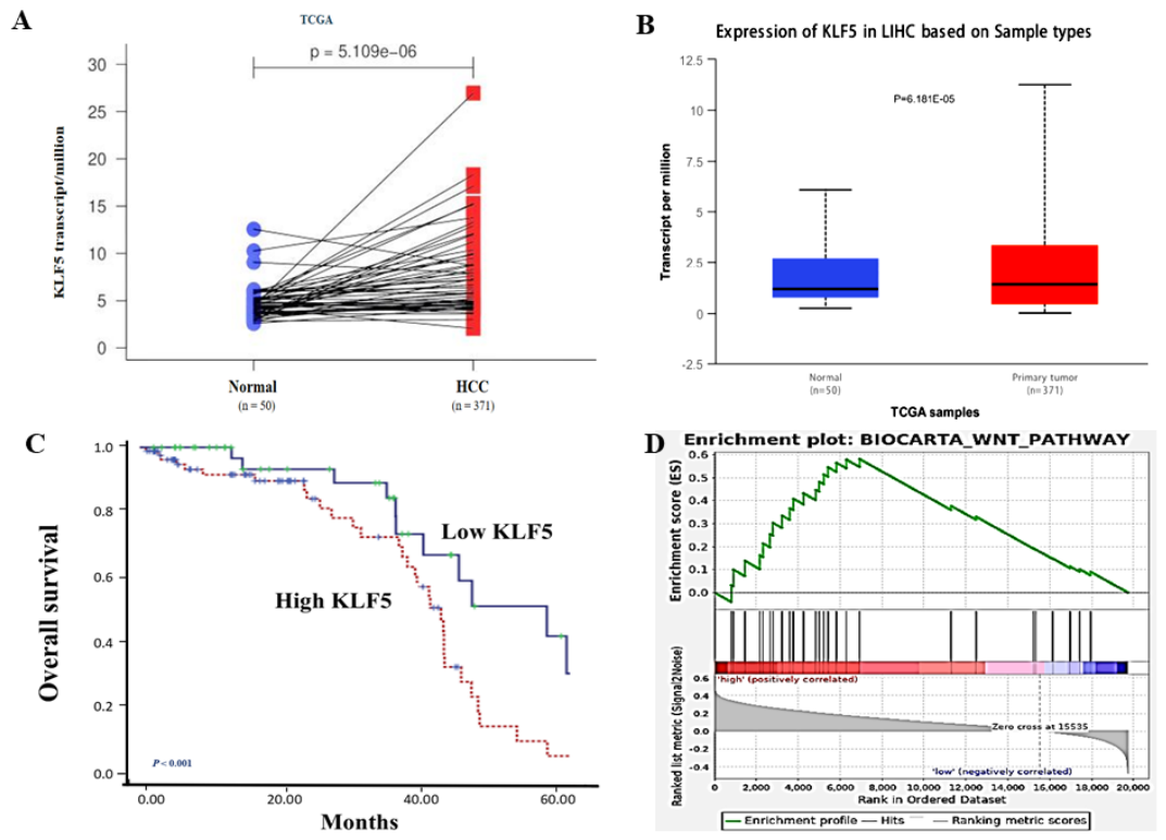
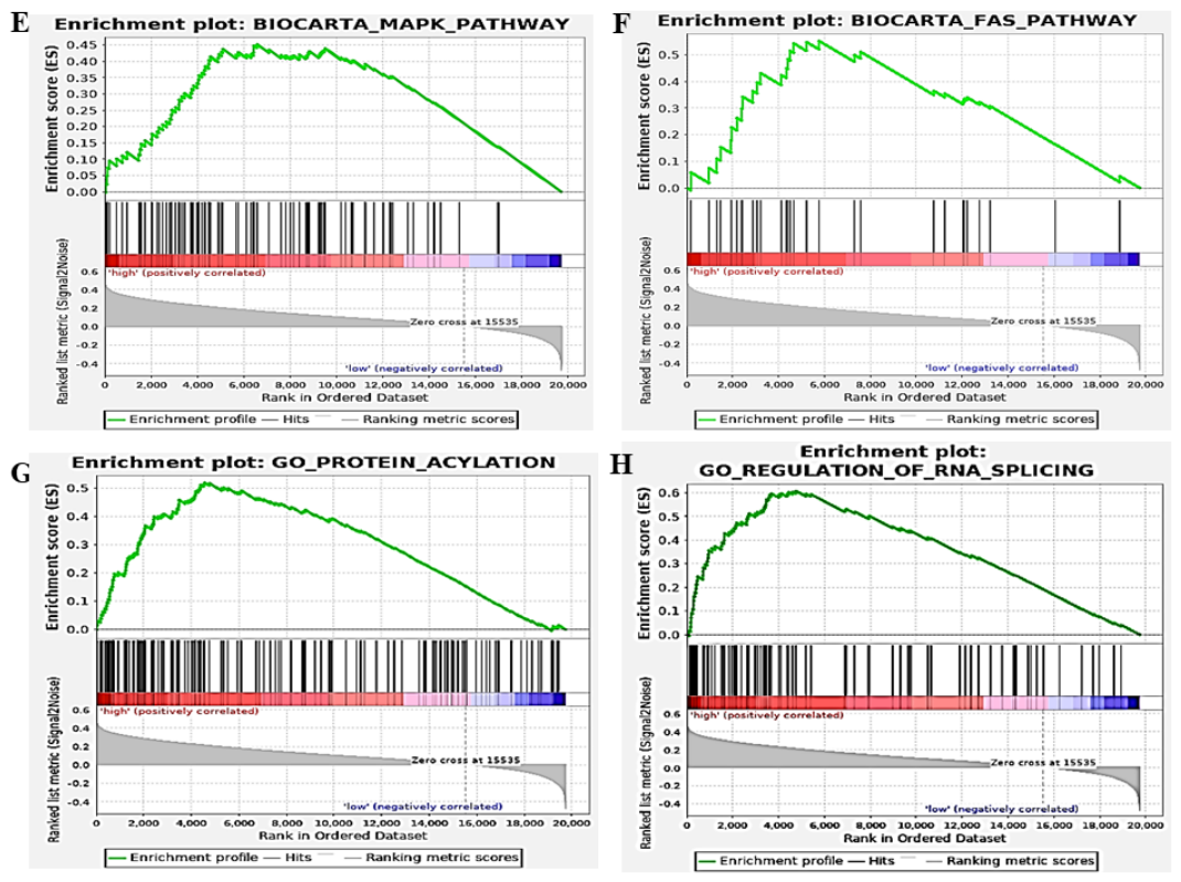
Figure 4.
Potential mechanism of KLF5 promoting CLD malignancy. (A) High KLF5 expression in HCC; (B) Wnt3a expression in HCC. (C) Merge of KLF5/Wnt3a expression in HCC. (D) low KLF5 expression in HCC tissues. (E) Wnt3a expression in HCC. (F) TUFT1 expression in HCC; (G) Merge of KLF5/Wnt3a/TUFT1 in HCC. (H) Mechanism of upregulated KLF5 interaction with other factors promoting CLD progression. AFP, alpha-fetoprotein. CLD, chronic liver diseases. IGF-II, insulin-like growth factor-II. KLF5, Krüppel-like factor 5. mtDNA, mitochondrial DNA. NF-kB, nuclear factor-Kappa B. ROS, Reactive oxygen species. TGF-B1, Transforming growth factor-beta 1. TUFT1, tuftelin1 as a transcription factor. Wnt3a, a number of Wnt signal pathway. Yao/Sai, et al.
Figure 4.
Potential mechanism of KLF5 promoting CLD malignancy. (A) High KLF5 expression in HCC; (B) Wnt3a expression in HCC. (C) Merge of KLF5/Wnt3a expression in HCC. (D) low KLF5 expression in HCC tissues. (E) Wnt3a expression in HCC. (F) TUFT1 expression in HCC; (G) Merge of KLF5/Wnt3a/TUFT1 in HCC. (H) Mechanism of upregulated KLF5 interaction with other factors promoting CLD progression. AFP, alpha-fetoprotein. CLD, chronic liver diseases. IGF-II, insulin-like growth factor-II. KLF5, Krüppel-like factor 5. mtDNA, mitochondrial DNA. NF-kB, nuclear factor-Kappa B. ROS, Reactive oxygen species. TGF-B1, Transforming growth factor-beta 1. TUFT1, tuftelin1 as a transcription factor. Wnt3a, a number of Wnt signal pathway. Yao/Sai, et al.
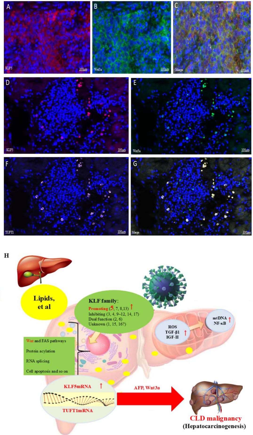

Table 1.
KLF5 incidence and expressing intensity in HCC and para-HCC groups.
| Group | n | TUFT1 |
χ2 value |
P value |
KLF5 score |
Z value |
P value |
||||
| Neg. | Pos. | 0 | 1 | 2 | 3 | ||||||
| HCC | 132 | 16 | 120 | 72.310 | <0.001 | 6 | 25 | 74 | 27 | 11.341 | < 0.001 |
| Para-HCC | 132 | 81 | 50 | 81 | 31 | 19 | 1 | ||||
HCC, hepatocellular carcinoma tissues; KLF5, Krüppel-like factor 5; Para-HCC, para-noncancerous tissues; KLF5, Krüppel-like factor 5; Neg., negative cases; Pos., positive cases.
Table 2.
Clinicopathological characteristics of KLF5 expression in HCC patients.
| Group | n | KLF5 Expression |
t value |
P value |
|
| Mean ± SD | |||||
| Age | |||||
| < 60 | 101 | 135.05 ± 68.41 | 1.886 | 0.061 | |
| ≥ 60 | 31 | 107.14 ± 74.53 | |||
| Gender | |||||
| Male | 107 | 128.23 ± 71.44 | 0.838 | 0.404 | |
| Female | 25 | 141.82 ± 58.69 | |||
| AFP (ng/mL) | |||||
| < 25 | 28 | 114.11 ± 49.05 | 2.245 | 0.021 | |
| ≥ 25 | 104 | 141.86 ± 58.36 | |||
| HBsAg | |||||
| Negative | 12 | 90.06 ± 77.83 | 2.971 | 0.002 | |
| Positive | 120 | 140.94 ± 96.45 | |||
| HBV-DNA | |||||
| Negative | 85 | 80.16 ± 72.12 | 3.587 | <0.001 | |
| Positive | 47 | 158.89 ± 94.66 | |||
| Tumor size (cm) | |||||
| <3 | 26 | 118.23 ± 59.32 | 2.257 | 0.024 | |
| ≥3 | 106 | 146.88 ± 68.74 | |||
| Degree of differentiation | |||||
| Well/moderately | 19 | 128.83 ± 69.12 | 0.904 | 0.517 | |
| Poor | 113 | 138.58 ± 89.91 | |||
| extrahepatic metastasis | |||||
| Without | 74 | 132.16 ± 60.82 | 2.026 | 0.034 | |
| With | 58 | 168.90 ± 55.56 | |||
| TNM stage | |||||
| I-II | 59 | 109.06 ± 73.44 | 3.350 | <0.001 | |
| III-IV | 73 | 147.64 ± 61.47 | |||
AFP, α-fetoprotein; HBsAg, hepatitis B virus surface antigen; HCC, hepatocellular carcinoma; KLF5, Krüppel-like factor 5; TNM, tumor/node/metastasis.
Table 3.
Univariate/multivariate analysis of KLF5 expression in HCC prognosis.
| Variable | Univariate | Multivariate | |||||
| HR | 95% CI | P >|Z| | HR | 95% CI | P >|Z| | ||
| Age, y | |||||||
| ≤ 60 vs. > 60 | 0.867 | 0.747~1.597 | 0.725 | ||||
| Gender | |||||||
| Male vs. Female | 1.415 | 0.558~2.083 | 0.987 | ||||
| KLF5 | |||||||
| Strong vs Low | 3.794 | 1.802-3.568 | <0.001 | 1.897 | 1.032-3.846 | 0.015 | |
| AFP ng/mL | |||||||
| <25 vs ≥25 | 2.102 | 1.534-4.950 | 0.018 | ||||
| HBsAg | |||||||
| Positive vs Negative | 2.786 | 0.124-0.987 | <0.001 | ||||
| Tumor size | |||||||
| <3 cm vs ≥3 cm | 1.828 | 0.496-1.864 | <0.001 | 0.351 | 0.174-0.789 | <0.001 | |
| Extrahepatic metastasis | |||||||
| With vs Without | 2.592 | 1.648-4.076 | <0.001 | 2.482 | 1.459-4.222 | 0.001 | |
| TNM stage | |||||||
| I - II vs III- IV | 1.045 | 1.187-3.609 | <0.001 | ||||
AFP, alpha-fetoprotein; HBsAg, hepatitis B virus surface antigen; HCC, hepatocellular carcinoma; KLF5, Krüppel-like factor 5; TNM, tumor/node/metastasis.
Table 4.
Diagnosis and differential diagnosis based on serum KLF5 or AFP in HCC.
| Group | n |
KLF5 (ng/mL, M ± SD) |
q Value* |
P Value |
AFP (ng/mL, M ± SD) |
q Value* |
P Value |
|
| HCC | 132 | 991.2 ± 103.8 | 39.87 | < 0.001 | 858.9 ± 821.7 | 7.298 | < 0.001 | |
| CH | 50 | 528.6 ± 51.6 | 14.42 | < 0.001 | 41.1 ± 46.2 | 4.647 | 0.005 | |
| LC | 50 | 684.8 ± 48.2 | 30.01 | < 0.001 | 63.6 ± 52.8 | 7.065 | 0.010 | |
| NC | 50 | 375.1 ± 54.8 | --- | --- | 10.4 ± 6.9 | --- | --- | |
| F Value | 892.500 | 49.460 | ||||||
| P Value | < 0.001 | < 0.001 | ||||||
AFP, alpha-fetoprotein; CH, chronic hepatitis; HBsAg, hepatitis B virus surface antigen; HCC, hepatocellular carcinoma; KLF5, Krüppel-like factor 5; LC, liver cirrhosis; NC, normal control.
Disclaimer/Publisher’s Note: The statements, opinions and data contained in all publications are solely those of the individual author(s) and contributor(s) and not of MDPI and/or the editor(s). MDPI and/or the editor(s) disclaim responsibility for any injury to people or property resulting from any ideas, methods, instructions or products referred to in the content. |
© 2026 by the authors. Licensee MDPI, Basel, Switzerland. This article is an open access article distributed under the terms and conditions of the Creative Commons Attribution (CC BY) license (http://creativecommons.org/licenses/by/4.0/).
Copyright: This open access article is published under a Creative Commons CC BY 4.0 license, which permit the free download, distribution, and reuse, provided that the author and preprint are cited in any reuse.