

Submitted:
26 August 2026
Posted:
27 August 2026
You are already at the latest version
Abstract
Purpose: Some sudden infant death syndrome (SIDS) and sudden unexpected infant death (SUID) cases remain unexplained after medicolegal investigation. The Metabolic Vulnerability Index (MVI) was proposed as a specimen-aware framework in which the total score summarizes measured multidomain findings and archetype classification organizes patterns of domain involvement. This review proposes a post-MVI framework for organizing candidate non-metabolic vulnerabilities after Stage 1 MVI classification. Methods: Literature on molecular autopsy, cardiac conduction-system pathology, concealed cardiomyopathy, epilepsy/SUDEP, autonomic or respiratory developmental disorders, and postmortem specimen constraints was synthesized into a staged eligibility framework, tiered escalation pathway, and pilot-implementation elements. Results: The framework distinguishes MVI-positive, confirmed low-MVI, and specimen-limited evaluations before routing eligible cases into a tiered pathway. Tier 1 prioritizes targeted cardiac channelopathy testing because of DNA robustness, family actionability, and relevance to autopsy-negative sudden death. Conditional Tier 2 addresses cardiac, neurologic, autonomic/respiratory-control, and structural mechanisms when supported by history, autopsy context, specimen readiness, or predefined triggers. Tier 3 is reserved for unresolved cases when research-level genomic expansion or specialized assessment is feasible and authorized. Stage 1 classification preserves indeterminate or not-evaluable results rather than treating missing findings as normal and separates exposure-context abstraction from score-eligible analytic findings and research-only measurements. Conclusions: The staged MVI/post-MVI model organizes unexplained SIDS/SUID cases into specimen-aware research profiles that integrate abnormal domain patterns, domain evaluability, exposure context, and post-MVI findings. These profiles can generate testable hypotheses about the biologic or physiologic pathways underlying candidate vulnerability. Pilot studies can establish the feasibility and reproducibility of domain collection; subsequent adequately powered studies can determine whether recurring patterns distinguish biologically meaningful subgroups within heterogeneous SIDS/SUID. The resulting classifications remain research constructs and do not constitute diagnoses or individual cause-of-death determinations.
Keywords:
Metabolic Vulnerability Index
; sudden infant death syndrome (SIDS)
; molecular autopsy
; cardiac channelopathy
; sudden unexpected infant death (SUID)
; postmortem genetic testing
; cardiac conduction system pathology
; autonomic regulation disorders
; specimen adequacy
; exposure-contextualized vulnerability
Introduction
Sudden infant death syndrome (SIDS) and sudden unexpected infant death (SUID) remain among the most challenging problems in pediatric pathology and forensic medicine [1,2]. Despite scene investigation, clinical history review, toxicology, and complete autopsy examination, many cases remain unexplained [1,2]. This persistent uncertainty likely reflects the convergence of multiple biologic pathways, including metabolic, cardiac electrical, neurologic, autonomic, and environmental contributors. Accordingly, improved postmortem evaluation requires frameworks capable of integrating diverse biologic domains rather than assessing isolated mechanisms.
Conventional postmortem evaluation of suspected SIDS/SUID emphasizes structural abnormalities, toxicologic exposures, and environmental risk factors. Although these approaches identify contributors in some cases, they often do not provide a mechanism. Increasing evidence suggests that sudden infant death may involve interactions among intrinsic vulnerability, developmental state, and external stressors, supporting structured multidomain investigation.
The Metabolic Vulnerability Index (MVI) was recently proposed as a specimen-aware interpretive framework for organizing measured vulnerability findings in sudden infant death [3]. In the present operational framework, the MVI evaluates five domains: hepatic CYP450 metabolic capacity, cytokine/inflammatory state, redox/oxidative injury, a limited medullary autonomic-network assessment based on whole-section H&E and one protocol-defined serotonergic-IHC classification and analytically supported xenobiotic/exposure findings with separate exposure-context abstraction. Domain 4 is an operationally simplified assessment without nucleus-specific IHC, named-nucleus sampling, or nucleus-by-assay or region-level scoring; it does not represent a comprehensive evaluation of medullary anatomy, serotonergic function, or respiratory/autonomic physiology. Domains 1–3 are primary vulnerability domains, whereas Domains 4 and 5 function as modifiers. Domain 3 reflects reproducible oxidative-injury findings rather than direct measurement of mitochondrial energetics. Domain 5 separates exposure-context abstraction from score-eligible analytical findings; protocol-defined research measurements remain separately reported unless prospectively validated for scoring. The total MVI score summarizes scored multidomain findings, whereas archetype classification organizes patterns of domain involvement without establishing mechanism, causal sequence, or cause of death.
The present manuscript extends the MVI framework by describing a conditional second-stage pathway primarily for confirmed low-MVI cases and selected borderline or specimen-limited cases in which non-metabolic mechanisms remain plausible. Here, “post-MVI” means structured evaluation following Stage 1 five domain MVI classification. It is an operational extension, not a replacement for the MVI. The low-MVI gateway is intended to prioritize escalation during pilot implementation, not to imply that metabolic and non-metabolic contributors are biologically mutually exclusive. MVI-positive cases may still undergo separately reported targeted post-MVI assessment when independent family-history, clinical, autopsy, or specimen-supported triggers exist.
This distinction is important because a confirmed low-MVI profile does not imply the absence of another candidate vulnerability. Confirmed low-MVI requires all five MVI domains to be scorable and within expected limits. Plausible residual pathways may include primary cardiac electrical instability, cardiac conduction-system abnormalities, concealed structural cardiac disease, epilepsy/SUDEP-associated mechanisms, or developmental abnormalities affecting respiratory or autonomic regulation. The post-MVI framework therefore introduces a tiered escalation strategy using targeted molecular autopsy, conditional structural cardiac evaluation, neurologic or autonomic assessment, and selected genomic expansion when appropriate.
The framework is provisional and depends on future validation of both the MVI gateway and the post-MVI pathway. Throughout this review, terms such as mechanism, pathway, and archetype denote biologically plausible vulnerability patterns rather than established causal explanations.
The objective of this narrative review is to describe a structured post-MVI escalation framework for autopsy-negative SIDS/SUID cases with confirmed low-MVI profiles, selected borderline findings, or specimen-limited evaluations in which non-metabolic mechanisms remain plausible. The sections that follow outline the tiered escalation pathway, candidate non-metabolic archetypes, conditional triggers, and practical considerations for pilot implementation.
Methods
Review Design
This study is a narrative, literature-informed framework paper, not a systematic review or meta-analysis. Its aim was to develop a provisional postmortem escalation framework for evaluating candidate non-metabolic mechanisms in autopsy-negative SIDS/SUID cases after first-stage MVI screening.
Search Strategy
PubMed and Google Scholar were searched for articles published through March 2026 using combinations of the terms “sudden infant death syndrome,” “sudden unexpected infant death,” “autopsy-negative sudden death,” “molecular autopsy,” “channelopathy,” “cardiac conduction system,” “cardiomyopathy,” “SUDEP,” “brainstem,” “autonomic,” and “central hypoventilation.” Additional references were identified by citation chaining from key reviews and primary studies.
Eligibility and Evidence Handling
Priority was given to studies addressing autopsy-negative infant death, postmortem genetic testing, cardiac conduction-system or myocardial substrate pathology, epilepsy/SUDEP-related mechanisms, and autonomic or respiratory developmental disorders. Because infant-only cohorts are limited, broader pediatric and young sudden-death evidence was included when mechanistically informative and treated as indirect rather than infant-specific prevalence evidence.
Because this is a narrative, literature-informed framework rather than a systematic review or meta-analysis, sources were not pooled quantitatively. Evidence was interpreted according to directness to autopsy-negative infant SIDS/SUID, specimen feasibility, family actionability, and relevance to pilot classification; broader evidence supported only biologic plausibility, specimen planning, or tier prioritization—not infant prevalence, diagnostic yield, or cause of death.
Framework Construction and Prioritization Logic
Candidate findings were grouped into five non-metabolic categories when they recurred as operationally distinct abnormality classes. Escalation tiers were ordered qualitatively by anticipated diagnostic-yield potential, postmortem specimen robustness, family actionability, relevance to inherited channelopathy/cardiomyopathy evaluation [4], medicolegal practicality, and approximate cost/resource burden.
Case-Record Standardization
A standardized Operational MVI/post-MVI Case Record Entry Form was developed as an implementation aid for prospective pilot use (Online Resource 1). The form records specimen availability and evaluability, first-stage MVI findings, post-MVI escalation assessments, protocol-defined research measurements, missingness, and final research classification. It does not replace standard SUID/SUIDI investigative documentation, routine autopsy and toxicology records, death-certification procedures, or jurisdiction-specific medicolegal forms. Inclusion of the form does not establish clinical or forensic validation of the proposed framework.
Operational MVI 2.0 Specification
Detailed Operational MVI 2.0 scoring criteria, specimen-evaluability requirements, and minimum assay anchors are provided in Online Resource 2. These operational rules are proposed for pilot implementation and do not constitute validated clinical or forensic diagnostic criteria.
Results
Framework Outputs from Literature Synthesis
Narrative synthesis produced four outputs: a staged eligibility framework, a tiered escalation pathway, a taxonomy of candidate non-metabolic findings, and pilot-implementation elements addressing specimen adequacy, exposure context, conditional triggers, and outcome categories (Table 1, Table 2, Table 3, Table 4 and Table 5; Figure 1, Figure 2 and Figure 3; appendices).
Component 1: Stage 1 MVI Classification and Eligibility for Post-MVI Evaluation
Table 1 and Table 2 summarize the linked classification layers: Stage 1 five-domain MVI classification, including confirmed low-MVI and specimen-limited evaluations, and candidate post-MVI mechanisms for eligible cases. Specimen targets and fallback matrices appear in Table 6; detailed domain-specific scoring rules, Domain 5 exposure triggers and triggered assessments, and minimum-assay and not-scorable conditions are provided in Online Resource 2.
Within confirmed low-MVI cases, and in selected separately reported specimen-limited cases meeting predefined escalation criteria, candidate non-metabolic mechanisms may include inherited cardiac channelopathies, concealed cardiomyopathy variants, epilepsy/SUDEP-associated variants, respiratory or autonomic developmental gene disorders, microscopic conduction-system abnormalities, occult myocardial substrate, rare developmental disorders, or unresolved mechanisms. These categories are summarized in Table 2.
In this framework, the MVI domains are treated as metabolically related and exposure-contextualized vulnerability domains rather than solely as markers of classical inherited metabolic disease. The three primary domains—CYP450 capacity, inflammatory cytokine signaling, and redox/oxidative injury—represent measured metabolic capacity, inflammatory activation, and oxidative molecular injury, respectively. The neurochemical/autonomic and xenobiotic/exposure domains function as contextual modifier domains [3]. This distinction is important because classical inborn errors of metabolism account for only a small minority of sudden infant deaths [23], whereas brainstem serotonergic abnormalities, tobacco or other xenobiotic exposures, inflammatory activation, and postmortem multiomic evidence of occult infection or neuroinflammation have been reported in SIDS/SUID cohorts [24,25,26,27,28,29]. These findings support using the MVI as a broad candidate-vulnerability classification framework rather than as a test for classical metabolic disease or as a cause-of-death determination.
Figure 1 summarizes how MVI-domain evaluability and Stage 1 classification determine eligibility for post-MVI evaluation.
Component 2: Development of the Tiered Post-MVI Escalation Framework
After Stage 1 classification, confirmed low-MVI cases and selected specimen-limited cases meeting predefined escalation criteria may proceed to structured post-MVI evaluation (Figure 2). The tiered pathway prioritizes diagnostic-yield potential, specimen robustness, family actionability, medicolegal feasibility, and cost/resource burden while avoiding indiscriminate testing. Tier 1 prioritizes targeted cardiac channelopathy testing because postmortem DNA is relatively robust and pathogenic/likely pathogenic arrhythmia-gene variants may have implications for relatives. Tier 2 conditionally expands to arrhythmia/cardiomyopathy, epilepsy/SUDEP, respiratory/autonomic gene panels, myocardial-substrate review, conduction-system preservation/mapping, or expanded neuropathology when supported by history, autopsy context, or specimen availability. Tier 3 is reserved for selected unresolved cases where research-level genomic expansion, specialized neuropathology, or discovery-oriented testing is feasible, authorized, and reported with research-use limitations.
Table 3 and Table 4 summarize the tiered post-MVI investigations and the conditional case features that guide their selection.
Component 3: Non-Metabolic Mechanistic Archetypes for Low-MVI Cases
Confirmed low-MVI does not exclude candidate vulnerability outside the five MVI domains. Candidate pathways include cardiac electrical or structural disorders, seizure-associated neurologic or autonomic vulnerability, and developmental abnormalities of respiratory or autonomic control [19,30]. Table 5 presents operational post-MVI archetypes aligned with the tiered escalation strategy.
Appendix B provides a complementary reference taxonomy for these archetypes and their relationship to non-metabolic domains.
Figure 3 places the original metabolic MVI archetypes and candidate post-MVI non-metabolic mechanisms into a shared domain matrix.
Discussion
The post-MVI pathway places conditional molecular, structural, neurologic, and autonomic evaluation after five-domain MVI classification. Confirmed low-MVI is the primary gateway; selected borderline, specimen-limited, and independently triggered MVI-positive cases are reported separately.
The proposed framework is not premised on inventing new autopsy procedures or analytically validating each laboratory assay as a new technology. Rather, its purpose is to organize existing postmortem pathology, toxicology, molecular, neuropathologic, and specialty laboratory outputs into a standardized specimen-aware workflow that distinguishes scorable, non-scorable, indeterminate, positive, negative, actionable, and unresolved findings.
A central principle of the framework is prioritization of investigations with the strongest anticipated combination of diagnostic-yield potential, technical reliability, family actionability, and feasibility within medicolegal practice. For this reason, targeted molecular autopsy focused on cardiac ion-channel genes is prioritized as the first-line post-MVI escalation strategy. Inherited channelopathies have been reported in a meaningful minority of autopsy-negative sudden-death cohorts, often in the approximate range of 5–10% [13,14,15,16,17]. DNA-based testing is also relatively robust under postmortem conditions compared with RNA- or protein-dependent assays [31,32]. When a pathogenic or likely pathogenic ion-channel variant is identified, the result may support provisional assignment to a cardiac electrical instability archetype and may have clinical implications for surviving relatives through evaluation of inherited arrhythmia risk [4].
Genetic-Testing Governance and Return-Of-Results Safeguards
Postmortem molecular testing in the proposed post-MVI pathway is intended to support pilot classification of candidate vulnerability patterns, not to independently determine cause or manner of death. Variants should be classified according to ACMG/AMP standards and interpreted in relation to case context, family history, autopsy findings, specimen quality, and independent pathology review [33]. Only pathogenic or likely pathogenic variants in genes with established disease validity and case-context concordance should be treated as positive candidate findings; variants of uncertain significance should be documented separately and should not be used as causal findings, cause-of-death evidence, or a basis for cascade testing unless later reclassified. Research findings should remain research-only unless confirmed through an appropriately accredited clinical laboratory and interpreted through qualified genetics, counseling, and, when relevant, inherited-arrhythmia or cardiology pathways. Prospective protocols should define authorization, family testing, trio sequencing, data sharing, secondary findings, recontact, and variant-reclassification procedures in advance.
Recent Immune and Medical Exposures as Contextual Modifiers
A practical benefit of the staged MVI/post-MVI workflow is that recent immune and medical exposures can be recorded as contextual modifiers of candidate physiologic vulnerability rather than as presumed causes of death. Variables such as vaccination, intercurrent illness, fever, reduced feeding, lethargy, medication exposure, antibiotics, acetaminophen use, smoke or nicotine exposure, and recent medical contact may be coded within predefined time windows and interpreted only in relation to independently measured MVI or post-MVI findings. This distinction is especially important for vaccination-related temporal associations: population-level reviews have not found childhood vaccination to cause or be linked to SIDS, while some evidence remains insufficient for selected vaccine or SUID outcomes and proposed mechanisms remain largely theoretical [34,35]. Within this framework, immunization history is therefore treated as one standardized recent immune or medical exposure variable, not as a causal trigger. Rare case reports or case-series observations involving temporally proximate exposures should be interpreted as low-certainty and hypothesis-generating unless supported by independent biologic, pathologic, genetic, toxicologic, or case-context evidence [36,37,38]. The exposure module supports prespecified group-level analyses of whether quantitative hepatic aluminum concentration varies with verified immunization product and timing and whether it co-occurs with independently measured inflammatory/redox findings or other vulnerability features. Feeding/formula and water history are retained as background oral-exposure context. Separately, illness or inflammatory context may be evaluated in relation to measured hepatic CYP450 protein abundance. Because total hepatic aluminum is not source-specific and CYP450 protein abundance does not directly measure catalytic function, these group-level concordance analyses cannot attribute aluminum to a particular source, establish phenoconversion, or support causal inference in an individual case [39,40,41,42,43].
Prospective pilot studies could record event-specific timing, including a complete immunization inventory for the 0–60 days before death with exact product and interval-to-death, together with shorter protocol-defined windows for symptoms, feeding changes, medication exposure, and recent medical contact. Analyses could assess whether these events co-occur with predefined MVI or post-MVI vulnerability patterns. The 0–60-day interval is an ascertainment window, not a validated biological or causal risk window.
Interpretive Taxonomy and Conditional Application
A pathogenic ion-channel variant, for example, would support provisional classification within an electrical-instability pattern, whereas subtle myocardial fibrosis with arrhythmia-associated genetic findings might support a structural-electrical convergence pattern. Because categories may overlap and are not causally definitive, the taxonomy supplies a consistent vocabulary for biologically plausible pathways that might otherwise remain grouped under unexplained sudden infant death.
The framework also emphasizes conditional investigation based on clinical context, autopsy findings, specimen quality, and resources. Family history of unexplained syncope or sudden death may support arrhythmia and cardiomyopathy testing, whereas recurrent apnea, BRUE, or prematurity-related instability may support respiratory/autonomic evaluation. Severe decomposition may limit protein- or RNA-dependent assays while still permitting DNA-based testing from suitable preserved tissue. This structure is intended to improve feasibility and potentially candidate-finding yield, while avoiding indiscriminate testing.
Positive findings should be reported as candidate contributing vulnerabilities unless supported by additional evidence meeting accepted clinical, genetic, pathologic, or medicolegal standards for stronger interpretation. Illustrative application scenarios are provided in Appendix C.
Limitations
Several limitations should be acknowledged. First, the mechanistic distributions presented here are literature-informed estimates rather than pooled prevalence values from a single prospective cohort. Reported ranges may vary by population, investigative protocol, specimen availability, and case-selection criteria. Similarly, the multidomain heatmap and archetype structure are conceptual tools rather than statistically derived clusters.
Second, the post-MVI framework depends on a parent MVI screening construct whose operating characteristics have not yet been prospectively established. The low-MVI gateway, including treatment of selected borderline or specimen-limited cases, should therefore be regarded as provisional. In particular, when Domain 2 is indeterminate because of limited or analytically unreliable blood availability, conditional post-MVI escalation is permitted only when the remaining scorable domains are unrevealing. Whether this specimen-adaptive rule improves gateway performance will require prospective evaluation.
Third, metabolic and non-metabolic contributors may coexist within the same case. The staged design prioritizes post-MVI escalation after confirmed low-MVI classification, but this operational sequence should not be interpreted as biological mutual exclusivity. MVI-positive cases should remain within the MVI analytic cohort; however, when an independent family-history, clinical, autopsy, or specimen-supported trigger exists, targeted post-MVI findings may be collected and reported separately. Future studies should measure the frequency and interpretive value of such cross-stage convergence rather than forcing cases into mutually exclusive categories.
Fourth, some mechanisms of sudden infant death may remain difficult or impossible to detect with currently validated postmortem tools. Transient ion-channel dysfunction, epigenetic or transcriptomic changes, neuroimmune activation, receptor-level abnormalities, subtle developmental abnormalities of brainstem or autonomic circuits, and stress-dependent physiologic disturbances may leave no clear structural or routine genetic signature. Such mechanisms are not excluded by the framework but are currently subsumed within the unresolved category.
Finally, the previously published MVI framework does not specifically evaluate classical inborn errors of metabolism, such as fatty-acid oxidation disorders, which are typically addressed through newborn screening or separate metabolic investigation. Future pilot studies should distinguish candidate-finding yield, causally persuasive yield, and clinically actionable diagnostic yield because these endpoints are not interchangeable.
Conclusions
The complementary MVI/post-MVI framework provides a staged, specimen-aware method for investigating candidate vulnerability in autopsy-negative SIDS/SUID cases when routine investigation remains unrevealing. Its purpose is not simply to catalogue individual abnormalities, but to organize combinations of abnormal MVI-domain findings, domain evaluability, exposure context, and post-MVI findings into interpretable research profiles. Recurring patterns may identify the biologic or physiologic systems in which vulnerability resides and may generate testable hypotheses about mechanisms, particularly when the patterns show concordance with independent contextual or post-MVI evidence. Initial studies must establish whether the required domains can be collected and evaluated reproducibly. If feasibility is demonstrated, larger adequately powered studies can test whether specific abnormal-domain patterns are reproducible and distinguish biologically meaningful subgroups within the heterogeneous SIDS/SUID population. These profiles remain provisional research classifications –not diagnoses, established mechanisms, or individual cause-of-death determinations.
Abbreviations
4-HNE: 4-hydroxynonenal; 5-HT: 5-hydroxytryptamine (serotonin); ALTE: Apparent Life-Threatening Event; AUT: autonomic/respiratory control; AV: atrioventricular; BRUE: Brief Resolved Unexplained Event; CACNA1C: Calcium Voltage-Gated Channel Subunit Alpha1 C; CAR-C: cardiac conduction system integrity; CAR-E: cardiac electrical function; CAR-S: cardiac structural integrity; COHb: carboxyhemoglobin; CPVT: catecholaminergic polymorphic ventricular tachycardia; CRP: C-reactive protein; CSF: cerebrospinal fluid; CYT: inflammatory cytokine signaling; CYP: CYP450-mediated metabolism; CYP450: cytochrome P450 enzyme superfamily; DEPDC5: DEP domain containing 5; DNA: deoxyribonucleic acid; DSP: desmoplakin gene; ED: emergency department; EDTA: ethylenediaminetetraacetic acid; ELISA: enzyme-linked immunosorbent assay; EMS: emergency medical services; EPI: neurologic/epileptic vulnerability; FFPE: formalin-fixed paraffin-embedded; GC-MS: gas chromatography-mass spectrometry; GC-MS/MS: gas chromatography-tandem mass spectrometry; GFAP: glial fibrillary acidic protein; GI: gastrointestinal; GSH/GSSG: reduced-to-oxidized glutathione ratio; H&E: hematoxylin and eosin; ICP-MS: inductively coupled plasma mass spectrometry; IHC: immunohistochemistry; IL-1β: interleukin-1 beta; IL-6: interleukin-6; IL-8: interleukin-8; IL-10: interleukin-10; KCNH2: Potassium Voltage-Gated Channel Subfamily H Member 2; KCNQ1: Potassium Voltage-Gated Channel Subfamily Q Member 1; KCNQ2: Potassium Voltage-Gated Channel Subfamily Q Member 2; LC-MS/MS: liquid chromatography-tandem mass spectrometry; LMNA: lamin A/C gene; LQTS: long QT syndrome; ME: medical examiner; MRM: multiple reaction monitoring; MVI: Metabolic Vulnerability Index; MYBPC3: myosin-binding protein C, cardiac; MYH7: myosin heavy chain 7; NEU: neurochemical regulation; NICU: neonatal intensive care unit; NS: not scorable; NSAID: nonsteroidal anti-inflammatory drug; OTC: over-the-counter; PFAS: per- and polyfluoroalkyl substances; PHOX2B: paired-like homeobox 2B gene; PKP2: plakophilin-2 gene; PMI: postmortem interval; QC: quality control; RED: redox/oxidative function; RNA: ribonucleic acid; RYR2: ryanodine receptor 2; SA: sinoatrial; SCN1A: Sodium Voltage-Gated Channel Alpha Subunit 1; SCN5A: Sodium Voltage-Gated Channel Alpha Subunit 5; SERT: serotonin transporter; SIDS: sudden infant death syndrome; SOP: standard operating procedure; STXBP1: syntaxin binding protein 1; SUDEP: sudden unexpected death in epilepsy; SUID: sudden unexpected infant death; TNF-α: tumor necrosis factor-alpha; TPH2: tryptophan hydroxylase 2; USD: United States dollars; VOC: volatile organic compound; VUS: variant of uncertain significance; XEN: xenobiotic exposure/burden.
Appendix A. Proposed Operational Considerations for Pilot Implementation of the MVI/Post-MVI Workflow and Research-Escalation Pathway
This appendix provides a proposed operational guide for pilot implementation of the Metabolic Vulnerability Index (MVI), the conditional post-MVI investigation pathway, and Tier 3 research-escalation pathway in autopsy-negative SIDS/SUID investigations. It is intended as a practical specimen-planning and workflow aid rather than a validated laboratory standard. Because specimen availability, postmortem interval, local autopsy practice, and laboratory platform requirements may vary across institutions, the targets listed below should be interpreted as proposed operational goals for pilot implementation and may require refinement in consultation with participating medical examiner offices and laboratories.
General Specimen Adequacy and Preservation Considerations
Before enhanced postmortem testing is undertaken, specimen adequacy should be assessed on a case-by-case basis because specimen quantity, postmortem interval, autolysis, hemolysis, and preservation conditions may all affect feasibility. In very young infant autopsies, usable postmortem blood may be extremely limited in both quantity and analytic reliability, and in some cases the obtainable specimen may be insufficient for multiple serum- or plasma-based assays. Accordingly, the workflow should be treated as specimen-adaptive and should not assume routine availability of adequate peripheral-quality blood.
Blood-based assays should be used only when a sufficient and analytically suitable specimen is genuinely available. DNA-based investigations may use EDTA blood when sufficient, but appropriately preserved frozen tissue should be accepted as the principal fallback DNA source in many infant autopsies. Tissue intended for molecular testing should be collected promptly, frozen when long-term storage is required, and evaluated for quality threats such as delayed refrigeration, hemolysis, cross-contamination, poor DNA yield, and inadequate preservation.
Proposed Specimen Hierarchy for Stage 1 MVI Testing
The Stage 1 MVI workflow should be implemented using a specimen hierarchy rather than assuming uniform blood availability in all infant cases. When a sufficient and analytically suitable postmortem blood specimen is genuinely obtainable, blood may be used selectively for CRP and, where validated and feasible, IL-6 and toxicology. However, because usable blood volume and quality may be severely limited in infant autopsies, the core MVI should not depend on blood-based testing for full implementation.
Domain 1 CYP450 capacity is primarily anchored to liver tissue protein abundance rather than blood. Domain 3 redox/oxidative injury is anchored mainly to tissue-based markers such as liver F2-isoprostanes. Domain 4 neurochemical integrity is primarily evaluated through brainstem histology and immunohistochemistry, with frozen brain material used only when available. Domain 5 xenobiotic and metal assessment may incorporate blood for toxicology when obtainable, but retained toxicology specimens and tissue-based confirmatory testing, especially liver-based testing, may be required in selected cases. Accordingly, where blood is absent, minimal, or analytically unreliable, preferred fallback specimens for the metabolic workflow include frozen liver, FFPE brainstem, retained toxicology specimens, and other tissue matrices appropriate to the relevant domain. For several Stage 1 blood-based assays, especially CRP, IL-6, and toxicology testing, exact minimum specimen volumes are platform-dependent and should be finalized in consultation with participating laboratories before pilot launch.
Proposed Specimen Requirements for Stage 2 Post-MVI Testing
The post-MVI Tier 1/Tier 2 workflow includes both DNA-based and tissue-based investigations. Targeted cardiac, arrhythmia/cardiomyopathy, epilepsy/SUDEP, and respiratory/autonomic gene panels may use EDTA blood when sufficient, but should also accept validated frozen tissue DNA sources under the specimen hierarchy above. Broader exome-based approaches should remain Tier 3 research-level or unresolved-vulnerability analyses rather than routine Tier 1/Tier 2 testing.
Several non-metabolic investigations are tissue-based rather than blood-dependent, including conduction-system mapping and serial sectioning, myocardial substrate evaluation with fibrosis or collagen stains, and expanded brainstem/autonomic neuropathology. These investigations require appropriate anatomic preservation and sampling rather than a defined blood volume.
For post-MVI cardiac evaluation, specimen planning should distinguish ordinary myocardial sampling from conduction-system preservation. Routine heart dissection may be sufficient for myocardial substrate evaluation, fibrosis stains, cardiomyopathy review, and coronary assessment, but it may not preserve the anatomic landmarks needed for later SA-node, AV-node, His-bundle, or proximal bundle-branch evaluation. Therefore, when a prospective post-MVI workflow anticipates possible conduction-system review, the heart should be retained intact when feasible or dissected according to a protocol that preserves the relevant conduction-system regions before routine sectioning compromises them.
Proposed Specimen Requirements for Tier 3 Research-Level / Unresolved-Vulnerability Analysis
The Tier 3 research-level pathway is reserved for broad genomic discovery or unresolved-vulnerability analysis when targeted Tier 1/Tier 2 testing is negative, indeterminate, not evaluable, or insufficient to explain the candidate vulnerability pattern. Proband exome sequencing and trio exome sequencing should therefore be treated as conditional research-escalation approaches rather than routine Tier 1/Tier 2 molecular autopsy tests. These approaches require adequate proband DNA from frozen tissue or EDTA blood when available; trio testing additionally requires separately obtained family samples and an appropriate consent or authorization pathway. Tier 3 interpretation should include governance for variants of uncertain significance, secondary findings, clinical confirmation before return of potentially actionable results, and clear distinction among actionable, research-only, and unresolved findings.

Table 6.
Proposed specimen targets for pilot implementation of the MVI/post-MVI workflow and tier 3 research-level escalation pathway. Summary of preferred specimens, adequacy considerations, fallback options, and interpretive notes for selected investigations in autopsy-negative SIDS/SUID cases.
Table 6.
Proposed specimen targets for pilot implementation of the MVI/post-MVI workflow and tier 3 research-level escalation pathway. Summary of preferred specimens, adequacy considerations, fallback options, and interpretive notes for selected investigations in autopsy-negative SIDS/SUID cases.
| Workflow stage | Domain / profile / investigation | Primary test or evaluation | Preferred specimen / adequacy target | Fallback / interpretive note |
| Stage 1 MVI | Hepatic CYP450 metabolic capacity | Age-adaptive targeted LC-MS/MS/MRM of applicable hepatic CYP isoforms, with age-specific requirements and scoring as specified in Online Resource 2 | Qualified frozen liver with sufficient material for assay QC and repeat analysis from an independent aliquot or preparation when needed | No equivalent fallback and no core genotyping substitute. If an age-required isoform is unavailable or uninterpretable, Domain 1 is NS/not evaluable. |
| Stage 1 MVI | Cytokine / inflammatory state | IL-6 and conventional CRP using marker-specific laboratory limits; at least one valid result is required for scoring | Compatible femoral serum or plasma with documented matrix, specimen quality, volume, and assay QC | No equivalent tissue fallback. If neither component produces a valid result, Domain 2 is NS/not evaluable or indeterminate rather than 0. |
| Stage 1 MVI | Redox / oxidative injury | Three-anchor liver panel: frozen-liver F2-isoprostanes and 8-OHdG/8-oxo-dG, plus FFPE-liver 4-HNE IHC | Qualified frozen liver for both quantitative assays and FFPE liver for IHC; sufficient material for confirmation if needed. | All three anchors must be scoreable. Kidney or other corroborative tissue does not replace a required liver anchor; otherwise Domain 3 is NS/not evaluable or indeterminate. |
| Stage 1 MVI | Medullary autonomic-network integrity modifier | Whole-section H&E plus one protocol-specified serotonergic stain—TPH2 preferred or validated 5-HT IHC; both are not required | Adequately oriented FFPE medulla with sufficient preservation, general landmarks, staining controls, and tissue for confirmation when needed | No microdissection, named-nucleus sampling, or frozen-tissue neurochemistry. No equivalent fallback; unavailable or uninterpretable evaluation is NS/not evaluable or indeterminate. |
| Stage 1 MVI | Xenobiotic / exposure modifier | Standard medical-examiner toxicology plus structured exposure-context abstraction; additional toxicology, metals, product, water, or environmental testing only when triggered | Validated toxicology specimen or matrix plus relevant records; retained specimen, product, residue, water, or environmental sample when required by a defined trigger | Alternative matrices may be used only when validated by the laboratory. Context alone does not produce an analytic score; an unavailable or uninterpretable required assessment is indeterminate or not evaluable rather than 0. |
| Stage 2 Post-MVI (Tiers 1–2) |
Targeted molecular autopsy / genomic escalation | Targeted cardiac channelopathy panel; expanded arrhythmia / cardiomyopathy panel; epilepsy/SUDEP panel; respiratory / autonomic gene panel, as indicated | Frozen spleen or liver tissue, or EDTA blood if sufficient quantity is available; adequate DNA-yielding specimen required | Alternative frozen tissue source may be used. Tissue may be the practical default in infant autopsies. Findings require variant-classification and return-of-results safeguards. |
| Stage 2 Post-MVI (Tiers 1–2) |
Conduction-system abnormality | Serial sectioning / conduction mapping | Whole heart when feasible, or targeted conduction-system blocks; preserved SA-node and AV-node/His-bundle landmarks | No equivalent fallback; not blood-dependent. Requires prospective cardiac landmark preservation. |
| Stage 2 Post-MVI (Tiers 1–2) |
Occult myocardial substrate | Histology, fibrosis assessment, collagen stains | Adequate cardiac tissue blocks/slides | No equivalent fallback; not blood-dependent. |
| Stage 2 Post-MVI (Tier 2; trigger-based) |
Expanded brainstem / autonomic neuropathology beyond the Stage 1 Domain 4 assessment | Case-triggered specialist evaluation using additional whole-section H&E, serial or deeper sections, and targeted IHC as indicated | Adequately oriented, preserved FFPE brainstem/medulla with landmarks and tissue for additional sections or stains | No equivalent fallback; frozen tissue is not a substitute. No routine microdissection or named-nucleus scoring. If tissue or interpretation is inadequate, classify as not evaluable; report separately without changing the Stage 1 Domain 4 score. |
| Stage 2 Post-MVI (Tier 3 Research-level) |
Broad genomic discovery | Proband or trio exome sequencing when targeted Tier 1–2 testing does not resolve the candidate vulnerability pattern. | Qualified proband frozen-tissue DNA or EDTA blood; trio testing additionally requires authorized family samples. | Research-only escalation requiring consent/authorization, VUS and secondary-finding governance, and clinical confirmation before return; distinguish actionable, research-only, and unresolved findings. |
Note. Specimen adequacy should follow validated local assay-platform requirements. Blood-based testing is scorable only when sufficient volume, quality, and the correct matrix are available. MVI, metabolic vulnerability index; SIDS, sudden infant death syndrome; SUID, sudden unexpected infant death; VUS, variant of uncertain significance.
Limited postmortem blood availability should not be treated as an automatic barrier to MVI/post-MVI implementation. The workflow is specimen-adaptive: Stage 1 relies heavily on liver and brainstem tissue anchors, and Stage 2 may proceed with tissue-based cardiac, conduction-system, myocardial, neuropathologic, and validated tissue-DNA approaches when EDTA blood is unavailable. If neither adequate blood nor DNA-suitable frozen tissue is available, molecular escalation should be recorded as not evaluable or deferred, while structural or tissue-based investigations may still proceed when the relevant tissues were retained.
Because several cardiac post-MVI investigations require different specimens, triggers, and interpretive standards, Table 7 summarizes how cardiac electrical, inherited cardiomyopathy, myocardial-substrate, and conduction-system findings should be evaluated and classified during pilot implementation. Cardiac post-MVI findings should be interpreted categorically rather than incorporated into the numeric Stage 1 MVI score. Unless otherwise specified, positive indicates either a pathogenic or likely pathogenic genetic finding, or a microscopic cardiac abnormality judged potentially relevant by appropriate pathology review. Negative indicates that the relevant specimen or domain was adequately evaluated without a reportable abnormality. Indeterminate indicates that findings are present but weak, nonspecific, variant-of-uncertain-significance (VUS)-based, incompletely correlated with the case context, or limited by specimen quality or sampling. Not evaluable indicates that the necessary DNA, tissue, anatomic landmarks, authorization, or technical conditions were unavailable or insufficient for reliable interpretation.
Table 7.
Conditional cardiac post-MVI investigations: triggers, target findings, and provisional interpretation. Operational summary of the cardiac components of the post-MVI pathway, including trigger conditions, target genetic or pathologic findings, and corresponding provisional post-MVI interpretations. These classifications are intended for pilot implementation and research interpretation; they do not contribute to the numeric Stage 1 MVI score and should not be interpreted as cause-of-death determinations.
Table 7.
Conditional cardiac post-MVI investigations: triggers, target findings, and provisional interpretation. Operational summary of the cardiac components of the post-MVI pathway, including trigger conditions, target genetic or pathologic findings, and corresponding provisional post-MVI interpretations. These classifications are intended for pilot implementation and research interpretation; they do not contribute to the numeric Stage 1 MVI score and should not be interpreted as cause-of-death determinations.
| Post-MVI cardiac domain | What triggers evaluation? | What is the laboratory / pathologist looking for? | Provisional post-MVI interpretation |
| Cardiac electrical vulnerability / channelopathy | Confirmed low-MVI status, structurally normal autopsy-negative sudden death, sleep-related death without clear asphyxial explanation, family history of sudden death, syncope, unexplained drowning, unexplained accidents, or suspected arrhythmia context | Pathogenic or likely pathogenic variants in established arrhythmia / channelopathy genes, such as long-QT, CPVT, Brugada, or related ion-channel pathways; VUS findings are documented but not treated as positive causal findings | Positive findings support provisional post-MVI classification as Cardiac Channelopathy / Electrical Instability. Findings remain candidate vulnerability markers and do not prove cause of death by themselves |
| Concealed cardiomyopathy / inherited cardiomyopathy substrate | Low-MVI status plus family history, borderline cardiac findings, mild myocardial abnormality, suspected arrhythmia context, or negative channelopathy panel with persistent cardiac concern | Pathogenic or likely pathogenic variants in cardiomyopathy or arrhythmogenic cardiomyopathy genes, including sarcomeric, desmosomal, nuclear-envelope, or related cardiac-structural genes | Positive findings support provisional classification as Concealed Cardiomyopathy. When cardiomyopathy-related findings are paired with electrical, myocardial substrate, or conduction-system findings, they may additionally support provisional classification as Structural-Electrical Convergence |
| Occult myocardial substrate | Borderline cardiac findings, structurally normal gross heart with unexplained sudden death, family history, suspected arrhythmia context, or preserved cardiac tissue available in a low-MVI case | Microscopic myocardial substrate not evident on gross examination, including fibrosis, collagen abnormality, myocarditis-like changes if present, subtle cardiomyopathic features, or other arrhythmogenic myocardial abnormalities | Positive findings support provisional classification as Occult Myocardial Substrate or Structural Cardiac Substrate, depending on associated findings |
| Cardiac conduction-system vulnerability | Low-MVI status plus structurally normal unexplained sudden death, family history of sudden death or syncope, suspected arrhythmia context, borderline cardiac findings, or protocol-defined need to preserve cardiac landmarks prospectively | Microscopic abnormalities involving the sinoatrial node region, atrioventricular node/His-bundle region, proximal bundle branches, or adjacent atrial and septal landmarks, including nodal developmental anomaly, conduction-system fibrosis, accessory pathway, abnormal nodal architecture, or other conduction substrate | Positive findings support provisional post-MVI classification as Conduction System Abnormality. This is a Tier 2 conditional post-MVI finding, not part of the numeric MVI score |
| Combined structural-electrical cardiac pattern | More than one cardiac-domain signal, such as pathogenic arrhythmia / cardiomyopathy variant plus myocardial fibrosis, conduction-system abnormality, or family-history concordance | Concordance between genetic, microscopic structural, conduction-system, family-history, or case-context findings | Concordant findings may support provisional classification as Structural-Electrical Convergence or Cardio-Neural / Cardiac-Autonomic Interaction, depending on associated non-cardiac findings |
Exposure-Context Data Collection Module
For pilot implementation, the specimen-adaptive workflow should include a structured exposure-context module. The module should record recent immune or medical events, immunization history within 0-60 days before death, medication exposure, feeding or intake changes, smoke/nicotine or other environmental exposures, prematurity-related instability, prior BRUE/apnea/cyanosis, resuscitation-related interventions, and sleep-environment variables using predefined timing windows.
When a specific exposure concern and a suitable specimen or retained sample are available, context-triggered assessments may include testing for well-water contaminants such as nitrate/nitrite, combustion toxicants such as carbon monoxide or hydrogen cyanide, pesticides, and lead [44,45,46,47,48,49].
These variables are context fields for exposure-vulnerability concordance review. They do not assign causation from temporal proximity alone and should be interpreted only alongside independently measured MVI or post-MVI findings.
Operational Study Modules for Pilot Implementation
More broadly, future pilot implementation of the complementary MVI/post-MVI framework could be organized around prespecified operational study modules, including recent immune and medical exposures, specimen adequacy and postmortem degradation, resuscitation and agonal-state modifiers, feeding/reduced-intake and perimortem nutritional context, medication–metabolic and xenobiotic exposure context, sleep-environment and external stressor integration, inherited-risk and family-actionability assessment, domain-concordance scoring, tier-specific incremental yield, clinically actionable versus research-only findings, reference-distribution analysis, and standardized specimen-retention protocols. Framed in this way, the workflow is not simply an expanded testing panel, but a structured data system for interpreting why a case is positive, negative, indeterminate, actionable, or unresolved while preserving the distinction between candidate vulnerability and cause-of-death determination.
Tier 1 Prioritization and Practical Feasibility Considerations
At present, the framework is best understood as a research-oriented or specialized referral model for use in medicolegal settings with adequate specimen preservation, pathology support, and access to postmortem molecular testing, rather than as a universal minimum standard for all routine death investigations.
Before molecular autopsy testing is ordered, confirm that a DNA-suitable specimen is available under the specimen hierarchy above and that the testing laboratory can validate the submitted matrix. This should be documented before classifying a molecular result as positive, negative, indeterminate, or not evaluable.
A targeted postmortem cardiac channelopathy panel, typically including 5–15 genes such as SCN5A, KCNQ1, KCNH2, RYR2, and CACNA1C, represents the highest-impact first-line escalation in autopsy-negative SIDS/SUID low-MVI cases. At an estimated cost of approximately $1,500–$4,000 per case, this focused DNA-based approach is substantially less expensive than proband exome or trio sequencing while offering the highest documented diagnostic yield among currently recognized non-metabolic mechanisms, approximately 5–10% in autopsy-negative cohorts.
Identification of a pathogenic ion-channel variant provides a clear mechanistic classification consistent with primary electrical instability and, in some cases, may reveal an underlying inherited arrhythmia syndrome. This should not be interpreted to mean that SIDS itself is broadly inherited or that recurrence is likely in future infants. Rather, when such a variant is inherited, it may identify previously unrecognized electrical vulnerability in a parent or sibling and thereby support surveillance or preventive intervention in relatives who carry the same variant. In this context, even a modest diagnostic yield may confer meaningful clinical value.
Compared with broader genomic approaches, the targeted channelopathy panel focuses on genes with well-established links to malignant arrhythmia and avoids the greater variant burden, interpretive uncertainty, and expense associated with exome-based sequencing. It also requires minimal additional procedural complexity, helps evaluate candidate primary electrical vulnerability relative to metabolic or structural findings, and remains compatible with cost-conscious prioritization in a tiered postmortem investigation. For these reasons, targeted channelopathy testing is prioritized as Tier 1 in the post-MVI escalation framework before broader genomic or more labor-intensive structural investigations.
Appendix B. Taxonomy of Post-MVI Candidate Vulnerability Patterns and Unresolved Outcome
This appendix provides a reference taxonomy for candidate post-MVI vulnerability patterns in confirmed low-MVI cases and selected separately reported specimen-limited cases meeting predefined escalation criteria. Refs. #15–22 identify provisional candidate non-metabolic vulnerability patterns that may coexist within a case. Ref. #23 is assigned only when no reportable candidate pattern is identified after the indicated evaluable escalation. These classifications are organizing constructs rather than diagnoses, causal mechanisms, or cause-of-death determinations. The numbering follows MVI archetypes #1–14.
Appendix C. Illustrative Application Scenarios for the Post-MVI Framework
The following hypothetical scenarios are provided solely to illustrate how low-MVI classification may direct conditional post-MVI escalation and mechanistic archetype assignment within the proposed framework; they are not intended as validation data or representative case frequencies. An infant is found unresponsive during sleep in a crib environment without a clear asphyxial explanation. Routine autopsy, toxicology, and scene investigation do not identify a cause of death. Evaluation across the five MVI domains reveals no measurable abnormalities, and the case is therefore classified as low-MVI (MVI = 0). However, the family history discloses unexplained syncope in a first-degree relative and a sudden unexplained death in a young maternal relative. Under the conditional trigger framework, these findings prioritize Tier 1 post-MVI escalation with targeted cardiac channelopathy testing. Molecular autopsy identifies a pathogenic ion-channel variant consistent with inherited arrhythmia susceptibility. The finding would support provisional assignment to the Cardiac Channelopathy archetype in Table 5 and would be most compatible with Ref. #15 Electrical Instability in Table 8 of Appendix B. In this example, the framework does not infer mechanism from the absence of metabolic findings alone; rather, low-MVI status directs structured escalation toward the most plausible non-metabolic pathway on the basis of case context and targeted postmortem testing.
Table 8.
Reference taxonomy of post-MVI candidate vulnerability patterns and unresolved outcome in eligible cases.Refs. #15–22 organize provisional candidate vulnerability patterns identified during post-MVI escalation; Ref. #23 denotes an unresolved outcome after the indicated evaluable investigations have been completed or appropriately attempted.
Table 8.
Reference taxonomy of post-MVI candidate vulnerability patterns and unresolved outcome in eligible cases.Refs. #15–22 organize provisional candidate vulnerability patterns identified during post-MVI escalation; Ref. #23 denotes an unresolved outcome after the indicated evaluable investigations have been completed or appropriately attempted.
| Ref No. | Mechanism Pattern | Mechanistic Class | Mechanism |
| #15 | Electrical Instability / Cardiac Channelopathy | Cardiac Channelopathy Vulnerability |
Genetic or functional disturbance of cardiac ion channels producing malignant arrhythmia susceptibility without overt structural heart disease. This pattern is most consistent with inherited channelopathy syndromes, including long QT syndrome, Brugada syndrome, and CPVT, capable of producing lethal arrhythmia during sleep or autonomic transition. |
| #16 | Structural Cardiac Substrate | Subclinical Myocardial Vulnerability |
Microscopic myocardial abnormalities or concealed cardiomyopathy substrate capable of destabilizing electrical conduction. This pattern indicates structural arrhythmogenic substrate without overt cardiomyopathy detectable at routine autopsy. |
| #17 | Conduction-System Abnormality | Cardiac Conduction System Vulnerability |
Structural or developmental abnormalities affecting the cardiac conduction system that disrupt impulse propagation and predispose to lethal arrhythmia despite otherwise normal cardiac morphology. |
| #18 | Neuro-Excitability / Epilepsy-SUDEP-Associated Vulnerability | Seizure-Associated Autonomic Instability |
Genetic or functional abnormalities affecting neuronal excitability associated with epilepsy or SUDEP-like mechanisms. Seizure-mediated disruption of autonomic regulation may precipitate respiratory or cardiac arrest. |
| #19 | Central Autonomic / Respiratory-Control Vulnerability | Developmental Respiratory / Autonomic Control Failure |
Developmental abnormalities affecting central autonomic or respiratory control pathways that impair ventilatory response, arousal, or chemosensory regulation during sleep. |
| #20 | Rare Developmental / Genetic Finding | Rare Congenital Mechanistic Pathway |
Rare or novel developmental genetic mechanisms not captured by targeted channelopathy, cardiomyopathy, epilepsy, or respiratory-control panels, typically identified through broader genomic sequencing approaches. |
| #21 | Cardio-Neural Interaction | Electrical-Autonomic Convergence |
Combined cardiac electrical vulnerability and autonomic regulatory instability producing increased susceptibility to arrhythmia during sleep-related autonomic transitions. |
| #22 | Structural-Electrical Convergence | Arrhythmogenic Substrate Interaction |
Structural myocardial or conduction-system abnormalities interacting with electrical channel dysfunction, thereby increasing susceptibility to malignant arrhythmia. |
| #23 | Unresolved After Post-MVI Evaluation | No Candidate Vulnerability Identified After Completion of Escalation. |
No pathogenic structural, genetic, or neuropathologic abnormality is identified despite completion of the structured escalation protocol. This classification reflects the current limits of mechanistic detection rather than absence of mechanism and may include functional, neuroimmune, receptor-level, epigenetic, transcriptomic, or other state-dependent disturbances not captured by currently validated postmortem assays. |
In a separate hypothetical low-MVI case, a history of recurrent apnea/BRUE and prematurity-related cardiorespiratory instability would trigger Tier 2 respiratory/autonomic escalation, including PHOX2B-focused testing and expanded brainstem/autonomic neuropathology. A positive finding would support provisional assignment to the Central Autonomic/Respiratory-Control vulnerability archetype and would be most compatible with Ref. #19 in Table 8 of Appendix B.
These illustrative scenarios show how low-MVI classification does not terminate investigation, but instead redirects it toward context-guided non-metabolic testing and provisional candidate-pattern classification within the post-MVI framework.
Beyond its conceptual value, the framework also has practical implications for specimen handling, assay prioritization, and postmortem workflow design. These operational issues are important if the complementary MVI and post-MVI frameworks are to be applied consistently in pilot or referral settings, but they may vary across medicolegal systems, specimen availability, and participating laboratories. Accordingly, proposed operational considerations, including preferred and fallback specimens, specimen adequacy targets, and blood-dependent versus tissue-based testing requirements, are summarized in Appendix A. This approach allows the main manuscript to remain focused on the conceptual framework while providing a practical guide for future pilot implementation.
References
- Kinney, H.C.; Thach, B.T. The sudden infant death syndrome. N Engl. J. Med. 2009, 361, 795–805. [Google Scholar] [CrossRef] [PubMed]
- Chatzinikolaou, F.; Vavoulidis, E.; Koutsoukis, D.; Margioula-Siarkou, C.; Dinas, K.; Petousis, S. Forensic pathology and infant deaths: A recent update. Leg. Med. 2025, 77, 102686. [Google Scholar] [CrossRef] [PubMed]
- Goldman, G.S. CYP450 constraints in infants: a multidomain convergence framework for investigating mechanistic vulnerability. Int. J. Med. Sci. 2026, 23, 1285–1319. [Google Scholar] [CrossRef] [PubMed]
- Ackerman, M.J.; Priori, S.G.; Willems, S.; Berul, C.; Brugada, R.; Calkins, H.; et al. HRS/EHRA expert consensus statement on the state of genetic testing for the channelopathies and cardiomyopathies. Heart Rhythm. 2011, 8, 1308–1339. [Google Scholar] [CrossRef] [PubMed]
- Hines, R.N. Ontogeny of human hepatic cytochromes P450. J. Biochem Mol. Toxicol. 2007, 21, 169–175. [Google Scholar] [CrossRef] [PubMed]
- Kearns, G.L.; Abdel-Rahman, S.M.; Alander, S.W.; Blowey, D.L.; Leeder, J.S.; Kauffman, R.E. Developmental pharmacology—drug disposition, action, and therapy in infants and children. N Engl. J. Med. 2003, 349, 1157–1167. [Google Scholar] [CrossRef] [PubMed]
- Blackwell, C.C.; Moscovis, S.M.; Gordon, A.E.; Al Madani, O.M.; Hall, S.T.; Gleeson, M.; et al. Cytokine responses and sudden infant death syndrome: genetic, developmental, and environmental risk factors. J. Leukoc. Biol. 2005, 78, 1242–1254. [Google Scholar] [CrossRef] [PubMed]
- Ferrante, L.; Opdal, S.H.; Vege, Å.; Rognum, T.O. Cytokine gene polymorphisms and sudden infant death syndrome. Acta Paediatr. 2010, 99, 384–388. [Google Scholar] [CrossRef] [PubMed]
- Opdal, S.H.; Rognum, T.O. Mitochondrial DNA point mutations detected in four cases of sudden infant death syndrome. Acta Paediatr. 1999, 88, 957–960. [Google Scholar] [CrossRef] [PubMed]
- Duncan, J.R.; Paterson, D.S.; Hoffman, J.M.; Mokler, D.J.; Borenstein, N.S.; Belliveau, R.A.; et al. Brainstem serotonergic deficiency in sudden infant death syndrome. JAMA 2010, 303, 430–437. [Google Scholar] [CrossRef] [PubMed]
- Slotkin, T.A. Cholinergic systems in brain development and disruption by neurotoxicants: nicotine, environmental tobacco smoke, organophosphates. Toxicol. Appl. Pharmacol. 2004, 198, 132–151. [Google Scholar] [CrossRef] [PubMed]
- Duncan, J.R.; Garland, M.; Myers, M.M.; Fifer, W.P.; Yang, M.; Kinney, H.C.; et al. Prenatal nicotine exposure alters fetal autonomic activity and medullary neurotransmitter receptors: implications for sudden infant death syndrome. J. Appl. Physiol. 2009, 107, 1579–1590. [Google Scholar] [CrossRef] [PubMed]
- Tester, D.J.; Ackerman, M.J. Postmortem long QT syndrome genetic testing for sudden unexplained death in the young. J. Am. Coll. Cardiol. 2007, 49, 240–246. [Google Scholar] [CrossRef] [PubMed]
- Tester, D.J.; Medeiros-Domingo, A.; Will, M.L.; Haglund, C.M.; Ackerman, M.J. Cardiac channel molecular autopsy: Insights from 173 consecutive cases of autopsy-negative sudden unexplained death referred for postmortem genetic testing. Mayo Clin. Proc. 2012, 87, 524–539. [Google Scholar] [CrossRef] [PubMed]
- Bagnall, R.D.; Weintraub, R.G.; Ingles, J.; Duflou, J.; Yeates, L.; Lam, L.; et al. A prospective study of sudden cardiac death among children and young adults. N Engl. J. Med. 2016, 374, 2441–2452. [Google Scholar] [CrossRef] [PubMed]
- Lahrouchi, N.; Raju, H.; Lodder, E.M.; Papatheodorou, E.; Ware, J.S.; Papadakis, M.; et al. Utility of post-mortem genetic testing in cases of sudden arrhythmic death syndrome. J. Am. Coll. Cardiol. 2017, 69, 2134–2145. [Google Scholar] [CrossRef] [PubMed]
- Tomassini, L.; Ricchezze, G.; Fedeli, P.; Lancia, M.; Gambelunghe, C.; De Micco, F.; et al. New insights on molecular autopsy in sudden death: A systematic review. Diagnostics 2024, 14, 1151. [Google Scholar] [CrossRef] [PubMed]
- Semsarian, C.; Ingles, J.; Wilde, A.A.M. Sudden cardiac death in the young: the molecular autopsy and a practical approach to surviving relatives. Eur. Heart J. 2015, 36, 1290–1296. [Google Scholar] [CrossRef] [PubMed]
- Devinsky, O.; Hesdorffer, D.C.; Thurman, D.J.; Lhatoo, S.; Richerson, G. Sudden unexpected death in epilepsy: epidemiology, mechanisms, and prevention. Lancet Neurol. 2016, 15, 1075–1088. [Google Scholar] [CrossRef] [PubMed]
- Weese-Mayer, D.E.; Berry-Kravis, E.M.; Ceccherini, I.; Keens, T.G.; Longhmanee, D.A.; Trang, H. An official ATS clinical policy statement: Congenital central hypoventilation syndrome. Am. J. Respir. Crit. Care Med. 2010, 181, 626–644. [Google Scholar] [CrossRef] [PubMed]
- Ottaviani, G.; Matturri, L.; Rossi, L. Sudden infant death syndrome (SIDS): a study of cardiac conduction system. Cardiovasc Pathol. 2000, 9, 137–145. [Google Scholar] [CrossRef] [PubMed]
- Gulino, S.P.; Burns, K.; Gunther, W.M.; MacLeod, H. Improving forensic pathologic investigation of sudden death in the young: tools, guidance, and methods of cardiovascular dissection from the Sudden Death in the Young Case Registry. Acad. Forensic Pathol. 2018, 8, 347–391. [Google Scholar] [CrossRef] [PubMed]
- van Rijt, W.J.; Koolhaas, G.D.; Bekhof, J.; Heiner-Fokkema, M.R.; de Koning, T.J.; Visser, G.; et al. Inborn Errors of Metabolism That Cause Sudden Infant Death: A Systematic Review with Implications for Population Neonatal Screening Programmes. Neonatology 2016, 109, 297–302. [Google Scholar] [CrossRef] [PubMed]
- Kinney, H.C. Abnormalities of the Brainstem Serotonergic System in the Sudden Infant Death Syndrome: a review. Pediatr. Dev. Pathol. 2005, 8, 507–524. [Google Scholar] [CrossRef] [PubMed]
- Shah, T.; Sullivan, K.; Carter, J. Sudden infant death syndrome and reported maternal smoking during pregnancy. Am. J. Public Health 2006, 96, 1757–1759. [Google Scholar] [CrossRef] [PubMed]
- Rognum, I.J.; Haynes, R.L.; Vege, A.; Yang, M.; Rognum, T.O.; Kinney, H.C. Interleukin-6 and the serotonergic system of the medulla oblongata in the sudden infant death syndrome. Acta Neuropathol. 2009, 118, 519–530. [Google Scholar] [CrossRef] [PubMed]
- Vege, A.; Rognum, T.O. Inflammatory responses in sudden infant death syndrome—past and present views. FEMS Immunol. Med. Microbiol. 1999, 25, 67–78. [Google Scholar] [CrossRef] [PubMed]
- Vege, A.; Rognum, T.O.; Scott, H.; Aasen, A.O.; Saugstad, O.D. SIDS cases have increased levels of interleukin-6 in cerebrospinal fluid. Acta Paediatr. 1995, 84, 193–196. [Google Scholar] [CrossRef] [PubMed]
- Ramachandran, P.S.; Okaty, B.W.; Riehs, M.; Wapniarski, A.; Hershey, D.; Harb, H.; et al. Multiomic Analysis of Neuroinflammation and Occult Infection in Sudden Infant Death Syndrome. JAMA Neurol. 2024, 81, 240–247. [Google Scholar] [CrossRef] [PubMed]
- Jiang, R.Y.; Varughese, R.T.; Kothare, S.V. Sudden unexpected death in epilepsy: A narrative review of mechanism, risks, and prevention. J. Clin. Med. 2025, 14, 3329. [Google Scholar] [CrossRef] [PubMed]
- Alonso, A.; Martín, P.; Albarrán, C.; García, P.; García, O.; Fernández de Simón, L.; et al. Real-time PCR designs to estimate nuclear and mitochondrial DNA copy number in forensic and ancient DNA studies. Forensic Sci. Int. 2004, 139, 141–149. [Google Scholar] [CrossRef] [PubMed]
- Heinrich, M.; Matt, K.; Lutz-Bonengel, S.; Schmidt, U. Successful RNA extraction from various human postmortem tissues. Int. J. Leg. Med. 2007, 121, 136–142. [Google Scholar] [CrossRef] [PubMed]
- Richards, S.; Aziz, N.; Bale, S.; et al. Standards and guidelines for the interpretation of sequence variants: a joint consensus recommendation of the American College of Medical Genetics and Genomics and the Association for Molecular Pathology. Genet Med. 2015, 17, 405–423. [Google Scholar] [CrossRef] [PubMed]
- Institute of Medicine (US) Immunization Safety Review Committee. Immunization Safety Review: Vaccinations and Sudden Unexpected Death in Infancy; Stratton, K., Almario, D.A., Wizemann, T.M., McCormick, M.C., Eds.; National Academies Press: Washington, DC, 2003. [Google Scholar] [CrossRef] [PubMed]
- Centers for Disease Control and Prevention. Sudden Infant Death Syndrome (SIDS) and Vaccines. CDC Vaccine Safety. Updated 20 Dec 2024. Available online: https://www.cdc.gov/vaccine-safety/about/sids.html (accessed on 9 Jun 2026).
- Ottaviani, G.; Lavezzi, A.M.; Matturri, L. Sudden infant death syndrome (SIDS) shortly after hexavalent vaccination: another pathology in suspected SIDS? Virchows Arch. 2006, 448, 100–104. [Google Scholar] [CrossRef] [PubMed]
- Matturri, L.; Del Corno, G.; Lavezzi, A.M. Sudden infant death following hexavalent vaccination: a neuropathologic study. Curr. Med. Chem. 2014, 21, 941–946. [Google Scholar] [CrossRef] [PubMed]
- Traversa, G.; Spila-Alegiani, S.; Bianchi, C.; Ciofi degli Atti, M.; Frova, L.; Massari, M.; et al. Hera Study Group. Sudden unexpected deaths and vaccinations during the first two years of life in Italy: a case series study. PLoS ONE 2011, 6, e16363. [Google Scholar] [CrossRef] [PubMed]
- Morgan, E.T. Impact of infectious and inflammatory disease on cytochrome P450-mediated drug metabolism and pharmacokinetics. Clin. Pharmacol. Ther. 2009, 85, 434–438. [Google Scholar] [CrossRef] [PubMed]
- Pellegrino, P.; Perrotta, C.; Clementi, E.; Radice, S. Vaccine–drug interactions: cytokines, cytochromes, and molecular mechanisms. Drug Saf. 2015, 38, 781–787. [Google Scholar] [CrossRef] [PubMed]
- U.S. Food and Drug Administration. Common Ingredients in FDA-Approved Vaccines. Content current as of March 2, 2026. Available online: https://www.fda.gov/vaccines-blood-biologics/safety-availability-biologics/common-ingredients-fda-approved-vaccines (accessed on 25 June 2026).
- U.S. Food and Drug Administration. FDA’s Infant Formula Product Testing Results. Content current as of April 29, 2026. Available online: https://www.fda.gov/food/infant-formula-homepage/fdas-infant-formula-product-testing-results (accessed on 25 June 2026).
- Centers for Disease Control and Prevention. Infant Formula Preparation and Storage. 14 May 2026. Available online: https://www.cdc.gov/infant-toddler-nutrition/formula-feeding/preparation-and-storage.html (accessed on 25 June 2026).
- U.S. Environmental Protection Agency. Potential Well Water Contaminants and Their Impacts. Last updated July 21, 2025. Available online: https://www.epa.gov/privatewells/potential-well-water-contaminants-and-their-impacts (accessed on 25 June 2026).
- Agency for Toxic Substances and Disease Registry. Nitrate and Nitrite: ToxFAQs. Page last reviewed July 1, 2017. Available online: https://wwwn.cdc.gov/TSP/ToxFAQs/ToxFAQsDetails.aspx?faqid=1186&toxid=258 (accessed on 25 June 2026).
- Centers for Disease Control and Prevention. Carbon Monoxide Poisoning Basics. 12 January 2026. Available online: https://www.cdc.gov/carbon-monoxide/about/index.html (accessed on 25 June 2026).
- National Institute for Occupational Safety and Health. NIOSH Pocket Guide to Chemical Hazards: Hydrogen Cyanide. Page last reviewed October 30, 2019. Available online: https://www.cdc.gov/niosh/npg/npgd0333.html (accessed on 25 June 2026).
- U.S. Environmental Protection Agency. Recognition and Management of Pesticide Poisonings. Last updated September 10, 2025. Available online: https://www.epa.gov/pesticide-worker-safety/recognition-and-management-pesticide-poisonings (accessed on 25 June 2026).
- Centers for Disease Control and Prevention. CDC Updates Blood Lead Reference Value. 2 April 2024. Available online: https://www.cdc.gov/lead-prevention/php/news-features/updates-blood-lead-reference-value.html (accessed on 25 June 2026).
- Stevens, J.C.; Hines, R.N.; Gu, C.; Koukouritaki, S.B.; Manro, J.R.; Tandler, P.J.; Zaya, M.J. Developmental expression of the major human hepatic CYP3A enzymes. J. Pharmacol. Exp. Ther. 2003, 307(2), 573–582. [Google Scholar] [CrossRef] [PubMed]
- Koukouritaki, S.B.; Manro, J.R.; Marsh, S.A.; Stevens, J.C.; Rettie, A.E.; McCarver, D.G.; Hines, R.N. Developmental expression of human hepatic CYP2C9 and CYP2C19. J. Pharmacol. Exp. Ther. 2004, 308(3), 965–974. [Google Scholar] [CrossRef] [PubMed]
- Goodwin, B.; Hodgson, E.; Liddle, C. The orphan human pregnane X receptor mediates the transcriptional activation of CYP3A4 by rifampicin through a distal enhancer module. Mol. Pharmacol. 1999, 56(6), 1329–1339. [Google Scholar] [CrossRef] [PubMed]
- Raucy, J.L.; Mueller, L.; Duan, K.; Allen, S.W.; Strom, S.; Lasker, J.M. Expression and induction of CYP2C P450 enzymes in primary cultures of human hepatocytes. J. Pharmacol. Exp. Ther. 2002, 302(2), 475–482. [Google Scholar] [CrossRef] [PubMed]
- Rana, R.; Chen, Y.; Ferguson, S.S.; Kissling, G.E.; Surapureddi, S.; Goldstein, J.A. Hepatocyte nuclear factor 4α regulates rifampicin-mediated induction of CYP2C genes in primary cultures of human hepatocytes. Drug Metab. Dispos. 2010, 38(4), 591–599. [Google Scholar] [CrossRef] [PubMed]
- Achour, B.; Al Feteisi, H.; Lanucara, F.; Rostami-Hodjegan, A.; Barber, J. Global proteomic analysis of human liver microsomes: rapid characterization and quantification of hepatic drug-metabolizing enzymes. Drug Metab. Dispos. 2017, 45(6), 666–675. [Google Scholar] [CrossRef] [PubMed]
- Prasad, B.; Achour, B.; Artursson, P.; Hop, C.E.C.A.; Lai, Y.; Smith, P.C.; et al. Toward a consensus on applying quantitative liquid chromatography-tandem mass spectrometry proteomics in translational pharmacology research: a white paper. Clin. Pharmacol. Ther. 2019, 106(3), 525–543. [Google Scholar] [CrossRef] [PubMed]
- Gaedigk, A.; Sangkuhl, K.; Whirl-Carrillo, M.; Klein, T.; Leeder, J.S. Prediction of CYP2D6 phenotype from genotype across world populations. Genet Med. 2017, 19(1), 69–76. [Google Scholar] [CrossRef] [PubMed]
- Birdwell, K.A.; Decker, B.; Barbarino, J.M.; Peterson, J.F.; Stein, C.M.; Sadee, W.; et al. Clinical Pharmacogenetics Implementation Consortium (CPIC) guidelines for CYP3A5 genotype and tacrolimus dosing. Clin. Pharmacol. Ther. 2015, 98, 19–24. [Google Scholar] [CrossRef] [PubMed]
- Subash, S.; Prasad, B. Age-dependent changes in cytochrome P450 abundance and composition in human liver. Drug Metab. Dispos. 2024, 52(12), 1363–1372. [Google Scholar] [CrossRef] [PubMed]
- Parvez, M.M.; Thakur, A.; Mehrotra, A.; Stancil, S.; Pearce, R.E.; Basit, A.; Leeder, J.S.; Prasad, B. Age-dependent abundance of CYP450 enzymes involved in metronidazole metabolism: application to pediatric PBPK modeling. Clin. Pharmacol. Ther. 2024, 116(4), 1090–1099. [Google Scholar] [CrossRef] [PubMed]
- Hines, R.N. The ontogeny of drug metabolism enzymes and implications for adverse drug events. Pharmacol. Ther. 2008, 118(2), 250–267. [Google Scholar] [CrossRef] [PubMed]
- Lacroix, D.; Sonnier, M.; Moncion, A.; Cheron, G.; Cresteil, T. Expression of CYP3A in the human liver—evidence that the shift between CYP3A7 and CYP3A4 occurs immediately after birth. Eur. J. Biochem. 1997, 247(2), 625–634. [Google Scholar] [CrossRef] [PubMed]
- Rehmel, J.; Ferguson-Sells, L.; Morse, B.L.; Li, B.; Dickinson, G.L. Physiologically based pharmacokinetic modeling of tadalafil to inform pediatric dose selection in children with pulmonary arterial hypertension. CPT Pharmacomet. Syst. Pharmacol. 2022, 11(2), 173–184. [Google Scholar] [CrossRef] [PubMed]
- Liu, W.; Morrow, J.D.; Yin, H. Quantification of F2-isoprostanes as a reliable index of oxidative stress in vivo using gas chromatography-mass spectrometry. Free Radic. Biol. Med. 2009, 47, 1101–1107. [Google Scholar] [CrossRef] [PubMed]
- Ayala, A.; Muñoz, M.F.; Argüelles, S. Lipid peroxidation: production, metabolism, and signaling mechanisms of malondialdehyde and 4-hydroxy-2-nonenal. Oxid. Med. Cell Longev. 2014, 2014, 360438. [Google Scholar] [CrossRef] [PubMed]
- Uchida, K. Role of reactive aldehyde in cardiovascular diseases. Free Radic. Biol. Med. 2000, 28, 1685–1696. [Google Scholar] [CrossRef] [PubMed]
- Zarkovic, N. 4-Hydroxynonenal as a bioactive marker of pathophysiological processes. Mol. Asp. Med. 2003, 24, 281–291. [Google Scholar] [CrossRef] [PubMed]
- Podszun, M.C.; Chung, J.Y.; Ylaya, K.; Kleiner, D.E.; Hewitt, S.M.; Rotman, Y. 4-HNE immunohistochemistry and image analysis for detection of lipid peroxidation in human liver samples using vitamin E treatment in NAFLD as a proof of concept. J. Histochem Cytochem. 2020, 68, 635–643. [Google Scholar] [CrossRef] [PubMed]
- Valavanidis, A.; Vlachogianni, T.; Fiotakis, C. 8-Hydroxy-2′-deoxyguanosine: a critical biomarker of oxidative stress and carcinogenesis. J. Environ. Sci. Health C 2009, 27, 120–139. [Google Scholar] [CrossRef] [PubMed]
- Cooke, M.S.; Evans, M.D.; Dizdaroglu, M.; Lunec, J. Oxidative DNA damage: mechanisms, mutation, and disease. FASEB J. 2003, 17, 1195–1214. [Google Scholar] [CrossRef] [PubMed]
- Gedik, C.M.; Collins, A.; European Standards Committee on Oxidative DNA Damage. Establishing the background level of base oxidation in human lymphocyte DNA: results of an interlaboratory validation study. FASEB J. 2005, 19, 82–84. [Google Scholar] [CrossRef] [PubMed]
- Murphy, M.P. How mitochondria produce reactive oxygen species. Biochem J. 2009, 417, 1–13. [Google Scholar] [CrossRef] [PubMed]
- Sies, H. Oxidative stress: a concept in redox biology and medicine. Redox Biol. 2015, 4, 180–183. [Google Scholar] [CrossRef] [PubMed]
- Paterson, D.S.; Trachtenberg, F.L.; Thompson, E.G. Multiple serotonergic brainstem abnormalities in sudden infant death syndrome. JAMA 2006, 296(17), 2124–2132. [Google Scholar] [CrossRef] [PubMed]
Figure 1.
Non-proportional staged classification pathway for metabolic vulnerability index (MVI) and post-MVI evaluation in autopsy-negative SIDS/SUID cases.The schematic shows how MVI-positive, confirmed low-MVI, and specimen-limited cases are routed through the proposed staged workflow. It is not intended to represent prevalence, diagnostic yield, or cause-of-death attribution.
Figure 1.
Non-proportional staged classification pathway for metabolic vulnerability index (MVI) and post-MVI evaluation in autopsy-negative SIDS/SUID cases.The schematic shows how MVI-positive, confirmed low-MVI, and specimen-limited cases are routed through the proposed staged workflow. It is not intended to represent prevalence, diagnostic yield, or cause-of-death attribution.
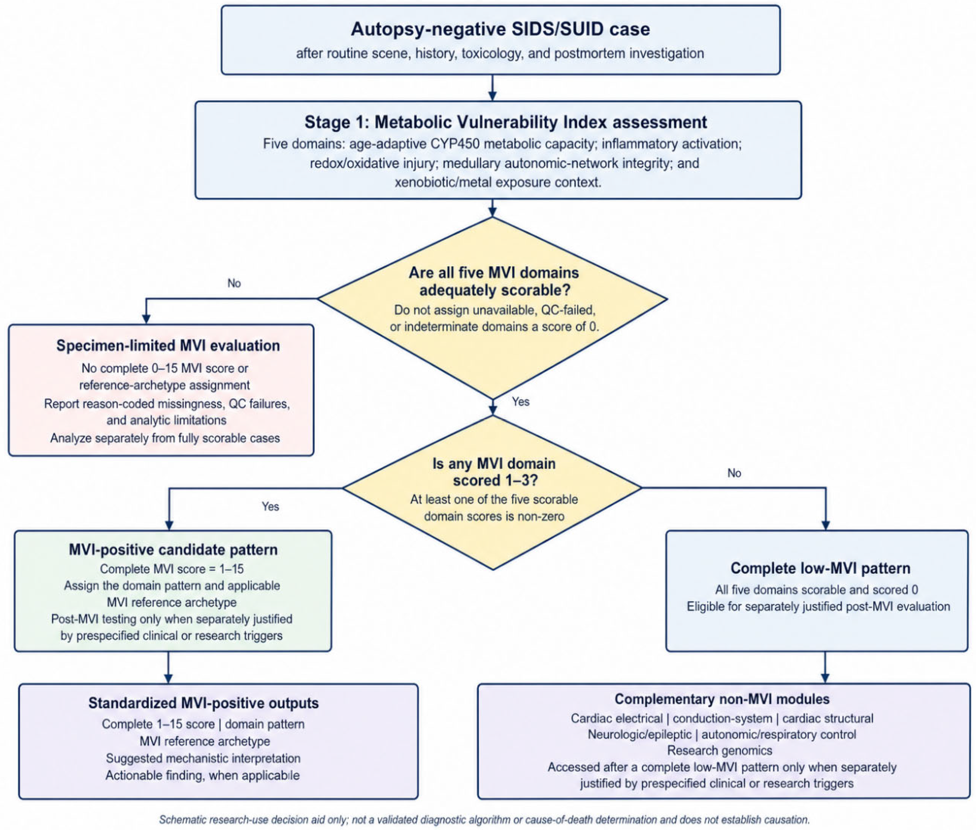
Figure 2.
Tiered post-MVI escalation pathway for eligible low-MVI or specimen-limited cases. The schematic shows how eligible cases proceed through targeted Tier 1 testing, conditional Tier 2 expansion, and selected Tier 3 research-level evaluation. It is intended to guide pilot classification and should not be interpreted as diagnostic yield, prevalence, or cause-of-death determination.
Figure 2.
Tiered post-MVI escalation pathway for eligible low-MVI or specimen-limited cases. The schematic shows how eligible cases proceed through targeted Tier 1 testing, conditional Tier 2 expansion, and selected Tier 3 research-level evaluation. It is intended to guide pilot classification and should not be interpreted as diagnostic yield, prevalence, or cause-of-death determination.
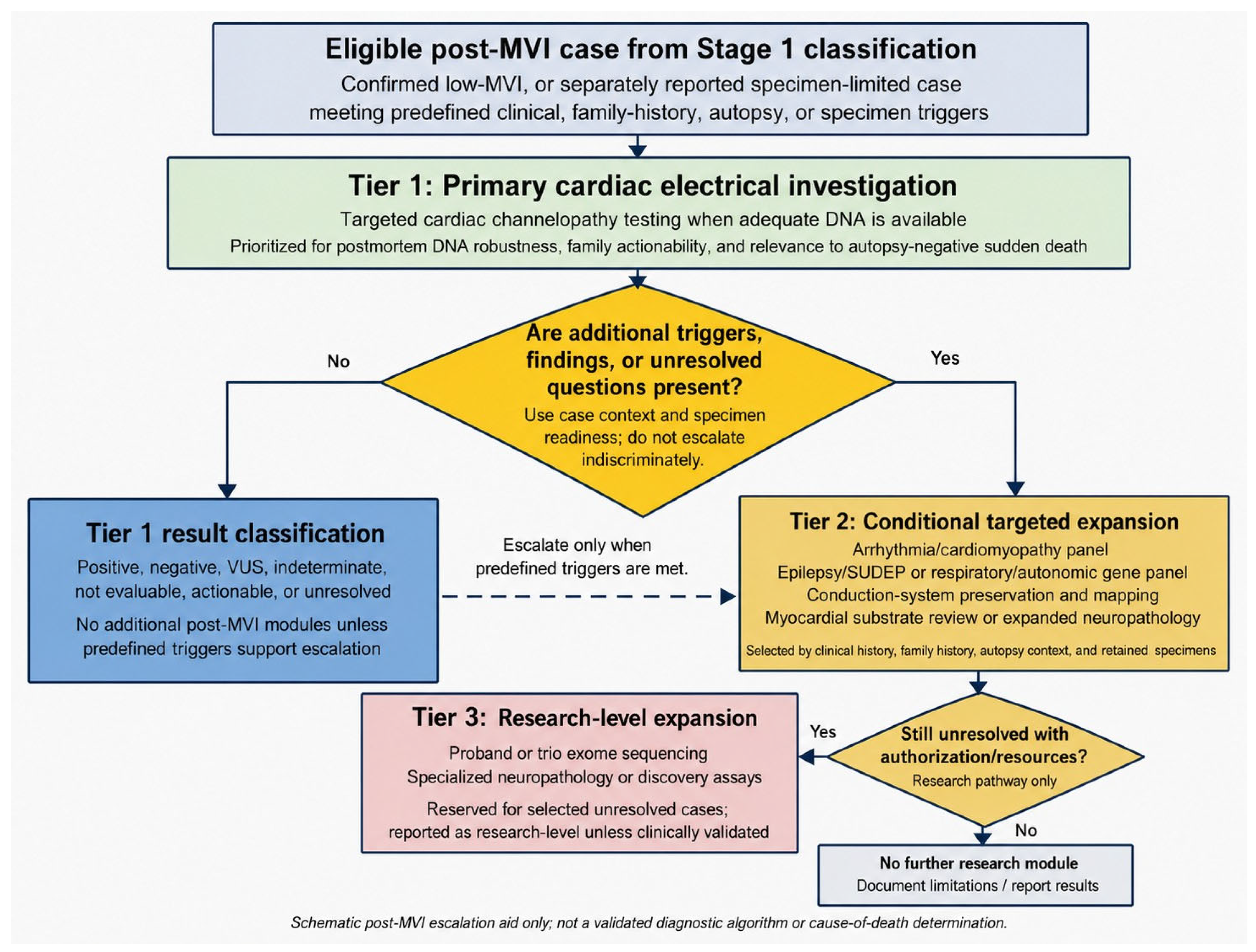
Figure 3.
Conceptual taxonomy aid linking metabolic vulnerability domains and candidate post-MVI non-metabolic domains.Archetypes 1–14 represent MVI vulnerability patterns. Refs. #15–22 summarize candidate non-metabolic vulnerability patterns considered after five-domain MVI classification and conditional post-MVI escalation, whereas Ref. #23 denotes an unresolved outcome after the indicated evaluable escalation. Shading indicates domain participation only and does not represent effect size, quantitative weighting, prevalence, diagnostic certainty, or data-derived clustering.
Figure 3.
Conceptual taxonomy aid linking metabolic vulnerability domains and candidate post-MVI non-metabolic domains.Archetypes 1–14 represent MVI vulnerability patterns. Refs. #15–22 summarize candidate non-metabolic vulnerability patterns considered after five-domain MVI classification and conditional post-MVI escalation, whereas Ref. #23 denotes an unresolved outcome after the indicated evaluable escalation. Shading indicates domain participation only and does not represent effect size, quantitative weighting, prevalence, diagnostic certainty, or data-derived clustering.
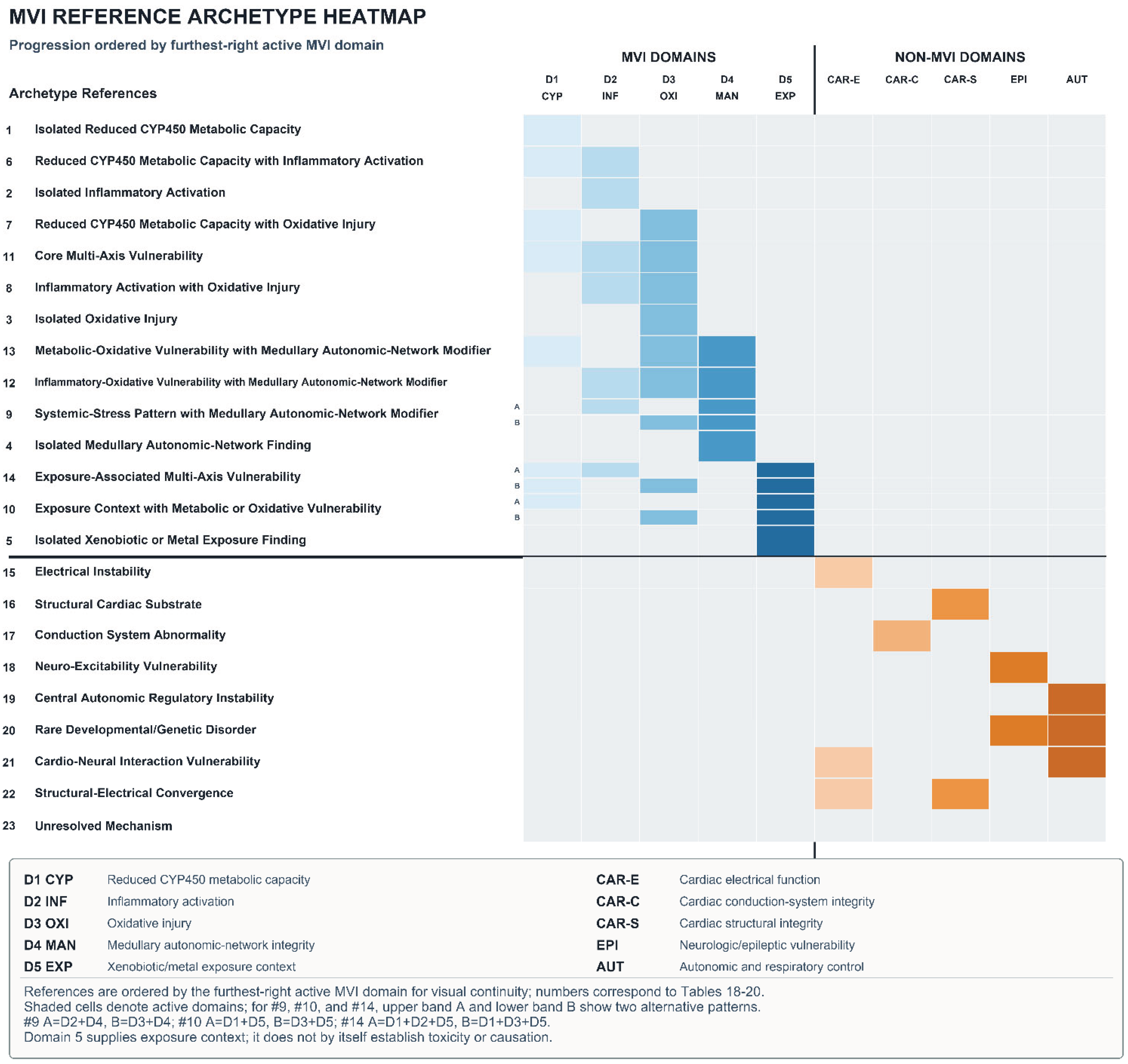
Table 1.
Stage 1 MVI Domains and pilot classification roles in autopsy-negative SIDS/SUID cases. Stage 1 MVI categories proposed for pilot classification. This table summarizes the conceptual role of each MVI domain and the operational meaning of low-MVI and specimen-limited categories. Entries are not mutually exclusive and should not be interpreted as pooled prevalence estimates, diagnostic-yield estimates, or cause-of-death determinations.
Table 1.
Stage 1 MVI Domains and pilot classification roles in autopsy-negative SIDS/SUID cases. Stage 1 MVI categories proposed for pilot classification. This table summarizes the conceptual role of each MVI domain and the operational meaning of low-MVI and specimen-limited categories. Entries are not mutually exclusive and should not be interpreted as pooled prevalence estimates, diagnostic-yield estimates, or cause-of-death determinations.
| Stage 1 domain / classification category | What it represents | Pilot classification role | Reference |
| Domain 1: CYP450 / Metabolic Capacity | Measured hepatic CYP450 protein abundance as a provisional indicator of metabolic capacity, interpreted using protocol-defined age- and specimen-appropriate anchors. | Candidate metabolic-capacity pattern when required hepatic measurements are adequate and interpretable; does not distinguish developmental, inherited, inhibitory, inflammatory, or other causes of reduced abundance. | [5,6] |
| Domain 2: Cytokine / Inflammatory State | Inflammatory activation, cytokine signaling, recent illness, immune stress, or resuscitation-relevant inflammatory context | Candidate inflammatory vulnerability when measured with validated or protocol-accepted specimens and interpreted with illness, postmortem interval, and resuscitation context | [7,8] |
| Domain 3: Redox / Oxidative Injury | Reproducible oxidative injury reflected by protocol-defined measures of lipid peroxidation, protein-adduct formation, and oxidative nucleic-acid damage | Candidate oxidative-injury pattern when required tissue measurements are interpretable and not better explained by specimen, postmortem, or resuscitation artifact; does not establish mitochondrial dysfunction, causation, or cause of death. | [9] |
| Domain 4: Medullary / Autonomic-Network Integrity | Limited whole-section medullary assessment using H&E and one protocol-defined serotonergic-IHC classification, without named-nucleus sampling or region-level scoring. | Medullary autonomic-network modifier when a reproducible abnormality is identified; does not establish serotonergic dysfunction, autonomic or respiratory failure, causation, or cause of death. | [1,10] |
| Domain 5: Xenobiotic / Exposure Findings and Context | Structured exposure-context abstraction plus standard or triggered analytical assessment using appropriate specimens, products, samples, or records. Context alone and separately reported research measurements do not generate a score. | Exposure modifier only when analyte-, method-, matrix-, and postmortem-appropriate analytical findings are interpretable. Context-negative is not equivalent to analytic-negative; an independently sufficient toxicologic explanation is exclusionary. | [11,12] |
| Confirmed Low-MVI Classification | No detectable abnormality across all five adequately scorable MVI domains | Confirmed low-MVI is the primary post-MVI gateway when all five domains are scorable and within expected limits; independently triggered assessment of MVI-positive cases is reported separately. | — |
| Specimen-Limited MVI Evaluation | One or more MVI domains unavailable, incomplete, analytically unreliable, or not evaluable under protocol-defined conditions | Report separately; do not assign missing or uninterpretable domains a default score of zero, and do not classify as confirmed low-MVI | — |
Table 2.
Candidate non-metabolic mechanisms for post-MVI pilot classification in eligible autopsy-negative SIDS/SUID cases1. Candidate non-metabolic mechanisms evaluated during post-MVI escalation. Categories are intended for pilot classification and research interpretation; they are not mutually exclusive and should not be interpreted as pooled prevalence estimates, diagnostic-yield estimates, or cause-of-death determinations.
Table 2.
Candidate non-metabolic mechanisms for post-MVI pilot classification in eligible autopsy-negative SIDS/SUID cases1. Candidate non-metabolic mechanisms evaluated during post-MVI escalation. Categories are intended for pilot classification and research interpretation; they are not mutually exclusive and should not be interpreted as pooled prevalence estimates, diagnostic-yield estimates, or cause-of-death determinations.
| Category | Definition | Primary Evaluation Context | Pilot-Use Interpretation | Reference |
| Cardiac Channelopathy |
Pathogenic or likely pathogenic variants associated with inherited cardiac electrical instability, including LQTS, CPVT, Brugada syndrome, or related inherited arrhythmia pathways | Confirmed low-MVI status, structurally normal autopsy-negative sudden death, sleep-related death without clear asphyxial explanation, or family history of sudden death/syncope | Candidate cardiac electrical vulnerability when supported by a pathogenic or likely pathogenic variant in an established arrhythmia gene; VUS findings should not be treated as positive causal findings | [13,14,15,16,17] |
| Concealed Cardiomyopathy | Genetic cardiomyopathy susceptibility without overt gross structural heart disease | Borderline cardiac findings, subtle myocardial abnormalities, family history of cardiomyopathy or sudden death, or persistent suspicion after Tier 1 testing | Candidate structural/electrical vulnerability when supported by pathogenic or likely pathogenic cardiomyopathy-associated findings and case-context concordance | [15,18] |
| Epilepsy / SUDEP-Associated Mechanism |
Neuronal excitability variants or epilepsy/SUDEP-associated neuro-excitability or autonomic vulnerability | Unwitnessed nocturnal death, possible seizure markers, prior seizure history, or family history of epilepsy | Candidate neuro-excitability vulnerability when supported by clinical/family context and relevant genetic or neuropathologic findings | [19] |
| Respiratory / Autonomic Developmental Mechanism | Disorders affecting central chemosensitivity, respiratory drive, arousal, or autonomic regulation | Prior apnea, BRUE/ALTE, cyanotic spells, prematurity-related apnea, or autonomic/respiratory instability | Candidate autonomic/respiratory-control vulnerability when supported by relevant history, genetic findings, or brainstem/autonomic assessment | [20] |
| Microscopic Conduction-System Abnormality | Developmental, fibrotic, or structural abnormalities of the cardiac conduction system detectable only with targeted preservation and serial sectioning | Structurally normal heart with suspected arrhythmic mechanism, borderline cardiac findings, or family history suggesting inherited electrical disease | Candidate conduction-system vulnerability when relevant anatomic landmarks are preserved and abnormalities are identified by appropriate pathology review | [21,22] |
| Occult Myocardial Substrate | Fibrosis, collagen abnormalities, or subtle myocardial substrate not evident on gross examination | Borderline histology, subtle cardiac findings, or suspicion of arrhythmogenic substrate despite routine autopsy-negative status | Candidate cardiac structural substrate when targeted histology or special stains identify potentially relevant myocardial abnormalities | [15,18,22] |
| Rare Developmental or Genetic Mechanism2 | Rare or novel developmental / genetic pathway not captured by targeted cardiac, epilepsy, or respiratory/autonomic evaluation | Persistently unresolved cases with adequate authorization, specimen quality, and research resources | Research-level candidate mechanism; rate and interpretive value should be measured prospectively | — |
| Unresolved After Post-MVI Evaluation3 | No candidate vulnerability identified after completed evaluable post-MVI assessment | Completed or appropriately attempted tiered evaluation with no reportable candidate mechanism | Residual research classification reflecting current detection limits, specimen limits, or absence of reportable findings; not evidence that no mechanism existed | — |
1 Categories are candidate vulnerability classifications for pilot use. They are not mutually exclusive and do not represent pooled prevalence, diagnostic yield, or cause-of-death attribution. 2 Rare developmental or genetic mechanisms should be reported as research-level findings unless clinically validated and interpreted under appropriate genetic-testing standards. 3 “Unresolved” is an outcome category to be measured prospectively, not a predicted frequency.
Table 3.
Post-MVI escalation investigations for non-metabolic mechanisms in autopsy-negative SIDS/SUID cases. Representative investigations, candidate pathways, tier rationale, and approximate U.S.-context planning costs for the post-MVI escalation framework.
Table 3.
Post-MVI escalation investigations for non-metabolic mechanisms in autopsy-negative SIDS/SUID cases. Representative investigations, candidate pathways, tier rationale, and approximate U.S.-context planning costs for the post-MVI escalation framework.
|
Investigation (Tier) |
Category | Candidate Pathway / Vulnerability Target | Primary rationale for tier placement | Typical Cost Range (USD)1 | ||
| Targeted Cardiac Channelopathy Panel (Tier 1) | Cardiac Electrical Function | Primary electrical instability (ion-channel-mediated arrhythmia risk) | Highest family-actionable yield with robust DNA and moderate cost | $1,500–$4,000 | ||
| Expanded Cardiac Genetics Panel (Arrhythmia + Cardio-myopathy) (Tier 2) | Cardiac Electrical / Structural Integrity | Structural + electrical substrate abnormalities | Broader potential yield but greater interpretive burden. | $2,500–$6,000 | ||
| Epilepsy/SUDEP Panel (Tier 2) | Neuro-Excitability (Epilepsy/SUDEP) | epilepsy/SUDEP-associated candidate pathway | Phenotypically targeted yield with robust DNA and moderate cost | $1,500–$4,000 | ||
| Respiratory/Autonomic Gene Panel (Tier 2) | Central autonomic / Respiratory Control | Central control instability | Targeted yield in selected clinical histories with moderate family actionability | $1,500–$4,000 | ||
| Structural Cardiac (Conduction System) (Tier 2 Conditional) | Cardiac Conduction System Integrity | Microscopic conduction system disease | Conditional structural clarification when cardiac clues are present, but with lower routine feasibility | $1,000–$5,000 | ||
| Structural Cardiac (Myocardial Substrate) (Tier 2 Conditional) | Cardiac Structural Integrity | Occult myocardial substrate | Low-cost targeted clarification when subtle structural or histologic clues are present | $35–$100 per slide | ||
| Expanded Brainstem / Autonomic Neuropathology (Tier 2) | Central Autonomic / Respiratory Control | Subtle autonomic defects | Conditional phenotypic relevance but lower routine feasibility | $800–$3,500 | ||
| Proband Exome Sequencing (Tier 3) | Rare Developmental / Genetic Mechanisms | Broad discovery (single-proband genomic evaluation) | Broad discovery potential but higher cost and greater interpretive uncertainty | $3,000–$8,000 | ||
| Trio Exome (Tier 3) | Rare Developmental / Genetic Mechanisms | De novo variant identification (family-based analysis) | Broad discovery value but highest cost and VUS burden | $6,000–$12,000+ | ||
1 Costs are planning ranges only and may vary by laboratory, institutional pricing, batching, specimen requirements, pathology services, and pilot-study contracting.
Table 4.
Conditional triggers for selection of post-MVI escalation investigations. Clinical history, autopsy findings, and specimen quality may guide prioritization of specific investigations within the post-MVI escalation framework.
Table 4.
Conditional triggers for selection of post-MVI escalation investigations. Clinical history, autopsy findings, and specimen quality may guide prioritization of specific investigations within the post-MVI escalation framework.
| Trigger / Case feature | Priority Consideration | Rationale | Tests potentially deferred unless clinically indicated |
| Autopsy-negative, sleep-related death without an independently sufficient asphyxial explanation | | Targeted arrhythmia / channelopathy panel | High family actionability; DNA-based testing is often feasible when a suitable specimen is available | Broad exome sequencing and extensive conduction-system mapping unless independently indicated by clinical, family-history, or autopsy findings |
| Family history of sudden death, syncope, “seizures,” drowning, unexplained accidents | Arrhythmia + cardiomyopathy panel (or exome if available) | Increases pretest probability of inherited cardiac disease | Brainstem expansion unless respiratory history |
| Prior BRUE / apnea / cyanosis; prematurity with recurrent apnea; “quiet death” | Expanded brainstem / autonomic neuropathologic evaluation + consider respiratory-control genes | Mechanism points to autonomic/respiratory instability | Conduction mapping unless cardiac clues |
| Recent immune or medical exposure1 | Exposure-contextualized MVI/Post-MVI review | Evaluates exposure-vulnerability concordance without causal inference | Causal attribution from temporal proximity alone; broad escalation absent supportive findings |
| Unwitnessed nocturnal death; possible seizure markers; family epilepsy history | Epilepsy/SUDEP panel (or exome) | Mechanistic plausibility and likelihood of adequate DNA quality | Conduction mapping unless cardiac signs |
| Borderline cardiac findings, structurally normal heart with unexplained sudden death, family history of sudden death/syncope, or possible arrhythmia context | Preserve heart or targeted conduction-system regions where feasible; consider conduction-system mapping, myocardial fibrosis/collagen stains, and expanded arrhythmia / cardiomyopathy panel | Subtle cardiac structural, electrical, or conduction-system vulnerability may not be evident on routine gross examination, and later conduction-system evaluation requires preservation of SA-node and AV-node/His-bundle landmarks | Epilepsy panel unless seizure history |
| Independently sufficient asphyxial explanation established after complete scene, autopsy, and medicolegal review | Exit the autopsy-negative post-MVI pathway unless another investigation is independently indicated | An explained death does not qualify for the primary autopsy-negative analytic pathway | Post-MVI escalation unless separately justified |
| Significant decomposition / poor cold chain | Prefer DNA-only strategies if feasible | DNA is most resilient | PMI-sensitive protein/RNA-based methods; extensive IHC if autolysis severe |
1 Recent immune or medical exposures are recorded as contextual timing variables only and should be interpreted with independent MVI/post-MVI findings, not as stand-alone causal triggers.
Table 5.
Provisional post-MVI interpretive archetypes and unresolved outcome for eligible cases. Refs. #15–22 organize provisional candidate non-metabolic vulnerability patterns identified during post-MVI escalation and may coexist within a case. Ref. #23 is assigned only when no reportable candidate pattern is identified after the indicated evaluable escalation. These classifications do not establish a diagnosis, causal sequence, or cause of death.
Table 5.
Provisional post-MVI interpretive archetypes and unresolved outcome for eligible cases. Refs. #15–22 organize provisional candidate non-metabolic vulnerability patterns identified during post-MVI escalation and may coexist within a case. Ref. #23 is assigned only when no reportable candidate pattern is identified after the indicated evaluable escalation. These classifications do not establish a diagnosis, causal sequence, or cause of death.
| Ref. no. | Archetype or outcome | Candidate vulnerability pattern | Key genetic or structural targets | Typical clinical or autopsy context | Primary investigation | |
| #15 | Electrical Instability / Cardiac Channelopathy | Inherited arrhythmia-associated electrical vulnerability without an explanatory structural abnormality | Established arrhythmia genes, including SCN5A, KCNQ1, KCNH2, RYR2, and CACNA1C | Sleep-related autopsy-negative death; structurally normal heart; family history of syncope, sudden death, unexplained drowning, or unexplained accidents | Targeted cardiac channelopathy panel (Tier 1) | |
| #16 | Structural Cardiac Substrate | Occult myocardial abnormality or concealed cardiomyopathy substrate not apparent on routine gross examination | Microscopic fibrosis, collagen abnormalities, subtle myocardial substrate, and established cardiomyopathy genes such as MYH7, MYBPC3, LMNA, DSP, and PKP2 | Borderline cardiac findings, subtle myocardial abnormalities, or family history of cardiomyopathy or sudden death | Targeted myocardial histology and/or expanded arrhythmia-cardiomyopathy panel (Tier 2 conditional) | |
| #17 | Conduction-System Abnormality | Structural or developmental abnormality of the cardiac conduction system | Accessory pathways, nodal developmental abnormalities, conduction-system fibrosis, or other abnormalities identified by appropriate pathology review | Structurally normal heart with suspected arrhythmic mechanism, borderline cardiac findings, or relevant family history | Conduction-system preservation, mapping, and serial sectioning (Tier 2 conditional) | |
| #18 | Neuro-Excitability / Epilepsy-SUDEP-Associated Vulnerability | Epilepsy/SUDEP-associated neuro-excitability or autonomic vulnerability | Established epilepsy-associated genes, including SCN1A, DEPDC5, KCNQ2, and STXBP1, together with relevant clinical or neuropathologic findings | Unwitnessed nocturnal death, possible seizure markers, prior seizure history, or family history of epilepsy | Epilepsy/SUDEP gene panel and clinically indicated neurologic review (Tier 2) | |
| #19 | Central Autonomic / Respiratory-Control Vulnerability | Developmental abnormality affecting respiratory drive, chemosensitivity, arousal, or autonomic regulation | PHOX2B and other established respiratory/autonomic developmental genes; relevant brainstem or autonomic findings | Prior apnea, BRUE/ALTE, cyanotic episodes, prematurity-related apnea, or documented respiratory/autonomic instability | Respiratory/autonomic gene panel and conditionally expanded brainstem/autonomic evaluation (Tier 2) | |
| #20 | Rare Developmental / Genetic Finding | Rare or novel developmental/genetic finding not captured by targeted evaluation | Broader developmental or disease-associated genes identified through appropriately governed genomic analysis | Persistently unresolved case with adequate specimen quality, authorization, and research resources | Proband or trio exome sequencing (Tier 3) | |
| #21 | Cardio-Neural Interaction | Co-occurring cardiac electrical and neurologic/autonomic candidate findings | Independently supported arrhythmia-associated and neuro-excitability or autonomic findings | Separate cardiac and neurologic/autonomic assessments identify potentially relevant findings in the same case | Integration of independently indicated cardiac and neurologic/autonomic investigations (Tiers 1–2) | |
| #22 | Structural-Electrical Convergence | Co-occurring myocardial or conduction-system substrate and cardiac electrical vulnerability | Interpretable structural or conduction-system abnormality together with a supported arrhythmia- or cardiomyopathy-associated finding | Targeted pathology and molecular evaluation identify potentially relevant structural and electrical findings in the same case | Integrated cardiac genetics, myocardial histology, and/or conduction-system assessment (Tiers 1–2) | |
| #23 | Unresolved After Post-MVI Evaluation | No reportable candidate vulnerability identified after completion or appropriate attempted completion of the indicated evaluable escalation | None identified | Indicated post-MVI investigations are completed or appropriately attempted without a reportable candidate finding | Integrated review of the indicated evaluable investigations | |
Disclaimer/Publisher’s Note: The statements, opinions and data contained in all publications are solely those of the individual author(s) and contributor(s) and not of MDPI and/or the editor(s). MDPI and/or the editor(s) disclaim responsibility for any injury to people or property resulting from any ideas, methods, instructions or products referred to in the content. |
© 2026 by the authors. Licensee MDPI, Basel, Switzerland. This article is an open access article distributed under the terms and conditions of the Creative Commons Attribution (CC BY) license (http://creativecommons.org/licenses/by/4.0/).
Copyright: This open access article is published under a Creative Commons CC BY 4.0 license, which permit the free download, distribution, and reuse, provided that the author and preprint are cited in any reuse.