

Submitted:
26 August 2026
Posted:
27 August 2026
You are already at the latest version
Abstract
Myofibrillar loss during skeletal muscle atrophy is often described as selective. MuRF1 is considered to remove thick-filament proteins preferentially, whereas thin-filament proteins are assigned to other ligases or appear relatively spared. We reassess whether this pattern demonstrates molecular specificity or restricted access. Four lines of evidence support an access-based interpretation. Actin can be polyubiquitinated by MuRF1, although loss of assembled thin-filament proteins is not MuRF1-dependent. This separates biochemical competence from net removal. Apparent preferential myosin loss also varies with extraction chemistry, normalization, and the combination of catabolic stimuli with inactivity. KLHL40 and KLHL41 preserve nebulin and leiomodin-3, indicating active maintenance of thin-filament stability. Turnover and ubiquitin-remnant proteomics quantify only molecules recovered from defined fractions. Neither method alone distinguishes between degradation and redistribution, or between analytical invisibility and degradation. We describe three levels of restricted access. They comprise assembly-state occlusion, dedicated molecular shielding, and analytical access imposed by extraction and fractionation. Oxidation, mechanical damage, end capping, and control of pointed-end length may alter access before bulk protein loss. This framework retains ligase specificity while defining the controls needed to establish it.
Keywords:
skeletal muscle atrophy
; thin filament
; actin
; MuRF1
; ubiquitin-proteasome system
; KLHL40
; KLHL41
; MICAL
; protein turnover
; myofibril extraction
1. Introduction
Skeletal muscle atrophy reduces force-generating capacity by removing a large protein mass while leaving recognizable sarcomeric order, at least during early and moderate wasting. Genetic and transcriptional studies established MuRF1 and atrogin-1/MAFbx as central atrogenes [1,2,3]. FoxO signaling coordinates proteasomal and autophagic programs [4,5]. Autophagy also has a homeostatic role. Its loss damages sarcomeres and can worsen muscle loss during denervation or fasting [6]. Reviews of the ubiquitin-proteasome system and its E3 ligases define the major degradative framework [7,8,9]. Calcium-dependent proteases, caspases, and chaperone systems provide additional routes [10,11,12]. Ordered sarcomeres can persist during early wasting. This raises a central question. Which step limits access to a structural protein before its final route of disposal is assigned?
The most influential experimental formulation of this asymmetry came from Cohen and colleagues, who showed that MuRF1-dependent ubiquitylation drives an ordered loss of thick-filament proteins during denervation and fasting, while actin, tropomyosin, troponins, and alpha-actinin declined similarly in wild-type and ligase-defective muscle [13]. In later summaries, this result is often compressed into the statement that MuRF1 recognizes thick, but not thin, filament substrates. The compression is understandable, but it removes a distinction made in the original study. Purified or released proteins can be competent substrates while the same molecules are protected when incorporated into a highly ordered myofibril. The measured phenotype reflects at least two variables. They are molecular recognition and physical access.
Three observations make this distinction consequential. Polge and colleagues reported MuRF1-dependent polyubiquitination and degradation of muscle actin in cellular, biochemical, and biopsy-associated experiments [14]. Low-ionic-strength extraction can also generate or magnify apparent preferential myosin depletion by leaving thick filaments in insoluble material [15]. A third line of evidence comes from BTB-BACK-kelch proteins. KLHL40 and KLHL41 can preserve major thin-filament proteins [16,17,18]. Studies of KBTBD13 and CUL3 connect related adaptors to sarcomeric function [19,20,21], while genetic and clinical work extends this association to nemaline myopathy [22,23,24,25]. These observations do not invalidate the founding genetic work. They show that absence of net loss in an assay cannot be equated with absence of substrate recognition.
This review does not attempt another catalog of myofibrillar proteolysis. Aweida and Cohen have already provided a detailed stepwise account of how the UPS dismantles filamentous myofibrils, including desmin depolymerization, ubiquitination, extraction by AAA-ATPases, and proteasomal degradation [26]. Our distinct question is why a component is not reached, or is not observed to be reached, in the first place. We focus on skeletal muscle, use cardiac work only when it clarifies a conserved sarcomeric quality-control principle, and distinguish biological restriction of access from analytical restriction. Therapeutic interventions and fiber-type-specific responses are outside the main scope.
The argument develops from the evidence rather than from a prespecified taxonomy. Section 2 revisits the apparent conflict between MuRF1-dependent thick-filament loss and MuRF1-mediated actin ubiquitination. Section 3 asks whether selective contractile-protein loss is reproducible across extraction methods and atrophy models. Section 4 considers active stabilization by kelch proteins and chaperone systems. Section 5 defines what turnover studies measure and identifies the unmeasured regulatory layer. Only then, in Section 6, are the convergent observations named as three levels of access restriction. These are assembly-state occlusion, dedicated shielding, and extraction/fractionation bias.
2. The Founding Claim and the Tension Inside It
2.1. Ordered Thick-Filament Loss Is Already an Accessibility Model
Cohen et al. generated mice expressing a RING-deleted MuRF1 that retained binding capacity but lacked effective ubiquitin-ligase activity [13]. Following denervation or fasting, wild-type muscle lost myosin-binding protein C (MyBP-C) and myosin light chains before major loss of myosin heavy chain (MyHC). The corresponding losses were blunted when MuRF1 function was disrupted. Thin-filament and Z-disk proteins, by contrast, declined without the same dependence on MuRF1. These findings established both a ligase-dependent component of atrophy and an ordered sequence within the thick filament.
The sequence matters as much as the substrate list. MyHC in an intact filament is not equivalent to soluble MyHC. Associated proteins, lattice packing, and the supramolecular environment can conceal lysines, degrons, or binding surfaces. Removing MyBP-C and light chains can expose MyHC to subsequent ubiquitination and extraction. Thus, even within the thick filament, the first event is not unrestricted ligase recognition of every component. A structural transition precedes efficient degradation. Commentary and later mechanistic work have accordingly described myofibril destruction as loss of stabilizing pieces without immediate collapse of the entire lattice [26,27,28].
This principle is supported by older biochemical observations that actin and myosin are rapidly degraded when purified but resist ubiquitin-dependent proteolysis in actomyosin complexes or myofibrils [13]. It is also compatible with proteomic mapping. In denervated muscle, ubiquitin-remnant enrichment identified a strong signal in sarcomeric proteins, including MyHCs, myomesins, and titin [29]. Titin carried an early concentration of upregulated diglycine sites near its C-terminus, which anchors at the M-band, creating a spatial gradient rather than a uniform protein-wide pattern [29]. Position within the assembly can influence where degradation begins.
2.2. Actin Is a Competent MuRF1 Substrate Under Other Conditions
Polge et al. directly challenged the idea that muscle actin lies outside the MuRF1 substrate repertoire [14]. In dexamethasone-treated myotubes, actin accumulated polyubiquitin conjugates, MuRF1 depletion stabilized epitope-tagged actin, and recombinant MuRF1 ubiquitinated actin in vitro. The study also detected ubiquitinated actin in human skeletal muscle material. These results demonstrate ubiquitination competence, but the experimental contexts included tagged actin, soluble or myofibrillar-enriched lysates, dexamethasone-treated cells, and recombinant reactions. They do not address the same structural and temporal questions as the in vivo knock-in experiments.
The two reports are best treated as measurements of different conditional probabilities. Cohen et al. asked whether loss of proteins incorporated into native myofibrils during defined atrophy models requires MuRF1 [13]. Polge et al. asked whether actin can bind, become polyubiquitinated by, and undergo degradation through a MuRF1-dependent route under biochemical and cellular conditions [14]. A protein may answer yes to the second question and no to the first if assembly suppresses access. Conversely, a loss-of-function phenotype in vivo can persist even when MuRF1 is biochemically capable of modifying the isolated protein.
This distinction also prevents an overly sharp division between thick- and thin-filament ligases. TRIM32 has been implicated in removal of desmin, actin, tropomyosin, troponins, and alpha-actinin, with desmin depolymerization facilitating later myofibrillar breakdown [26,27,28,30]. MuRF1 ubiquitylome studies likewise identify a broader set of candidate proteins than the small group whose steady-state loss has been genetically demonstrated [31]. Interactomes, in vitro ubiquitination, ubiquitin-remnant enrichment, and genotype-dependent abundance each supply different evidence. They should not be collapsed into a single label of “substrate.”
2.3. What Survives the Apparent Contradiction
Three conclusions remain secure. First, MuRF1 is required for the efficient loss of specific thick-filament components in several atrophy settings [13,32]. Second, actin can be polyubiquitinated in a MuRF1-dependent manner under particular cellular and biochemical conditions [14]. Third, incorporation into an ordered myofibril changes the degradability of contractile proteins [13,26,27,28,30]. The unresolved variable is access. The structural state in which the ligase encounters the protein, the molecular partners that cover it, and the extraction procedure that makes it visible to the investigator.
Accordingly, thin-filament sparing should be described operationally. It means that a measured pool declined less, or declined independently of the tested ligase, under a specified model and extraction method. It does not, by itself, prove that the ligase lacks affinity for every biochemical form of that protein. This more limited wording preserves the genetic evidence while avoiding a specificity claim that the actin data do not support.
2.4. Dismantling Requires Release, Extraction, and Disposal
Recognition by an E3 ligase is only one event in the process of myofibril loss. The proteasome accepts unfolded polypeptides through a narrow axial channel and cannot ingest an intact actomyosin lattice. Pharmacological inhibition established that proteasome activity accounts for a large share of the accelerated proteolysis in atrophying muscle, but that result locates the terminal disposal system rather than the step that makes a sarcomeric protein available to it [26,33]. An access model must separate four transitions. These are structural loosening, covalent marking, extraction from the assembly, and final destruction.
Caspase-3 provides one proposed route into this sequence. In catabolic models, limited actomyosin cleavage generated a characteristic approximately 14-kDa actin fragment and increased subsequent proteasome-dependent degradation [34]. Caspase-3 deficiency also attenuated denervation atrophy in vivo [11]. These findings do not mean that caspase-3 universally initiates myofibril breakdown. Calpains, desmin remodeling, and other release mechanisms differ among models. They do show why a full-length actin band, a cleavage fragment, and loss of total actin are distinct readouts. Cleavage may increase access without yet demonstrating either ubiquitination or complete degradation.
Calcium-dependent myofilament release supplies a complementary example. Septic muscle showed Z-band disruption and release of actin- and myosin-containing material, while interventions that limited calcium-dependent proteolysis reduced the response [10,35,36]. Importantly, the calpain-inhibitor phenotype occurred without proportional suppression of atrogin-1/MAFbx or MuRF1 expression [36]. Expression of an atrogene and physical availability of its substrates can be regulated on different axes. A strongly induced ligase may have little effect on a filament that has not been released, whereas modest ligase abundance may suffice once an assembly barrier is removed.
Extraction from a marked assembly is itself an active step. Dominant-negative p97/VCP reduced denervation- and fasting-induced atrophy and trapped ubiquitinated thin- and thick-filament components together with p97 cofactors, supporting a segregase function at the myofibril [37]. The delayed denervation program described by Volodin et al. similarly required desmin depolymerization, PAX4-dependent induction, and p97/VCP [28]. METTL21C-dependent trimethylation of p97 altered its assembly and ATPase activity and modified protein-quality-control phenotypes in skeletal muscle [38]. These studies make extraction capacity a biological variable rather than a technical afterthought.
3. Whether the Asymmetry Is Reproducible at All
3.1. Ionic Strength Can Create Apparent Preferential Myosin Loss
The strongest warning comes from a basic measurement issue. Myosin is poorly extracted from intact thick filaments by low-ionic-strength buffers and becomes more recoverable as ionic strength rises. Cosper and Leinwand compared conventional low-salt lysis with a high-salt myosin extraction buffer and showed that low-salt supernatants substantially underestimate MyHC [15]. Studies reporting extreme preferential MyHC depletion often quantified only these supernatants.
The underlying accounting is important. Myosin constitutes roughly 30% of total muscle protein, but a band normalized to equal lysate protein reports concentration within the recovered material, not total MyHC content per muscle. A 50% fall in muscle mass can produce a 50% fall in total MyHC content even if the MyHC-to-total-protein ratio is unchanged. Conversely, an approximately 80% fall in a normalized MyHC band implies additional disproportionate depletion and is highly sensitive to protein-specific recovery. With adequate high-salt extraction, Cosper and Leinwand did not observe selective MyHC depletion in the cachexia model studied [15]. The result does not show that preferential myosin loss never occurs. It shows that extraction and denominator must be treated as part of the biological inference.
This issue is particularly important because Western blots are frequently normalized within the soluble material recovered after lysis. If the fraction of MyHC entering that material changes with filament packing, post-translational modification, or buffer composition, the normalized band reports both extractability and abundance. Total-protein staining cannot repair a protein-specific recovery failure. Pellet analysis, high-salt extraction, or quantitative recovery controls are required.
3.2. Preferential Loss Depends on the Combination of Insults
Model dependence provides a second qualification. Yamada et al. found that cancer cachexia alone did not produce preferential myosin loss under their analytical conditions, and dexamethasone alone likewise did not reproduce the phenotype. Preferential loss appeared when the systemic catabolic stimulus was combined with denervation [39]. Electrical stimulation can attenuate preferential myosin loss in steroid-denervation models, further linking the phenotype to inactivity and neuromuscular disconnection rather than to a universal cachectic program [40].
Clinical and extreme-physiology observations also resist a single rule. Acute quadriplegic myopathy is often characterized as an acquired myosinopathy, and intensive-care interventions can produce marked motor-protein dysfunction [41,42]. Spaceflight alters thick- and thin-filament isoforms in a muscle- and fiber-type-dependent manner [43]. Hibernating mammals, in contrast, preserve muscle mass and function despite prolonged inactivity by coordinating the suppression of proteolysis and maintenance programs [44]. These settings differ in neural drive, mechanical load, glucocorticoid exposure, inflammation, nutrition, and time. A substrate-selectivity statement detached from the model discards variables that may determine whether the myofibril becomes accessible.
The practical conclusion is modest but important. “preferential myosin loss” is a phenotype, not a constitutive property of muscle atrophy. It should be reported together with the catabolic stimulus, neural and loading state, duration, muscle, extraction buffer, analyzed fraction, and denominator.
3.3. Fraction Redistribution Can Mimic Synthesis or Negative Breakdown
Dynamic proteome profiling adds a more subtle example. Camera and Burniston combined protein-specific synthesis with changes in abundance to estimate breakdown after a high-fat diet and resistance exercise [45]. Several proteins generated mathematically negative breakdown estimates. Negative degradation is not biologically meaningful. The authors interpreted these values as evidence that proteins or premyofibrillar complexes had moved into the analyzed myofibrillar fraction. Alpha-actin and troponin C2 were among proteins for which assembly or redistribution could explain an excess increase in the myofibrillar pool [45].
This phenomenon reveals that fractionation is not a neutral window onto turnover. A soluble regulatory protein can appear to be lost when it enters the filament, whereas an incorporated protein can appear stable when a newly assembled pool replaces degraded molecules. Similarly, a component released from damaged myofibrils may disappear from the myofibrillar fraction before it is degraded. Without paired analysis of soluble, insoluble, and total pools, compartment transfer is mathematically indistinguishable from synthesis or breakdown in a single fraction.
Table 1 summarizes the interpretive boundaries. The point is not that the cited assays are unreliable. Each assay answers a legitimate question, but the answer is narrower than the biological label often attached to it.
3.4. Fraction Completeness and Mass Balance
The denominator creates a second accounting problem. Equal-protein loading answers how much target is present per unit of recovered protein. It does not answer how much target remains per muscle, per fiber, or per original tissue mass. A smaller atrophic muscle can retain the same target-to-total-protein ratio while losing a large absolute amount. Conversely, poor extraction of a single highly abundant filament can disproportionately lower its band. Matched high- and low-salt extraction, analysis of both supernatant and pellet, and normalization back to the starting tissue are necessary when preferential loss is the claim [15,33,45].
Ubiquitin-remnant proteomics requires a parallel denominator. A larger diglycine-peptide signal may reflect higher site occupancy, greater abundance of the parent protein, improved peptide recovery, or movement of the modified species into the analyzed fraction. Quantifying the corresponding unmodified peptide or total protein in every fraction does not solve all chain-topology and E3-attribution questions, but it distinguishes modification stoichiometry from simple abundance change [29,31]. Spike-in recovery standards and orthogonal immunoblotting of pellets can further reveal protein-specific extraction failures.
4. Protection as a Dedicated Function
4.1. KLHL40 Stabilizes Rather Than Delivers Key Thin-Filament Proteins
BTB-BACK-kelch proteins commonly act as substrate adaptors for CUL3-RING ubiquitin ligases. KBTBD13 and related adaptors connect this family to sarcomeric mechanics and CUL3 activity [19,20,21]. Other genetic and clinical studies link kelch proteins to myofibrillar preservation and nemaline myopathy [22,23,24,25]. KLHL40 localizes to sarcomeric regions, binds nebulin (NEB) and leiomodin-3 (LMOD3), and increases their protein abundance without corresponding mRNA changes [17]. KLHL40-deficient mice and patients with severe KLHL40 mutations show marked reductions in NEB and LMOD3, muscle weakness, and sarcomeric abnormalities [17].
The mechanism is not identical for both targets. KLHL40 decreases K48-linked polyubiquitination of LMOD3 and protects it from proteasome-associated loss [17]. NEB stabilization appears less directly linked to proteasome inhibition and may involve proper folding or the prevention of aggregation. Thus, the safe conclusion is not that KLHL40 blocks ubiquitination of all thin-filament proteins. It is that KLHL40 creates a pro-stability state for NEB and LMOD3, with direct evidence for reduced LMOD3 ubiquitination and a distinct, less resolved mechanism for NEB.
This is active restriction of access in a functional sense. The substrate and proteolytic system are present, and removal occurs when the protector is absent. Thin-filament abundance is governed not only by the affinity of a degradative E3, but also by proteins that alter folding, assembly, recognition, and exposure.
4.2. KLHL41 Separates Adaptor Ubiquitination from Substrate Ubiquitination
KLHL41 provides a second, mechanistically different case. It binds nebulin repeats and stabilizes a nebulin fragment that otherwise has a short half-life of approximately 6 h in cycloheximide-chase experiments [16]. KLHL41 itself undergoes K48-linked polyubiquitination on its BTB domain, and this modification is required for efficient nebulin stabilization. The nebulin substrate is not thereby marked for degradation. The term “nonproteolytic ubiquitination” in the original title refers to a ubiquitination-dependent stabilizing mechanism centered on KLHL41, not to nonproteolytic ubiquitination of nebulin.
The data favor a chaperone-like mechanism in which KLHL41 prevents nebulin aggregation and supports sarcomere integrity [16]. Nevertheless, the mechanistic boundary should remain explicit. Peer review of the study noted that an indirect model, in which KLHL41 degrades a third factor that would otherwise destabilize nebulin, was not completely ruled out. Proteasome or autophagy inhibition did not rescue the nebulin fragment in the absence of KLHL41, arguing against a simple version of that model, but not proving that every indirect route is absent [16]. The appropriate claim is that KLHL41-dependent autoubiquitination is required for nebulin stabilization and is consistent with chaperone-like protection.
4.3. The Same Adaptor Can Protect One Partner and Destroy Another
KLHL41 is not simply an anti-degradation protein. It also regulates nebulin-related anchoring protein (NRAP), a developmental chaperone of myofibril assembly. KLHL41 promotes NRAP ubiquitination and degradation. When KLHL41 is deficient, NRAP persists, disrupts the KLHL40-nebulin interaction, and worsens nemaline-myopathy pathology [18]. Reducing NRAP improves aspects of the disease phenotype [18]. The same molecular environment uses controlled degradation to end one chaperone program while preserving a mature structural protein.
This bidirectionality undermines a binary classification of proteins as degradative E3 adaptors or stabilizing chaperones. The functional outcome depends on the partner, developmental time, complex composition, and assembly state. KBTBD13 extends the overlap. It binds actin and modulates thin-filament mechanics while also interacting with CUL3 [20,21]. Cullin-3 activity is broadly required for striated-muscle function, and mutations in several kelch proteins cause myofibrillar disruption [19,22,23,24,25]. The boundary between structural constituent, access regulator, and ubiquitin-system component is permeable.
4.4. Chaperone Systems Define Substrates by Damage State
CASA supplies a parallel principle. BAG3-containing complexes recognize mechanically damaged or unfolded proteins, such as filamin C, and coordinate chaperone-assisted handling, ubiquitination, autophagic delivery, and replacement [46]. In cardiomyocytes, reduced BAG3-mediated sarcomeric turnover impairs contractility, illustrating that protection cannot mean indefinite retention. Damaged components must be made accessible at the appropriate time [47]. Titin quality control similarly combines chaperones, ubiquitin pathways, autophagy, and mechanically exposed domains [48]. Ozz-dependent regulation of embryonic myosin replacement further demonstrates that E3 ligases can control exchange rates within myofibrils rather than simply bulk destruction [49].
These examples shift specificity from protein identity to protein state. A folded, force-bearing, properly incorporated molecule can be shielded, while the same sequence becomes a substrate after unfolding, release, oxidation, cleavage, or failure of chaperone support. For the thin filament, this means that “spared” may describe successful quality control rather than lack of degradative capacity.
The main limitation is equally important. Direct evidence for KLHL40 and KLHL41 comes mainly from developmental and nemaline myopathy systems [16,17,18]. Related genetic studies support a broader role for kelch proteins in sarcomeric biology [19,20,21]. Reviews of kelch proteins [22,23,24,25] and thin-filament disease [50,51,52] provide additional context, but none establishes an adult atrophy pathway. Extension to denervation, cachexia, or aging remains a hypothesis. Dedicated protection clearly exists in muscle. The available evidence does not show that KLHL proteins explain thin-filament sparing in every wasting condition.
4.5. Oxidation and Force-Dependent Damage Can Switch Access Without Changing Identity
A substrate can change degradability without changing its amino-acid identity or binding partner. MICAL-family monooxygenases provide a particularly clear biochemical example. Mical binds F-actin and promotes disassembly, and direct oxidation of conserved actin Met44 both severs filaments and limits repolymerization [53,54]. The modification is not inevitably terminal. The methionine sulfoxide reductase SelR/MsrB can reduce Mical-oxidized actin and restore its polymerization properties [55]. Oxidation creates a reversible state transition that could precede exposure to quality-control machinery.
The transition is amplified by actin-disassembly proteins. Mical-mediated oxidation markedly increases cofilin binding and severing, while MICAL1 oxidation can overcome the protection normally supplied by tropomyosin and can permit rapid severing even by a phosphomimetic cofilin species [56,57]. Single-filament measurements further show that ADF/cofilin can promote depolymerization from both filament ends and alter the behavior of capping proteins [58]. These experiments establish that oxidation, side-binding proteins, and end regulation are mechanistically coupled rather than independent layers. These biochemical and cellular results define plausible access switches, not evidence that the same sequence initiates adult skeletal-muscle atrophy.
Mechanical damage produces an analogous state definition. Tension can unfold cytoskeletal proteins and recruit BAG3-centered CASA for ubiquitin-dependent autophagic sorting [59]. BAG3 and Hsc70 also stabilize the barbed-end capping protein CapZ under mechanical stress. BAG3 dysfunction increased CapZ degradation and myofibrillar vulnerability in a cardiac model [60]. These findings suggest that quality control may recognize damaged conformations or destabilized complexes rather than a constitutively exposed sequence. A force-bearing filament can be shielded at baseline and become accessible after oxidation, uncapping, unfolding, or partner loss.
5. What Has and Has Not Been Measured
5.1. Protein-Specific Turnover Provides a Baseline, Not a Direct Degradation Readout
Stable-isotope labeling and mass spectrometry have advanced muscle biology beyond the limitations of mixed-protein synthesis rates. In a 2025 BMC Methods study, Stansfield et al. quantified fractional turnover rates from 3944 peptides representing 935 proteins in vivo in three physically active young men [61]. The median fractional turnover rate was 4.3% per day, with first- and third-quartile rates of 2.52% and 7.84% per day, respectively. The study further argued that mole and absolute turnover rates can change the biological interpretation because fractional values do not account for protein abundance [61].
The dataset is valuable but should not be overextended. It is a small physiological-methods study in healthy participants, not a measurement of atrophic muscle. Fractional turnover describes flux through a pool. It does not identify the protease, distinguish degradation from inter-pool transfer, or reveal whether a low value reflects molecular shielding, slow synthesis, limited peptide recovery, or averaging across proteoforms [61,62]. Protein-specific estimates are also sensitive to the definition of the pool and to changes in abundance.
Exercise studies provide complementary magnitudes. Camera and Burniston reported average myofibrillar synthesis values around 1.09% per day without resistance exercise and 1.58% per day with resistance exercise in their experimental design [45]. Other studies place mixed or myofibrillar synthesis in a similar approximate range, but synthesis cannot be used as a synonym for turnover when abundance is changing [45,61,63]. Dynamic SILAC studies in non-muscle cells demonstrate the broader feasibility of proteome-wide half-life estimation while also showing that cell-culture values cannot be imported uncritically into adult muscle [62]. Developmental imaging of Z-band proteins and work on sarcomeric actin dynamics further show that incorporation and exchange are protein- and location-specific [64,65].
5.2. Denervation Ubiquitinomics Maps Modification, Not Fate
Lang et al. examined early denervation and observed approximately 20% gastrocnemius mass loss by day 7 [29]. Enrichment with a lysine-epsilon-diglycine antibody mapped 2328 sites on 667 proteins, including extensive sarcomeric modification. Titin alone carried 418 mapped remnants, with early regulated sites concentrated toward its M-band-associated C-terminus [29]. These data independently support spatially ordered access within the sarcomere.
Yet a diglycine remnant does not specify the chain topology, the responsible E3, proteasomal commitment, or the degradation rate. Highly abundant proteins generate more detectable peptides, and missing sites may reflect digestion, ionization, peptide length, or database assignment. Comparing ubiquitination with total abundance and assembly state is essential. MuRF1 overexpression-based ubiquitylome mapping broadens the candidate substrate set, but likewise cannot on its own prove direct ligase-substrate pairing or destruction within an intact myofibril [31].
5.3. The Missing Layer Is Turnover of the Proteins That Set Access
Thin-filament stability depends on polymerization, severing, capping, monomer handling, anchoring, and repair. Cofilin-2 is required for normal sarcomere organization and muscle maintenance. CFL2 mutations cause nemaline myopathy, and loss models produce progressive actin disorganization [66,67,68,69]. Non-muscle cofilin-1 also contributes to sarcomeric integrity under specific disease conditions [70]. Twinfilin can drive actin-barbed-end depolymerization, COTL1 antagonizes cofilin in actin-network remodeling, and drebrin has a demonstrated role in myotube formation [71,72,73]. Reviews of thin-filament assembly and length control explain how related regulators influence filament state and disease [50,51,52].
What remains poorly characterized is their degradation in atrophy. In the literature set assembled for this review, targeted searches through 26 August 2026 did not identify a study that directly measured an atrophy-dependent half-life and degradation route for CFL2, twinfilin, COTL1, or drebrin in skeletal muscle. Searches combined each protein name and aliases with “skeletal muscle,” “atrophy,” “half-life,” “turnover,” “degradation,” “ubiquitin,” “proteasome,” and “autophagy.” This is a bounded search result, not proof that no relevant experiment exists. The closest quantitative thin-filament-associated observation in the reviewed literature is the fraction-redistribution problem described for profilin-1, actin, and troponin C2 after resistance exercise [45], which is not an atrophy study and does not yield an interpretable degradation rate.
The gap is mechanistically important because these proteins can determine the assembly state that controls access. If an actin-severing or monomer-handling protein turns over rapidly during denervation, the resulting filament-state transition could precede visible actin loss. Conversely, long-lived capping or stabilizing proteins could preserve filament architecture despite strong induction of ubiquitin ligases. Actin and tubulin abundance are themselves subject to multilayered autoregulation, including transcriptional and post-transcriptional feedback [74]. Such homeostatic control further decouples steady-state abundance from degradation rate.
5.4. End Regulators Define the Accessible Thin-Filament State
The ends of the sarcomeric actin filament are small molecular territories with disproportionate control over stability. CapZ occupies the barbed end at the Z-line, and inhibition of CapZ-actin binding delays or disrupts patterned thin-filament assembly without equivalently blocking thick-filament organization [75,76]. Tropomodulin occupies the pointed end, where its interaction with actin, tropomyosin, and the N-terminus of nebulin limits subunit exchange [77,78]. These proteins are not simply static labels for filament ends. They define whether addition, loss, and access by disassembly factors are kinetically permitted.
Thin-filament length is also not a universal constant. Human biopsies show muscle- and fiber-type-associated variation, including nebulin-free pointed-end extensions [79]. Nebulin-deficient mouse muscles have shorter filaments, impaired force, and structural abnormalities, and related patient and mouse models connect reduced nebulin abundance or altered nebulin structure to nonuniform length and weakness [80,81,82,83]. These observations establish that the same actin sequence can reside in distinct structural neighborhoods depending on fiber type, nebulin coverage, and disease state.
Pointed-end control is buffered by isoform compensation and competition. Tmod4 deletion can be compensated by Tmod1 and a leiomodin isoform switch, whereas depletion of Tmod1 elongates skeletal-muscle thin filaments [84]. Leiomodin has strong actin-nucleating activity, LMOD3 loss shortens and disorganizes skeletal thin filaments, and structural work supports a ‘leaky cap’ that competes with tropomodulin and permits limited pointed-end elongation [85,86,87,88]. The functional state of the pointed end is determined by relative occupancy and complex composition, not merely by the presence of actin.
Nebulin also alters the filament’s physical state along its length. Conditional nebulin loss reduces thin-filament stiffness and perturbs tropomyosin-troponin movement and cross-bridge recruitment [89]. A disease-linked substitution in the shared leucine-rich-repeat architecture of tropomodulin and leiomodin disrupts actin regulation and abolishes the expected length-control functions [90]. Together, these results define multiple ways in which covering, capping, stiffness, and end geometry can alter the exposure of actin surfaces without altering E3-ligase identity.
5.5. Minimum Reporting Needed to Interpret Sparing
Studies claiming selective sparing should report enough information to reconstruct the accessible pool. Reports should specify the buffer ionic strength, the detergents used, and each fraction analyzed. Recovery of a known insoluble thick-filament marker should be shown. Muscle and fiber composition, neural and loading state, and time after the insult should also be provided. Abundance should be normalized to total protein or to starting tissue mass, and ubiquitination should be interpreted in the context of protein abundance. Tagged substrates must be shown to enter native filaments. When an E3 is assigned, binding, site-specific ubiquitination, loss of function, and rescue should be treated as separate evidentiary layers.
6. An Access-Based Framework and the Experiments That Would Test It
6.1. Three Levels of Restricted Access
The reviewed evidence converges on three operational levels (Figure 1). The first level is assembly-state occlusion. Polymerization, lattice packing, and associated proteins can limit the interaction of E3 ligases, segregases, proteases, or antibodies with a substrate. Ordered thick-filament loss provides one example [13]. Delayed myofibril breakdown and resistance of actomyosin assemblies provide further evidence that structural state matters [26,27,28]. The spatial gradient of titin ubiquitination offers a related example [29,30].
The second level is dedicated molecular shielding. Chaperones, kelch proteins, or stabilizing partners actively prevent misfolding, aggregation, ubiquitination, or inappropriate removal. KLHL40-mediated preservation of LMOD3 and NEB and KLHL41-dependent nebulin stabilization exemplify this level [16,17]. Shielding is dynamic, because KLHL41 also promotes NRAP destruction and CASA exposes damaged proteins to turnover [18,46,47,48].
The third level is analytical access. Extraction, fractionation, peptide detectability, and normalization determine which molecules are included in the dataset. Low-salt under-recovery of MyHC and negative breakdown estimates from fraction redistribution indicate that analytical access can mimic biological selectivity [15,45]. This third level is not a biological barrier around the filament. It is a barrier between the specimen and the observer.
These levels can coexist. A shielded protein can remain in an insoluble assembly and then be under-recovered by the assay. Conversely, an unshielded protein can be released from the myofibril, ubiquitinated, and removed from the analyzed filament fraction before degradation is complete. Figure 1 therefore places analytical access outside the biological system rather than representing it as another molecular layer.
6.2. The Framework Changes the Meaning of a Negative Result
Under this framework, a preserved band is evidence of the recovery pool’s preservation, not of intrinsic resistance. Failure to detect ubiquitination is evidence that the modified peptide was not detected under those conditions, not proof that no site was modified. The lack of genotype dependence is evidence that the tested ligase is not required for the net loss in that model, not proof that the ligase cannot recognize the protein. These distinctions are more than semantics. They determine which experiment logically follows.
Figure 2 maps the principal claims by evidence type without converting them into a composite grade. MuRF1-dependent thick-filament loss has direct in vivo genetic and biochemical support, whereas relative thin-filament sparing remains model-specific and is qualified by biochemical actin ubiquitination and analytical sensitivity. Model and extraction dependence have direct experimental support but limited clinical testing. Active stabilization by KLHL40/KLHL41 has direct genetic and biochemical support in developmental or myopathy systems, not adult atrophy. The final row identifies the absence of protein-specific half-life and degradation-route measurements for access-setting actin-binding proteins during atrophy.
6.3. Discriminating Experiments
The most informative experiment would combine pulse-chase labeling with exhaustive fraction accounting. In the same muscle and time course, soluble, myofibrillar, detergent-insoluble, and total homogenate pools should be quantified for the same peptides. Protein abundance, synthesis, ubiquitin-site occupancy, and appearance in released fractions should be measured concurrently. High- and low-salt extraction could be performed on matched aliquots to estimate method-dependent recovery. This design would distinguish genuine destruction from release, assembly, and analytical loss.
A second experiment would stratify ubiquitination by assembly state. Candidate proteins could be expressed at near-endogenous levels and separated into monomeric, oligomeric, and filament-incorporated pools. E3 binding, site occupancy, chain topology, segregase recruitment, and degradation would then be compared across states. For actin, it is not sufficient to show that MuRF1 ubiquitinates purified protein. The critical test is whether the same sites and kinetics occur when actin is incorporated into a native thin filament.
A third priority is the access-setting proteins. CFL2, twinfilin, COTL1, drebrin, profilin, and selected capping proteins should undergo protein-specific half-life measurement across denervation, immobilization, fasting, glucocorticoid exposure, and cachexia, with neural and loading variables separated. Perturbing each regulator should be followed by actin polymerization state, filament incorporation, ubiquitin-remnant mapping, and muscle function. If regulatory-protein turnover precedes bulk actin loss and causally changes its accessibility, the access framework gains direct support.
A causal design should also separate recognition from access genetically. An inducible degron can acutely remove a candidate shield or end regulator after sarcomeres have matured, avoiding developmental confounding. In parallel, an E3-binding-defective but assembly-competent substrate, an extraction-defective p97 perturbation, and a proteasome or autophagy blockade can locate the rate-limiting step. Epistasis can locate the relevant step. If removal of the shield exposes actin but p97 inhibition prevents loss, shielding acts upstream of extraction. If the substrate remains stable despite verified exposure and extraction, a downstream specificity determinant is required.
6.4. Falsification and Limitations
An access-based account is falsifiable. Its strongest form would be weakened if a native thin-filament component were ubiquitinated with equal site occupancy and kinetics in a fully assembled myofibril and in solution, recruited segregases equally, yet remained selectively stable in vivo under validated quantitative extraction. Such a result would require a downstream determinant independent of access. Likewise, if paired whole-tissue and fraction-specific measurements reproduced a large selective loss under multiple extraction chemistries and across distinct atrophy models, analytical access would no longer be an adequate explanation for that phenotype.
The present synthesis has limitations. The apparent Cohen-Polge tension rests heavily on two landmark studies that used different systems [13,14]. The extraction argument is compelling, but it does not invalidate every report of preferential myosin loss [15,39]. Direct KLHL40 and KLHL41 studies are mainly developmental or myopathic [16,17,18]. Related kelch and CUL3 studies broaden the biological context [19,20,21], while genetic and review literature describes the associated disease spectrum [22,23,24,25]. The direct-chaperone interpretation of KLHL41 still does not exclude every indirect mechanism [16]. Human protein-turnover datasets remain small and physiologically heterogeneous [45,61,63]. The evidence base for the selected actin regulators remains incomplete and should be reassessed as new studies emerge.
7. Conclusions
The thin filament does not appear spared for one reason. In some models, specific ligases are not required for its net loss. In other contexts, actin is a competent ubiquitination substrate. Structural incorporation, stabilizing partners, damage state, extraction chemistry, and movement between fractions determine which observation is obtained. Access should not replace specificity as an explanation. It should be defined first. Substrate specificity can be claimed only after the accessible biochemical and analytical pools are established.
This ordering clarifies several long-standing discrepancies without declaring any single study incorrect. It also yields a concrete research program. Measure all fractions, map ubiquitination to assembly state, and determine the turnover of the actin regulators that open or close access to the filament. Until those measurements are made, a sparing phenotype should be treated as a conditional observation rather than a molecular identity. End-capping, oxidation, force-dependent damage, and segregase activity now provide specific candidate gates that can be placed experimentally upstream or downstream of ligase recognition.
Author Contributions
Conceptualization, W.L.; investigation, T.H.P.N. and W.L.; writing - original draft preparation, W.L.; writing - review and editing, W.L.; visualization, T.H.P.N. and W.L.; funding acquisition, W.L. The authors have read and agreed to the published version of the manuscript.
Funding
This study was supported by the National Research Foundation of Korea (NRF), which is funded by the Korean government (grant no. RS-2026-25480711).
Institutional Review Board Statement
Not applicable.
Informed Consent Statement
Not applicable.
Data Availability Statement
No new data were created in this study. All data discussed are available in the cited primary publications.
Acknowledgments
OpenAI ChatGPT was used to assist with the preparation of preliminary figure schematics, which were subsequently redrawn in BioRender, and OpenAI ChatGPT and Anthropic Claude were used for language, structural, citation placement, and internal consistency checks. All AI-assisted outputs were reviewed and verified by the authors.
Conflicts of Interest
The authors declare no conflict of interest.
References
- Bodine, S.C.; Latres, E.; Baumhueter, S.; Lai, V.K.; Nunez, L.; Clarke, B.A.; Poueymirou, W.T.; Panaro, F.J.; Na, E.; Dharmarajan, K.; et al. Identification of ubiquitin ligases required for skeletal muscle atrophy. Science 2001, 294, 1704–1708. [Google Scholar] [CrossRef] [PubMed]
- Gomes, M.D.; Lecker, S.H.; Jagoe, R.T.; Navon, A.; Goldberg, A.L. Atrogin-1, a muscle-specific F-box protein highly expressed during muscle atrophy. Proc. Natl. Acad. Sci. USA 2001, 98, 14440–14445. [Google Scholar] [CrossRef] [PubMed]
- Sandri, M.; Sandri, C.; Gilbert, A.; Skurk, C.; Calabria, E.; Picard, A.; Walsh, K.; Schiaffino, S.; Lecker, S.H.; Goldberg, A.L. Foxo transcription factors induce the atrophy-related ubiquitin ligase atrogin-1 and cause skeletal muscle atrophy. Cell 2004, 117, 399–412. [Google Scholar] [CrossRef] [PubMed]
- Mammucari, C.; Milan, G.; Romanello, V.; Masiero, E.; Rudolf, R.; Del Piccolo, P.; Burden, S.J.; Di Lisi, R.; Sandri, C.; Zhao, J.; et al. FoxO3 controls autophagy in skeletal muscle in vivo. Cell Metab. 2007, 6, 458–471. [Google Scholar] [CrossRef] [PubMed]
- Zhao, J.; Brault, J.J.; Schild, A.; Cao, P.; Sandri, M.; Schiaffino, S.; Lecker, S.H.; Goldberg, A.L. FoxO3 coordinately activates protein degradation by the autophagic/lysosomal and proteasomal pathways in atrophying muscle cells. Cell Metab. 2007, 6, 472–483. [Google Scholar] [CrossRef] [PubMed]
- Masiero, E.; Agatea, L.; Mammucari, C.; Blaauw, B.; Loro, E.; Komatsu, M.; Metzger, D.; Reggiani, C.; Schiaffino, S.; Sandri, M. Autophagy is required to maintain muscle mass. Cell Metab. 2009, 10, 507–515. [Google Scholar] [CrossRef] [PubMed]
- Hnia, K.; Clausen, T.; Moog-Lutz, C. Shaping striated muscles with ubiquitin proteasome system in health and disease. Trends Mol. Med. 2019. [Google Scholar] [CrossRef] [PubMed]
- Peris-Moreno, D.; Cussonneau, L.; Combaret, L.; Polge, C.; Taillandier, D. Ubiquitin ligases at the heart of skeletal muscle atrophy control. Molecules 2021, 26, 407. [Google Scholar] [CrossRef] [PubMed]
- Kitajima, Y.; Yoshioka, K.; Suzuki, N. The ubiquitin-proteasome system in regulation of skeletal muscle homeostasis and atrophy: From basic science to disorders. J. Physiol. Sci. 2020, 70. [Google Scholar] [CrossRef] [PubMed]
- Huang, J.; Zhu, X. The molecular mechanisms of calpains action on skeletal muscle atrophy. Physiol. Res. 2016. [Google Scholar] [CrossRef] [PubMed]
- Plant, P.J.; Bain, J.R.; Correa, J.; Woo, M.; Batt, J. Absence of caspase-3 protects against denervation-induced skeletal muscle atrophy. J. Appl. Physiol. 2009. [Google Scholar] [CrossRef] [PubMed]
- Acquarone, D.; Bertero, A.; Brancaccio, M. Chaperone proteins: The rising players in muscle atrophy. J. Cachexia Sarcopenia Muscle 2024. [Google Scholar] [CrossRef] [PubMed]
- Cohen, S.; Brault, J.J.; Gygi, S.P.; Glass, D.J.; Valenzuela, D.M.; Gartner, C.; Latres, E.; Goldberg, A.L. During muscle atrophy, thick, but not thin, filament components are degraded by MuRF1-dependent ubiquitylation. J. Cell Biol. 2009, 185, 1083–1095. [Google Scholar] [CrossRef] [PubMed]
- Polge, C.; Heng, A.-E.; Jarzaguet, M.; Ventadour, S.; Claustre, A.; Combaret, L.; Béchet, D.; Matondo, M.; Uttenweiler-Joseph, S.; Monsarrat, B.; et al. Muscle actin is polyubiquitinylated in vitro and in vivo and targeted for breakdown by the E3 ligase MuRF1. FASEB J. 2011, 25, 3790–3802. [Google Scholar] [CrossRef] [PubMed]
- Cosper, P.F.; Leinwand, L.A. Myosin heavy chain is not selectively decreased in murine cancer cachexia. Int. J. Cancer 2012, 130, 2722–2727. [Google Scholar] [CrossRef] [PubMed]
- Ramirez-Martinez, A.; Cenik, B.K.; Bezprozvannaya, S.; Chen, B.; Bassel-Duby, R.; Liu, N.; Olson, E.N. KLHL41 stabilizes skeletal muscle sarcomeres by nonproteolytic ubiquitination. eLife 2017, 6, e26439. [Google Scholar] [CrossRef] [PubMed]
- Garg, A.; O’Rourke, J.; Long, C.; Doering, J.; Ravenscroft, G.; Bezprozvannaya, S.; Nelson, B.R.; Beetz, N.; Li, L.; Chen, S.; et al. KLHL40 deficiency destabilizes thin filament proteins and promotes nemaline myopathy. J. Clin. Investig. 2014, 124. [Google Scholar] [CrossRef] [PubMed]
- Jirka, C.; Pak, J.H.; Grosgogeat, C.A.; Marchetii, M.M.; Gupta, V.A. Dysregulation of NRAP degradation by KLHL41 contributes to pathophysiology in nemaline myopathy. Hum. Mol. Genet. 2019. [Google Scholar] [CrossRef] [PubMed]
- Blondelle, J.; Tallapaka, K.; Seto, J.T.; Ghassemian, M.; Clark, M.; Laitila, J.M.; Bournazos, A.; Singer, J.D.; Lange, S. Cullin-3-dependent deregulation of ACTN1 represents a pathogenic mechanism in nemaline myopathy. JCI Insight 2019, 4. [Google Scholar] [CrossRef] [PubMed]
- de Winter, J.M.; Molenaar, J.P.; Yuen, M.; van der Pijl, R.; Shen, S.; Conijn, S.; van de Locht, M.; Willigenburg, M.; Bogaards, S.J.; van Kleef, E.S.; et al. KBTBD13 is an actin-binding protein that modulates muscle kinetics. J. Clin. Investig. 2020, 130. [Google Scholar] [CrossRef] [PubMed]
- Sambuughin, N.; Swietnicki, W.; Techtmann, S.; Matrosova, V.; Wallace, T.; Goldfarb, L.; Maynard, E. KBTBD13 interacts with Cullin 3 to form a functional ubiquitin ligase. Biochem. Biophys. Res. Commun. 2012. [Google Scholar] [CrossRef] [PubMed]
- Gupta, V.A.; Ravenscroft, G.; Shaheen, R.; Todd, E.J.; Swanson, L.C.; Shiina, M.; Ogata, K.; Hsu, C.; Clarke, N.F.; Darras, B.T.; et al. Identification of KLHL41 mutations implicates BTB-kelch-mediated ubiquitination as an alternate pathway to myofibrillar disruption in nemaline myopathy. Am. J. Hum. Genet. 2013. [Google Scholar] [CrossRef] [PubMed]
- Papizan, J.B.; Vidal, A.H.; Bezprozvannaya, S.; Bassel-Duby, R.; Olson, E.N. Cullin-3-RING ubiquitin ligase activity is required for striated muscle function in mice. J. Biol. Chem. 2018. [Google Scholar] [CrossRef] [PubMed]
- Blondelle, J.; Lange, S. The role of Cullin-RING ligases in striated muscle development, function, and disease. Int. J. Mol. Sci. 2020, 21, 7936. [Google Scholar] [CrossRef] [PubMed]
- Gupta, V.A.; Beggs, A.H. Kelch proteins: Emerging roles in skeletal muscle development and diseases. Skelet. Muscle 2014, 4, 11. [Google Scholar] [CrossRef] [PubMed]
- Aweida, D.; Cohen, S. Breakdown of filamentous myofibrils by the UPS - step by step. Biomolecules 2021, 11, 110. [Google Scholar] [CrossRef] [PubMed]
- Schiaffino, S. Losing pieces without disintegrating: Contractile protein loss during muscle atrophy. Proc. Natl. Acad. Sci. USA 2017, 114, 1753–1755. [Google Scholar] [CrossRef] [PubMed]
- Volodin, A.; Kosti, I.; Goldberg, A.L.; Cohen, S. Myofibril breakdown during atrophy is a delayed response requiring the transcription factor PAX4 and desmin depolymerization. Proc. Natl. Acad. Sci. USA 2017, 114. [Google Scholar] [CrossRef] [PubMed]
- Lang, F.; Aravamudhan, S.; Nolte, H.; Türk, C.; Hölper, S.; Müller, S.; Günther, S.; Blaauw, B.; Braun, T.; Krüger, M. Dynamic changes in the skeletal muscle proteome during denervation-induced atrophy. Dis. Model. Mech. 2017, 10. [Google Scholar] [CrossRef] [PubMed]
- Cohen, S.; Zhai, B.; Gygi, S.P.; Goldberg, A.L. Ubiquitylation by Trim32 causes coupled loss of desmin, Z-bands, and thin filaments in muscle atrophy. J. Cell Biol. 2012, 198, 575–589. [Google Scholar] [CrossRef] [PubMed]
- Baehr, L.M.; Hughes, D.C.; Lynch, S.A.; Van Haver, D.; Maia, T.M.; Marshall, A.G.; Radoshevich, L.; Impens, F.; Waddell, D.S.; Bodine, S.C. Identification of the MuRF1 skeletal muscle ubiquitylome through quantitative proteomics. Function 2021, 2, zqab029. [Google Scholar] [CrossRef] [PubMed]
- Clarke, B.A.; Drujan, D.; Willis, M.S.; Murphy, L.O.; Corpina, R.A.; Burova, E.; Rakhilin, S.V.; Stitt, T.N.; Patterson, C.; Latres, E.; et al. The E3 ligase MuRF1 degrades myosin heavy chain protein in dexamethasone-treated skeletal muscle. Cell Metab. 2007, 6, 376–385. [Google Scholar] [CrossRef] [PubMed]
- Tawa, N.E., Jr.; Odessey, R.; Goldberg, A.L. Inhibitors of the proteasome reduce the accelerated proteolysis in atrophying rat skeletal muscles. J. Clin. Investig. 1997, 100, 197–203. [Google Scholar] [CrossRef] [PubMed]
- Du, J.; Wang, X.; Miereles, C.; Bailey, J.L.; Debigare, R.; Zheng, B.; Price, S.R.; Mitch, W.E. Activation of caspase-3 is an initial step triggering accelerated muscle proteolysis in catabolic conditions. J. Clin. Investig. 2004, 113, 115–123. [Google Scholar] [CrossRef] [PubMed]
- Williams, A.B.; Decourten-Myers, G.M.; Fischer, J.E.; Luo, G.; Sun, X.; Hasselgren, P.O. Sepsis stimulates release of myofilaments in skeletal muscle by a calcium-dependent mechanism. FASEB J. 1999, 13, 1435–1443. [Google Scholar] [CrossRef] [PubMed]
- Fareed, M.U.; Evenson, A.R.; Wei, W.; Menconi, M.; Poylin, V.; Petkova, V.; Pignol, B.; Hasselgren, P.-O. Treatment of rats with calpain inhibitors prevents sepsis-induced muscle proteolysis independent of atrogin-1/MAFbx and MuRF1 expression. Am. J. Physiol. Regul. Integr. Comp. Physiol. 2006, 290, R1589–R1597. [Google Scholar] [CrossRef] [PubMed]
- Piccirillo, R.; Goldberg, A.L. The p97/VCP ATPase is critical in muscle atrophy and the accelerated degradation of muscle proteins. EMBO J. 2012, 31, 3334–3350. [Google Scholar] [CrossRef] [PubMed]
- Wiederstein, J.L.; Nolte, H.; Gunther, S.; Piller, T.; Baraldo, M.; Kostin, S.; Bloch, W.; Schindler, N.; Sandri, M.; Blaauw, B.; et al. Skeletal muscle-specific methyltransferase METTL21C trimethylates p97 and regulates autophagy-associated protein breakdown. Cell Rep. 2018, 23, 1342–1356. [Google Scholar] [CrossRef] [PubMed]
- Yamada, T.; Ashida, Y.; Tatebayashi, D.; Abe, M.; Himori, K. Cancer cachexia induces preferential skeletal muscle myosin loss when combined with denervation. Front. Physiol. 2020, 11, 445. [Google Scholar] [CrossRef] [PubMed]
- Yamada, T.; Himori, K.; Tatebayashi, D.; Yamada, R.; Ashida, Y.; Imai, T.; Akatsuka, M.; Masuda, Y.; Kanzaki, K.; Watanabe, D.; et al. Electrical stimulation prevents preferential skeletal muscle myosin loss in steroid-denervation rats. Front. Physiol. 2018, 9, 1111. [Google Scholar] [CrossRef] [PubMed]
- Larsson, L. Acute quadriplegic myopathy: An acquired myosinopathy. Adv. Exp. Med. Biol. 2008. [Google Scholar] [CrossRef] [PubMed]
- Friedrich, O.; Diermeier, S.; Larsson, L. Weak by the machines: Muscle motor protein dysfunction - a side effect of intensive care unit treatment. Acta Physiol. 2017. [Google Scholar] [CrossRef] [PubMed]
- Ulanova, A.D.; Gritsyna, Y.; Vikhlyantsev, I.M.; Salmov, N.; Bobylev, A.; Abdusalamova, Z.; Rogachevsky, V.; Shenkman, B.; Podlubnaya, Z. Isoform composition and gene expression of thick and thin filament proteins in striated muscles of mice after 30-day space flight. BioMed Res. Int. 2015, 104735. [Google Scholar] [CrossRef] [PubMed]
- Porczak, A.E.; Feng, N.Y. Hibernation as a model for skeletal muscle preservation. Ann. N. Y. Acad. Sci. 2025. [Google Scholar] [CrossRef] [PubMed]
- Camera, D.M.; Burniston, J.G.; Pogson, M.; Smiles, W.J.; Hawley, J.A. Dynamic proteome profiling of individual proteins in human skeletal muscle after a high-fat diet and resistance exercise. FASEB J. 2017. [Google Scholar] [CrossRef] [PubMed]
- Arndt, V.; Dick, N.; Tawo, R.; Dreiseidler, M.; Wenzel, D.; Hesse, M.; Fürst, D.O.; Saftig, P.; Saint, R.; Fleischmann, B.K.; et al. Chaperone-assisted selective autophagy is essential for muscle maintenance. Curr. Biol. 2010, 20. [Google Scholar] [CrossRef] [PubMed]
- Martin, T.G.; Myers, V.D.; Dubey, P.; Dubey, S.; Perez, E.; Moravec, C.S.; Willis, M.S.; Feldman, A.M.; Kirk, J.A. Cardiomyocyte contractile impairment in heart failure results from reduced BAG3-mediated sarcomeric protein turnover. Nat. Commun. 2021, 12. [Google Scholar] [CrossRef] [PubMed]
- Kotter, S.; Kruger, M. Protein quality control at the sarcomere: Titin protection and turnover and implications for disease development. Front. Physiol. 2022, 13, 914296. [Google Scholar] [CrossRef] [PubMed]
- Ichimura, E.; Ojima, K.; Muroya, S.; Suzuki, T.; Kobayashi, K.; Nishimura, T. The ubiquitin ligase Ozz decreases the replacement rate of embryonic myosin in myofibrils. Physiol. Rep. 2021, 9, e15003. [Google Scholar] [CrossRef] [PubMed]
- Prill, K.; Dawson, J.F. Assembly and maintenance of sarcomere thin filaments and associated diseases. Int. J. Mol. Sci. 2020, 21, 542. [Google Scholar] [CrossRef] [PubMed]
- de Winter, J.M.; Joureau, B.; Lee, E.-J.; Kiss, B.; Yuen, M.; Gupta, V.A.; Pappas, C.T.; Gregorio, C.C.; Stienen, G.J.M.; Edvardson, S.; et al. Mutation-specific effects on thin filament length in thin filament myopathy. Ann. Neurol. 2016. [Google Scholar] [CrossRef] [PubMed]
- Laitila, J.; Wallgren-Pettersson, C. Recent advances in nemaline myopathy. Neuromuscul. Disord. 2021. [Google Scholar] [CrossRef] [PubMed]
- Hung, R.-J.; Yazdani, U.; Yoon, J.; Wu, H.; Yang, T.; Gupta, N.; Huang, Z.; van Berkel, W.J.H.; Terman, J.R. Mical links semaphorins to F-actin disassembly. Nature 2010, 463, 823–827. [Google Scholar] [CrossRef] [PubMed]
- Hung, R.-J.; Pak, C.W.; Terman, J.R. Direct redox regulation of F-actin assembly and disassembly by Mical. Science 2011, 334, 1710–1713. [Google Scholar] [CrossRef] [PubMed]
- Hung, R.-J.; Spaeth, C.S.; Yesilyurt, H.G.; Terman, J.R. SelR reverses Mical-mediated oxidation of actin to regulate F-actin dynamics. Nat. Cell Biol. 2013, 15, 1445–1454. [Google Scholar] [CrossRef] [PubMed]
- Grintsevich, E.E.; Yesilyurt, H.G.; Rich, S.K.; Hung, R.-J.; Terman, J.R.; Reisler, E. F-actin dismantling through a redox-driven synergy between Mical and cofilin. Nat. Cell Biol. 2016, 18, 876–885. [Google Scholar] [CrossRef] [PubMed]
- Wioland, H.; Fremont, S.; Guichard, B.; Echard, A.; Jegou, A.; Romet-Lemonne, G. Actin filament oxidation by MICAL1 suppresses protections from cofilin-induced disassembly. EMBO Rep. 2021, 22, e50965. [Google Scholar] [CrossRef] [PubMed]
- Wioland, H.; Guichard, B.; Senju, Y.; Myram, S.; Lappalainen, P.; Jégou, A.; Romet-Lemonne, G. ADF/cofilin accelerates actin dynamics by severing filaments and promoting their depolymerization at both ends. Curr. Biol. 2017, 27, 1956–1967.e7. [Google Scholar] [CrossRef] [PubMed]
- Ulbricht, A.; Eppler, F.J.; Tapia, V.E.; van der Ven, P.F.M.; Hampe, N.; Hersch, N.; Vakeel, P.; Stadel, D.; Haas, A.; Saftig, P.; et al. Cellular mechanotransduction relies on tension-induced and chaperone-assisted autophagy. Curr. Biol. 2013, 23, 430–435. [Google Scholar] [CrossRef] [PubMed]
- Hishiya, A.; Kitazawa, T.; Takayama, S. BAG3 and Hsc70 interact with actin capping protein CapZ to maintain myofibrillar integrity under mechanical stress. Circ. Res. 2010, 107, 1220–1231. [Google Scholar] [CrossRef] [PubMed]
- Stansfield, B.N.; Barrett, J.S.; Bennett, S.; Stead, C.A.; Pugh, J.; Shepherd, S.O.; Strauss, J.A.; Louis, J.; Close, G.L.; Lisboa, P.J.; et al. Turnover rates of human muscle proteins in vivo reported in fractional, mole and absolute units. BMC Methods 2025, 2, 9. [Google Scholar] [CrossRef]
- Doherty, M.K.; Hammond, D.E.; Clague, M.J.; Gaskell, S.J.; Beynon, R.J. Turnover of the human proteome: Determination of protein intracellular stability by dynamic SILAC. J. Proteome Res. 2008. [Google Scholar] [CrossRef] [PubMed]
- Nishimura, Y.; Musa, I.; Holm, L.; Lai, Y.-C. Recent advances in measuring and understanding the regulation of exercise-mediated protein degradation in skeletal muscle. Am. J. Physiol. Cell Physiol. 2021. [Google Scholar] [CrossRef] [PubMed]
- Wang, J.; Shaner, N.C.; Mittal, B.; Zhou, Q.; Chen, J.; Sanger, J.M.; Sanger, J.W. Dynamics of Z-band based proteins in developing skeletal muscle cells. Cell Motil. Cytoskelet. 2005. [Google Scholar] [CrossRef] [PubMed]
- Ono, S. Dynamic regulation of sarcomeric actin filaments in striated muscle. Cytoskeleton 2010. [Google Scholar] [CrossRef] [PubMed]
- Agrawal, P.B.; Greenleaf, R.S.; Tomczak, K.K.; Lehtokari, V.-L.; Wallgren-Pettersson, C.; Wallefeld, W.; Laing, N.G.; Darras, B.T.; Maciver, S.K.; Dormitzer, P.R.; et al. Nemaline myopathy with minicores caused by mutation of the CFL2 gene encoding the skeletal muscle actin-binding protein, cofilin-2. Am. J. Hum. Genet. 2007. [Google Scholar] [CrossRef] [PubMed]
- Balakrishnan, M.; Yu, S.F.; Chin, S.M.; Soffar, D.B.; Windner, S.E.; Goode, B.L.; Baylies, M.K. Cofilin loss in Drosophila muscles contributes to muscle weakness through defective sarcomerogenesis during muscle growth. Cell Rep. 2020, 32, 107893. [Google Scholar] [CrossRef] [PubMed]
- Miyauchi-Nomura, S.; Obinata, T.; Sato, N. Cofilin is required for organization of sarcomeric actin filaments in chicken skeletal muscle cells. Cytoskeleton 2012. [Google Scholar] [CrossRef] [PubMed]
- Fattori, F.; Fiorillo, C.; Rodolico, C.; Tasca, G.; Verardo, M.; Bellacchio, E.; Pizzi, S.; Ciolfi, A.; Fagiolari, G.; Lupica, A.; et al. Expanding the histopathological spectrum of CFL2-related myopathies. Clin. Genet. 2018. [Google Scholar] [CrossRef] [PubMed]
- Vignier, N.; Chatzifrangkeskou, M.; Pinton, L.; Wioland, H.; Marais, T.; Lemaitre, M.; Le Dour, C.; Peccate, C.; Cardoso, D.; Schmitt, A.; et al. The non-muscle ADF/cofilin-1 controls sarcomeric actin filament integrity and force production in striated muscle laminopathies. Cell Rep. 2021, 36, 109601. [Google Scholar] [CrossRef] [PubMed]
- Shekhar, S.; Hoeprich, G.J.; Gelles, J.; Goode, B.L. Twinfilin bypasses assembly conditions and actin filament aging to drive barbed end depolymerization. J. Cell Biol. 2020. [Google Scholar] [CrossRef] [PubMed]
- Kim, J.; Shapiro, M.J.; Bamidele, A.O.; Gurel, P.; Thapa, P.; Higgs, H.N.; Hedin, K.E.; Shapiro, V.S.; Billadeau, D.D. Coactosin-like 1 antagonizes cofilin to promote lamellipodial protrusion at the immune synapse. PLoS ONE 2014, 9, e85090. [Google Scholar] [CrossRef] [PubMed]
- Mancini, A.; Sirabella, D.; Zhang, W.; Yamazaki, H.; Shirao, T.; Krauss, R.S. Regulation of myotube formation by the actin-binding factor drebrin. Skelet. Muscle 2011, 1, 36. [Google Scholar] [CrossRef] [PubMed]
- Cho, Y.; Kim, Y.K. Multilayered regulation of cytoskeletal protein abundance: Autoregulatory mechanisms of actin and tubulin. Exp. Mol. Med. 2026. [Google Scholar] [CrossRef] [PubMed]
- Casella, J.F.; Craig, S.W.; Maack, D.J.; Brown, A.E. Cap Z(36/32), a barbed end actin-capping protein, is a component of the Z-line of skeletal muscle. J. Cell Biol. 1987, 105, 371–379. [Google Scholar] [CrossRef] [PubMed]
- Schafer, D.A.; Hug, C.; Cooper, J.A. Inhibition of CapZ during myofibrillogenesis alters assembly of actin filaments. J. Cell Biol. 1995, 128, 61–70. [Google Scholar] [CrossRef] [PubMed]
- Fowler, V.M.; Sussmann, M.A.; Miller, P.G.; Flucher, B.E.; Daniels, M.P. Tropomodulin is associated with the free (pointed) ends of the thin filaments in rat skeletal muscle. J. Cell Biol. 1993, 120, 411–420. [Google Scholar] [CrossRef] [PubMed]
- McElhinny, A.S.; Kolmerer, B.; Fowler, V.M.; Labeit, S.; Gregorio, C.C. The N-terminal end of nebulin interacts with tropomodulin at the pointed ends of the thin filaments. J. Biol. Chem. 2001, 276, 583–592. [Google Scholar] [CrossRef] [PubMed]
- Gokhin, D.S.; Kim, N.E.; Lewis, S.A.; Hoenecke, H.R.; D’Lima, D.D.; Fowler, V.M. Thin-filament length correlates with fiber type in human skeletal muscle. Am. J. Physiol. Cell Physiol. 2012, 302, C555–C565. [Google Scholar] [CrossRef] [PubMed]
- Bang, M.-L.; Li, X.; Littlefield, R.; Bremner, S.; Thor, A.; Knowlton, K.U.; Lieber, R.L.; Chen, J. Nebulin-deficient mice exhibit shorter thin filament lengths and reduced contractile function in skeletal muscle. J. Cell Biol. 2006, 173, 905–916. [Google Scholar] [CrossRef] [PubMed]
- Gokhin, D.S.; Bang, M.-L.; Zhang, J.; Chen, J.; Lieber, R.L. Reduced thin filament length in nebulin-knockout skeletal muscle alters isometric contractile properties. Am. J. Physiol. Cell Physiol. 2009, 296, C1123–C1132. [Google Scholar] [CrossRef] [PubMed]
- Ottenheijm, C.A.C.; Witt, C.C.; Stienen, G.J.; Labeit, S.; Beggs, A.H.; Granzier, H. Thin filament length dysregulation contributes to muscle weakness in nemaline myopathy patients with nebulin deficiency. Hum. Mol. Genet. 2009, 18, 2359–2369. [Google Scholar] [CrossRef] [PubMed]
- Ottenheijm, C.A.C.; Buck, D.; de Winter, J.M.; Ferrara, C.; Piroddi, N.; Tesi, C.; Jasper, J.R.; Malik, F.I.; Meng, H.; Stienen, G.J.M.; et al. Deleting exon 55 from the nebulin gene induces severe muscle weakness in a mouse model for nemaline myopathy. Brain 2013, 136, 1718–1731. [Google Scholar] [CrossRef] [PubMed]
- Gokhin, D.S.; Ochala, J.; Domenighetti, A.A.; Fowler, V.M. Tropomodulin 1 directly controls thin filament length in both wild-type and tropomodulin 4-deficient skeletal muscle. Development 2015, 142, 4351–4362. [Google Scholar] [CrossRef] [PubMed]
- Chereau, D.; Boczkowska, M.; Skwarek-Maruszewska, A.; Fujiwara, I.; Hayes, D.B.; Rebowski, G.; Lappalainen, P.; Pollard, T.D.; Dominguez, R. Leiomodin is an actin filament nucleator in muscle cells. Science 2008, 320, 239–243. [Google Scholar] [CrossRef] [PubMed]
- Yuen, M.; Sandaradura, S.A.; Dowling, J.J.; Kostyukova, A.S.; Moroz, N.; Quinlan, K.G.; Lehtokari, V.-L.; Ravenscroft, G.; Todd, E.J.; Ceyhan-Birsoy, O.; et al. Leiomodin-3 dysfunction results in thin filament disorganization and nemaline myopathy. J. Clin. Investig. 2014, 124, 4693–4708. [Google Scholar] [CrossRef] [PubMed]
- Tolkatchev, D.; Smith, G.E., Jr.; Schultz, L.E.; Colpan, M.; Helms, G.L.; Cort, J.R.; Gregorio, C.C.; Kostyukova, A.S. Leiomodin creates a leaky cap at the pointed end of actin-thin filaments. PLoS Biol. 2020, 18, e3000848. [Google Scholar] [CrossRef] [PubMed]
- Kiss, B.; Gohlke, J.; Tonino, P.; Hourani, Z.; Kolb, J.; Strom, J.; Alekhina, O.; Smith, J.E., III; Ottenheijm, C.; Gregorio, C.; et al. Nebulin and Lmod2 are critical for specifying thin-filament length in skeletal muscle. Sci. Adv. 2020, 6, eabc1992. [Google Scholar] [CrossRef] [PubMed]
- Kiss, B.; Lee, E.-J.; Ma, W.; Li, F.W.; Tonino, P.; Mijailovich, S.M.; Irving, T.C.; Granzier, H.L. Nebulin stiffens the thin filament and augments cross-bridge interaction in skeletal muscle. Proc. Natl. Acad. Sci. USA 2018, 115, 10369–10374. [Google Scholar] [CrossRef] [PubMed]
- Schultz, L.E.; Colpan, M.; Smith, G.E., Jr.; Mayfield, R.M.; Larrinaga, T.M.; Kostyukova, A.S.; Gregorio, C.C. A nemaline myopathy-linked mutation inhibits the actin-regulatory functions of tropomodulin and leiomodin. Proc. Natl. Acad. Sci. USA 2023, 120, e2315820120. [Google Scholar] [CrossRef] [PubMed]
Figure 1.
Three access gates between recognition and an observed “spared” pool. Assembly-state occlusion and molecular shielding operate within the biological specimen, whereas analytical access is determined by extraction, fraction selection, detectability, and normalization. Recognition, ubiquitination, release, and destruction are distinct steps. Restriction at any step can preserve the recovered pool. Created in BioRender. Lee, W. (2026) https://BioRender.com/hodue8b.
Figure 1.
Three access gates between recognition and an observed “spared” pool. Assembly-state occlusion and molecular shielding operate within the biological specimen, whereas analytical access is determined by extraction, fraction selection, detectability, and normalization. Recognition, ubiquitination, release, and destruction are distinct steps. Restriction at any step can preserve the recovered pool. Created in BioRender. Lee, W. (2026) https://BioRender.com/hodue8b.
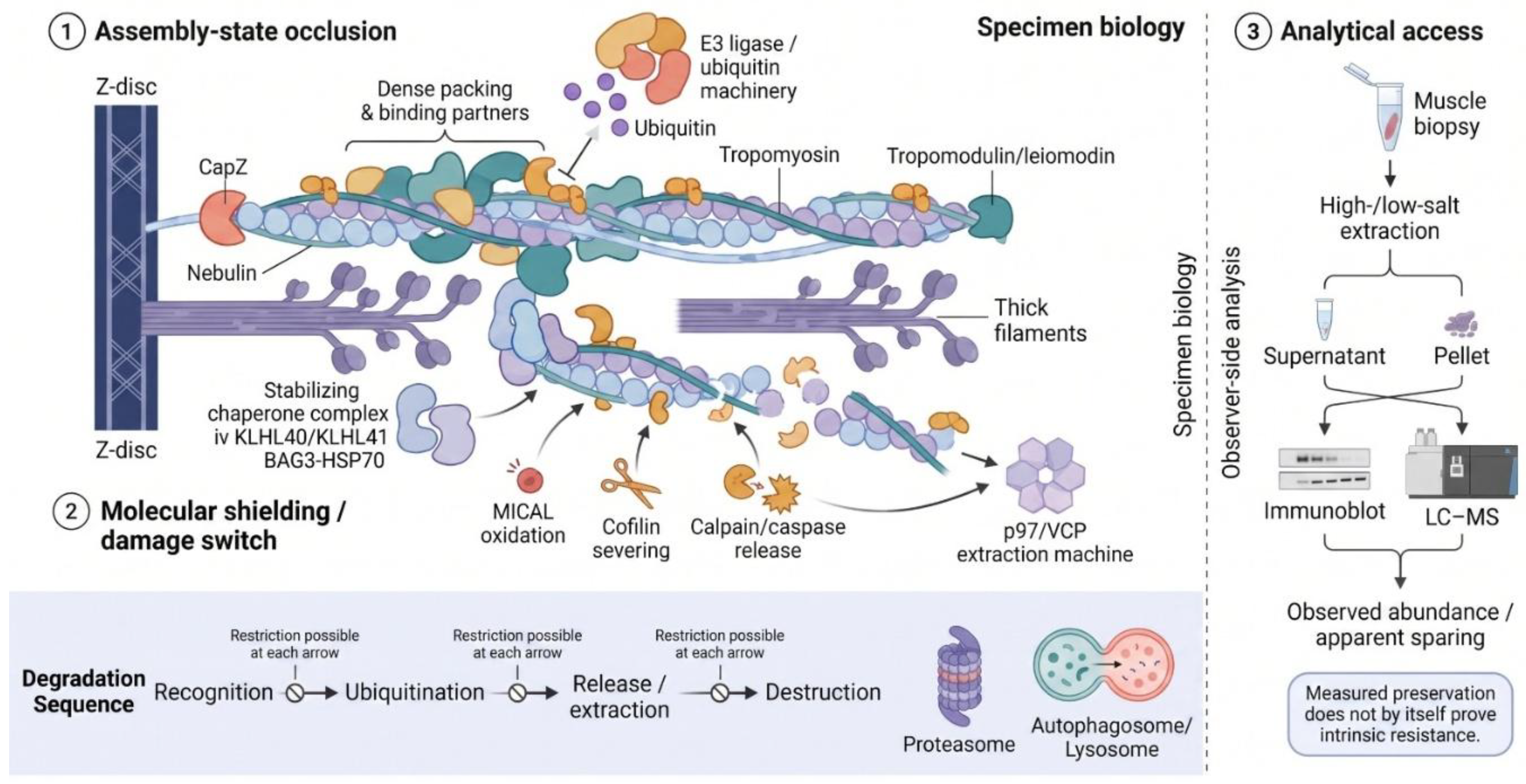
Figure 2.
Evidence map for access-related claims in muscle atrophy. Cells identify the strongest evidence type without assigning a composite quality score. Direct evidence is confined to the stated evidence class. Context-specific findings should not be generalized across atrophy models. The largely empty final row highlights the absence of protein-specific half-life and degradation-route measurements for access-setting actin-binding proteins during atrophy. Primary studies are cited with the corresponding discussion in Section 2, Section 3, Section 4 and Section 5. Created in BioRender. Lee, W. (2026) https://BioRender.com/98yg1sx.
Figure 2.
Evidence map for access-related claims in muscle atrophy. Cells identify the strongest evidence type without assigning a composite quality score. Direct evidence is confined to the stated evidence class. Context-specific findings should not be generalized across atrophy models. The largely empty final row highlights the absence of protein-specific half-life and degradation-route measurements for access-setting actin-binding proteins during atrophy. Primary studies are cited with the corresponding discussion in Section 2, Section 3, Section 4 and Section 5. Created in BioRender. Lee, W. (2026) https://BioRender.com/98yg1sx.
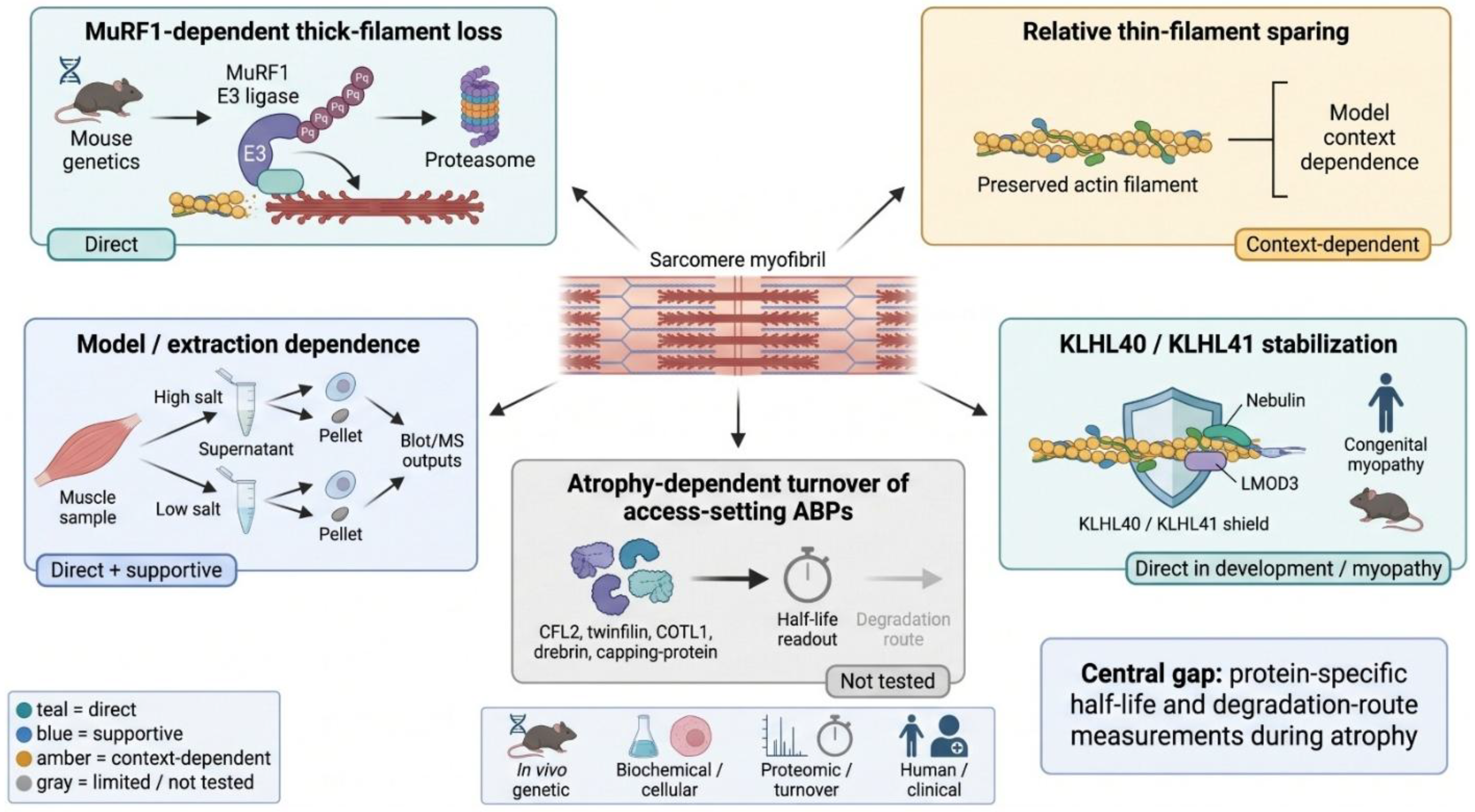

Table 1.
What common measurements establish and what they do not.
| Measurement | Positive result supports | Negative or sparing result does not prove | Access variable that must be controlled |
|---|---|---|---|
| In vitro ubiquitination | Biochemical competence of an E3-substrate pair | Modification of the assembled native protein in vivo | Protein conformation, binding partners, polymerization state |
| Ligase loss-of-function with immunoblot | Ligase dependence of abundance in the recovered pool | Absence of ligase binding or ubiquitination in another pool | Extraction efficiency, fraction recovery, model and time |
| Diglycine-remnant proteomics | Presence and regulation of ubiquitinated lysine sites | Proteasomal destruction or complete substrate attribution to one E3 | Protein abundance, peptide detectability, site occupancy, assembly state |
| Dynamic labeling of one fraction | Appearance or replacement within that fraction | Direct degradation when inter-fraction transfer is possible | Soluble-to-myofibrillar redistribution and abundance normalization |
| Preserved thin-filament band | Stability of the assayed, extractable pool | Intrinsic resistance to every ubiquitin ligase | Insoluble material, masked epitopes, filament packing |
Disclaimer/Publisher’s Note: The statements, opinions and data contained in all publications are solely those of the individual author(s) and contributor(s) and not of MDPI and/or the editor(s). MDPI and/or the editor(s) disclaim responsibility for any injury to people or property resulting from any ideas, methods, instructions or products referred to in the content. |
© 2026 by the authors. Licensee MDPI, Basel, Switzerland. This article is an open access article distributed under the terms and conditions of the Creative Commons Attribution (CC BY) license.
Copyright: This open access article is published under a Creative Commons CC BY 4.0 license, which permit the free download, distribution, and reuse, provided that the author and preprint are cited in any reuse.