

Submitted:
26 August 2026
Posted:
27 August 2026
You are already at the latest version
Abstract
Rhus pentaphylla Desf., a medicinal plant traditionally used in North Africa, remains insufficiently investigated for its glycosylated metabolites and biological properties. This study evaluated the influence of extraction solvent on the LC–ESI–MS profile and multifunctional bioactivities of aqueous, ethanolic, and hydroethanolic extracts of Moroccan R. pentaphylla. Thirty-nine metabolites were tentatively identified, predominantly hydroxycinnamic acid derivatives and flavonol glycosides. Chlorogenic acid, cryptochlorogenic acid, and quercetin glucuronide were among the most prominent constituents. The occurrence of several flavonoid glycosides highlights R. pentaphylla as a source of structurally diverse plant glycoconjugates. The hydroethanolic extract exhibited the highest phenolic content and the strongest antioxidant activity (DPPH IC50 = 32 µg/mL; FRAP = 81.57 mM FeSO4/g), whereas the aqueous and ethanolic extracts showed the most pronounced tyrosinase inhibition. All three extracts displayed comparable α-amylase inhibitory activity. Antibacterial screening revealed selective activity against Pseudomonas aeruginosa. At sub-inhibitory concentrations, the extracts also significantly reduced extracellular protein secretion and exopolysaccharide production, indicating antivirulence effects. Overall, the findings demonstrate that the extraction solvent affects the distribution of glycosylated and other bioactive metabolites in R. pentaphylla and influences the resulting biological activities. This work provides new insights into the phytochemical and biological significance of this medicinal species and supports further isolation, structural confirmation, and in vivo investigation of its active glycoconjugates.
Keywords:
Rhus pentaphylla
; glycoconjugates
; flavonol glycosides
; LC–ESI–MS
; quercetin glucuronide
; antioxidant activity
; enzyme inhibition
; Pseudomonas aeruginosa
; antivirulence activity
1. Introduction
Plant-derived bioactive compounds continue to fuel drug discovery efforts, largely because of the chemical and structural diversity they display and the range of pharmacological effects they can exert. Natural products, particularly phenolic secondary metabolites, have attracted tremendous interest for their enzyme-modulatory and antimicrobial properties, which are relevant to both metabolic disorders and infectious diseases. Within this context, species belonging to the genus Rhus (Anacardiaceae) have attracted considerable scientific interest owing to their diverse phytochemical composition and broad spectrum of biological activities [1]. Rhus pentaphylla Desf. is a xerophytic shrub native to Mediterranean and North African ecosystems, where traditional uses include treatments for inflammatory and infectious conditions [2,3,4]. Previous studies on Rhus species have revealed high levels of phenolic acids, flavonoids, and tannins, compounds frequently associated with antioxidant, antimicrobial, and enzyme inhibitory activities, supporting the therapeutic potential of the genus [5,6,7]. Phenolic phytochemicals are particularly noteworthy for their ability to interact with key enzymes implicated in metabolic and dermatological pathways. Among these, α-amylase inhibition represents an established strategy for controlling postprandial hyperglycemia through the reduction of carbohydrate digestion and glucose absorption [8,9,10,11]. Likewise, tyrosinase, the rate-limiting enzyme in melanogenesis, is an important target for managing hyperpigmentation disorders and developing cosmetic formulations. Several Rhus species have demonstrated promising inhibitory effects against these enzymes, suggesting that their phenolic constituents may contribute to multifunctional biological activities [12]. In addition to enzyme modulation, species of the genus Rhus have frequently demonstrated antibacterial activity against a broad panel of clinically relevant pathogens [13,14,15,16,17]. More recently, increasing attention has been directed toward antivirulence strategies targeting P. aeruginosa, particularly through the inhibition of quorum sensing, virulence factor production, and biofilm formation, thereby reducing the selective pressure associated with conventional antibiotics [18,19,20]. However, such properties remain largely unexplored in R. pentaphylla. Despite its ethnopharmacological relevance and growing evidence supporting the biological potential of Rhus species, comprehensive investigations integrating detailed phytochemical characterization with antioxidant, enzyme inhibitory, antibacterial, and antivirulence evaluations remain scarce for R. pentaphylla, particularly for Moroccan populations. In addition to enzyme modulation, species of the genus Rhus have frequently demonstrated antibacterial activity against a broad panel of clinically relevant pathogens [13,14,15,16,17]. More recently, growing attention has turned toward antivirulence strategies that attenuate bacterial pathogenicity by targeting virulence factor production and biofilm-associated processes, rather than bacterial viability itself, thereby limiting the selective pressure associated with conventional antibiotics [18,19,20]. Such properties, however, remain largely unexplored in R. pentaphylla, and no study to date has combined detailed phytochemical characterization with antioxidant, enzyme inhibitory, antibacterial, and antivirulence evaluations for this species, particularly in Moroccan populations. This gap is especially relevant given that secondary metabolite profiles in plants are known to vary with local environmental and geographical conditions, so that chemotypes from a given region cannot simply be assumed to reflect those already described elsewhere [21,22,23]. Against this background, the present study aimed to characterize the phytochemical composition of aqueous, hydroethanolic, and ethanolic extracts of Moroccan R. pentaphylla by LC-ESI-MS, and to evaluate their antioxidant, α-amylase inhibitory, tyrosinase inhibitory, antibacterial, and antivirulence activities. A secondary objective was to explore potential relationships between phytochemical composition and biological responses, in order to support the scientific validation of this species and inform its future valorization as a source of multifunctional natural bioactive compounds.
2. Material and Methods
2.1. Sample Preparation and Extraction
2.1.1. Plant Material
The aerial parts of R. pentaphylla Desf. were harvested in October 2023 from Douar Oued Baht, located approximately 15 km from the city of Khemisset, Morocco. Botanical identification was initially performed by Prof. Rouane (École Normale Supérieure, Rabat) and subsequently verified by Prof. Khamar (Department of Botany, Scientific Institute of Rabat). A voucher specimen (RAB114123) was deposited in the herbarium of the Scientific Institute of Rabat for future reference. The plant material was air-dried at room temperature, away from light, for 10 days to limit the degradation of light-sensitive metabolites. The dried material was then finely pulverized using a laboratory grinder (Retsch GM200) and stored in airtight containers at room temperature until extraction.
2.1.2. Ultrasound-Assisted Extraction
Extraction was performed by ultrasound-assisted extraction (UAE) using three solvent systems differing in polarity: distilled water, absolute ethanol, and a hydroethanolic mixture (ethanol/water, 50:50, v/v). Briefly, 10 g of the powdered aerial parts were mixed with 100 mL of extraction solvent, corresponding to a solid-to-solvent ratio of 1:10 (w/v). The suspensions were sonicated in a 50 kHz ultrasonic bath maintained at 45 °C for 20 min. Extractions were performed in triplicate for each solvent. Following extraction, the mixtures were centrifuged at 4000 rpm for 10 min and the supernatants filtered through Whatman No. 1 filter paper to remove residual plant debris. The filtrates were concentrated under reduced pressure using a BUCHI R-300 rotary evaporator (BÜCHI Labortechnik AG, Switzerland). The resulting crude extracts were weighed to determine extraction yield and stored at 4 °C until subsequent analyses.
2.1.3. Chemicals and Reagents
All chemicals and analytical reagents used in this study were of analytical grade. Folin–Ciocalteu reagent, gallic acid, quercetin, anthrone, bovine serum albumin (BSA), Bradford reagent, 2,2-diphenyl-1-picrylhydrazyl (DPPH), α-amylase, soluble starch, 3,5-dinitrosalicylic acid (DNS), mushroom tyrosinase, L-3,4-dihydroxyphenylalanine (L-DOPA), kojic acid, acarbose, crystal violet, and azithromycin were purchased from Sigma-Aldrich, St. Louis, USA. Unless otherwise specified, all aqueous solutions were prepared using distilled water.
2.2. Phytochemical Analysis
2.2.1. Determination of Total Phenolic Content (TPC)
The total phenolic content of the extracts was quantified using the Folin–Ciocalteu colorimetric assay according to the method reported by [24] with slight modifications. Briefly, 20 µL of each extract (1 mg/mL) was transferred into a 96-well microplate, followed by the addition of 100 µL of Folin–Ciocalteu reagent diluted to 10% (v/v). After an initial reaction period of 5 min at room temperature in the dark, 80 µL of a 7.5% (w/v) sodium carbonate solution was added. The reaction mixtures were allowed to develop for an additional 30 min before absorbance was recorded at 765 nm against the corresponding reagent blank using a BMG LABTECH microplate reader (Bath, UK). Gallic acid served as the calibration standard, and the results were expressed as milligrams of gallic acid equivalents per gram of dry extract (mg GAE/g DE). All determinations were carried out in triplicate.
2.2.2. Determination of Total Flavonoid Content (TFC)
Flavonoid concentration was estimated by the aluminum chloride colorimetric assay using quercetin as the reference compound [25]. For each analysis, 100 µL of extract solution (1 mg/mL) was combined with an aluminum chloride solution (1.2%) and a potassium acetate solution (120 mM). Following incubation at room temperature for 30 min, the absorbance of the resulting complexes was measured at 415 nm against the corresponding reagent blank. Flavonoid levels were calculated from a quercetin calibration curve and expressed as milligrams of quercetin equivalents per gram of dry extract (mg QE/g DE). Each sample was analyzed in triplicate.
2.2.3. Determination of Total Sugar Content (TSC)
Total soluble sugars were quantified using the anthrone assay according to the method reported by [26]. Briefly, 100 µL of extract was mixed with 900 µL of freshly prepared anthrone reagent, after which the reaction mixtures were heated in a boiling water bath (100 °C) for 7 min. After cooling to room temperature, 200 µL from each reaction mixture was transferred to a 96-well microplate, and the absorbance was measured at 620 nm against the corresponding reagent blank using a microplate reader (BMG LABTECH). Glucose was employed to generate the calibration curve, and sugar content was expressed as milligrams of glucose equivalents per gram of dry extract (mg GE/g DE). All measurements were performed in triplicate.
2.2.4. Determination of Total Protein Content (TP)
Protein concentration was determined using the Bradford colorimetric method, according to the procedure described by [27]. Briefly, 40 µL of extract solution (1 mg/mL) was transferred into the wells of a 96-well microplate and combined with 160 µL of Bradford reagent. Following a 5 min incubation at room temperature, absorbance was measured at 595 nm against the corresponding reagent blank. Quantification was achieved using a bovine serum albumin (BSA) standard curve, and the results were expressed as milligrams of bovine serum albumin equivalents per gram of dry extract (mg BSAE/g DE). Each determination was conducted in triplicate.
2.2.5. LC-ESI-MS/MS Analysis
The phytochemical composition of the extracts was characterized using a Shimadzu LCMS-8050 liquid chromatography–tandem mass spectrometry system (Shimadzu, Kyoto, Japan) equipped with a triple quadrupole mass analyzer and an electrospray ionization (ESI) source. Chromatographic separation was performed on a Zorbax Eclipse XDB-C18 reversed-phase column (4.6 × 150 mm, 3.5 µm; Agilent Technologies, Santa Clara, CA, USA). The mobile phase consisted of water (solvent A) and acetonitrile (solvent B), both containing 0.1% formic acid. A linear gradient was applied from 5% to 90% solvent B over 60 min at a flow rate of 1.0 mL min−1. Samples (10 µL, 10 mg mL−1) were introduced using a SIL-40C XS autosampler, and instrument operation and data acquisition were managed with LCsolution software (Shimadzu, Japan). Mass spectrometric analyses were performed in the negative ionization mode, with spectra acquired over an m/z range of 100–1500. MS/MS fragmentation experiments were conducted using collision energies of 35 and 45 eV. Tentative identification of the detected metabolites was achieved by comparing retention times, precursor ions, and MS/MS fragmentation patterns with previously published data and spectral information available in the literature [28].
2.3. Antioxidant Activity
2.3.1. DPPH Radical Scavenging Assay
The free radical scavenging capacity of the extracts was evaluated using the DPPH assay following the method described by [29]. In brief, serial dilutions of each extract were prepared to obtain concentrations ranging from 0.001 to 1 mg/mL. An aliquot of 20 µL from each dilution was transferred to a 96-well microplate and combined with 180 µL of a freshly prepared 0.1 mM methanolic DPPH solution. The reaction mixtures were then incubated in the dark at room temperature for 30 minutes before the absorbance was recorded at 517 nm using a BMG LABTECH microplate reader (Bath, UK). Methanol was used as the blank and ascorbic acid as the positive control. Radical scavenging activity was calculated using the following equation:
where Acontrol and Asample represent the absorbance of the control and sample, respectively. The concentration required to inhibit 50% of the DPPH radicals (IC50) was determined from the corresponding dose–response curves. All measurements were performed in triplicate.
DPPH inhibition effect (%)= ({Acontrol - Asample}/{Acontrol}) ×100
2.3.2. Ferric Reducing Antioxidant Power (FRAP) Assay
The ferric reducing capacity of the extracts was determined according to the potassium ferricyanide method described by [30], adapted from Oyaizu (1986). In brief, 30 μL of each extract was transferred to a 96-well microplate and successively mixed with 100 μL of distilled water, 45 μL of 1 M hydrochloric acid (HCl), 45 μL of 1% potassium ferricyanide (w/v), 15 μL of 0.2% sodium dodecyl sulfate (SDS), and 15 μL of 1% ferric chloride (FeCl3) (w/v). The reaction mixtures were then incubated at 50 °C for 20 minutes. The absorbance was subsequently recorded at 700 nm using a BMG LABTECH microplate reader (Bath, UK). The reducing power of the extracts was quantified using a ferrous sulphate (FeSO4) calibration curve and expressed as millimolar FeSO4 equivalents per milligram of extract (mM FeSO4/g). All analyses were performed in triplicate.
2.4. Enzymatic Activity
2.4.1. α-. Amylase Inhibition
The inhibitory activity of the extracts against α-amylase was evaluated according to the procedure described by [31], with minor modifications. In brief, 100 μL of each extract solution was mixed with 100 μL of an α-amylase solution (1 U/mL), which was prepared in phosphate-buffered saline (PBS), and the mixture was pre-incubated at 37 °C for 10 minutes. Then, 100 μL of a 1% soluble starch solution was added as the enzyme substrate, and the reaction mixtures were incubated for a further 30 minutes at the same temperature. The reaction was then terminated by the addition of 200 μL of 3,5-dinitrosalicylic acid (DNS) reagent, after which the tubes were heated in a boiling water bath for 10 minutes. Once cooled to room temperature, 1.5 mL of distilled water was added to each tube, and absorbance was measured at 540 nm using a UV–Vis spectrophotometer. Acarbose was included as the positive control. The percentage of enzyme inhibition was calculated according to the following equation:
Where control corresponds to 100% enzyme activity. All assays were performed in triplicate.
α-amylase inhibition (%) = ({Acontrol - Asample}/{Acontrol}) ×100
2.4.2. Tyrosinase Inhibition
Tyrosinase inhibitory activity was assessed using mushroom tyrosinase and L-3,4-dihydroxyphenylalanine (L-DOPA) as the substrate, following the method reported by [32]. Each reaction mixture contained 20 μL of extract, 20 μL of a 2500 U/mL mushroom tyrosinase solution, and 100 μL of a 0.05 M phosphate buffer solution at pH 6.5. The enzymatic reaction was initiated by adding 20 μL of a 5 mM L-DOPA solution. After a further 20 min incubation period, dopachrome formation was monitored by measuring the absorbance at 475 nm against the corresponding reagent blank, using a microplate reader (BMG LABTECH). Kojic acid served as the reference inhibitor.
where Acontrol and Asample represent the absorbance of the control and sample, respectively
Tyrosinase inhibition (%) = [(Acontrol − Asample)/Acontrol] × 100,
All experiments were carried out in triplicate.
2.5. Antibacterial Activities
2.5.1. Disc Diffusion Test
The antibacterial potential of the extracts was investigated using an agar disc diffusion assay against five bacterial strains: Pseudomonas aeruginosa, Escherichia coli, Enterococcus faecalis, Staphylococcus aureus, and Salmonella typhi. An aliquot of 100 µL from an overnight bacterial culture was uniformly distributed over the surface of Mueller–Hinton agar plates. Sterile paper discs (6 mm in diameter) were loaded with 30 µL of the 100 mg/mL extract solution and carefully positioned on the inoculated agar. The plates were then incubated at 37 ± 2 °C for 24 hours. Ampicillin (40 µg/mL) was used as the reference antibacterial agent, while the corresponding extraction solvents served as negative controls. Antibacterial efficacy was determined by measuring the diameter of the growth inhibition zones surrounding each disc, and was expressed in millimetres (mm) [33]. All assays were conducted in triplicate
2.5.2. MIC Determination for Extracts
Based on the preliminary agar disc diffusion screening, only the ethanolic and hydroethanolic extracts of R. pentaphylla were selected for MIC determination against P. aeruginosa, while the aqueous extract was excluded due to its weak antibacterial activity. The minimum inhibitory concentration of the extracts was established using the broth microdilution technique in 96-well microplates, in accordance with the procedures outlined by [30,31,32,33,34]. The extracts were subjected to two-fold serial dilutions in Mueller–Hinton broth to obtain final concentrations ranging from 100 to 1.562 mg mL−1. Prior to testing, all extract solutions were sterilized by passage through 0.22 µm syringe filters. Aliquots (200 µL) of each dilution were dispensed into the microplate wells, followed by inoculation with 2 µL of a bacterial suspension adjusted to an OD600 of 0.6. Wells containing bacterial inoculum without extract served as growth controls, while uninoculated Mueller–Hinton broth was included as the negative control. After incubation at 37 °C for 18 h with shaking (150 rpm), bacterial growth was assessed visually and by measuring absorbance at 600 nm. The MIC was defined as the lowest extract concentration showing complete inhibition of visible bacterial growth.
2.5.3. Biofilm Inhibition Assay
The antibiofilm activity of R. pentaphylla extracts against P. aeruginosa was evaluated using the crystal violet microplate assay [35,36]. Based on MIC results, only the ethanolic and hydroethanolic extracts were tested at sub-inhibitory concentrations at 1/8 and 1/4 MIC. Sterile extract solutions were prepared in MHB, inoculated with a standardized bacterial suspension, and incubated under the same conditions as the MIC assay. Following incubation, non-adherent cells were removed, and the remaining biofilm was stained with 1% crystal violet for 15 minutes. After washing and air-drying, the retained dye was solubilized with 95% ethanol, and biofilm biomass was quantified by measuring absorbance at 595 nm using a microplate reader. Wells containing inoculated medium without extract supplementation served as positive controls, while uninoculated medium served as the negative control. Azithromycin (2 µg/mL) was included as the reference antibiofilm agent. All experiments were performed in triplicate.
2.5.4. Outer Membrane Protein Quantification
The effect of R. pentaphylla extracts on the production of virulence-associated proteins by P. aeruginosa was assessed using the Bradford colorimetric assay [37,38]. Based on MIC results, only ethanolic and hydroethanolic extracts were tested at sub-inhibitory concentrations of 1/8 and 1/4 of the MIC. Bacterial cultures were incubated in the presence or absence of the extracts for 24 hours under the same conditions used for the MIC assay. After centrifugation at 10,000×g for 12 minutes, the resulting supernatant was filtered through a 0.45 µm syringe filter. The extracellular protein content was then determined by measuring the absorbance at 595 nm. Azithromycin (2 µg/mL) was included as the reference control, while uninoculated culture medium served as the blank. All assays were performed in triplicate [39].
2.5.5. Exopolysaccharide Estimation
In addition to total protein quantification, extracellular polymeric substances (EPS) were extracted from the collected supernatants by precipitation with cold 95% ethanol, followed by overnight incubation at 4 °C. The resulting EPS pellets were used to determine the total carbohydrate content according to established methods. Azithromycin (2 µg/mL) was included as a reference control, while uninoculated media served as blanks [39].
2.6. Statistical Analysis
All experiments were performed in triplicate, and the results are expressed as mean ± standard deviation (SD). Statistical analyses were carried out using GraphPad Prism version 8.0.1 (GraphPad Software, San Diego, CA, USA). Differences between the extracts were evaluated using one-way analysis of variance (ANOVA), followed by a Tukey’s multiple-comparisons test. Prior to ANOVA, the assumption of homogeneity of variances was verified using the Brown–Forsythe test. Statistical significance was established at P < 0.05.
3. Results
3.1. Phytochemical Composition and Extraction Yield
The yields of the three dry crude extracts from the aerial part of R. pentaphylla—namely the aqueous, the ethanolic, and the 50% ethanolic extracts—are expressed as percentages and shown in Table 1, along with their phenolic content. The hydroethanolic extract consistently exhibited the highest yield of total phenolics (TPC) and flavonoids (TFC), compared with aqueous and ethanolic extracts, underlining the superior extraction efficiency of water/ ethanol (50:50, v/v) for phenolic compounds.
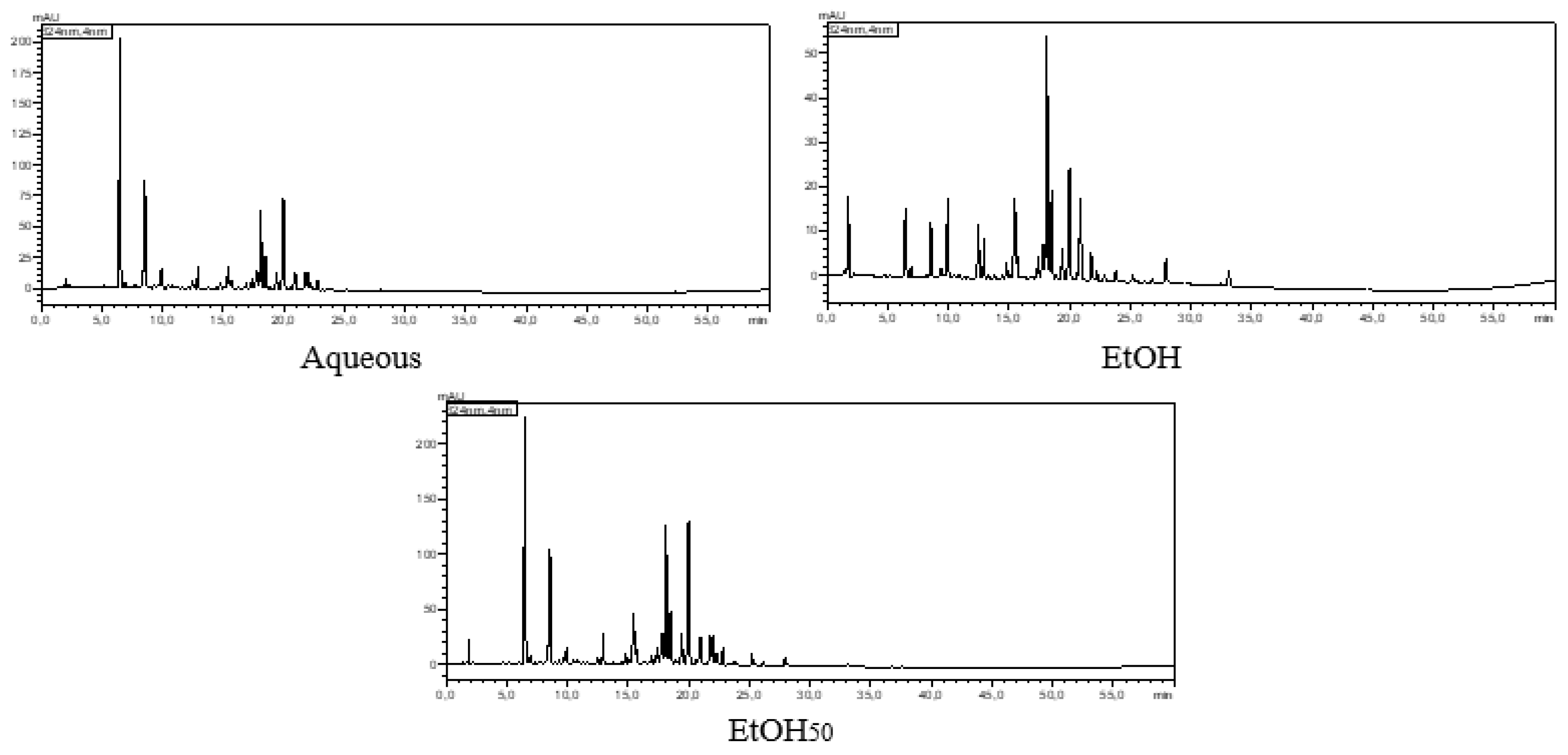
LC–ESI–MS analysis enabled the tentative identification of 39 metabolites, predominantly phenolic acids, flavan-3-ols, coumarins, and flavonoid glycosides (Table 2). The phytochemical profile of R. pentaphylla was characterized by a high abundance of hydroxycinnamic acid derivatives and flavonol glycosides, with notable qualitative and semi-quantitative differences among the aqueous, hydroethanolic, and ethanolic extracts. The detection of several glycosylated phenolic compounds, including caffeic acid glucoside, coumaric acid glycosides, and flavonol glycosides, highlights R. pentaphylla as a potential source of structurally diverse plant glycoconjugates. Glycosylation can influence the polarity, solubility, stability, extractability, and biological properties of phenolic aglycones, which may partly explain their differential distribution among the three extraction solvents.
Among the phenolic acids identified, chlorogenic acid and cryptochlorogenic acid were the most abundant, particularly in the aqueous and hydroethanolic extracts, where they exhibited the highest relative intensities. Other hydroxycinnamic acid derivatives, including caffeic acid, caffeic acid glucoside, p-coumaroylquinic acid, coumaric acid, and coumaric acid glycosides, were also detected, confirming the richness of R. pentaphylla in phenylpropanoid metabolites and their conjugated forms. In contrast, several coumaric acid derivatives and esculetin were more abundant in the ethanolic extract, suggesting that solvent polarity selectively influenced the extraction of moderately polar constituents. Overall, these findings indicate that the extraction solvent plays a decisive role in the recovery and distribution of free and glycosylated phenolic metabolites from R. pentaphylla.
Figure 1.
LC-MS/MS chromatograms of R. pentaphylla extracts.
3.2. Antioxidant Activity
The antioxidant capacity of each extract, assessed by DPPH and FRAP assays (Table 3), revealed that the hydroethanolic extract exhibited the strongest radical scavenging activity (IC50 = 32.01 μg/mL), as well as ferric-reducing antioxidant power (81.57 mM of FeSO4/g), followed by the ethanolic extract (IC50 = 39.60 μg/mL,70.44 mM FeSO4/g). In contrast, the aqueous extract showed significantly lower activity (IC50 = 87.01 μg/mL). Although all extracts were less active than the reference compound, ascorbic acid (IC50 = 3.60 μg/mL), these findings highlight the strong antioxidant potential of the hydroethanolic extract of R. pentaphylla, which aligns with its higher phenolic content, supporting the well-established correlation between phenolic compounds and antioxidant activity.
3.3. Enzyme Inhibitory Activities
Table 4 presents the inhibitory activities of the aqueous, 50% EtOH, and 100% EtOH extracts of Rhus pentaphylla against α-amylase and tyrosinase. The inhibitory activity varied depending on both the extraction solvent and the target enzyme. For α-amylase, the aqueous extract exhibited the lowest IC50 value (1.25 ± 0.08 mg/mL), followed by the 50% ethanolic (1.49 ± 0.09 mg/mL) and 100% ethanolic (1.61 ± 0.12 mg/mL) extracts. However, one-way ANOVA revealed no statistically significant differences among the three extracts (P = 0.0968), although all were less potent than the reference inhibitor, acarbose (IC50 = 0.61 ± 0.03 mg/mL). In contrast, significant differences were observed for tyrosinase inhibition (P = 0.0091). The aqueous (IC50 = 0.279 ± 0.01 mg/mL) and 100% ethanolic (IC50 = 0.270 ± 0.05 mg/mL) extracts exhibited comparable inhibitory activities and were significantly more active than the 50% ethanolic extract (IC50 = 0.420 ± 0.04 mg/mL). Nevertheless, kojic acid remained the most potent tyrosinase inhibitor (IC50 = 0.060 ± 0.05 mg/mL). These findings indicate that the extraction solvent influences the recovery of bioactive compounds involved in enzyme inhibition. Interestingly, the strongest tyrosinase inhibitory activity was observed for the aqueous and 100% EtOH extracts rather than the 50% EtOH extract, suggesting that the compounds responsible for tyrosinase inhibition differ from those primarily contributing to antioxidant activity and total phenolic content.
3.4. Antibacterial Activity
The antibacterial properties of R. pentaphylla extracts were evaluated against five bacterial strains using both qualitative and quantitative approaches. Initial screening was performed using the agar disc diffusion assay to measure inhibition zone diameters (ZD). The minimum inhibitory concentration (MIC) was then determined using the broth microdilution method to assess the inhibitory potency of the most active extracts. The antibacterial results are summarized in Table 5.
As shown in Table 5, the ethanolic extract exhibited the highest antibacterial activity against P. aeruginosa, producing an inhibition zone of around 8 mm, while the hydroethanolic extract followed closely behind. In contrast, the aqueous extract showed little or no activity. All three extracts displayed negligible inhibitory effects against E. coli, E. faecalis, S. aureus, and S. typhi, as reflected by the small inhibition zones recorded. The positive control (ampicillin) produced markedly larger inhibition zones against all the tested strains, confirming its superior antibacterial activity compared to the plant extracts. These results suggest that R. pentaphylla extracts have a selective antibacterial effect, with P. aeruginosa being the most susceptible microorganism among the tested strains.
Following the preliminary screening by the agar disc diffusion test, the ethanolic and hydroethanolic extracts of R. pentaphylla, which demonstrated antibacterial activity against P. aeruginosa, were selected for MIC determination. As shown in Table 6, both extracts exhibited an MIC of 100 mg/mL, indicating their ability to completely inhibit visible bacterial growth at this concentration. Nevertheless, differences in antibacterial potency were observed, as the ethanolic extract achieved a growth inhibition rate of 98.25%, while the hydroethanolic extract produced an inhibition rate of 81.56% at the MIC concentration. In contrast, the reference antibiotic ampicillin displayed a markedly lower MIC value (31.5 µg/mL), reflecting its superior antibacterial efficacy against P. aeruginosa.
Anti-biofilm and Virulence-Related Activities
In recent years, targeting bacterial virulence rather than bacterial viability has gained increasing attention as an alternative strategy to address antimicrobial resistance. Among the virulence traits of P. aeruginosa, biofilm formation is a key factor contributing to persistence and reduced susceptibility to antimicrobial treatments. Therefore, the antibiofilm activity of the ethanolic and hydroethanolic extracts of R. pentaphylla was investigated at 1/8 and 1/4 MIC. As illustrated in Figure 2, both extracts produced only a modest reduction in biofilm biomass relative to the untreated control, indicating a limited inhibitory effect on biofilm formation.
Besides biofilm formation, P. aeruginosa secretes numerous virulence factors that contribute to its pathogenic potential, including extracellular proteases and other proteinaceous compounds involved in host colonization and tissue damage. Since the suppression of bacterial virulence has emerged as a promising alternative to conventional antimicrobial approaches, the effect of R. pentaphylla extracts on the production of extracellular proteins was investigated as an indicator of virulence attenuation. The ability of P. aeruginosa to simultaneously produce biofilms and virulence-associated proteins is a major factor underlying its persistence and resistance in clinical settings.
Given the contribution of extracellular proteins to the virulence of P. aeruginosa, the effect of R. pentaphylla extracts on protein secretion was investigated at sub-MIC concentrations. As shown in Figure 3, the ethanolic fraction significantly reduced extracellular protein production at 1/4 MIC (25 mg/mL), whereas the hydroethanolic extract produced only a slight reduction relative to the untreated control. Notably, the inhibitory effect of the ethanolic extract was comparable to that observed with azithromycin (2 μg/mL). Overall, the ethanolic extract displayed a greater capacity to suppress protein secretion than the hydroethanolic extract.
In addition to extracellular proteins, the effect of Rhus pentaphylla extracts on the production of exopolysaccharides, another key virulence component involved in biofilm establishment and stability, was evaluated. As shown in Figure 4, the ethanolic extract at 1/4 MIC (25 mg/mL) significantly decreased EPS production compared with the untreated control, reaching levels comparable to those obtained with azithromycin (2 µg/mL). In contrast, the hydroethanolic extract exhibited a weaker inhibitory effect on EPS secretion. Overall, the ethanolic extract demonstrated a greater capacity to reduce major virulence-associated factors of P. aeruginosa than the hydroethanolic extract.
4. Discussion
Oxidative stress is increasingly recognized as a central pathogenic mechanism underlying a wide spectrum of chronic disorders, including metabolic diseases, dermatological conditions, microbial infections, and neurodegenerative, cardiovascular, hepatic, and renal pathologies [40,41,42,43,44,45]. Excessive generation of reactive oxygen species (ROS) promotes lipid peroxidation, protein oxidation, DNA damage, and dysregulation of cellular signaling pathways, ultimately leading to chronic inflammation and tissue dysfunction [46]. Consequently, medicinal plants capable of simultaneously modulating oxidative stress and multiple disease-associated biological targets have attracted considerable attention as potential sources of multifunctional therapeutic agents. Within this context, the present study provides a comprehensive evaluation of Moroccan R. pentaphylla, integrating extraction efficiency, phytochemical characterization by LC-MS, and the assessment of antioxidant, enzyme inhibitory, antibacterial, and antivirulence activities. Extraction efficiency was markedly influenced by solvent composition, with the hydroethanolic extract yielding substantially higher recovery than the aqueous and ethanolic extracts. Such findings are consistent with previous studies reporting that mixtures of water and ethanol improve the extraction of phytochemicals with diverse polarities through complementary physicochemical mechanisms, whereby water facilitates plant tissue swelling and metabolite release while ethanol enhances solubilization of moderately polar compounds [47,48,49,50,51]. Although the hydroethanolic extract yielded the greatest recovery, biological activity was more closely associated with phytochemical composition than extraction efficiency. LC-MS analysis revealed chlorogenic acid, cryptochlorogenic acid, and quercetin glucuronide as the predominant metabolites in all extracts. These metabolites are recognized for their diverse biological properties. Chlorogenic acid and its isomers have demonstrated potent radical-scavenging activity, metal-chelating capacity, and protection against oxidative DNA damage [52,53,54], whereas quercetin 3-O-β-D-glucuronide, the major flavonoid conjugate identified in the present study, has been reported to exhibit antioxidant, anti-inflammatory, moisturizing, and antimelanogenic activities [55]. Therefore, the biological activities observed in R. pentaphylla extracts may be partially attributed to these metabolites. However, synergistic interactions among major and minor constituents likely contribute to the overall bioactivity of the extracts. The significant antioxidant activities exhibited by the hydroethanolic extract (IC50 = 32 µg/mL and 81.57 mM FeSO4/g in FRAP) are consistent with previous reports describing R. pentaphylla as a rich source of phenolic compounds with strong radical-scavenging and reducing capacities [56,57]. Nevertheless, the antioxidant effectiveness of plant extracts depends on more than just the amount of phenolic compounds present; it also depends on their chemical structure, relative abundance, and potential interactions among individual constituents [58,59,60]. This may explain why the extract presenting the highest extraction yield did not necessarily exhibit the strongest activity across all biological assays, emphasizing that different phytochemical classes may selectively contribute to antioxidant and enzyme inhibitory properties. Beyond direct antioxidant protection, modulation of carbohydrate metabolism represents an important strategy for limiting oxidative stress-associated damage. Chronic hyperglycemia promotes excessive ROS generation, protein glycation, and inflammatory responses, thereby contributing to the development of diabetic complications [61,62]. In this context, the α-amylase inhibitory activity observed in R. pentaphylla extracts suggests the ability to moderate carbohydrate digestion and attenuate postprandial glucose excursions [8]. Although the inhibitory effects remained lower than those of acarbose, the three extracts exhibited comparable inhibitory activities, with the aqueous extract showing the lowest IC50 value. However, the differences among the extracts were not statistically significant, indicating that α-amylase inhibition was only marginally influenced by the extraction solvent. To our knowledge, this study constitutes the first report describing α-amylase inhibition in R. pentaphylla. Similar α-amylase inhibitory activity has been reported in Rhus coriaria, where flavonoids and gallotannins were identified as major contributors to enzyme inhibition [63]. Beyond carbohydrate-hydrolyzing enzymes, members of the genus Rhus have also attracted considerable attention for their tyrosinase inhibitory properties, which have been largely associated with their phenolic constituents [64,65]. The inhibitory activity of flavonoids toward tyrosinase is strongly influenced by their structural characteristics, including the number and position of hydroxyl groups, their capacity to chelate the copper ions present in the enzyme active site, and the degree of glycosylation [66,67]. Therefore, the tyrosinase-inhibitory activity observed in R. pentaphylla, particularly in the aqueous and ethanolic extracts, further supports its multifunctional potential, extending beyond metabolic regulation to the prevention of oxidative stress-associated skin disorders. Interestingly, despite the hydroethanolic extract possessing the highest phenolic content, it exhibited the lowest tyrosinase inhibitory activity, suggesting that enzyme inhibition depends not only on the total phenolic concentration but also on the qualitative composition and relative abundance of individual phenolic constituents. The increasing emergence of antibiotic-resistant pathogens has stimulated the search for alternative antimicrobial strategies, particularly those targeting bacterial virulence rather than exclusively inhibiting bacterial growth. P. aeruginosa is a clinically relevant opportunistic pathogen characterized by intrinsic resistance mechanisms and its capacity to establish persistent infections through biofilm formation and the secretion of numerous virulence factors. In the present study, the antibacterial activity of R. pentaphylla extracts was strain-dependent, with the ethanolic and hydroethanolic extracts exhibiting activity predominantly against P. aeruginosa. However, negligible inhibition was observed against E. coli, E. faecalis, S. aureus, and S. typhi. Variations in bacterial susceptibility to phenolic-rich plant extracts have been attributed to differences in cell envelope architecture, membrane permeability, and intrinsic resistance mechanisms, which collectively influence the accessibility and effectiveness of bioactive compounds [68,69]. Previous studies on R. pentaphylla have also reported variable antibacterial activities depending on extraction methods, solvent polarity, and tested microorganisms [23,56,57]. The MIC evaluation demonstrated that both ethanolic and hydroethanolic extracts inhibited the visible growth of P. aeruginosa at 100 mg/mL. Although this activity remained considerably lower than that of ampicillin (MIC = 31.5 µg/mL), the ethanolic extract exhibited a greater inhibitory efficacy (98.25%) compared with the hydroethanolic extract (81.56%). Such relatively high MIC values are frequently reported for crude plant extracts because their activity depends on the concentration and interactions among numerous constituents rather than on purified antimicrobial molecules [70]. Besides growth inhibition, one of the most noteworthy findings of the present study was the ability of R. pentaphylla extracts to attenuate key virulence-associated traits of P. aeruginosa. While biofilm inhibition remained limited, both extracts significantly reduced extracellular protein secretion and exopolysaccharide production at subinhibitory concentrations, with the ethanolic extract displaying the most pronounced effects. These observations are particularly relevant because extracellular proteins and exopolysaccharides constitute essential components of the P. aeruginosa virulence arsenal and biofilm matrix, contributing to bacterial persistence, host colonization, and resistance to antimicrobial treatments. The suppression of these factors suggests that R. pentaphylla may interfere with bacterial pathogenicity independently of direct bactericidal activity. This antivirulence activity may be associated with the phenolic profile of the extract, particularly the presence of quercetin glucuronide and chlorogenic acid derivatives. Quercetin has been shown to inhibit quorum sensing, biofilm formation, and virulence-factor production in P. aeruginosa [71], while comparable anti-quorum-sensing activities have also been reported for flavonoid-rich plant fractions [72]. Although quercetin glucuronide was identified in the present study, further investigations are required to determine its specific contribution to these biological effects. Among the metabolites identified in R. pentaphylla, chlorogenic acid has been widely reported to possess antibacterial activity through alterations of bacterial membrane integrity and cellular function [73]. However, its specific contribution to the quorum-sensing inhibition and antibiofilm effects observed in the present study remains to be elucidated. Taken together, these results indicate that solvent polarity governs not only the extraction efficiency of R. pentaphylla phytochemicals but also which biological activities are preferentially expressed, so that no single extract performs best across all the endpoints tested. This solvent-dependent selectivity, rather than total metabolite yield, appears to be the main driver of the multifunctional profile observed in this species, and supports a rationale for combining or sequentially using different extracts depending on the intended application. These findings should nonetheless be interpreted in light of the exploratory, in vitro nature of the study: metabolite identifications remain tentative, the tested extracts are crude mixtures, and no isolated compound or in vivo model was evaluated. Bioassay-guided fractionation, mechanistic investigations, and in vivo validation are therefore needed to confirm the active constituents and the therapeutic relevance of these effects.
5. Conclusion
This study provides the first integrated characterization of the phytochemical composition and multifunctional biological properties of Moroccan R. pentaphylla extracts. LC–ESI–MS analysis enabled the tentative identification of 39 metabolites, predominantly hydroxycinnamic acid derivatives and flavonol glycosides. Caffeic acid glucoside, coumaric acid glycosides, quercetin glucuronide, and other flavonoid glycosides highlight this species as a potential source of structurally diverse plant glycoconjugates. Their differential distribution among the aqueous, ethanolic, and hydroethanolic extracts indicates that solvent polarity strongly influences the recovery of free and glycosylated phenolic constituents. The hydroethanolic extract exhibited the highest total phenolic content and the strongest antioxidant activity, whereas the aqueous and ethanolic extracts showed the most pronounced tyrosinase inhibition. Although the three extracts displayed comparable α-amylase inhibitory effects, their antibacterial and antivirulence properties differed substantially. The ethanolic extract showed the strongest activity against P. aeruginosa and markedly reduced extracellular protein secretion and exopolysaccharide production at sub-inhibitory concentrations, while biofilm inhibition remained modest. Overall, no single extract combined all the optimal properties: the hydroethanolic extract was the most promising for antioxidant applications, whereas the ethanolic extract exhibited the greatest antibacterial and antivirulence potential. These solvent-dependent effects may reflect qualitative and semi-quantitative differences in glycosylated and non-glycosylated metabolites; however, direct compound–activity relationships cannot be established from the present data. Bioassay-guided fractionation, isolation, complete structural elucidation, and quantitative analysis of the principal glycoconjugates are therefore required. Mechanistic studies and in vivo validation will also be necessary to confirm the therapeutic and cosmetological potential of R. pentaphylla.
Author Contributions
Fatima-ezzahra EN-NACIRI: Conceptualization, Methodology, Investigation, Formal analysis, Data curation, Visualization, Writing—original draft. Patrick MARTIN: Supervision, Methodology, Resources, Writing—review and editing. Maryline ABERT VIAN and Nicolas JOLY: Resources, Writing—review and editing. Nihad SAHRI: Methodology, Investigation (LC–MS analysis), Data curation. Asmae ALAOUI: Conceptualization, Supervision, Validation, Resources, Review & editing the original draft.
Funding
The authors received no external funding to conduct this study.
Data Availability Statement
The original contributions presented in this study are included in the article. Further inquiries can be directed to the corresponding authors.
Acknowledgments
The authors gratefully acknowledge the technical support and laboratory facilities provided by their respective institutions.
Conflicts of Interest
The authors declare no conflict of interest.
References
- Opiyo SA, Njoroge PW, Ndirangu EG, Kuria KM. A review of biological activities and phytochemistry of Rhus species. Am. J. Chem. 2021;11(2):28–36. [CrossRef]
- Benamar H, Boulekbache-Makhlouf L, Meddah B, Meddour A, Mahdjoub M, Madani K, et al. Bioactive compound contents and biological activities of the Algerian medicinal plant Rhus pentaphylla (Jacq.) Desf. (Anacardiaceae). Biol. Life Sci. Forum. 2021;6(1):68. [CrossRef]
- Elmarrkechy S, Mahdi I, Fahsi N, El Herradi EH, Sahri N, Tourabi M, et al. Could Searsia pentaphylla be a source of new bioactive compounds? A bibliometric overview. Food Biosci. 2024;62:105349. [CrossRef]
- Messaoudi N, et al. Vasorelaxant and antihypertensive effects of Rhus pentaphylla (Searsia pentaphylla). Int. J. Pharm. Sci. Res. 2019;10(10):4430–4443. [CrossRef]
- Elmakaoui A, Damour H, Bourais I, Badaoui B, Imtara H, El Kabous K, et al. Chemical composition, antioxidant potential and antibacterial activity of Pistacia atlantica Desf. essential oil leaves, with a focus on variations in the main trunk diameter. Rec. Nat. Prod. 2024;18(2):220–236. [CrossRef]
- Uuh-Narvaez JJ, Segura-Campos MR, Sytar O. Antioxidant potential and known secondary metabolites of rare or underutilized plants of the Yucatan region. Future Pharmacol. 2023;3(4):664–685. [CrossRef]
- El Kamari F, El Omari H, El-Mouhdi K, Chlouchi A, Harmouzi A, Lhilali I, et al. Effects of different solvents on the total phenol content, total flavonoid content, antioxidant, and antifungal activities of Micromeria graeca L. from the Middle Atlas of Morocco. Biochem. Res. Int. 2024;2024:9027997. [CrossRef]
- Kaur N, Kumar V, Nayak SK, Wadhwa P, Kaur P, Sahu SK. Alpha-amylase as a molecular target for the treatment of diabetes mellitus: a comprehensive review. Chem. Biol. Drug Des. 2021;98(4):539–560. [CrossRef]
- Mahmud DA, Islam MM, Rahaman MM, Ripon MAR, Quarishi SB, Hossain MS. Relation of serum α-amylase level with glycated hemoglobin and trace elements in patients with diabetes mellitus. AACE Endocrinol. Diabetes. 2026;13(2):253–258. [CrossRef]
- Kashtoh H, Baek KH. New insights into the latest advances in plant-derived α-amylase inhibitors with antidiabetic effects. Plants. 2023;12(16):2944. [CrossRef]
- Nakajima K. Low serum amylase and obesity, diabetes and metabolic syndrome: a novel interpretation. World J. Diabetes. 2016;7(6):112–121. [CrossRef]
- Park SJ, Kim MS, Han JW, Kim WS, Lee JE, Jang MK. Bioactivity of supercritical fluid extracts from Rhus chinensis. J. Agric. Life Environ. Sci. 2024;36(4):396–409. [CrossRef]
- Mutuku A, Wanjala C, Kigondu E, Kipyegon C, Lagat R, Mwangi J, et al. Evaluation of the antimicrobial activity and safety of Rhus vulgaris (Anacardiaceae) extracts. BMC Complement. Med. Ther. 2020;20:272. [CrossRef]
- Rayne S, Mazza G. Biological activities of extracts from sumac (Rhus spp.): a review. Nat. Preced. 2007. [CrossRef]
- Çalışkan R, Sari SP, Altınbaşak BB, Dinç HÖ, Issa G, Yüksel Mayda P. Bioactive components and antioxidant and antimicrobial activities of Rhus coriaria, a sumac species found in Turkey. Bezmialem Sci. 2022;10(6):796–804. [CrossRef]
- Abdel-Mawgoud M, Khedr FG, Mohammed EI. Phenolic compounds, antioxidant and antibacterial activities of Rhus flexicaulis Baker. Jordan J. Biol. Sci. 2019;12(1):17–21.
- Belew A, Hanan G, Meshesha D, Teshome G, Kassa T, Yimer EM, et al. Antibacterial activity of a novel phytocomplex of Rhus coriaria extract against early- and late-colonizer cariogenic bacteria. Discover Plants. 2025;2(1):141. [CrossRef]
- Thi MTT, Wibowo D, Rehm BHA. Pseudomonas aeruginosa biofilms. Int. J. Mol. Sci. 2020;21(22):8671. [CrossRef]
- Pang Z, Raudonis R, Glick BR, Lin TJ, Cheng Z. Antibiotic resistance in Pseudomonas aeruginosa: mechanisms and alternative therapeutic strategies. Biotechnol. Adv. 2019;37(1):177–192. [CrossRef]
- Asfour HZ. Anti-virulence activities of natural products against Pseudomonas aeruginosa: A review. Saudi J. Biol. Sci. 2018;25(2):253–264. [CrossRef]
- Itidel C, Chokri M, Mohamed B, Yosr Z. Antioxidant activity, total phenolic and flavonoid content variation among Tunisian natural populations of Rhus tripartita (Ucria) Grande and Rhus pentaphylla Desf. Ind. Crops Prod. 2013;51:171–177. [CrossRef]
- Mansour H, Yatouji S, Mbarek S, Houas I, Delai A, Dridi D. Correlation between antibutyrylcholinesterase and antioxidant activities of three aqueous extracts from Tunisian Rhus pentaphyllum. Ann. Clin. Microbiol. Antimicrob. 2011;10:32. [CrossRef]
- Fadhil H, Mraihi F, Cherif JK, Sökmen M. Comparative study of the total polyphenol content of Tunisian wild Rhus pentaphylla fruit extracts and evaluation of their biological activities. Ital. J. Food Sci. 2019;31(2):219–225. [CrossRef]
- Lamuela-Raventós RM. Folin–Ciocalteu method for the measurement of total phenolic content and antioxidant capacity. In: Measurement of Antioxidant Activity and Capacity: Recent Trends and Applications. Wiley; 2017. p. 107–115. [CrossRef]
- Matić P, Sabljić M, Jakobek L. Validation of spectrophotometric methods for the determination of total polyphenol and total flavonoid content. J. AOAC Int. 2017;100(6):1795–1803. [CrossRef]
- Yemm EW, Willis AJ. The estimation of carbohydrates in plant extracts by anthrone. Biochem. J. 1954;57(3):508–514. [CrossRef]
- Valipour Nouroozi R, Valipour Noroozi M, Ahmadizadeh M. Determination of protein concentration using Bradford microplate protein quantification assay. Int. Electron. J. Med. 2015;4(1):11–17.
- Tawfeek N, Mahdi I, Fahsi N, Tourabi M, El Herradi EH, Sahri N, et al. Cupressus arizonica Greene: phytochemical profile and cosmeceutical and dermatological properties of its leaf extracts. Molecules. 2023;28(3):1036. [CrossRef]
- Blois MS. Antioxidant determinations by the use of a stable free radical. Nature. 1958;181(4617):1199–1200. [CrossRef]
- Alaoui A, Sahri N, Mahdi I, Fahsi N, El Herradi EH, Sobeh M. Argan: phytochemical profiling and evaluation of the antioxidant, hypoglycemic, and antibacterial properties of its fruit pulp extracts. Heliyon. 2024;10(1):e23612. [CrossRef]
- Telagari M, Hullatti K. In vitro α-amylase and α-glucosidase inhibitory activities of Adiantum caudatum Linn. and Celosia argentea Linn. extracts and fractions. Indian J. Pharmacol. 2015;47(4):425–429. [CrossRef]
- Elgamal AM, et al. Phytochemical profiling and anti-aging activities of Euphorbia retusa extract: in silico and in vitro studies. Arab. J. Chem. 2021;14(6):103159. [CrossRef]
- Zaidan MRS, Rain AN, Badrul AR, et al. In vitro screening of five local medicinal plants for antibacterial activity using the disc diffusion method. Trop. Biomed. 2005;22(2):165–170.
- Abbas HA, Elsherbini AM, Shaldam MA. Repurposing metformin as a quorum-sensing inhibitor in Pseudomonas aeruginosa. Afr. Health Sci. 2017;17(3):808–819. [CrossRef]
- Mahdi I, Fahsi N, Hafidi M, Benjelloun S, Allaoui A, Biskri L. Rhizospheric phosphate-solubilizing Bacillus atrophaeus GQJK17 S8 enhances quinoa seedling growth, withstands heavy metals, and mitigates salt stress. Sustainability. 2021;13(6):3307. [CrossRef]
- Mostafa I, Abbas HA, Ashour ML, Yasri A, El-Shazly AM, Singab ANB, et al. Polyphenols from Salix tetrasperma impair virulence and inhibit quorum sensing in Pseudomonas aeruginosa. Molecules. 2020;25(6):1341. [CrossRef]
- Bradford MM. A rapid and sensitive method for the quantitation of microgram quantities of protein utilizing the principle of protein–dye binding. Anal. Biochem. 1976;72(1–2):248–254. [CrossRef]
- Adonizio A, Kong KF, Mathee K. Inhibition of quorum sensing-controlled virulence factor production in Pseudomonas aeruginosa by South Florida plant extracts. Antimicrob. Agents Chemother. 2008;52(1):198–203. [CrossRef]
- Mahdi I, Fahsi N, Tourabi M, El Herradi EH, Sahri N, Sobeh M, et al. Thymus satureioides Coss.: mineral composition, nutritional value, phytochemical profiling, and dermatological properties. Molecules. 2023;28(12):4636. [CrossRef]
- Płóciniczak A, Bukowska-Olech E, Wysocka E. The complexity of oxidative stress in human age-related diseases: a review. Metabolites. 2025;15(7):479. [CrossRef]
- Nattagh-Eshtivani E, et al. The role of Pycnogenol in the control of inflammation and oxidative stress in chronic diseases: molecular aspects. Phytother. Res. 2022;36:2352–2374. [CrossRef]
- Núñez-Sellés AJ, Nuñez-Musa R, Guillen-Marmolejos R. Linking oxidative stress biomarkers to disease progression and antioxidant therapy in hypertension and diabetes mellitus. Front. Mol. Biosci. 2025;12. [CrossRef]
- Jin S, Kang P. A systematic review on advances in the management of oxidative stress-associated cardiovascular diseases. Antioxidants. 2024;13(8):923. [CrossRef]
- Houldsworth A. Role of oxidative stress in neurodegenerative disorders: a review of reactive oxygen species and prevention by antioxidants. Brain Commun. 2024;6. [CrossRef]
- Ho HJ, Shirakawa H. Oxidative stress and mitochondrial dysfunction in chronic kidney disease. Cells. 2023;12(1):88. [CrossRef]
- Masenga SK, Kabwe L, Chakulya M, Kirabo A. Mechanisms of oxidative stress in metabolic syndrome. Int. J. Mol. Sci. 2023;24(9):7898. [CrossRef]
- Tourabi M, et al. Efficacy of various extraction solvents on the phytochemical composition and biological properties of Mentha longifolia L. leaf extracts. Sci. Rep. 2023;13:19078. [CrossRef]
- Tourabi M, et al. Optimization of the extraction process and solvent polarity to enhance the recovery of phytochemical compounds, nutritional composition, and biofunctional properties of Mentha longifolia L. extracts. Bioresour. Bioprocess. 2025;12:59. [CrossRef]
- Tsakona S, Galanakis CM, Gekas V. Hydroethanolic mixtures for the recovery of phenols from Mediterranean plant materials. Food Bioprocess Technol. 2012;5:1384–1393. [CrossRef]
- Spaggiari C, et al. Metabolite profiling and bioactivities of leaves, stems, and flowers of Rumex usambarensis (Dammer) Dammer, a traditional African medicinal plant. Plants. 2023;12(3):482. [CrossRef]
- Fadhil H, Mraihi F, Zouied D, Ayadi M, Cherif J. Anticorrosion inhibition behavior of Rhus pentaphylla fruit extracts in 1 M HCl against carbon steel and their chemical characterization using HPLC–ESI–MS. ChemistrySelect. 2021;6(17):4316–4326. [CrossRef]
- Liang N, Dupuis J, Yada RY, Kitts DD. Chlorogenic acid isomers directly interact with Keap1–Nrf2 signaling in Caco-2 cells. Mol. Cell. Biochem. 2019;457(1–2):105–118. [CrossRef]
- Xu JG, Hu Q, Liu Y. Antioxidant and DNA-protective activities of chlorogenic acid isomers. J. Agric. Food Chem. 2012;60(46):11625–11630. [CrossRef]
- Naveed M, Hejazi V, Abbas M, Kamboh AA, Khan GJ, Shumzaid M, et al. Chlorogenic acid (CGA): A pharmacological review and call for further research. Biomed. Pharmacother. 2018;97:67–74. [CrossRef]
- Ha AT, Rahmawati L, You L, Hossain MA, Kim JH, Cho JY. Anti-inflammatory, antioxidant, moisturizing, and antimelanogenesis effects of quercetin 3-O-β-D-glucuronide in human keratinocytes and melanoma cells via activation of NF-κB and AP-1 pathways. Int. J. Mol. Sci. 2022;23(1):433. [CrossRef]
- Agouram F, Sokar Z, Chait A. Evaluation of antinociceptive activity, antioxidant properties, and total phenolic content of the ethanolic extracts of Rhus pentaphylla leaves and fruits from Morocco. J. Anim. Plant Sci. 2023;33(6):1304–1313. [CrossRef]
- Agouram F, Kabdy H, Oubella K, Oufquir S, Aitbaba A, Azraida H, et al. Estimation of the phenolic content and in vivo evaluation of the anti-inflammatory and anti-ulcer activities of Rhus pentaphylla leaf and fruit extracts from Morocco. Nat. Prod. Res. 2026;40(6):1570–1579. [CrossRef]
- Moazzen A, Öztinen N, Ak-Sakalli E, Koşar M. Structure–antiradical activity relationships of 25 natural antioxidant phenolic compounds from different classes. Heliyon. 2022;8(9):e10467. [CrossRef]
- Rice-Evans CA, Miller NJ, Paganga G. Structure–antioxidant activity relationships of flavonoids and phenolic acids. Free Radic. Biol. Med. 1996;20(7):933–956. [CrossRef]
- Chen Z, Świsłocka R, Choińska R, Marszałek K, Dąbrowska A, Lewandowski W, et al. Exploring the correlation between molecular structure and biological activities of metal–phenolic compound complexes: research and description of the role of metal ions in improving the antioxidant activities of phenolic compounds. Int. J. Mol. Sci. 2024;25(21):11775. [CrossRef]
- Pageon H. Reaction of glycation and human skin: Effects on the skin and its components, reconstructed skin as a model. Pathol. Biol. (Paris). 2010;58(3):226–231. [CrossRef]
- Yamagishi S, Matsui T. Advanced glycation end products (AGEs), oxidative stress, and diabetic retinopathy. Curr. Pharm. Biotechnol. 2011;12(3):362–368. [CrossRef]
- Gök HN, Deliorman Orhan D, Gürbüz İ, Aslan M. Activity-guided isolation of α-amylase, α-glucosidase, and pancreatic lipase inhibitory compounds from Rhus coriaria L. J. Food Sci. 2020;85(10):3220–3228. [CrossRef]
- Chen HX, Wang CZ, Ye JZ, Zhou H, Tao R, Zhang YS. Inhibitory effect of the ethyl acetate fraction of the ethanol extract from Rhus verniciflua Stokes wood on mushroom tyrosinase activity. BioResources. 2014;9(4):7243–7256.
- Parvez S, Amin MH, Bae H. Tyrosinase inhibitors of Galla Rhois and its derivative components. Adv. Tradit. Med. 2021;21(2):267–280. [CrossRef]
- Fan M, Ding H, Zhang G, Hu X, Gong D. Relationships of dietary flavonoid structure with its tyrosinase inhibitory activity and affinity. LWT. 2019;107:25–34. [CrossRef]
- Abd El-Mageed HR, et al. Insights on the inhibitory power of flavonoids on tyrosinase activity: A survey from 2016 to 2021. Molecules. 2022;27:85.
- Cushnie TPT, Lamb AJ. Antimicrobial activity of flavonoids. Int. J. Antimicrob. Agents. 2005;26(5):343–356. [CrossRef]
- Daglia M. Polyphenols as antimicrobial agents. Curr. Opin. Biotechnol. 2012;23(2):174–181. [CrossRef]
- Ríos JL, Recio MC. Medicinal plants and antimicrobial activity. J. Ethnopharmacol. 2005;100(1–2):80–84. [CrossRef]
- Ouyang J, et al. Quercetin is an effective inhibitor of quorum sensing, biofilm formation, and virulence factors in Pseudomonas aeruginosa. J. Appl. Microbiol. 2016;120(4):966–974. [CrossRef]
- Vasavi HS, Arun AB, Rekha PD. Anti-quorum sensing activity of the flavonoid-rich fraction from Centella asiatica L. against Pseudomonas aeruginosa PAO1. J. Microbiol. Immunol. Infect. 2016;49(1):8–15. [CrossRef]
- Lou Z, Wang H, Zhu S, Ma C, Wang Z. Antibacterial activity and mechanism of action of chlorogenic acid. J. Food Sci. 2011;76(6):M398–M403. [CrossRef]
Figure 2.
Effect of ethanolic and hydroethanolic extracts of R. pentaphylla on P. aeruginosa biofilm formation at sub-MIC concentrations. Values are expressed as mean ± SD (n = 3). 0 MIC corresponds to the untreated control. Biofilm biomass was quantified by the crystal violet assay and measured at OD595.
Figure 2.
Effect of ethanolic and hydroethanolic extracts of R. pentaphylla on P. aeruginosa biofilm formation at sub-MIC concentrations. Values are expressed as mean ± SD (n = 3). 0 MIC corresponds to the untreated control. Biofilm biomass was quantified by the crystal violet assay and measured at OD595.
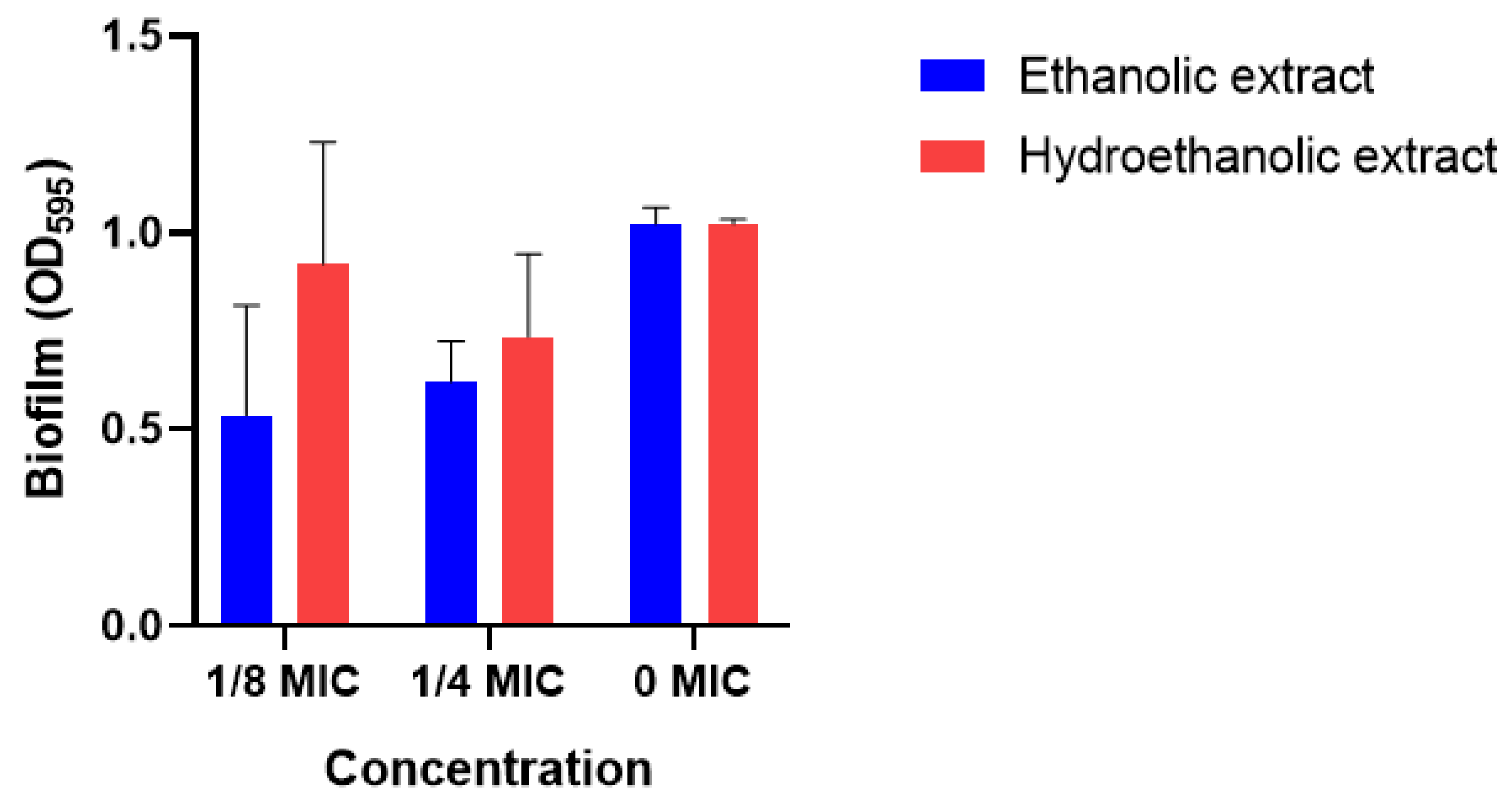
Figure 3.
Effects of ethanolic and hydroethanolic extracts of Rhus pentaphylla on Pseudomonas aeruginosa virulence factors. (A) Extracellular protein content (OD595). (B) Exopolysaccharide (EPS) content (OD480). Values are expressed as mean ± SD (n = 3). Bacteria were treated with extract concentrations corresponding to 1/8 MIC and 1/4 MIC. The untreated culture (0 MIC) served as the control.
Figure 3.
Effects of ethanolic and hydroethanolic extracts of Rhus pentaphylla on Pseudomonas aeruginosa virulence factors. (A) Extracellular protein content (OD595). (B) Exopolysaccharide (EPS) content (OD480). Values are expressed as mean ± SD (n = 3). Bacteria were treated with extract concentrations corresponding to 1/8 MIC and 1/4 MIC. The untreated culture (0 MIC) served as the control.
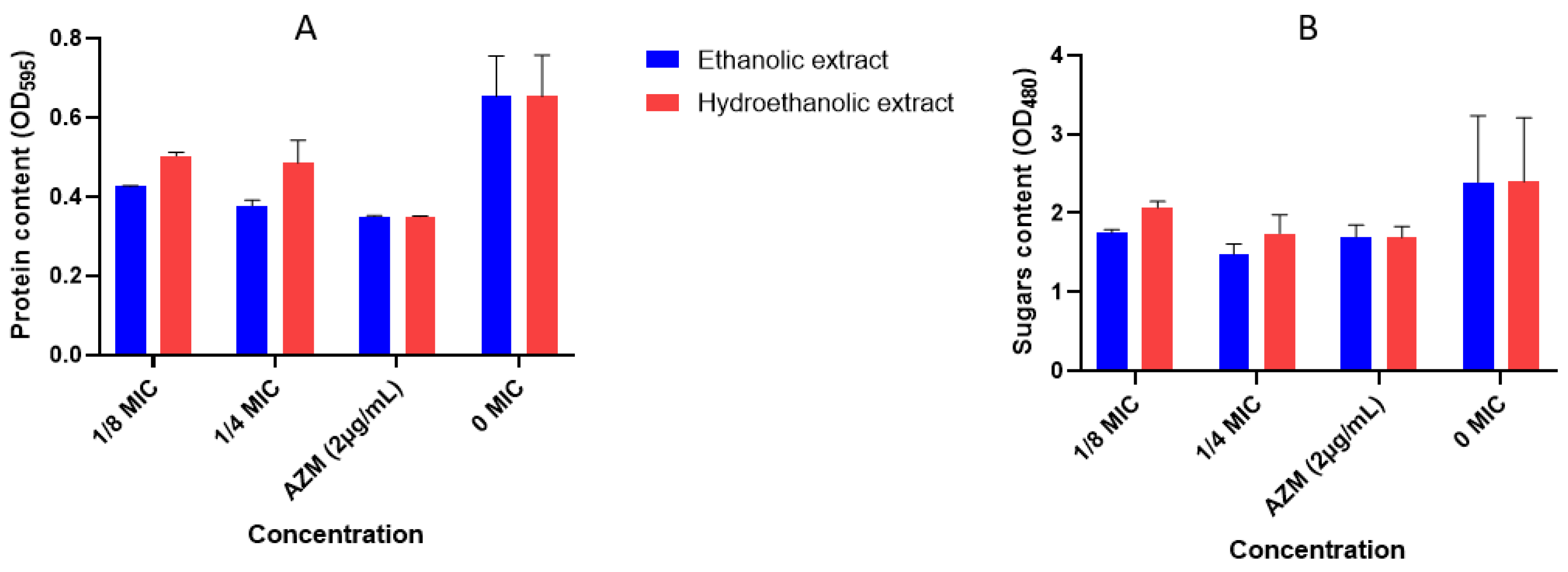

Table 1.
Extraction yield and phytochemicals of R. pentaphylla.
| Extract | Yield | TPC | TFC | TP | TS | ||||
| % | mg GAE/g | mg QE/g | mg BSAE/g | mg GLU/g | |||||
| Aqueous | 6.77 | 116.69 ± 0.006c | 36.94 ± 0.001c | 15.99 ± 0.016c | 144.82 ± 0.016a | ||||
| EtOH50 | 37.95 | 161.05 ± 0.011a | 51.34 ± 0.004b | 38.02 ± 0.005b | 84.40 ± 0.010b | ||||
| EtOH100 | 24.5 | 142.99 ± 0.012b | 66.50 ± 0.005a | 90.19 ± 0.013a | 135.48 ± 0.026a |
Values are expressed as mean ± SD (n = 3). According to one-way ANOVA followed by Tukey’s multiple comparisons test (P < 0.05), means sharing different superscript letters within the same column are significantly different.
Table 2.
Tentatively identified compounds in R. pentaphylla extracts using LC-ESI-MS analysis.
| Rt (min) | [M-H]- | MS/MS | Suggested component | ETOH | ETOH50 | Water |
|---|---|---|---|---|---|---|
| 0.91 | 193 | 193 | Glucuronic acid | Tr | Tr | Tr |
| 1.47 | 191 | 111, 191 | Citric acid | + | + | + |
| 1.53 | 191 | 108, 191 | Quinic acid | + | + | + |
| 1.61 | 353 | 173, 191 | Quinic glucoside | ++ | +++ | +++ |
| 2.80 | 331 | 169, 331 | Galloyl glucose | + | Tr | + |
| 3.07 | 609 | 177, 305 | (epi)Gallocatechin-(epi) gallocatechin | + | Tr | + |
| 3.25 | 169 | 125 | Gallic acid | + | Tr | + |
| 3.56 | 315 | 109, 153 | Protocatechuic acid glucoside | + | Tr | + |
| 5.22 | 305 | 125, 179, 305 | (epi)Gallocatechin | + | Tr | + |
| 5.49 | 593 | 177,289, 305 | (epi) Gallocatechin-(epi)catechin | + | + | + |
| 6.32 | 353 | 135, 179, 191 | Chlorogenic acid | +++ | ++++ | ++++ |
| 6.90 | 341 | 135 | Caffeic acid glucoside | + | + | + |
| 8.27 | 337 | 119, 191 | p-Coumaroylquinic acid | + | + | + |
| 9.22 | 325 | 119, 145 | Coumaric acid glucoside | +++ | + | + |
| 9.48 | 357 | 137, 195 | Dihydroferulic acid glucoside | +++ | + | + |
| 9.85 | 353 | 135, 179, 191 | Cryptochlorogenic acid | +++ | ++++ | ++++ |
| 10.32 | 177 | 109 | Esculetin | +++ | ++ | ++ |
| 10.51 | 457 | 119, 163 | Coumaric acid glucoside arabinoside | +++ | ++ | ++ |
| 10.78 | 179 | 135 | caffeic acid | + | + | + |
| 12.23 | 289 | 109, 158 | Catechin | + | + | + |
| 14.47 | 163 | 119, 163 | Coumaric acid | +++ | ++ | ++ |
| 14.56 | 625 | 271, 317 | Myricetin rutinoside | +++ | ++ | ++ |
| 14.75 | 625 | 301 | Quercetin diglucoside | +++ | ++ | ++ |
| 15.29 | 479 | 271, 317 | Myricetin Glucoside | +++ | ++ | + |
| 15.53 | 493 | 317 | Myricetin Glucuronide | +++ | ++ | + |
| 15.58 | 463 | 301 | Quercetin Glucoside | +++ | +++ | + |
| 16.39 | 449 | 271, 317 | Myricetin arabinoside | +++ | ++ | + |
| 17.38 | 609 | 301 | Quercetin rutinoside (Rutin) | +++ | +++ | ++ |
| 18.32 | 477 | 301 | Quercetin Glucuronide | ++++ | +++ | +++ |
| 19.20 | 435 | 301 | Quercetin Arabinoside | ++ | ++ | + |
| 19.98 | 593 | 285 | Kaempferol rutinoside | ++ | ++ | + |
| 20.49 | 623 | 301, 315 | Isorhamnetin rutinoside (Narcissin) | ++++ | ++++ | +++ |
| 21.39 | 477 | 301, 315 | Isorhamnetin glucoside | ++++ | ++++ | ++ |
| 22.95 | 447 | 285 | Kaempferol glucoside | +++ | +++ | ++ |
| 23.79 | 431 | 285 | Kaempferol Rhamnoside | ++ | ++ | ++ |
| 27.96 | 301 | 151, 179 | Quercetin | + | + | + |
| 27.97 | 317 | 163, 301, 317 | Myricetin | +++ | ++ | ++ |
| 33.04 | 285 | Kaempferol | ++ | ++ | Tr | |
| 33.90 | 315 | 151, 271, 301 | Isorhamnitin | ++ | ++ | Tr |
Rt (min): retention time (minute), Tr: trace; +: low; ++: moderate; +++: high; ++++: predominant.
Table 3.
Antioxidant activity of R. pentaphylla extracts.
| Extract | DPPH | FRAP | ||
| IC50, µg/mL | mM FeSO4/g | |||
| Aqueous | 87.01 ± 0.005a | 66.57 ± 0.012b | ||
| EtOH50 | 32 ± 0.005c | 81.56 ± 0.069a | ||
| EtOH100 | 39.60 ± 0.019b | 70.44 ± 0.059ab | ||
| Ascorbic acid | 3.60 ± 0.003 | – | ||
| Quercetin | – | 57.48 ± 0.55 |
Values are expressed as mean ± SD. Different letters within the same column indicate significant differences (P < 0.05; one-way ANOVA followed by Tukey’s test).
Table 4.
α-amylase and tyrosinase inhibitory activities of R. pentaphylla extracts expressed as IC50 values (mg/mL).
Table 4.
α-amylase and tyrosinase inhibitory activities of R. pentaphylla extracts expressed as IC50 values (mg/mL).
| Extract | α-amylase | Tyrosinase | ||
| IC50 (mg/mL) | IC50 (mg/mL) | |||
| Aqueous | 1.25 ± 0.08a | 0.279 ± 0.01b | ||
| EtOH50 | 1.49 ± 0.09a | 0.42 ± 0.04a | ||
| EtOH100 | 1.60 ± 0.12a | 0.27 ± 0.05b | ||
| Acarbose | 0.61 ± 0.03 | – | ||
| Kojic acid | – | 0.06 ± 0.05 |
Values are expressed as mean ± SD (n = 3). Different superscript letters within the same column indicate statistically significant differences among R. pentaphylla extracts as determined by one-way ANOVA followed by Tukey’s multiple comparisons test (P < 0.05). Lower IC50 values indicate stronger enzyme inhibitory activity.
Table 5.
Antibacterial activity of R. pentaphylla extracts against the tested bacterial strains expressed as inhibition zone diameters (mm).
Table 5.
Antibacterial activity of R. pentaphylla extracts against the tested bacterial strains expressed as inhibition zone diameters (mm).
| Diameters of the inhibition zones (mm) | ||||
|---|---|---|---|---|
| EtOH extract | Water extract | EtOH50 extract | Ampicillin | |
| P. aeruginosa | 14,017 ± 0,625a | 6,827 ± 0,058b | 7,444 ± 0,013b | 15,01 ± 0,03 |
| E. coli | 6,470 ± 0,026a | 6,488 ± 0,060a | 6,523 ± 0,033a | 14,71 ± 0,013 |
| E. faecalis | 6,319 ± 0,014a | 6,377 ± 0,014a | 6,436 ± 0,082a | 15,81 ± 0,021 |
| S. aureus | 6,448 ± 0,094a | 6,394 ± 0,029a | 6,316 ± 0,022a | 17,22 ± 0,045 |
| S. typhi | 6,396 ± 0,060a | 6,309 ± 0,004a | 6,471 ± 0,056a | 16,45 ± 0,067 |
Values are expressed as mean ± SD (n = 3). Different superscript letters within the same row indicate significant differences among Rhus pentaphylla extracts, as determined by one-way ANOVA followed by a Tukey’s multiple comparisons test (P < 0.05). Inhibition zone diameters include the 6-mm paper disc.
Table 6.
Minimum inhibitory concentration (MIC) and percentage of bacterial growth inhibition at the MIC of R. pentaphylla extracts against P. aeruginosa.
Table 6.
Minimum inhibitory concentration (MIC) and percentage of bacterial growth inhibition at the MIC of R. pentaphylla extracts against P. aeruginosa.
| Extract | MIC(mg/mL) | Growth inhibition at MIC (%) | ||
| EtOH50 | 100 | 81.56 ± 0.07 | ||
| EtOH100 | 100 | 98.25 ± 0.01 | ||
| Ampicillin | 0.031 | ND |
ND: Not Determined.
Disclaimer/Publisher’s Note: The statements, opinions and data contained in all publications are solely those of the individual author(s) and contributor(s) and not of MDPI and/or the editor(s). MDPI and/or the editor(s) disclaim responsibility for any injury to people or property resulting from any ideas, methods, instructions or products referred to in the content. |
© 2026 by the authors. Licensee MDPI, Basel, Switzerland. This article is an open access article distributed under the terms and conditions of the Creative Commons Attribution (CC BY) license (http://creativecommons.org/licenses/by/4.0/).
Copyright: This open access article is published under a Creative Commons CC BY 4.0 license, which permit the free download, distribution, and reuse, provided that the author and preprint are cited in any reuse.