
Submitted:
25 August 2026
Posted:
27 August 2026
You are already at the latest version
Abstract
Kobuviruses are emerging enteric pathogens with broad host ranges, yet their pathogenic potential in wild large felids remains largely unexplored. Here, we report the first case of fatal systemic infection caused by a novel Aichivirus A (AiV-A) strain, designated FeKoV0813, in a captive juvenile white lion (Panthera leo). The animal presented with acute respiratory distress and severe diarrhea, culminating in death despite supportive care. Comprehensive diagnostic workflows, including necropsy, histopathology, fluorescence in situ hybridization (FISH), and absolute quantitative RT-PCR, were employed to determine tissue tropism and viral dissemination. Whole-genome sequencing, phylogenetic analysis, and selection pressure assessment were conducted to characterize the viral evolutionary dynamics and host-specific constraints. The 8,213-nucleotide genome exhibited typical Kobuvirus organization, clustering within the Aichivirus A clade alongside carnivore-derived strains from dogs, cats, and foxes. High viral RNA loads were detected primarily in the small intestine (~7.4 log₁₀ copies/100 ng RNA) and trachea, with dissemination to the kidney and pancreas. Histopathological and FISH analyses localized viral RNA to epithelial lesions, while transcriptomic profiling revealed a robust upregulation of pro-inflammatory cytokines (particularly CXCL8) in the heavily infected intestine. Notably, selection pressure analysis indicated strong purifying selection acting on the feline VP1 gene, with no positively selected sites identified, contrasting with signatures of episodic positive selection observed in human and rodent lineages. This study provides the first definitive evidence that AiV-A can act as a fatal systemic pathogen in a large felid, significantly expanding the known host range of kobuviruses. Our findings underscore the need for vigilant surveillance of kobuviruses in captive wildlife and highlight the potential risk of spillover infections from domestic carnivore reservoirs.
Keywords:
Kobuvirus
; Aichivirus A
; Panthera leo
; transcriptomic analysis
; cross-species transmission
1. Introduction
The genus Kobuvirus, within the family Picornaviridae, comprises genetically diverse, non-enveloped viruses with positive-sense, single-stranded RNA genomes. Kobuviruses are primarily associated with enteric infection, although extra-intestinal dissemination has been reported in some hosts[1,2] According to the International Committee on Taxonomy of Viruses (ICTV), the genus currently comprises six species, designated Aichivirus A–F[3]. Among these, Aichivirus A (AiV-A) has been identified in a broad range of hosts, including humans, domestic carnivores, livestock, birds, and rodents [4,5,6,7,8,9].
The AiV-A genome is approximately 8.2–8.4 kb in length, consisting of a 5′ untranslated region (UTR), a single polyprotein-encoding open reading frame (ORF) organized as L-VP0-VP3-VP1-2A-2B-2C-3A-3B-3C-3D, and a 3′ UTR[10]. VP1, a surface-exposed capsid protein, contributes to host-cell recognition. Structural studies of human AiV have identified a C-terminal polyproline helix within VP1 that may participate in receptor interactions, suggesting a potential role for capsid architecture in determining host or cellular tropism[11].
In domestic carnivores, AiV-A has been detected predominantly in faecal samples from dogs and cats with enteric disease, with reported prevalence varying among populations[12,13,14,15]. Its contribution to disease, however, remains incompletely defined. Evidence from wild carnivores further indicates that kobuvirus infection may extend beyond the gastrointestinal tract. In a fox co-infected with canine distemper virus, canine kobuvirus RNA was detected in peripheral tissues and the central nervous system[16,17]
Here, we describe FeKoV0813, an AiV-A strain identified in a captive juvenile white lion presenting with acute respiratory distress, severe diarrhoea, and anorexia that subsequently died. Post-mortem metatranscriptomic and molecular analyses detected viral RNA in multiple tissues, with the highest loads in the small intestine and trachea. Histopathology, quantitative RT-PCR, fluorescence in situ hybridization, whole-genome sequencing, and host transcriptomic profiling were used to characterize viral distribution, tissue involvement, host responses, and the evolutionary relationship of FeKoV0813 to other AiV-A strains. This case extends the known host range of AiV-A to a large felid and provides evidence of multi-tissue viral involvement in a fatal disease case, establishing a basis for further investigation of kobuvirus infection in captive and wild felids.
2. Materials and Methods
2.1. Animal and Sample Collection
A captive juvenile white lion (Panthera leo) housed at a zoological facility operated by a wildlife conservation agency in Henan Province, China, presented with respiratory signs, including wheezing and coughing, together with anorexia and severe diarrhoea. Despite supportive treatment, the animal died 20 days after the onset of clinical signs. A complete necropsy was performed immediately after death.
Samples of the trachea, lung, heart, kidney, liver, intestine, and pancreas were collected. For histopathological examination and in situ hybridization, tissues were fixed in 4% paraformaldehyde for 24–48 h at 4°C. Parallel tissue samples were snap-frozen in liquid nitrogen and stored at −80°C until RNA extraction and subsequent molecular analyses. All procedures were conducted in accordance with institutional animal welfare guidelines.
2.2. RNA Extraction and Metagenomic
Total RNA was extracted from tissue samples using TRIzol Reagent (Invitrogen Life Technologies). RNA concentration and quality were assessed using a NanoDrop spectrophotometer (Thermo Scientific), and 3 μg of total RNA per sample was used for library preparation. Poly(A)+ RNA was enriched using poly-T oligo-attached magnetic beads and fragmented prior to first- and second-strand cDNA synthesis. Double-stranded cDNA was end-repaired, 3′-adenylated, ligated to Illumina paired-end adapters, and size-selected to obtain fragments of approximately 400–500 bp using the AMPure XP system (Beckman Coulter). Libraries were amplified for 15 PCR cycles, purified, and quantified using the Agilent High Sensitivity DNA Assay on a Bioanalyzer 2100 system (Agilent Technologies). The final libraries were sequenced on an Illumina NovaSeq 6000 platform at Shanghai Personal Biotechnology Co., Ltd.
2.3. Metatranscriptomic Screening for Co-Infecting Pathogens
To investigate the presence of potential co-infecting pathogens, a retrospective metatranscriptomic analysis was performed using the sequencing dataset deposited under BioProject accession number PRJNA1510436. Raw sequencing reads were quality-filtered using fastp v0.23.2 to generate clean reads.
For bacterial profiling, clean reads were analysed using MetaPhlAn v4.0.6 with its marker-gene database. For broader pathogen screening, clean reads were assembled de novo into contigs using MEGAHIT v1.2.9. The resulting contigs were searched against the NCBI non-redundant (nr) protein database, the Fungi ITS RefSeq database, and the MIDORI2 database, which contains curated reference sequences from diverse eukaryotic taxa, to identify sequences of potential viral, fungal, and parasitic origin[18,19].
2.4. Histopathology
Tissue samples from the trachea, lung, kidney, liver, heart, pancreas, and intestine were fixed in 4% paraformaldehyde at 4°C for 24–48 h, dehydrated through a graded ethanol series, cleared in xylene, and embedded in paraffin. Paraffin-embedded tissues were sectioned at 4 μm, mounted on glass slides, deparaffinized, rehydrated, and stained with hematoxylin and eosin (H&E) according to standard procedures. Histological sections were examined using an Olympus BX53 light microscope.
Histopathological evaluation focused on epithelial integrity, inflammatory cell infiltration, necrosis, and glandular alterations, where applicable. Serial sections were prepared on RNase-free slides for subsequent in situ hybridization.
2.5. Dual-Fluorescence In Situ Hybridization (FISH)
Paraffin-embedded tissue sections (4 μm) from the trachea, kidney, small intestine, and rectum were mounted on RNase-free, poly-L-lysine-coated slides. Sections were deparaffinized in xylene, rehydrated through a graded ethanol series, and subjected to heat-mediated antigen retrieval in citrate buffer (pH 6.0) at 95°C for 15 min. Sections were then digested with proteinase K (20 μg/mL) at 37°C for 20 min, post-fixed in 4% paraformaldehyde for 10 min, and acetylated with 0.25% acetic anhydride in 0.1 M triethanolamine for 10 min.
Fluorescence in situ hybridization (FISH) was performed using a commercial kit (Servicebio, Wuhan, China) according to the manufacturer's instructions[20]. A probe targeting the VP1 gene of FeKoV0813 (5′-CCAGACCAGTCCTCCCACCCGTAG-3′) was synthesized by Servicebio; probe sequences are provided in Table S1. Hybridization was performed overnight at 42°C in a humidified chamber. Sections were subsequently subjected to stringent washes in 2× SSC at 42°C and 0.5× SSC at 60°C. Nuclei were counterstained with DAPI, and images were acquired using an Olympus IX73 inverted fluorescence microscope (Olympus, Tokyo, Japan). Sense-strand probes were applied to adjacent sections as negative controls.
2.6. Viral Isolation Attempts
Viral isolation was attempted from homogenates of trachea and ileum using multiple cell lines, including CRFK, F81, MDCK, and Vero E6 cells, following standard protocols as previously described.
2.7. Quantitative Analysis of Viral Expression with qPCR in Different Tissues
To quantify FeKoV RNA across tissues, an absolute RT-qPCR assay was established using a plasmid-based standard curve targeting the VP1 gene. A 208-bp VP1 fragment was cloned into the pMD19-T vector (Takara) to generate the standard plasmid. Plasmid concentration was determined spectrophotometrically, and the corresponding copy number was calculated from the plasmid molecular weight. Tenfold serial dilutions ranging from 10¹ to 10⁸ copies per reaction were used to generate the standard curve.Total RNA extracted from each tissue was reverse-transcribed into cDNA using the PrimeScript RT Reagent Kit (Takara) with random hexamer primers. Quantitative PCR was performed using SYBR Green Master Mix (Vazyme) and specific primers (Supplementary Table 1) on a CFX Connect Real-Time PCR Detection System (Bio-Rad, Hercules, CA, USA). The cycling conditions consisted of an initial denaturation at 95°C for 30 s, followed by 40 cycles of 95°C for 5 s and 60°C for 30 s. All reactions were performed in triplicate.
Viral RNA copy numbers were calculated by interpolation from the standard curve using the corresponding threshold cycle (Ct) values. To enable comparison among tissues, viral loads were normalized to 100 ng of total RNA input and expressed as log₁₀ copies per 100 ng of total RNA.
2.8. Viral Genome Assembly and Phylogenetic Analysis
Raw sequencing reads were processed using Trimmomatic v0.39 to remove adapter sequences and low-quality bases. The resulting clean reads were assembled de novo using MEGAHIT v1.2.9 with default parameters. Putative viral contigs were identified by BLASTn and BLASTx searches against the NCBI non-redundant nucleotide and protein databases. The complete FeKoV genome was reconstructed by iterative mapping of sequencing reads to the candidate viral contig using Bowtie2 v2.4.5, followed by manual inspection and curation in Geneious Prime v2023.2. Open reading frames were predicted using ORFfinder.
For phylogenetic analysis, representative reference sequences from the six recognized species within the genus Kobuvirus (Aichivirus A–F) were retrieved from GenBank. Multiple sequence alignments were generated using MAFFT v7.520 with the L-INS-i algorithm[21]. Maximum-likelihood phylogenetic trees were inferred using IQ-TREE v2.2.0 under the best-fitting nucleotide substitution model selected by ModelFinder. Branch support was assessed using 1,000 ultrafast bootstrap replicates. Phylogenetic trees were visualized and annotated using FigTree v1.4.4 and Adobe Illustrator.
2.9. Selection Pressure Analysis
The VP1 coding sequences of FeKoV0813 and representative Aichivirus A (AiV-A) strains were aligned using MAFFT v7.520 with the L-INS-i algorithm. The alignment was manually inspected to preserve codon integrity, and sequences containing internal stop codons or excessive gaps were excluded from subsequent analyses. Selection pressure was evaluated by estimating the ratio of nonsynonymous to synonymous substitution rates (dN/dS) using the Datamonkey server and four complementary methods implemented in the HyPhy framework: Single-Likelihood Ancestor Counting (SLAC)[22], Fixed-Effects Likelihood (FEL)[22], Mixed-Effects Model of Evolution (MEME)[23], and Fast Unconstrained Bayesian Approximation (FUBAR)[24]. Sites were considered significant at P < 0.1 for SLAC, FEL, and MEME, or a posterior probability ≥ 0.9 for FUBAR. To increase confidence in site-specific inferences, only residues supported by at least two methods were considered to show evidence of selection.To examine amino acid variation among host-associated AiV-A lineages, the VP1 amino acid sequence of FeKoV0813 was compared with representative human-, feline-, and rodent-associated strains to identify host-associated amino acid differences.
2.10. Transcriptomic Analysis of Viral and Host Gene Expression
Clean reads from each tissue sample were aligned to the assembled FeKoV0813 reference genome using Bowtie2 v2.4.5 with default settings. Viral read abundance was normalized to the total number of mapped reads and reported as reads per million (RPM), enabling quantitative comparison of viral RNA abundance across tissues.
For host transcriptome analysis, clean reads were aligned to the domestic cat reference genome (Felis catus; Felis_catus_9.0) using STAR v2.7.10a with the --quantMode GeneCounts option. Gene expression levels were quantified as transcripts per million (TPM) using RSEM v1.3.3. The expression profiles of selected immune-related genes, including the pro-inflammatory cytokines IL1A, IL1B, IL6 and IL18 and the chemokines CXCL8 and CXCL10, were extracted from the TPM matrix and compared across tissues.
2.11. Statistical Analysis
All statistical analyses were performed using R version 4.2.0. Data are presented as mean ± standard deviation (SD). Differences among groups were assessed by one-way analysis of variance (ANOVA), followed by Tukey’s multiple-comparisons test. A two-sided P value < 0.05 was considered statistically significant.
3. Results
3.1. Clinical Presentation and Gross Pathology
The affected white lion developed acute respiratory distress, characterized by tachypnea and open-mouth breathing. Moist rales were detected on thoracic auscultation, and these respiratory signs were accompanied by anorexia and watery diarrhea. Despite supportive treatment, the animal's condition deteriorated rapidly and culminated in death. At necropsy, the tracheal mucosa was markedly congested and covered by diffuse hemorrhagic exudate. Segmental congestion and mild thickening of the intestinal wall were observed in the ileum, whereas the kidneys exhibited multifocal petechial hemorrhages. No gross abnormalities were detected in the heart, lungs, liver, or pancreas.
3.2. Detection of Aichivirus A in the White Lion and Screening for Co-Infecting Pathogens
Metatranscriptomic sequencing, followed by RT–PCR validation, detected Aichivirus A (AiV A) nucleic acids in tissue samples collected from the white lion. To investigate the potential contribution of other infectious agents to the disease, the metatranscriptomic dataset was retrospectively screened for common feline pathogens, including feline herpesvirus 1 (FHV-1), feline parvovirus (FPV), feline coronavirus (FCoV), feline immunodeficiency virus (FIV), feline bocavirus (FBoV), feline leukemia virus (FeLV), feline rotavirus (FRV), Mycoplasma felis, Bordetella bronchiseptica, Chlamydia felis, and Salmonella. No reads mapping to these pathogens were detected above the predefined background thresholds, providing no metatranscriptomic evidence for co-infection with these agents in this case.
3.3. Histopathology and in Situ Localization of FeKoV0813 in Tissues
We examined tissue pathology by H&E staining and localized FeKoV0813 RNA by dual-fluorescence in situ hybridization (FISH) targeting the viral VP1 gene in the trachea, kidney, small intestine and rectum. In the trachea, H&E staining revealed moderate epithelial disorganization and mild submucosal mononuclear infiltration (Figure 1A). FISH detected prominent Cy3-labelled signals localized to the surface epithelium and submucosal glands, consistent with the high viral load quantified in this tissue(Figure 2). In the kidney, H&E staining showed no significant histopathological alterations(Figure 1B). Despite the absence of overt tissue damage, FISH detected distinct Cy3-positive signals in the cortical region, associated with renal tubular epithelial cells; glomeruli were consistently negative. In the small intestine and rectum, H&E staining revealed mild villus blunting and epithelial shedding in the small intestine, with scattered mononuclear cell infiltration in the lamina propria; no ulceration or necrosis was observed(Figure 1C and 1D). In the rectum, similar epithelial changes were noted. FISH analysis showed Cy3-labelled signals concentrated in the mucosal epithelium, predominantly at villus tips in the small intestine and along the luminal surface in the rectum. Signal intensity was higher in the small intestine than in the rectum, correlating with the viral load gradient measured by qPCR.
3.4. Viral Load in Different Tissues
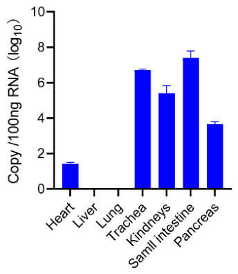
To characterize the tissue distribution of FeKoV0813 RNA, viral loads were quantified in seven tissues by absolute RT–qPCR, including the heart, liver, lung, trachea, kidney, small intestine, and pancreas (Fig. 3). Viral RNA was detected in five of the seven tissues examined. The highest viral load was observed in the small intestine (7.4 log₁₀ copies per 100 ng total RNA), followed by the trachea (6.7 log₁₀ copies per 100 ng RNA). Intermediate viral loads were detected in the kidney (5.4 log₁₀ copies) and pancreas (3.7 log₁₀ copies), whereas the heart contained substantially lower levels (1.3 log₁₀ copies). In contrast, viral RNA was not detected in the liver or lung across replicates.
The marked enrichment of FeKoV0813 RNA in the small intestine and trachea, together with its detection in the kidney, pancreas, and heart, indicates a tissue distribution extending beyond the gastrointestinal tract. No viral RNA was detected in the liver or lung under the conditions of the assay.
Figure 3.
Tissue distribution of FeKoV0813 RNA. Viral RNA loads in seven tissues were quantified by absolute RT–qPCR. Viral RNA abundance was normalized to 100 ng of total RNA per reaction and expressed as log₁₀ copies per 100 ng total RNA. Data are presented as mean ± s.d. from three independent biological replicates.
Figure 3.
Tissue distribution of FeKoV0813 RNA. Viral RNA loads in seven tissues were quantified by absolute RT–qPCR. Viral RNA abundance was normalized to 100 ng of total RNA per reaction and expressed as log₁₀ copies per 100 ng total RNA. Data are presented as mean ± s.d. from three independent biological replicates.
3.5. Genomic Characterization of FeKoV0813 Phylogenetic Analysis
3.5.1. Genomic Structure Characteristics
The complete genome of FeKoV0813 was determined to be 8,213 nucleotides (nt) in length, excluding the 3' terminal poly(A) tail (Figure 4A). The genome exhibited a canonical Kobuvirus genomic organization, comprising a 5’ untranslated region (5’ UTR, 648 nt), a leader protein (L, 504 nt), a structural protein region (P1: VP0, VP3, and VP1, at 1,131, 669, and 762 nt, respectively), two non-structural protein regions (P2: 2A, 2B, and 2C, at 408, 495, and 1,005 nt, respectively; P3: 3A, 3B, 3C, and 3D, at 282, 81, 570, and 1,404 nt, respectively), and a 3'UTR (251 nt). The GC content across distinct genomic regions varied from 50.6% to 61.7%, with the highest observed in the VP3 region (61.7%) and the lowest in the 3'UTR (50.6%), consistent with observations in other reported kobuviruses (Table 1) [25,26].
3.5.2. Sequence Homology Analysis
To elucidate the genetic relatedness of FeKoV0813, pairwise nucleotide (nt) and deduced amino acid (aa) sequence identities were calculated against representative reference strains of Kobuvirus species A to F (Table 2). The result showed that FeKoV0813 shared the highest genetic identity with members of Aichivirus A (AiV-A). Within the AiV-A species, the closest relationship was observed with a feline-derived strain (Cat/FK13), with complete genome nt identity of 87.5%, and ORF nt/aa identities of 87.8% and 93.8%, respectively. Lower identities were observed with the rat-derived strain (Rat/Rt386), sharing 74.5% nt identity across the complete genome. At the structural protein level, VP0 and VP3 displayed notably high conservation (nt/aa identities of 84.0–93.4% and 87.9–93.8%, respectively), whereas VP1 was comparatively less conserved (81.8% nt and 71.6% aa). In contrast, identities with AiV-B to AiV-F strains were significantly lower, with complete genome nt identities ranging from 48.0% to 54.3% and ORF aa identities from 43.8% to 57.6%. These data strongly corroborate the classification of FeKoV0813 within Kobuvirus species A.
3.5.3. Phylogenetic Analysis
Maximum-likelihood phylogenetic trees were reconstructed using the P1 region, VP1 gene and complete polyprotein sequence to examine the evolutionary placement of FeKoV0813 (Fig. 4b). Across all three datasets, FeKoV0813 consistently clustered within the AiV-A clade, with strong bootstrap support (>99%). Within this clade, FeKoV0813 grouped with AiV-A strains detected in carnivorous mammals, including cats, dogs and foxes. The concordant phylogenetic placement across genomic regions, together with the sequence identity analyses, supports the classification of FeKoV0813 as a member of Aichivirus A. Its clustering with viruses from other carnivorous hosts is consistent with shared evolutionary relationships among these kobuviruses, although the available phylogenetic data alone do not establish cross-species transmission or host-specific co-evolution.
3.5.4. Selection Pressure Analysis
Selection pressure on the VP1 gene was assessed across feline-, human- and rodent-associated kobuvirus lineages using four codon-based methods implemented in HyPhy: SLAC, FEL, MEME and FUBAR. Sites were considered significant at P < 0.1 for SLAC, FEL and MEME, and at a posterior probability ≥0.9 for FUBAR.
No positively selected sites were identified in the feline lineage by any of the four methods(Supplementary Table 2). In contrast, evidence of pervasive purifying selection was detected, with FEL and SLAC identifying 79 and 6 negatively selected sites, respectively. These findings indicate that the analyzed feline kobuvirus VP1 sequences are predominantly subject to purifying selection. Evidence of episodic positive selection was detected in the human and rodent lineages. MEME identified six candidate sites in human VP1, including site 216, which was also supported by FUBAR, whereas three candidate sites were detected in rodent VP1, with site 243 additionally supported by FEL and FUBAR. Because the analyses were restricted to internal branches, lineage-specific selection acting on terminal branches was not evaluated.
3.6. Viral Transcript Abundance and Host Immune Responses Across Tissues
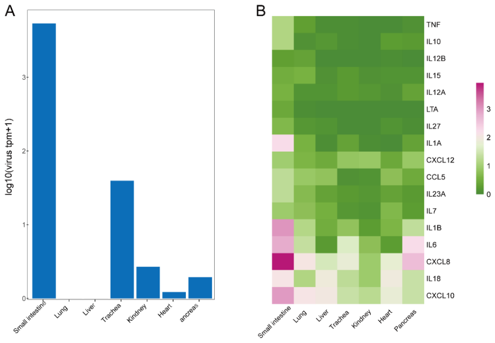
The relative abundance of kobuvirus-derived transcripts varied across the seven tissues examined (Fgure 5 ). Viral transcript abundance was highest in the intestine (BS0809_C), followed by the trachea (BS0809_QG). Lower viral signals were detected in the kidney, pancreas and heart, whereas no viral transcripts were detected in the lung or liver. Among the immune-related genes examined, the intestine showed the highest transcript abundance of the pro-inflammatory cytokines IL1A, IL1B, IL6 and IL18 and the chemokines CXCL8 and CXCL10. Of these genes, CXCL8 exhibited the strongest transcriptional signal in the intestine.
3.7. Viral Isolation Attempts
Infectious FeKoV was not recovered from tissue homogenates despite repeated isola-tion attempts using CRFK, F81, MDCK, and Vero E6 cells under different culture condi-tions, including incubation at 37°C with or without trypsin. No cytopathic effects were observed duringafter three subsequent blind passages and RT-PCR of culture supernatants remained negative throughout.
Figure 5.
iral transcript abundance and host immune gene expression profiles across tissues.
4. Discussion
Kobuviruses have been identified in a broad range of mammalian hosts, including humans, cattle, swine, dogs, cats, sheep, rodents, and bats[1,2,5,10,27,28,29]. but their biological and pathogenic significance in carnivores remains incompletely understood. In felids, current knowledge is derived almost exclusively from domestic cats, in which kobuviruses have been detected predominantly in faecal samples from animals with enteric disease.[8,30,31]. The present case therefore extends the available evidence to a large felid and provides an opportunity to assess the tissue distribution and pathological relevance of AiV-A infection in a previously uncharacterized host. The intestinal distribution of FeKoV0813 is consistent with the predominantly enteric association reported for kobuviruses in domestic carnivores. Feline kobuviruses have been identified mainly in faecal samples from cats with diarrhoea[7,14], although their contribution to disease has remained difficult to establish because most previous studies relied primarily on molecular detection rather than tissue-based analyses.In contrast, the present case provides convergent evidence from viral load quantification, in situ localization, and histopathology, supporting biologically relevant involvement of FeKoV0813 in the intestinal lesions.
The prominent tracheal involvement represents an important feature of this case. Kobuviruses in domestic carnivores are generally investigated in the context of enteric disease, and respiratory tract involvement has received comparatively little attention[7]. In the present case, the trachea contained one of the highest viral RNA burdens, and viral RNA was localized to epithelial and glandular structures. indicating dissemination beyond the gastrointestinal tract. Extra-intestinal detection and systemic distribution of kobuviruses have previously been reported in dogs and wild carnivores Porcine kobuvirus has been demonstrated to escape the gut, resulting in viremia in clinically healthy pigs[4], and systemic canine kobuvirus was detected in the brain, bladder, and lymph nodes of a fox co-infected with canine distemper virus[17]. Together with the respiratory signs observed clinically, these findings support substantial involvement of the tracheal mucosa during infection. However, the presence of viral RNA alone does not demonstrate productive replication or direct viral injury. In this case, the concordance between viral load, tissue localization, and pathological changes provides the strongest evidence for viral involvement in the intestine and trachea.
FeKoV0813 clustered with carnivore-associated AiV-A strains and showed the highest sequence identity to a feline-derived strain[6,31]. This relationship is consistent with the broad host range and genetic diversity reported for AiV-A[10,30]. Nevertheless, phylogenetic relatedness does not establish the source or direction of transmission. The present data therefore support a close evolutionary relationship between FeKoV0813 and other carnivore-associated AiV-A strains but do not determine whether the virus originated from domestic carnivores, wildlife, or another reservoir[16]. VP1 was more variable than VP0 and VP3, and selection-pressure analysis identified predominant purifying selection within the feline-associated VP1 lineage, in contrast to the episodic positive selection detected in the human and rodent lineages[32]. The predominance of purifying selection within the feline-associated VP1 lineage suggests that this capsid protein is subject to evolutionary constraint in these viruses. However, the limited number and host diversity of available sequences preclude conclusions regarding host adaptation or the evolutionary history of FeKoV0813 in large felids.
transcriptomic profiling revealed marked upregulation of CXCL8 and pro-inflammatory cytokines (IL1A, IL1B, IL6, IL18) in the intestine, with CXCL8 exhibiting the strongest signal. This transcriptional pattern was consistent with the high intestinal viral burden and epithelial localization of FeKoV0813, suggesting a localized inflammatory response associated with infection. In feline viral infections, excessive neutrophil recruitment driven by CXCL8 is strongly associated with mucosal barrier disruption, a mechanism well-documented in conditions such as feline infectious peritonitis and feline panleukopenia [33,34]. Alongside viral RNA detection in tracheal and renal epithelia, we hypothesize that this CXCL8-driven inflammatory burst compromised the mucosal integrity, thereby facilitating hematogenous seeding of FeKoV0813 to the kidney and pancreas[31,35,36]. However, transcriptomic analysis was based on tissues from a single animal and lacked uninfected white-lion controls. In addition, host reads were mapped to the domestic cat reference genome. The observed transcriptional differences should therefore be interpreted as tissue-associated responses rather than as evidence of a defined pathogenic mechanism.
Several limitations should be considered. This investigation was based on a single animal, and infectious virus was not recovered despite repeated isolation attempts. In addition, although metatranscriptomic screening was performed to identify potential co-infecting pathogens, alternative or concurrent causes of disease cannot be completely excluded. Consequently, the available evidence does not establish FeKoV0813 as the sole cause of death. Nevertheless, the combined clinical, pathological, molecular, and genomic findings support a substantial association between FeKoV0813 infection and the fatal disease observed in this white lion. The detection of high viral loads and epithelial localization in the intestine and trachea, together with viral RNA in additional tissues, indicates multi-tissue involvement during infection. These findings support the inclusion of AiV-A in diagnostic investigations of enteric disease in captive and wild felids and highlight the need for broader surveillance to define its prevalence, host range, and pathogenic significance.
5. Conclusions
This study reports the detection and genomic characterization of FeKoV0813, an Aichivirus A strain, in a captive juvenile white lion (Panthera leo) that died following severe gastrointestinal and respiratory disease. FeKoV0813 RNA was detected in multiple tissues, with the highest levels in the small intestine and trachea, and was localized to epithelial cells in both tissues. Phylogenetic analysis placed FeKoV0813 within a carnivore-associated AiV-A lineage, extending the known host range of AiV-A to a large felid. Given the single-case design and unsuccessful virus isolation, the contribution of FeKoV0813 to the observed disease and death cannot be determined. Further investigations are required to clarify the prevalence, tissue tropism, and pathogenic significance of AiV-A in captive and wild felids.
Supplementary Materials
The following supporting information can be downloaded at the website of this paper posted on Preprints.org.
Author Contributions
Conceptualization, Y.D. and Y.W.; methodology, Y.D.,Y.W, F.S; validation and ormal analysis, Y.D,.; investigation, N.Z.,C.W.,M.L.WC; data curation, writing—original draft preparation, writing—review and editingY.D ; project administration, W.L.; funding acquisition, Z.C. All authors have read and agreed to the published version of the manuscript.
Funding
This research was funded by Prevention and Control of Emerging and Major Infectious Diseases-National Science and Technology Major Project (2026ZD01999702), Major Project of Guangzhou National Laboratory(GZNL2023A01001) , Guangzhou Postdoctoral Research Funding Project(YL202501).
Institutional Review Board Statement
No laboratory animals were involved in this study. and the study protocol was reviewed and approved by the Animal Ethics and Management Committee of Guangzhou Zoo and Guangzhou Wildlife Research Center (permit number:GZDWY202504 ).
Data Availability Statement
The complete genome sequence of the FeKoV strain reported in this study has been deposited in GenBank under accession number [PZ353127]. The transcriptomic data have been deposited in the NCBI Sequence Read Archive under BioProject accession number PRJNA1510436. https://www.ncbi.nlm.nih.gov/bioproject/PRJNA1510436.
Conflicts of Interest
The authors declare no conflicts of interest.
References
- Yamashita, T.; Kobayashi, S.; Sakae, K.; Nakata, S.; Chiba, S.; Ishihara, Y.; Isomura, S. Isolation of Cytopathic Small Round Viruses with BS-C-1 Cells from Patients with Gastroenteritis. J Infect Dis 1991, 164, 954–957. [CrossRef]
- Reuter, G.; Boros, Á.; Pankovics, P. Kobuviruses – a Comprehensive Review. Reviews in Medical Virology 2011, 21, 32–41. [CrossRef]
- Simmonds, P.; Adriaenssens, E.M.; Lefkowitz, E.J.; Oksanen, H.M.; Zerbini, F.M.; Alfenas-Zerbini, P.; Aylward, F.O.; Dempsey, D.M.; Freitas-Astúa, J.; Hendrickson, R.C.; et al. Changes to Virus Taxonomy, the International Code of Virus Classification and Nomenclature, and the ICTV Statutes Ratified by the International Committee on Taxonomy of Viruses (2025). Arch Virol 2025, 171, 23. [CrossRef]
- Reuter, G.; Kecskeméti, S.; Pankovics, P. Evolution of Porcine Kobuvirus Infection, Hungary. Emerg Infect Dis 2010, 16, 696–698. [CrossRef]
- Reuter, G.; Boros, Á.; Pankovics, P.; Egyed, L. Kobuvirus in Domestic Sheep, Hungary. Emerg Infect Dis 2010, 16, 869–870. [CrossRef]
- Kapoor, A.; Simmonds, P.; Dubovi, E.J.; Qaisar, N.; Henriquez, J.A.; Medina, J.; Shields, S.; Lipkin, W.I. Characterization of a Canine Homolog of Human Aichivirus. Journal of Virology 2011, 85, 11520–11525. [CrossRef]
- Li, L.; Pesavento, P.A.; Shan, T.; Leutenegger, C.M.; Wang, C.; Delwart, E. Viruses in Diarrhoeic Dogs Include Novel Kobuviruses and Sapoviruses. Journal of General Virology 2011, 92, 2534–2541. [CrossRef]
- Di Martino, B.; Di Profio, F.; Melegari, I.; Marsilio, F.; Martella, V. Detection of Feline Kobuviruses in Diarrhoeic Cats, Italy. Veterinary Microbiology 2015, 176, 186–189. [CrossRef]
- Pankovics, P.; Boros, Á.; Kiss, T.; Reuter, G. Identification and Complete Genome Analysis of Kobuvirus in Faecal Samples of European Roller (Coracias Garrulus): For the First Time in a Bird. Arch Virol 2015, 160, 345–351. [CrossRef]
- Khamrin, P.; Maneekarn, N.; Okitsu, S.; Ushijima, H. Epidemiology of Human and Animal Kobuviruses. VirusDis. 2014, 25, 195–200. [CrossRef]
- Zhu, L.; Wang, X.; Ren, J.; Kotecha, A.; Walter, T.; Yuan, S.; Yamashita, T.; Tuthill, T.; Fry, E.; Rao, Z.; et al. Structure of Human Aichi Virus and Implications for Receptor Binding. Nature Microbiology 2016, 1(11):16150. Published 2016 Sep 5. [CrossRef]
- Oem, J.; Choi, J.-W.; Lee, M.-H.; Lee, K.; Choi, K.-S. Canine Kobuvirus Infections in Korean Dogs. Archives of Virology 2014, 159(10):2751-2755. [CrossRef]
- Olarte-Castillo, X.A.; Heeger, F.; Mazzoni, C.; Greenwood, A.; Fyumagwa, R.; Moehlman, P.; Hofer, H.; East, M. Molecular Characterization of Canine Kobuvirus in Wild Carnivores and the Domestic Dog in Africa. Virology 2015, 477:89-97. [CrossRef]
- Kong, N.; Zuo, Y.; Wang, Z.; Yu, H.; Zhou, E.; Shan, T.; Tong, G. Molecular Characterization of New Described Kobuvirus in Dogs with Diarrhea in China. Springerplus 2016, 5, 2047. [CrossRef]
- Niu, T.; Yi, S.; Wang, X.; Wang, L.; Guo, B.; Zhao, L.-Y.; Zhang, S.; Dong, H.; Wang, K.; Hu, X. Detection and Genetic Characterization of Kobuvirus in Cats: The First Molecular Evidence from Northeast China. Infection, Genetics and Evolution 2018, 68:58-67. [CrossRef]
- Lu, L.; Van Dung, N.; Ivens, A.; Bogaardt, C.; O’Toole, A.; Bryant, J.E.; Carrique-Mas, J.; Van Cuong, N.; Anh, P.H.; Rabaa, M.A.; et al. Genetic Diversity and Cross-Species Transmission of Kobuviruses in Vietnam. Virus Evol 2018, 4, vey002. [CrossRef]
- Kaiser, F.K.; van Dyck, L.; Jo, W.K.; Schreiner, T.; Pfankuche, V.M.; Wohlsein, P.; Baumann, I.; Peters, M.; Baumgärtner, W.; Osterhaus, A.D.M.E.; et al. Detection of Systemic Canine Kobuvirus Infection in Peripheral Tissues and the Central Nervous System of a Fox Infected with Canine Distemper Virus. Microorganisms 2021, 9, 2521. [CrossRef]
- Cui, X.; Fan, K.; Liang, X.; Gong, W.; Chen, W.; He, B.; Chen, X.; Wang, H.; Wang, X.; Zhang, P.; et al. Virus Diversity, Wildlife-Domestic Animal Circulation and Potential Zoonotic Viruses of Small Mammals, Pangolins and Zoo Animals. Nat Commun 2023, 14, 2488. [CrossRef]
- Wang, H.; Meng, Y.; Chen, X.; Cui, X.; Zuo, Q.; Han, N.; Liang, X.; Shen, X.; Li, C.; Li, D.; et al. Unveiling Novel Viral Diversity, Biogeography, and Host Networks in Wildlife Through High-Throughput Sequencing Data Mining. Adv Sci (Weinh) 2025, 12, e11920. [CrossRef]
- Yu, X.; Wang, X.; Xu, F.; Zhang, X.; Wang, M.; Zhou, R.; Sun, Z.; Pan, X.; Feng, L.; Zhang, W.; et al. Mir-615-3p Promotes Osteosarcoma Progression via the SESN2/AMPK/mTOR Pathway. Cancer Cell Int 2024, 24, 411. [CrossRef]
- Katoh, K.; Standley, D.M. MAFFT Multiple Sequence Alignment Software Version 7: Improvements in Performance and Usability. Mol Biol Evol 2013, 30, 772–780. [CrossRef]
- Kosakovsky Pond, S.L.; Frost, S.D.W. Not so Different after All: A Comparison of Methods for Detecting Amino Acid Sites under Selection. Mol Biol Evol 2005, 22, 1208–1222. [CrossRef]
- Murrell, B.; Wertheim, J.O.; Moola, S.; Weighill, T.; Scheffler, K.; Kosakovsky Pond, S.L. Detecting Individual Sites Subject to Episodic Diversifying Selection. PLoS Genet 2012, 8, e1002764. [CrossRef]
- Murrell, B.; Moola, S.; Mabona, A.; Weighill, T.; Sheward, D.; Kosakovsky Pond, S.L.; Scheffler, K. FUBAR: A Fast, Unconstrained Bayesian Approximation for Inferring Selection. Mol Biol Evol 2013, 30, 1196–1205. [CrossRef]
- Yan, N.; Yue, H.; Liu, Q.; Wang, G.; Tang, C.; Liao, M. Isolation and Characteristics of a Novel Aichivirus D from Yak. Microbiology Spectrum 2023, 11, e00099-23. [CrossRef]
- Wang, G.; Deng, Q.; Zhu, P.; Punyapornwithaya, V.; Shi, X.; Liu, Y.; Duan, X.; Li, Z.; Li, W. Genomic Sequencing and Evolutionary Analysis of Bovine Kobuvirus in Yunnan Province, China. BMC Vet Res 2025, 21, 367. [CrossRef]
- Yan, N.; Xu, D.; Yue, H.; Tang, C. Isolation and Molecular Characteristics of D2 Genotype of Aichivirus D in Dairy Cattle in China. Front. Vet. Sci. 2025, 12. [CrossRef]
- Reuter, G.; Boldizsár, A.; Kiss, I.; Pankovics, P. Candidate New Species of Kobuvirus in Porcine Hosts. Emerg Infect Dis 2008, 14, 1968–1970. [CrossRef]
- You, F.-F.; Zhang, M.-Y.; He, H.; He, W.-Q.; Li, Y.-Z.; Chen, Q. Kobuviruses Carried by Rattus Norvegicus in Guangdong, China. BMC Microbiol 2020, 20, 94. [CrossRef]
- Reuter, G.; Boros, Á.; Pankovics, P. Kobuviruses – a Comprehensive Review. Reviews in Medical Virology 2011, 21, 32–41. [CrossRef]
- Di Martino, B.; Di Profio, F.; Melegari, I.; Marsilio, F. Feline Virome—A Review of Novel Enteric Viruses Detected in Cats. Viruses 2019, 11, 908. [CrossRef]
- Van Nguyen, T.; Kasantikul, T.; Piewbang, C.; Techangamsuwan, S. Evolutionary Dynamics of Canine Kobuvirus in Vietnam and Thailand Reveal the Evidence of Viral Ability to Evade Host Immunity. Sci Rep 2024, 14, 12037. [CrossRef]
- Dean, G.A.; Olivry, T.; Stanton, C.; Pedersen, N.C. In Vivo Cytokine Response to Experimental Feline Infectious Peritonitis Virus Infection. Vet Microbiol 2003, 97, 1–12. [CrossRef]
- Berg, A.-L.; Ekman, K.; Belák, S.; Berg, M. Cellular Composition and Interferon-γ Expression of the Local Inflammatory Response in Feline Infectious Peritonitis (FIP). Vet Microbiol 2005, 111, 15–23. [CrossRef]
- Stuetzer, B.; Hartmann, K. Feline Parvovirus Infection and Associated Diseases. The Veterinary Journal 2014, 201, 150–155. [CrossRef]
- In Veterinary Immunology; 2024; ISBN 978-0-443-10975-1. 36. Veterinary Immunology; 2024; ISBN 978-0-443-10975-1.
Figure 1.
Histopathological findings in FeKoV0813-infected tissues. Representative H&E-stained sections of trachea, kidney, small intestine and rectum from the FeKoV0813-infected white lion. (A) In the trachea, extensive epithelial necrosis and shedding (black arrowheads) expose the underlying lamina propria, with necrotic glandular epithelial cells visible in the submucosa (red arrowheads). (B) In the kidney, cortical architecture is disorganized, with widespread necrosis and sloughing of tubular epithelial cells (black arrowheads); glomeruli are relatively unaffected. (C-D) In the small intestine and rectum, the mucosa shows extensive epithelial necrosis (black arrowheads), focal glandular dilatation (green arrowheads), epithelial flattening and intraluminal eosinophilic debris. Scale bars, 100 μm.
Figure 1.
Histopathological findings in FeKoV0813-infected tissues. Representative H&E-stained sections of trachea, kidney, small intestine and rectum from the FeKoV0813-infected white lion. (A) In the trachea, extensive epithelial necrosis and shedding (black arrowheads) expose the underlying lamina propria, with necrotic glandular epithelial cells visible in the submucosa (red arrowheads). (B) In the kidney, cortical architecture is disorganized, with widespread necrosis and sloughing of tubular epithelial cells (black arrowheads); glomeruli are relatively unaffected. (C-D) In the small intestine and rectum, the mucosa shows extensive epithelial necrosis (black arrowheads), focal glandular dilatation (green arrowheads), epithelial flattening and intraluminal eosinophilic debris. Scale bars, 100 μm.
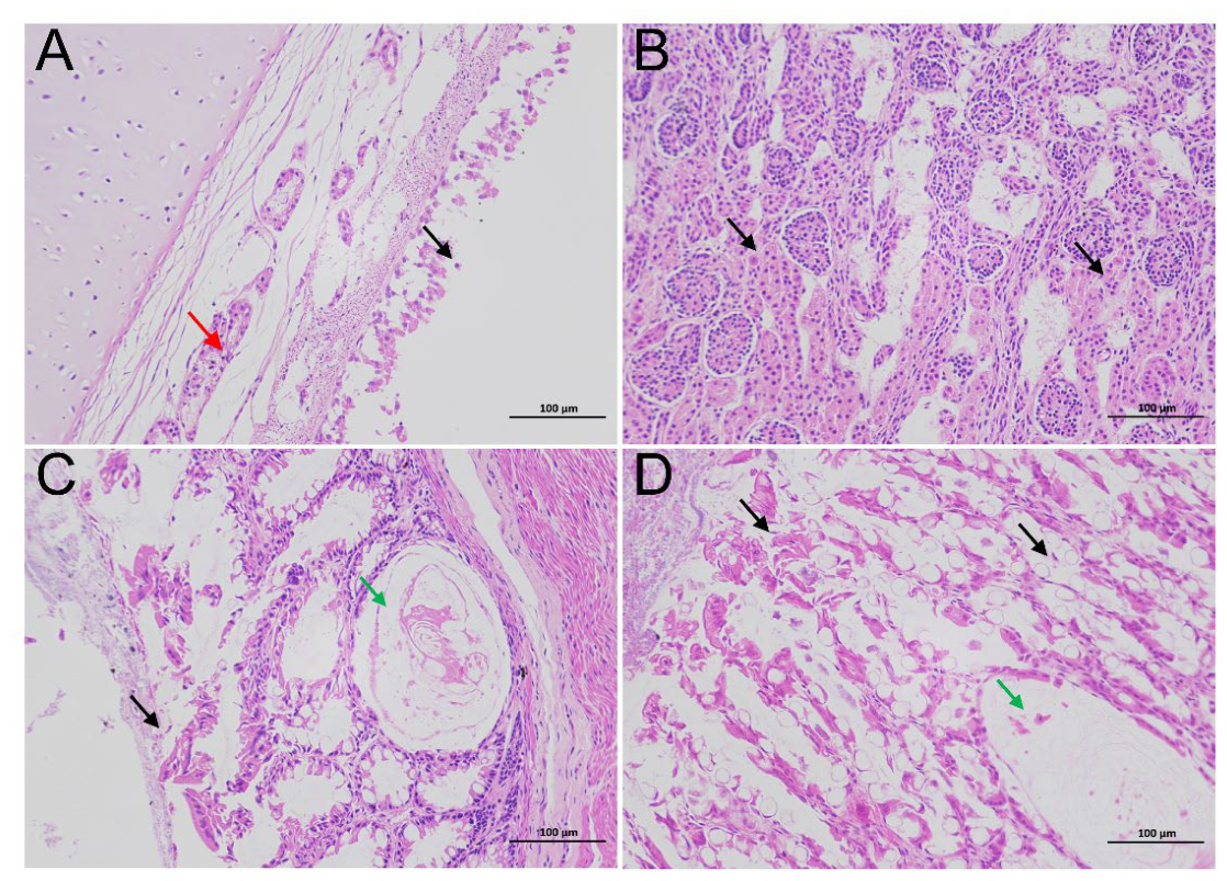
Figure 2.
In situ localization of FeKoV0813 RNA in infected tissues. Dual-fluorescence in situ hybridization (FISH) was performed on sections of trachea, kidney, small intestine and rectum using a Cy3-labelled probe targeting the FeKoV VP1 gene (red); nuclei were counterstained with DAPI (blue). Merged images show the spatial relationship between viral RNA and cellular nuclei. In the trachea, Cy3 signals are concentrated in the surface epithelium and submucosal glands. In the kidney, labelling is restricted to renal tubular epithelial cells in the cortical region, with no signal detected in glomeruli. In the small intestine, intense signals localize predominantly to villus tips. In the rectum, signals are confined to the surface epithelium. Scale bars, 50 μm.
Figure 2.
In situ localization of FeKoV0813 RNA in infected tissues. Dual-fluorescence in situ hybridization (FISH) was performed on sections of trachea, kidney, small intestine and rectum using a Cy3-labelled probe targeting the FeKoV VP1 gene (red); nuclei were counterstained with DAPI (blue). Merged images show the spatial relationship between viral RNA and cellular nuclei. In the trachea, Cy3 signals are concentrated in the surface epithelium and submucosal glands. In the kidney, labelling is restricted to renal tubular epithelial cells in the cortical region, with no signal detected in glomeruli. In the small intestine, intense signals localize predominantly to villus tips. In the rectum, signals are confined to the surface epithelium. Scale bars, 50 μm.
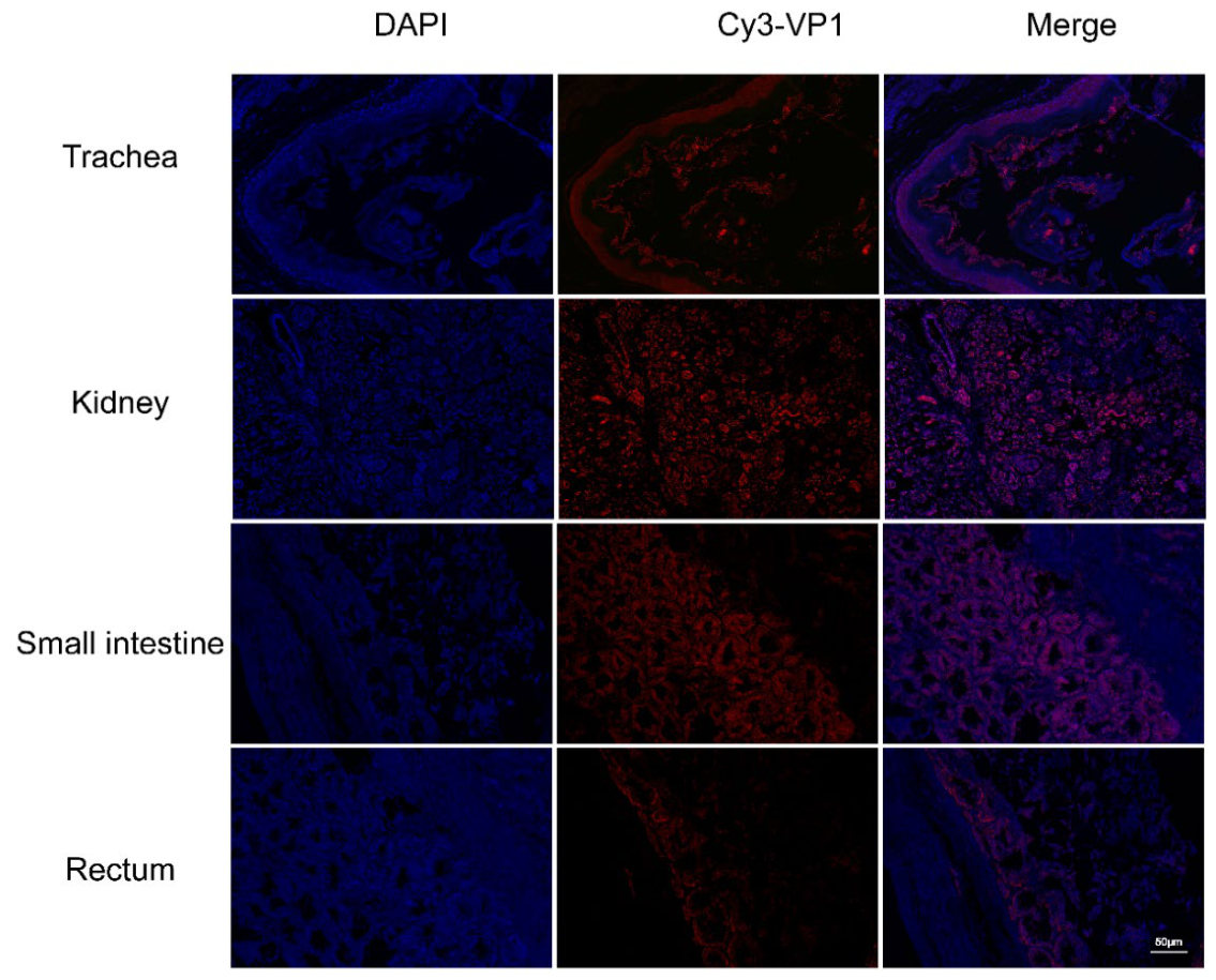
Figure 4.
Genomic architecture and phylogenetic characterization of the lion-derived kobuvirus FeKoV0813.(A) Schematic of the FeKoV0813 genome, comprising a 5′ UTR, a single polyprotein-encoding ORF, and a 3′ UTR. The polyprotein is processed into the leader protein (L), structural capsid proteins (VP0, VP3, and VP1), and non-structural proteins (2A–2C, 3A–3D). B, Maximum-likelihood phylogenies inferred with IQ-TREE (1,000 ultrafast bootstrap replicates) based on the complete genome , the P1 capsid region and the VP1 gene.
Figure 4.
Genomic architecture and phylogenetic characterization of the lion-derived kobuvirus FeKoV0813.(A) Schematic of the FeKoV0813 genome, comprising a 5′ UTR, a single polyprotein-encoding ORF, and a 3′ UTR. The polyprotein is processed into the leader protein (L), structural capsid proteins (VP0, VP3, and VP1), and non-structural proteins (2A–2C, 3A–3D). B, Maximum-likelihood phylogenies inferred with IQ-TREE (1,000 ultrafast bootstrap replicates) based on the complete genome , the P1 capsid region and the VP1 gene.
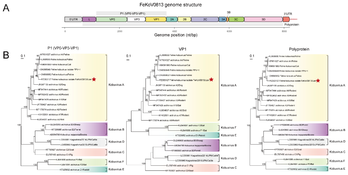

Table 1.
The location and size information of each gene of the FeKoV0813 strain.
| Gene | Location | size/bp | GC% | |
|---|---|---|---|---|
| 5'UTR | 1~648 | 648 | 56.9 | |
| L | 649~1152 | 504 | 56.5 | |
| P1 | VP0 | 1153~2283 | 1131 | 59.9 |
| VP3 | 2284~2952 | 669 | 61.7 | |
| VP1 | 2953~3714 | 762 | 60.4 | |
| P2 | 2A | 3715~4122 | 408 | 61.3 |
| 2B | 4123~4617 | 495 | 57.8 | |
| 2C | 4618~5622 | 1005 | 57.7 | |
| P3 | 3A | 5623~5904 | 282 | 59.6 |
| 3B | 5905~5985 | 81 | 58 | |
| 3C | 5986~6555 | 570 | 59.8 | |
| 3D | 6556~7959 | 1404 | 55.7 | |
| 3'UTR | 7963~8213 | 251 | 50.6 |
Table 2.
Nucleotide(nt) and amino acid (aa)identities between FeKoV0813 and representative kobuvirus reference strains.
Table 2.
Nucleotide(nt) and amino acid (aa)identities between FeKoV0813 and representative kobuvirus reference strains.
| Shared nt identity (%)/aa identity (%) | |||||||||
|---|---|---|---|---|---|---|---|---|---|
| Gene region |
AiV-A Cat/FK-13 |
AiV-A Rat/Rt386 |
AiV-B Ferret/MpKoV38 |
AiV-B Sheep/TB3 |
AiV-C Pig/S-1 |
AiV-C Goat/12Q108 |
AiV-D Cattle/Kagoshima |
AiV-E Bat/JX2010 |
AiV-F Bat/GD2012 |
| KF831027 nt/aa |
MF352432 nt/aa |
KF006985 nt/aa |
GU245693 nt/aa |
EU787450 nt/aa |
KF793927 nt/aa |
LC055961 nt/aa |
KJ641686\ nt/aa | KJ641691 nt/aa |
|
| 5‘UTR | 85.2/- | 70.9/- | -/- | 28.6/- | 22.9/- | 27.1/- | -/- | -/- | -/- |
| L | 86.1/88.7 | 52.1/49.7 | 31.7/26.8 | 34/25 | 31.8/26.7 | 33.1/23.6 | 32.1/25.3 | 29.6/9.3 | 31.1/0 |
| VP0 | 84/93.4 | 74.9/83.6 | 61.8/59.9 | 59.8/59.9 | 60.5/60.4 | 63.5/64.2 | 52.4/50.3 | 48.4/47.6 | 47.8/47 |
| VP3 | 87.9/93.8 | 78.4/83.9 | 65.8/58 | 66.5/58.5 | 63.4/59.4 | 62.1/59.8 | 58.8/50.4 | 64.9/65.2 | 62.1/65.2 |
| VP1 | 81/84.6 | 71.6/69.3 | 43.6/32 | 42/32.7 | 43.6/36.8 | 44.6/33.6 | 40.9/33.1 | 41.2/34.2 | 42/31.9 |
| 2A | 91.6/96.4 | 75.4/70.3 | 57.7/55.4 | 55.9/58 | 55.9/53.6 | 54.7/53.6 | 56.2/54 | 46.2/36.4 | 48.6/33.6 |
| 2B | 92.1/98.8 | 80.6/82.4 | 47.6/50.3 | 46.7/52.1 | 49.6/45.6 | 45.8/54.5 | 43.4/38.5 | 40.3/29.6 | 40.2/32.8 |
| 2C | 90.9/97.6 | 80.9/89.9 | 67.6/69 | 65.8/68.2 | 64/67.9 | 63.6/67.9 | 58.4/55.1 | 53.4/48.7 | 50/47.2 |
| 3A | 89.4/97.6 | 71.9/89.9 | 47.7/69 | 46.4/68.2 | 52.7/67.9 | 50.5/67.9 | 48.8/55.1 | 37.2/48.7 | 35.9/47.2 |
| 3B | 90.1/96.3 | 76.5/77.8 | 38.7/50 | 40.9/53.3 | 33.3/35.3 | 37.3/38.2 | 25/31.3 | 29.2/16.7 | 14.4/25.9 |
| 3C | 87.9/94.7 | 75.8/81.1 | 57.2/49.8 | 53.8/48.7 | 53.5/52.1 | 57.8/53.1 | 54.1/46.9 | 41.3/34.9 | 41.6/33.2 |
| 3D | 90.2/95.5 | 81.2/89.3 | 70.9/74.8 | 70.7/74.8 | 70.4/74 | 68.3/73.8 | 66/68.5 | 62.2/62.2 | 60.2/60.5 |
| 3‘UTR | 87.6/- | 60.5/- | 18.3/- | 21.1/- | 18.1/- | 19.1/- | 19.7/- | 29/- | 25.2/- |
| complete | 87.5/- | 74.5/- | 54.3/- | 53.5/- | 53.1/- | 53.4/- | 50.7/- | 48.2/- | 48/- |
| ORF | 87.8/93.8 | 75.6/80.3 | 57.6/55.5 | 56.7/55.4 | 56.6/55.4 | 56.7/56 | 52.5/49 | 48.9/42.1 | 48.7/43.8 |
“-” indicates that it does not apply to identity comparison.
Disclaimer/Publisher’s Note: The statements, opinions and data contained in all publications are solely those of the individual author(s) and contributor(s) and not of MDPI and/or the editor(s). MDPI and/or the editor(s) disclaim responsibility for any injury to people or property resulting from any ideas, methods, instructions or products referred to in the content. |
© 2026 by the authors. Licensee MDPI, Basel, Switzerland. This article is an open access article distributed under the terms and conditions of the Creative Commons Attribution (CC BY) license (http://creativecommons.org/licenses/by/4.0/).
Copyright: This open access article is published under a Creative Commons CC BY 4.0 license, which permit the free download, distribution, and reuse, provided that the author and preprint are cited in any reuse.