

Submitted:
24 August 2026
Posted:
26 August 2026
You are already at the latest version
Abstract
The recent emergence and rapid spread of Shamonda-like virus (SHAV/DE/2026) across several European countries raises questions about the mechanisms facilitating the circulation of this and related arboviruses. Although Culicoides biting midges are considered the most plausible vectors, the ecological pathways by which viruses may enter and persist within these insects remain incompletely understood. Here, we examine a potentially overlooked component of arbovirus transmission ecology: interactions among viruses, nematodes, and Culicoides. Published evidence demonstrates that mermithid nematodes can penetrate Culicoides larvae, develop within the insect, and in some systems persist through metamorphosis into adult midges. Importantly, experimental studies have also demonstrated nematode-associated enhancement of viral infection in Culicoides and virus carriage by nematodes in other arthropod systems. Based on these observations, we propose a testable environmental pathway in which virus-associated nematodes may facilitate viral entry into immature Culicoides, potentially linking environmental virus sources with adult vectors and susceptible vertebrate hosts. This hypothesis warrants targeted experimental investigation. If confirmed, such a pathway could expand current concepts of arbovirus transmission and should be considered in future biosurveillance, biorisk assessment, and preventive strategies against emerging arbovirus-associated biological threats.
Keywords:
Shamonda virus
; Culicoides
; free-living nematodes
; arbovirus transmission
; virus–vector interactions
; environmental virus reservoir
; emerging viral infections
1. Introduction the 2026 Emergence of Shamonda-like Virus in Europe
Shamonda virus (SHAV) is not a newly discovered pathogen. It was first isolated in 1965 from cattle in Nigeria and was subsequently detected only sporadically outside its original West African setting. Evidence of SHAV circulation has been reported in East Africa, including serological evidence in cattle in Tanzania, in southern Africa, and in Japan; in southern Japan, SHAV was isolated from Culicoides biting midges and sentinel cattle in 2002 [1,2,3,4]. In the summer of 2026, however, an orthobunyavirus of the Simbu serogroup closely related to SHAV emerged for the first time as a recognized disease event in Western and Central European cattle [5,6,7,8]. Since mid-June 2026, an acute, non-fatal syndrome with high herd morbidity was observed in dairy cattle in southern Germany and Switzerland; “high herd morbidity” is the wording used by Wernike et al.(2026) in the first scientific report of the outbreak [5]. The predominant clinical signs were fever, diarrhoea, lethargy and a marked reduction in milk production [5,6]. Similar clinical cases were subsequently confirmed in France, including dairy herds in the eastern departments of Doubs, Jura, Ain, Haute-Savoie and Savoie [8].
In Germany, the first investigated affected herds were located in Baden-Württemberg, with samples originating from four cattle farms in southern Germany; contemporaneous reports specified the Schwarzwald-Baar and Tuttlingen districts [5,6]. On 31 July 2026, the Institute of Virology and Immunology (IVI) detected Shamonda-like virus in samples from Swiss cattle with fever, diarrhoea and reduced milk yield [7]. The Friedrich-Loeffler-Institut (FLI) reported the German detection on 3 August 2026 after Pan-Simbuvirus PCR and further characterization of samples that had been submitted to the FLI by the Chemisches und Veterinäruntersuchungsamt Freiburg (CVUA Freiburg) [6]. The precise timing, geographic origin and route of introduction of SHAV into Europe remain unknown. Clinical disease had been reported in southern Germany and Switzerland since mid-June 2026, while the first laboratory-confirmed Swiss detection documented by IVI was obtained on 31 July 2026 [5,7].
Pan-Simbuvirus PCR followed by metagenomic sequencing enabled recovery of the complete genome of the newly detected virus [5]. Whole-genome analysis demonstrated the three-segmented negative-sense RNA genome characteristic of orthobunyaviruses and showed the closest relationship to known SHAV strains in all three genome segments. According to the FLI, amino-acid sequence similarities with SHAV were approximately 99% for the S segment, 95% for the M segment and 97% for the L segment [6]. The accompanying scientific preprint reported nucleotide identities of 97% for the S segment and 93% for both the M and L segments relative to SHAV isolate Ib An 5550 [5]. Although the S and L segments are also closely related to Schmallenberg virus (SBV), the M segment shows substantially lower amino-acid similarity to SBV (<50%) [6]. The M segment is of particular biological interest because it encodes the viral surface glycoproteins Gn and Gc, as well as the non-structural protein NSm [10]. Full-genome sequencing performed by IVI on viruses from several individual cattle from different Swiss cantons provided definitive confirmation of Shamonda-like virus and showed that the Swiss sequences were practically identical to the German virus, with only minor differences among individual herds [7]. In Germany, the initial detections were concentrated in Baden-Württemberg; on 17 August the FLI announced additional confirmed infections in Rhineland-Palatinate and Hesse [9]. Initially referred to as “Shamonda-like virus,” the European variant is now also designated by the FLI as Shamonda virus/DE/2026. The FLI retained the name Shamonda virus because the European virus is too closely related to SHAV known since the 1960s to justify a separate name, although the current European variant shows genetic differences from previously characterized African and Asian SHAV strains [9].
By 20 August, IVI reported detections in 22 of the 26 Swiss cantons, including Ticino, Glarus and Graubünden. The same IVI update reported that, following outbreaks notified in the Netherlands, the presence of the virus in Austria had been confirmed by the Austrian Agency for Health and Food Safety (AGES), with detections in Vorarlberg and Upper Austria [7,13]. Information on the consequences for sheep and goats remains limited. Dutch agricultural guidance therefore called for increased vigilance for abortions, stillbirths and congenital abnormalities and advised farmers wishing to reduce exposure during the current vector season to consider delaying mating of ewes until October [12].
The reproductive consequences of the 2026 European emergence are of particular concern. Viruses of the Simbu serogroup include established causes of transplacental infection and congenital disease in ruminants, most notably Akabane virus and SBV [14,15]. Importantly, SHAV itself has previously been associated directly with congenital disease: during 2015–2016, 15 malformed calves exposed to intrauterine SHAV infection were investigated in southern Japan, with characteristic abnormalities including arthrogryposis and gross central nervous system lesions, leading the authors to identify SHAV as a teratogenic virus of ruminants [14]. The date 6 August 2026 refers specifically to the first detection of SHAV-like virus in aborted bovine fetuses, not to the beginning of the European outbreak. IVI detected the virus in samples from several aborted bovine fetuses originating from different herds [7]. These findings demonstrate fetal infection and support transplacental transmission of the newly emerged European virus. However, IVI is simultaneously investigating other possible causes of abortion and associated factors; consequently, a causal relationship between SHAV infection and the observed abortions has not yet been established [7]. The available evidence therefore raises concern about possible reproductive losses, but it remains premature to attribute all reported abortions or future congenital abnormalities directly to SHAV/DE/2026.
The regulatory status of SHAV differs from that of several other important Culicoides-borne livestock infections. As of 23 August 2026, SHAV is not included among the diseases categorised under the European Union Animal Health Law implementing list and is therefore not subject to harmonised EU disease-specific surveillance or control measures under that framework [16]. Reference [16] is the current consolidated version of Commission Implementing Regulation (EU) 2018/1882, in force from 15 July 2026; it is cited here solely to establish the regulatory status of SHAV and not as evidence for the August 2026 outbreak events. SHAV is likewise absent from the current WOAH list of diseases adopted under Chapter 1.3 of the Terrestrial Code [17]. This is consistent with national information from Austria, where the 2026 SHAV-like infection is described as non-notifiable and not subject to official control measures [13]. WOAH nevertheless provides technical guidance on bunyaviral diseases of animals, including orthobunyaviruses, and WAHIS serves as the official international platform for publication of animal-disease notifications and other epidemiologically important information [19,20]. A search of the publicly accessible WOAH and WAHIS resources conducted on 23 August 2026 did not identify an immediate notification or other public WAHIS event report specifically documenting the 2026 European SHAV emergence. This statement should therefore be understood as the result of a dated search of the public database, rather than as evidence that no communication with WOAH has occurred. Current publicly available outbreak information is being provided principally by national reference institutes, veterinary authorities and animal-health organisations [6,7,8,9,11,12,13].
2. Culicoides Biting Midges as the Most Likely Vectors of the European Shamonda-like Virus
The vector responsible for transmission of the newly emerged European Shamonda-like virus (SHAV/DE/2026) has not yet been established. Nevertheless, biting midges of the genus Culicoides (Diptera: Ceratopogonidae) are currently the most plausible candidates. This assumption is supported not only by analogy with other ruminant arboviruses, but also by direct historical observations for SHAV itself. The virus was originally isolated in Nigeria in the 1960s from cattle and Culicoides biting midges [1,2]. After several decades without reported isolation, SHAV re-emerged in southern Japan in 2002, where six viral isolates were obtained from Culicoides biting midges and blood samples of sentinel cattle in Kagoshima and Miyazaki Prefectures [2]. Thus, the association of SHAV with Culicoides has been demonstrated independently in geographically distant regions, although isolation of virus from field-collected midges does not by itself identify a particular species as a competent vector.
This interpretation is strongly supported by the established vector role of Culicoides for other economically important ruminant viruses. The genus comprises more than 1,300 described extant species worldwide, but only a fraction are known or suspected to participate in arbovirus transmission [21,22]. In Europe, Culicoides are the established biological vectors of bluetongue virus (BTV), while several species and species complexes have also been implicated in transmission of Schmallenberg virus (SBV), an orthobunyavirus of the Simbu serogroup closely related to SHAV. For SBV, vector competence or strong field evidence has particularly involved members of the Obsoletus complex, including C. obsoletus and C. scoticus, as well as C. chiopterus and C. dewulfi; C. imicola has also been implicated, whereas the role of some other species remains less certain [23,24]. For BTV, confirmed or vector-competent European species include C. imicola, C. obsoletus, C. scoticus, C. nubeculosus, C. impunctatus and C. pulicaris, with C. chiopterus, C. dewulfi and C. punctatus also considered vector-competent [24]. The extensive distribution and abundance of several of these taxa in Europe provide a biologically plausible basis for rapid vector-borne dissemination of a newly introduced Simbu-group virus.
Culicoides are, however, more than mechanical carriers of vertebrate viruses. Successful biological transmission requires infection and replication within the midge, dissemination from the midgut into the haemocoel and subsequent access to tissues involved in virus transmission [21,24]. Moreover, the biological community associated with Culicoides extends beyond vertebrate arboviruses. Biting midges may harbour insect-specific viruses and a variety of other microorganisms and parasites. Particularly relevant is the well-documented parasitism of Culicoides by mermithid nematodes. Heleidomermis magnapapula has been demonstrated to infect and develop within larvae of C. variipennis, and field studies in California documented substantial seasonal levels of parasitism in natural C. variipennis populations [26,27,28]. More recently, mermithid nematodes were detected directly in adult C. huffi collected in southern Thailand; 35 of 263 examined midges were parasitized (13.3%), and the nematodes were characterized morphologically, ultrastructurally and molecularly [29].
Particularly relevant to the present hypothesis is the demonstration of an insect-specific iridescent virus in naturally infected larvae of C. barbosai in Florida. The virus infected the larval fat body, formed crystalline arrays of viral particles and was associated with death before pupation [25]. The same study also detected the microsporidium Nosema sp. in the larval fat body [25]. These findings demonstrate that Culicoides can simultaneously occupy two biologically distinct roles: they can act as vectors of vertebrate arboviruses and as hosts for microorganisms and viruses that replicate within their own tissues. This complex internal and ecological environment is important when considering additional biological interactions that might influence the maintenance or transmission of SHAV/DE/2026.
3. Nematodes Associated with Culicoides and a Potential Overlooked Component of Virus Transmission Ecology
3.1. Established Associations Between Nematodes and Culicoides Biting Midges
Parasitic nematodes are well documented in biting midges of the genus Culicoides, with the best-characterized associations involving mermithid nematodes (Mermithidae) [28,31]. These interactions are not limited to incidental detection: experimental and field studies have demonstrated host penetration, intrahost development, emergence from the midge, and free-living stages in the same aquatic or semi-aquatic habitats in which Culicoides larvae develop [26,27,28,30]. The most extensively investigated model is Heleidomermis magnapapula, a mermithid parasite of the Culicoides variipennis complex [26,27,28].
The life cycle of H. magnapapula in Culicoides has been reproduced experimentally. Mullens and Velten maintained the nematode in laboratory cultures of C. variipennis for approximately 20 months, demonstrating that infective preparasitic juveniles penetrate midge larvae and subsequently develop within the insect host [26]. Experimental host-range studies further showed that penetration is not restricted to a single Culicoides taxon. Preparasitic H. magnapapula penetrated larvae of all Culicoides species tested, although successful development and emergence varied among host species. Complete development was demonstrated in C. variipennis occidentalis, while maturation or limited emergence was also observed in C. lahontan, C. boydi and C. cacticola [28]. These findings indicate that the nematode–Culicoides association can extend across multiple biting-midge species, although host suitability differs substantially.
The ecological interface between mermithids and Culicoides is particularly important. Field investigations of H. magnapapula showed that parasitism occurs in manure-polluted aquatic habitats used by immature C. variipennis. Free-living adult nematodes and infective stages occur in the substrate in close spatial association with developing midge larvae [27]. Studies of age-dependent parasitism indicated that infection is concentrated in early larval stages: young parasitic nematodes were detected predominantly in first- and second-instar Culicoides larvae, consistent with penetration occurring early in larval development, whereas mature post-parasitic nematodes generally emerged from later larval stages. Reported prevalence in individual larval stages was high, reaching approximately 71% in first instars and 64% in both second and third instars in one intensively studied habitat [30]. In broader field surveys in California, parasitism in some C. variipennis populations reached 69%, demonstrating that the association can be common under favourable ecological conditions [27].
After penetration, mermithid juveniles develop within the host body and obtain nutrients from the insect. Morphological studies describe a trophosome involved in nutrient storage and a stylet associated with the infective juvenile stage [26,29]. Development culminates in the emergence of a post-parasitic juvenile from the Culicoides host, after which the nematode enters a free-living phase in the external environment [26,27]. Thus, the mermithid life cycle repeatedly connects the internal tissues and haemolymph of Culicoides with the aquatic or moist substrate occupied by the immature stages of the vector.
Importantly, mermithid infection is not necessarily confined to Culicoides larvae. A recent field study in southern Thailand demonstrated mermithid nematodes directly in adult C. huffi. Of 263 adult biting midges examined, 35 (13.3%) were parasitized, including females, males and intersex individuals; 39 nematodes were recovered in total [29]. Nematodes were detected by dissection of the abdomen, and emergence of post-parasitic juveniles from adult midges was also observed. Ultrastructural and molecular analyses confirmed their affiliation with Mermithidae [29]. This observation provides evidence that, at least in some Culicoides–mermithid systems, a nematode acquired during the immature aquatic stage may remain associated with the insect through metamorphosis and be present in the terrestrial, flying adult stage [29].
Taken together, these studies establish a biologically intimate and ecologically recurrent association between nematodes and Culicoides. Infective nematode stages and midge larvae share the same environmental habitats; nematodes actively penetrate Culicoides larvae, develop within the host, interact with internal host compartments, and subsequently return to the external environment. In some systems, they can also persist into the adult midge. These established features provide the factual basis for considering whether the nematode–Culicoides interface might have additional consequences for the ecology of microorganisms associated with biting midges.
3.2. Viral Infections of Culicoides and Evidence for Nematode-Associated Virus Transmission
The biological interface between nematodes and Culicoides becomes particularly relevant when the viral infections of biting midges are considered. The host-range experiments described above involved nine additional Culicoides taxa—C. variipennis occidentalis, C. boydi, C. lahontan, C. freeborni, C. brookmani, the C. jacksoni group, C. cacticola, an undescribed species near C. lahillei, and an undescribed species of the biguttatus group—and all nine were penetrated by preparasitic Heleidomermis magnapapula, although subsequent nematode development differed markedly among hosts [28]. Thus, penetration by mermithids is not restricted to a single Culicoides species, an observation that broadens the potential ecological interface in which nematode–midge–microorganism interactions could occur.
Culicoides larvae themselves are known natural hosts of insect-specific iridescent viruses (Iridoviridae). Natural iridescent-virus infections have been reported from Culicoides barbosai, C. clastrieri, C. cubitalis, C. odibilis and C. variipennis sonorensis, as well as from an earlier unidentified Culicoides population considered to represent C. arboricola or C. guttipennis [25,31,32]. In C. barbosai collected in Florida, patent iridescent-virus infection occurred in 2.3–7.1% of larvae in positive collections. Viral infection was localized primarily in the fat body, where large crystalline arrays of approximately 99-nm particles produced the characteristic blue iridescence [25]. Patently infected larvae died before pupation [25]. Therefore, in this system there is no evidence that a patent larval iridovirus infection survives metamorphosis into the adult midge. More generally, published Culicoides studies have not yet demonstrated reliable transstadial persistence of these iridescent viruses from infected larvae into viable adults [25,32].
Viruses infecting adult Culicoides can disseminate widely through internal tissues. This is well documented for vertebrate arboviruses. In experimentally infected adult C. sonorensis, epizootic haemorrhagic disease virus serotype 2 (EHDV-2) was first detected in the posterior midgut and subsequently in the salivary glands, fat body, eyes, neural tissues and other secondary sites, consistent with dissemination through the haemolymph [33]. The fat body showed particularly extensive infection, while virus was also detected in the ovarian sheath but not in oocytes or nurse cells [33]. These findings illustrate that once a virus crosses the midgut barrier, the haemocoel and internal tissues of Culicoides provide access to multiple anatomical compartments—including compartments through which a developing mermithid passes or from which it obtains nutrients.
A critical gap emerges from comparison of these two bodies of evidence. On one side, mermithid nematodes can penetrate multiple Culicoides taxa, develop in the haemocoel, and in some cases persist through metamorphosis into the adult flying stage [26,28,29]. On the other, Culicoides species acquire, support replication of and transmit viruses of warm-blooded vertebrates such as SBV, BTV and EHDV [24,33,37,38,39,40]. Yet, in the literature examined here, no study was identified that specifically tested a single Culicoides individual for simultaneous infection with a vertebrate arbovirus (including BTV, SBV, EHDV or SHAV) and a mermithid nematode, or that investigated whether such viruses can associate with the nematode surface or tissues. This absence should not be interpreted as evidence that such an interaction does not occur; rather, it defines an apparently unexamined interface and therefore a testable research question.
For epizootic haemorrhagic disease virus (EHDV), the within-vector pathway has been resolved in substantially greater anatomical detail. Experimental infection of adult C. sonorensis with EHDV-2 showed initial infection in the posterior midgut followed by dissemination through the haemolymph to the salivary glands, fat body, eyes and neural tissues; viral antigen was also detected in the ovarian sheath and spermathecal epithelium [33]. The particularly extensive infection of the fat body is noteworthy because mermithid nematodes developing in Culicoides occupy the haemocoel and obtain nutrients from internal host compartments. Thus, although no interaction between EHDV and a Culicoides-associated nematode has been demonstrated, the documented tissue distribution establishes anatomical opportunity for contact should both agents occur in the same midge.
Bluetongue virus (BTV), an orbivirus of ruminants, provides a second relevant example. During the emergence of BTV serotype 3 in western Germany in 2023, BTV-3 RNA was detected in a pool of field-collected Culicoides, while SBV was simultaneously detected in numerous Culicoides pools from the same surveillance programme [40]. Although detection of viral RNA in a field pool alone does not establish vector competence for every constituent species, it documents the contemporaneous presence of two economically important ruminant viruses within local Culicoides populations. This is particularly relevant to the current European emergence of Shamonda-like orthobunyaviruses, for which Culicoides are considered the most plausible vectors by analogy with other Simbu-serogroup viruses, while the exact vector species and within-vector biology remain to be established.
The relevance of this anatomical overlap extends beyond insect-specific viruses because Culicoides are biological vectors of several viruses of warm-blooded vertebrates. Schmallenberg virus (SBV), an orthobunyavirus of the Simbu serogroup infecting ruminants, has been detected in field-collected C. scoticus, C. obsoletus sensu stricto and C. chiopterus in the Netherlands [37]. Subsequent European investigations expanded the list of SBV-positive or experimentally competent taxa to include C. dewulfi, C. punctatus, C. pulicaris, C. nubeculosus and C. imicola, among others [38,39]. More recent surveillance in Germany (2019–2023) detected SBV RNA every year and identified positive pools predominantly within the subgenus Avaritia (C. obsoletus, C. scoticus, C. dewulfi and C. chiopterus), but also in C. punctatus, C. pulicaris, C. lupicaris, C. selandicus and C. griseidorsum [24]. These data demonstrate that infection with a ruminant orthobunyavirus is distributed across a taxonomically diverse assemblage of Culicoides species rather than being restricted to a single vector species.
Most importantly, a direct interaction between an iridescent virus, Culicoides and a mermithid nematode has already been demonstrated. Mullens, Velten and Federici studied an iridescent virus naturally infecting C. variipennis sonorensis together with H. magnapapula [31]. In field material, 24 of 25 patently iridescent-virus-infected midge larvae were also parasitized by the mermithid. Laboratory experiments further showed that exposure to iridescent-virus inoculum in the presence of preparasitic nematodes produced visible viral infection in approximately 40–100% of midge larvae, whereas the same virus concentration produced no visible infections in the absence of nematodes [31]. These observations were interpreted as evidence that nematode penetration markedly facilitates viral entry into the Culicoides host. Thus, the general concept of nematode-associated enhancement of virus transmission is not purely hypothetical in biting midges; it has experimental precedent in a Culicoides–mermithid–iridovirus system.
Evidence from other dipteran–mermithid systems strengthens this interpretation and helps distinguish possible mechanisms. In a mermithid parasitizing terrestrial isopods, iridovirus particles and virogenic areas were demonstrated within nematode tissues, providing direct evidence that an iridovirus can replicate in a nematode [34]. In contrast, studies of the mosquito Culex pipiens and the mermithid Strelkovimermis spiculatus demonstrated a mechanical mechanism. Dual MIV–nematode infections were strongly associated in natural populations, and laboratory exposure to virus plus nematodes produced high infection rates whereas virus suspension alone produced no patent infection [35]. Subsequent transmission experiments showed that viral particles adhered to the cuticle of infective second-stage nematodes without evidence of replication inside the nematode; the nematode then functioned as a vector introducing the virus during penetration of the mosquito larva [36]. These studies establish at least two biologically plausible nematode–virus relationships: external carriage on the nematode cuticle and, in another host system, true viral replication within nematode tissues.
Whether comparable mechanisms operate for vertebrate arboviruses carried by Culicoides remains unknown. No study identified in the present literature search demonstrated that BTV, SBV, SHAV or another vertebrate arbovirus is carried externally or internally by a Culicoides-associated nematode, nor was evidence found that such a virus is released into the environment by an emerging mermithid. Likewise, direct environmental release of iridescent virus from infected Culicoides has not been demonstrated. Patent iridovirus infections can kill midge larvae before pupation [25], making infected larval cadavers a conceivable environmental source, but this remains an inference rather than an established transmission route in Culicoides. The experimentally demonstrated Culicoides–H. magnapapula–iridovirus interaction nevertheless provides a critical precedent for asking whether nematodes may influence the acquisition, entry, persistence or dissemination of other viruses within Culicoides.
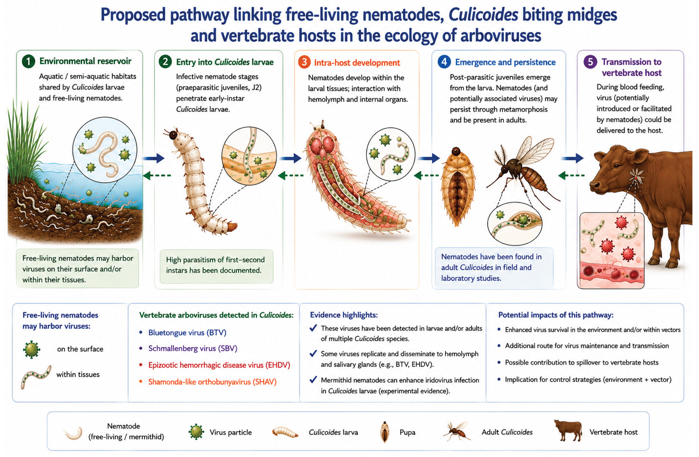
The scheme in Figure 1 integrates established observations on nematode–Culicoides associations and arbovirus infection of Culicoides with a proposed, yet experimentally unverified, pathway in which virus-associated nematodes may contribute to viral entry, persistence, and transmission through the midge developmental cycle. Solid blue arrows indicate established components of the pathway, whereas green dashed arrows indicate biologically plausible bidirectional interactions between nematodes and Culicoides at different developmental stages; red dotted arrows identify the experimentally unverified steps proposed here.
In particular, it remains to be established whether free-living nematodes carrying virus particles on their surface or within their tissues can provide a link between an environmental virus reservoir and Culicoides, whether the virus can subsequently persist through the midge developmental cycle, and whether it can ultimately reach a vertebrate host during blood feeding. Experimental testing of this pathway may reveal a previously unrecognized environmental component of Culicoides-borne virus transmission.
Discussion
Arbovirus transmission is conventionally considered within a vector–host framework in which hematophagous arthropods, particularly mosquitoes, ticks, sand flies, and biting midges, acquire viruses during blood feeding on an infected vertebrate host and subsequently transmit them to another susceptible host. In the case of Culicoides-borne arboviruses, most studies have therefore focused on vector competence, virus replication and dissemination within the insect, and transmission during subsequent blood meals [23,24]. However, acquisition of a virus from an infected vertebrate during blood feeding may not necessarily represent the only ecological route by which a virus can enter an arthropod vector.
In the present study, we propose that an additional, currently unexplored environmental pathway should be considered. Free-living or environmentally exposed nematode stages may potentially interact with virus particles present in aquatic or moist substrates and subsequently encounter immature Culicoides occupying the same habitats. If virus particles can remain associated with the nematode surface or tissues and gain access to the midge during nematode penetration or development, this would create a conceptually different transmission sequence involving two consecutive biological carriers rather than a single conventional arthropod vector: environment → virus-associated nematode → Culicoides → susceptible vertebrate host. At present, this pathway remains hypothetical for vertebrate arboviruses and should not be interpreted as an established mechanism of SHAV, BTV, SBV or EHDV transmission.
Nevertheless, several observations discussed above provide biological grounds for testing this hypothesis. Mermithid nematodes can penetrate Culicoides larvae, develop within the haemocoel, interact with internal host compartments and, in some systems, remain associated with the insect through metamorphosis into the adult stage. Independently, Culicoides support infection and replication of several arboviruses of vertebrates, with viral dissemination involving the haemocoel, fat body, salivary glands and other internal tissues. Most importantly, the interaction between nematodes and viruses within biting midges is not without experimental precedent: in the Culicoides variipennis sonorensis–Heleidomermis magnapapula system, nematode penetration markedly facilitated infection by an iridescent virus [31]. Studies in other nematode–arthropod systems have additionally demonstrated both external carriage of viral particles on the nematode cuticle and, in another system, viral replication within nematode tissues [34,35,36]. What remains unknown is whether comparable interactions can occur with arboviruses of vertebrates.
Environmental Change May Modify the Proposed Pathway
Environmental changes may further modify the epidemiological conditions under which such interactions could occur. Climate change and habitat alteration can influence vector abundance, geographical distribution, seasonality, breeding-site availability, and contacts among vectors, animal hosts and environmental reservoirs. These changes may create new opportunities for pathogen circulation and emergence. At the same time, delayed recognition of emerging infections, including clinically inapparent infections, together with the limited availability of specific vaccines and therapeutics for many newly emerging viruses, can complicate timely surveillance and control, as recently emphasized by Fatima et al. (2025) [41].
These considerations may be particularly relevant to the proposed nematode–virus–Culicoides pathway. Environmental conditions that increase the spatial and temporal overlap between free-living nematodes and immature Culicoides could increase opportunities for interactions among nematodes, viruses and biting midges. Temperature and precipitation, soil and sediment moisture, organic enrichment of breeding substrates, livestock density, and changes in vector seasonality may therefore influence not only conventional Culicoides-borne transmission but also the probability of an environmental route involving nematodes. Because immature Culicoides, free-living nematode stages and potentially virus-contaminated biological material may coexist in aquatic or moist substrates, environmental change could alter both the frequency and duration of these contacts.
This possibility may be especially relevant when considering the emergence and future spread of SHAV/DE/2026. The vector species responsible for its current circulation in Europe has not yet been established, and the potential contribution of environmental stages to virus maintenance or acquisition remains unknown. Consequently, surveillance focused exclusively on adult Culicoides and conventional vertebrate–vector transmission may overlook interactions occurring earlier in the vector life cycle or within the breeding environment.
Experimental Testing of the Proposed Pathway
The hypothesis proposed here is experimentally testable. We suggest that future surveillance and experimental studies should investigate Culicoides, nematodes and viruses as components of the same ecological system rather than examining them independently. Several complementary approaches could be particularly informative:
- Field-collected Culicoides should be examined simultaneously for SHAV RNA and nematode infection, allowing the frequency of nematode association to be compared between virus-positive and virus-negative midges.
- Free-living and mermithid nematodes collected directly from Culicoides breeding sites should be tested for SHAV, BTV, SBV and EHDV RNA, with appropriate methods used to distinguish viral material associated with the nematode surface from that present within nematode tissues.
- The prevalence and intensity of nematode infection should be compared between virus-positive and virus-negative Culicoides in naturally exposed populations. Such studies should ideally identify both the Culicoides and nematode species molecularly, because host–parasite compatibility differs among taxa.
- Controlled laboratory experiments should test the complete proposed sequence: virus-associated nematodes → Culicoides larvae → pupae → adult Culicoides. These experiments should determine whether virus associated with a nematode can enter the midge, whether it remains detectable and infectious during development, whether it survives metamorphosis, and whether it subsequently reaches tissues required for transmission, particularly the salivary glands.
Ultimately, the decisive experiment would be to determine whether virus initially associated with an environmentally exposed nematode can remain infectious throughout this sequence and subsequently be transmitted by an adult Culicoides during blood feeding to a susceptible vertebrate host. Demonstration of such a pathway would extend the conventional one-vector model of Culicoides-borne arbovirus transmission to a more complex ecological system in which an environmental nematode may act as an upstream carrier connecting an environmental virus source with the established arthropod vector. Conversely, failure to demonstrate individual steps of this sequence would be equally informative, allowing the biological limits of the proposed pathway to be defined.
If experimentally confirmed, the proposed pathway would broaden the current understanding of arbovirus transmission ecology by identifying an additional environmental component linking virus reservoirs, nematodes, arthropod vectors, and susceptible vertebrate hosts. Recognition of such a pathway would have implications extending beyond SHAV/DE/2026, as environmentally mediated virus acquisition could potentially be relevant to other Culicoides-borne arboviruses. Importantly, evidence for this mechanism would need to be incorporated into future biosurveillance, biorisk assessment, and the development of preventive and control strategies aimed at limiting the emergence and spread of arbovirus-associated biological threats.
Conclusions
The emergence of SHAV/DE/2026 in Europe highlights the need to consider not only established Culicoides-borne transmission mechanisms but also potentially overlooked ecological interactions that may contribute to arbovirus circulation. The evidence reviewed here demonstrates close associations between nematodes and Culicoides, including persistence of mermithid nematodes into adult midges, as well as experimental precedents for nematode-mediated enhancement of viral infection in arthropods. On this basis, we propose a testable hypothesis in which virus-associated nematodes may provide an additional environmental route for viral entry into Culicoides and potentially contribute to subsequent transmission to susceptible vertebrate hosts. Experimental verification or rejection of this pathway is now required. If confirmed, this mechanism should be considered in future arbovirus biosurveillance, biorisk assessment, and preventive strategies aimed at reducing the emergence and spread of arbovirus-associated biological threats.
Funding
This work was not financially supported.
Conflicts of Interest
The author declares no conflict of interest.
References
- Causey OR, Kemp GE, Causey CE, Lee VH. Isolations of Simbu-group viruses in Ibadan, Nigeria 1964–69, including the new types Sango, Shamonda, Sabo and Shuni. Ann Trop Med Parasitol. 1972;66(3):357–362. [CrossRef]
- Yanase T, Maeda K, Kato T, Nyuta S, Kamata H, Yamakawa M, Tsuda T. The resurgence of Shamonda virus, an African Simbu group virus of the genus Orthobunyavirus, in Japan. Arch Virol. 2005;150(2):361–369. [CrossRef]
- Sturgill TL, Walsh T, Ulziibat G, Clavijo A, Sturgill DM. Revisiting the importance of orthobunyaviruses for animal health: a scoping review of livestock disease, diagnostic tests, and surveillance strategies for the Simbu serogroup. Viruses. 2024;16(2):294. [CrossRef]
- van der Walt M, Rakaki ME, MacIntyre C, Mendes A, Junglen S, Theron C, Anthony T, O’Dell N, Venter M. Identification and Molecular Characterization of Shamonda Virus in an Aborted Goat Fetus in South Africa. Pathogens. 2023;12(9):1100. [CrossRef]
- Wernike K, Hoffmann B, Link EK, Rotheneder S, Eshak M, Pfaff F, Höper D, Beer M. It happened again – emergence of a Shamonda-like orthobunyavirus in Central Europe, 2026. bioRxiv. 2026. [CrossRef]
- Friedrich-Loeffler-Institut (FLI). FLI detects a new orthobunyavirus of the Simbu serogroup (“Shamonda-like”) in cattle in Germany. Greifswald-Insel Riems: FLI; 3 Aug Available from: https://www.fli.de/en/press/press-releases/press-singleview/fli-detects-a-new-orthobunyavirus-of-the-simbu-serogroup-shamonda-like-in-cattle-in-germany/.
- Institute of Virology and Immunology (IVI). New orthobunyavirus of the Simbu serogroup detected in cattle. Published 3 Aug 2026; updates of 10, 13, 17 and 20 Aug Mittelhäusern/Bern: IVI. Available from: https://www.ivi.admin.ch/en/neues-orthobunyavirus-der-simbu-serogruppe-bei-rindern-entdeckte.
- Ministère de l’Agriculture, de l’Agro-alimentaire et de la Souveraineté alimentaire. Cas d’infections de vaches laitières par un orthobunyavirus. Paris; 7 Aug Available from: https://agriculture.gouv.fr/cas-dinfections-de-vaches-laitieres-par-un-orthobunyavirus.
- Friedrich-Loeffler-Institut (FLI). Shamonda virus: Newly introduced cattle virus continues to spread. Greifswald-Insel Riems: FLI; 17 Aug Available from: https://www.fli.de/en/news/short-messages/short-message/shamonda-virus-newly-introduced-cattle-virus-continues-to-spread/.
- Suda Y, Murota K, Yanase T. Complete genome sequences of Sathuperi and Shamonda viruses isolated in Japan. J Vet Med Sci. 2023;85(12):1324–1326. [CrossRef]
- Koninklijke Nederlandse Maatschappij voor Diergeneeskunde (KNMvD). Shamonda-achtig virus aangetroffen bij Nederlandse runderen. 19 Aug Available from: https://www.knmvd.nl/shamonda-achtig-virus-aangetroffen-bij-nederlandse-runderen/.
- Land- en Tuinbouw Organisatie Nederland (LTO Nederland). Nieuw orthobunyavirus vastgesteld in Nederland: blijf alert op ziekteverschijnselen. 17 Aug Available from: https://www.lto.nl/nieuw-orthobunyavirus-vastgesteld-in-nederland-blijf-alert-op-ziekteverschijnselen/.
- Landwirtschaftskammer Oberösterreich. Shamonda-like-Virus bei Rindern: Erste Fälle in Österreich. 20 Aug Available from: https://ooe.lko.at/shamonda-like-virus-bei-rindern-erste-f%C3%A4lle-in-%C3%B6sterreich%2B2400%2B4448266.
- Hirashima Y, Kitahara S, Kato T, Shirafuji H, Tanaka S, Yanase T. Congenital malformations of calves infected with Shamonda virus, Southern Japan. Emerg Infect Dis. 2017;23(6):993–996. [CrossRef]
- World Organisation for Animal Health (WOAH). Schmallenberg disease. Paris: WOAH. Accessed 23 Aug Available from: https://www.woah.org/en/disease/schmallenberg-disease/.
- European Commission. Commission Implementing Regulation (EU) 2018/1882 of 3 December 2018 on the application of certain disease prevention and control rules to categories of listed diseases and establishing a list of species and groups of species posing a considerable risk for the spread of those listed diseases. Current consolidated version: 15 July EUR-Lex. Accessed 23 Aug Available from: https://eur-lex.europa.eu/eli/reg_impl/2018/1882/oj.
- World Organisation for Animal Health (WOAH). Animal Diseases: WOAH-listed diseases portal. Paris: WOAH. Accessed 23 Aug Available from: https://www.woah.org/en/what-we-do/animal-health-and-welfare/animal-diseases/.
- British Agriculture Bureau. Novel Shamonda-like orthobunyavirus in western Europe. 17 Aug Available from: https://www.britishagriculturebureau.co.uk/updates-and-information/novel-shamonda-like-orthobunyavirus-in-western-europe/.
- World Organisation for Animal Health (WOAH). Disease Data Collection: World Animal Health Information System (WAHIS). Paris: WOAH. Accessed 23 Aug Available from: https://www.woah.org/en/what-we-do/animal-health-and-welfare/disease-data-collection/.
- World Organisation for Animal Health (WOAH). Bunyaviral diseases of animals (excluding Rift Valley fever and Crimean–Congo haemorrhagic fever). In: Manual of Diagnostic Tests and Vaccines for Terrestrial Animals. Chapter 3.10.Adopted May 2023; WOAH Terrestrial Manual. Accessed 23 Aug Available from: https://www.woah.org/fileadmin/Home/eng/Health_standards/tahm/3.10.01_BUNYAVIRAL_DISEASES.pdf.
- Sick F, Beer M, Kampen H, Wernike K. Culicoides biting midges—underestimated vectors for arboviruses of public health and veterinary importance. Viruses. 2019;11(4):376. [CrossRef]
- Kampen H, Werner D. Biting midges (Diptera: Ceratopogonidae) as vectors of viruses. Microorganisms. 2023;11(11):2706. [CrossRef]
- Balenghien T, Pagès N, Goffredo M, Carpenter S, Augot D, Jacquier E, et al. The emergence of Schmallenberg virus across Culicoides communities and ecosystems in Europe. Prev Vet Med. 2014;116(4):360–369. [CrossRef]
- Monitoring of Schmallenberg virus, bluetongue virus and epizootic haemorrhagic disease virus in biting midges in Germany 2019–Parasites Vectors. 2025. PMID:40618098. [CrossRef]
- Fukuda T, Kline DL, Day JF. An iridescent virus and a microsporidium in the biting midge Culicoides barbosai from Florida. J Am Mosq Control Assoc. 2002;18(2):128–PMID:12083356.
- Mullens BA, Velten RK. Laboratory culture and life history of Heleidomermis magnapapula in its host, Culicoides variipennis (Diptera: Ceratopogonidae). J Nematol. 1994;26(1):1–10.
- Paine EO, Mullens BA. Distribution, seasonal occurrence, and patterns of parasitism of Heleidomermis magnapapula (Nematoda: Mermithidae), a parasite of Culicoides variipennis (Diptera: Ceratopogonidae) in California. Environ Entomol. 1994;23(1):154–160. [CrossRef]
- Mullens BA, Luhring KA, Breidenbaugh MS. Experimental host range studies with Heleidomermis magnapapula (Mermithidae), a parasite of Culicoides variipennis (Ceratopogonidae). J Am Mosq Control Assoc. 1997;13(4):398–PMID:9474570.
- Promrangsee C, Sanprasert V, Thepparat A, et al. First record of mermithid parasitism in adult biting midges, Culicoides huffi (Diptera: Ceratopogonidae), collected from Southern Thailand, with ultrastructural and molecular characterization. Parasites Vectors. 2025;18:303. [CrossRef]
- Mullens BA, Paine EO, Velten RK. Age-dependent parasitism of Culicoides variipennis sonorensis (Diptera: Ceratopogonidae) by Heleidomermis magnapapula (Nematoda: Mermithidae). Biological Control. 1997.
- Mullens BA, Velten RK, Federici BA. Iridescent virus infection in Culicoides variipennis sonorensis and interactions with the mermithid parasite Heleidomermis magnapapula. J Invertebr Pathol. 1999;73(2):231–233. PMID:10066406. [CrossRef]
- Williams T. Natural invertebrate hosts of iridoviruses (Iridoviridae). Neotrop Entomol. 2008;37(6):615–632. PMID:19169548. [CrossRef]
- Mills MK, Ruder MG, Nayduch D, Michel K, Drolet BS. Dynamics of epizootic hemorrhagic disease virus infection within the vector, Culicoides sonorensis (Diptera: Ceratopogonidae). PLoS One. 2017;12(11):e0188865. PMID:29176848. [CrossRef]
- Poinar GO Jr, Hess RT, Cole A. Replication of an iridovirus in a nematode (Mermithidae). Intervirology. 1980;14(5–6):316–320. PMID:7251336. [CrossRef]
- Muttis E, Micieli MV, García JJ. Culex pipiens affected by joint infection of a mosquito iridescent virus and Strelkovimermis spiculatus. J Invertebr Pathol. 2013;114(3):295–297. PMID:24012500. [CrossRef]
- Muttis E, Micieli MV, Urrutia MI, García JJ. Transmission of a pathogenic virus (Iridoviridae) of Culex pipiens larvae mediated by the mermithid Strelkovimermis spiculatus (Nematoda). J Invertebr Pathol. 2015;129:40–44. PMID:26031563. [CrossRef]
- Elbers ARW, Meiswinkel R, van Weezep E, van Oldruitenborgh-Oosterbaan MMS, Kooi EA. Schmallenberg virus in Culicoides spp. biting midges, the Netherlands, Emerg Infect Dis. 2013;19(1):106–109. [CrossRef]
- Goffredo M, Monaco F, Capelli G, et al. Schmallenberg virus in Italy: a retrospective survey in Culicoides stored during the bluetongue Italian surveillance program. Prev Vet Med. 2013;111(3–4):230–236. [CrossRef]
- Pagès N, Talavera S, Verdún M, et al. Schmallenberg virus detection in Culicoides biting midges in Spain: first laboratory evidence for highly efficient infection of Culicoides of the Obsoletus complex and Culicoides imicola. Transbound Emerg Dis. 2018;65(1):e1–e6. [CrossRef]
- Voigt A, Kampen H, Heuser E, et al. Bluetongue virus serotype 3 and Schmallenberg virus in Culicoides biting midges, Western Germany, Emerg Infect Dis. 2024;30(7):1438–1441. [CrossRef]
- Fatima M, An T, Park P-G, Hong K-J. Advancements and Challenges in Addressing Zoonotic Viral Infections with Epidemic and Pandemic Threats. Viruses. 2025;17(3). [CrossRef]
Figure 1.
Proposed pathway linking free-living nematodes, Culicoides biting midges, and vertebrate hosts in the ecology of arboviruses. Legend: Solid blue arrows represent established pathways supported by experimental and/or field observations. Green dashed arrows represent biologically plausible interactions inferred from currently available evidence. Red dotted arrows represent hypothetical pathways proposed in this study that have not yet been experimentally demonstrated.
Disclaimer/Publisher’s Note: The statements, opinions and data contained in all publications are solely those of the individual author(s) and contributor(s) and not of MDPI and/or the editor(s). MDPI and/or the editor(s) disclaim responsibility for any injury to people or property resulting from any ideas, methods, instructions or products referred to in the content. |
© 2026 by the authors. Licensee MDPI, Basel, Switzerland. This article is an open access article distributed under the terms and conditions of the Creative Commons Attribution (CC BY) license (http://creativecommons.org/licenses/by/4.0/).
Copyright: This open access article is published under a Creative Commons CC BY 4.0 license, which permit the free download, distribution, and reuse, provided that the author and preprint are cited in any reuse.