

Submitted:
23 August 2026
Posted:
25 August 2026
You are already at the latest version
Abstract
Oxidative stress accumulates early in the central nervous system (CNS) and is one major driver of age-related neurodegeneration. Here we compare major oxidative-stress models, highlighting their distinct mechanisms and disease relevance. We summarize therapeutic strategies ranging from radical scavengers and endogenous antioxidant enhancers to mitochondrial-targeted agents and rejuvenation approaches that restore intrinsic cytoprotective programs. We further discuss emerging oxidative-stress profiling strategies to define individual redox vulnerabilities. Together, mechanistically informed modeling, multidimensional profiling, and targeted restoration of oxidative resilience may enable more precise interventions for age-related CNS degeneration.
Keywords:
oxidative stress
; neurodegeneration
; disease modeling
; antioxidant
; profiling
; rejuvenation
1. Introduction
Age-related central nervous system (CNS) degeneration has become a major global health challenge, driven by an aging population, increasing healthcare costs, increased caregiving demands, and limited treatment options. Within the CNS, including the brain and eyes, oxidative stress begins accumulating early in life [1]. As aging progresses, additional stressors such as lifestyle factors and mitochondrial dysfunction can overwhelm cellular defense systems, leading to pathology. Three prominent aging theories highlight the importance of oxidative stress in the aging process: (1) the Free Radical Theory [2], which suggests that aging results from the excessive accumulation of reactive oxygen species (ROS) and reactive nitrogen species (RNS), which promote free radical reactions and cause oxidative damage to cells and tissues; (2) the Mitochondrial Theory [3], which posits that aging is driven by the gradual buildup of mitochondrial dysfunction and mitochondrial ROS accumulation; and, the epigenetic Information Theory of Aging, [4] that we proposed in 2023, which states that aging is driven by the progressive loss of youthful epigenetic information. The link to oxidative stress is raised by our recent work and others which revealed that the epigenetic regulation of oxidative-resilience genes, including GSTA4 [5], GSTM1, GSTM5 [6], becomes disrupted with aging in the eye, a developmental extension of the CNS. CNS cells, particularly dopaminergic neurons in the brain and the retina and RPE in the eye, are particularly vulnerable to oxidative stress due to their high oxygen consumption and lipid-rich content [7]. This intrinsic vulnerability positions oxidative stress as a central driver of age-related neurodegenerative disorders in the brain and eye.
Various oxidative insults have been developed for animal and cell culture models and are sometimes used interchangeably, despite inducing oxidative injury through distinct mechanisms. Yet, comprehensive overviews detailing the mechanisms underlying these different insults remain limited. Existing reviews also tend to focus on either the eye or the brain, despite their shared biology and the potential for experimental and therapeutic insights to translate across these CNS tissues.
Here, we review (i) environmental and lifestyle contributors to oxidative burden (Figure 1), (ii) the molecular mechanisms underlying common oxidative insults (Table 1), and (iii) antioxidant interventions related to CNS age-related disease (Table 2). This framework should guide the selection of appropriate experimental models and inform strategies to counter oxidative damage across a broad spectrum of age-related diseases.
2. In Vivo Models of Oxidative Stress: Environmental and Lifestyle Insults
2.1. High-Fat Diet or Obesity
According to 2022 WHO data, 1 in 8 people worldwide were living with obesity. Excessive fat accumulation in obesity increases mortality from non-communicable diseases, including cardiovascular diseases, diabetes and neurological disorders such as Parkinson’s disease (PD) and Alzheimer’s disease (AD). A key but often overlooked contributor to obesity-related pathology is the oxidative stress driven by lipid accumulation [8]. A recent systematic review further links high-fat diet (HFD) to retinal oxidative stress, inflammation, and neurodegeneration, supporting lipotoxicity as a systemic driver of ocular and CNS aging [9]. Human epidemiological and genetic evidence associates high-fat dietary patterns with increased glaucoma risk, whereas antioxidant-rich diets appear protective, supporting oxidative-stress-mediated retinal ganglion cell (RGC) vulnerability as a plausible mechanism [10].
A HFD increases intestinal chylomicron production, generating free fatty acids (FFAs) that are subsequently taken up by the liver. In hepatocytes, FFAs undergo mitochondrial β-oxidation and feed into the tricarboxylic acid (TCA) cycle [11]. The resulting increase in electron flux through the respiratory chain promotes mitochondrial ROS production and lipid peroxidation, including the generation of 4-hydroxy-2-nonenal (4-HNE) and malondialdehyde (MDA). These processes impair ATP production and mitochondrial respiration, ultimately contributing to mitochondrial dysfunction [12]. Excess ROS and lipotoxicity can then initiate a broader stress response by upregulating NADPH oxidase (NOX) subunits, inducing ER stress, disrupting Ca2+ homeostasis, and depleting antioxidant defenses, thereby establishing a self-amplifying cycle of oxidative damage [13]. FFAs can also be incorporated into triglycerides and very low-density lipoproteins (VLDL) [11]. When oxidized to oxLDL, lipoproteins further exacerbate oxidative stress (see oxLDL section below). Collectively, HFD-induced oxidative stress is initiated by excess lipid flux and mitochondrial β-oxidation and subsequently amplified by mitochondrial ROS production and lipotoxicity (Figure 2).
HFD is therefore a useful model for investigating how dietary lipid excess contributes to CNS and retinal degeneration. For example, an HFD providing 60% of calories from fat for 2 months can induce glial-cell senescence, contributing to neuroinflammation and cognitive decline [14]. Similarly, HFD regimens containing 60% kcal fat for 3 months or 45% kcal fat for 12 months induce oxidative stress in the retinal pigment epithelium (RPE), accompanied by inflammation and features of age-related macular degeneration (AMD) [15,16].
2.2. Radiation
Radiation is the emission or transmission of energy that can either induce atomic and molecular motion that produces heat (non-ionizing radiation) or remove tightly bound electrons from atoms to generate ions (ionizing radiation). Humans have real-world exposure to either of these. Non-ionizing radiation spans a broad range of wavelengths, including ultraviolet (UV), visible, near-infrared, and infrared light. Beyond our planet, astronauts encounter significant ionizing radiation exposures (especially during solar events) on spacecraft and on the International Space Station, creating a unique exposure that maybe related to CNS alterations seen in Spaceflight-Associated Neuro-ocular Syndrome (SANS) [17,18]. Together, ionizing radiation is a potent and long-lasting oxidative stressor to the CNS.
How radiation damages cells have also been characterized. Ionizing radiation, from photons to heavy particles like protons or carbon ions, can damage CNS cells, which are particularly vulnerable, by generating free radicals that harm DNA and induce metabolic stress [19]. Data show that even low-dose (<1Gy) and short-duration (<1 week) exposure to charged particles can trigger persistent oxidative stress for weeks to months, leading to cognitive impairment [20]. In vitro, oxidative stress is elevated for up to 80 cell generations after 10 Gy of X-ray exposure [21].
Then in contrast, non-ionizing radiation generates ROS primarily through energy-transfer mechanisms rather than direct molecular ionization. Ultraviolet radiation, particularly UVB (290–320 nm), contains high-energy photons that promote ROS production through photosensitization mechanisms [22]. Infrared laser radiation can induce oxidative stress by directly converting triplets oxygen (3O2) to singlet oxygen (1O2), a highly reactive form of ROS [23]. Visible light, especially blue light (415-455 nm), has been strongly linked to oxidative stress in the RPE in AMD [24]. This phototoxicity is driven in part by light-induced excitation of N-retinylidene-N-retinylethanolamine (A2E), a major fluorophore of lipofuscin, which generates ROS, particularly 1O2 [25,26].
These different wavelengths have consequently been used to model distinct forms of ocular oxidative injury. UVB irradiation (e.g., 35 mJ/cm2 daily for 4 days in mice [27]) is a well-established model of photoaging; however, because most UVB is absorbed by anterior ocular structures such as the cornea and lens, its penetration to the retina is limited. In contrast, blue light reaches the retina more efficiently and is commonly used to model RPE phototoxicity and AMD-like damage (e.g., blue LED exposure for 3 days [28]). Laser exposure is also widely used to induce choroidal neovascularization as a model of exudative AMD [29]. However, this approach primarily disrupts Bruch’s membrane and triggers a strong inflammatory and angiogenic response, with oxidative stress arising largely as a secondary consequence.
Importantly, low levels of radiation and particularly of non-ionizing radiation (light) has also been shown to be protective. This has primarily been attributed to the induction of low or chronic levels of cellular stress, at levels the cell can recover from using endogenous repair and antioxidant mechanisms in mitochondria and anti-oxidation pathways, these repair mechanisms, building resilience against future insults. This is discussed further in relevant sections regarding therapeutic interventions below at Section 4.4.
2.3. Cigarette Smoke
According to the WHO, tobacco smoking kills more than 8 million people each year and up to half of its users. Smoking is a well-established risk factor for aging and age-related diseases such as AMD, AD, and PD, with oxidative stress playing a central role in their pathogenesis [30,31].
Cigarette smoke contains a complex mixture of nearly 5000 chemical oxidants [32], including quinone/hydroquinone complexes in the tar phase and reactive oxygen- and carbon-centered radicals in the gas phase [32]. Some of these oxidants enter the bloodstream, causing DNA damage, protein modifications, and lipid peroxidation, which subsequently increase ROS production in the CNS, deplete antioxidants, and trigger inflammation [32].
Smoking-related ROS may facilitate the formation of amyloid-β (Aβ) oligomers and abnormal tau phosphorylation-key hallmarks of AD neuropathology [30]. Genetic susceptibility amplifies this risk: a Finnish study found that apolipoprotein E (APOE) ε4 carriers who smoke heavily in midlife more than double their risk of developing AD [33]. Importantly, smoking cessation reduced AD risk, underscoring the reversibility of smoking-related neurotoxic effects [34].
Smoking also increases the risk of age-related eye diseases, including cataract and AMD [31,35]. This risk arises through multiple mechanisms: smoking-related ROS production, reduction of macular pigment and plasma levels of antioxidants, inadequate peripheral blood flow [36]. Chronic exposure to cigarette smoke leads to impaired cytoprotective mechanisms, mitochondrial dysfunction, oxidative damage and structural degeneration of the RPE and Bruch’s membrane in mice, as well as RPE cell apoptosis in in vitro models. These changes mirror the early features of AMD, especially mitochondrial dysfunction as well as proteostasis and autophagy impairment that leads to RPE dysfunction and cell death [37]. Consistent with gene–environment interactions in AMD, iPSC-RPE studies suggest that the CFH Y402H risk background alters RPE metabolism and mitochondrial responses to chronic cigarette smoke extract [38].
Smoking may also worsen glaucomatous damage through vascular and oxidative mechanisms. Greater cumulative smoking exposure, measured by pack-years, has been associated with lower optic nerve head vessel density and larger choroidal microvasculature dropout, both of which are biomarkers of glaucoma severity and progression [39]. Emerging evidence suggests that smoking may contribute more strongly to glaucoma progression than disease onset, while smoking cessation may help mitigate this risk [40].
Cigarette smoke can effectively generate an oxidative stress model. For instance, exposure to cigarette smoke in mice - 24 hours/day, 5 days/week for 3.5 months, or 5 hours/day, 5 days/week for 6 months - induces oxidative damage to the RPE, leading to AMD-like features [31,41]. In in vitro culture systems, researchers use cigarette smoke extract to establish oxidative stress models [42].
2.4. Air Pollution
WHO data reveal that nearly the entire global population (99%) is exposed to air pollution levels that exceed WHO guideline limits. This polluted air contains harmful substances that contribute to strokes, heart diseases, lung cancer, and both acute and chronic respiratory conditions. Oxidative stress is a primary mechanism underlying these health effects [43,44,45,46]. Key pollutants of significant public health concern include not only fine particulate matter (PM2.5), but also strong oxidants such as nitrogen dioxide (NO2), ozone (O3), hydroxyl radicals (OH•), and transition metal-containing particles (e.g., copper and iron) [47]. These oxidants and metals can generate ROS within cells through redox reactions, while PM2.5 can also trigger inflammation via antigen-presenting pathways [48]. Air pollution (e.g., dust storms, PM2.5, O3) thus represents a multifaceted environmental hazard for studying age-related CNS degeneration and chronic eye diseases [49,50].
2.5. Aging (Mitochondrial Aging, Lysosomal Aging and Inflammaging)
Aging is characterized by mitochondrial dysfunction, lysosomal dysfunction, and chronic low-grade inflammation, which together create a feedback loop that accelerates oxidative stress, cellular senescence, and age-related diseases. With advanced age, mitochondrial function declines partly due to the accumulation of mutations in mitochondrial DNA (mtDNA). These defects impair oxidative phosphorylation, reduce adenosine triphosphate (ATP) production, and increase ROS generation. Mitochondria also serve as key hubs for innate immune signaling. Mitochondrial injury can trigger the release of mtDNA and oxidized mtDNA into the cytoplasm, where they activate cGAS-STING signaling and inflammasome pathways [51]. In the retina, Ferrington and colleagues have provided strong human-donor and iPSC-RPE evidence that AMD is associated with RPE mitochondrial dysfunction, increased mtDNA damage, altered bioenergetics, and impaired responses to oxidative stress, supporting mitochondrial decline as a central driver of retinal aging and AMD [52].
Mitochondrial dynamics, particularly the balance between fission and fusion and mitochondrial transport, are also strongly linked to neurodegeneration in the eye and brain. Fission/fusion genes are identified as specific source mutations in Parkinson’s disease (e.g. parkin), autosomal dominant optic atrophy (e.g. OPA1), and Charcot-Marie-Tooth disease type 2A (e.g. mitofusin-2). In vitro in retinal ganglion cells, the balance between fission and fusion strongly regulates axon growth [53,54,55,56], and in vivo in animal models, the increased intraocular pressure associated with glaucoma risk directly increases mitochondrial fission and stasis [57], while restoring mitochondrial fusion or transport strongly promotes neuroprotection and axon regeneration [58,59,60].
Lysosomal function also deteriorates with age. Our recent longitudinal profiling of lysosomal metabolites in the brain across the lifespan revealed age-dependent accumulation of glycerophosphodiesters and the amino acid derivative cystine. These metabolites are also elevated in lysosomal storage disorders and can predict chronological age [61], raising the possibility that progressive lysosomal metabolite accumulation contributes to aging-associated cellular dysfunction.
Chronic inflammation [62] further amplifies oxidative stress through redox-sensitive pathways such as NF-κB. Although these interconnected processes are difficult to model independently, restoring mitochondrial function, limiting lysosomal metabolite accumulation, and suppressing inflammation may help counter aging and age-related CNS disease. For example, IL-11 inhibition has been shown to extend both health span and lifespan [63].
3. In Vitro Oxidative Stress Models: Mechanistic Insights
3.1. Oxidized Low-Density Lipoprotein (oxLDL)
The in vivo HFD model we discussed before is complemented in vitro by exposure to oxidized oxLDL. oxLDL is generated when free radicals attack the polyunsaturated fatty acids (PUFAs) in LDL, producing reactive lipid peroxides such as 4-HNE [64]. These products modify apolipoprotein B (apoB), converting native LDL into oxLDL, which is recognized by scavenger receptors including LOX-1, CD36, CD68, and SR-A. Because these receptors are differentially expressed across cell and tissue types, oxLDL can trigger distinct downstream responses.
Lipoprotein receptor 1 (LOX-1), highly expressed on vascular endothelial cells, plays a key role in atherosclerosis and related vascular pathology [65,66]. HNE–histidine adducts on oxLDL act as LOX-1 ligands and facilitate receptor-mediated uptake. LOX-1 activation triggers three major responses: (1) ROS production, through p66Shc phosphorylation and PKC-mediated NOX activation [67], generating O2•- and H2O2. (2) apoptosis, through activation of caspase-3 and caspase-9 and suppression of Bcl-2 and c-IAP-1 [68,69]; (3) inflammation, through NF-κB, NLRP3, and MAPK signaling, inducing VCAM-1, ICAM-1, MCP-1, and pro-inflammatory cytokines [69,70] (Figure 3). SR-A and CD36 are particularly abundant in macrophages [71], where they promote NLRP3 inflammasome activation, ROS production, and foam-cell formation. Their expression can be further reinforced through a positive-feedback SR-A/CD36/PKCα/PPARγ pathway [72]. oxLDL can also activate receptor tyrosine kinase (RTK) signaling, influencing cell proliferation, differentiation, migration, and apoptosis [73]. In RPE cells, OxLDL is also taken up in a CD36-dependent manner [74]. OxLDL is then transported to the lysosomes where it induces lysosomal stabilization and subsequent inflammasome. Inflammasome activation, in turn, leads to the release of activated IL-1ß, a potent inflammatory cytokine and/or pyroptosis.
Malek and colleagues helped establish the AMD relevance of this lipid-stress pathway by showing that cholesterol- and apoB-containing lipoproteins accumulate in Bruch’s membrane, basal deposits, and drusen, supporting mechanistic parallels between AMD and atherosclerosis [75]. Their later work further implicated nuclear-receptor pathways, including PPAR and LXR signaling, in regulating RPE lipid metabolism, inflammation, and AMD-like pathology [76]. Complementing this framework, Miller and colleagues showed that shifting RPE lipid fate toward autophagy/lipophagy and mitochondrial fatty-acid oxidation can reduce apolipoprotein secretion, lipofuscin accumulation, and senescence [77,78]. Together, these studies suggest that impaired RPE lipid handling converts oxLDL/lipoprotein stress into lysosomal dysfunction, inflammation, and drusen-associated oxidative injury. Together, these findings suggest that shifting RPE lipid handling from extracellular lipoprotein secretion toward intracellular lipid degradation and mitochondrial metabolism may reduce drusen-associated lipid burden and oxidative stress.
3.2. Sodium Iodate (NaIO3)
Sodium iodate (NaIO3) was first found to induce fundus pigmentary changes and blindness in 1926 [81], and then developed as a model of oxidative injury because it can induce potent and rapid RPE-initiated retinal degeneration [82]. To investigate retinal degeneration in vivo, NaIO3 can be given through a single intraperitoneal injection [83], orbital venous plexus injection [84], or tail vein injection in mice [85,86]. However, the extent of RPE damage and the type of cell death differ: apoptosis is induced at 20 mg/kg [86], while necroptosis is induced at 100 mg/kg [83]. RPE death is followed by photoreceptors apoptosis, blood-retinal barrier breakdown, and retinal neurodegeneration, mirroring the pathological features seen in AMD [83,85,87,88]. In vitro, 2-5 mM NaIO3 induces apoptosis in ARPE-19 cells, 5-10 mM induces necrosis, and ≥10 mM triggers ferroptosis, all after 24-48 hours of exposure [87,89,90,91,92].
Mechanistically, NaIO3 oxidizes thiol-containing proteins and polyunsaturated fatty acids, depleting glutathione (GSH), increasing glutathione disulfide (GSSG), and generating lipid hydroperoxides [92]. It can also suppress GSH synthesis by inhibiting system Xc− [93], further promoting ROS accumulation and lipid peroxidation. This oxidative stress activates multiple downstream pathways, including NOX-dependent ER stress and Ca2+ overload [89,92], ERK signaling and epithelial-to-mesenchymal transition [89,91,94], NRF2-related antioxidant responses and ferroptotic signaling [94], mitochondrial dysfunction driven in part by lipid peroxidation products such as 4-HNE [95], and NF-κB/NLRP3-mediated inflammation [96] (Figure 4).
Importantly, RPE susceptibility may be further enhanced by melanin-dependent conversion of glycine to glyoxylate, a process increased by NaIO3 [97]. Glycine depletion and glyoxylate accumulation can promote Ca2+ influx, ROS production, lipid peroxidation, inflammatory cytokine release, and macrophage infiltration [98,99].
Overall, NaIO3 is a robust model of RPE-centered oxidative injury in which direct thiol oxidation, glutathione depletion, lipid peroxidation, mitochondrial stress, and inflammatory signaling converge to drive dose-dependent apoptosis, necrosis, and ferroptosis.
3.3. Amyloid Beta (Aβ)
Amyloid β (Aβ) oligomer accumulation correlates with cognitive impairment, AD and AD-related retinopathy [100,101]. Aβ peptide is produced through the cleavage of amyloid precursor protein (APP) by β- and γ-secretases, with Aβ monomers aggregating into oligomers, protofibrils, and amyloid fibrils, a process accelerated by 4-HNE [102]. Among these species, soluble Aβ oligomers are considered particularly neurotoxic and strongly linked to oxidative injury.
Aβ oligomers promote oxidative stress through several mechanisms. First, they can insert into lipid bilayers and drive lipid peroxidation, generating products such as 4-HNE [103,104]. In turn, 4-HNE promotes formation of cytotoxic protofibrillar Aβ species, impairs Aβ clearance, disrupts mitochondrial and synaptic function, inhibits Na+/K+-ATPase, and elevates intracellular Ca2+ [105,106]. Second, Aβ oligomers engage cell-surface receptors, including NMDAR, p75NTR, and LilrB2/PirB, triggering Ca2+ dysregulation, wNOX activation, apoptosis, and synaptic damage [107,108,109,110]. Third, Aβ binds redox-active metals such as Cu, Fe, and Zn, promoting aggregation and ROS generation [111,112]. Fourth, Aβ can accumulate in mitochondria [113], where it disrupts cytochrome c oxidase assembly [114,115], interacts with ABAD [116], and cyclophilin D [117], and promotes mitochondrial permeability transition and mtROS production (Figure 5).
Experimentally, soluble Aβ42 oligomers are commonly used to model Aβ-induced neurotoxicity [118]. Because wild-type rodents do not readily develop amyloid plaques, transgenic models such as Tg2576, which overexpresses Swedish-mutant human APP, are widely used to study amyloidosis and test anti-Aβ therapies [119].
3.4. Iron and Iron-Induced Oxidative Species
Aging disrupts iron homeostasis, leading to reduced intestinal iron absorption and systemic iron deficiency while simultaneously promoting iron accumulation in tissues such as the brain and retina, where it contributes to oxidative stress and neurodegeneration [120,121].
Iron enters cells through transferrin-dependent and transferrin-independent pathways. Intracellular ferric iron (Fe3+) is reduced to ferrous iron (Fe2+) and released into the cytosolic labile iron pool [122]. Fe2+ can generate highly reactive hydroxyl radicals (HO•) through Fenton and related redox reactions, causing oxidative damage to lipids, proteins, and DNA and promoting ferroptosis (Figure 6) [123].
Iron overload is a feature of several age-related neurodegenerative diseases, including AMD, AD, and PD [124,125], and is therefore widely used to model ferroptotic injury. In vitro, commonly used approaches include ferric ammonium citrate (10–100 mg/L) [126], hemin (5-20 μM) [127], and FeCl3 (10–500 μM) [128]. In vivo models include high-iron diets (14 g Fe/kg) [129], intracerebral iron injection [130], and intravenous iron-sucrose administration [131]. Ferroptosis can also be induced by impairing iron export, for example through hepcidin-mediated inhibition of ferroportin [132].
3.5. Other Oxidative Insults
Byproducts of lipid peroxidation, including 4-HNE and MDA, are widely used markers of oxidative stress. They also act as secondary mediators that amplify oxidative damage and can contribute to cell death. However, their high reactivity and instability limit their use as direct experimental oxidants. 4-HNE rapidly forms adducts with proteins and nucleic acids, making it difficult to quantify, whereas MDA is less reactive, less toxic, and more stable, and is therefore commonly used as a lipid peroxidation marker [133,134].
Hydrogen peroxide (H2O2) is widely used to model oxidative stress in age-related diseases [135]. Through Fenton and Haber–Weiss chemistry, H2O2 can generate highly reactive hydroxyl radicals. However, H2O2 is relatively unstable and rapidly decomposes into water and oxygen, causing rapid changes in effective concentration that can complicate experimental interpretation [136].
Tert-butyl hydroperoxide (t-BHP), a short-chain organic hydroperoxide, provides a more stable alternative to H2O2 [137]. t-BHP induces oxidative stress by generating peroxyl and alkoxyl radicals that initiate lipid peroxidation [138]; and by consuming reduced glutathione (GSH) during glutathione peroxidase-mediated detoxification [139]. It can be administered intracerebroventricularly to induce oxidative stress in vivo [140] and is also widely used in cell culture models [141].
1-Methyl-4-phenyl-1,2,3,6-tetrahydropyridine (MPTP) is a lipophilic neurotoxin that readily crosses the blood–brain barrier. In the brain, MPTP is converted by astrocytes and other glial cells into 1-methyl-4-phenylpyridinium (MPP+) [142,143]. MPP+ is selectively taken up by dopaminergic neurons, accumulates in mitochondria, and inhibits respiratory complex I, reducing ATP production and increasing ROS generation [144,145]. MPTP is therefore a gold-standard model of dopaminergic neurodegeneration and Parkinson’s disease (PD) [144,146].
Oxidopamine (6-hydroxydopamine, 6-OHDA) is another selective dopaminergic neurotoxin for studying PD [147]. Unlike MPTP, 6-OHDA does not cross the blood–brain barrier and must be delivered by stereotaxic injection. It damages both dopaminergic and noradrenergic neurons, largely through rapid auto-oxidation and ROS generation [148].
N-methyl-D-aspartic acid (NMDA) is not a direct oxidant but induces oxidative stress through excitotoxic NMDA receptor activation. NMDA receptors are Ca2+-permeable glutamate-gated ion channels expressed throughout the CNS [149]. Sustained activation causes mitochondrial Ca2+ overload and ROS production [150]. Intravitreal NMDA induces excitotoxic degeneration of retinal ganglion and amacrine cells [151], while intrahippocampal injection induces neuronal degeneration in the brain [149,152].
Tau is likewise not a direct oxidant. Although physiological tau stabilizes microtubules, pathological aggregation into neurofibrillary tangles contributes to neuronal dysfunction in AD [153]. Tauopathy can promote oxidative stress by disrupting mitochondrial fission–fusion dynamics, activating NOX signaling, and interacting with redox-active copper to promote H2O2 generation [154,155].
4. Therapeutic Oxidant Resistant Interventions: Mechanisms and Clinical Outcomes
4.1. AREDS/AREDS2, Vitamin C, and Vitamin E
The Age-Related Eye Disease Study (AREDS), initiated in 1999, evaluated nutritional interventions for AMD and age-related cataract [156]. The original AREDS formulation contained vitamin C (VC), vitamin E (VE), β-carotene, zinc, and copper and was shown to reduce progression to advanced AMD and vision loss [157], leading to its use in patients with intermediate AMD [158]. Because β-carotene was associated with increased lung cancer risk, particularly in former smokers [159,160], AREDS2 replaced β-carotene with lutein and zeaxanthin. AREDS2 has been shown to reduce progression to advanced AMD and slow geographic atrophy progression toward the fovea [161,162,163].
The protective effects of AREDS/AREDS2 involve free-radical scavenging, support of endogenous antioxidant enzymes, and maintenance of retinal function (Figure 7). VC and VE are direct free-radical scavengers [164]. and have also shown potential benefit in PD [165] and AD [166] (Table 2). Lutein and zeaxanthin are concentrated in the macula, where they filter blue light and provide antioxidant protection [167,168]. Zinc and copper support antioxidant enzymes, including SOD, catalase, and metallothionein, with copper also included to prevent potential zinc-induced copper deficiency [156,169].
4.2. GSH, NAC, GlyNAC
Glutathione (GSH) is the most abundant intracellular antioxidant and a major defense against oxidative damage. GSH directly scavenges reactive species and serves as a reducing substrate for detoxification of lipid hydroperoxides [170]. Glutathione S-transferases (GSTs) further protect cells by conjugating GSH to electrophilic lipid-peroxidation products. In particular, GSTA4 efficiently conjugates 4-HNE to GSH, generating GS-HNE for subsequent export from the cell [171,172,173]. Importantly, our recent study [5] showed that transient expression of the reprogramming factors Oct4–Sox2–Klf4 (OSK) selectively induces GSTA4 in aged retinal pigment epithelium (RPE): GSTA4 knockout abolishes OSK-mediated oxidative protection, whereas AAV-GSTA4 alone recapitulates its cytoprotective and vision-preserving effects in vivo. These findings identify GSTA4 as a key effector of GSH-dependent detoxification of lipid peroxidation products (Figure 8).
Intravenous GSH showed minimal clinical benefit in PD (Table 2) [166], potentially because of limited blood–brain barrier penetration. Subsequent studies therefore explored intranasal delivery, which can access the CNS through olfactory and trigeminal pathways. Intranasal GSH has been reported to be safe and well tolerated, with some studies suggesting symptomatic improvement in PD [174,175].
To further optimize intracellular GSH levels, the combination supplement GlyNAC- composed of the GSH precursors N-acetylcysteine (NAC) and glycine-was developed to support and enhance the body’s natural GSH synthesis. This approach enables cells to efficiently produce GSH based on their metabolic needs, especially during periods of increased oxidative stress or GSH deficiency. Additionally, GlyNAC can efficiently cross the blood-brain barrier. In mice, GlyNAC supplementation reduced oxidative stress and improved age-related phenotypes, with one study reporting lifespan extension [176]. Clinical studies in older adults further found GlyNAC to be safe and associated with improved mitochondrial function, GSH availability, and markers of oxidative stress and aging [177,178]. In human RPE models, NAC has also been shown to protect against oxidative damage, supporting glutathione-precursor supplementation as a rational retinal antioxidant strategy [179].
4.3. Coenzyme Q10 and Its Analogues
Unlike VC, VE, and GSH/GlyNAC, which directly scavenge reactive species, Coenzyme Q10 (CoQ10) refers to a mitochondrial redox-active coenzyme that cycles between oxidized ubiquinone and reduced ubiquinol forms. In its oxidized ubiquinone form, CoQ10 functions in the electron-transport chain by shuttling electrons from complexes I/II to complex III. In its reduced ubiquinol form, CoQ10 acts as a lipophilic antioxidant, limiting membrane lipid peroxidation by quenching lipid peroxyl radicals [180,181].
Clinical trials of CoQ10 in Parkinson’s disease (PD) began in the late 1990s. Early studies found CoQ10 to be safe but largely ineffective at lower doses [182], while later trials using up to 1,200 mg/day suggested it slows functional decline [183]. However, subsequent studies failed to confirm meaningful clinical benefit in PD [184,185,186]. Similarly, although CoQ10 improved cognitive outcomes in animal models of Alzheimer’s disease, clinical efficacy has not been demonstrated [187]. Its therapeutic potential is limited in part by poor bioavailability and inefficient mitochondrial uptake [188].
Preclinical studies in glaucoma further support CoQ10 as a mitochondrial-protective antioxidant in the visual CNS. In a mouse model of glaucoma, CoQ10 reduced glutamate excitotoxicity, oxidative stress, and mitochondrial alteration, thereby improving retinal ganglion cell survival [189]. A later study showed that ubiquinol, the reduced form of CoQ10 with improved bioavailability, also attenuated oxidative stress and preserved retinal ganglion cell survival and visual function. These findings suggest that CoQ10-based interventions may be particularly relevant in optic neuropathies where mitochondrial dysfunction and oxidative injury contribute to retinal ganglion cell degeneration.
To improve mitochondrial delivery, analogues such as mitoquinone (MitoQ) were developed. MitoQ links ubiquinone to the lipophilic triphenylphosphonium (TPP) cation, enabling preferential mitochondrial accumulation [190]. Additionally, MitoQ is absorbed into cells faster than CoQ10 due to its smaller size, water solubility, and positive charge, allowing for rapid uptake from circulation without the need for fat or food [136].
Despite improved uptake, a double-blind, placebo-controlled trial found no effect of MitoQ on PD progression [191]. Further studies are needed to determine whether mitochondrial-targeted CoQ analogues can provide benefit in other neurodegenerative settings.
4.4. Other Noteworthy Oxidation-Resistant Interventions
Sterically hindered phenols have been shown in models of ox-LDL induced injury to provide potent protection of RPE from oxidative stress [192]. In RPE cultures, hindered phenol–containing antioxidants such as troglitazone, PMC (2,2,5,7,8-pentamethyl-6-chromanol), and trolox prevent ~90% of ox-LDL–induced cell death, whereas several non-phenolic antioxidants with strong general radical-scavenging capacity fail to confer similar protection, indicating a structure-specific effect. These compounds accumulate in lysosomes where they suppress ROS, stabilize lysosomal membranes, and facilitate the clearance of ox-LDL, in part by increasing levels of NCP1, a cholesterol-export protein [192]. Mechanistically, PMC blunts ox-LDL induced upregulation of oxidative stress-responsive genes such as HMOX-1 and other genes downstream of NRF2 [193]. Collectively, these studies support hindered phenol as promising candidates for pharmacologic protection in dry AMD.
Alpha-lipoic acid (ALA) is a mitochondrial antioxidant containing two thiol groups that support redox reactions and directly scavenge hydroxyl radicals, hypochlorous acid, and singlet oxygen [194]. Although ALA did not protect against expansion of the geographic atrophy form of AMD in a small clinical trial [195], and its clinical efficacy remains debated when used for diabetic peripheral neuropathy [196,197,198]. However, small clinical studies have suggested cognitive stabilization in mild AD [199] and benefit from combining ALA with omega-3 fatty acids [200].
Deprenyl (selegiline) irreversibly inhibits monoamine oxidase B (MAO-B), reducing dopamine breakdown and associated H2O2 generation. Clinical trials suggested that deprenyl can delay disability and the need for levodopa in Parkinson’s disease (PD) [201,202,203], although later reviews found no clear evidence that MAO-B inhibitors slow disease progression and noted more treatment withdrawals due to adverse events [204].
Deuterated docosahexaenoic acid (D-DHA) has emerged as a promising oxidation-resistant supplement because deuterium substitution markedly slows the initiation of DHA oxidation and subsequent lipid-peroxidation chain reactions. Photoreceptor outer-segment membranes are exceptionally rich in native DHA - accounting for ~60% of their polyunsaturated lipid content - making them especially susceptible to iron-catalyzed peroxidative damage that underlies age-related retinal degeneration. Joshua Dunaief’s group demonstrated that orally delivered D-DHA efficiently incorporates into retinal membranes and, in an iron-overload mouse model of geographic atrophy, virtually abolished carboxyethlypyrrole adduct formation, preserved photoreceptor and RPE architecture, and maintained electroretinographic function - benefits not achieved with unmodified DHA [205]. Similarly, D-DHA protects RPE from degeneration in a mouse model of chronic oxidative stress [206]. These findings establish D-DHA as a mechanism-based antioxidant that acts upstream to curb lipid peroxidation, holding translational promise for dry AMD and other oxidative retinopathies. A clinical trial of D-DHA for geographic atrophy has been initiated [NCT07029945].
Memantine, an uncompetitive NMDA receptor antagonist, was evaluated for glaucoma neuroprotection by targeting glutamate-mediated RGC excitotoxicity. In non-human primate glaucoma models, memantine reduced structural RGC/optic nerve damage and protected lateral geniculate nucleus neurons from shrinkage, supporting glaucoma as a broader CNS neurodegenerative process [207,208]. However, two large Phase 3 trials in open-angle glaucoma failed to meet their primary endpoints, highlighting the translational challenges of neuroprotection trials, including endpoint selection and trial-execution limitations [209].
Several metabolic interventions may also enhance oxidative resilience. Caloric restriction and lithocholic acid (LCA) can reduce mitochondrial ROS and extend lifespan in model organisms [210,211,212]. Alpha-ketoglutarate (AKG) can imitate a state of energy scarcity by regulate tricarboxylic acid cycle and limit ATP synthase activity [213]. Besides, AKG can directly consume ROS by reacting with ammonia and generates glutamine [214] or H2O2 decomposition [215]. AKG has been reported to extend lifespan and exhibit anti-aging effects in worms, flies, and mice [216,217,218].
Photobiomodulation (PBM) using red and near-infrared (NIR) light has emerged as a non-invasive strategy to enhance mitochondrial resilience. Red/NIR wavelengths (~600–1000 nm) are thought to act primarily through mitochondrial cytochrome c oxidase, enhancing electron transport and ATP production while activating antioxidant and pro-survival pathways [219,220];. In vitro and in vivo studies show that PBM can improve mitochondrial function and reduce ROS, lipid peroxidation, and retinal degeneration following oxidative injury [221]. Glaucoma-relevant studies further demonstrate that 670-nm red light protects retinal ganglion cell dendrites following axonal injury and increases the redox-protective protein DJ-1, suggesting activation of endogenous oxidative resilience [222]. More broadly, PBM has shown therapeutic potential across retinal diseases, including AMD [223]. Most recently, the FDA authorized the Valeda Light Delivery System, combining 590-, 660-, and 850-nm wavelengths, for treatment of dry AMD, marking an important clinical translation of PBM. These support red/NIR light as a drug-free strategy to reinforce mitochondrial function and endogenous antioxidant defenses.
Finally, controlled hypoxia has emerged as a strategy for mitochondrial disease [224]. Supporting this concept, the small-molecule compound HypoxyStat - which alters hemoglobin-oxygen dissociation dynamics to reduce tissue oxygen delivery - demonstrated remarkable efficacy in a preclinical model of mitochondrial disease. Daily oral administration of HypoxyStat induced systemic hypoxia under normoxic conditions and prolonged survival in Ndufs4 knockout mice, while ameliorating neurobehavioral deficits and pathological hallmarks [225].
4.5. Rejuvenating Endogenous Oxidative Resilience with Gene Therapy
An emerging strategy is to use AAV gene therapy to rejuvenate endogenous oxidative resilience that deteriorates with age, rather than relying solely on repeated administration of exogenous antioxidants. NRF2 is a central regulator of antioxidant and detoxification programs, and AAV-mediated restoration of NRF2 signaling could reinforce multiple endogenous defense pathways simultaneously [226]. We discovered that epigenetic restoration provides a broader approach: AAV delivery of epigenetic reprogramming factors Oct4–Sox2–Klf4 (OSK) can restore youthful cellular programs in retinal neurons [227], benefiting animal models of glaucoma [227,228], multiple sclerosis [229], NAION [230], and AD [231], and even extending mouse lifespan [232]. AAV-OSK has now entered into clinical trial to examine efficacy in restoring vision of glaucoma and NAION patients (NCT07290244). Notably, whereas the effects of OSK in neurons are largely mediated through epigenetic remodeling, including mechanisms involving the DNA demethylase TET2, a distinct non-canonical function of OSK has recently been identified in RPE: enhancing cellular resilience to diverse oxidative insults, including NaIO3, ox-LDL, and H2O2 [5]. Mechanistic dissection of this response identified GSTA4 as a direct downstream effector of OSK, whose promoter is activated by OSK and whose expression enhances oxidative resilience in the RPE by detoxifying the lipid peroxidation product 4-HNE [5]. These findings suggest that individual cytoprotective effectors downstream of reprogramming factors may capture selected therapeutic benefits of rejuvenation while avoiding risks associated with prolonged expression of the reprogramming factors themselves. Together, NRF2, OSK, and GSTA4 illustrate a developing paradigm in which gene therapy is used not simply to neutralize ROS, but to restore the intrinsic stress-response networks that enable young cells to withstand oxidative injury.
5. Measuring Oxidative Stress - Biomarkers Aiding Translation to Human Therapies
Particularly in the retina, we have a unique opportunity to measure oxidative stress and related biomarkers through imaging modalities and liquid biopsy sampling [233]. Imaging is particularly motivating through hyperspectral and fluorescent wavelengths that return reliable data on mitochondrial redox stress balance, as well as blood flow and oxygenation, both at rest and in response to visual/light stimuli [234,235]. In the eye, aqueous humor liquid-biopsy approaches such as TEMPO enable proteomic and metabolomic profiling from small-volume samples, providing a more direct molecular readout of the retinal environment for inflammation, cellular stress, and biological aging [236]. Novel visual stimuli combined with advanced approaches to the analysis of visually evoked potentials in the brain can parse out the eye-brain connection in glaucoma [237]. Such biomarkers in the eye are also proving useful as biomarkers for neurodegenerative disease in the brain and even healthspan more broadly [238,239], and are discussed at length elsewhere.
More broadly, we propose a multilayered oxidative-stress profiling framework (Table 3; Figure 9) combining markers of lipid, protein, and DNA oxidation; iron homeostasis; endogenous antioxidant capacity; and inflammation with retinal imaging, MRI/MRS, genetics, and longitudinal wearable data. Integration of these measurements through AI could help define individual redox vulnerabilities, stratify disease risk, and guide selection and monitoring of antioxidant or cytoprotective interventions.
6. Summary and Future Directions — From Antioxidant Therapy to Restoring Endogenous Redox Resilience
Oxidative stress is a major driver of age-related CNS degeneration. Here, we categorize commonly used oxidative insults by mechanism and application to facilitate selection of appropriate experimental models. In vivo models—including high-fat diet, radiation, cigarette smoke, air pollution, and aging itself—capture interactions among environmental exposures, mitochondrial dysfunction, inflammation, and neurodegeneration. Complementary in vitro models provide greater mechanistic precision: NaIO3 models RPE-centered oxidative injury; NMDA induces calcium overload and secondary oxidative damage; iron dysregulation and oxLDL model ferroptotic and vascular stress; Aβ and tau reproduce components of Alzheimer’s disease pathology; and MPTP and 6-OHDA model distinct mechanisms of dopaminergic degeneration. Despite this mechanistic diversity, a recent study identified ROS-induced disulfide bond formation at surface-exposed cysteines and consequent loss of protein mobility as a potentially shared mechanism of oxidative damage [240], raising the possibility that diverse oxidative insults ultimately converge on common cellular vulnerabilities.
Antioxidant interventions are similarly diverse, ranging from direct radical scavengers such as vitamins C and E to approaches that augment endogenous antioxidant systems, including NAC, GlyNAC, alpha-lipoic acid, and mitochondrial-targeted interventions such as CoQ10 and MitoQ. Clinical studies have provided evidence of benefit in selected settings, most notably AREDS/AREDS2 for AMD, but overall efficacy across age-related CNS diseases remains variable. A major challenge is therefore to determine who is experiencing oxidative stress, which form of oxidative damage predominates, and which antioxidant pathway is insufficient. As discussed above, multimodal oxidative-stress profiling may help identify which redox pathways are impaired in individual patients and thereby guide more rational therapeutic selection.
Beyond supplying exogenous antioxidants, an emerging therapeutic opportunity is to rejuvenate the endogenous oxidative resilience that deteriorates with age. NRF2 exemplifies such a cytoprotective hub, coordinating antioxidant and detoxification programs while supporting mitochondrial function, lysosomal homeostasis, and proteostasis [226]. Its dysregulation during aging and diseases such as AMD may compromise the ability of cells to adapt to oxidative stress. Similarly, our recent work identified GSTA4 as a downstream effector of OSK-mediated rejuvenation to enhance oxidative resilience in the aged RPE [5], demonstrating that rejuvenation programs can be mechanistically dissected to identify individual cytoprotective effectors. Such approaches may circumvent limitations of conventional antioxidants, including poor delivery to the relevant cells and subcellular compartments, by restoring or augmenting intrinsic defense programs.
Along this line, a further challenge is delivering redox-modulating therapies to the appropriate CNS cell type and subcellular compartment. This is particularly relevant in the eye, where systemic therapies such as oral or intravenous drugs often achieve limited intraocular exposure because of the blood–retinal barrier, while topical formulations primarily reach anterior tissues and penetrate poorly to the posterior retina. For diseases involving the retina, such as glaucoma, where retinal ganglion cells are the primary therapeutic target, and AMD, effective delivery may therefore require local approaches such as intravitreal administration of drugs, intraocular drug delivery platforms, or AAV vectors for gene therapy approaches. Alternatively, sustained-release ocular implants [241] could provide prolonged local exposure while reducing the need for repeated dosing. For brain delivery, intranasal administration has also emerged as a promising strategy to access the CNS while bypassing the blood–brain barrier, with studies in PD showing improved delivery and encouraging clinical outcomes [174,175]. Thus, therapeutic efficacy will depend not only on identifying the appropriate antioxidant or cytoprotective pathway, but also on achieving sufficient and sustained delivery to the vulnerable cell population.
Together, the next generation of redox medicine may move beyond broad antioxidant use toward defining disease-specific oxidative vulnerabilities, rejuvenating endogenous defenses, and delivering therapies to the right cells. Combining mechanistically appropriate models, multidimensional biomarkers, rejuvenation of cytoprotective networks, and tissue-targeted delivery could enable more precise interventions for age-related CNS degeneration while creating opportunities for discoveries in the eye to inform the brain, and vice versa.
Contributions
H.S., J.A., Z.C., A.S.H., V.B.M., J.L.G., J.L.D., J.T.H., R.N.W., P.A.D., J.S.W., B.R.K., Y.R.L., jointly contributed to the conceptualization, writing, and revision of this review. H.S., J.A., Z.C., Y.R.L. prepared the illustrations.
Acknowledgments
We thank James Cameron for assistance with illustrations. H.S. received support from the First Affiliated Hospital with Nanjing Medical University. A.S.H. received support from NASA (80NNSSC20K1034), NIH/NEI (R01EY030501, R01EY038008), David L. Epstein Career Advancement Award in Glaucoma Research sponsored by Alcon, American Glaucoma Society Mid-Career Physician Research Grant, and an unrestricted grant by Research to Prevent Blindness (New York). J.L.G. received support NIH/NEI P30EY026877, R01EY033816, K12EY033745, and Research to Prevent Blindness (New York). J.L.D. was supported by an unrestricted grant from Research to Prevent Blindness (New York). R.N.W received research support from NIH/NEI R01EY09058, R01MD014850, K12EY024255, and an unrestricted grant from Research to Prevent Blindness, (New York). J.S.W. is an H.H.M.I. investigator. B.R.K is supported by NIH grants R01EY03691, R01EY025794, P01AG071463. Y.R.L. was supported by postdoctoral fellowships from Glenn/AFAR (by Michael Shen, MIT’13), the Life Sciences Research Foundation (by Ming Lei-Chang Luo Life Science Fund) and the NIH/NEI Pathway to Independence Award (K99EY037340).
Declare of interests
Y.R.L., B.R.K., J.S.W. are inventors on a patent applied for by Whitehead Institute and Mass General Brigham related to using novel methods of cellular rejuvenation to treat oxidative stress-related injury and age-related degeneration. A.S.H. is a consultant to Amydis, Celanese, Balance, Iantrek, QLARIS, Santen, Spinogenix, Topcon, Vialase, and has financial interest in Diagnosys and Heidelberg Engineering. R.N.W. is a consultant to Amydis, Spinogenix, Merck Research Laboratories, Boehringer-Ingelheim, Thea, Qlaris, Topcon; receives diagnostic instruments for research from Vasoptic, Optina, ICare; and is on Board of Directors of Remoni Health, Iantrek, Machine MD, Cascader, and EyeGo. J.S.W. declares outside interest in 5AM Ventures, Amgen, nChroma, DEM Biosciences, KSQ Therapeutics, Maze Therapeutics, Tenaya Therapeutics, Tessera Therapeutics, Thermo Fisher, Xaira, and TRV.
Declaration of use of generative AI
The author(s) used ChatGPT for manuscript proof reading, after which the author(s) reviewed and edited the content as needed and take(s) full responsibility for the content.
Abbreviations
CNS, central nervous system; mtDNA, mitochondrial DNA; ROS, reactive oxygen species; RNS, reactive nitrogen species; O2•−, superoxide anion; H2O2, hydrogen peroxide; OH•, hydroxyl radical; ROO•, peroxyl radical; 1O2, singlet oxygen; 3O2, triplet oxygen; O3, ozone; NO2, nitrogen dioxide; PM2.5, fine particulate matter; VC, vitamin C; VE, vitamin E; SOD, superoxide dismutase; NAC, N-acetylcysteine; CoQ10, coenzyme Q10; AMD, age-related macular degeneration; HFD, high-fat diet; FFA, free fatty acid; TCA, tricarboxylic acid; 4-HNE, 4-hydroxy-2-nonenal; MDA, malondialdehyde; NADH, nicotinamide adenine dinucleotide; NADPH, nicotinamide adenine dinucleotide phosphate; NOX, NADPH oxidase; LDL, low-density lipoprotein; VLDL, very-low-density lipoprotein; oxLDL, oxidized low-density lipoprotein; RPE, retinal pigment epithelium; UV, ultraviolet; UVB, ultraviolet B; LED, light-emitting diode; A2E, N-retinylidene-N-retinylethanolamine; SANS, Spaceflight-Associated Neuro-ocular Syndrome; AD, Alzheimer’s disease; PD, Parkinson’s disease; APOE, apolipoprotein E; apoB, apolipoprotein B; Aβ, amyloid-β; ATP, adenosine triphosphate; PUFA, polyunsaturated fatty acid; LOX-1, lectin-like oxidized low-density lipoprotein receptor-1; SR, scavenger receptor; NF-κB, nuclear factor kappaB; TNFα, tumor necrosis factor alpha; IL-1β, interleukin-1β; IL-6, interleukin-6; NLRP3, NOD-, LRR-, and pyrin domain-containing protein 3; VCAM-1, vascular cell adhesion molecule-1; ICAM-1, intercellular adhesion molecule-1; MCP-1, monocyte chemoattractant protein-1; Bcl-2, B-cell lymphoma 2; PKC, protein kinase C; PKCβ, protein kinase Cβ; JNK, c-Jun N-terminal kinase; MAPK, mitogen-activated protein kinase; RTK, receptor tyrosine kinase; PI3K, phosphatidylinositol-3-kinase; AKT, protein kinase B; NaIO3, sodium iodate; NMDA, N-methyl-D-aspartic acid; NMDAR, N-methyl-D-aspartate receptor; GSH, glutathione; GSSG, glutathione disulfide; GST, glutathione S-transferase; GSTA4, glutathione S-transferase alpha 4; GS-HNE, glutathione-conjugated 4-HNE; CAT, catalase; GPx, glutathione peroxidase; ER, endoplasmic reticulum; ERK, extracellular signal-related kinase; NRF2, nuclear factor erythroid 2–related factor 2; HO-1, heme oxygenase-1; EMT, epithelial-to-mesenchymal transition; APP, amyloid precursor protein; p75NTR, p75 neurotrophin receptor; LilrB2, leukocyte immunoglobulin-like receptor B2; PirB, paired immunoglobulin-like receptor B; ABAD, Aβ-binding alcohol dehydrogenase; mPTP, mitochondrial permeability transition pore; Fe3+, ferric iron; Fe2+, ferrous iron; MPTP, 1-methyl-4-phenyl-1,2,3,6-tetrahydropyridine; MPP+, 1-methyl-4-phenylpyridinium; 6-OHDA, 6-hydroxydopamine; AREDS, Age-Related Eye Disease Study; DHA, docosahexaenoic acid; D-DHA, deuterated docosahexaenoic acid; EPA, eicosapentaenoic acid; DHAsc, dehydroascorbate; SDHAsc, semidehydroascorbate; LOO•, lipid peroxyl radical; LOOH, lipid hydroperoxide; Toc, tocopherol; Toc•, tocopheroxyl radical; IRBP, interphotoreceptor retinoid-binding protein; FA-OOH, fatty-acid hydroperoxide; PL-OOH, phospholipid hydroperoxide; CU-OOH, cholesterol hydroperoxide; GlyNAC, glycine and N-acetylcysteine; GSH/GSSG, reduced-to-oxidized glutathione ratio; CoQ10, coenzyme Q10; MitoQ, mitoquinone; TPP, triphenylphosphonium; ALA, alpha-lipoic acid; MAO-B, monoamine oxidase B; RGC, retinal ganglion cell; LCA, lithocholic acid; AKG, alpha-ketoglutarate; AAV, adeno-associated virus; OSK, Oct4, Sox2, and Klf4; TET2, ten-eleven translocation methylcytosine dioxygenase 2; TEMPO, tracing expression of multiple protein origins; CRP, C-reactive protein; CSF, cerebrospinal fluid; 8-OHdG, 8-hydroxy-2′-deoxyguanosine.
References
- Tanase, M.; et al. Role of Carbonyl Modifications on Aging-Associated Protein Aggregation. Sci. Rep. 2016, 6, 19311. [Google Scholar] [CrossRef] [PubMed]
- D, H. Aging: A theory based on free radical and radiation chemistry. J. Gerontol. 1956, 11. [Google Scholar] [CrossRef] [PubMed]
- D, H. The biologic clock: The mitochondria? J. Am. Geriatr. Soc. 1972, 20. [Google Scholar] [CrossRef] [PubMed]
- Lu, Y.R.; Tian, X.; Sinclair, D.A. The Information Theory of Aging. Nat. Aging 2023, 3, 1486–1499. [Google Scholar] [CrossRef] [PubMed]
- Lu, Y.R.; et al. Reprogramming Factors Activate a Non-Canonical Oxidative Resilience Pathway That Can Rejuvenate RPEs and Restore Vision. bioRxiv Prepr. Serv. Biol. 2025, 2030, 673239. [Google Scholar] [CrossRef] [PubMed]
- Hunter, A.; et al. DNA methylation is associated with altered gene expression in AMD. Invest Ophthalmol. Vis. Sci. 2012, 53, 2089–2105. [Google Scholar] [CrossRef] [PubMed]
- Salim, S. Oxidative Stress and the Central Nervous System. J. Pharmacol. Exp. Ther. 2017, 360, 201–205. [Google Scholar] [CrossRef] [PubMed]
- Hang, Y.; et al. The phytochemical epigallocatechin gallate prolongs the lifespan by improving lipid metabolism, reducing inflammation and oxidative stress in high-fat diet-fed obese rats. Aging Cell 2020, 19. [Google Scholar] [CrossRef] [PubMed]
- Pieńczykowska, K.; Bryl, A.; Mrugacz, M. The Impact of a High-Fat Diet on Eye Health. Nutrients 2025, 17, 3271. [Google Scholar] [CrossRef] [PubMed]
- Shengnan, Z.; Tao, W.; Yanan, Z.; Chao, S. Exploring the impact of diet, sleep, and metabolomic pathways on Glaucoma subtypes: Insights from Mendelian randomization and cross-sectional analyses. Nutr. Metab. 2025, 22, 74. [Google Scholar] [CrossRef] [PubMed]
- D, P.; A, B.; B, F.; A, M. Mitochondria in steatohepatitis. Semin Liver Dis. 2001, 21. [Google Scholar] [CrossRef] [PubMed]
- Naoto, M.-N.; et al. Increased oxidative stress precedes the onset of high-fat diet-induced insulin resistance and obesity. Metabolism 2008, 57. [Google Scholar] [CrossRef] [PubMed]
- O’Brien, P.D.; Hinder, L.M.; Callaghan, B.C.; Feldman, E.L. Neurological consequences of obesity. Lancet Neurol. 2017, 16, 465–477. [Google Scholar] [CrossRef] [PubMed]
- Ogrodnik, M.; et al. Obesity-Induced Cellular Senescence Drives Anxiety and Impairs Neurogenesis. Cell Metab. 2019, 29, 1061–1077.e1068. [Google Scholar] [CrossRef] [PubMed]
- Keeling, E.; et al. A High Fat “Western-style” Diet Induces AMD-Like Features in Wildtype Mice. Mol. Nutr. Food Res. 2022, 66, e2100823. [Google Scholar] [CrossRef] [PubMed]
- Sterling, J.K.; et al. Inflammatory adipose activates a nutritional immunity pathway leading to retinal dysfunction. Cell Rep. 2022, 39, 110942. [Google Scholar] [CrossRef] [PubMed]
- Sung, J.K.; Kassel, R.; Hargens, A.; Huang, A.S. Spaceflight Associated Neuro-Ocular Syndrome; Ong, J., Ed.; Academic Press: Cambridge, MA, USA, 2022; pp. 155–173. [Google Scholar]
- Stenger, M.B.; et al. Focus on the Optic Nerve Head in Spaceflight-Associated Neuro-ocular Syndrome. Ophthalmology 2019, 126, 1604–1606. [Google Scholar] [CrossRef] [PubMed]
- Smart, D. Radiation Toxicity in the Central Nervous System: Mechanisms and Strategies for Injury Reduction. Semin. Radiat. Oncol. 2017, 27, 332–339. [Google Scholar] [CrossRef] [PubMed]
- Tseng, B.P.; et al. Functional Consequences of Radiation-Induced Oxidative Stress in Cultured Neural Stem Cells and the Brain Exposed to Charged Particle Irradiation. Antioxid. Redox Signal. 2014, 20, 1410–1422. [Google Scholar] [CrossRef] [PubMed]
- C L, L.; et al. Apoptosis, reproductive failure, and oxidative stress in Chinese hamster ovary cells with compromised genomic integrity. Cancer Res. 1998, 58. [Google Scholar]
- de Jager, T.L.; Cockrell, A.E.; Du Plessis, S.S. Ultraviolet Light Induced Generation of Reactive Oxygen Species. Adv. Exp. Med. Biol. 2017, 996, 15–23. [Google Scholar] [CrossRef] [PubMed]
- Sokolovski, S.G.; et al. Infrared laser pulse triggers increased singlet oxygen production in tumour cells. Sci. Rep. 2013, 3, 3484. [Google Scholar] [CrossRef] [PubMed]
- Emilie, A.; et al. Phototoxic action spectrum on a retinal pigment epithelium model of age-related macular degeneration exposed to sunlight normalized conditions. PLoS ONE 2013, 8. [Google Scholar] [CrossRef] [PubMed]
- Roxana A, R.; Nathan L, M.; Aarti, B.; Gabriel H, T. Light exposure stimulates formation of A2E oxiranes in a mouse model of Stargardt’s macular degeneration. Proc. Natl. Acad. Sci. U S A 2004, 101. [Google Scholar] [CrossRef] [PubMed]
- M, R.; et al. Blue light-induced reactivity of retinal age pigment. In vitro generation of oxygen-reactive species. J. Biol. Chem. 1995, 270. [Google Scholar] [CrossRef] [PubMed]
- An, M.J.; et al. Transcriptome analysis for UVB-induced phototoxicity in mouse retina. Environ. Toxicol. 2018, 33, 52–62. [Google Scholar] [CrossRef] [PubMed]
- Nakamura, M.; et al. Exposure to excessive blue LED light damages retinal pigment epithelium and photoreceptors of pigmented mice. Exp. Eye Res. 2018, 177, 1–11. [Google Scholar] [CrossRef] [PubMed]
- Lambert, V.; et al. Laser-induced choroidal neovascularization model to study age-related macular degeneration in mice. Nat. Protoc. 2013, 8, 2197–2211. [Google Scholar] [CrossRef] [PubMed]
- Durazzo, T.C.; Mattsson, N.; Weiner, M.W. Smoking and increased Alzheimer’s disease risk: A review of potential mechanisms. Alzheimer’s Dement. J. Alzheimer’s Assoc. 2014, 10, S122–S145. [Google Scholar] [CrossRef] [PubMed]
- Espinosa-Heidmann, D.G.; et al. Cigarette smoke-related oxidants and the development of sub-RPE deposits in an experimental animal model of dry AMD. Invest Ophthalmol. Vis. Sci. 2006, 47, 729–737. [Google Scholar] [CrossRef] [PubMed]
- Church, D.F.; Pryor, W.A. Free-radical chemistry of cigarette smoke and its toxicological implications. Environ. Health Perspect. 1985, 64, 111–126. [Google Scholar] [CrossRef] [PubMed]
- Rusanen, M.; et al. Midlife smoking, apolipoprotein E and risk of dementia and Alzheimer’s disease: A population-based cardiovascular risk factors, aging and dementia study. Dement. Geriatr. Cogn. Disord. 2010, 30, 277–284. [Google Scholar] [CrossRef] [PubMed]
- Choi, D.; Choi, S.; Park, S.M. Effect of smoking cessation on the risk of dementia: A longitudinal study. Ann. Clin. Transl. Neurol. 2018, 5, 1192–1199. [Google Scholar] [CrossRef] [PubMed]
- Hurley, S.; Purcell, K.; Greenhalgh, E.M.; Winstanley, M.H. Tobacco in Australia: Facts & Issues; (eds E. M. Greenhalgh, M. M. Scollo, & M. H. Winstanley) Ch. Chapter 3: The health effects of active smoking, (Cancer Council Victoria, 2020).
- Zarbin, M. Current concepts in the pathogenesis of age-related macular degeneration. Arch. Ophthalmol. 2004, 122, 598–614. [Google Scholar] [CrossRef] [PubMed]
- Singh, K.K.; et al. Molecular underpinnings of induced degenerative heterogeneity in the retinal pigment epithelium. Proc. Natl. Acad. Sci. 2026, 123, e2505412123. [Google Scholar] [CrossRef] [PubMed]
- Shang, P.; et al. The Complement Factor H (Y402H) risk polymorphism for age-related macular degeneration affects metabolism and response to oxidative stress in the retinal pigment epithelium. Free Radic. Biol. Med. 2024, 225, 833–845. [Google Scholar] [CrossRef] [PubMed]
- Eslani, M.; et al. Effects of Smoking on Optic Nerve Head Microvasculature Density in Glaucoma. J. Glaucoma 2022, 31, 710–716. [Google Scholar] [CrossRef] [PubMed]
- Mahmoudinezhad, G.; et al. Impact of Smoking on Visual Field Progression in a Long-term Clinical Follow-up. Ophthalmology 2022, 129, 1235–1244. [Google Scholar] [CrossRef] [PubMed]
- Masashi, F.; Norihiro, N.; Thomas E, S.; Shyam, B.; James T, H. Chronic cigarette smoke causes oxidative damage and apoptosis to retinal pigmented epithelial cells in mice. PLoS ONE 2008, 3. [Google Scholar] [CrossRef] [PubMed]
- Wang, L.; et al. Nrf2 signaling modulates cigarette smoke-induced complement activation in retinal pigmented epithelial cells. Free Radic. Biol. Med. 2014, 70, 155–166. [Google Scholar] [CrossRef] [PubMed]
- Charalambos, M.; et al. Molecular mechanisms of oxidative stress in asthma. Mol. Asp. Med. 2021, 85. [Google Scholar] [CrossRef] [PubMed]
- Michelle L, B.; Lilian, C.-G. Air pollution: Mechanisms of neuroinflammation and CNS disease. Trends Neurosci. 2009, 32. [Google Scholar] [CrossRef] [PubMed]
- Austin, L.; Kelly, O.; Surya, P.; Matilda, N. The Impact of Air Pollution on Atopic Dermatitis. Curr. Allergy Asthma Rep. 2023, 23. [Google Scholar] [CrossRef] [PubMed]
- Xiaoquan, R.; Jixin, Z.; Robert D, B.; Sanjay, R. Effect of Particulate Matter Air Pollution on Cardiovascular Oxidative Stress Pathways. Antioxid. Redox Signal 2017, 28. [Google Scholar] [CrossRef] [PubMed]
- Lakey, P.S.; et al. Chemical exposure-response relationship between air pollutants and reactive oxygen species in the human respiratory tract. Sci. Rep. 2016, 6, 32916. [Google Scholar] [CrossRef] [PubMed]
- Lim, E.Y.; Kim, G.D. Particulate Matter-Induced Emerging Health Effects Associated with Oxidative Stress and Inflammation. Antioxidants 2024, 13. [Google Scholar] [CrossRef] [PubMed]
- Hajipour, S.; et al. Exposure to ambient dusty particulate matter impairs spatial memory and hippocampal LTP by increasing brain inflammation and oxidative stress in rats. Life Sci. 2020, 242, 117210. [Google Scholar] [CrossRef] [PubMed]
- Millen, A.E.; et al. Air Pollution and Chronic Eye Disease in Adults: A Scoping Review. Ophthalmic Epidemiol. 2024, 31, 1–10. [Google Scholar] [CrossRef] [PubMed]
- Kim, J.; Kim, H.-S.; Chung, J.H. Molecular mechanisms of mitochondrial DNA release and activation of the cGAS-STING pathway. Exp. Mol. Med. 2023, 55, 510–519. [Google Scholar] [CrossRef] [PubMed]
- Ferrington, D.A.; et al. Altered bioenergetics and enhanced resistance to oxidative stress in human retinal pigment epithelial cells from donors with age-related macular degeneration. Redox Biol. 2017, 13, 255–265. [Google Scholar] [CrossRef] [PubMed]
- Yokota, S.; et al. Kif5a Regulates Mitochondrial Transport in Developing Retinal Ganglion Cells In Vitro. Invest Ophthalmol. Vis. Sci. 2023, 64, 4. [Google Scholar] [CrossRef] [PubMed]
- Kreymerman, A.; et al. Light-evoked activity and BDNF regulate mitochondrial dynamics and mitochondrial localized translation in CNS axons. iScience 2025, 28, 113563. [Google Scholar] [CrossRef] [PubMed]
- Kreymerman, A.; et al. MTP18 is a Novel Regulator of Mitochondrial Fission in CNS Neuron Development, Axonal Growth, and Injury Responses. Sci. Rep. 2019, 9, 10669. [Google Scholar] [CrossRef] [PubMed]
- Steketee, M.B.; et al. Mitochondrial dynamics regulate growth cone motility, guidance, and neurite growth rate in perinatal retinal ganglion cells in vitro. Invest Ophthalmol. Vis. Sci. 2012, 53, 7402–7411. [Google Scholar] [CrossRef] [PubMed]
- Takihara, Y.; et al. In vivo imaging of axonal transport of mitochondria in the diseased and aged mammalian CNS. Proc. Natl. Acad. Sci. U S A 2015, 112, 10515–10520. [Google Scholar] [CrossRef] [PubMed]
- Liu, D.; et al. Optineurin-facilitated axonal mitochondria delivery promotes neuroprotection and axon regeneration. Nat. Commun. 2025, 16, 1789. [Google Scholar] [CrossRef] [PubMed]
- Shah, S.H.; Goldberg, J.L. The Role of Axon Transport in Neuroprotection and Regeneration. Dev. Neurobiol. 2018, 78, 998–1010. [Google Scholar] [CrossRef] [PubMed]
- Shah, S.H.; et al. Quantitative transportomics identifies Kif5a as a major regulator of neurodegeneration. Elife 2022, 11. [Google Scholar] [CrossRef] [PubMed]
- Puszynska, A.M.; et al. Atlas of lysosomal aging reveals a metabolite signature shared with lysosomal storage disorders. Science 2026, 393, eady0832. [Google Scholar] [CrossRef] [PubMed]
- Franceschi, C.; Campisi, J. Chronic inflammation (inflammaging) and its potential contribution to age-associated diseases. J. Gerontol. A Biol. Sci. Med. Sci. 2014, 69, S4–S9. [Google Scholar] [CrossRef] [PubMed]
- Widjaja, A.A.; et al. Inhibition of IL-11 signalling extends mammalian healthspan and lifespan. Nature 2024, 632, 157–165. [Google Scholar] [CrossRef] [PubMed]
- Nègre-Salvayre, A.; et al. Proatherogenic effects of 4-hydroxynonenal. Free Radic. Biol. Med. 2017, 111, 127–139. [Google Scholar] [CrossRef] [PubMed]
- Cominacini, L.; et al. Oxidized low density lipoprotein (ox-LDL) binding to ox-LDL receptor-1 in endothelial cells induces the activation of NF-kappaB through an increased production of intracellular reactive oxygen species. J. Biol. Chem. 2000, 275, 12633–12638. [Google Scholar] [CrossRef] [PubMed]
- Lubrano, V.; Balzan, S. LOX-1 and ROS, inseparable factors in the process of endothelial damage. Free Radic. Res. 2014, 48, 841–848. [Google Scholar] [CrossRef] [PubMed]
- Kun-Ling, T.; et al. Coenzyme Q10 suppresses oxLDL-induced endothelial oxidative injuries by the modulation of LOX-1-mediated ROS generation via the AMPK/PKC/NADPH oxidase signaling pathway. Mol. Nutr. Food Res. 2011. [Google Scholar] [CrossRef] [PubMed]
- Chen, J.; et al. Role of caspases in Ox-LDL-induced apoptotic cascade in human coronary artery endothelial cells. Circulation Research 2004, 94, 370–376. [Google Scholar] [CrossRef]
- Barreto, J.; Karathanasis, S.; Remaley, A.; Sposito, A. Role of LOX-1 (Lectin-Like Oxidized Low-Density Lipoprotein Receptor 1) as a Cardiovascular Risk Predictor: Mechanistic Insight and Potential Clinical Use. Arterioscler. Thromb. Vasc. Biol. 2021, 41, 153–166. [Google Scholar] [CrossRef] [PubMed]
- Mentrup, T.; et al. Atherogenic LOX-1 signaling is controlled by SPPL2-mediated intramembrane proteolysis. J. Exp. Med. 2019, 216, 807–830. [Google Scholar] [CrossRef] [PubMed]
- Hughes, D.; Fraser, I.; Gordon, S. Murine macrophage scavenger receptor: In vivo expression and function as receptor for macrophage adhesion in lymphoid and non-lymphoid organs. Eur. J. Immunol. 1995, 25, 466–473. [Google Scholar] [CrossRef] [PubMed]
- Jung-Suk, C.; et al. Dietary compound quercitrin dampens VEGF induction and PPARgamma activation in oxidized LDL-exposed murine macrophages: Association with scavenger receptor CD36. J. Agric. Food Chem. 2009, 58. [Google Scholar] [CrossRef] [PubMed]
- Du, Z.; Lovly, C. Mechanisms of receptor tyrosine kinase activation in cancer. Mol. Cancer 2018, 17, 58. [Google Scholar] [CrossRef] [PubMed]
- Gopalan, G.; Ariel R, C.; Dhanesh, A.; Patricia A, D.A. Oxidized Lipoprotein Uptake Through the CD36 Receptor Activates the NLRP3 Inflammasome in Human Retinal Pigment Epithelial Cells. Invest Ophthalmol. Vis. Sci. 2016, 57. [Google Scholar] [CrossRef] [PubMed]
- Malek, G.; Li, C.M.; Guidry, C.; Medeiros, N.E.; Curcio, C.A. Apolipoprotein B in cholesterol-containing drusen and basal deposits of human eyes with age-related maculopathy. Am. J. Pathol. 2003, 162, 413–425. [Google Scholar] [CrossRef] [PubMed]
- Choudhary, M.; et al. LXRs regulate features of age-related macular degeneration and may be a potential therapeutic target. JCI Insight 2020, 5. [Google Scholar] [CrossRef] [PubMed]
- Zhang, Q.; et al. Pharmacologic activation of autophagy without direct mTOR inhibition as a therapeutic strategy for treating dry macular degeneration. Aging 2021, 13, 10866–10890. [Google Scholar] [CrossRef] [PubMed]
- Hass, D.T.; et al. Acetyl-CoA carboxylase inhibition increases retinal pigment epithelial cell fatty acid flux and restricts apolipoprotein efflux. J. Biol. Chem. 2024, 300, 107772. [Google Scholar] [CrossRef] [PubMed]
- Yu, A.L.; Lorenz, R.L.; Haritoglou, C.; Kampik, A.; Welge-Lussen, U. Biological effects of native and oxidized low-density lipoproteins in cultured human retinal pigment epithelial cells. Exp. Eye Res. 2009, 88, 495–503. [Google Scholar] [CrossRef] [PubMed]
- Draczynska-Lusiak, B.; Doung, A.; Sun, A.Y. Oxidized lipoproteins may play a role in neuronal cell death in Alzheimer disease. Mol. Chem. Neuropathol. 1998, 33, 139–148. [Google Scholar] [CrossRef] [PubMed]
- Sorsby, A. EXPERIMENTAL PIGMENTARY DEGENERATION OF THE RETINA BY SODIUM IODATE. Br. J. Ophthalmol. 1941, 25, 58–62. [Google Scholar] [CrossRef] [PubMed]
- Nie, Q.; et al. Sodium Iodate-Induced Mouse Model of Age-Related Macular Degeneration Displayed Altered Expression Patterns of Sumoylation Enzymes E1, E2 and E3. Curr. Mol. Med. 2018, 18, 550–555. [Google Scholar] [CrossRef] [PubMed]
- Kiuchi, K.; Yoshizawa, K.; Shikata, N.; Moriguchi, K.; Tsubura, A. Morphologic characteristics of retinal degeneration induced by sodium iodate in mice. Curr. Eye Res. 2002, 25, 373–379. [Google Scholar] [CrossRef] [PubMed]
- Machalińska, A.; et al. Sodium iodate selectively injuries the posterior pole of the retina in a dose-dependent manner: Morphological and electrophysiological study. Neurochem. Res. 2010, 35, 1819–1827. [Google Scholar] [CrossRef] [PubMed]
- Balmer, J.; Zulliger, R.; Roberti, S.; Enzmann, V. Retinal Cell Death Caused by Sodium Iodate Involves Multiple Caspase-Dependent and Caspase-Independent Cell-Death Pathways. Int. J. Mol. Sci. 2015, 16, 15086–15103. [Google Scholar] [CrossRef] [PubMed]
- Peng, Z.; et al. Protection of retina by αB crystallin in sodium iodate induced retinal degeneration. PLoS ONE 2014, 9. [Google Scholar] [CrossRef] [PubMed]
- Hanus, J.; Anderson, C.; Sarraf, D.; Ma, J.; Wang, S. Retinal pigment epithelial cell necroptosis in response to sodium iodate. Cell Death Discov. 2016, 2, 16054. [Google Scholar] [CrossRef] [PubMed]
- Wang, J.; Iacovelli, J.; Spencer, C.; Saint-Geniez, M. Direct effect of sodium iodate on neurosensory retina. Investig. Ophthalmol. Vis. Sci. 2014, 55, 1941–1953. [Google Scholar] [CrossRef] [PubMed]
- Yang, Y.; et al. Inhibition of oxidative stress-induced epithelial-mesenchymal transition in retinal pigment epithelial cells of age-related macular degeneration model by suppressing ERK activation. J. Adv. Res. 2024, 60, 141–157. [Google Scholar] [CrossRef] [PubMed]
- Mao, X.; et al. The rescue effect of mesenchymal stem cell on sodium iodate-induced retinal pigment epithelial cell death through deactivation of NF-κB-mediated NLRP3 inflammasome. Biomed. Pharmacother. Biomed. Pharmacother. 2018, 103, 517–523. [Google Scholar] [CrossRef] [PubMed]
- Hsu, M.; et al. Quercetin Alleviates the Accumulation of Superoxide in Sodium Iodate-Induced Retinal Autophagy by Regulating Mitochondrial Reactive Oxygen Species Homeostasis through Enhanced Deacetyl-SOD2 via the Nrf2-PGC-1α-Sirt1 Pathway. Antioxidants 2021, 10. [Google Scholar] [CrossRef] [PubMed]
- Liu, B.; et al. Sodium iodate induces ferroptosis in human retinal pigment epithelium ARPE-19 cells. Cell Death Dis. 2021, 12, 230. [Google Scholar] [CrossRef] [PubMed]
- Chen, C.; et al. Sodium Iodate-Induced Ferroptosis in Photoreceptor-Derived 661W Cells Through the Depletion of GSH. Int. J. Mol. Sci. 2025, 26, 2334. [Google Scholar] [CrossRef] [PubMed]
- Tang, Z.; et al. HO-1-mediated ferroptosis as a target for protection against retinal pigment epithelium degeneration. Redox Biol. 2021, 43, 101971. [Google Scholar] [CrossRef] [PubMed]
- Carlström, K.E.; et al. Gsta4 controls apoptosis of differentiating adult oligodendrocytes during homeostasis and remyelination via the mitochondria-associated Fas-Casp8-Bid-axis. Nat. Commun. 2020, 11, 4071. [Google Scholar] [CrossRef] [PubMed]
- Mao, X.; et al. The rescue effect of mesenchymal stem cell on sodium iodate-induced retinal pigment epithelial cell death through deactivation of NF-κB-mediated NLRP3 inflammasome. BioMed Pharmacother. 2018, 103, 517–523. [Google Scholar] [CrossRef] [PubMed]
- Baich, A.; Ziegler, M. The effect of sodium iodate and melanin on the formation of glyoxylate. Pigment Cell Res. 1992, 5, 394–395. [Google Scholar] [CrossRef] [PubMed]
- Wang, W.; et al. Glycine metabolism in animals and humans: Implications for nutrition and health. Amino Acids 2013, 45, 463–477. [Google Scholar] [CrossRef] [PubMed]
- Wang, X.; Xie, L.; Liu, C. CCR2 antagonist attenuates calcium oxalate-induced kidney oxidative stress and inflammation by regulating macrophage activation. Exp. Anim. 2024, 73, 211–222. [Google Scholar] [CrossRef] [PubMed]
- Reed, T.; Pierce, W.; Markesbery, W.; Butterfield, D. Proteomic identification of HNE-bound proteins in early Alzheimer disease: Insights into the role of lipid peroxidation in the progression of AD. Brain Res. 2009, 1274, 66–76. [Google Scholar] [CrossRef] [PubMed]
- Qiuchen, C.; et al. Transport of β-amyloid from brain to eye causes retinal degeneration in Alzheimer’s disease. J. Exp. Med. 2024, 221. [Google Scholar] [CrossRef] [PubMed]
- Chen, G.-F.; et al. Amyloid beta: Structure, biology and structure-based therapeutic development. Acta Pharmacol. Sin. 2017, 38, 1205–1235. [Google Scholar] [CrossRef] [PubMed]
- Butterfield, D.A.; Hensley, K.; Harris, M.; Mattson, M.; Carney, J. Β-Amyloid Peptide Free Radical Fragments Initiate Synaptosomal Lipoperoxidation in a Sequence-Specific Fashion: Implications to Alzheimer′s Disease. Biochem. Biophys. Res. Commun. 1994, 200, 710–715. [Google Scholar] [CrossRef] [PubMed]
- Butterfield, D. Brain lipid peroxidation and alzheimer disease: Synergy between the Butterfield and Mattson laboratories. Ageing Res. Rev. 2020, 64, 101049. [Google Scholar] [CrossRef] [PubMed]
- Siegel, S.; Bieschke, J.; Powers, E.; Kelly, J. The oxidative stress metabolite 4-hydroxynonenal promotes Alzheimer protofibril formation. Biochemistry 2007, 46, 1503–1510. [Google Scholar] [CrossRef] [PubMed]
- Owen, J.B.; et al. Oxidative modification to LDL receptor-related protein 1 in hippocampus from subjects with Alzheimer disease: Implications for Aβ accumulation in AD brain. Free Radic. Biol. Med. 2010, 49, 1798–1803. [Google Scholar] [CrossRef] [PubMed]
- Alberdi, E.; et al. Amyloid β oligomers induce Ca2+ dysregulation and neuronal death through activation of ionotropic glutamate receptors. Cell Calcium 2010, 47, 264–272. [Google Scholar] [CrossRef] [PubMed]
- Shankar, G.; et al. Natural oligomers of the Alzheimer amyloid-beta protein induce reversible synapse loss by modulating an NMDA-type glutamate receptor-dependent signaling pathway. J. Neurosci. Off. J. Soc. Neurosci. 2007, 27, 2866–2875. [Google Scholar] [CrossRef] [PubMed]
- Hashimoto, Y.; et al. Molecular characterization of neurohybrid cell death induced by Alzheimer’s amyloid-beta peptides via p75NTR/PLAIDD. J. Neurochem 2004, 90, 549–558. [Google Scholar] [CrossRef] [PubMed]
- Kim, T.; et al. Human LilrB2 is a β-amyloid receptor and its murine homolog PirB regulates synaptic plasticity in an Alzheimer’s model. Science 2013, 341, 1399–1404. [Google Scholar] [CrossRef] [PubMed]
- Boom, A.; et al. Astrocytic calcium/zinc binding protein S100A6 over expression in Alzheimer’s disease and in PS1/APP transgenic mice models. Biochim. Et. Biophys. Acta (BBA) -Mol. Cell Res. 2004, 1742, 161–168. [Google Scholar] [CrossRef] [PubMed]
- Crnich, E.; Lullo, R.; Tabaka, A.; Havens, M.A.; Kissel, D.S. Interactions of copper and copper chelate compounds with the amyloid beta peptide: An investigation into electrochemistry, reactive oxygen species and peptide aggregation. J. Inorg. Biochem. 2021, 222, 111493. [Google Scholar] [CrossRef] [PubMed]
- Wilkins, H.M. Interactions between amyloid, amyloid precursor protein, and mitochondria. Biochem. Soc. Trans. 2023, 51, 173–182. [Google Scholar] [CrossRef] [PubMed]
- Anandatheerthavarada, H.; Biswas, G.; Robin, M.; Avadhani, N. Mitochondrial targeting and a novel transmembrane arrest of Alzheimer’s amyloid precursor protein impairs mitochondrial function in neuronal cells. J. Cell Biol. 2003, 161, 41–54. [Google Scholar] [CrossRef] [PubMed]
- Devi, L.; Prabhu, B.; Galati, D.; Avadhani, N.; Anandatheerthavarada, H. Accumulation of amyloid precursor protein in the mitochondrial import channels of human Alzheimer’s disease brain is associated with mitochondrial dysfunction. J. Neurosci. Off. J. Soc. Neurosci. 2006, 26, 9057–9068. [Google Scholar] [CrossRef] [PubMed]
- Lustbader, J.W.; et al. ABAD directly links Abeta to mitochondrial toxicity in Alzheimer’s disease. Science 2004, 304, 448–452. [Google Scholar] [CrossRef] [PubMed]
- Du, H.; Yan, S.S. Mitochondrial permeability transition pore in Alzheimer’s disease: Cyclophilin D and amyloid beta. Biochim Biophys. Acta 2010, 1802, 198–204. [Google Scholar] [CrossRef] [PubMed]
- Puzzo, D.; et al. Picomolar amyloid-beta positively modulates synaptic plasticity and memory in hippocampus. J. Neurosci. Off. J. Soc. Neurosci. 2008, 28, 14537–14545. [Google Scholar] [CrossRef] [PubMed]
- Hsiao, K.; et al. Correlative Memory Deficits, Aβ Elevation, and Amyloid Plaques in Transgenic Mice. Science 1996, 274, 99–103. [Google Scholar] [CrossRef] [PubMed]
- Rola S, Z.; Sung Min, H.; Christiaan, L.; Rui, X. Iron homeostasis and organismal aging. Ageing Res. Rev. 2021, 72. [Google Scholar] [CrossRef] [PubMed]
- Dunaief, J.; Beard, J.; Ying, G.-S.; Hahn, P. Iron levels in human retina: Sex difference and increase with age. NeuroReport 2006, 17, 1803–1806. [Google Scholar] [CrossRef] [PubMed]
- Kaplan, J.; Ward, D.M. The essential nature of iron usage and regulation. Curr. Biol. CB 2013, 23, R642–R646. [Google Scholar] [CrossRef] [PubMed]
- Kehrer, J.P. The Haber–Weiss reaction and mechanisms of toxicity. Toxicology 2000, 149, 43–50. [Google Scholar] [CrossRef] [PubMed]
- Everett, J.; et al. Nanoscale synchrotron X-ray speciation of iron and calcium compounds in amyloid plaque cores from Alzheimer’s disease subjects. Nanoscale 2018, 10, 11782–11796. [Google Scholar] [CrossRef] [PubMed]
- Błasiak, J.; Skłodowska, A.; Ulińska, M.; Szaflik, J.P. Iron and age-related macular degeneration. Klin. Ocz. 2009, 111, 174–177. [Google Scholar]
- Zhang, P.; et al. Ferroptosis was more initial in cell death caused by iron overload and its underlying mechanism in Parkinson’s disease. Free Radic. Biol. Med. 2020, 152, 227–234. [Google Scholar] [CrossRef] [PubMed]
- Yu, Y.; et al. Sodium hydrosulfide inhibits hemin-induced ferroptosis and lipid peroxidation in BV2 cells via the CBS/H(2)S system. Cell. Signal. 2023, 104, 110594. [Google Scholar] [CrossRef] [PubMed]
- Nday, C.M.; Malollari, G.; Petanidis, S.; Salifoglou, A. In vitro neurotoxic Fe(III) and Fe(III)-chelator activities in rat hippocampal cultures. From neurotoxicity to neuroprotection prospects. J. Inorg. Biochem. 2012, 117, 342–350. [Google Scholar] [CrossRef] [PubMed]
- Chen, M.; et al. High Dietary Iron Disrupts Iron Homeostasis and Induces Amyloid-β and Phospho-τ Expression in the Hippocampus of Adult Wild-Type and APP/PS1 Transgenic Mice. J. Nutr. 2019, 149, 2247–2254. [Google Scholar] [CrossRef] [PubMed]
- Li, Q.; et al. Baicalein Exerts Neuroprotective Effects in FeCl(3)-Induced Posttraumatic Epileptic Seizures via Suppressing Ferroptosis. Front. Pharmacol. 2019, 10, 638. [Google Scholar] [CrossRef] [PubMed]
- Song, D.; et al. AMD-like retinopathy associated with intravenous iron. Exp. Eye Res. 2016, 151, 122–133. [Google Scholar] [CrossRef] [PubMed]
- Zhang, H.; et al. Hepcidin Promoted Ferroptosis through Iron Metabolism which Is Associated with DMT1 Signaling Activation in Early Brain Injury following Subarachnoid Hemorrhage. Oxid. Med. Cell Longev. 2021, 2021, 9800794. [Google Scholar] [CrossRef] [PubMed]
- Haro Girón, S.; et al. Prognostic Value of Malondialdehyde (MDA) in the Temporal Progression of Chronic Spinal Cord Injury. J. Pers. Med. 2023, 13. [Google Scholar] [CrossRef] [PubMed]
- Barrera, G.; et al. Lipid Peroxidation-Derived Aldehydes, 4-Hydroxynonenal and Malondialdehyde in Aging-Related Disorders. Antioxidants 2018, 7. [Google Scholar] [CrossRef] [PubMed]
- Zhe, W.; Dandan, W.; Hengyi, X. Methods of cellular senescence induction using oxidative stress. In Methods Mol Biol; 1048 2013. [Google Scholar] [CrossRef] [PubMed]
- Porteous, C.M.; et al. Rapid uptake of lipophilic triphenylphosphonium cations by mitochondria in vivo following intravenous injection: Implications for mitochondria-specific therapies and probes. Biochim. Et. Biophys. Acta 2010, 1800, 1009–1017. [Google Scholar] [CrossRef] [PubMed]
- Zhao, W.; et al. Tert-butyl hydroperoxide (t-BHP) induced apoptosis and necroptosis in endothelial cells: Roles of NOX4 and mitochondrion. Redox Biol. 2017, 11, 524–534. [Google Scholar] [CrossRef] [PubMed]
- Davies, M.J. Detection of peroxyl and alkoxyl radicals produced by reaction of hydroperoxides with rat liver microsomal fractions. Biochem. J. 1989, 257, 603–606. [Google Scholar] [CrossRef] [PubMed]
- Crane, D.; Häussinger, D.; Graf, P.; Sies, H. Decreased Flux through Pyruvate Dehydrogenase by Thiol Oxidation during t-Butyl Hydroperoxide Metabolism in Perfused Rat Liver. Biol. Chem. 1983, 364, 977–988. [Google Scholar] [CrossRef] [PubMed]
- Adams, J.D., Jr.; et al. New aspects of brain oxidative stress induced by tert-butylhydroperoxide. Free Radic. Biol. Med. 1993, 15, 195–202. [Google Scholar] [CrossRef] [PubMed]
- Jung, W.K.; Park, S.B.; Yu, H.Y.; Kim, Y.H.; Kim, J. Effect of Esculetin on Tert-Butyl Hydroperoxide-Induced Oxidative Injury in Retinal Pigment Epithelial Cells In Vitro. Molecules 2022, 27. [Google Scholar] [CrossRef] [PubMed]
- Langston, J.W.; Irwin, I.; Langston, E.B.; Forno, L.S. 1-Methyl-4-phenylpyridinium ion (MPP+): Identification of a metabolite of MPTP, a toxin selective to the substantia nigra. Neurosci. Lett. 1984, 48, 87–92. [Google Scholar] [CrossRef] [PubMed]
- Markey, S.P.; Johannessen, J.N.; Chiueh, C.C.; Burns, R.S.; Herkenham, M.A. Intraneuronal generation of a pyridinium metabolite may cause drug-induced parkinsonism. Nature 1984, 311, 464–467. [Google Scholar] [CrossRef] [PubMed]
- Langston, J.W.; Ballard, P.; Tetrud, J.W.; Irwin, I. Chronic Parkinsonism in humans due to a product of meperidine-analog synthesis. Science 1983, 219, 979–980. [Google Scholar] [CrossRef] [PubMed]
- Sriram, K.; Pai, K.S.; Boyd, M.R.; Ravindranath, V. Evidence for generation of oxidative stress in brain by MPTP: In vitro and in vivo studies in mice. Brain Res. 1997, 749, 44–52. [Google Scholar] [CrossRef] [PubMed]
- Davis, G.C.; et al. Chronic Parkinsonism secondary to intravenous injection of meperidine analogues. Psychiatry Res. 1979, 1, 249–254. [Google Scholar] [CrossRef] [PubMed]
- Ungerstedt, U. 6-Hydroxy-dopamine induced degeneration of central monoamine neurons. Eur. J. Pharmacol. 1968, 5, 107–110. [Google Scholar] [CrossRef] [PubMed]
- Cadet, J.L.; Brannock, C. Free radicals and the pathobiology of brain dopamine systems. Neurochem. Int. 1998, 32, 117–131. [Google Scholar] [CrossRef] [PubMed]
- Dong, X.-X.; Wang, Y.; Qin, Z.-H. Molecular mechanisms of excitotoxicity and their relevance to pathogenesis of neurodegenerative diseases. Acta Pharmacol. Sin. 2009, 30, 379–387. [Google Scholar] [CrossRef] [PubMed]
- Dugan, L.L.; et al. Mitochondrial production of reactive oxygen species in cortical neurons following exposure to N-methyl-D-aspartate. J. Neurosci. 1995, 15, 6377–6388. [Google Scholar] [CrossRef] [PubMed]
- T T, L.; A S, A.; J M, K.; M O, T. N-methyl-D-aspartate (NMDA)--induced apoptosis in rat retina. Invest Ophthalmol. Vis. Sci. 1999, 40. [Google Scholar]
- Rambousek, L.; et al. Rat intra-hippocampal NMDA infusion induces cell-specific damage and changes in expression of NMDA and GABAA receptor subunits. Neuropharmacology 2016, 105, 594–606. [Google Scholar] [CrossRef] [PubMed]
- Long, J.M.; Holtzman, D.M. Alzheimer Disease: An Update on Pathobiology and Treatment Strategies. Cell 2019, 179, 312–339. [Google Scholar] [CrossRef] [PubMed]
- Cheng, Y.; Bai, F. The Association of Tau With Mitochondrial Dysfunction in Alzheimer’s Disease. Front. Neurosci. 2018, 12, 163. [Google Scholar] [CrossRef] [PubMed]
- Su, X.-Y.; et al. Hydrogen peroxide can be generated by tau in the presence of Cu (II). Biochem. Biophys. Res. Commun. 2007, 358, 661–665. [Google Scholar] [CrossRef] [PubMed]
- The Age-Related Eye Disease Study (AREDS): design implications. AREDS report no. 1. Controlled clinical trials 1999, 20, 573–600. [CrossRef]
- A randomized, placebo-controlled, clinical trial of high-dose supplementation with vitamins C and E, beta carotene, and zinc for age-related macular degeneration and vision loss: AREDS report no. 8. Arch. Ophthalmol. 2001, 119, 1417–1436. [CrossRef]
- Serge, C.; Mathilde, L.; Stanislas, V.; Pierre J, D.; René, L. Beyond AREDS Formulations, What Is Next for Intermediate Age-Related Macular Degeneration (iAMD) Treatment? Potential Benefits of Antioxidant and Anti-inflammatory Apocarotenoids as Neuroprotectors. Oxid. Med. Cell Longev. 2020, 2021. [Google Scholar] [CrossRef] [PubMed]
- G S, O.; et al. Effects of a combination of beta carotene and vitamin A on lung cancer and cardiovascular disease. N Engl. J. Med. 1996, 334. [Google Scholar] [CrossRef] [PubMed]
- Jessie A, S.; Alyson, L.; Christopher G, S.; Joseph A, G.; Emily, W. Long-term use of beta-carotene, retinol, lycopene, and lutein supplements and lung cancer risk: Results from the VITamins And Lifestyle (VITAL) study. Am. J. Epidemiol. 2009, 169. [Google Scholar] [CrossRef] [PubMed]
- Emily, Y.; et al. Long-term Outcomes of Adding Lutein/Zeaxanthin and ω-3 Fatty Acids to the AREDS Supplements on Age-Related Macular Degeneration Progression: AREDS2 Report 28. JAMA Ophthalmol. 2022, 140. [Google Scholar] [CrossRef] [PubMed]
- Group*, T.A.-R.E.D.S.R. Lutein + Zeaxanthin and Omega-3 Fatty Acids for Age-Related Macular Degeneration: The Age-Related Eye Disease Study 2 (AREDS2) Randomized Clinical Trial. JAMA 2013, 309, 2005–2015. [Google Scholar] [CrossRef] [PubMed]
- Keenan, T.D.L.; Agrón, E.; Keane, P.A.; Domalpally, A.; Chew, E.Y. Oral Antioxidant and Lutein/Zeaxanthin Supplements Slow Geographic Atrophy Progression to the Fovea in Age-Related Macular Degeneration. Ophthalmology 2025, 132, 14–29. [Google Scholar] [CrossRef] [PubMed]
- Chan, A.C. Partners in defense, vitamin E and vitamin C. Can. J. Physiol. Pharmacol. 1993, 71, 725–731. [Google Scholar] [CrossRef] [PubMed]
- Fahn, S. A pilot trial of high-dose alpha-tocopherol and ascorbate in early Parkinson’s disease. Ann. Neurol. 1992, 32, S128–S132. [Google Scholar] [CrossRef] [PubMed]
- Sano, M.; et al. A controlled trial of selegiline, alpha-tocopherol, or both as treatment for Alzheimer’s disease. The Alzheimer’s Disease Cooperative Study. N Engl. J. Med. 1997, 336, 1216–1222. [Google Scholar] [CrossRef] [PubMed]
- Jayashree, M.; Sundaram, C.; M K, P.; Ashok, G. Modulatory Effect of Carotenoid Supplement Constituting Lutein and Zeaxanthin (10:1) on Anti-oxidant Enzymes and Macular Pigments Level in Rats. Pharmacogn. Mag. 2018, 14. [Google Scholar] [CrossRef] [PubMed]
- Conn, P.F.; Schalch, W.; Truscott, T.G. The singlet oxygen and carotenoid interaction. J. Photochem. Photobiol. B Biol. 1991, 11, 41–47. [Google Scholar] [CrossRef] [PubMed]
- Chi Kwan, T.; Yuan, L.; Janice, T.; Yanjie, Z.; X F S, Z. Superoxide dismutase 1 acts as a nuclear transcription factor to regulate oxidative stress resistance. Nat. Commun. 2014, 5. [Google Scholar] [CrossRef] [PubMed]
- Guoyao, W.; Yun-Zhong, F.; Sheng, Y.; Joanne R, L.; Nancy D, T. Glutathione metabolism and its implications for health. J. Nutr. 2004, 134. [Google Scholar] [CrossRef] [PubMed]
- Yogesh C, A.; et al. Regulation of 4-hydroxynonenal-mediated signaling by glutathione S-transferases. Free Radic. Biol. Med. 2004, 37. [Google Scholar] [CrossRef] [PubMed]
- Awasthi, Y.C.; Ramana, K.V.; Chaudhary, P.; Srivastava, S.K.; Awasthi, S. Regulatory roles of glutathione-S-transferases and 4-hydroxynonenal in stress-mediated signaling and toxicity. Free Radic. Biol. Med. 2017, 111, 235–243. [Google Scholar] [CrossRef] [PubMed]
- Sharad S, S.; Sushma, Y.; Cherice, R.; Jyotsana, S. RLIP76: A novel glutathione-conjugate and multi-drug transporter. Biochem Pharmacol. 2008, 77. [Google Scholar] [CrossRef] [PubMed]
- Mischley, L.K.; et al. A randomized, double-blind phase I/IIa study of intranasal glutathione in Parkinson’s disease. Mov. Disord. 2015, 30, 1696–1701. [Google Scholar] [CrossRef] [PubMed]
- Mischley, L.K.; Lau, R.C.; Shankland, E.G.; Wilbur, T.K.; Padowski, J.M. Phase IIb Study of Intranasal Glutathione in Parkinson’s Disease. J. Park. Dis. 2017, 7, 289–299. [Google Scholar] [CrossRef] [PubMed]
- Premranjan, K.; Ob W, O.; Rajagopal V, S. GlyNAC (Glycine and N-Acetylcysteine) Supplementation in Mice Increases Length of Life by Correcting Glutathione Deficiency, Oxidative Stress, Mitochondrial Dysfunction, Abnormalities in Mitophagy and Nutrient Sensing, and Genomic Damage. Nutrients 2022, 14. [Google Scholar] [CrossRef] [PubMed]
- Premranjan, K.; et al. Glycine and N-acetylcysteine (GlyNAC) supplementation in older adults improves glutathione deficiency, oxidative stress, mitochondrial dysfunction, inflammation, insulin resistance, endothelial dysfunction, genotoxicity, muscle strength, and cognition: Results of a pilot clinical trial. Clin. Transl. Med. 2021, 11. [Google Scholar] [CrossRef] [PubMed]
- Premranjan, K.; et al. Supplementing Glycine and N-Acetylcysteine (GlyNAC) in Older Adults Improves Glutathione Deficiency, Oxidative Stress, Mitochondrial Dysfunction, Inflammation, Physical Function, and Aging Hallmarks: A Randomized Clinical Trial. J. Gerontol. A Biol. Sci. Med. Sci. 2022, 78. [Google Scholar] [CrossRef] [PubMed]
- Terluk, M.R.; et al. N-Acetyl-L-cysteine Protects Human Retinal Pigment Epithelial Cells from Oxidative Damage: Implications for Age-Related Macular Degeneration. Oxidative Med. Cell. Longev. 2019, 2019, 5174957. [Google Scholar] [CrossRef] [PubMed]
- Acosta, M.J.; et al. Coenzyme Q biosynthesis in health and disease. Biochim. Et. Biophys. Acta (BBA) -Bioenerg. 2016, 1857, 1079–1085. [Google Scholar] [CrossRef] [PubMed]
- Ernster, L.; Dallner, G. Biochemical, physiological and medical aspects of ubiquinone function. Biochim. Et. Biophys. Acta 1995, 1271, 195–204. [Google Scholar] [CrossRef] [PubMed]
- Ghodsi, H.; Rahimi, H.R.; Aghili, S.M.; Saberi, A.; Shoeibi, A. Evaluation of curcumin as add-on therapy in patients with Parkinson’s disease: A pilot randomized, triple-blind, placebo-controlled trial. Clin. Neurol. Neurosurg. 2022, 218, 107300. [Google Scholar] [CrossRef] [PubMed]
- Shults, C.W.; et al. Effects of Coenzyme Q10 in Early Parkinson Disease: Evidence of Slowing of the Functional Decline. Arch. Neurol. 2002, 59, 1541–1550. [Google Scholar] [CrossRef] [PubMed]
- Investigators, T.P.S.G.Q. A Randomized Clinical Trial of High-Dosage Coenzyme Q10 in Early Parkinson Disease: No Evidence of Benefit. JAMA Neurol. 2014, 71, 543–552. [Google Scholar] [CrossRef] [PubMed]
- Yoritaka, A.; et al. Randomized, double-blind, placebo-controlled pilot trial of reduced coenzyme Q10 for Parkinson’s disease. Park. Relat. Disord. 2015, 21, 911–916. [Google Scholar] [CrossRef] [PubMed]
- Prasuhn, J.; et al. An omics-based strategy using coenzyme Q10 in patients with Parkinson’s disease: Concept evaluation in a double-blind randomized placebo-controlled parallel group trial. Neurol. Res. Pract. 2019, 1, 31. [Google Scholar] [CrossRef] [PubMed]
- Galasko, D.R.; et al. Antioxidants for Alzheimer Disease: A Randomized Clinical Trial With Cerebrospinal Fluid Biomarker Measures. Arch. Neurol. 2012, 69, 836–841. [Google Scholar] [CrossRef] [PubMed]
- Suárez-Rivero, J.M.; et al. Coenzyme Q(10) Analogues: Benefits and Challenges for Therapeutics. In Antioxidants (Basel, Switzerland); 2021. [Google Scholar] [CrossRef] [PubMed]
- Lee, D.; et al. Coenzyme Q10 inhibits glutamate excitotoxicity and oxidative stress-mediated mitochondrial alteration in a mouse model of glaucoma. Invest Ophthalmol. Vis. Sci. 2014, 55, 993–1005. [Google Scholar] [CrossRef] [PubMed]
- Andrew M, J.; Helena M, C.; Robin A J, S.; Michael P, M. Interactions of mitochondria-targeted and untargeted ubiquinones with the mitochondrial respiratory chain and reactive oxygen species. Implications for the use of exogenous ubiquinones as therapies and experimental tools. J. Biol. Chem. 2005, 280. [Google Scholar] [CrossRef] [PubMed]
- Barry J, S.; et al. A double-blind, placebo-controlled study to assess the mitochondria-targeted antioxidant MitoQ as a disease-modifying therapy in Parkinson’s disease. Mov. Disord. 2010, 25. [Google Scholar] [CrossRef] [PubMed]
- Gnanaguru, G.; et al. Discovery of sterically-hindered phenol compounds with potent cytoprotective activities against ox-LDL-induced retinal pigment epithelial cell death as a potential pharmacotherapy. Free Radic. Biol. Med. 2022, 178, 360–368. [Google Scholar] [CrossRef] [PubMed]
- Chaudhary, S.; et al. The Mechanism of PMC (2,2,5,7,8-Pentamethyl-6-chromanol), a Sterically Hindered Phenol Antioxidant, in Rescuing Oxidized Low-Density-Lipoprotein-Induced Cytotoxicity in Human Retinal Pigment Epithelial Cells. Antioxidants 2025, 14. [Google Scholar] [CrossRef] [PubMed]
- L, P.; E H, W.; H J, T. Alpha-Lipoic acid as a biological antioxidant. Free Radic. Biol. Med. 1995, 19. [Google Scholar] [CrossRef] [PubMed]
- Kim, B.J.; et al. Orally Administered Alpha Lipoic Acid as a Treatment for Geographic Atrophy: A Randomized Clinical Trial. Ophthalmol. Retin. 2020, 4, 889–898. [Google Scholar] [CrossRef] [PubMed]
- Orellana-Donoso, M.; et al. Effectiveness of alpha-lipoic acid in patients with neuropathic pain associated with type I and type II diabetes mellitus: A systematic review and meta-analysis. Medicine 2023, 102, e35368. [Google Scholar] [CrossRef] [PubMed]
- Abubaker, S.A.; Alonazy, A.M.; Abdulrahman, A. Effect of Alpha-Lipoic Acid in the Treatment of Diabetic Neuropathy: A Systematic Review. Cureus 2022, 14, e25750. [Google Scholar] [CrossRef] [PubMed]
- Ziegler, D. Pathogenetic treatments for diabetic peripheral neuropathy. Diabetes Res. Clin. Pract. 2023, 206, 110764. [Google Scholar] [CrossRef] [PubMed]
- Hager, K.; Kenklies, M.; McAfoose, J.; Engel, J.; Münch, G. Alpha-lipoic acid as a new treatment option for Alzheimer’s disease--a 48 months follow-up analysis. J. Neural Transm. Suppl. 2007, 189–193. [Google Scholar] [CrossRef] [PubMed]
- Shinto, L.; et al. A randomized placebo-controlled pilot trial of omega-3 fatty acids and alpha lipoic acid in Alzheimer’s disease. J. Alzheimer’s Dis. JAD 2014, 38, 111–120. [Google Scholar] [CrossRef] [PubMed]
- Effect of deprenyl on the progression of disability in early Parkinson’s disease. N Engl. J. Med. 1989, 321, 1364–1371. [CrossRef] [PubMed]
- Shoulson, I. DATATOP: A decade of neuroprotective inquiry. Parkinson Study Group. Deprenyl And Tocopherol Antioxidative Therapy Of Parkinsonism. Ann. Neurol. 1998, 44, S160–S166. [Google Scholar] [PubMed]
- Effects of Tocopherol and Deprenyl on the Progression of Disability in Early Parkinson’s Disease. N. Engl. J. Med. 1993, 328, 176–183. [CrossRef] [PubMed]
- Macleod, A.D.; Counsell, C.E.; Ives, N.; Stowe, R. Monoamine oxidase B inhibitors for early Parkinson’s disease. Cochrane Database Syst. Rev. 2005, 2005, Cd004898. [Google Scholar] [CrossRef] [PubMed]
- Liu, Y.; et al. Deuterated docosahexaenoic acid protects against oxidative stress and geographic atrophy-like retinal degeneration in a mouse model with iron overload. Aging Cell 2022, 21, e13579. [Google Scholar] [CrossRef] [PubMed]
- Foshe, S.; et al. Oral Deuterated Docosahexaenoic Acid Protects Against Onset and Progression of RPE Degeneration in a Mouse Model of Chronic Oxidative Stress. Invest Ophthalmol. Vis. Sci. 2026, 67, 28. [Google Scholar] [CrossRef] [PubMed]
- Hare, W.A.; et al. Efficacy and safety of memantine treatment for reduction of changes associated with experimental glaucoma in monkey, II: Structural measures. Invest Ophthalmol. Vis. Sci. 2004, 45, 2640–2651. [Google Scholar] [CrossRef] [PubMed]
- Yücel, Y.H.; et al. Memantine protects neurons from shrinkage in the lateral geniculate nucleus in experimental glaucoma. Arch. Ophthalmol. 2006, 124, 217–225. [Google Scholar] [CrossRef] [PubMed]
- Weinreb, R.N.; et al. Oral Memantine for the Treatment of Glaucoma: Design and Results of 2 Randomized, Placebo-Controlled, Phase 3 Studies. Ophthalmology 2018, 125, 1874–1885. [Google Scholar] [CrossRef] [PubMed]
- Qu, Q.; et al. Lithocholic acid binds TULP3 to activate sirtuins and AMPK to slow down ageing. Nature 2024. [Google Scholar] [CrossRef] [PubMed]
- Qu, Q.; et al. Lithocholic acid phenocopies anti-ageing effects of calorie restriction. Nature 2024. [Google Scholar] [CrossRef] [PubMed]
- Gredilla, R.; Barja, G. Minireview: The role of oxidative stress in relation to caloric restriction and longevity. Endocrinology 2005, 146, 3713–3717. [Google Scholar] [CrossRef] [PubMed]
- Rhoads, T.W.; Anderson, R.M. Alpha-Ketoglutarate, the Metabolite that Regulates Aging in Mice. Cell Metab. 2020, 32, 323–325. [Google Scholar] [CrossRef] [PubMed]
- Wu, N.; et al. Alpha-Ketoglutarate: Physiological Functions and Applications. Biomol. Ther. 2016, 24, 1–8. [Google Scholar] [CrossRef] [PubMed]
- Liu, S.; He, L.; Yao, K. The Antioxidative Function of Alpha-Ketoglutarate and Its Applications. BioMed Res. Int. 2018, 2018, 3408467. [Google Scholar] [CrossRef] [PubMed]
- Chin, R.M.; et al. The metabolite α-ketoglutarate extends lifespan by inhibiting ATP synthase and TOR. Nature 2014, 510, 397–401. [Google Scholar] [CrossRef] [PubMed]
- Su, Y.; et al. Alpha-ketoglutarate extends Drosophila lifespan by inhibiting mTOR and activating AMPK. Aging 2019, 11, 4183–4197. [Google Scholar] [CrossRef] [PubMed]
- Asadi Shahmirzadi, A.; et al. Alpha-Ketoglutarate, an Endogenous Metabolite, Extends Lifespan and Compresses Morbidity in Aging Mice. Cell Metab. 2020, 32, 447–456.e446. [Google Scholar] [CrossRef] [PubMed]
- Ahn, S.H.; Suh, J.S.; Lim, G.H.; Kim, T.J. The Potential Effects of Light Irradiance in Glaucoma and Photobiomodulation Therapy. Bioengineering 2023, 10. [Google Scholar] [CrossRef] [PubMed]
- Chichan, H.; Aldujaly, I.H.; Michalakis, K.; Kanal, L. Photobiomodulation in ocular therapy: Current status and future perspectives. Int. J. Ophthalmol. 2025, 18, 351–357. [Google Scholar] [CrossRef] [PubMed]
- Heinig, N.; et al. Photobiomodulation Mediates Neuroprotection against Blue Light Induced Retinal Photoreceptor Degeneration. Int. J. Mol. Sci. 2020, 21. [Google Scholar] [CrossRef] [PubMed]
- Beirne, K.; Freeman, T.J.; Rozanowska, M.; Votruba, M. Red Light Irradiation In Vivo Upregulates DJ-1 in the Retinal Ganglion Cell Layer and Protects against Axotomy-Related Dendritic Pruning. Int. J. Mol. Sci. 2021, 22. [Google Scholar] [CrossRef] [PubMed]
- Rodriguez, D.A.; Song, A.; Bhatnagar, A.; Weng, C.Y. Photobiomodulation Therapy for Non-exudative Age-related Macular Degeneration. Int. Ophthalmol. Clin. 2025, 65, 47–52. [Google Scholar] [CrossRef] [PubMed]
- Jain, I.H.; et al. Hypoxia as a therapy for mitochondrial disease. Science 2016, 352, 54–61. [Google Scholar] [CrossRef] [PubMed]
- Blume, S.; et al. HypoxyStat, a small-molecule form of hypoxia therapy that increases oxygen-hemoglobin affinity. Cell 2025. [Google Scholar] [CrossRef] [PubMed]
- Wu, D.M.; et al. Nrf2 overexpression rescues the RPE in mouse models of retinitis pigmentosa. JCI Insight 2021, 6. [Google Scholar] [CrossRef] [PubMed]
- Lu, Y.; et al. Reprogramming to recover youthful epigenetic information and restore vision. Nature 2020, 588, 124–129. [Google Scholar] [CrossRef] [PubMed]
- Karg, M.M.; et al. Sustained Vision Recovery by OSK Gene Therapy in a Mouse Model of Glaucoma. Cell Reprogram 2023, 25, 288–299. [Google Scholar] [CrossRef] [PubMed]
- Drake, S.S.; et al. Cellular rejuvenation protects neurons from inflammation-mediated cell death. Cell Rep. 2025, 44, 115298. [Google Scholar] [CrossRef] [PubMed]
- Ksander, B.; et al. Epigenetic reprogramming- A novel gene therapy that restores vision loss in a nonhuman primate model of NAION. Investig. Ophthalmol. Vis. Sci. 2023, 64, 474. [Google Scholar]
- Berdugo-Vega, G.; et al. Cognitive rejuvenation through partial reprogramming of engram cells. Neuron 2026, 114, 1102–1116.e1107. [Google Scholar] [CrossRef] [PubMed]
- Macip, C.C.; et al. Gene Therapy-Mediated Partial Reprogramming Extends Lifespan and Reverses Age-Related Changes in Aged Mice. Cell Reprogram 2024, 26, 24–32. [Google Scholar] [CrossRef] [PubMed]
- Beykin, G.; Goldberg, J.L. Molecular Biomarkers for Glaucoma. Curr. Ophthalmol. Rep. 2019, 7, 171–176. [Google Scholar] [CrossRef] [PubMed]
- Li, L.; et al. Longitudinal in vivo Ca(2+) imaging reveals dynamic activity changes of diseased retinal ganglion cells at the single-cell level. Proc. Natl. Acad. Sci. U S A 2022, 119, e2206829119. [Google Scholar] [CrossRef] [PubMed]
- Beykin, G.; Norcia, A.M.; Srinivasan, V.J.; Dubra, A.; Goldberg, J.L. Discovery and clinical translation of novel glaucoma biomarkers. Prog. Retin Eye Res. 2021, 80, 100875. [Google Scholar] [CrossRef] [PubMed]
- Wolf, J.; et al. Liquid-biopsy proteomics combined with AI identifies cellular drivers of eye aging and disease in vivo. Cell 2023, 186, 4868–4884.e4812. [Google Scholar] [CrossRef] [PubMed]
- Norcia, A.M.; Yakovleva, A.; Jehangir, N.; Goldberg, J.L. Preferential Loss of Contrast Decrement Responses in Human Glaucoma. Invest Ophthalmol. Vis. Sci. 2022, 63, 16. [Google Scholar] [CrossRef] [PubMed]
- Le Yi Wong, J.; et al. Oculomics and AI: The eye as a biomarker for health span. Asia Pac. J. Ophthalmol. (Phila) 2026, 15, 100282. [Google Scholar] [CrossRef] [PubMed]
- Cheng, H.; et al. Ophthalmic drug discovery and development using artificial intelligence and digital health technologies. npj Digit Med. 2025, 8, 573. [Google Scholar] [CrossRef] [PubMed]
- Dall’Agnese, A.; et al. Proteolethargy is a pathogenic mechanism in chronic disease. Cell 2025, 188, 207–221.e230. [Google Scholar] [CrossRef] [PubMed]
- Tan, S.S.; Wong, T.T. Drug delivery systems in glaucoma – Current innovations and future perspectives. Asia-Pac. J. Ophthalmol. 2025, 14, 100163. [Google Scholar] [CrossRef] [PubMed]
- Storch, A.; et al. Randomized, double-blind, placebo-controlled trial on symptomatic effects of coenzyme Q(10) in Parkinson disease. Arch. Neurol. 2007, 64, 938–944. [Google Scholar] [CrossRef] [PubMed]
- Petersen, R.C.; et al. Vitamin E and donepezil for the treatment of mild cognitive impairment. N Engl. J. Med. 2005, 352, 2379–2388. [Google Scholar] [CrossRef] [PubMed]
- Lloret, A.; et al. Vitamin E paradox in Alzheimer’s disease: It does not prevent loss of cognition and may even be detrimental. J. Alzheimer’s Dis. JAD 2009, 17, 143–149. [Google Scholar] [CrossRef] [PubMed]
- Dysken, M.W.; et al. Effect of vitamin E and memantine on functional decline in Alzheimer disease: The TEAM-AD VA cooperative randomized trial. Jama 2014, 311, 33–44. [Google Scholar] [CrossRef] [PubMed]
- Hauser, R.A.; Lyons, K.E.; McClain, T.; Carter, S.; Perlmutter, D. Randomized, double-blind, pilot evaluation of intravenous glutathione in Parkinson’s disease. Mov. Disord. 2009, 24, 979–983. [Google Scholar] [CrossRef] [PubMed]
- Monti, D.A.; et al. N-Acetyl Cysteine May Support Dopamine Neurons in Parkinson’s Disease: Preliminary Clinical and Cell Line Data. PLoS ONE 2016, 11, e0157602. [Google Scholar] [CrossRef] [PubMed]
- Monti, D.A.; et al. N-Acetyl Cysteine Is Associated With Dopaminergic Improvement in Parkinson’s Disease. Clin. Pharmacol. Ther. 2019, 106, 884–890. [Google Scholar] [CrossRef] [PubMed]
- Adair, J.C.; Knoefel, J.E.; Morgan, N. Controlled trial of N-acetylcysteine for patients with probable Alzheimer’s disease. Neurology 2001, 57, 1515–1517. [Google Scholar] [CrossRef] [PubMed]
- Remington, R.; Chan, A.; Paskavitz, J.; Shea, T.B. Efficacy of a vitamin/nutriceutical formulation for moderate-stage to later-stage Alzheimer’s disease: A placebo-controlled pilot study. Am. J. Alzheimer’s Dis. Other Dement. 2009, 24, 27–33. [Google Scholar] [CrossRef] [PubMed]
- Kumar, P.; et al. Supplementing Glycine and N-Acetylcysteine (GlyNAC) in Older Adults Improves Glutathione Deficiency, Oxidative Stress, Mitochondrial Dysfunction, Inflammation, Physical Function, and Aging Hallmarks: A Randomized Clinical Trial. J. Gerontol. A Biol. Sci. Med. Sci. 2023, 78, 75–89. [Google Scholar] [CrossRef] [PubMed]
Figure 1.
Environmental and lifestyle factors drive oxidative stress, accelerating age-related diseases. Factors commonly encountered in daily life - including high-fat diet, radiation, air pollution, visible light exposure, aging, and smoking - contribute significantly to increased oxidative stress. This stress initiates a cascade of cellular changes, including cell death, senescence, inflammation, metabolic dysregulation, telomere attrition, altered proliferative signaling, epigenetic alterations, impaired proteostasis, and dysregulated autophagy. Collectively, these molecular and cellular disruptions drive aging and promote various age-associated diseases, such as dementia, age-related macular degeneration (AMD), glaucoma, Parkinson’s disease, diabetes, vascular diseases, and cancer.
Figure 1.
Environmental and lifestyle factors drive oxidative stress, accelerating age-related diseases. Factors commonly encountered in daily life - including high-fat diet, radiation, air pollution, visible light exposure, aging, and smoking - contribute significantly to increased oxidative stress. This stress initiates a cascade of cellular changes, including cell death, senescence, inflammation, metabolic dysregulation, telomere attrition, altered proliferative signaling, epigenetic alterations, impaired proteostasis, and dysregulated autophagy. Collectively, these molecular and cellular disruptions drive aging and promote various age-associated diseases, such as dementia, age-related macular degeneration (AMD), glaucoma, Parkinson’s disease, diabetes, vascular diseases, and cancer.
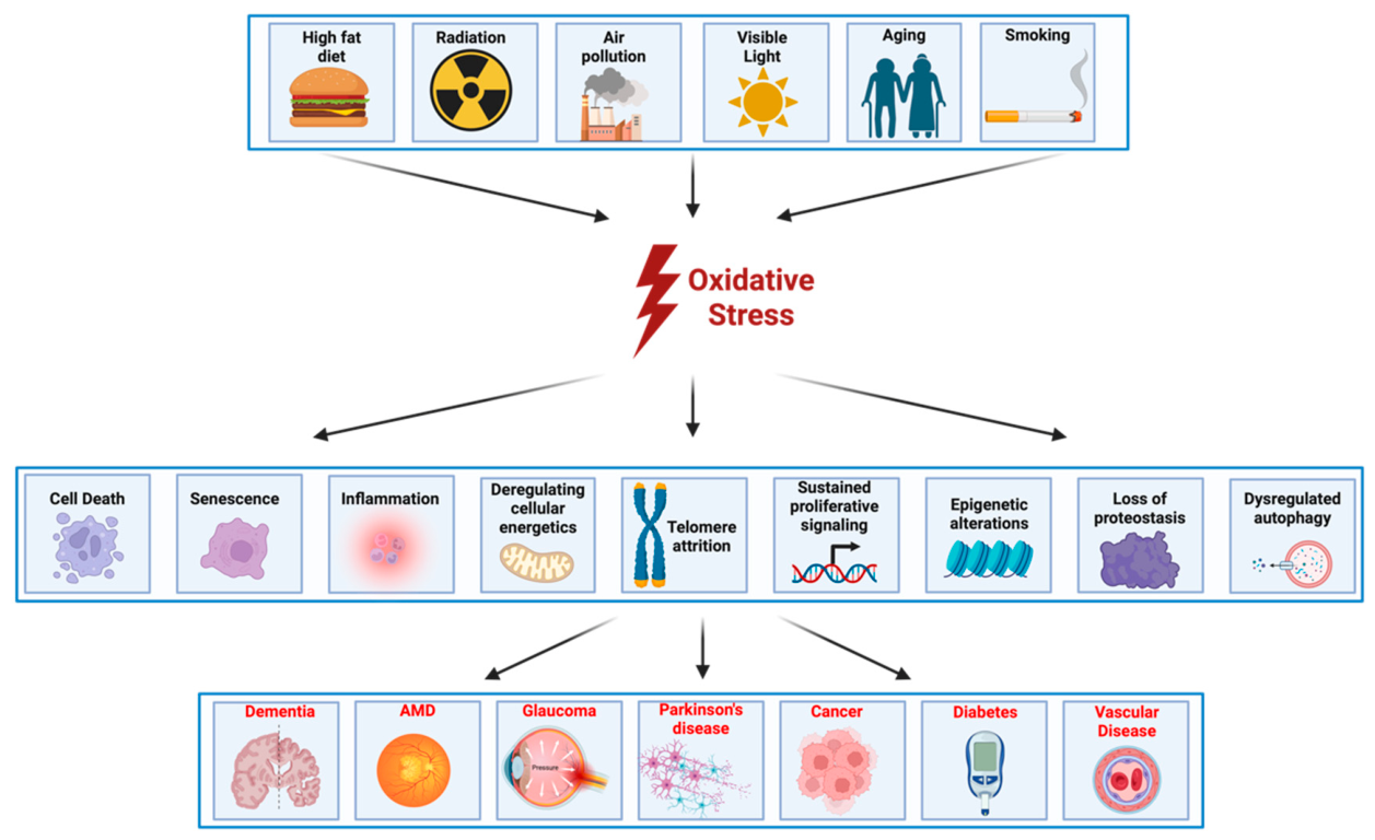
Figure 2.
High-fat diet-induced oxidative stress and mitochondrial dysfunction in metabolically active tissues. HFD promotes oxidative stress, inflammation, and cellular senescence, particularly in metabolically active tissues such as the liver, adipose tissue, brain, and eye. Dietary fats are absorbed as chylomicrons and free fatty acids (FFAs), which undergo mitochondrial β-oxidation, elevating reactive oxygen species (ROS) and lipid peroxidation products (4-HNE, MDA), leading to mitochondrial dysfunction. Excess lipids impair parkin-dependent mitophagy, while enhanced NADPH oxidase (NOX) activity further elevates ROS production, ER stress, mitochondrial calcium overload, and decreased mitochondrial respiration. Additionally, FFAs converted into triglycerides and very-low-density lipoproteins (VLDL) are oxidized into oxLDL, exacerbating oxidative damage.
Figure 2.
High-fat diet-induced oxidative stress and mitochondrial dysfunction in metabolically active tissues. HFD promotes oxidative stress, inflammation, and cellular senescence, particularly in metabolically active tissues such as the liver, adipose tissue, brain, and eye. Dietary fats are absorbed as chylomicrons and free fatty acids (FFAs), which undergo mitochondrial β-oxidation, elevating reactive oxygen species (ROS) and lipid peroxidation products (4-HNE, MDA), leading to mitochondrial dysfunction. Excess lipids impair parkin-dependent mitophagy, while enhanced NADPH oxidase (NOX) activity further elevates ROS production, ER stress, mitochondrial calcium overload, and decreased mitochondrial respiration. Additionally, FFAs converted into triglycerides and very-low-density lipoproteins (VLDL) are oxidized into oxLDL, exacerbating oxidative damage.
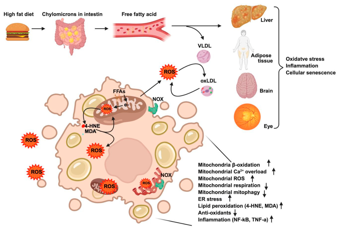
Figure 3.
Mechanisms linking high-fat diet (HFD) to oxidative stress and cellular dysfunction Oxidized LDL (oxLDL), containing reactive aldehydes such as 4-HNE, binds to LOX-1 receptors, triggering multiple signaling cascades: (1) activation of NF-κB, NLRP3 inflammasome, and MAPK pathways, leading to inflammation; (2) induction of apoptosis via increased caspase activation and reduced Bcl-2 expression; and (3) enhanced ROS production through phosphorylation of p66Shc and activation of NADPH oxidase (NOX). These events establish self-amplifying loops involving LOX-1/NOX/ROS/NF-κB and ROS/PKCβ/JNK/p66Shc/NOX pathways. OxLDL also enters cells through scavenger receptors (CD36, SR-A), further exacerbating oxidative stress, lipid droplet accumulation, and lipofuscin formation. Additionally, oxLDL interactions with receptor tyrosine kinases (RTKs) modulate cellular proliferation and migration pathways.
Figure 3.
Mechanisms linking high-fat diet (HFD) to oxidative stress and cellular dysfunction Oxidized LDL (oxLDL), containing reactive aldehydes such as 4-HNE, binds to LOX-1 receptors, triggering multiple signaling cascades: (1) activation of NF-κB, NLRP3 inflammasome, and MAPK pathways, leading to inflammation; (2) induction of apoptosis via increased caspase activation and reduced Bcl-2 expression; and (3) enhanced ROS production through phosphorylation of p66Shc and activation of NADPH oxidase (NOX). These events establish self-amplifying loops involving LOX-1/NOX/ROS/NF-κB and ROS/PKCβ/JNK/p66Shc/NOX pathways. OxLDL also enters cells through scavenger receptors (CD36, SR-A), further exacerbating oxidative stress, lipid droplet accumulation, and lipofuscin formation. Additionally, oxLDL interactions with receptor tyrosine kinases (RTKs) modulate cellular proliferation and migration pathways.
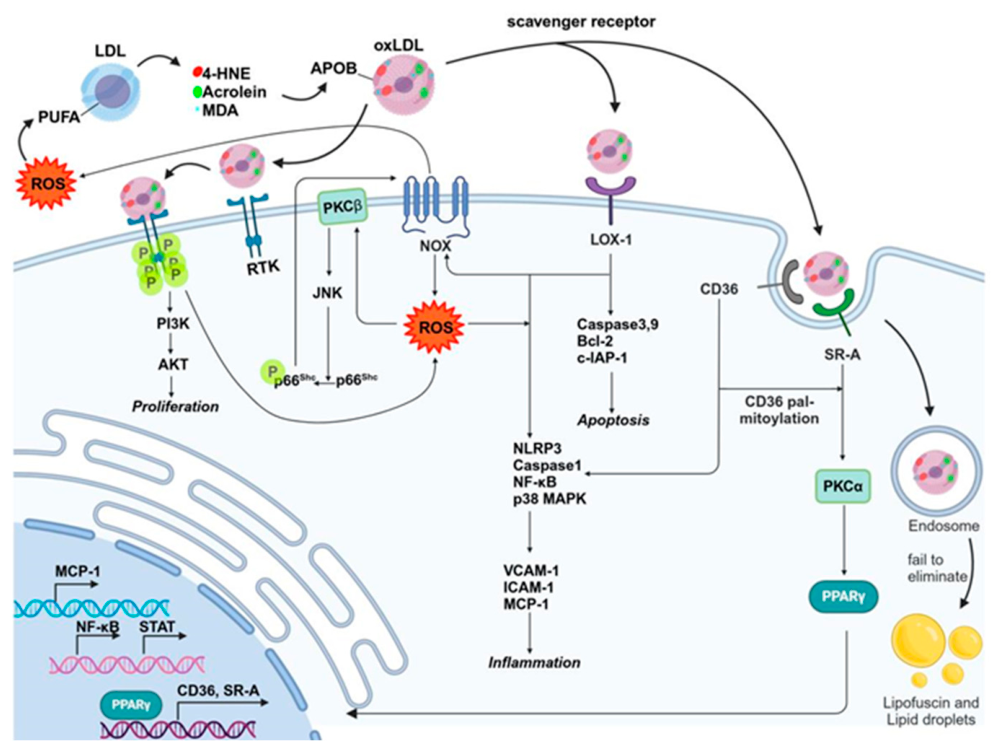
Figure 4.
Mechanisms of NaIO₃-induced oxidative injury and cell death in retinal pigment epithelium (RPE) NaIO₃ induces retinal pigment epithelium (RPE) cell injury through multiple oxidative pathways. It directly oxidizes polyunsaturated fatty acids (PUFAs) and thiol-containing proteins, leading to lipid peroxidation and mitochondrial dysfunction. NaIO3 also inhibits the cystine/glutamate transporter (system Xc⁻), depleting glutathione (GSH) and amplifying oxidative stress. Elevated ROS triggers NADPH oxidase (NOX) activation, ER stress, ERK phosphorylation, calcium overload, and epithelial-to-mesenchymal transition (EMT). Activation of the Nrf2-HO-1 axis leads to iron accumulation via ferroportin/transferrin-mediated uptake, contributing to ferroptosis. Additionally, melanin metabolism in vivo converts glycine into glyoxylate even at low concentration, further depleting antioxidants like glutathione peroxidase (GPX), catalase (CAT), superoxide dismutase (SOD) and exacerbating calcium dysregulation. Ultimately, these mechanisms culminate in various forms of cell death, including apoptosis, necroptosis, and ferroptosis, depending on NaIO3 concentration.
Figure 4.
Mechanisms of NaIO₃-induced oxidative injury and cell death in retinal pigment epithelium (RPE) NaIO₃ induces retinal pigment epithelium (RPE) cell injury through multiple oxidative pathways. It directly oxidizes polyunsaturated fatty acids (PUFAs) and thiol-containing proteins, leading to lipid peroxidation and mitochondrial dysfunction. NaIO3 also inhibits the cystine/glutamate transporter (system Xc⁻), depleting glutathione (GSH) and amplifying oxidative stress. Elevated ROS triggers NADPH oxidase (NOX) activation, ER stress, ERK phosphorylation, calcium overload, and epithelial-to-mesenchymal transition (EMT). Activation of the Nrf2-HO-1 axis leads to iron accumulation via ferroportin/transferrin-mediated uptake, contributing to ferroptosis. Additionally, melanin metabolism in vivo converts glycine into glyoxylate even at low concentration, further depleting antioxidants like glutathione peroxidase (GPX), catalase (CAT), superoxide dismutase (SOD) and exacerbating calcium dysregulation. Ultimately, these mechanisms culminate in various forms of cell death, including apoptosis, necroptosis, and ferroptosis, depending on NaIO3 concentration.
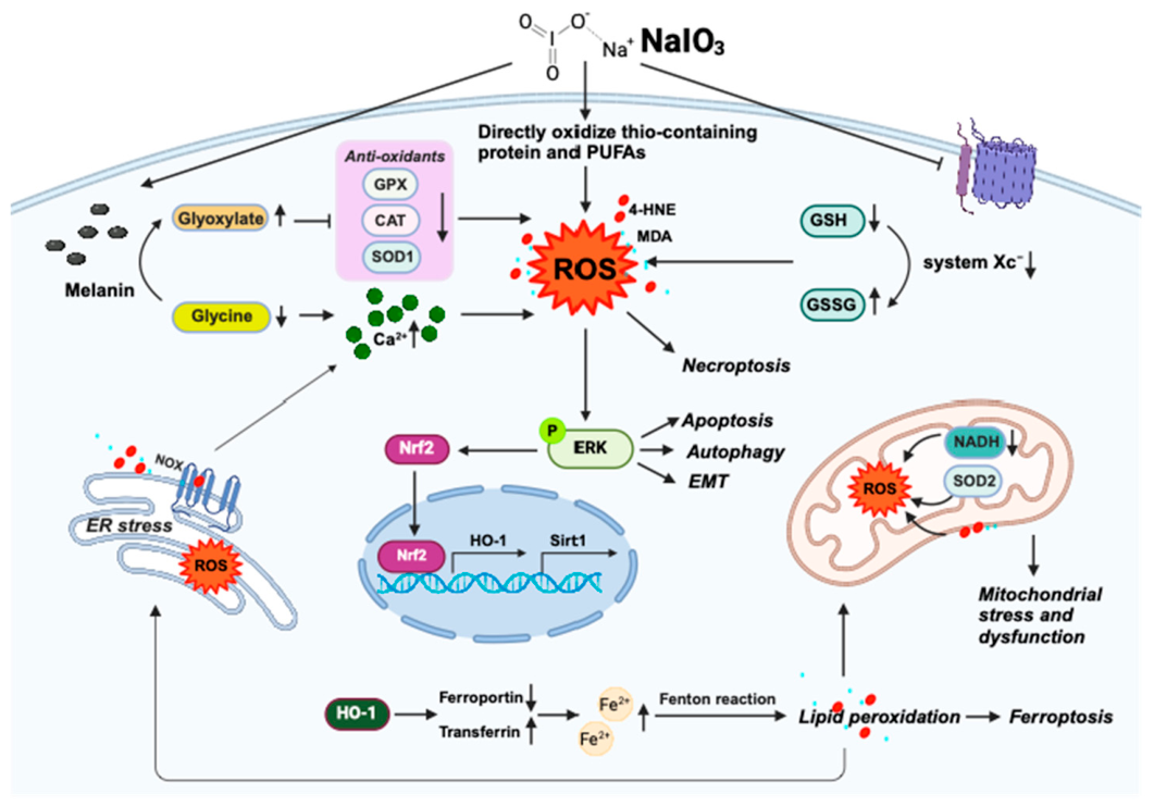
Figure 5.
Mechanisms of amyloid-β (Aβ)-induced oxidative stress in neurodegeneration Amyloid-β (Aβ) peptides induce oxidative stress through multiple pathways. First, Aβ oligomers insert into neuronal lipid membranes, initiating lipid peroxidation and generating reactive aldehydes such as 4-HNE, exacerbating neuronal injury. Second, Aβ interactions with cell-surface receptors (NMDAR, p75NTR, LilrB2/PirB) activate signaling cascades that elevate reactive oxygen species (ROS), promoting neuronal dysfunction and apoptosis. Third, binding of Aβ to metal ions (Cu, Fe, Zn) further amplifies ROS production and facilitates protein misfolding. Finally, Aβ directly impairs mitochondrial function by inhibiting critical enzymes and inducing mitochondrial permeability transition pore (mPTP) formation, accelerating mitochondrial failure and oxidative stress in Alzheimer’s disease pathology.
Figure 5.
Mechanisms of amyloid-β (Aβ)-induced oxidative stress in neurodegeneration Amyloid-β (Aβ) peptides induce oxidative stress through multiple pathways. First, Aβ oligomers insert into neuronal lipid membranes, initiating lipid peroxidation and generating reactive aldehydes such as 4-HNE, exacerbating neuronal injury. Second, Aβ interactions with cell-surface receptors (NMDAR, p75NTR, LilrB2/PirB) activate signaling cascades that elevate reactive oxygen species (ROS), promoting neuronal dysfunction and apoptosis. Third, binding of Aβ to metal ions (Cu, Fe, Zn) further amplifies ROS production and facilitates protein misfolding. Finally, Aβ directly impairs mitochondrial function by inhibiting critical enzymes and inducing mitochondrial permeability transition pore (mPTP) formation, accelerating mitochondrial failure and oxidative stress in Alzheimer’s disease pathology.
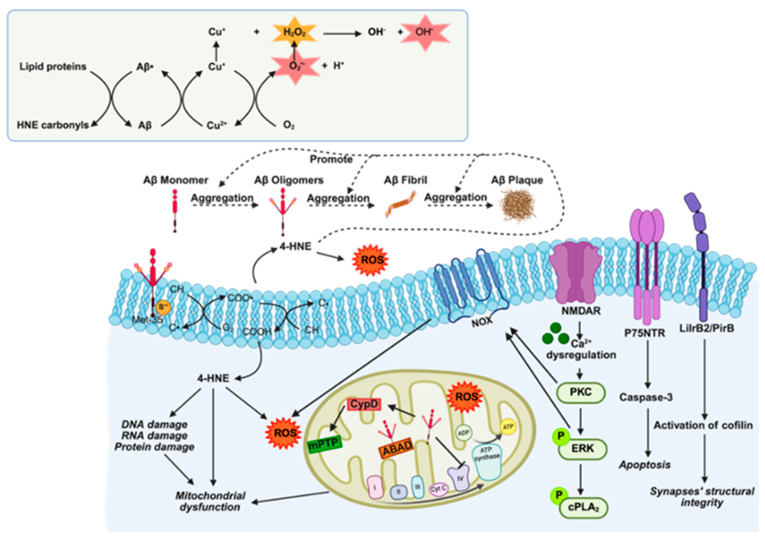
Figure 6.
Age-related iron dysregulation and oxidative stress in neurodegeneration Iron crosses cellular barriers via transferrin-dependent and transferrin-independent pathways, and its dysregulation promotes oxidative damage through hydroxyl radical generation by Fenton, Fenton-like, and Haber-Weiss reactions. This cascade leads to lipid peroxidation (MDA, 4-HNE), DNA, and protein damage, ultimately driving neuronal death.
Figure 6.
Age-related iron dysregulation and oxidative stress in neurodegeneration Iron crosses cellular barriers via transferrin-dependent and transferrin-independent pathways, and its dysregulation promotes oxidative damage through hydroxyl radical generation by Fenton, Fenton-like, and Haber-Weiss reactions. This cascade leads to lipid peroxidation (MDA, 4-HNE), DNA, and protein damage, ultimately driving neuronal death.
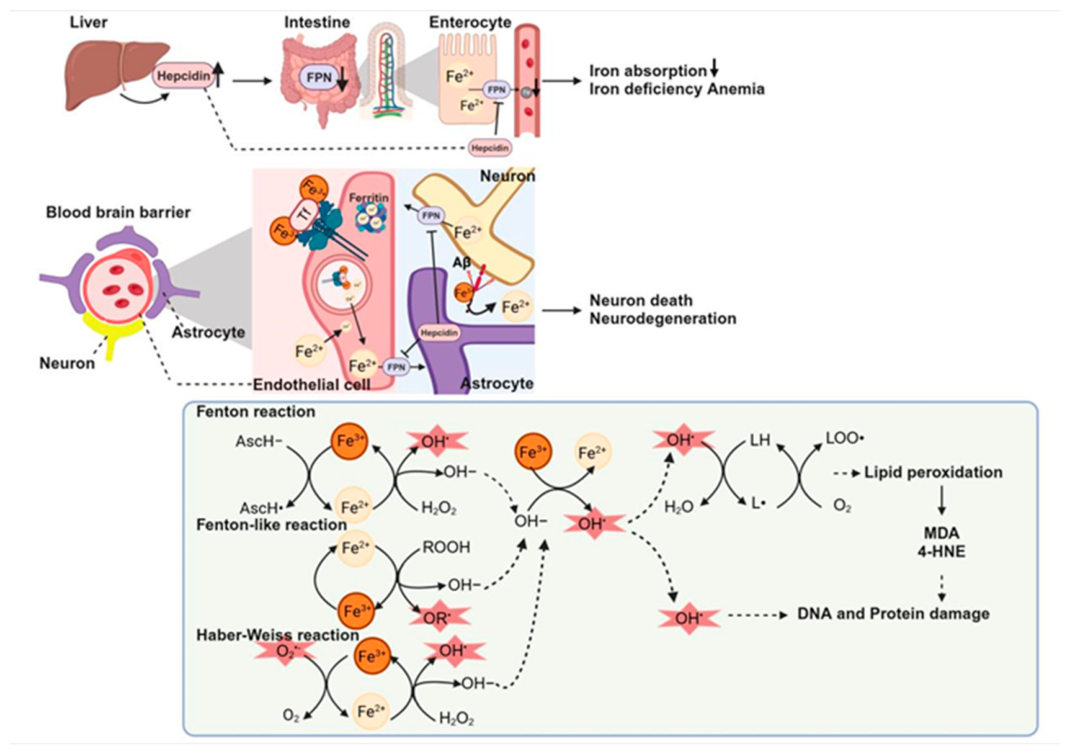
Figure 7.
Antioxidant pathways targeted by the Age-Related Eye Disease Study (AREDS) and AREDS2. Vitamins C and E. α-Tocopherol (vitamin E) donates a hydrogen atom to lipid peroxyl radicals (LOO•), yielding the inert lipid hydroperoxide (LOOH) and the tocopheroxyl radical (Toc•). Ascorbate (vitamin C) promptly reduces Toc• back to tocopherol, while itself becoming SDHAsc or DHAsc. GSH and NADH recycle SDHAsc/DHAsc to regenerate ascorbate, completing the redox loop and suppressing lipid-peroxidation chains. Carotenoids. β-Carotene from the diet is converted to retinol (vitamin A) in the intestine, transported to retinal pigment epithelial (RPE) cells, and delivered by interphotoreceptor retinoid-binding protein (IRBP) to photoreceptors for the visual cycle. Lutein and zeaxanthin additionally filter high-energy blue light and quench singlet oxygen, providing frontline antioxidant defense. Zinc and copper. These trace metals serve as cofactors for superoxide dismutases SOD1 and SOD3, which convert superoxide (O₂•–) to hydrogen peroxide (H₂O₂). Zinc also modulates catalase activity and forms zinc-metallothionein complexes that scavenge reactive species; oxidation releases zinc and produces thionin, amplifying radical clearance. Together, these pathways reduce oxidative stress implicated in AMD progression.
Figure 7.
Antioxidant pathways targeted by the Age-Related Eye Disease Study (AREDS) and AREDS2. Vitamins C and E. α-Tocopherol (vitamin E) donates a hydrogen atom to lipid peroxyl radicals (LOO•), yielding the inert lipid hydroperoxide (LOOH) and the tocopheroxyl radical (Toc•). Ascorbate (vitamin C) promptly reduces Toc• back to tocopherol, while itself becoming SDHAsc or DHAsc. GSH and NADH recycle SDHAsc/DHAsc to regenerate ascorbate, completing the redox loop and suppressing lipid-peroxidation chains. Carotenoids. β-Carotene from the diet is converted to retinol (vitamin A) in the intestine, transported to retinal pigment epithelial (RPE) cells, and delivered by interphotoreceptor retinoid-binding protein (IRBP) to photoreceptors for the visual cycle. Lutein and zeaxanthin additionally filter high-energy blue light and quench singlet oxygen, providing frontline antioxidant defense. Zinc and copper. These trace metals serve as cofactors for superoxide dismutases SOD1 and SOD3, which convert superoxide (O₂•–) to hydrogen peroxide (H₂O₂). Zinc also modulates catalase activity and forms zinc-metallothionein complexes that scavenge reactive species; oxidation releases zinc and produces thionin, amplifying radical clearance. Together, these pathways reduce oxidative stress implicated in AMD progression.
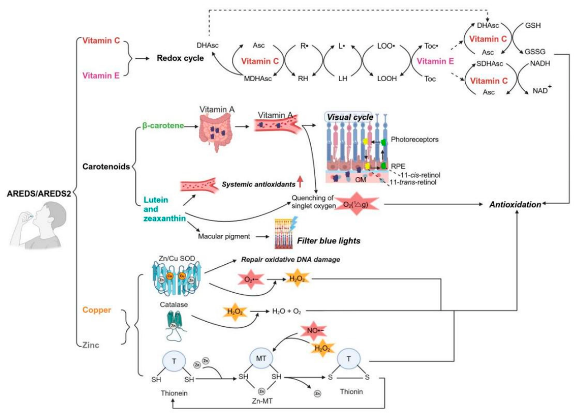
Figure 8.
Glutathione (GSH)-mediated detoxification of lipid peroxidation products Environmental and metabolic stressors (e.g., high-fat diet, UV light, H2O2, NaIO3) lead to ROS, lipid hydroperoxide and toxic aldehyde formation, such as 4-HNE. These hydroperoxides (FA-OOH, PL-OOH, CU-OOH) are reduced to alcohols by GSH, producing oxidized glutathione (GSSG) and water as by-products. These procedures are catalyzed by glutathione S-transferases (GSTs, e.g., GSTA1-1, GSTA2-2, GSTA4-4, GST5.8).
Figure 8.
Glutathione (GSH)-mediated detoxification of lipid peroxidation products Environmental and metabolic stressors (e.g., high-fat diet, UV light, H2O2, NaIO3) lead to ROS, lipid hydroperoxide and toxic aldehyde formation, such as 4-HNE. These hydroperoxides (FA-OOH, PL-OOH, CU-OOH) are reduced to alcohols by GSH, producing oxidized glutathione (GSSG) and water as by-products. These procedures are catalyzed by glutathione S-transferases (GSTs, e.g., GSTA1-1, GSTA2-2, GSTA4-4, GST5.8).
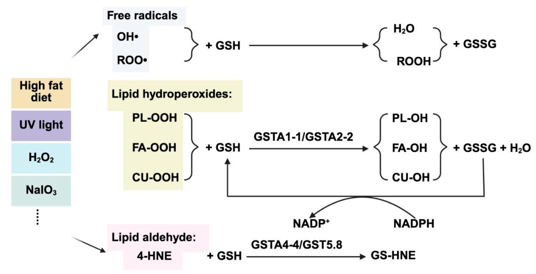
Figure 9.
Schematic of a personalized oxidative stress profiling and intervention framework. A comprehensive, AI-powered framework integrates patient-specific factors, blood- and fluid-based oxidative and inflammatory biomarkers, and longitudinal monitoring to assess oxidative burden and endogenous defense capacity. In the eye, emerging aqueous humor liquid biopsy approaches such as TEMPO can provide proteomic and metabolomic readouts that more directly reflect the retinal microenvironment. AI can integrate these data to stratify risk and guide personalized interventions, including direct free-radical scavengers (e.g., vitamins C and E), augmentation of endogenous antioxidant systems (e.g., GSH and N-acetylcysteine), mitochondrial-targeted antioxidants (e.g., coenzyme Q10), and restoration of endogenous defense programs such as NRF2 and GSTA4. Wearable devices add longitudinal information on activity, sleep, circadian rhythm, physiological stress, and environmental exposures. Together, this framework aims to match individual redox vulnerabilities with more precise and dynamically monitored interventions.
Figure 9.
Schematic of a personalized oxidative stress profiling and intervention framework. A comprehensive, AI-powered framework integrates patient-specific factors, blood- and fluid-based oxidative and inflammatory biomarkers, and longitudinal monitoring to assess oxidative burden and endogenous defense capacity. In the eye, emerging aqueous humor liquid biopsy approaches such as TEMPO can provide proteomic and metabolomic readouts that more directly reflect the retinal microenvironment. AI can integrate these data to stratify risk and guide personalized interventions, including direct free-radical scavengers (e.g., vitamins C and E), augmentation of endogenous antioxidant systems (e.g., GSH and N-acetylcysteine), mitochondrial-targeted antioxidants (e.g., coenzyme Q10), and restoration of endogenous defense programs such as NRF2 and GSTA4. Wearable devices add longitudinal information on activity, sleep, circadian rhythm, physiological stress, and environmental exposures. Together, this framework aims to match individual redox vulnerabilities with more precise and dynamically monitored interventions.
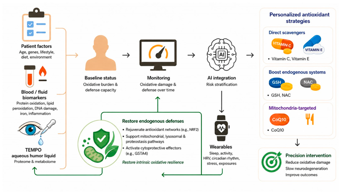

Table 1.
Primary mechanism behind different oxidative stress agents that are suitable for studying age-related CNS and eye diseases.
Table 1.
Primary mechanism behind different oxidative stress agents that are suitable for studying age-related CNS and eye diseases.
| Primary mechanisms of ROS production | Age-related neurodegenerative disease | Age-related eye disease | |
|---|---|---|---|
| Oxidative stress agents in vivo | |||
| High fat diet | Free fatty acids undergo mitochondrial β-oxidation and tricarboxylic acid cycle | Indicate the role of dietary lipids in aging progress | |
| Ultraviolet radiation | Generate ROS through photosensitization mechanisms | × | Models photoaging |
| Laser radiation | × | Models exudative form of AMD | |
| Blue light | Indirectly generate ROS through light-induced excitation of N-retinylidene-N-retinylethanolamine | × | Models RPE phototoxicity |
| Smoking | Contains free radicals | Indicate the role of smoke in aging progress | |
| Age | Mitochondrial function declines and increased chronic inflammation | Models age-related mitochondrial dysfunction and chronic inflammation | |
| Air pollution | Generate ROS through Redox reactions and inducing inflammation | Indicate the role of air pollution in aging progress | |
| Oxidative stress agents in vitro | |||
| oxLDL | Establish an oxLDL/LOX-1/NOX/ROS self-amplifying feedback loop | Modeling atherosclerosis and vascular oxidative stress as a shared pathogenic mechanisms in age-related CNS diseases | |
| NaIO3 | Directly oxidize PUFAs and thio-containing protein | × | Models oxidative-stress induced RPE-initiated retinal degeneration |
| Aβ | Induce lipid peroxidation by inserting into the lipid bilayer, interact with cell surface receptors, metal ion interactions and mitochondrial outer membrane translocase | Modeling neurotoxicity in Alzheimer’s disease | × |
| Iron | Intracellular iron overload generates ROS through Fenton, Fenton-like and Haber-Weiss reactions | Models iron homeostasis in age-related disease | |
| 4-HNE | Modify proteins/DNA, enhance oxLDL and Aβ production | 4-HNE models lipid peroxidation-induced senescence in age-related disease. More commonly, both 4-HNE and MDA serve as critical biomarkers to assess oxidative stress in studying age-related diseases. | |
| H2O2 | Produce ROS through Fenton and Haber-Weiss reactions | Models oxidative stress-induced premature senescence in age-related disease | |
| Tau | Induce mitochondrial dysfunction | Models tauopathy in Alzheimer’s disease | × |
| MPTP | Selectively inhibit Complex I and produce mitochondrial ROS in dopaminergic neurons | Systemic (cross blood-brain barrier) models mitochondrial oxidative stress in Parkinson’s disease | × |
| 6-OHDA | Auto-oxidation to generate ROS in dopaminergic and noradrenergic neurons | Models dopaminergic and noradrenergic toxicity in Parkinson’s disease | × |
| NMDA | Excessive activation of NMDA receptors leading to Ca2+ overload | × | Modeling excitotoxicity in retinal amacrine cells |
Table 2.
Antioxidant agents that have undergone randomized clinical trials.
| Participants (Year) | Dose | Treatment period | Conclusions | |
|---|---|---|---|---|
| Coenzyme Q10 (CoQ10) | Mid-stage PD (2007) | PO 300 mg/d | 3 months | No significant symptomatic effects [242]. |
| Mild to moderate AD (2012) | PO 1200 mg/d | 16 weeks | No significant change [187]. | |
| Early PD (2002) | PO 300/600/1200 mg/d + 1200 IU/d VE | 16 months | 1200 mg/d demonstrated a significant reduction in disability progression. CoQ10 seems to slow the progressive decline in function [183]. | |
| Early PD (2014) | PO 1200/2400 mg/d + 1200 IU/d VE | 16 months | No clinical benefit [184]. | |
| PD with wearing off (2015) | PO 300 mg/d | 48 weeks | Showing symptom improvement [185]. | |
| Early PD (2015) | PO 300 mg/d | 96 weeks | No clinical benefit [185]. | |
| PD (2019) | PO 156 mg/d | 6 months | Ongoing [186]. | |
| MitoQ | PD | PO 40/80 mg/d | 12 months | No effect on PD progression [191]. |
| Vitamin E (VE) | Moderately severe AD (1997) | PO 2000 IU/d | 2 years | Slowing the progression of AD [166]. |
| AD (2005) | PO 2000 IU/d | 3 years | VE had no benefit in AD patient with mild cognitive impairment [243]. | |
| AD (2009) | PO 800 IU/d | 6 months | VE does not prevent cognition loss and may even be detrimental [244]. | |
| Mild to moderate AD (2012) | PO 800 IU/d + 500 mg/d VC + 900 mg/d ALA | 16 weeks | VC/VE/ALA reduced oxidative stress in brain, but bring a potential safety concern about faster cognitive decline [187]. | |
| Mild to moderate AD (2014) | PO 2000 IU/d | More than 2 years | Slowing functional decline [245]. | |
| PD (1993) | PO 2000 IU/d | 14 months | No beneficial effect [203]. | |
| Early PD (1998) | PO 2000 IU/d | 14 months | No beneficial effect [202]. | |
| Deprenyl | PD (1993) | PO 10 mg/d | 14 months | Delay the onset of disability associated with PD [203]. |
| Early PD (1989, 1998) | PO 10 mg/d | 14 months, 8.2 years | Delay the onset of disability associated with PD [201]. Delay the time needed for levodopa therapy [202]. |
|
| Glutathione (GSH) | PD (2009) | IV 1400 mg, 3 times/week | 4 weeks | No significant clinical benefit [246]. |
| PD (2015, 2017) | Intranasal 300, 600 mg/d | 3 months | PD symptoms improved [174,175]. | |
| N-acetylcysteine (NAC) | PD (2016) | IV 50 mg/kg, 1 time/week + PO 600 mg/day on rest of days. | 3 months | Effective in improving PD symptoms [247]. |
| PD (2019) | IV 50 mg/kg, 1 time/week + PO 1000 mg/day on rest of days. | 3 months | Effective in improving PD symptoms [248]. | |
| AD (2001) | PO 50 mg/kg/d | 6 months | Some of the cognitive tests results improved [249]. | |
| Moderate to late-stage AD (2009) | PO 1200 mg/d + Vitamin B12 ug/d + VE 60 IU/d+ S-adenosyl methionine 800 mg/d + acetyl- L -carnitine 1000 mg/d |
9 months | Improving mood, behavior, and support continued performance of daily activities [250]. | |
| GlyNAC | Aging (2023) | PO 100 mg/kg/d glycine, 100mg/kg/d N-acetylcysteine, 200mg/kg/d alanine | 16 weeks | Effective in improving and reversing multiple age-associated abnormalities to promote health in aging humans [251] |
| Alpha lipoic acid (ALA) | AD (2014) | PO 600 mg/d ALA + 3 g/d omega-3 fatty acid | 12 months | The combination of ω-3 and ALA slowed cognitive and functional decline in AD [200]. |
| AREDS | AMD (2001) | PO 15 mg beta carotene + 80 mg zinc oxide + 2 mg cupric oxide + 500 mg VC + 400 IU VE | Averaged 6.3 years | Reduced the risk of progression to advanced AMD [157]. |
| AREDS2 | Intermediate AMD (2013) | PO 10 mg lutein + 2 mg zeaxanthin and/or 350 mg DHA +650 mg EPA | Mediate follow-up was 5 years | Addition of lutein+zeaxanthin, DHA+EPA, or both to the AREDS formulation in primary analyses did not further reduce risk of progression to advanced AMD [162]. |
| AREDS2 | Intermediate AMD (2022) | PO 10 mg lutein + 2 mg zeaxanthin+ 80 mg zinc oxide + 2 mg cupric oxide + 500 mg VC + 400 IU VE. | 5 years | Beneficial with late AMD progression [161]. |
| AREDS/AREDS2 | AMD (2024) | PO AREDS or AREDS2 | 6 years | Slow geographic atrophy progression to the fovea [163]. |
Abbreviations: PO, oral administration; IV, intravenous.
Table 3.
Oxidative stress related parameters for AI modeling.
| Checklist | Examination method |
|---|---|
| 1. Patient-specific factors | |
| Age | Questionnaire (QNR), medical records |
| Family history | QNR, medical records |
| Cancer history | QNR, medical records |
| Auto-immune system disease history | QNR, medical records |
| 2. Lifestyle-specific factors | |
| High-fat diet | QNR, dietary assessment |
| Calorie restriction | QNR, dietary assessment |
| Cigarette smoke habit | QNR, biomarker (e.g., nicotine) |
| Irradiation history | QNR, medical records |
| Blue light exposure | QNR, environmental assessment |
| Air pollution exposure | QNR, environmental assessment |
| 3. Oxidation markers | |
| 1.1. Protein oxidation markers | |
| Aβ | Blood, CSF |
| Phosphorylated tau protein (p-tau217) | Plasma, CSF |
| oxLDL | Blood |
| 1.2. Lipid Peroxidation Markers | |
| MDA/4-HNE | Blood, urine, eye liquid biopsy |
| F2-Isoprostanes | Urine, plasma, eye liquid biopsy |
| 1.3. DNA oxidative damage | |
| 8-Hydroxydeoxyguanosine (8-OHdG) | Blood, urine, eye liquid biopsy |
| 1.4. Iron level | |
| Ferritin | Serum, eye liquid biopsy |
| 4. Anti-oxidation markers | |
| 1.1. Antioxidant enzyme | |
| SOD | Serum, RBC lysate, eye liquid biopsy |
| Catalase | Serum, RBC lysate, eye liquid biopsy |
| Glutathione peroxidase (GPx) | Serum, RBC lysate, eye liquid biopsy |
| 4.2. Non-enzyme antioxidant | |
| GSH/GSSG ratio | Blood, eye liquid biopsy |
| Vitamin C | Plasma, eye liquid biopsy |
| Vitamin E | Plasma, eye liquid biopsy |
| ALA | Plasma, eye liquid biopsy |
| CoQ10 | Plasma, eye liquid biopsy |
| 5. Inflammation | |
| Interleukin-6 (IL-6) | Blood, eye liquid biopsy |
| Tumor necrosis factor alpha (TNFα) | Blood, eye liquid biopsy |
| CRP (C-reactive protein) | Blood, eye liquid biopsy |
Disclaimer/Publisher’s Note: The statements, opinions and data contained in all publications are solely those of the individual author(s) and contributor(s) and not of MDPI and/or the editor(s). MDPI and/or the editor(s) disclaim responsibility for any injury to people or property resulting from any ideas, methods, instructions or products referred to in the content. |
© 2026 by the authors. Licensee MDPI, Basel, Switzerland. This article is an open access article distributed under the terms and conditions of the Creative Commons Attribution (CC BY) license.
Copyright: This open access article is published under a Creative Commons CC BY 4.0 license, which permit the free download, distribution, and reuse, provided that the author and preprint are cited in any reuse.