

Submitted:
21 August 2026
Posted:
24 August 2026
You are already at the latest version
Abstract
Alzheimer’s disease (AD) is characterised by progressive accumulation of misfolded proteins and failure of cellular clearance pathways, particularly the autophagy–lysosomal system. Given the limited disease-modifying efficacy of current pharma-cological strategies, intermittent fasting (IF) has emerged as a potentially relevant non-pharmacological intervention capable of modulating energy metabolism, proteo-stasis and cellular stress responses. This review analyses the mechanisms through which IF and fasting-like metabolic states may stimulate macroautophagy and chap-erone-mediated autophagy, and evaluates their potential neuroprotective relevance in AD. Evidence from in vitro models and animal studies suggests that fast-ing-induced metabolic switching, particularly β-hydroxybutyrate production, can modulate AMPK–mTOR signalling, autophagic flux, oxidative stress, synaptic plas-ticity and Aβ/Tau-related proteostasis. However, the translation of these findings to humans remains incomplete. Current clinical studies suggest potential benefits on se-lected cognitive, sleep and metabolic outcomes, but rely mainly on indirect readouts such as neuropsychological testing, peripheral biomarkers, neuroimaging, metabo-lomics and microbiota-derived signatures. Larger, longer and biomarker-integrated randomised controlled trials using nutritionally supervised protocols are required to determine whether IF can safely produce clinically meaningful neuroprotection in AD without increasing frailty, sarcopenia or hypoglycaemic risk in older adults.
Keywords:
intermittent fasting
; time-restricted eating
; autophagy
; chaperone-mediated autophagy
; β-hydroxybutyrate
; Alzheimer’s disease
; cognitive decline
; neuroprotection
1. Introduction
1.1. Alzheimer’s Disease (AD): Etiology, Epidemiology and Clinical Challenges
An ageing population and the rising prevalence of neurodegenerative diseases represent one of the greatest public health challenges of the 21st century [1]. Among these, Alzheimer’s disease (AD) is the most common form of dementia, accounting for approximately 60–70 per cent of all cases worldwide [2]. It is estimated to affect more than 50 million people worldwide, a figure that could triple by 2050 due to increased life expectancy [3]. Although cognitive decline is frequently associated with ageing, this condition is characterised by progressive neurological degeneration affecting memory, thinking and behaviour; this stems from impaired synaptic plasticity and neuronal death [2,4]. In Alzheimer’s disease (AD), unlike other forms of dementia, various factors—including genetic, environmental and metabolic factors—contribute to the accumulation of toxic protein aggregates [5], severely impacting patients’ daily lives and placing an enormous burden on healthcare systems and carers.
From an etiological perspective, the disease can be classified into two main types: the familial form, which has an early onset and accounts for less than 5 per cent of cases, associated with specific genetic mutations—such as those in the amyloid precursor protein (APP), presenilin 1 (PSEN1) and presenilin 2 (PSEN2) genes, and the sporadic form, which has a late onset and accounts for more than 95% of diagnoses [3]. The sporadic form is multifactorial, with ageing being the main risk factor, interacting with genetic factors such as the apolipoprotein E ε4 allele and environmental or lifestyle factors [3,5].
Under physiological conditions, the brain is an organ with high metabolic demands that requires a constant energy supply, relying primarily on glucose to maintain its homeostasis [1]. However, during ageing and AD, a reduction in cerebral glucose metabolism is observed, associated with mitochondrial dysfunction and alterations in energy metabolism. This energy crisis increases the production of reactive oxygen species (ROS), leading to a state of chronic oxidative stress, damageing lipids, proteins and DNA, and exacerbating neuroinflammation [6]. Oxidative stress and inflammation compound the aggregation of proteins such as Tau and amyloid-β (Aβ), accelerating neurodegeneration [5]. It is in this state of energy deficit that the brain’s ability to maintain its homeostasis and eliminate damaged cellular components diminishes. Despite advances in our understanding of the disease, current pharmacological options are limited. Approved treatments such as cholinesterase inhibitors are primarily symptomatic and do not halt the progression of the disease. Meanwhile, the most recent therapies aimed at removing amyloid plaques (monoclonal antibodies) have shown inconclusive results and side effects, without manageing to reverse the cognitive damage that has already occurred [3].
1.2. Molecular Pathology: The Tau and Amyloid-β Protein Hypothesis
At the pathophysiological level, Alzheimer’s disease is defined as a proteinopathy, that is, a disorder characterised by the accumulation and aggregation of misfolded proteins.
At the neuropathological level, AD is defined by the presence of two distinctive histological hallmarks that lead to synaptic loss and neuronal death [5]: amyloid-β plaque pathology, also known as senile plaques, which are extracellular deposits formed by the abnormal accumulation of the amyloid-β peptide. This peptide is generated by incorrect proteolytic processing of the amyloid precursor protein (APP) via the amyloidogenic pathway [5]. Although this was historically considered the initiating event—known as the ‘amyloid cascade hypothesis’—current evidence suggests that the accumulation of Aβ alone does not fully explain neurodegeneration or cognitive decline but rather acts as a trigger for other toxic processes.
Furthermore, there is the pathology of Tau neurofibrillary tangles, which are intracellular aggregates of hyperphosphorylated Tau protein. Under normal conditions, the Tau protein is highly soluble and essential for stabilising microtubules, regulating the axonal transport of organelles and vesicles, and maintaining genomic integrity within the nucleus [5]; however, in AD, Tau undergoes modifications such as hyperphosphorylation, which cause it to detach from the cytoskeleton, losing its stabilising function and allowing the formation of toxic oligomers that disrupt axonal transport before becoming trapped in tangles, leading to cell death [7].
The neuropathological progression of AD can also be described using topographical staging systems that distinguish the spread of Aβ deposits [8] from that of Tau-related neurofibrillary pathology [9]. Thal and colleagues proposed five phases of Aβ deposition, beginning in the neocortex, followed by allocortical regions, then diencephalic nuclei, striatum and basal forebrain, subsequently brainstem nuclei, and finally the cerebellum, indicating a hierarchical expansion of amyloid pathology across the brain [8]. In contrast, Braak and Braak staging describes the progression of neurofibrillary tangles and neuropil threads in six stages: early transentorhinal involvement in stages I-II, limbic involvement including the entorhinal cortex and hippocampal formation in stages III-IV, and widespread isocortical association-area involvement in stages V-VI [9]. This distinction is important because amyloid deposition and Tau pathology do not necessarily progress in parallel, and Tau pathology shows a stronger relationship with neurodegeneration and clinical decline [10,11]. It is important to emphasize that Tau, particularly when hyperphosphorylated and aggregated, is the main component of neurofibrillary tangles, that its pathological progression correlates with neuronal dysfunction and disease advancement, and that mechanisms such as GSK-3-mediated Tau phosphorylation and extracellular Tau propagation may contribute to the anatomical spread of Tau pathology in AD [10,11,12].
Although AD was historically considered to be caused by amyloid-β pathology, it is important to note that more recent studies show that Tau pathology correlates more closely with cognitive decline than amyloid burden [7,9,10,11]. Furthermore, there is current scientific evidence suggesting that senile plaques and neurofibrillary tangles are capable of binding free radicals, thereby exerting a potential antioxidant effect against the severe oxidative stress characteristic of the disease [13]. Along the same lines, they could act as a cellular defence mechanism, functioning as a biological barrier capable of sequestering and neutralising intermediate aggregation species or soluble oligomers, which are the truly neurotoxic elements responsible for the spread of the pathology. Therefore, the formation of macroscopic aggregates could represent a desperate attempt by the neuron to reduce the concentration of highly reactive soluble species in an environment of high oxidative stress.
1.3. Cellular Clearance Systems: Macroautophagy and Chaperone-Mediated Autophagy
In a healthy brain, autophagy acts as the primary mechanism for cellular maintenance and clearance to promote proteostasis, responsible for engulfing and degrading these defective intracellular proteins. However, oxidative stress, mitochondrial dysfunction and metabolic imbalances lead to a progressive deterioration of this pathway. It is precisely this failure of the cellular clearance systems that prevents the proper degradation of the Tau protein, leading to its toxic accumulation in the form of neurofibrillary tangles. Furthermore, the lack of efficient recycling of organelles, particularly mitochondria—a process known as mitophagy—leads to increased oxidative stress, which in turn exacerbates neuronal damage [14]. Furthermore, although Aβ accumulates mainly in the extracellular space, it has been reported that its presence can negatively interfere with the mechanisms of autophagy itself, exacerbating the breakdown of cerebral homeostasis [5].
This study focuses on autophagy as a central, key mechanism in the pathogenesis of Alzheimer’s disease. Autophagy is the primary cellular clearance system responsible for degrading damaged organelles and misfolded protein aggregates via the lysosomes. Autophagy initiation is primarily governed by the metabolic sensors mechanistic target of rapamycin complex 1 (mTORC1) and AMP-activated protein kinase (AMPK), which integrate nutrient availability, growth factor signalling and cellular energy status. Under nutrient-rich conditions, active mTORC1 phosphorylates components of the unc-51-like kinase 1 (ULK1)/autophagy-related protein 13 (ATG13)/FAK family-interacting protein of 200 kDa (FIP200)/autophagy-related protein 101 (ATG101) complex, maintaining autophagy in an inhibited state. During starvation, AMPK simultaneously inhibits mTORC1 and directly phosphorylates ULK1, thereby activating the autophagic flux. Activated ULK1 subsequently phosphorylates multiple downstream targets, including Beclin-1 and ATG9, initiating phagophore formation [15].
Phagophore nucleation depends on activation of the class III phosphatidylinositol-3 kinase (PI3KC3) complex I, which generates phosphatidylinositol-3-phosphate (PI3P) at specialized membrane domains known as omegasomes. Membrane expansion requires continuous lipid delivery from multiple intracellular compartments through ATG9-positive vesicles while ATG12-ATG5-ATG16L1 complex functions as an E3-like ligase and catalyzes the lipidation of microtubule-associated protein 1 light chain 3 (LC3) and GABA type A receptor-associated protein (GABARAP) family proteins. Cytosolic LC3-I undergoes conjugation to phosphatidylethanolamine, generating membrane-bound LC3-II, which becomes inserted into both the inner and outer autophagosomal membranes. Cargo selection is mediated by autophagy receptors including p62/sequestosome 1 (p62/SQSTM1), neighbour of BRCA1 gene 1 (NBR1) or optineurin (OPTN), which simultaneously recognize ubiquitinated substrates through their ubiquitin-binding domains and interact with LC3 proteins. This molecular bridge allows the selective sequestration of aggregated proteins, damaged mitochondria [16,17].
Autophagosome maturation culminates in its fusion with lysosomes through coordinated interactions between Rab GTPases and SNARE proteins. Ras-related protein Rab7 (Rab7), syntaxin-17, synaptosomal-associated protein 29 (SNAP29) and vesicle-associated membrane protein 8 (VAMP8) cooperate to mediate membrane docking and fusion, generating the autolysosome. Within this compartment, the acidic environment maintained by the vacuolar H+-ATPase activates lysosomal hydrolases, including Cathepsins B, D and L, which degrade proteins, lipids and organelles [18,19] (Figure 1).
In the brains of patients with Alzheimer’s disease, a massive accumulation of immature autophagic vacuoles has been observed, indicating a failure in the autophagic flux, whereby the cellular ‘rubbish bin’ system is activated but is unable to complete degradation, thereby blocking an evolutionarily conserved catabolic process that acts as the cellular ‘recycling’ system [20]. In addition, severe lysosomal dysfunction occurs, where the various clearance pathways converge. This collapse of the lysosomes not only prevents the effective removal of intracellular Tau tangles via autophagy, but also drastically impairs phagocytosis [21]. Furthermore, the malfunctioning of this lysosomal degradation network impairs the ability of glial cells to phagocytose and eliminate already-formed extracellular Aβ plaques, creating a vicious cycle of cellular toxicity. Therefore, restoring or enhancing autophagy is considered a promising therapeutic strategy for halting the progression of the disease.
In contrast, chaperone-mediated autophagy (CMA) is a highly selective, precision tool which, unlike macroautophagy, does not require the formation of double-membrane vesicles or autophagosomes [21]. In this system, the chaperone protein Hsc70 (HSPA8) recognises a specific biochemical motif in the cytosol—similar to the KFERQ sequence—present in target proteins such as soluble Tau monomers [22,23]. Upon recognition, heat shock protein 40 (HSP40) and heat shock protein 90 (HSP90) families assist the stabilization before the chaperone transports them directly to the lysosomal membrane, where they interact with the Lysosome-associated membrane protein type 2A (LAMP-2A) monomers, which dynamically assemble into multimeric translocation complexes within the lysosomal membrane. Lysosomal HSC70 together with luminal co-chaperones actively drives substrate unfolding and vectorial translocation into the lysosomal lumen in an ATP-dependent manner. Following complete translocation, substrates are rapidly degraded by lysosomal proteases, while LAMP-2A multimers disassemble into inactive monomers available for subsequent transport cycles [24,25] (Figure 1). This differentiation is vital in the pathogenesis, as the early collapse of the CMA promotes the initial accumulation of Tau, forcing macroautophagy to attempt to compensate for and clear the resulting large tangles, often without success [26,27].
Neurofibrillary tangles are large aggregates of the Tau protein which, due to their intracellular nature, cannot be easily eliminated by the immune system or conventional drugs; their removal depends almost exclusively on macroautophagy. Precisely because of its translocation mechanism, which requires the proteins to be unfolded beforehand, CMA lacks the ability to degrade insoluble aggregates; instead, it acts preventively by clearing individual pathological proteins in their soluble form, such as hyperphosphorylated Tau monomers. And when it fails to degrade soluble Tau, these proteins accumulate and eventually form insoluble aggregates [3].
Although both are lysosomal degradation systems, they have mechanisms of action and molecular targets that operate very differently, as can be seen in Figure 1. Macroautophagy acts as a general clean-up system by forming double-membrane vesicles (autophagosomes) responsible for engulfing entire portions of the cytoplasm, dysfunctional organelles and large insoluble protein aggregates.
In the context of Alzheimer’s disease, CMA plays a dual role that is essential for neuronal survival. Mutant, hyperphosphorylated tau protein, the main cause of neurofibrillary tangles in AD, contains recognition motifs for Hsc70 [28]. This interaction is made possible by the functional structure of the chaperone, which utilises two essential domains: the nucleotide-binding domain (NBD), with ATPase activity responsible for providing the energy required for protein unfolding; and the substrate-binding domain (SBD), which acts as a molecular anchor to selectively recognise and physically bind to KFERQ-like motives (a sequence of five amino acids) in Tau near the C-terminal end (336QVEVK340 and 347KDRVQ351) [22,29] (Figure 1). In a young, healthy brain, the CMA can capture and degrade defective Tau proteins before they aggregate. However, recent research shows that, during ageing and the progression of Alzheimer’s disease—as demonstrated in transgenic mouse models and in the analysis of post-mortem brain tissue from AD patients— the activity of the LAMP-2A receptor on the lysosomal membrane decreases dramatically, causing this degradation pathway to fail and accelerating the accumulation of Tau, thereby leading to neurotoxicity [26,30].
Microautophagy is another lysosomal degradation pathway, the dysfunction and mutation of several genes involved in which have recently been linked to the development of various neurodegenerative diseases. Unlike macroautophagy, which requires the formation of intermediate vesicles and the CMA (which utilises chaperone proteins), in microautophagy it is the lysosomal or late endosomal membrane itself that invaginates directly to engulf portions of the cytosol and trap protein waste within it (Figure 1). This membrane remodelling process is orchestrated predominantly by the Endosomal Sorting Complex Required for Transport (ESCRT) machinery. Sequential recruitment of ESCRT-0, ESCRT-I, ESCRT-II and ESCRT-III complexes coordinates membrane deformation, cargo concentration, membrane scission and vesicle release into the endosomal lumen. In addition to protein turnover, microautophagy contributes to lipid homeostasis, organelle quality control and membrane remodelling, thereby complementing the degradative functions of both macroautophagy and CMA [31,32,33].
1.4. Intermittent Fasting as a Neuroprotective Intervention
Against this backdrop, non-pharmacological lifestyle-based interventions, such as nutrition and intermittent fasting, are emerging as preventive tools capable of acting on multiple molecular targets simultaneously.
Intermittent fasting (IF), defined as a dietary pattern that alternates periods of food intake with periods of voluntary calorie restriction, has emerged in recent years as a promising non-pharmacological dietary intervention, which aims not only at calorie restriction but also at metabolic modulation [1]. Unlike chronic calorie restriction, IF seeks to activate adaptive cellular stress responses without necessarily reducing total calorie intake in the long term. Among the most widely studied and applied protocols in the scientific literature are alternate-day fasting (ADF, alternating days of normal eating with full 24-hour fasting periods), time-restricted feeding (such as 16:8, which concentrates food intake within an 8-hour window) and continuous periodic fasting (such as the 5:2 diet).
The biological basis of IF lies in the ‘metabolic switch’, one of the most profound physiological adaptations induced by this practice. Under normal eating conditions, the human brain relies almost exclusively on circulating glucose as an energy source. However, following a prolonged period of fasting (lasting 12–16 hours or more), hepatic glycogen stores are depleted, and the body undergoes a massive neuroendocrine transition. The liver begins to mobilise fatty acids to produce ketone bodies, primarily acetoacetate (Ac) and β-hydroxybutyrate (BHB) [1,34], which then become the primary source of energy.
In the pathogenesis of Alzheimer’s disease (AD), this metabolic shift takes on crucial therapeutic significance, as AD is characterised early on by cerebral glucose hypometabolism. Neurons develop insulin resistance and lose their ability to utilise glucose, leading to an ATP deficit and, progressively, to cell death [34]. However, recent clinical reviews highlight that ketone body transporters in the blood-brain barrier are not impaired in patients with Alzheimer’s. Thus, the β-hydroxybutyrate generated during fasting is able to cross into the brain and provide an alternative and highly efficient energy source, rescuing neurons from starvation [34].
Beyond its role as a simple fuel, current medical knowledge has shown that BHB acts as a potent epigenetic and metabolic signalling molecule that directly interacts with cellular clearance mechanisms, as illustrated in Figure 2 [23]. This metabolic shift, characterised by a drop in ATP levels, increases the cellular AMP/ATP ratio. This energy imbalance activates adaptive cellular signalling pathways, specifically by activating AMPK (a low-energy sensor) and simultaneously inhibiting the mTOR pathwayway (a nutrient abundance sensor) [20].
The simultaneous action of both pathways is essential for adaptive autophagy to occur—a quality control and repair mechanism whereby the cell isolates and degrades damaged organelles and misfolded proteins via lysosomal recycling, thereby preserving homeostasis [4]. This signalling cascade has been extensively demonstrated and validated across various experimental models. Specifically, in in vitro models, the direct addition of BHB to the culture medium has been shown to inhibit mTOR phosphorylation and induce key markers of autophagy, such as the conversion of LC3-I to LC3-II. Complementarily, in in vivo models using transgenic Alzheimer’s mice subjected to fasting protocols, histological and Western blot analyses of brain tissue confirm this suppression of mTOR and reveal a physical increase in the formation of autophagosomes in neurons. Finally, although ethical constraints in humans prevent the use of invasive methods such as brain biopsies to measure these markers directly, clinical trials have detected activation of the AMPK pathway and suppression of mTOR by measuring biomarkers in peripheral blood mononuclear cells (PBMCs) and circulating exosomes in patients undergoing calorie restriction [35].
This is where intermittent fasting demonstrates its enormous clinical potential. Unlike macroautophagy, which responds rapidly to the initial lack of nutrients, CMA is activated and regulated in a sustained manner following longer periods of fasting [20]. Continued nutritional stress induces a compensatory and significant increase in the expression of Hsc70 and LAMP-2A in the central nervous system, an effect that has been extensively demonstrated in animal models (in vivo), such as mice subjected to 24-to 48-hour fasting protocols, forcing the reactivation of this pathway [20,22]. By activating the proteostasis network through scheduled calorie restriction—and as has also been observed in neuronal cultures (in vitro) under serum deprivation [26] —neurons are able to resume the selective identification and destruction of pathological proteins. Consequently, the pharmacological or dietary modulation of the CMA through fasting protocols is currently regarded as one of the most promising targets for halting the structural progression of cognitive decline in AD [27,28].
This autophagic activation, triggered by transient nutritional stress, enables the neuron to degrade intracellular toxic components more efficiently—primarily tau protein oligomers and dysfunctional mitochondria—before the collapse of proteostasis accelerates the pathology (Figure 1). Furthermore, although the accumulation of Aβ occurs in extracellular plaques, the stimulation of macroautophagy is crucial for curbing it at its intracellular source. Autophagy acts by capturing and degrading APP and its intermediate fragments in the lysosomes before they complete their processing. By directly eliminating this ‘raw material’ within the cell, the abnormal production and secretion of toxic Aβ monomers into the interstitial space is reduced [5]; this is essential for preserving synaptic integrity and halting the spread of toxicity in the neuronal microenvironment [5,6].
If intermittent fasting is capable of enhancing this lysosomal degradation pathway, it could tackle the pathology at its intracellular root—something that anti-amyloid therapies have not yet succeeded in achieving [4]. Taken together, this reaffirms that the neuroprotective effect of fasting is not a passive phenomenon, but a concerted response in which the switch in energy substrate from glucose to ketones activates survival processes, such as intracellular clearance systems and neuronal proteostasis [21]. Nevertheless, it is essential to control the duration of the stimulus, as, in the short term, it exerts a protective role and promotes cellular longevity; however, prolonged or severe restriction can lead to an excessive and harmful autophagic response that may induce programmed cell death, known as type II autophagic cell death [20].
However, translating these findings into human clinical practice presents considerable challenges due to interspecies metabolic differences. Experimental studies have shown that short-term fasting (24–48 hours in mice) induces profound neuronal autophagy through inhibition of the mTOR pathwayway, increasing the number and size of autophagosomes and facilitating their fusion with lysosomes for the degradation of toxic material [4]. Furthermore, this change in metabolic state in the brain stimulates the production of neurotrophic factors such as BDNF (brain-derived neurotrophic factor), promoting synaptic plasticity and neuronal resilience to metabolic and oxidative stress [6].
Studies have compared 24–48-hour fasting in mouse models, where it is capable of activating profound neuronal autophagy in the short term via mTOR inhibition [4], whilst in humans an equivalent period barely alters basal homeostasis so drastically, requiring different physiological adaptations [34]. Furthermore, recent research suggests that the activation of autophagic markers (such as LC3 or Beclin-1) observed in mouse tissues is not always replicated in human tissues under similar calorie-restriction protocols [20]. Therefore, given the profound modulatory effect that diet has on disease development [5], it is vital to critically analyse whether popular fasting protocols in humans (such as the 16:8 method) are sufficient to activate these cellular clearance mechanisms, or whether a different approach is required to prevent cognitive decline (Figure 3).
This manuscript aims to analyse the molecular mechanisms by which intermittent fasting stimulates autophagy (both macroautophagy and CMA), to evaluate its ability to reduce the burden of neurotoxic proteins such as Tau and Aβ, and to discuss the limitations of extrapolating results from animal models to the prevention of cognitive decline in humans.
2. Comparison of Study Models in Intermittent Fasting and Neurodegeneration
The assessment of the efficacy of intermittent fasting (IF) as a neuroprotective strategy shows significant differences and variations depending on the study model used. The main findings, mechanisms studied and inherent limitations in in vitro models, in vivo models using mice and clinical trials in humans are detailed below, highlighting the differences and limitations between them.
2.1. In Vitro Models: Cell Cultures
Studies performed in living cells, including primary neuronal cultures and immortalized cell lines, are essential for dissecting fasting-related signaling pathways in a controlled environment [36,37]. In this context, “fasting” cannot be reproduced in a physiological sense; instead, it is commonly mimicked by serum deprivation, amino acid withdrawal, glucose restriction, or incubation in nutrient-poor media such as Hank’s Balanced Salt Solution (HBSS) for defined periods ranging from a few hours to 24 h. These paradigms have been widely applied in human neuroblastoma-derived SH-SY5Y cells, human embryonic kidney 293 (HEK293) cells, Neuro-2a cells, cortical neurons, and primary rodent neuronal cultures, allowing the study of acute nutrient sensing, autophagic flux, and stress resistance in the absence of systemic confounders. These experimental approaches activate nutrient-sensing pathways, particularly the AMPK–ULK1 axis, while reducing mTORC1-dependent inhibition of autophagy. Because isolated cells lack hepatic ketogenesis, hormonal crosstalk, and whole-body metabolic buffering, their response to nutrient deprivation is rapid and reflects cell-autonomous mechanisms rather than organismal adaptation [38].
These in vitro models are particularly valuable for evaluating the core machinery of autophagy. Autophagy is not a single static event but a dynamic lysosomal degradation pathway that includes autophagosome biogenesis, cargo sequestration, autophagosome-lysosome fusion, and lysosomal breakdown of the cargo. Therefore, a rigorous interpretation of autophagy requires flux-based approaches rather than reliance on a single marker. In practice, studies typically assess LC3-I to LC3-II conversion, p62/SQSTM1 turnover, and lysosomal competence, often in combination with pharmacological blockade of lysosomal degradation using bafilomycin A1 [39]. This is important because LC3-II accumulation may reflect either induction of autophagosome formation or impaired degradation, whereas a decrease in p62 after starvation or ketone exposure more directly supports increased autophagic flux. In parallel, markers of chaperone-mediated autophagy, particularly Hsc70 and LAMP2A, provide additional mechanistic information because tau and other soluble misfolded proteins can be selectively routed to the lysosome through CMA.
At the mechanistic level, nutrient deprivation activates autophagy through canonical nutrient-sensing pathways, including inhibition of mTORC1 and activation of energy-stress signalling such as AMPK. This shifts the balance from anabolic growth toward catabolism and cellular recycling, thereby promoting survival under metabolic stress. In neurons, this response is especially relevant because these cells are highly dependent on proteostasis and mitochondrial quality control. Under fasting-like conditions, autophagy contributes not only to bulk cytoplasmic clearance but also to selective removal of damaged mitochondria and toxic protein assemblies [40]. This is of particular interest in Alzheimer’s disease and related tauopathies, where mitochondrial dysfunction, oxidative stress, and lysosomal insufficiency converge to drive synaptic failure and neuronal loss.
In SH-SY5Y neuroblastoma cells and primary hippocampal neurons, serum deprivation induces autophagy, as shown by the formation of LC3-positive vesicles and by the requirement for ATG7-dependent autophagosome formation. In an amyloidogenic context, exposure to Aβ25–35 or Aβ1–42 produced a strong autophagic response in SH-SY5Y cells, while inhibition of autophagy or ATG7 knockdown increased Aβ-induced neurotoxicity [41]. These findings support the view that autophagy initially functions as a protective proteostatic response to amyloid stress, promoting the intracellular sequestration and degradation of toxic Aβ species.
A key pathological issue in Alzheimer’s disease is that autophagy is often initiated but not completed efficiently, leading to accumulation of immature autophagic vacuoles and impaired lysosomal degradation. Several studies have shown that APP processing can itself exacerbate lysosomal dysfunction. In particular, intraneuronal accumulation of APP-βCTF, generated by the β-secretase β-site APP-cleaving enzyme 1 (BACE1) cleavage of APP, has been linked to defective acidification of the endolysosomal compartment through inhibition of vacuolar H+-ATPase activity. This creates a feed-forward loop in which endolysosomal impairment further compromises degradation of APP-derived fragments and other aggregated proteins [42]. In parallel, extracellular Aβ can be internalized by microglia and other glial cells, where chronic exposure promotes lysosomal membrane stress, impaired catabolic capacity, and inflammatory cell death responses [43]. Thus, the autophagy-lysosome system is not only a clearance route but also a vulnerability node in AD pathogenesis.
Importantly, several in vitro studies support the idea that metabolic interventions inspired by fasting can restore autophagy-dependent proteostasis. For example, β-hydroxybutyrate (BHB), the principal circulating ketone body elevated during fasting, has been shown to protect cortical neurons against glucose deprivation-induced death by improving autophagic flux, restoring ATP homeostasis, reducing ROS accumulation, and lowering pathological Aβ-related toxicity. In cultured neurons, BHB increases LC3-II turnover and promotes p62 degradation, consistent with enhanced autophagosome processing rather than simple vesicle accumulation [44]. These effects suggest that ketone bodies do not merely serve as alternative fuels; they also act as signalling metabolites that support lysosomal function and protein quality control. In AD-relevant models, BHB has additionally been linked to reduced monomeric Aβ burden and improved neuronal survival, indicating that metabolic switching may indirectly suppress amyloidogenic stress by facilitating intracellular processing of APP and improving proteostatic balance [41].
Beyond macroautophagy, fasting-like interventions also intersect with chaperone-mediated autophagy. This pathway is particularly relevant for tau biology because soluble and damaged tau species can become substrates for LAMP2A-dependent lysosomal import after recognition by Hsc70. However, in disease states, CMA can become overwhelmed or functionally blocked, contributing to the accumulation of hyperphosphorylated Tau and the formation of neurofibrillary pathology [28]. Experimental evidence indicates that agents capable of stimulating CMA, such as bromo-protopine, reduce pathological Tau species, supporting the concept that selective lysosomal clearance may be more effective than bulk degradation alone. More recently, studies in neuronal models have also highlighted an autophagic-lysosomal axis involving OPTN and LC3B in the control of Tau proteostasis, showing that ketone bodies can enhance tau clearance through ketolysis-independent mechanisms [45]. This is highly relevant to fasting biology, because it provides a mechanistic bridge between nutrient deprivation, ketone signalling, and selective degradation of aggregation-prone proteins.
Microglial and glial responses further extend the significance of these findings. Although neurons are often the main focus of in vitro studies, glial cells are essential determinants of the tissue-level outcome of Aβ exposure. Chronic Aβ accumulation impairs microglial autophagic flux and lysosomal competence, reducing their ability to process phagocytosed material and maintain an anti-inflammatory, clearance-competent phenotype. Under these conditions, the autophagy system may shift from a protective degradative pathway to a dysfunctional stress state [23,46]. Therefore, interventions that improve autophagic and lysosomal efficiency may have dual benefits: they may protect neurons directly and also enhance the capacity of glial cells to clear extracellular toxic species.
Recent studies have demonstrated the potential therapeutic properties of various autophagy inducers in AD models. For example, it has been shown that bromo-protopine, a protopine derivative that stimulates chaperone-mediated autophagy (CMA), reduces the presence of pathological Tau [47]. Similarly, it has been discovered that klotho, a single-pass transmembrane protein in the brain, is capable of inducing autophagy, leading to greater and more efficient clearance of Aβ via this lysosomal clearance pathway [48]. It should be borne in mind that, as Aβ is an extracellular aggregate, its clearance does not depend on direct neuronal macroautophagy. In fact, it has been shown that the overexpression of klotho acts on microglia, stimulating the phagocytosis of Aβ plaques from the extracellular space for subsequent intracellular digestion, thereby linking this clearance process to the autophagic and lysosomal machinery of the glial cells themselves.
Some studies also indicate that nutrient restriction induces broader morphological and transcriptional adaptations in neuronal cell lines. In differentiated SH-SY5Y cells, serum deprivation can increase neurite length and branching, together with changes in plasticity-associated genes, including pathways linked to BDNF signaling [37]. These observations suggest that nutrient stress may engage not only survival pathways but also structural plasticity programmes that could be relevant to neuronal resilience. Such data should be interpreted cautiously, however, because artificial deprivation protocols may produce non-physiological adaptations that are not equivalent to systemic intermittent fasting in vivo. Even so, these models remain indispensable for identifying cell-autonomous mechanisms connecting metabolic stress to autophagy, cytoprotection, and neuronal remodeling.
Microautophagy, for its part, constitutes another degradation pathway characterized by the direct invagination of the lysosomal membrane to engulf cytosolic material, a mechanical process tightly regulated by the ESCRT pathway [31]. Recent research has linked dysfunction of this machinery to the development of various neurodegenerative diseases. Mutations in various genes regulating microautophagy exacerbate the pathology of AD. For example, alterations in the vacuolar protein sorting-associated protein 4A (VPS4A) gene—which encodes a motor protein with ATPase activity that is essential to the ESCRT-III pathway—prevent the membrane from sealing correctly in neurons. When microautophagy is paralyzed, a severe bottleneck occurs in the endolysosomal pathway, leading to deficient clearance of intracellular waste [49]. This deficiency directly creates a toxic environment, promoting both thephosphorylation of the Tau protein and the aggregation of Aβ [49]. Physiologically, when the Tau protein becomes hyperphosphorylated, it undergoes a conformational change that exposes its Hsc70 recognition domain, marking it for degradation by the CMA. However, due to severe lysosomal blockage, a toxic and irreversible accumulation of Tau occurs. This means that the structural deficit in microautophagy is not merely a consequence of the disease, but acts as a primary risk factor in AD.
Taken together, the in vitro literature supports a model in which fasting-mimetic conditions and ketone bodies promote autophagy through coordinated regulation of macroautophagy, CMA, lysosomal acidification, and protein quality control. In the context of Alzheimer’s disease and tauopathies, these processes may reduce the burden of toxic APP fragments, improve Aβ handling by glial cells, facilitate Tau degradation, and preserve neuronal viability under metabolic stress (Table 1). Although cell culture systems cannot reproduce the full endocrine and systemic complexity of fasting, they provide strong mechanistic evidence that autophagy is a central effector of the neuroprotective response triggered by nutrient scarcity.
Challenges and limitations of the in vitro model: The main problem with in vitro models is the absence of a systemic environment. Neurons in a culture dish lack vascularization, full glial support and interaction with the gut-brain axis [50]. Furthermore, specific studies warn that artificially altering culture media to simulate fasting induces specific adaptations within the cell that may introduce confounding factors when measuring markers of plasticity [37]. Finally, at the metabolic level, the production of ketone bodies during fasting depends on ketogenesis in the liver, an endocrine factor that is not naturally present in an isolated neuronal cell culture, thereby limiting the translational physiological applicability of the results [51].
2.2. In Vivo Mice Models
Mouse studies provide the most complete preclinical framework for testing IF because they combine whole-organism metabolic adaptation with access to brain tissue for behavioural, histological and biochemical analyses. However, the interpretation of these studies depends strongly on the mouse model and fasting protocol used. APP/presenilin-1 (APP/PS1) mice, which overexpress mutant human amyloid precursor protein (APP, typically the Swedish mutation, KM670/671NL) and presenilin-1 (PSEN1), develop progressive amyloid deposition and memory deficits[52,53], whereas five familial AD mutations (5XFAD) mice show earlier and more aggressive intracellular and extracellular Aβ accumulation, neuroinflammation and neuronal loss [54,55]. The 3xTg-AD model is also especially useful for dietary-intervention studies because it combines amyloid and Tau pathology, allowing fasting- or restriction-related effects on Aβ accumulation, Tau phosphorylation, neuroinflammation, autophagy signalling and cognition to be assessed [56,57].
Before considering disease-specific models, short-term fasting studies provide important mechanistic evidence that fasting can activate neuronal macroautophagy in the mammalian brain. In GFP-LC3 transgenic mice, 24–48 h fasting decreased mTOR signalling and increased neuronal autophagosome formation, as shown by LC3-positive puncta, increased autophagosome number and size, and biochemical changes consistent with autophagy induction [4]. Although this study was not performed in an AD model, it provides direct in vivo proof that fasting is able to engage neuronal autophagy-related machinery in brain tissue.
A separate line of evidence comes from non-AD models of vascular cognitive impairment, which are relevant because cerebrovascular dysfunction, oxidative stress and blood–brain barrier disruption contribute to cognitive decline and may interact with AD pathology. In male C57BL/6NTac mice subjected to chronic cerebral hypoperfusion by bilateral common carotid artery stenosis (BCAS), 16 h daily fasting for four months reduced blood–brain barrier disruption, tight-junction protein loss, white-matter damage, hippocampal neuronal death and oxidative stress [58]. These findings support a neurovascular and antioxidant protective effect of IF, although they do not provide direct evidence of neuronal autophagy activation or AD-specific disease modification.
In AD-specific models, Pan et al. used 3-month-old 5XFAD mice to test whether IF protects against cognitive impairment and amyloid pathology through the gut–microbiota–metabolites–brain axis [59]. In this protocol, 5XFAD mice underwent alternate-day fasting for 10–12 weeks, with food provided or removed at 8:00 pm each day and unrestricted access to water, while age-matched wild-type and 5XFAD controls were maintained under ad libitum feeding. Behavioural testing at approximately 5.5–6 months showed that IF improved spatial learning and memory in the Morris water maze and improved performance in the novel object recognition test. Pathologically, IF reduced Aβ plaque deposition in the cortex and hippocampus, decreased soluble and insoluble Aβ1-40 and Aβ1-42 levels in hippocampal homogenates, and suppressed reactive gliosis, as shown by reduced Iba1-positive microglia and GFAP-positive astrocytes. At the molecular level, IF reduced hippocampal p-mTOR and increased autophagy-related markers, including p62 and a trend toward increased LC3-II/I ratio, suggesting suppression of mTOR signalling and activation of autophagy in 5XFAD mice. Importantly, 16S rRNA sequencing showed that IF remodelled the gut microbiota, including enrichment of Lactobacillaceae and Lactobacillus reuteri, while antibiotic-mediated microbiota depletion partially attenuated the cognitive and amyloid-lowering effects of IF. Cecal metabolomics further showed reduced carbohydrate-related metabolites and increased amino-acid-related metabolites, particularly sarcosine and dimethylglycine; administration of either metabolite mimicked several protective effects of IF, improving cognition and hippocampal long-term potentiation while reducing Aβ deposition and glial activation. These findings suggest that IF may protect against AD-like pathology through a combined mechanism involving mTOR/autophagy modulation, reduced amyloid burden, lower glial activation and gut microbiota-derived metabolic changes rather than through a single neuronal pathway.
Complementarily, Wu et al. examined the effect of IF in six-month-old APP/PS1 mice subjected to one month of fasting intervention compared with ad libitum feeding [60]. Molecular analyses including Iba1, CD68, Perilipin-2, LC3 and p62 suggested a cooperative mechanism in which fasting reduces intracellular lipid-droplet accumulation within microglia, restores microglial motility and lysosomal function, and enhances phagocytic clearance of extracellular Aβ plaques. In parallel, neuronal autophagy may reduce intracellular APP processing and the secretion of new Aβ monomers. Behaviourally, IF improved cognitive performance and spatial memory in Y-maze and Barnes maze tests compared with ad libitum-fed APP/PS1 mice. Together, this study strengthens the idea that IF may influence Aβ pathology through both neuronal autophagy-related APP handling and microglial lysosomal recovery.
The 3xTg-AD model provides a useful framework for separating the effects of calorie reduction from those of fasting because it develops both amyloid and Tau pathology. A particularly informative study by Babygirija et al. assigned six-month-old male and female 3xTg-AD mice, together with non-transgenic controls, to ad libitum feeding (AL), diluted ad libitum feeding (DL), which reduced caloric intake by approximately 30% without imposing prolonged fasting, or classical calorie restriction (CR), in which mice received 30% fewer calories once daily and therefore underwent a prolonged inter-meal fast for nine months [61]. This design showed that reducing calories alone improved body weight, adiposity and glucose tolerance, but that the fasting component was required for several key neuroprotective effects, including improved insulin sensitivity, suppression of brain mTORC1 signalling, induction of autophagy markers and stronger cognitive benefits. In female 3xTg-AD mice, both DL and CR reduced Aβ plaque burden, but only CR reduced phosphorylated Tau at Thr231 and decreased glial activation markers such as GFAP and IBA1. In males, plaque deposition was less evident, but CR reduced phosphorylated Tau and microglial activation. At the molecular level, CR reduced phosphorylation of mTORC1 substrates and increased autophagy-related markers, including p62 and LC3A/B, with BDNF also increased, particularly in males. Behaviourally, CR-fed 3xTg-AD mice showed superior performance in Barnes maze measures of spatial memory compared with AL- and DL-fed animals, indicating that fasting was necessary for the full cognitive benefit of calorie restriction. These findings suggest that the fasting interval, rather than calorie reduction alone, is a critical component of the protective effect of CR on Tau pathology, neuroinflammation, autophagy-related signalling and cognition in this AD model.
Taken together, animal studies support IF as a multimodal intervention capable of acting on metabolic, vascular, inflammatory, autophagy-related, microbiota-dependent and cognitive pathways. Nevertheless, the mechanisms are not identical across models. Short-term fasting provides direct evidence of neuronal macroautophagy activation, vascular models support neurovascular and antioxidant protection, 5XFAD and APP/PS1 studies emphasise Aβ burden, microglial function and gut-derived metabolic changes, and 3xTg-AD studies highlight the importance of Tau pathology and of the fasting interval within calorie-restriction protocols (Table 2). These distinctions are important because cognitive improvement does not always parallel direct reduction of Aβ or phosphorylated Tau burden, suggesting that fasting may also preserve cognition through improved metabolic flexibility, reduced neuroinflammation, synaptic resilience and glial recovery.
Challenges and limitations of in vivo studies: The main limitation of in vivo studies is the marked difference in metabolic rate between species. A 24 h fast in a mouse can induce substantial metabolic stress and hepatic glycogen depletion that are not directly comparable to moderate fasting protocols used in humans [62]. Moreover, most AD mouse models overexpress familial AD mutations, whereas most human AD is sporadic and arises from complex interactions among age, genetics, metabolism, vascular function, inflammation and lifestyle [63]. Therefore, animal evidence is essential for identifying mechanisms and testing causality, but fasting duration, molecular responses and behavioural outcomes should not be translated directly into clinical recommendations without human trials designed around feasible and nutritionally safe protocols.
2.3. Clinical Evidence in Humans
The clinical translation of IF as a neuroprotective intervention in AD remains promising but methodologically challenging. Current human evidence suggests that IF and related metabolic interventions may modulate cardiometabolic, inflammatory and cellular processes associated with ageing; however, the clinical literature is still heterogeneous, frequently indirect and insufficient to establish definitive conclusions for AD prevention or treatment [64] (Table 3). In the brain, fasting is thought to promote a metabolic switch from glucose dependence toward lipid mobilization and ketone-body production, thereby engageing adaptive responses linked to stress resistance, cellular repair and the recycling of damaged proteins and organelles. These mechanisms are particularly relevant to AD, a disease characterized by disrupted proteostasis, impaired lysosomal clearance and progressive accumulation of amyloid-β and tau pathology [65].
In addition to these metabolic and cellular responses, IF may influence the microbiota–gut–brain axis. Altered feeding–fasting cycles can reshape gut microbial composition and diversity, increasing taxa commonly associated with metabolic health, such as Akkermansia muciniphila, Lactobacillus and Bifidobacterium, while reducing communities linked to pro-inflammatory phenotypes. Such changes may increase the production of microbiota-derived metabolites, particularly short-chain fatty acids (SCFAs) such as butyrate, propionate and acetate, which have been implicated in immune modulation, intestinal barrier integrity and central nervous system signalling. Although this axis provides a plausible mechanistic bridge between IF and neuroprotection, its clinical confirmation in patients with AD remains limited [66].
A major barrier in human studies is the inability to directly monitor neuronal autophagic flux, mTOR inhibition or brain proteostasis in vivo without invasive procedures that are ethically unacceptable. While positron emission tomography (PET) can quantify amyloid and phosphorylated tau burden in living patients, intracellular mechanisms such as lysosomal flux and chaperone-mediated autophagy cannot yet be measured directly in the human brain. Consequently, clinical studies rely on indirect endpoints, including neuropsychological testing, neuroimageing, blood and cerebrospinal fluid biomarkers, neuron-derived extracellular vesicles, inflammatory profiles, peripheral metabolites and microbiota-derived signatures. Peripheral metabolic readouts, including ketone bodies, SCFAs and markers of insulin resistance, may therefore complement neuroimageing as non-invasive proxies of the potential neuroprotective effects of IF.
The strongest causal evidence to date comes from the assessor-blinded pilot randomised controlled trial by Wang et al. (2025), which enrolled 46 older adults with mild cognitive impairment (MCI) [67]. Participants were assigned for 12 weeks to either 15:9 time-restricted eating, with food intake between 08:00 and 17:00, or to a control group maintaining habitual eating patterns. Global cognition was assessed at baseline, 6 weeks and 12 weeks using the Montreal Cognitive Assessment (MoCA), while verbal episodic memory was evaluated with the Auditory Verbal Learning Test–Huashan version (AVLT-H). Compared with controls, the fasting group showed a more favourable trajectory in global cognition, with an estimated advantage of 1.22 MoCA points at 6 weeks and 1.70 points at 12 weeks. Among MoCA subdomains, attention improved significantly at 6 weeks, although this effect appeared to reflect stability in the fasting group rather than deterioration in controls. On the AVLT-H, the most consistent benefit was observed for recognition memory, which improved by 2.44 points relative to controls, whereas short- and long-delay recall did not show conclusive superiority. These data suggest modest cognitive benefits of time-restricted eating in MCI, but the findings should be interpreted as preliminary because of the small sample size, short duration and absence of biological confirmation of AD pathology.
A complementary line of evidence comes from the 36-month prospective observational study by Ooi et al. (2020), which followed 99 Muslim adults aged 60 years or older with MCI [68]. Participants were categorized according to their practice of Sunnah fasting, typically performed from dawn to sunset on Mondays and Thursdays, into regular fasting, irregular fasting and non-fasting groups. Cognitive outcomes were assessed using the MMSE and MoCA for global cognition, the Rey Auditory Verbal Learning Test for verbal learning and memory, Digit Span for attention and working memory, and Digit Symbol for processing speed and sustained attention. After 36 months, regular fasting was associated with a more favourable trajectory across cognitive measures, whereas persistence of MCI was most common in the non-fasting group. Notably, only 1 of 37 participants in the regular fasting group remained classified as MCI, compared with 18 of 27 participants in the non-fasting group. The study also incorporated metabolic, inflammatory, oxidative-stress and DNA-damage markers. Regular fasting was associated with lower C-reactive protein and malondialdehyde levels, higher superoxide dismutase activity, reduced DNA damage by comet assay and plasma metabolomic changes consistent with greater fat and ketone utilisation. Nevertheless, because the study was observational and groups may have differed at baseline in lifestyle and health behaviors, causal inference is limited.
James et al. (2024) conducted a remotely delivered single-group pre/post pilot study to evaluate whether prolonged nightly fasting could improve cognitive and cardiometabolic outcomes in older adults with self-reported memory decline [64,69]. The intervention consisted of an 8-week protocol of 14 h nightly fasting, performed 6 days per week, with one flexible day off per week; participants were asked to start fasting no later than 8:00 pm and to maintain a 10 h daytime eating window, while continuing their usual diet without prescribed changes in diet quantity or quality. A total of 20 participants enrolled and 18 completed the study. Cognitive function was assessed remotely using the composite score of the Memory and Attention Phone Screener, which included measures of immediate and delayed verbal memory, reverse digit span, serial subtraction, oral Trail Making Test A/B, verbal fluency and orientation. After the intervention, Memory and Attention Phone Screener composite score significantly increased from 30.47 ± 26.10 to 42.35 ± 17.01 (p = 0.02; Cohen’s d = 0.58), while insomnia severity decreased from 6.72 ± 5.65 to 5.00 ± 3.74 on the Insomnia Severity Index (p = 0.043; Cohen’s d = 0.52). body mass index (BMI) showed only a small non-significant reduction, and diet quality and routine behaviours did not significantly change. Engagement and adherence were high, with 67% attendance at all weekly check-ins, 78% attendance at at least 7 of 8 check-ins, and adherence ranging from 70% to 100%; no intervention-related adverse events were reported. However, because this was a small uncontrolled pilot study, mostly composed of non-Hispanic White female participants, and because no brain imaging, AD biomarkers, ketone measurements or autophagy markers were collected, the results should be interpreted as feasibility and preliminary cognitive/sleep evidence rather than proof that prolonged nightly fasting activates neuronal autophagy or modifies AD pathology in humans.
Evidence directly involving patients with AD is even more preliminary. Zhao et al. (2025) reported a small uncontrolled intervention in which ten patients with AD initiated a 16:8 time-restricted eating protocol for four months, with nine completing follow-up [70]. MoCA total score increased after the intervention, with a particularly marked improvement in executive function. However, the absence of a control group, very small sample size and lack of metabolomic assessment prevent causal attribution to fasting. The same study also reported lower fecal propionate and butyrate levels in patients with AD compared with non-demented controls. Although this comparison did not test a fasting intervention, it identifies a potentially relevant microbiota-derived metabolic alteration in AD. In parallel animal experiments, the authors proposed a microbiota–propionate–FFAR3 axis as a possible pathway through which time-restricted feeding could influence neuroinflammation, amyloid burden and cognition.
Kapogiannis et al. (2024) provide one of the most mechanistically informative human studies, although it was conducted in cognitively intact older adults with overweight and peripheral insulin resistance rather than in patients with AD [71]. Over eight weeks, the study compared 5:2 intermittent fasting with a healthy living diet based on US Department of Agriculture (USDA) recommendations. The multimodal design included neuron-derived extracellular vesicles to assess neuronal insulin signalling, magnetic resonance imageing to estimate brain age (BrainAGE), magnetic resonance spectroscopy to measure brain glucose and metabolites, and cerebrospinal fluid and extracellular-vesicle biomarkers related to AD pathology. Both interventions reduced estimated brain age in a region including the anterior cingulate and ventromedial prefrontal cortices, with no significant between-diet difference. IF showed more favourable signals in selected executive domains, including strategic planning/time organization and cognitive flexibility, and greater improvement in cued long-delay verbal recall, whereas logical memory improved in both groups. At the metabolic level, systemic insulin resistance and lipid profiles improved in both interventions, and ketone bodies increased within the fasting group, supporting a shift toward fat and ketone utilisation. However, classical AD biomarkers, including Aβ42, Aβ40 and phosphorylated Tau 181 (pTau181), did not change, indicating that the study links fasting to brain metabolism and cognition but does not demonstrate direct modification of AD pathology.
Several metabolically related interventions provide indirect evidence relevant to IF. Kim et al. (2020) compared four weeks of 5:2 intermittent energy restriction with continuous energy restriction in adults with central obesity [72]. Both groups improved performance on the Mnemonic Similarity Task, a measure related to hippocampal pattern separation, but there was no clear superiority of intermittent restriction. Brenton et al. (2019) evaluated a ketogenic diet for six months in relapsing-remitting multiple sclerosis [73]. Although not an IF protocol, the diet partially mimics the fasting metabolic state by promoting ketone utilisation. The intervention was associated with favourable metabolic and inflammatory changes, including reduced insulin and leptin, and improved fatigue, including subjective cognitive fatigue; however, objective cognitive tests did not show significant improvement, and relevance to AD remains indirect.
Beyond cognitive outcomes, studies in non-AD populations are useful because they illustrate methodological approaches for interrogating mechanisms that cannot be measured directly in the human brain. In relapsing-remitting multiple sclerosis, Cignarella et al. (2018) found that intermittent fasting produced immunometabolic changes, including reduced leptin, modulation of lymphocyte populations and signals compatible with altered gut microbiota [74]. Rahmani et al. (2023) used advanced neuroimageing to detect changes in brain regions and white-matter tracts compatible with reduced inflammation or microstructural alteration after intermittent calorie restriction [75]. Siavoshi et al. (2025) examined metabolomic age as a marker of biological ageing in multiple sclerosis [76]. These studies should not be interpreted as direct evidence of IF efficacy in AD; rather, they expand the methodological framework for future trials by showing how peripheral biomarkers, neuroimageing, metabolomics and immune profiling can be combined as indirect indicators of proposed neuroprotective mechanisms.
Taken together, the available human literature suggests a possible metabolic convergence across fasting studies: regular fasting has been linked to ketone-related metabolic shifts and reduced oxidative stress in MCI [68], time-restricted feeding has general microbiota- and SCFA-centred hypotheses in AD [70], and intermittent fasting can modify peripheral and brain metabolic parameters in older adults at metabolic risk [71]. Nevertheless, this evidence remains preliminary and heterogeneous. It does not yet demonstrate that fasting-induced metabolic changes directly activate neuronal autophagy in humans, improve proteostasis in the AD brain or modify the underlying amyloid and tau pathology.
Ongoing and recently registered studies indicate that the field is moving from exploratory and observational work toward trials designed to test feasibility, safety and potential efficacy more rigorously. The Time-Restricted Eating in Alzheimer’s Disease (TREAD-AD) pilot trial and related protocols primarily focus on the feasibility, acceptability and safety of time-restricted eating in patients with AD or related cognitive impairment [77,78]. Particularly important is the randomised controlled trial protocol by Chen et al. (2025), which aims to evaluate a 16:8 time-restricted eating intervention over 24 months in 160 patients with mild or moderate AD [79]. This trial is designed to assess whether time-restricted eating can slow cognitive and functional decline and whether response differs by APOE ε4 carrier status. It also plans to include blood-based AD biomarkers, ketone bodies, lipid profiles, microbiota, metabolomics and neuroimageing outcomes. Compared with previous studies, this design directly targets patients with AD, includes a larger sample, uses a control group and incorporates genotype as a potential modifier of response.
Until such trials are completed, intermittent fasting should be considered an experimental and hypothesis-generating strategy rather than an established intervention for preventing or treating AD (Figure 4). Future studies should prioritize standardized protocols, adequate dietary supervision, objective adherence monitoring, sufficient protein and micronutrient intake, frailty and sarcopenia prevention, stratification by metabolic status and APOE genotype, and multimodal biomarker panels capable of linking clinical outcomes to plausible mechanisms of autophagy, proteostasis, neuroinflammation and brain energy metabolism.
3. Discussion
This review supports the hypothesis that intermittent fasting (IF) may influence Alzheimer’s disease (AD)-related vulnerability through a coordinated metabolic and proteostatic response rather than through a single isolated mechanism. Across the studies analysed, the most consistent biological signal is the fasting-induced metabolic switch, which increases ketone-body availability, activates AMPK, inhibits mTOR signalling and favours autophagy-dependent recycling of damaged organelles and aggregation-prone proteins. This is particularly relevant in AD, where impaired energy metabolism, oxidative stress, neuroinflammation, autophagy-lysosomal dysfunction and progressive loss of proteostasis converge to promote neuronal vulnerability.
The strength of the evidence differs substantially across experimental models. In vitro systems are useful for dissecting molecular events such as LC3 lipidation, LC3-I to LC3-II conversion, p62/SQSTM1 turnover, LAMP-2A/Hsc70-dependent substrate handling, lysosomal acidification, APP-βCTF accumulation and APP/Aβ- or Tau-related clearance mechanisms. Nutrient-deprivation and HBSS-like protocols provide a controlled framework for modelling acute cellular nutrient stress, whereas BHB-treated Neuro-2a cells help isolate the contribution of ketone bodies to autophagic flux, ATP restoration, ROS modulation, apoptosis reduction and monomeric Aβ handling. However, these models cannot reproduce hepatic ketogenesis, endocrine regulation, vascular responses, integrated glial-neuronal interactions or the microbiota-gut-brain axis, all of which are central to the systemic response induced by IF in vivo. Therefore, in vitro evidence is mechanistically informative but physiologically incomplete.
An important point emerging from the reviewed studies is that the different forms of autophagy should not be interpreted as equivalent. Macroautophagy is mainly assessed through LC3-II formation, autophagosome accumulation and p62 turnover, whereas CMA depends on Hsc70/HSPA8 and LAMP-2A-mediated substrate translocation. This distinction is especially relevant for Tau pathology, since soluble pathological Tau species may be targeted by CMA before irreversible aggregation, while larger insoluble aggregates are more dependent on macroautophagic handling. In contrast, the evidence regarding microautophagy is currently linked mainly to ESCRT/VPS4A dysfunction, endolysosomal bottlenecks, Tau phosphorylation and Aβ aggregation, rather than to direct activation of microautophagy by IF-like treatments. Thus, the evidence supports IF-related modulation of macroautophagy and CMA more strongly than microautophagy.
Animal models provide stronger whole-organism evidence because they integrate metabolic adaptation, neuroimmune responses, vascular function, synaptic plasticity and behavioural outcomes. In mice, fasting or fasting-like interventions can inhibit mTOR, increase neuronal autophagosome formation, improve BDNF/CREB-related synaptic plasticity, reduce neuroinflammation and improve cognitive performance. However, cognitive benefit does not always imply direct reduction of Aβ or phosphorylated Tau burden. Some dietary interventions improve learning and memory without substantially reducing pathological aggregates, suggesting that IF may preserve cognition through a broader neuroprotective state involving metabolic flexibility, reduced inflammation, enhanced plasticity, chaperone activity, vascular protection and microglial recovery, rather than through aggregate clearance alone.
Nevertheless, preclinical evidence has important translational limitations. Rodents have a higher basal metabolic rate than humans, deplete glycogen faster and respond to 24-48 h fasting with a degree of metabolic stress that is not directly comparable to moderate human fasting protocols. In addition, most AD mouse models reproduce familial, mutation-driven or amyloid-centred pathology, whereas most human AD cases are sporadic and arise from complex interactions among age, genetics, metabolism, inflammation, vascular function and lifestyle. For this reason, animal models are essential for mechanistic insight, but their fasting durations, molecular effects and behavioural outcomes cannot be directly converted into clinical recommendations.
Human evidence remains encouraging but preliminary. The clinical studies included in this review suggest possible benefits on global cognition, recognition memory, executive function, sleep, insulin resistance, inflammatory profiles, ketone-related metabolism and microbiota-derived metabolites. These studies include controlled or observational work in MCI, pilot interventions in older adults with self-reported memory decline, small studies in patients with AD and trials in metabolically at-risk older adults. Taken together, they support the feasibility of moderate fasting protocols and suggest potential cognitive, sleep and metabolic benefits. However, most human studies remain small, heterogeneous, short in duration or indirect, and many lack AD biomarker confirmation, neuroimaging, ketone measurements or direct autophagy readouts. Therefore, the current human literature should be interpreted as evidence of feasibility and clinical signal, not as proof that IF activates neuronal autophagy or modifies Aβ/Tau pathology in the human AD brain.
This translational gap is central to the field (Figure 5). Unlike animal or cell models, neuronal autophagic flux and brain proteostasis cannot be directly measured in living patients. Clinical studies must therefore rely on indirect readouts such as cognition, PET imaging, blood or cerebrospinal-fluid biomarkers, extracellular vesicles, metabolomics, inflammatory markers, microbiota signatures and cardiometabolic outcomes. PET can quantify amyloid and phosphorylated Tau burden, but it does not directly measure intracellular lysosomal flux, macroautophagy, CMA or microautophagy. Future studies should therefore combine clinical outcomes with mechanistic biomarkers capable of linking cognitive or metabolic changes to plausible biological pathways. These should include ketone bodies, insulin sensitivity, inflammatory and oxidative-stress markers, SCFAs, microbiota composition, neuron-derived extracellular vesicles and amyloid/tau neuroimaging where feasible.
From a clinical perspective, IF should therefore be considered a promising but still experimental lifestyle-based strategy. Its potential advantages include low cost, broad metabolic effects, compatibility with multimodal prevention programmes and possible benefits on sleep, circadian alignment, metabolic flexibility and neuroinflammation. However, older adults with cognitive impairment are vulnerable to malnutrition, sarcopenia, frailty, hypoglycaemia and poor adherence.
Another nutritional axis that may be relevant when translating fasting-based interventions to older adults is one-carbon metabolism and homocysteine homeostasis. Hyperhomocysteinemia has been repeatedly associated with cognitive decline, MCI, AD and brain atrophy, although causality remains debated [80]. Mechanistically, elevated homocysteine may promote oxidative stress, vascular injury, amyloidogenic AβPP processing, Tau phosphorylation and protein N-homocysteinylation. Of particular relevance to proteostasis, homocysteine-thiolactone detoxification pathways involving PON1 have been linked to mTOR/autophagy dysregulation and Aβ accumulation in experimental models. Therefore, future IF trials in MCI or AD should not only monitor ketone bodies, insulin sensitivity, inflammatory markers and microbiota-derived metabolites, but also nutritional status, vitamin B6/B9/B12 levels, renal function and plasma homocysteine, especially in frail older adults or individuals following restrictive dietary patterns.
The available evidence supports prioritising moderate and nutritionally supervised protocols, such as prolonged overnight fasting or carefully monitored time-restricted eating, rather than aggressive or prolonged fasting regimens. Adequate protein and micronutrient intake, medication review, body-composition monitoring and objective adherence tracking should be essential components of future clinical implementation.
An optimal translational design would combine a randomised controlled clinical trial in individuals with MCI or early AD with mechanistic substudies. Such trials should include standardised TRE protocols, stratification by metabolic status and APOE genotype, serial cognitive testing, body-composition assessment, ketone and insulin-resistance markers, inflammatory and oxidative-stress biomarkers, microbiota and SCFA profiling, and amyloid/tau neuroimaging where feasible. Patient-derived iPSC neuronal models or organoids exposed to fasting-period serum could provide a complementary ex vivo platform to test whether systemic metabolic changes are accompanied by measurable activation of human neuronal autophagy. To this end, specific markers could be quantified using Western blot, immunofluorescence and confocal microscopy, including LC3-II/I ratio, p62 degradation, LAMP-2A/Hsc70-related CMA markers, APP/Aβ handling and phosphorylated Tau accumulation. This approach would make it possible to correlate an individual’s cognitive and metabolic response with demonstrable autophagy-related changes in patient-derived neural systems, thereby reducing the interspecies bias of murine models and the systemic limitations of conventional cell cultures. However, this design would be technically demanding, costly and dependent on strictly standardised organoid culture protocols, given the intrinsic variability and reproducibility limitations of these models.
4. Conclusions
In conclusion, IF is a biologically plausible but still experimental strategy for AD prevention or early intervention. The strongest evidence remains preclinical, where fasting or fasting-like metabolic states activate AMPK-mTOR-dependent macroautophagy, support CMA, improve synaptic plasticity and enhance cognitive performance. Human studies suggest potential benefits on selected cognitive, sleep and metabolic outcomes, but they do not yet demonstrate direct activation of neuronal autophagy or modification of amyloid and Tau pathology in the AD brain. Future work should therefore focus on safe, moderate and nutritionally supervised fasting protocols tested in larger and longer randomised clinical trials with integrated biomarker panels. Only through this translational approach will it be possible to determine whether feasible fasting interventions can produce clinically meaningful neuroprotection and slow cognitive decline in Alzheimer’s disease.
Funding
This research was funded by the Spanish Ministry of Economy and Competitiveness: PID2024-155447OB-I00 and PID2021-123859OB-100 from MCIN/AEI/10.13039/501100011033/FEDER, UE. It was also supported by the CSIC through an intramural grant (201920E104) and the Centre for Networked Biomedical Research on Neurodegenerative Diseases. The Centro de Biología Molecular Severo Ochoa (CBMSO) is a Severo Ochoa Center of Excellence (MICIN, award CEX2021-001154-S).
Institutional Review Board Statement
Not applicable.
Informed Consent Statement
Not applicable.
Data Availability Statement
Not applicable.
Acknowledgments
All figures were created with BioRender.co During the preparation of this manuscript, the authors used Microsoft M365 Copilot (GPT-5 chat model) for the purposes of improving the clarity of English language, refining phrasing, and summarizing selected sections of the manuscript. The authors have reviewed and edited the output and take full responsibility for the content of this publication.
Conflicts of Interest
The authors declare no conflicts of interest.
Abbreviations
The following abbreviations are used in this manuscript:
| 5XFAD | Five familial Alzheimer’s disease mutations mouse model. |
| Aβ | Amyloid-β peptide |
| AD | Alzheimer’s disease |
| ADF | Alternate-day fasting |
| AMPK | AMP-activated protein kinase |
| APOE | Apolipoprotein E. |
| APP | Apolipoprotein |
| APP-βCTF | Amyloid precursor proteinβ-carboxy-terminal fragment |
| APP/PS1 | Amyloid precursor protein/presenilin-1 mouse model |
| ATP | Adenosine triphosphate |
| BBB | Blood–brain barrier. |
| BDNF | Brain-derived neurotrophic factor. |
| BHB | β-hydroxybutyrate. |
| BMI | Body mass index. |
| CMA | Chaperone-mediated autophagy. |
| CR | Caloric restriction. |
| CREB | cAMP response element-binding protein. |
| DNA | Deoxyribonucleic acid. |
| ESCRT | Endosomal sorting complex required for transport. |
| HBSS | Hank’s Balanced Salt Solution. |
| Hsc70/HSPA8 | 70 kDa heat shock cognate protein. |
| IF | Intermittent fasting. |
| ISI | Insomnia Severity Index. |
| LAMP-2A | Lysosome-associated membrane protein type 2A. |
| LC3 | Microtubule-associated protein 1 light chain 3. |
| LC3-I/LC3-II | Cytosolic and lipidated forms of LC3. |
| MCI | Mild cognitive impairment. |
| MMSE | Mini-Mental State Examination. |
| MoCA | Montreal Cognitive Assessment. |
| mTOR | Mechanistic target of rapamycin. |
| mTORC1 | Mechanistic target of rapamycin complex 1. |
| NFTs | Neurofibrillary tangles. |
| p62/SQSTM1 | p62/sequestosome 1. |
| PBMCs | Peripheral blood mononuclear cells. |
| PET | Positron emission tomography. |
| PSEN1 | Presenilin 1. |
| PSEN2 | Presenilin 2. |
| pTau181 | Phosphorylated Tau 181. |
| RCT | Randomised controlled trial. |
| ROS | Reactive oxygen species. |
| SCFAs | Short-chain fatty acids. |
| TRE | Time-restricted eating. |
| TREAD-AD | Time-Restricted Eating in Alzheimer’s Disease. |
| V-ATPase | Vacuolar H+-ATPase. |
| VPS4A | vacuolar protein sorting-associated protein 4A. |
References
- Brocchi, A.; Rebelos, E.; Dardano, A.; Mantuano, M.; Daniele, G. Effects of intermittent fasting on brain metabolism. Nutrients 2022, 14, 1275. [Google Scholar] [CrossRef]
- Nandi, A.; Counts, N.; Chen, S.; Seligman, B.; Tortorice, D.; Vigo, D.; Bloom, D.E. Global and regional projections of the economic burden of Alzheimer’s disease and related dementias from 2019 to 2050: a value of statistical life approach. EClinicalMedicine 2022, 51. [Google Scholar] [CrossRef]
- Report, A.s.A. 2025 Alzheimer’s disease facts and figures. Alzheimers Dement. 2025, 21. [Google Scholar] [CrossRef]
- Alirezaei, M.; Kemball, C.C.; Flynn, C.T.; Wood, M.R.; Whitton, J.L.; Kiosses, W.B. Short-term fasting induces profound neuronal autophagy. Autophagy 2010, 6, 702–710. [Google Scholar] [CrossRef]
- Fernández-Sanz, P.; Ruiz-Gabarre, D.; García-Escudero, V. Modulating effect of diet on Alzheimer’s disease. Diseases 2019, 7, 12. [Google Scholar] [CrossRef]
- Butterfield, D.A.; Halliwell, B. Oxidative stress, dysfunctional glucose metabolism and Alzheimer disease. Nat. Rev. Neurosci. 2019, 20, 148–160. [Google Scholar] [CrossRef]
- Montalbano, M.; McAllen, S.; Cascio, F.L.; Sengupta, U.; Garcia, S.; Bhatt, N.; Ellsworth, A.; Heidelman, E.A.; Johnson, O.D.; Doskocil, S. TDP-43 and tau oligomers in Alzheimer’s disease, amyotrophic lateral sclerosis, and frontotemporal dementia. Neurobiol. Dis. 2020, 146, 105130. [Google Scholar] [CrossRef]
- Thal, D.R.; Rub, U.; Orantes, M.; Braak, H. Phases of Aβ-deposition in the human brain and its relevance for the development of AD. Neurology 2002, 58, 1791–1800. [Google Scholar] [CrossRef]
- Braak, H.; Braak, E. Neuropathological stageing of Alzheimer-related changes. Acta Neuropathol. 1991, 82, 239–259. [Google Scholar] [CrossRef]
- Avila, J. Intracellular and extracellular tau. Front. Neurosci. 2010, 4, 49. [Google Scholar] [CrossRef]
- Sayas, C.L.; Ávila, J. GSK-3 and tau: a key duet in Alzheimer’s disease. Cells 2021, 10, 721. [Google Scholar] [CrossRef]
- Pérez, M.; Avila, J.; Hernández, F. Propagation of tau via extracellular vesicles. Front. Neurosci. 2019, 13, 698. [Google Scholar] [CrossRef]
- Castellani, R.J.; Perry, G. The Teflon hypothesis. Brain Commun. 2023, 5, fcad203. [Google Scholar] [CrossRef]
- Barmaki, H.; Nourazarian, A.; Khaki-Khatibi, F. Proteostasis and neurodegeneration: a closer look at autophagy in Alzheimer’s disease. Front. Aging Neurosci. 2023, 15, 1281338. [Google Scholar] [CrossRef]
- Nakatogawa, H. Mechanisms governing autophagosome biogenesis. Nat. Rev. Mol. Cell Biol. 2020, 21, 439–458. [Google Scholar] [CrossRef]
- Nishimura, T.; Tooze, S.A. Emerging roles of ATG proteins and membrane lipids in autophagosome formation. Cell Discov. 2020, 6, 32. [Google Scholar] [CrossRef] [PubMed]
- Mizushima, N.; Yoshimori, T.; Ohsumi, Y. The role of Atg proteins in autophagosome formation. Annu. Rev. Cell Dev. Biol. 2011, 27, 107–132. [Google Scholar] [CrossRef] [PubMed]
- Lőrincz, P.; Juhász, G. Autophagosome-lysosome fusion. J. Mol. Biol. 2019. [Google Scholar] [CrossRef]
- Yim, W.W.-Y.; Mizushima, N. Lysosome biology in autophagy. Cell Discov. 2020, 6, 6. [Google Scholar] [CrossRef] [PubMed]
- Shabkhizan, R.; Haiaty, S.; Moslehian, M.S.; Bazmani, A.; Sadeghsoltani, F.; Bagheri, H.S.; Rahbarghazi, R.; Sakhinia, E. The beneficial and adverse effects of autophagic response to caloric restriction and fasting. Adv. Nutr. 2023, 14, 1211–1225. [Google Scholar] [CrossRef]
- Nixon, R.A. Autophagy–lysosomal-associated neuronal death in neurodegenerative disease. Acta Neuropathol. 2024, 148, 42. [Google Scholar] [CrossRef]
- Kaushik, S.; Cuervo, A.M. The coming of age of chaperone-mediated autophagy. Nat. Rev. Mol. Cell Biol. 2018, 19, 365–381. [Google Scholar] [CrossRef]
- Madhavan, S.S.; Diaz, S.R.; Peralta, S.; Nomura, M.; King, C.D.; Ceyhan, K.E.; Lin, A.; Bhaumik, D.; Foulger, A.C.; Shah, S. β-hydroxybutyrate is a metabolic regulator of proteostasis in the aged and Alzheimer disease brain. Cell Chem. Biol. 2025, 32, 174–191. e178. [Google Scholar] [CrossRef]
- Majeski, A.E.; Dice, J.F. Mechanisms of chaperone-mediated autophagy. Int. J. Biochem. Cell Biol. 2004, 36, 2435–2444. [Google Scholar] [CrossRef]
- Wang, Y.; Liu, J.; Wang, H.; Jiang, P.; Cao, L.; Lu, S.; Zhang, S.; Yang, R.; Feng, H.; Cao, L. Multiple regulatory mechanisms, functions and therapeutic potential of chaperone-mediated autophagy. Theranostics 2025, 15, 2778. [Google Scholar] [CrossRef]
- Bourdenx, M.; Martín-Segura, A.; Scrivo, A.; Rodriguez-Navarro, J.A.; Kaushik, S.; Tasset, I.; Diaz, A.; Storm, N.J.; Xin, Q.; Juste, Y.R. Chaperone-mediated autophagy prevents collapse of the neuronal metastable proteome. Cell 2021, 184, 2696–2714. e2625. [Google Scholar] [CrossRef]
- Jia, Q.; Li, J.; Guo, X.; Li, Y.; Wu, Y.; Peng, Y.; Fang, Z.; Zhang, X. Neuroprotective effects of chaperone-mediated autophagy in neurodegenerative diseases. Neural Regen. Res. 2024, 19, 1291–1298. [Google Scholar] [CrossRef]
- Wang, Y.-T.; Lu, J.-H. Chaperone-mediated autophagy in neurodegenerative diseases: molecular mechanisms and pharmacological opportunities. Cells 2022, 11, 2250. [Google Scholar] [CrossRef]
- Wang, L.; Sooram, B.; Kumar, R.; Schedin-Weiss, S.; Tjernberg, L.O.; Winblad, B. Tau degradation in Alzheimer’s disease: Mechanisms and therapeutic opportunities. Alzheimer’s Dement. 2025, 21, e70048. [Google Scholar] [CrossRef]
- Yu, L.; Pang, X.; Yang, L.; Jin, K.; Guo, W.; Wei, Y.; Pang, C. Sensitivity of substrate translocation in chaperone-mediated autophagy to Alzheimer’s disease progression. Aging 2024, 16, 9072. [Google Scholar] [CrossRef]
- Sakai, Y.; Oku, M. ATG and ESCRT control multiple modes of microautophagy. FEBS Lett. 2024, 598, 48–58. [Google Scholar] [CrossRef]
- Schuck, S. Microautophagy–distinct molecular mechanisms handle cargoes of many sizes. J. Cell Sci. 2020, 133, jcs246322. [Google Scholar] [CrossRef]
- Wang, L.; Klionsky, D.J.; Shen, H.-M. The emerging mechanisms and functions of microautophagy. Nat. Rev. Mol. Cell Biol. 2023, 24, 186–203. [Google Scholar] [CrossRef]
- Jang, J.; Kim, S.R.; Lee, J.E.; Lee, S.; Son, H.J.; Choe, W.; Yoon, K.-S.; Kim, S.S.; Yeo, E.-J.; Kang, I. Molecular mechanisms of neuroprotection by ketone bodies and ketogenic diet in cerebral ischemia and neurodegenerative diseases. Int. J. Mol. Sci. 2023, 25, 124. [Google Scholar] [CrossRef]
- Walter, S.; Jung, T.; Herpich, C.; Norman, K.; Pivovarova-Ramich, O.; Ott, C. Determination of the autophagic flux in murine and human peripheral blood mononuclear cells. Front. Cell Dev. Biol. 2023, 11, 1122998. [Google Scholar] [CrossRef]
- Cetin, S.; Knez, D.; Gobec, S.; Kos, J.; Pišlar, A. Cell models for Alzheimer’s and Parkinson’s disease: At the interface of biology and drug discovery. Biomed. Pharmacother. 2022, 149, 112924. [Google Scholar] [CrossRef]
- Thomson, A.C.; Schuhmann, T.; de Graaf, T.A.; Sack, A.T.; Rutten, B.P.; Kenis, G. The effects of serum removal on gene expression and morphological plasticity markers in differentiated SH-SY5Y cells. Cell. Mol. Neurobiol. 2022, 42, 1829–1839. [Google Scholar] [CrossRef]
- Kim, J.; Kundu, M.; Viollet, B.; Guan, K.-L. AMPK and mTOR regulate autophagy through direct phosphorylation of Ulk1. Nat. Cell Biol. 2011, 13, 132–141. [Google Scholar] [CrossRef]
- Gómez-Sánchez, R.; Yakhine-Diop, S.M.; Rodríguez-Arribas, M.; Bravo-San Pedro, J.M.; Martínez-Chacón, G.; Uribe-Carretero, E.; de Castro, D.C.P.; Pizarro-Estrella, E.; Fuentes, J.M.; González-Polo, R.A. mRNA and protein dataset of autophagy markers (LC3 and p62) in several cell lines. Data Brief. 2016, 7, 641–647. [Google Scholar] [CrossRef]
- Lee, J.-H.; Nixon, R.A. Autolysosomal acidification failure as a primary driver of Alzheimer disease pathogenesis. Autophagy 2022, 18, 2763–2764. [Google Scholar] [CrossRef]
- Hung, S.-Y.; Huang, W.-P.; Liou, H.-C.; Fu, W.-M. Autophagy protects neuron from Aβ-induced cytotoxicity. Autophagy 2009, 5, 502–510. [Google Scholar] [CrossRef]
- Chiang, Y.-F.; Nguyen, N.T.K.; Hsia, S.-M.; Chen, H.-Y.; Lin, S.-H.; Lin, C.-I. Protective potential of β-Hydroxybutyrate against glucose-deprivation-induced neurotoxicity involving the modulation of autophagic flux and the monomeric Aβ level in Neuro-2a cells. Biomedicines 2023, 11, 698. [Google Scholar] [CrossRef]
- Caccamo, A.; Ferreira, E.; Branca, C.; Oddo, S. Retracted article: p62 improves AD-like pathology by increasing autophagy. Mol. Psychiatry 2017, 22, 865–873. [Google Scholar] [CrossRef]
- Rossitto, L.-A.M.; Foo, J.; Di Silvestri, J.M.; Lehmann, L.; Brunelli, M.; Nie, Y.; Muñoz-Mayorga, D.; Xiao, Q.; Dai-Liu, A.Y.; Jati, S. Ketone body β-hydroxybutyrate restores neuronal Tau proteostasis via ketolysis-independent mechanism. bioRxiv 2026. [Google Scholar] [CrossRef]
- Gorantla, N.V.; Chinnathambi, S. Autophagic pathways to clear the tau aggregates in Alzheimer’s disease. Cell. Mol. Neurobiol. 2021, 41, 1175–1181. [Google Scholar] [CrossRef]
- Pomilio, C.; Gorojod, R.M.; Riudavets, M.; Vinuesa, A.; Presa, J.; Gregosa, A.; Bentivegna, M.; Alaimo, A.; Alcon, S.P.; Sevlever, G. Microglial autophagy is impaired by prolonged exposure to β-amyloid peptides: evidence from experimental models and Alzheimer’s disease patients. Geroscience 2020, 42, 613–632. [Google Scholar] [CrossRef]
- Sreenivasmurthy, S.G.; Iyaswamy, A.; Krishnamoorthi, S.; Reddi, R.N.; Kammala, A.K.; Vasudevan, K.; Senapati, S.; Zhu, Z.; Su, C.-F.; Liu, J. Bromo-protopine, a novel protopine derivative, alleviates tau pathology by activating chaperone-mediated autophagy for Alzheimer’s disease therapy. Front. Mol. Biosci. 2022, 9, 1030534. [Google Scholar] [CrossRef]
- Fung, T.Y.; Iyaswamy, A.; Sreenivasmurthy, S.G.; Krishnamoorthi, S.; Guan, X.-J.; Zhu, Z.; Su, C.-F.; Liu, J.; Kan, Y.; Zhang, Y. Klotho an autophagy stimulator as a potential therapeutic target for Alzheimer’s disease: a review. Biomedicines 2022, 10, 705. [Google Scholar] [CrossRef]
- Keeley, O.; Coyne, A.N. Nuclear and degradative functions of the ESCRT-III pathway: implications for neurodegenerative disease. Nucleus 2024, 15, 2349085. [Google Scholar] [CrossRef]
- Katt, M.E.; Shusta, E.V. In vitro models of the blood-brain barrier: building in physiological complexity. Curr. Opin. Chem. Eng. 2020, 30, 42–52. [Google Scholar] [CrossRef]
- Jensen, N.J.; Wodschow, H.Z.; Nilsson, M.; Rungby, J. Effects of ketone bodies on brain metabolism and function in neurodegenerative diseases. Int. J. Mol. Sci. 2020, 21, 8767. [Google Scholar] [CrossRef]
- Laboratory, T.J. APP/PS1. Available online: https://www.jax.org/strain/005864.
- Jankowsky, J.L.; Fadale, D.J.; Anderson, J.; Xu, G.M.; Gonzales, V.; Jenkins, N.A.; Copeland, N.G.; Lee, M.K.; Younkin, L.H.; Wagner, S.L. Mutant presenilins specifically elevate the levels of the 42 residue β-amyloid peptide in vivo: evidence for augmentation of a 42-specific γ secretase. Hum. Mol. Genet. 2004, 13, 159–170. [Google Scholar] [CrossRef]
- Laboratory, T.J. 5xFAD. Available online: https://www.jax.org/strain/008730#.
- Oakley, H.; Cole, S.L.; Logan, S.; Maus, E.; Shao, P.; Craft, J.; Guillozet-Bongaarts, A.; Ohno, M.; Disterhoft, J.; Van Eldik, L. Intraneuronal β-amyloid aggregates, neurodegeneration, and neuron loss in transgenic mice with five familial Alzheimer’s disease mutations: potential factors in amyloid plaque formation. J. Neurosci. 2006, 26, 10129–10140. [Google Scholar] [CrossRef]
- Laboratory, T.J. 3xTg-AD. Available online: https://www.jax.org/strain/004807.
- Oddo, S.; Caccamo, A.; Shepherd, J.D.; Murphy, M.P.; Golde, T.E.; Kayed, R.; Metherate, R.; Mattson, M.P.; Akbari, Y.; LaFerla, F.M. Triple-transgenic model of Alzheimer’s disease with plaques and tangles: intracellular Aβ and synaptic dysfunction. Neuron 2003, 39, 409–421. [Google Scholar]
- Rajeev, V.; Fann, D.Y.; Dinh, Q.N.; Kim, H.A.; De Silva, T.M.; Jo, D.-G.; Drummond, G.R.; Sobey, C.G.; Lai, M.K.; Chen, C.L.-H. Intermittent fasting attenuates hallmark vascular and neuronal pathologies in a mouse model of vascular cognitive impairment. Int. J. Biol. Sci. 2022, 18, 6052. [Google Scholar] [CrossRef]
- Pan, R.-Y.; Zhang, J.; Wang, J.; Wang, Y.; Li, Z.; Liao, Y.; Liao, Y.; Zhang, C.; Liu, Z.; Song, L. Intermittent fasting protects against Alzheimer’s disease in mice by altering metabolism through remodeling of the gut microbiota. Nat. Aging 2022, 2, 1024–1039. [Google Scholar] [CrossRef]
- Wu, L.; Zhao, Y.; Gong, X.; Liang, Z.; Yu, J.; Wang, J.; Zhang, Y.; Wang, X.; Shu, X.; Bao, J. Intermittent Fasting Ameliorates β-Amyloid Deposition and Cognitive Impairment Accompanied by Decreased Lipid Droplet Aggregation Within Microglia in an Alzheimer’s Disease Model. Mol. Nutr. Food Res. 2025, 69, e202400660. [Google Scholar] [CrossRef]
- Babygirija, R.; Han, J.H.; Sonsalla, M.M.; Matoska, R.; Calubag, M.F.; Green, C.L.; Tobon, A.; Yeh, C.-Y.; Vertein, D.; Schlorf, S. Fasting is required for many of the benefits of calorie restriction in the 3xTg mouse model of Alzheimer’s disease. Nat. Commun. 2025, 16, 7147. [Google Scholar] [CrossRef]
- Mattson, M.P.; Longo, V.D.; Harvie, M. Impact of intermittent fasting on health and disease processes. Ageing Res. Rev. 2017, 39, 46–58. [Google Scholar] [CrossRef]
- Andrade-Guerrero, J.; Santiago-Balmaseda, A.; Jeronimo-Aguilar, P.; Vargas-Rodríguez, I.; Cadena-Suárez, A.R.; Sánchez-Garibay, C.; Pozo-Molina, G.; Méndez-Catalá, C.F.; Maria-del-Carmen, C.-A.; Diaz-Cintra, S. Alzheimer’s disease: an updated overview of its genetics. Int. J. Mol. Sci. 2023, 24, 3754. [Google Scholar] [CrossRef]
- James, D.L.; Hawley, N.A.; Mohr, A.E.; Hermer, J.; Ofori, E.; Yu, F.; Sears, D.D. Impact of intermittent fasting and/or caloric restriction on aging-related outcomes in adults: a scoping review of randomized controlled trials. Nutrients 2024, 16, 316. [Google Scholar] [CrossRef]
- Gudden, J.; Arias Vasquez, A.; Bloemendaal, M. The effects of intermittent fasting on brain and cognitive function. Nutrients 2021, 13, 3166. [Google Scholar] [CrossRef]
- Guo, M.; Wang, X.; Li, Y.; Luo, A.; Zhao, Y.; Luo, X.; Li, S. Intermittent fasting on neurologic diseases: potential role of gut microbiota. Nutrients 2023, 15, 4915. [Google Scholar] [CrossRef]
- Wang, J.; Wang, F.; Shi, M.; Chen, H.; Liu, Q.; Liu, L.; Xu, X.; He, C.; Xu, L.; Hu, X. Effects of 15: 9 time-restricted eating on cognitive function and anthropometric parameters among the elderly with mild cognitive impairment: an assessor-blinded, pilot randomized controlled trial. BMC Geriatr. 2025, 25, 1063. [Google Scholar] [CrossRef]
- Ooi, T.C.; Meramat, A.; Rajab, N.F.; Shahar, S.; Ismail, I.S.; Azam, A.A.; Sharif, R. Intermittent fasting enhanced the cognitive function in older adults with mild cognitive impairment by inducing biochemical and metabolic changes: a 3-year progressive study. Nutrients 2020, 12, 2644. [Google Scholar] [CrossRef]
- James, D.L.; Larkey, L.K.; Maxfield, M.; Han, S.; Ofori, E.; Mohr, A.E.; Hawley, N.A.; Alperin, K.; Ahlich, E.; Vance, D.E. Prolonged nightly fasting in older adults with memory decline: A single-group pilot study exploring changes in cognitive function and cardiometabolic risk factors. J. Clin. Transl. Sci. 2025, 9, e1. [Google Scholar] [CrossRef]
- Zhao, Y.; Jia, M.; Ding, C.; Bao, B.; Li, H.; Ma, J.; Dong, W.; Gao, R.; Chen, X.; Chen, J. Time-restricted feeding mitigates Alzheimer’s disease-associated cognitive impairments via a B. pseudolongum-propionic acid-FFAR3 axis. Imeta 2025, 4, e70006. [Google Scholar] [CrossRef]
- Kapogiannis, D.; Manolopoulos, A.; Mullins, R.; Avgerinos, K.; Delgado-Peraza, F.; Mustapic, M.; Nogueras-Ortiz, C.; Yao, P.J.; Pucha, K.A.; Brooks, J. Brain responses to intermittent fasting and the healthy living diet in older adults. Cell Metab. 2024, 36, 1668–1678. e1665. [Google Scholar] [CrossRef]
- Kim, C.; Pinto, A.M.; Bordoli, C.; Buckner, L.P.; Kaplan, P.C.; Del Arenal, I.M.; Jeffcock, E.J.; Hall, W.L.; Thuret, S. Energy restriction enhances adult hippocampal neurogenesis-associated memory after four weeks in an adult human population with central obesity; a randomized controlled trial. Nutrients 2020, 12, 638. [Google Scholar] [CrossRef]
- Brenton, J.N.; Banwell, B.; Bergqvist, A.C.; Lehner-Gulotta, D.; Gampper, L.; Leytham, E.; Coleman, R.; Goldman, M.D. Pilot study of a ketogenic diet in relapsing-remitting MS. Neurol. Neuroimmunol. Neuroinflammation 2019, 6, e565. [Google Scholar] [CrossRef]
- Cignarella, F.; Cantoni, C.; Ghezzi, L.; Salter, A.; Dorsett, Y.; Chen, L.; Phillips, D.; Weinstock, G.M.; Fontana, L.; Cross, A.H. Intermittent fasting confers protection in CNS autoimmunity by altering the gut microbiota. Cell Metab. 2018, 27, 1222–1235. e1226. [Google Scholar] [CrossRef]
- Rahmani, F.; Ghezzi, L.; Tosti, V.; Liu, J.; Song, S.-K.; Wu, A.T.; Rajamanickam, J.; Obert, K.A.; Benzinger, T.L.; Mittendorfer, B. Twelve weeks of intermittent caloric restriction diet mitigates neuroinflammation in midlife individuals with multiple sclerosis: a pilot study with implications for prevention of Alzheimer’s disease. J. Alzheimer’s Dis. 2023, 93, 263–273. [Google Scholar] [CrossRef]
- Siavoshi, F.; Smith, M.D.; Cassard, S.; Moreau, G.B.; Brenton, J.N.; Mowry, E.M.; Fitzgerald, K.C.; Bhargava, P. Fasting mimicking diets reverse accelerated biological aging in multiple sclerosis. Ann. Neurol. 2025, 98, 1278–1282. [Google Scholar] [CrossRef]
- Racette, S.B.; Gunning, J.A.; Eagan, D.E.; Zaniletti, I.; Smith, T.L.; DeCuna, C.J.; Hettiwatte, Y.S.; Demeke, M.T.; Khan, N.A.; Aliskevich, E.L. Time-Restricted Eating in Alzheimer’s Disease: TREAD Pilot Trial Design. Contemp. Clin. Trials Commun. 2025, 101564. [Google Scholar] [CrossRef]
- TREAD: Time Restricted Eating Intervention for Alzheimer’s Disease (TREAD). Available online: https://clinicaltrials.gov/study/NCT06548191?term=TREAD&rank=2.
- Chen, J.; Zhang, X.; Chen, X.; Yu, W.; Jia, M.; Zhu, L.; Liu, Z.; Hu, J. Effect of time-restricted feeding on cognitive dysfunction in Alzheimer’s disease and the role of ApoE polymorphism: protocol for a randomised controlled trial. BMJ Open 2025, 15, e099591. [Google Scholar] [CrossRef]
- Clément, G.; Hansmannel, F.; Hopes, L.; Bossenmeyer-Pourié, C.; Renaud, M. Alzheimer’s disease and nutrition: Focus on the impact of hyperhomocysteinemia. J. Alzheimer’s Dis. 2025, 13872877261471813. [Google Scholar]
Figure 1.
Autophagy–lysosomal pathways under physiological conditions and in Alzheimer’s disease. Under physiological conditions, macroautophagy, chaperone-mediated autophagy (CMA) and microautophagy converge on the lysosome to maintain neuronal proteostasis. Macroautophagy is regulated by the AMPK–mTOR–ULK1 axis and involves phagophore formation, LC3-II-positive autophagosome maturation and subsequent fusion with the lysosome, enabling the degradation of damaged mitochondria and protein aggregates. In CMA, Hsc70/HSPA8 recognizes soluble proteins containing KFERQ-like motifs and delivers them to LAMP-2A, which mediates their translocation into the lysosomal lumen. Microautophagy directly incorporates cytosolic cargo through ESCRT-dependent invagination of the lysosomal or late-endosomal membrane. In Alzheimer’s disease, altered AMPK–mTOR–ULK1 signalling, impaired autophagosome–lysosome fusion, accumulation of immature autophagic vacuoles, reduced LAMP-2A-dependent substrate translocation and defective ESCRT-mediated membrane invagination compromise lysosomal clearance. These alterations promote the accumulation of pathological Tau species, damaged organelles and other cytosolic substrates, ultimately contributing to proteostasis failure and neurotoxicity.
Figure 1.
Autophagy–lysosomal pathways under physiological conditions and in Alzheimer’s disease. Under physiological conditions, macroautophagy, chaperone-mediated autophagy (CMA) and microautophagy converge on the lysosome to maintain neuronal proteostasis. Macroautophagy is regulated by the AMPK–mTOR–ULK1 axis and involves phagophore formation, LC3-II-positive autophagosome maturation and subsequent fusion with the lysosome, enabling the degradation of damaged mitochondria and protein aggregates. In CMA, Hsc70/HSPA8 recognizes soluble proteins containing KFERQ-like motifs and delivers them to LAMP-2A, which mediates their translocation into the lysosomal lumen. Microautophagy directly incorporates cytosolic cargo through ESCRT-dependent invagination of the lysosomal or late-endosomal membrane. In Alzheimer’s disease, altered AMPK–mTOR–ULK1 signalling, impaired autophagosome–lysosome fusion, accumulation of immature autophagic vacuoles, reduced LAMP-2A-dependent substrate translocation and defective ESCRT-mediated membrane invagination compromise lysosomal clearance. These alterations promote the accumulation of pathological Tau species, damaged organelles and other cytosolic substrates, ultimately contributing to proteostasis failure and neurotoxicity.
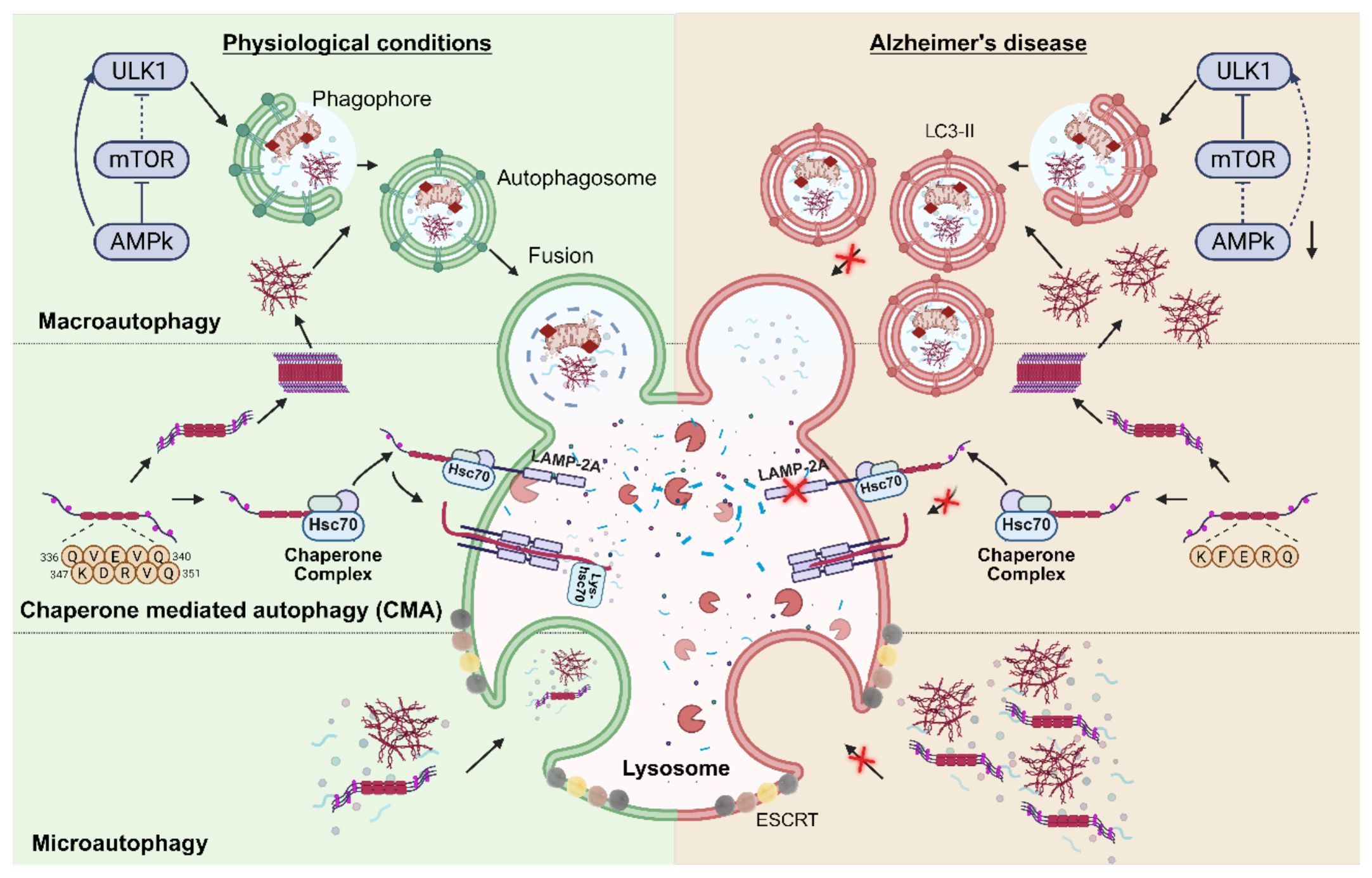
Figure 2.
Proposed link between intermittent fasting-induced metabolic switching and autophagy-related signalling in neurons. During intermittent fasting, depletion of hepatic glycogen promotes adipose-tissue lipolysis and hepatic ketogenesis, increasing the production of ketone bodie as acetoacetate or β-hydroxybutyrate (BHB). Circulating BHB crosses the blood–brain barrier and may act both as an alternative energy substrate under conditions of cerebral glucose hypometabolism and as a metabolic signalling molecule. The associated increase in the cellular AMP/ATP ratio activates AMPK, inhibits mTOR and promotes ULK1-dependent autophagy-related signalling, potentially enhancing macroautophagy and chaperone-mediated autophagy (CMA). In Alzheimer’s disease, these pathways remain mechanistically plausible but have not yet been directly demonstrated in the living human brain.
Figure 2.
Proposed link between intermittent fasting-induced metabolic switching and autophagy-related signalling in neurons. During intermittent fasting, depletion of hepatic glycogen promotes adipose-tissue lipolysis and hepatic ketogenesis, increasing the production of ketone bodie as acetoacetate or β-hydroxybutyrate (BHB). Circulating BHB crosses the blood–brain barrier and may act both as an alternative energy substrate under conditions of cerebral glucose hypometabolism and as a metabolic signalling molecule. The associated increase in the cellular AMP/ATP ratio activates AMPK, inhibits mTOR and promotes ULK1-dependent autophagy-related signalling, potentially enhancing macroautophagy and chaperone-mediated autophagy (CMA). In Alzheimer’s disease, these pathways remain mechanistically plausible but have not yet been directly demonstrated in the living human brain.
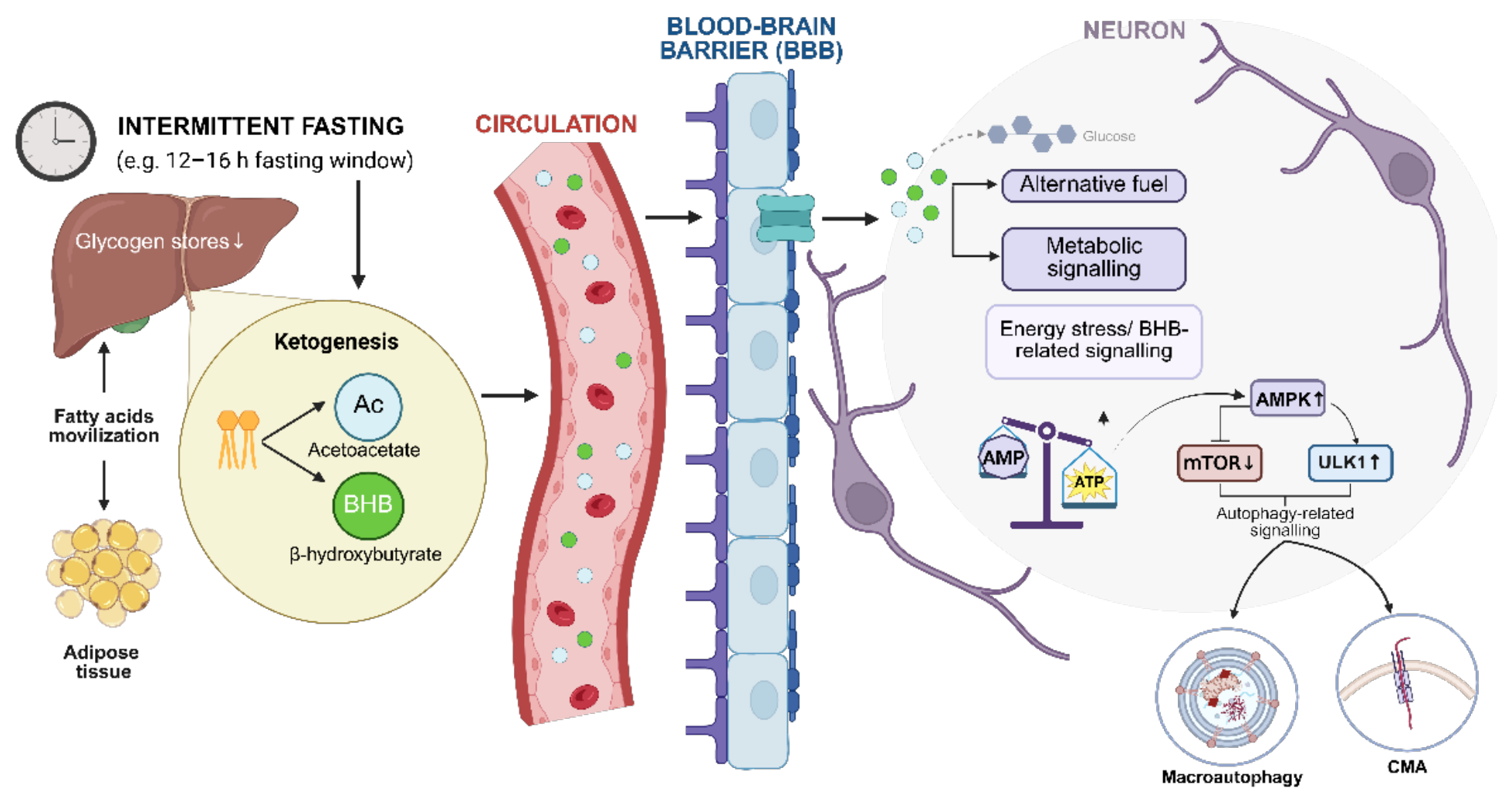
Figure 3.
Integrated neuroprotective mechanisms of intermittent fasting in Alzheimer’s disease. Intermittent fasting (IF) acts as a multimodal intervention that promotes neuroprotection through the convergence of metabolic, autophagic, mitochondrial, and microbiota-dependent pathways. Fasting-induced metabolic reprogramming facilitates a “metabolic switch” toward ketone-body utilization, primarily β-hydroxybutyrate (BHB), which activates the AMPK sensor and inhibits the mTOR pathway. This signaling cascade stimulates macroautophagy for the degradation of insoluble Tau aggregates, chaperone-mediated autophagy (CMA) for soluble Tau monomer clearance, and microautophagy—a process dependent on the VPS4A motor protein and ESCRT-III machinery to prevent lysosomal bottlenecks. Concurrently, IF enhances mitophagy, reducing reactive oxygen species (ROS) and oxidative damage. At the intersystemic level, IF modulates the microbiota–gut–brain axis through a propionate–FFAR3 signaling axis and increases the production of short-chain fatty acids (SCFAs), which support gut barrier integrity and reduce systemic inflammation. Notably, IF reduces intracellular lipid droplets within microglia, restoring their phagocytic capacity to manage the neural microenvironment. These combined mechanisms culminate in the activation of the CREB–BDNF axis, supporting synaptic plasticity and potential cognitive resilience while slowing the progression of cognitive decline.
Figure 3.
Integrated neuroprotective mechanisms of intermittent fasting in Alzheimer’s disease. Intermittent fasting (IF) acts as a multimodal intervention that promotes neuroprotection through the convergence of metabolic, autophagic, mitochondrial, and microbiota-dependent pathways. Fasting-induced metabolic reprogramming facilitates a “metabolic switch” toward ketone-body utilization, primarily β-hydroxybutyrate (BHB), which activates the AMPK sensor and inhibits the mTOR pathway. This signaling cascade stimulates macroautophagy for the degradation of insoluble Tau aggregates, chaperone-mediated autophagy (CMA) for soluble Tau monomer clearance, and microautophagy—a process dependent on the VPS4A motor protein and ESCRT-III machinery to prevent lysosomal bottlenecks. Concurrently, IF enhances mitophagy, reducing reactive oxygen species (ROS) and oxidative damage. At the intersystemic level, IF modulates the microbiota–gut–brain axis through a propionate–FFAR3 signaling axis and increases the production of short-chain fatty acids (SCFAs), which support gut barrier integrity and reduce systemic inflammation. Notably, IF reduces intracellular lipid droplets within microglia, restoring their phagocytic capacity to manage the neural microenvironment. These combined mechanisms culminate in the activation of the CREB–BDNF axis, supporting synaptic plasticity and potential cognitive resilience while slowing the progression of cognitive decline.
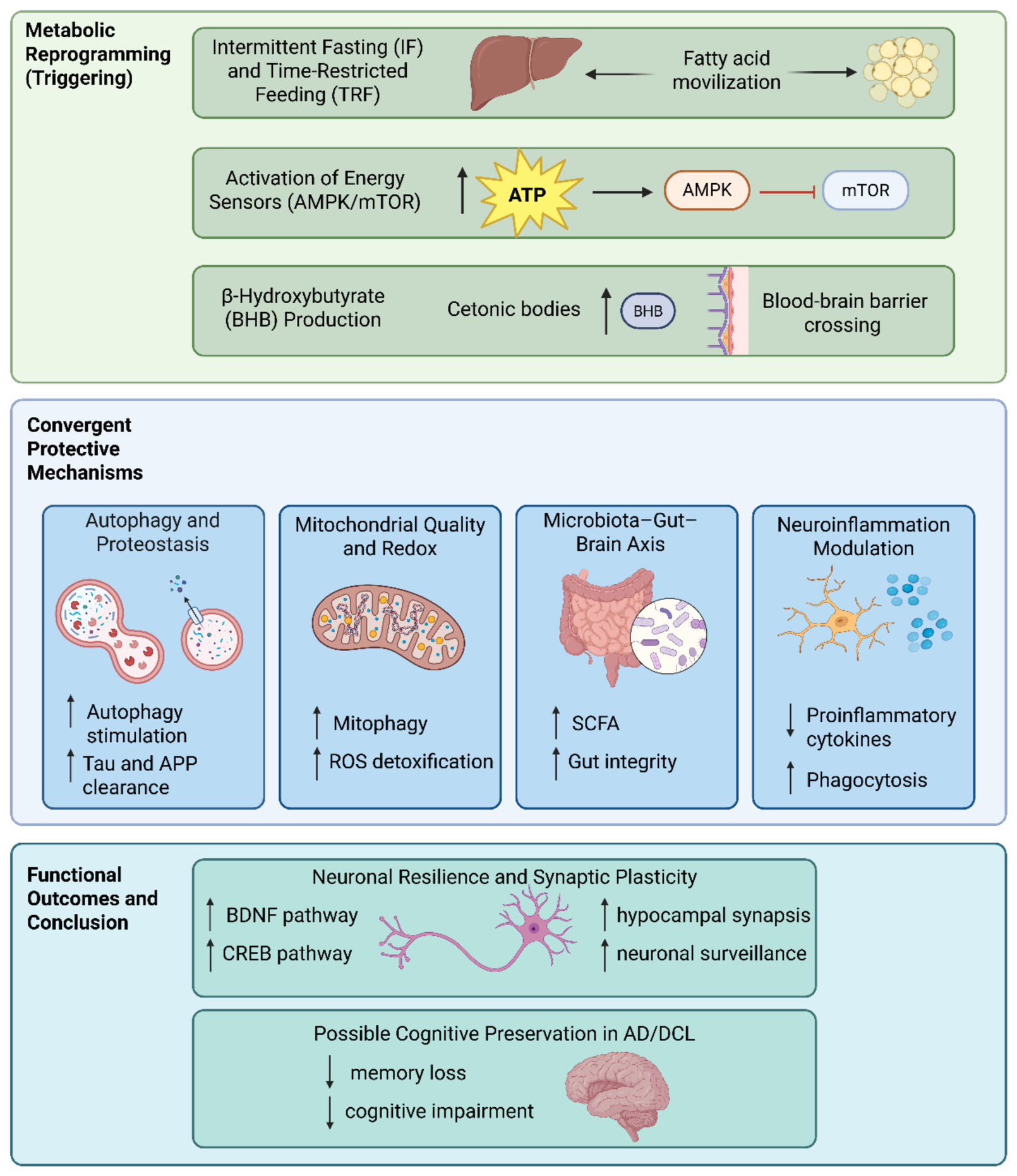
Figure 4.
Human clinical evidence and proposed mechanistic links between fasting-based interventions and Alzheimer’s disease-relevant outcomes. Human studies have evaluated time-restricted eating, intermittent fasting, caloric or energy restriction, and ketogenic or fasting-mimicking interventions, reporting systemic and peripheral changes in ketone metabolism, insulin sensitivity, inflammatory and oxidative-stress markers, and gut microbiota-derived short-chain fatty acids. These observations support proposed links with improved cerebral energy metabolism, AMPK–mTOR signalling, autophagy and proteostasis, reduced neuroinflammation, and enhanced synaptic resilience; however, these brain mechanisms remain largely indirect and have not been directly demonstrated in the living human Alzheimer’s disease brain. Human readouts currently include cognitive performance, peripheral biomarkers, microbiota-related measures, neuroimaging and brain-metabolism indicators, as well as feasibility, adherence and safety outcomes. Overall, the available evidence supports mechanistic plausibility but remains preliminary and insufficient to establish activation of neuronal autophagy or disease-modifying effects on amyloid-β and Tau pathology in humans.
Figure 4.
Human clinical evidence and proposed mechanistic links between fasting-based interventions and Alzheimer’s disease-relevant outcomes. Human studies have evaluated time-restricted eating, intermittent fasting, caloric or energy restriction, and ketogenic or fasting-mimicking interventions, reporting systemic and peripheral changes in ketone metabolism, insulin sensitivity, inflammatory and oxidative-stress markers, and gut microbiota-derived short-chain fatty acids. These observations support proposed links with improved cerebral energy metabolism, AMPK–mTOR signalling, autophagy and proteostasis, reduced neuroinflammation, and enhanced synaptic resilience; however, these brain mechanisms remain largely indirect and have not been directly demonstrated in the living human Alzheimer’s disease brain. Human readouts currently include cognitive performance, peripheral biomarkers, microbiota-related measures, neuroimaging and brain-metabolism indicators, as well as feasibility, adherence and safety outcomes. Overall, the available evidence supports mechanistic plausibility but remains preliminary and insufficient to establish activation of neuronal autophagy or disease-modifying effects on amyloid-β and Tau pathology in humans.
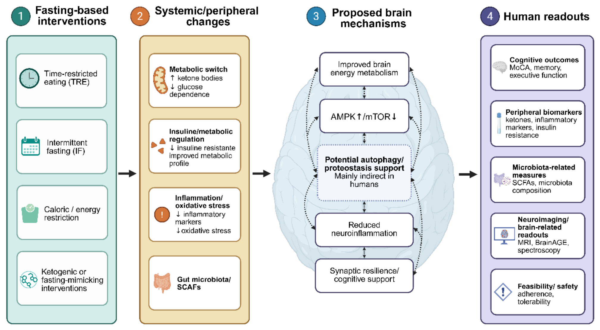
Figure 5.
Translational gap between experimental models and human clinical studies of intermittent fasting-induced neuroprotection. In vitro models allow precise manipulation of nutrient deprivation, ketone exposure and autophagy-related pathways, enabling the analysis of LC3 lipidation, p62 turnover, LAMP-2A-dependent CMA and lysosomal function. However, these models lack hepatic ketogenesis, vascular regulation, immune-endocrine interactions and the microbiota–gut–brain axis. Animal models provide whole-organism metabolic responses and access to brain tissue for histological, biochemical and behavioural analyses, but interspecies differences in metabolic rate and the predominance of transgenic familial AD models limit extrapolation to sporadic human disease. Human studies offer clinical relevance and allow assessment of cognition, adherence, safety, neuroimaging and peripheral biomarkers, but direct measurement of neuronal autophagic flux or brain proteostasis is not feasible. Future translation requires long-term, nutritionally supervised randomised controlled trials incorporating objective adherence monitoring, frailty and safety assessment, APOE/metabolic stratification, multimodal biomarkers and complementary patient-derived cellular models.
Figure 5.
Translational gap between experimental models and human clinical studies of intermittent fasting-induced neuroprotection. In vitro models allow precise manipulation of nutrient deprivation, ketone exposure and autophagy-related pathways, enabling the analysis of LC3 lipidation, p62 turnover, LAMP-2A-dependent CMA and lysosomal function. However, these models lack hepatic ketogenesis, vascular regulation, immune-endocrine interactions and the microbiota–gut–brain axis. Animal models provide whole-organism metabolic responses and access to brain tissue for histological, biochemical and behavioural analyses, but interspecies differences in metabolic rate and the predominance of transgenic familial AD models limit extrapolation to sporadic human disease. Human studies offer clinical relevance and allow assessment of cognition, adherence, safety, neuroimaging and peripheral biomarkers, but direct measurement of neuronal autophagic flux or brain proteostasis is not feasible. Future translation requires long-term, nutritionally supervised randomised controlled trials incorporating objective adherence monitoring, frailty and safety assessment, APOE/metabolic stratification, multimodal biomarkers and complementary patient-derived cellular models.
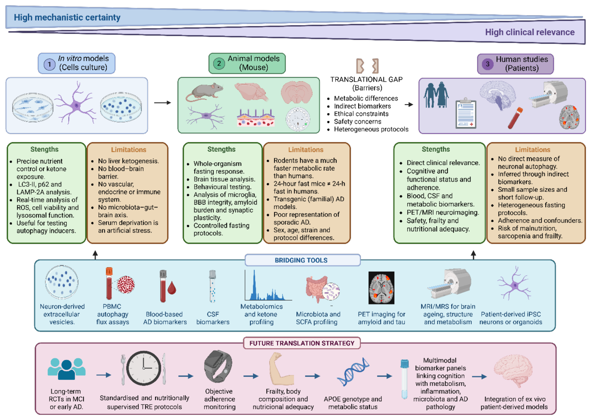

Table 1.
Cell-based evidence linking autophagy pathways with Aβ/Tau clearance-related readouts.
| Study | Mechanistic evidence | Cellular model | Experimental Manipulation | Main findings |
|---|---|---|---|---|
| Lee JH and Nixon RA 2022 [40] | Canonical macroautophagy activation (AMPK–mTOR pathway) | SH-SY5Y neuroblastoma cells; primary hippocampal neurons | Serum deprivation, amino acid withdrawal, glucose restriction or HBSS starvation | Nutrient deprivation activates AMPK–ULK1 signaling while inhibiting mTORC1, inducing ATG7-dependent autophagosome formation, LC3-positive vesicle accumulation and enhanced autophagic flux. |
| Hung SY, et al. 2009 [41] | Protective macroautophagy during amyloid stress | SH-SY5Y cells | Exposure to Aβ25–35 or Aβ1–42 with or without autophagy inhibition/ATG7 knockdown | Amyloid exposure induces autophagy as an adaptive response. Genetic or pharmacological inhibition of autophagy increases Aβ-induced neurotoxicity, indicating a protective role of macroautophagy during early amyloid stress. |
| Gómez-Sánchez R, et al. 2018 [39] | Autophagic flux and lysosomal competence | Cultured neurons | Assessment of LC3-I/LC3-II conversion, p62 turnover and lysosomal blockade (bafilomycin A1) | Accurate evaluation of autophagy requires flux measurements rather than static LC3-II accumulation. Combined analysis of LC3-II turnover, p62 degradation and lysosomal function distinguishes increased autophagy from impaired degradation. |
| Chiang YF, et al. 2023 [42] | Lysosomal dysfunction associated with APP processing | Neuronal models | Intracellular accumulation of APP-βCTF generated by BACE1 cleavage | APP-βCTF inhibits vacuolar H+-ATPase activity, impairing lysosomal acidification and endolysosomal degradation. This creates a feed-forward cycle promoting accumulation of APP-derived fragments and aggregated proteins. |
| Rossitto LM, et al. 2026 [44] | Ketone bodies restore autophagic flux | Primary cortical neurons | β-hydroxybutyrate (BHB) treatment under glucose deprivation | BHB enhances LC3-II turnover and p62 degradation, restores ATP homeostasis, decreases ROS accumulation and reduces Aβ toxicity, indicating improved autophagic flux rather than simple autophagosome accumulation. |
| Gorantla NV, et al. 2021 [45] | Ketone body-mediated Tau clearance | Neuronal models | BHB or ketone body treatment | Ketone bodies promote Tau degradation through an OPTN–LC3B-dependent autophagic-lysosomal pathway independently of ketolysis, linking fasting metabolism with selective Tau proteostasis. |
| Sreenivasmurthy, SG, et al. 2022 [47] | Chaperone-mediated autophagy (CMA) | Neuronal models | Pharmacological activation with bromo-protopine | Activation of Hsc70/LAMP2A-dependent CMA selectively enhances degradation of pathological Tau species, supporting CMA as a therapeutic target for tauopathies. |
| Pomilio C, et al. 2020 [46] | Glial autophagy and lysosomal function | Microglia and glial cells | Chronic Aβ exposure | Sustained Aβ accumulation impairs autophagic flux and lysosomal competence in microglia, reducing phagocytic capacity and promoting a dysfunctional inflammatory phenotype. |
| Fung TY, et al. 2022 [48] | Autophagy-inducing compounds | AD cellular models | Treatment with bromo-protopine or klotho overexpression | Bromo-protopine activates CMA and reduces pathological Tau. Klotho stimulates autophagy and enhances microglial phagocytosis and lysosomal degradation of extracellular Aβ. |
| Thomson AC, et al. 2022 [37] | Neuronal plasticity associated with fasting-mimetic conditions | Differentiated SH-SY5Y cells | Serum deprivation | Nutrient restriction increases neurite length and branching and induces expression of plasticity-related genes, including BDNF-associated pathways, suggesting enhanced neuronal resilience alongside autophagy activation. |
| Keeley O, el al. 2024 [49] | Microautophagy / ESCRT-dependent pathway | Neuronal models carrying VPS4A/ESCRT defects | Genetic disruption of VPS4A or ESCRT-III machinery | Impaired microautophagy blocks endolysosomal trafficking, leading to defective intracellular waste clearance, increased Tau hyperphosphorylation and Aβ accumulation, identifying microautophagy dysfunction as a potential upstream contributor to AD pathology. |
Table 2.
Animal studies evaluating fasting-related interventions, autophagy-related mechanisms and cognitive outcomes in models relevant to AD.
Table 2.
Animal studies evaluating fasting-related interventions, autophagy-related mechanisms and cognitive outcomes in models relevant to AD.
| Study | Animal model | IF / dietary protocol | Main measures | Autophagy / clearance relevance |
|---|---|---|---|---|
| Alirezaei et al.[4] | Non-transgenic/GFP-LC3 mice | Short-term fasting, 24-48 h | LC3-positive autophagosomes, autophagosome number/size, p-mTOR/p-S6, transmission electron microscopy | Direct neuronal macroautophagy activation in brain; not an AD disease-modification study. |
| Rajeev et al.[58] | Wild-type C57BL/6NTac mice with CCH/BCAS | 16 h fasting/day for 4 months | Evans blue, immunofluorescence, Luxol Fast Blue, tight junctions, MMP-2/MT1-MMP, oxidative stress, glutathione/SOD | Neurovascular and antioxidant protection; not direct neuronal autophagy endpoint. |
| Wu et al.[60] | APP/PS1 mice, 6 months old | IF for 1 month vs ad libitum | Y-maze, Barnes maze, Iba1, CD68, Perilipin-2, LC3, p62, Aβ deposition | Macroautophagy-related APP/Aβ handling and microglial phagocytic recovery. |
| Pan et al. [59] | 5XFAD mice , 3 months old | Alternate-day fasting for 10–12 weeks; food provided or removed at 8:00 pm; water ad libitum | Morris water maze, novel object recognition, Aβ plaque burden, soluble/insoluble Aβ1-40 and Aβ1-42, Iba1, GFAP, p-mTOR, p62, LC3-II/I, 16S rRNA microbiota sequencing, cecal metabolomics, antibiotic depletion, sarcosine/dimethylglycine supplementation | IF reduced Aβ pathology and gliosis, suppressed mTOR signalling, increased autophagy-related markers and remodelled the gut microbiota/metabolite profile; antibiotic depletion partially blocked IF benefits, and sarcosine/dimethylglycine mimicked IF-like protection |
| Babygirija et al. [61] | 3xTg AD mice and controls | CR, IF without total CR, prolonged fasting interval designs | Open field, Morris water maze, amyloid-beta, phospho-Tau, mTORC1, autophagy, neuroinflammation | Fasting interval required for mTORC1 inhibition, autophagy activation and full cognitive benefit. |
Table 3.
Human studies and related metabolic interventions relevant to intermittent fasting, cognitive impairment and Alzheimer’s disease.
Table 3.
Human studies and related metabolic interventions relevant to intermittent fasting, cognitive impairment and Alzheimer’s disease.
| Study | Population/design | Protocol | Main findings or outcomes | Interpretation |
|---|---|---|---|---|
| Wang et al. [67] | Older adults with MCI; assessor-blinded pilot RCT (n=46) | 15:9 TRE; 12 weeks | Improved MoCA trajectory and recognition memory compared with control. | Most direct causal evidence in MCI, but short and not AD-biomarker confirmed. |
| Ooi et al. [68] | Older Muslim adults with MCI; 36-month prospective observational study (n=99) | Regular or irregular Sunnah fasting vs non-fasting | Regular fasting associated with better cognitive trajectory and lower inflammatory/oxidative-stress markers. | Suggestive long-term signal, but non-randomised and potentially confounded. |
| James et al. [64,69] | Older adults ≥65 years with self-reported memory decline; remote single-group pre/post pilot study; 20 enrolled, 18 completed |
14 h prolonged nightly fasting, 6 days/week, for 8 weeks; fast started no later than 8:00 pm; 10 h daytime eating window; usual diet maintained |
Memory and Attention Phone Screener composite improved from 30.47 ± 26.10 to 42.35 ± 17.01, p = 0.02, d = 0.58; ISI decreased from 6.72 ± 5.65 to 5.00 ± 3.74, p = 0.043, d = 0.52; BMI, diet quality and routine did not significantly change; adherence 70–100%; no adverse events. | Feasibility and preliminary cognitive/sleep signal. Not AD-confirmed, no control group, small and homogeneous sample, no autophagy, ketone, PET or AD biomarker measures. |
| Zhao et al. [70] | Patients with AD; small uncontrolled human intervention (n=10; 9 completed) | 16:8 TRE; 4 months | MoCA total score increased, particularly executive function; AD associated with lower fecal propionate and butyrate. | Directly AD-related but preliminary, uncontrolled and very small. |
| Kapogiannis et al. [71] | Cognitively intact older adults with overweight and insulin resistance; RCT | 5:2 IF vs healthy living diet; 8 weeks | Both diets reduced estimated brain age; selected executive and verbal-memory outcomes favoured IF; AD biomarkers unchanged. | Mechanistically informative, but not conducted in AD patients. |
| Kim et al. [72] | Adults with central obesity; randomised trial | 5:2 intermittent energy restriction vs continuous energy restriction; 4 weeks | Both groups improved Mnemonic Similarity Task performance; no clear superiority of intermittent restriction. | Indirect evidence linking metabolic restriction to hippocampal-related memory. |
| Brenton et al. [73] | Relapsing-remitting multiple sclerosis; pilot dietary study | Ketogenic diet; 6 months | Reduced insulin and leptin and improved fatigue, including subjective cognitive fatigue; no objective cognitive improvement. | Indirect metabolic evidence; not an IF or AD study. |
| Cignarella et al. [74] | Relapsing-remitting multiple sclerosis; fasting-related intervention | Intermittent fasting protocol | Reduced leptin, modulated lymphocyte populations and altered gut-microbiota-related signals. | Useful for immunometabolic and microbiota mechanisms, not AD efficacy. |
| Rahmani et al. [75] | Midlife individuals with multiple sclerosis; pilot neuroimaging study | Intermittent caloric restriction; 12 weeks | Advanced neuroimaging suggested changes compatible with reduced neuroinflammation or microstructural alteration. | Methodological model for neuroimaging-based mechanistic assessment. |
| Siavoshi et al. [76] | Multiple sclerosis; metabolomic ageing analysis | Fasting-mimicking diet | Reported reversal of accelerated biological ageing based on metabolomic-age measures. | Indirect evidence for biological-ageing biomarkers. |
| Racette et al./TREAD [77,78] | Patients with AD or related cognitive impairment; pilot trial design and registered protocol | Time-restricted eating feasibility/safety intervention | Designed to assess feasibility, acceptability and safety; results not yet available in the manuscript. | Important framework for future AD-specific TRE implementation. |
| Chen et al. [79] | Planned RCT in mild/moderate AD (n=160) | 16:8 TRE; 24 months | Protocol includes cognition, function, APOE ε4, blood AD biomarkers, ketones, microbiota, metabolomics and neuroimaging. | Key study for testing longer-term safety and efficacy in AD. |
Disclaimer/Publisher’s Note: The statements, opinions and data contained in all publications are solely those of the individual author(s) and contributor(s) and not of MDPI and/or the editor(s). MDPI and/or the editor(s) disclaim responsibility for any injury to people or property resulting from any ideas, methods, instructions or products referred to in the content. |
© 2026 by the authors. Licensee MDPI, Basel, Switzerland. This article is an open access article distributed under the terms and conditions of the Creative Commons Attribution (CC BY) license (http://creativecommons.org/licenses/by/4.0/).
Copyright: This open access article is published under a Creative Commons CC BY 4.0 license, which permit the free download, distribution, and reuse, provided that the author and preprint are cited in any reuse.