

Submitted:
22 August 2026
Posted:
24 August 2026
You are already at the latest version
Abstract
Despite its potential clinical relevance, research into air-bacteria contamination in hospital settings is still in its early stages. In this study, we present the results of a one-year investigation into Gram-negative airborne bacteria in a medical ward in a hospital in southern Italy. Data show that multidrug-resistant Acinetobacter baumannii strains, which were also resistant to carbapenems, were isolated over the course of the study. Strains were characterised by the Pulsed Field Gel Electrophoresis (PFGE) molecular typing method, and the identified PFGE profiles were clustered into three groups based on genetic relatedness greater than 90%. These groups were termed A, B and C, with group A including the majority (71%) of isolates. The group A was further subdivided into three subgroups (A1, A2 and A3) based on the genetic relatedness of 97%, 99% and 94%, respectively. The presence of strains with distinct genetic relationships, as well as their detection in different rooms and at different sampling dates, may be explained through independent air contamination by specific bacteria groups or subgroups. This study also highlights the importance of air monitoring as a key control measure, particularly for the detection of ESKAPE pathogens such as A. baumannii, which has proven desiccation-resistant properties and can therefore survive in the air and on inanimate surfaces.
Keywords:
Acinetobacter baumannii
; air-bacteria contamination
; carbapenem resistant
; hospital settings
1. Introduction
Antimicrobial resistance (AMR) is a growing health challenge worldwide. Bacterial infections caused by multidrug-resistant (MDR) strains increase healthcare costs, prolong hospital stays, and are associated with higher mortality rates [1,2,3]. The impact of MDR and bacterial infections caused by MDR strains has recently been reported by a study that analysed 11 infectious syndromes caused by 84 pathogen-drug combinations in 204 countries and territories from 1990 to 2021. The study estimated that 4.71 million deaths were attributable to AMR bacteria, of which 1.14 million were directly related [4]. Such a figure was also confirmed by European data where the deaths associated with AMR bacteria accounted to 541,000 with 133,000 deaths attributable to AMR bacteria. [2]. The emerging impact on public health by infections caused by MDR strains has also been highlighted by the World Health Organization (WHO) which has included AMR as one of the top 10 global health threats [3]. In this context, the increasing frequency with which MDR bacteria have been isolated from clinical and environmental samples has increased the risk of ever more widespread AMR with serious implications for public health [5]. This is supported by the increasing number of hospital-acquired infections (HAIs), with an estimated 136 million cases occurring worldwide each year [6]. HAIs are among the most common adverse events with a high rate of morbidity and mortality when associated with healthcare ESKAPE pathogens (an acronym for Enterococcus faecium, Staphylococcus aureus, Klebsiella pneumoniae, Acinetobacter baumannii, Pseudomonas aeruginosa, and Enterobacter spp.) [7,8]. The 2024 WHO Bacterial Priority Pathogens List (https://www.who.int/publications/i/item/9789240103986), addressed the urgent threat posed by antibiotic-resistant pathogens, classifying A. baumannii and K. pneumoniae carbapenem-resistant and E. coli third-generation cephalosporin-resistant as critical priority human pathogens [9]. This is primarily due to the emerging resistance to carbapenems and third/fourth-generation cephalosporins, which are considered last-resort antibiotics [10,11]. The 2025 report from the WHO Global Antimicrobial Resistance and Use Surveillance System (GLASS) confirmed this emergency, stating a global median resistance of 17.2% across 93 pathogen–antibiotic combinations. Time-related analyses from 2018 to 2023 also reported annual increases in carbapenem resistance of 12.5% for E. coli and 15.3% for K. pneumoniae in specific regions. Taken together, these data provided a quantitative overview of the increasing burden posed by carbapenem-resistant Enterobacterales (CRE), Pseudomonas aeruginosa and A. baumannii. [12].
It is clear that one of the recommended measures to reduce the spread of MDR bacteria, particularly ESKAPE strains, is to monitor environments where pathogens can spread, such as hospitals [13,14]. A. baumannii strains are of particular concern due to their ability to persist in various environments, such as hospital settings, dry surfaces and air, thereby increasing the risk of infection. [15,16,17]. However, detection of airborne bacteria in hospitals is still a poorly explored field despite its growing importance in understanding the potential relationship between air bacteria contamination and risk of infection. [18]. The emerging interest in detection of airborne microorganisms has recently led to the development of new bioaerosol research tools and sampling devices. Generally, an air sampler should enable the collection of a representative bioaerosol sample that is suitable for both traditional and modern microbiological analysis techniques. Using a filter to collect the bioaerosol sample preserves the properties needed for microbiological analysis. Among the different types of filters, gelatine has been proven to fine preserve cultivability of the sample. Furthermore, gelatine filters can be dissolved in a buffer solution or placed directly onto agar for culturing or molecular-based analysis. [19].
In this study, we present data on air samples collected over the course of one year in an internal medicine ward of a tertiary hospital in southern Italy. Samples were harvested using an air sampler fitted with a gelatine filter, as well as via passive sampling methods. The study screened for the presence of MDR A. baumannii, P. aeruginosa, K. pneumoniae, E. coli, Serratia and Proteus strains. A. baumannii isolates were detected throughout the study. The strains were characterised using the Pulse Field Gel Electrophoresis (PFGE) genotyping technique and were clustered into three genetically distinct groups (A, B and C), with group A accounting for 71% of the total isolates.
2. Materials and Methods
2.1. Air Sampling, Bacteria Identification and Antimicrobial Susceptibility
A total of 280 air samples were collected in an internal medicine ward at a large tertiary hospital in southern Italy. Sampling was performed weekly for 12 months (June 2024-May 2025) by either an air sampler (n.31 samples), using the Airport Portable MD8 fitted with a gelatine filter (Sartorius AG, Göttingen, Germany) at a flow rate of 50 L/min and a suction volume of 2000L, or via a passive sampling method (n.249 samples) through air sedimentation for 1 hour. After sampling, the gelatine filters were immediately layered onto Mac Conkey agar plates (OXOID) and incubated at 37°C. Similarly, passive sampling was performed using Mac Conkey agar plates, which were then incubated immediately at 37°C. Active and passive sampling was conducted approximately one metre above the floor and one metre from the patient's bed. Priority was given to rooms in which patients who tested positive for bloodstream infections caused by A. baumannii or K. pneumoniae were admitted. Passive sampling was performed in the same rooms as the active sampling, as well as in other rooms in the ward that were progressively further away from the room where the active sampling took place. Detection of Acinetobacter, Pseudomonas and Enterobacteriaceae (K. pneumoniae, E. coli, Serratia, and Proteus) strains was initially assessed on Mac Conkey agar plates (OXOID). Suspected colonies found on Mac Conkey agar plates were successively isolated on Luria-Bertani (LB) agar plates. All colonies were characterised using matrix-assisted desorption/ionisation-time-of-flight mass spectrometry (MALDI-TOF-MS, BioMérieux, Marcy l'Etoile, France), a proteomic method that identifies microorganisms based on their protein fingerprint. Species identification was achieved with an accuracy rate ranging from 95% to 99.8%. Antimicrobial susceptibility versus amikacin (AK), ciprofloxacin (CIP), colistin (CO), gentamicin (GM), meropenem (MP), meropenem/vaborbactam(MEV), piperacillin/tazobactam (P/T) and tigecycline (TGC) was determined by the VITEK® automated identification system AST-N439 card (Biomérieux, Marcy- l’Etoile, France) and according to the European Committee on Antimicrobial Suscepitibility Testing (EUCAST 2025). Antimicrobial susceptibility was confirmed by bacteria growth on LB agar plates supplemented with either amikacin (30µg/ml), ciprofloxacin (1µg/ml), colistin (1µg/ml), gentamicin (10µg/ml), meropenem (4µg/ml), piperacillin (100µg/ml), or tigecycline (4µg/ml).
2.2. PFGE Analysis
The A. baumannii strains detected in this study were characterised using the PFGE molecular typing method, in line with previous studies [20,21], and in accordance with the PFGE protocols standardised for E. coli O157:H7, Salmonella serotypes and Shigella species [22]. The protocol included the initial preparation of a cell suspension by removing cells from the LB agar plate surface with a sterile cotton, followed by resuspension in 2 mL of Cell Suspension Buffer (CSB: 100 mM Tris and 100 mM EDTA at pH 8.0). The concentration of each cell suspension was adjusted to an absorbance value of approximately 1.3–1.4 at a wavelength of 610 nm, as measured using a spectrophotometer. A 400 µL aliquot of each adjusted cell suspension was transferred to a 1.5 mL centrifuge tube, to which 400 µL of 1% PFGE-quality agarose equilibrated to ~54 °C was quickly added. The mixture was gently mixed by pipetting up and down before being immediately dispensed into the wells of reusable or disposable PFGE plug moulds. The plugs were allowed to solidify at room temperature for 5–10 minutes, after which they were removed from the moulds and placed in a 50 mL polypropylene conical tube. The cells contained in the plugs were lysed by incubating them in 4 ml of lysis solution (CLS-1: 50 mM Tris–HCl, 50 mM EDTA, 2.5 mg/mL lysozyme and 0.1 mg/mL proteinase K) at 37 °C in a shaking incubator for 1 h. The plugs were then transferred to a fresh 50 mL polypropylene conical tube containing 4 mL of cell lysis solution 2 (CLS-2: 0.5 M EDTA, 1% sarcosyl and 400 μg/mL proteinase K) and placed in a shaking incubator at 55 °C for 2 h. The DNA in each plug was then digested with 40 U of Apa I at 37 °C for 4 h. The plugs containing the restricted Apa I DNA fragments were embedded in a 1% Pulsed Field Certified Agarose (Bio-Rad, Milan, Italy) gel in Tris-borate-EDTA buffer (44.5 mM Tris-borate, 1 mM EDTA; pH 8.0), and the DNA fragments were resolved at 14 °C using a CHEF-DRIII apparatus (Bio-Rad, Milan, Italy). The run electrophoresis parameters were set as follows: initial switch time, 1.8 s; final switch time, 18.6 s; voltage, 6 V; angle of migration, 120°; run time, 18 h. Universal DNA standard fragments were included in each gel by loading plugs harbouring cells of the Salmonella enterica serovar Braenderup strain H9812 digested with Xba I. Agarose gels were stained with ethidium bromide at a final concentration of 0.5 µg/mL, giving a detection limit of 1–5 ng/band. Images of the DNA fragments were acquired using the Gel Doc-It photo documentation system (UVP, Upland, CA, USA). PFGE patterns were analysed using GelJ fingerprinting software [23]. The phylogenetic tree of the PFGE patterns was produced using the Dice coefficient statistical tool, which measures the similarity or overlap between two sets of data. The unweighted pair-group method with arithmetic mean (UPGMA) and a tolerance of 2.0% for differences in band position were employed to build the dendrograms reported in Figure 1. Normalisation of the PFGE patterns across electrophoretic gels was achieved by using DNA fragments generated by the S. Braenderup strain H9812 XbaI-restriction as an external reference. There is no similarity cut-off as a threshold for PFGE profiles of A. baumannii strains to be defined as genetically related. Here, we assume a similarity value of ≥90% as a threshold to cluster PFGE profiles into genetically related groups.
3. Results
Over the one-year monitoring period, seventeen A. baumannii strains (hereafter referred to as ‘air-detected isolates’) were recovered from 31 active and 249 passive samples. All strains were found to be resistant to AK-CIP-GM-MP-PT (Table 1). Molecular typing by PFGE classified the air-detected isolates into thirteen pulsotypes, which were clustered into three groups based on genetic relatedness of at least 90%. These groups were termed groups A, B and C (Figure 1). Group A accounted for most of the isolates (71%), including nine of the thirteen identified pulsotypes. Group A was subdivided into three subgroups (A1, A2 and A3) based on the percentage of genetic similarity. These subgroups included strains with 97%, 99% and 94% similarity, respectively. Group B included three strains with genetic similarity ≥93%. Group C comprised two strains with the same pulsotype (100% of genetic similarity).
Detection of the air-detected isolates clustered within the genetically related groups A, B, and C was not related to the room or date of sampling (Table 1). Indeed, group A, the most prevalent group, was identified for strains isolated from six different rooms (1, 2, 3, 4, 8, 12). Likewise, the subgroups A1, A2 and A3 were detected for strains isolated from two (3 and 8), two (8 and 12) and three different rooms (1, 2 and 4), respectively. A similar picture emerged for the genetically related strains of group B and C detected in two (8 and 11) and two (5 and 9) different rooms, respectively. Furthermore, it is noteworthy that A. baumannii strains belonging to different groups or subgroups were detected in the same rooms. For example, strains belonging to groups A1, A2 and B were detected in the room 8.
There was also no correlation between the different PFGE groups or subgroups and the date of sampling. This is because the former's detection was limited in terms of both sampling time and the room sampled. Indeed, rooms in which strains of the same subgroup were detected on more than one occasion showed sampling dates that were either very close together (e.g. room 8 for strains of subgroup A1) or widely spaced (e.g. room 1, where strains of subgroup A3 were detected five weeks apart). Sampling type (active or passive) also showed no correlation between room and date of sampling when strain detection was considered. Nine out of seventeen samples were active and eight were passive. Even for the antimicrobial susceptibility showed by the detected A. baumannii strains it was not possible to relate the resistance pattern to specific PFGE groups or subgroups. All strains exhibited the same resistance pattern to AK-CIP-GM-MP-PT (with very close or identical MIC) and were susceptible to CO.
4. Discussion
A. baumannii is the species of the genus Acinetobacter that is most frequently involved in hospital infections. It is not a commensal of humans or animals, and its natural habitats are poorly understood. [24]. A. baumannii is an important pathogen responsible for severe ventilator-associated pneumonia and bloodstream, wounds, soft tissue and urinary tract infections that can cause nosocomial infections even leading to an increased hospital mortality rate [25,26]. Furthermore, the MDR exhibited by this pathogen, particularly with regard to carbapenems (one of the last available front-line antimicrobial agents), raises significant concerns regarding the antimicrobial therapy for treating the infections it causes. Acinetobacter's ability to resist desiccation further increases its survival period on inanimate surfaces, potentially facilitating transmission within and between hospital wards [27,28]. Air is a latent route for spreading infection. This has recently been receiving attention from the scientific community. [29,30,31]. For instance, some studies suggest that infected patients could release A. baumannii strains into hospital wards, where they could survive as airborne contaminants for a long time, increasing the risk of infecting new patients. [30,32]
Here we report a study aimed at assessing the presence of airborne pathogens in an internal medicine ward of a large tertiary hospital in southern Italy. Over one year (June 2024-May 2025) of monitoring our data revealed the detection of seventeen MDR A. baumannii strains. All strain exhibited the same resistance pattern to AK-CIP-GM-MP-PT (with very close or identical MIC) and susceptibility to CO (Table 1). The isolates were characterised by the PFGE molecular typing method and the 13 identified PFGE pulsotypes were clustered into three genetically related groups (A, B and C) (Figure 1). Group A was prevalent, accounting for nine of the 13 PFGE pulsotypes and 71% of the total isolates. The group A was further divided, based on genetic relatedness of 97%, 99% and 94%, into the subgroups A1, A2 and A3, respectively. Group B accounted for strains with genetic relatedness of more than 93%, while strains in group C were indistinguishable from each other. The presence of groups with genetic relatedness ranging from 93% to 100% may suggest a possible clonal origin for these populations.
Analysis of strain detection in relation to rooms and sampling dates (hereafter referred to as sampling features) revealed no correlation between sampling features and PFGE groups or subgroups. The same conclusion was reached when the antimicrobial susceptibility profiles exhibited by the A. baumannii isolates were matched with the sampling features, PFGE groups or subgroups. The sampling features included twelve rooms in which active and passive sampling was performed (for a total of 290 samples), as well as three additional rooms that were used as a meeting room, an infirmary and a ward for physicians, where passive samples were taken. The sampling features also included thirty sampling dates, spanning from June 2024 to May 2025.
The random distribution of strains belonging to different PFGE groups or subgroups among sampling features, and the absence of persistence of a specific clone, suggests that individual strains with distinct genetic relationships were introduced over the period of the study. The detection of MDR A. baumannii strains, including those resistant to carbapenems, raises a number of questions, such as the identification of environmental reservoirs in which the strains can persist and spread.
5. Conclusions
The link between airborne bacteria and infections is still a matter of debate, particularly with regard to transmission modalities [19,33]. Nevertheless, identifying and addressing routes of infection is a key step in reducing the spread of both pathogen bacteria and antimicrobial resistance. Therefore, detecting MDR pathogens such as A. baumannii in hospital ward aerosols can help to assess the risk of bacterial air contamination and implement the necessary measures to reduce the spread of these pathogens and their antibiotic resistance determinants. The WHO has emphasised the importance of air monitoring as a fundamental control measure (https://www.who.int/publications/i/item/WHO-UHL-IHS-IPC-2022.2). The current study supports these recommendations by demonstrating the effectiveness of air monitoring as a control measure.
The impact on public health of data presented in this study are on the detection of airborne multidrug resistant (included that to carbapenems) pathogen bacteria such as A. baumannii strains and its potential risk of transmission to new patients. Analysis of the molecular features (PFGE profiles) of the detected A. baumannii isolates, rooms and date of sampling has also suggested independent introduction of strains populations with unrelated genetic relationships. Data as a whole have strongly suggested the need of air monitoring as a routinely fundamental control measure to reduce the spread of bacteria pathogens and their antibiotic resistance determinants.
Author Contributions
Conceptualization and methodology, C.C., M.O., and C.P.; investigation C.C., M.O. and M.A., formal analysis C.C. and M.O., data curation P.B., G.G. and M.S., writing-review and editing P.P., M.C. and A.C., writing-original draft C.P. All authors have read and agreed to the published version of the manuscript.
Funding
This work was supported by the European Union Next-GenerationEU, PRIN2022 - project code “20229KTNRM” – CUP AH3D23006100006.
Institutional Review Board Statement
Not applicable.
Informed Consent Statement
Not applicable.
Data Availability Statement
No unpublished external datasets, proprietary databases, or additional data platforms were used for original data analysis in this article. The original contributions presented in the study are included in the article, further inquiries can be directed to the corresponding author.
Acknowledgments
we thank Karen Laxton for writing assistance.
Conflicts of Interest
The authors declare no conflicts of interest.
References
- Dadgostar, P. Antimicrobial Resistance: Implications and Costs. Infect. Drug Resist. 2019, 12, 3903–3910. [Google Scholar] [CrossRef] [PubMed]
- Mestrovic, T.; Aguilar, G.R.; Swetschinski, L.R.; Kevin, S.I.; Gray, Authia P; Weaver, Nicole Davis; Han, Chieh; Wool, Eve E; Hayoon, Anna Gershberg; Hay, Simon I. The burden of bacterial antimicrobial resistance in the WHO European region in 2019: a cross-country systematic analysis. Lancet Public Health 2022, 7, e897–e913. [Google Scholar] [CrossRef]
- Murray, C.J.I.; Ikuta, K.S.; Sharara, K.S.F.; Swetschinski, L.; Gray, A.; Han, C.; Bisignano, C.; Rao, P.; Wool, E.; Johnson, S.C.; et al. Global burden of bacterial antimicrobial resistance in 2019: a systematic analysis. Lancet 2022, 399, 629–655. [Google Scholar] [CrossRef] [PubMed]
- Collaborators. Global burden of bacterial antimicrobial resistance 1990-2021: a systematic analysis with forecasts to 2050. Lancet 2024, 404, 1199–1226. [Google Scholar] [CrossRef] [PubMed]
- Aljeldah, M.M. Antimicrobial Resistance and Its Spread Is a Global Threat. Antibiotics 2022, 11. [Google Scholar] [CrossRef]
- Balasubramanian, R.; Van Boeckel, T.P.; Carmeli, Y.; Cosgrove, S.; Laxminarayan, R. Global incidence in hospital-associated infections resistant to antibiotics: An analysis of point prevalence surveys from 99 countries. PLoS Med. 2023, 20, e1004178. [Google Scholar] [CrossRef] [PubMed]
- Abban, M.K.; Ayerakwa, E.A.; Mosi, L.; Isawumi, A. The burden of hospital acquired infections and antimicrobial resistance. Heliyon 2023, 9, e20561. [Google Scholar] [CrossRef]
- Raoofi, S.; Pashazadeh Kan, F.; Rafiei, S.; Hosseinipalangi, Z.; Noorani Mejareh, Z.; Khani, S.; Abdollahi, B.; Seyghalani Talab, F.; Sanaei, M.; Zarabi, F.; et al. Global prevalence of nosocomial infection: A systematic review and meta-analysis. PLoS ONE 2023, 18, e0274248. [Google Scholar] [CrossRef] [PubMed]
- Sati, H.; Carrara, E.; Savoldi, A.; Hansen, P.; Garlasco, J.; Campagnaro, E.; Boccia, S.; Castillo-Polo, J.A.; Magrini, E.; Garcia-Vello, P.; et al. The WHO Bacterial Priority Pathogens List 2024: a prioritisation study to guide research, development, and public health strategies against antimicrobial resistance. Lancet. Infect. Dis. 2025, 25, 1033–1043. [Google Scholar] [CrossRef]
- Avershina, E.; Shapovalova, V.; Shipulin, G. Fighting Antibiotic Resistance in Hospital-Acquired Infections: Current State and Emerging Technologies in Disease Prevention, Diagnostics and Therapy. Front Microbiol. 2021, 12, 707330. [Google Scholar] [CrossRef]
- Ma, Y.X.; Wang, C.Y.; Li, Y.Y.; Li, J.; Wan, Q.Q.; Chen, J.H.; Tay, F.R.; Niu, L.N. Considerations and Caveats in Combating ESKAPE Pathogens against Nosocomial Infections. Adv. Sci. (Weinh) 2020, 7, 1901872. [Google Scholar] [CrossRef]
- Miftode, I.L.; Radu, V.D.; Jigoranu, R.A.; Leca, D.A.; Prepeliuc, C.S.; Pasare, M.A.; Miftode, R.S.; Grigoriu, M.G.; Paranga, T.G.; Miftode, E.G. Novel Insights into Carbapenem Resistance: Mechanisms, Diagnostics, and Future Directions. Antibiotics 2026, 15. [Google Scholar] [CrossRef]
- Ajayi, A.O.; Odeyemi, A.T.; Akinjogunla, O.J.; Adeyeye, A.B.; Ayo-Ajayi, I. Review of antibiotic-resistant bacteria and antibiotic resistance genes within the one health framework. Infect. Ecol. Epidemiol. 2024, 14, 2312953. [Google Scholar] [CrossRef]
- Samreen; Ahmad, I.; Malak, H.A.; Abulreesh, H.H. Environmental antimicrobial resistance and its drivers: a potential threat to public health. J. Glob. Antimicrob. Resist. 2021, 27, 101–111. [Google Scholar] [CrossRef]
- Ibrahim, S.; Al-Saryi, N.; Al-Kadmy, I.M.S.; Aziz, S.N. Multidrug-resistant Acinetobacter baumannii as an emerging concern in hospitals. Mol. Biol. Rep. 2021, 48, 6987–6998. [Google Scholar] [CrossRef]
- Dijkshoorn, L.; Nemec, A.; Seifert, H. An increasing threat in hospitals: multidrug-resistant Acinetobacter baumannii. Nat. Rev. Microbiol. 2007, 5, 939–951. [Google Scholar] [CrossRef]
- Jawad, A.; Seifert, H.; Snelling, A.M.; Heritage, J.; Hawkey, P.M. Survival of Acinetobacter baumannii on dry surfaces: comparison of outbreak and sporadic isolates. J. Clin. Microbiol. 1998, 36, 1938–1941. [Google Scholar] [CrossRef]
- Hiwar, W.; King, M.F.; Shuweihdi, F.; Fletcher, L.A.; Dancer, S.J.; Noakes, C.J. What is the relationship between indoor air quality parameters and airborne microorganisms in hospital environments? A systematic review and meta-analysis. Indoor Air 2021, 31, 1308–1322. [Google Scholar] [CrossRef] [PubMed]
- Mainelis, G. Bioaerosol Sampling: Classical Approaches, Advances, and Perspectives. Aerosol Sci. Technol. 2020, 54, 496–519. [Google Scholar] [CrossRef] [PubMed]
- Durmaz, R.; Otlu, B.; Koksal, F.; Hosoglu, S.; Ozturk, R.; Ersoy, Y.; Aktas, E.; Gursoy, N.C.; Caliskan, A. The optimization of a rapid pulsed-field gel electrophoresis protocol for the typing of Acinetobacter baumannii, Escherichia coli and Klebsiella spp. Jpn. J. Infect. Dis. 2009, 62, 372–377. [Google Scholar] [CrossRef]
- Calia, C.; Pazzani, C.; Oliva, M.; Scrascia, M.; Lovreglio, P.; Capolongo, C.; Dionisi, A.M.; Chiarelli, A.; Monno, R. Carbapenemases-producing Klebsiella pneumoniae in hospitals of two regions of Southern Italy. Apmis 2017, 125, 491–498. [Google Scholar] [CrossRef]
- Ribot, E.M.; Fair, M.A.; Gautom, R.; Cameron, D.N.; Hunter, S.B.; Swaminathan, B.; Barrett, T.J. Standardization of pulsed-field gel electrophoresis protocols for the subtyping of Escherichia coli O157:H7, Salmonella, and Shigella for PulseNet. Foodborne Pathog. Dis. 2006, 3, 59–67. [Google Scholar] [CrossRef]
- Heras, J.; Dominguez, C.; Mata, E.; Pascual, V.; Lozano, C.; Torres, C.; Zarazaga, M. GelJ--a tool for analyzing DNA fingerprint gel images. BMC Bioinform. 2015, 16, 270. [Google Scholar] [CrossRef]
- Wilharm, G.; Skiebe, E.; Michalska, A.; Higgins, P.G.; Weber, K.; Schaudinn, C.; Neugebauer, C.; Gorlitz, K.; Meimers, G.; Rizova, Y.; et al. Acinetobacter baumannii's lifestyle includes soil-dwelling colonization of decaying plant material and airborne spread. Nat. Commun. 2026, 17. [Google Scholar] [CrossRef]
- Redruello-Guerrero, P.; Fernandez-Martinez, N.F.; Garcia-Marin, C.; Martin-delosReyes, L.M.; Lorusso, N.; Millan-Cachinero, C.; Valero-Ubierna, M.D.C.; Rivera-Izquierdo, M. Carbapenemase production and in-hospital mortality associated with multidrug-resistant bacteria: a retrospective study conducted in Granada, Spain. Infect. Dis. (Lond) 2025, 57, 542–550. [Google Scholar] [CrossRef]
- El-Ashry, A.H.; Abdrabou, A.M.; El-Tantawy, N.; Mahmoud, R.; El-Mahdy, R. In vitro activity of meropenem-vaborbactam combinations and eravacycline against carbapenem-resistant Acinetobacter baumannii. Sci. Rep. 2025, 15, 34109. [Google Scholar] [CrossRef]
- Lucidi, M.; Capecchi, G.; Spagnoli, C.; Basile, A.; Artuso, I.; Persichetti, L.; Fardelli, E.; Capellini, G.; Visaggio, D.; Imperi, F.; et al. The response to desiccation in Acinetobacter baumannii. Virulence 2025, 16, 2490209. [Google Scholar] [CrossRef]
- Muller, C.; Reuter, S.; Wille, J.; Xanthopoulou, K.; Stefanik, D.; Grundmann, H.; Higgins, P.G.; Seifert, H. A global view on carbapenem-resistant Acinetobacter baumannii. mBio 2023, 14, e0226023. [Google Scholar] [CrossRef]
- Liu, Y.; Wang, Y.; Hao, C.; Li, Y.; Lou, H.; Hong, Q.; Dong, H.; Zhu, H.; Lai, B.; Liu, Y.; et al. Pathogenic bacteria and antibiotic resistance genes in hospital indoor bioaerosols: pollution characteristics, interrelation analysis, and inhalation risk assessment. Env. Pollut. 2025, 374, 126243. [Google Scholar] [CrossRef]
- Yakupogullari, Y.; Otlu, B.; Ersoy, Y.; Kuzucu, C.; Bayindir, Y.; Kayabas, U.; Togal, T.; Kizilkaya, C. Is airborne transmission of Acinetobacter baumannii possible: A prospective molecular epidemiologic study in a tertiary care hospital. Am. J. Infect. Control 2016, 44, 1595–1599. [Google Scholar] [CrossRef]
- Jiang, M.; Mu, Y.; Li, N.; Zhang, Z.; Han, S. Carbapenem-resistant Acinetobacter baumannii from Air and Patients of Intensive Care Units. Pol. J. Microbiol. 2018, 67, 333–338. [Google Scholar] [CrossRef]
- Mousa, M.; Schwartz, D.; Carmeli, Y.; Nutman, A. Droplet aerosol dissemination of carbapenem-resistant Acinetobacter baumannii surrounding ventilated patients. Infect. Control Hosp. Epidemiol. 2019, 40, 365–367. [Google Scholar] [CrossRef]
- Bing, Y.; Zhang, Y.H.; Leung, N.H.L.; Cowling, B.J.; Yang, Z.F. Role of viral bioaerosols in nosocomial infections and measures for prevention and control. J. Aerosol Sci. 2018, 117, 200–211. [Google Scholar] [CrossRef]
Figure 1.
Dendrogram of the PFGE profiles of the airborne A. baumannii isolates.
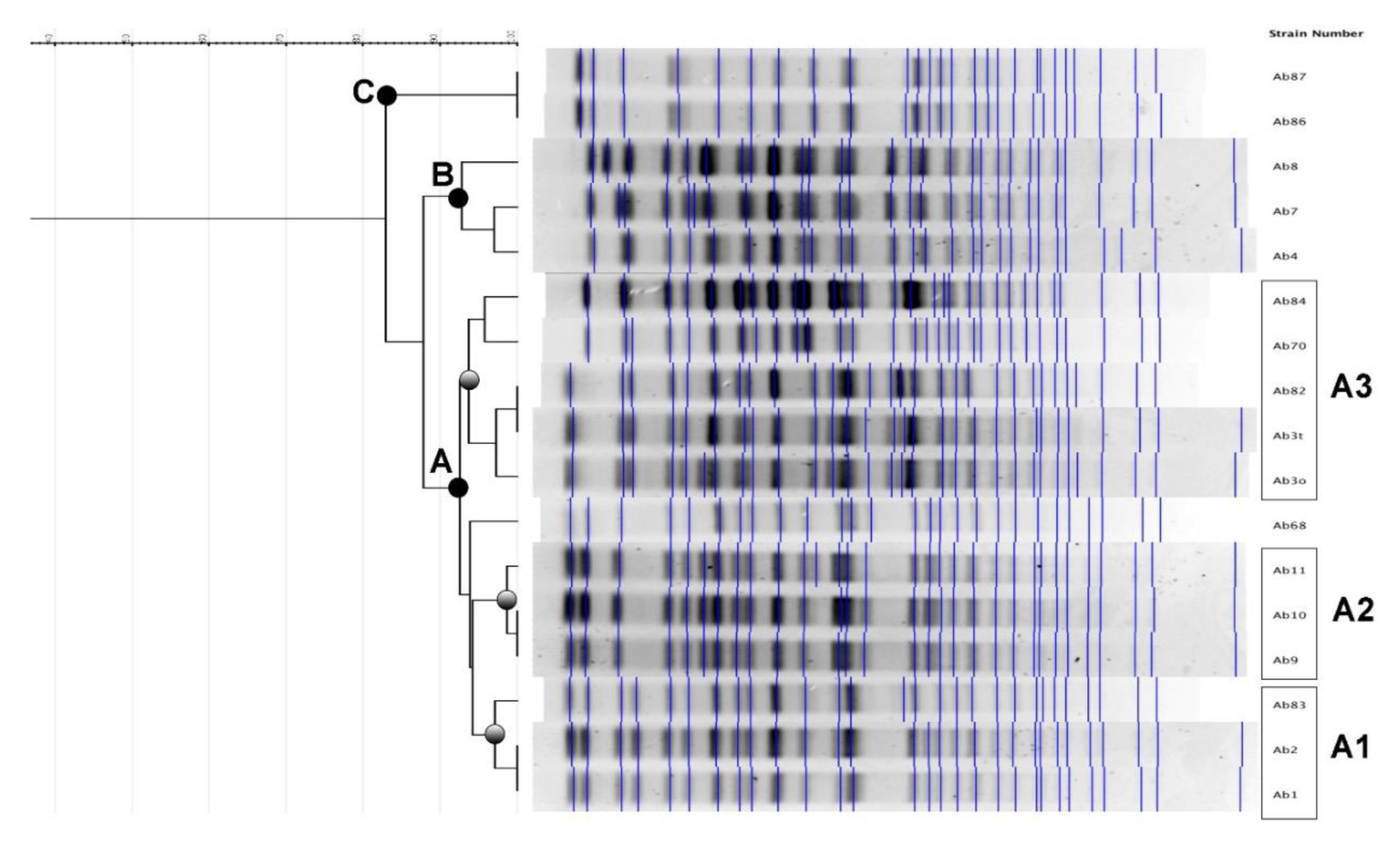

Table 1.
A. baumannii airborne isolates: sampling and MIC.
| Date of sampling | BI positive for (1) | Type of sampling | Room | Strain | Genetically related group | MIC | |||||||
|---|---|---|---|---|---|---|---|---|---|---|---|---|---|
| AK | CIP | CO | GM | MP | MEV | P/T | TGC | ||||||
| 19/09/2024 | Ab | Active | 8 | Ab1 | A1 | ≥ 64 | ≥ 4 | 1 | ≥ 16 | ≥ 16 | 16 | ≥ 128 | 2 |
| 25/09/2024 | Kp | Active | 8 | Ab2 | A1 | ≥ 64 | ≥ 4 | 1 | ≥ 16 | ≥ 16 | 16 | ≥ 128 | 2 |
| " | Ab | Passive | 4 | Ab3o | A3 | ≥ 64 | ≥ 4 | 1 | ≥ 16 | ≥ 16 | 16 | ≥ 128 | 4 |
| " | Ab | " | 4 | Ab3t | A3 | ≥ 64 | ≥ 4 | 0,5 | ≥ 16 | 8 | 8 | 32 | 4 |
| 16/10/2024 | Kp | Passive | 11 | Ab4 | B | 32 | ≥ 4 | 1 | ≥ 16 | ≥ 16 | 16 | ≥ 128 | 4 |
| 30/10/2024 | Kp | Passive | 8 | Ab7 | B | 32 | ≥ 4 | 1 | ≥ 16 | ≥ 16 | ≥ 64 | ≥ 128 | 2 |
| " | Kp | Active | 11 | Ab8 | B | 32 | ≥ 4 | 1 | ≥ 16 | ≥ 16 | 32 | ≥ 128 | 2 |
| 20/11/2024 | Kp | Active | 12 | Ab9 | A2 | ≥ 64 | ≥ 4 | 1 | ≥ 16 | ≥ 16 | 16 | ≥ 128 | 1 |
| 27/11/2024 | Kp | Passive | 12 | Ab10 | A2 | ≥ 64 | ≥ 4 | 2 | ≥ 16 | ≥ 16 | 16 | ≥ 128 | 1 |
| 11/12/2024 | Kp | Active | 8 | Ab11 | A2 | ≥ 64 | ≥ 4 | 1 | ≥ 16 | ≥ 16 | 16 | ≥ 128 | 1 |
| " | Kp | Passive | 4 | Ab68 | A | ≥ 64 | ≥ 4 | 1 | ≥ 16 | ≥ 16 | 16 | ≥ 128 | 1 |
| 08/01/2025 | Kp | Active | 2 | Ab70 | A3 | 32 | ≥ 4 | 1 | 8 | ≥ 16 | 32 | ≥ 128 | 1 |
| 27/02/2025 | Ab | Active | 1 | Ab82 | A3 | ≥ 64 | ≥ 4 | 1 | ≥ 16 | ≥ 16 | ≥ 64 | ≥ 128 | 2 |
| 05/03/2025 | Ab | Active | 3 | Ab83 | A1 | ≥ 64 | ≥ 4 | 4 | ≥ 16 | ≥ 16 | ≥ 64 | ≥ 128 | 1 |
| 09/04/2025 | Kp | Active | 1 | Ab84 | A3 | 32 | ≥ 4 | 1 | 8 | ≥ 16 | 16 | ≥ 128 | 1 |
| 15/04/2025 | Ab | Passive | 9 | Ab86 | C | ≥ 64 | ≥ 4 | 2 | ≥ 16 | ≥ 16 | 16 | ≥ 128 | 4 |
| 28/04/2025 | Kp | Passive | 5 | Ab87 | C | ≥ 64 | ≥ 4 | 2 | ≥ 16 | ≥ 16 | 16 | ≥ 128 | 2 |
MIC: minimal inhibitory concentration. AK: amikacin; CIP: ciprofloxacin; CO: colistin; GM: gentamicin; MP: meropenem; MEV: meropenem/vaborbactam; P/T: piperacillin/tazobactam; TGC: tigecycline. (1) BI: bloodstream infections; Ab: A. baumannii; Kp: K. pneumoniae.
Disclaimer/Publisher’s Note: The statements, opinions and data contained in all publications are solely those of the individual author(s) and contributor(s) and not of MDPI and/or the editor(s). MDPI and/or the editor(s) disclaim responsibility for any injury to people or property resulting from any ideas, methods, instructions or products referred to in the content. |
© 2026 by the authors. Licensee MDPI, Basel, Switzerland. This article is an open access article distributed under the terms and conditions of the Creative Commons Attribution (CC BY) license.
Copyright: This open access article is published under a Creative Commons CC BY 4.0 license, which permit the free download, distribution, and reuse, provided that the author and preprint are cited in any reuse.