Preprint
Article

This version is not peer-reviewed.

Cinncassiol A-19 Glucoside from Cinnamomum cassia Targets Digestive Lipases Through Covalent Inhibition: A Promising Natural Strategy Against Obesity

Submitted:

21 August 2026

Posted:

24 August 2026

You are already at the latest version

Abstract

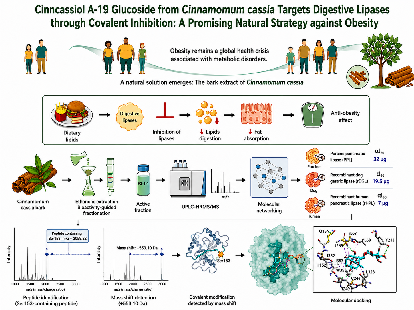
Natural inhibitors of digestive lipases offer promising alternatives to conventional obesity medications. This study screened nine medicinal plant extracts identifying the ethanolic bark extract of Cinnamomum cassia as the most potent inhibitor for gastric and pancreatic lipases. Using an integrated approach combining bioactivity-guided fractionation and mass spectrometry, we isolated the highly active fraction, F3-1-1, which exhibited dose- and time-dependent inhibitory effects against human and porcine pancreatic lipases, as well as dog gastric lipase (DGL). Further mass spectrometry and peptide mass fingerprint analysis identified cinncassiol A-19 glucoside as the main bioactive constituent. Notably, this compound forms covalent complexes with the catalytic peptide containing the Ser153 residue in DGL, characterized by a mass shift of +553.10 Da. Molecular docking simulations supported these results, demonstrating stable interactions within the catalytic pocket of DGL through hydrogen and hydrophobic bond interactions. These findings highlight C. cassia as a promising source of natural anti-obesity agents.
Keywords: 
;  ;  ;  ;  

1. Introduction

Obesity is a chronic, multifactorial, and relapsing disease that has emerged as one of the most significant global public health challenges (Gómez-Ambrosi et al., 2025). Characterized by an excess and abnormal accumulation of body fat, obesity is strongly associated with an increased risk of various comorbidities, with a cumulative effect from a young age, correlated with several diseases, including cardiovascular diseases and type 2 diabetes, dyslipidaemia, metabolic dysfunction-associated liver disease, hypertension, and several cancers (Abdelaal et al., 2017). The prevalence of obesity has increased alarmingly in the past decades across all age groups and has become a global epidemic (Blüher, 2019). In 2022, 43% of adults aged 18 years and over were overweight, of whom 16% were obese (Phelps et al., 2024). A recent epidemiological study showed that the overall prevalence of obesity among children and adolescents aged 5 to 19 years was 8.5% in 2022, compared to 5% in 2015 (The GBD 2015 Obesity Collaborators, 2017; Zhang et al., 2024). Among the geriatric population, the prevalence has also increased considerably, reaching 25.3% globally (Khaleghi et al., 2025). This pathology not only has devastating health consequences but also entails considerable financial costs for individuals and communities (Spieker & Pyzocha, 2016). To address this worldwide challenge, scientific research has focused on treating and preventing further increasing obesity rates. To this end, several research studies and pharmaceutical companies have concentrated on discovering and developing natural and synthetic drugs to reduce the damage caused by obesity.
Current pharmacological strategies for obesity management target central and peripheral pathways involved in appetite regulation and energy homeostasis. Early anti-obesity agents such as sibutramine and rimonabant were withdrawn due to severe cardiovascular and neuropsychiatric adverse effects, highlighting the limitations of centrally acting therapies (Ruiz et al., 2024; Scheen, 2010; Weill et al., 2010). More recently, glucagon-like peptide-1 receptor agonists, including semaglutide and liraglutide, have demonstrated robust efficacy in inducing weight loss by modulating appetite, gastric emptying, and insulin secretion (Chao et al., 2023; Wilding et al., 2021).
Despite their clinical success, these therapies remain limited by high cost, parenteral administration, long-term dependency, and gastrointestinal side effects, restricting their accessibility at the population level. Therefore, there is a growing interest in complementary or alternative approaches, particularly those derived from natural sources, to support safer and more sustainable obesity management strategies. Inhibition of human digestive gastric (HGL) and pancreatic (HPL) lipases to reduce fat absorption has become the main pharmacological approach for the treatment of obesity over the last decade (Liu et al., 2020; Subramaniyan & Hanim, 2025). The lipase inhibitor, Orlistat, marketed as Xenical® or Alli® exemplifies this approach. Orlistat or tetrahydrolipstatin (THL) is a derivative of lipstatin that was isolated from Streptomyces toxytricini (Borgström, 1988). It is a potent anti-obesity drug which inhibits the hydrolysis of the dietary triglycerides (TGs) into absorbable fatty acids and monoglycerides by inhibiting digestive lipases (Müller & Petry, 2006; Tiss et al., 2010). Orlistat has been reported to cause weight loss, inhibit fat absorption by 30%, and limit caloric intake (Phillips & Roberts, 2007). In addition to its anti-obesity effect, Orlistat has been shown to have numerous undesirable effects, including liver damage (Sall et al., 2014). Given the several side effects and the current prescribing guidelines for medications, it’s crucial to discover new drugs with a high level of safety and efficacy.
For the management of obesity, and in addition to synthetic drugs, it has been reported that natural products, particularly extracts and isolated compounds, have been widely developed as anti-obesity agents in traditional medicine to treat and remediate obesity through different mechanisms, including the inhibition of digestive lipases. These products were used in the management of obesity and other pathologies due to various bioactive compounds including terpenoids, flavonoids, polyphenols. Many medicinal, herbal and edible plants have shown potent anti-obesity effects through lipase inhibitory activity, such as phaseolus vulgaris (Peddio et al., 2022), Invingia gabonensis (Omonkhua et al., 2024), chili pepper (Watcharachaisoponsiri et al., 2017) and some Lauraceae such as cinnamon (Jaradat et al., 2023; Saleem et al., 2024). However, although medicinal plants are often considered safer than synthetic drugs, they may contain toxic compounds that can compromise their safety (Bahmani et al., 2016). Therefore, considerable efforts have been devoted to identifying natural lipase-inhibiting compounds and developing reliable approaches to isolate active molecules while reducing the potential adverse effects with crude extracts.
This study aimed to screen digestive lipase inhibitors from ethanolic extracts prepared from nine medicinal plants. The extract exhibiting the highest inhibitory activity was further fractionated to identify the compounds responsible for lipase inhibition. The most active fraction was analyzed by high-resolution tandem mass spectrometry (HRMS/MS). The identification of the putative active inhibitor and the proposed inhibition mechanism were investigated using proteomic approaches coupled with mass spectrometry, including peptide mass fingerprinting (PMF), together with molecular docking.

2. Material and Methods

2.1. Chemicals and Reagents

Sodium taurodeoxycholate (NaTDC), Glyceryl tributyrate (TC4), olive oil, bovine serum albumin (BSA) and gum Arabic (GA) were obtained from Sigma-Aldrich-Fluka (Saint-Quentin-Fallavier, France). Orlistat was purchased from Hoffmann-La-Roche Ltd. in Basel, Switzerland). Ethanol absolute anhydrous, methanol, petroleum ether, hexane, diethyl ether, dichloromethane (DCM) and acetonitrile (HPLC gradient grade) were obtained from Carlo Erba reagents (Val de Reuil, France). Normal-phase thin-layer chromatography (NP-TLC) aluminium sheets pre-coated with silica gel 60 and reversed-phase (RP)-18 TLC plates were from Merck (Darmstadt, Germany). All other chemicals and solvents, of the highest quality, were obtained from local suppliers. Recombinant dog gastric lipase (rDGL, molecular mass, 49 kDa) was produced and purified as described previously (Roussel et al., 2002). Recombinant human pancreatic lipase (rHPL, molecular mass, 50 kDa) was expressed and purified in the laboratory. Porcine pancreatic lipase (PPL, Type II, L3126, molecular mass, 50 kDa) was purchased from Sigma-Aldrich-Fluka Chimie (Saint-Quentin-Fallavier, France) and pancreatic colipase was purified from lipid-free porcine pancreatic powder as described previously (Fernandez et al., 2007). In all the experiments, water was obtained from the Milli-Q Gradient water purification system.

2.2. Preparation of Plant Crude Extracts

Nine dried plants, collected from different regions of Tunisia (Table S1), were finely ground using a high-speed laboratory blender and then sieved to obtain a powder. Each dried powder was extracted by maceration for 24h with ethanol (1:10, w/v). The different extracts were filtered through No. 3 sintered glass and concentrated using a vacuum rotary evaporator. The dried powders were stored at 4 °C until analysis.
Prior to biological assays, for each extract, a stock solution with a final concentration of 100 mg.mL- 1 was prepared in ethanol prior to the experiments. These extracts were then tested in vitro for their inhibitory activity against PPL, which was used as the initial inhibition target for the initial screening of digestive lipase inhibitors.

2.3. Preparation of the Different Cinnamomum cassia Extracts

Dried bark C. cassia powder was extracted, separately, by maceration with each of the following solvents (1:10, w/v): hexane, DCM, ethanol and water. The different extracts were filtrated through No. 3 sintered glass and concentrated using a vacuum rotary evaporator. In the case of aqueous extract, it was lyophilized. The inhibitory potential of Hexane extract (HE), DCM extract (DCME), Ethanol Extract (EE) and Water Extract (WE) were then tested against PPL.

2.4. Lipolytic Activity Measurement

Lipolytic activity was measured at 37 °C by automated, continuous titration of fatty acids released, under the catalytic action of lipase on a mechanically stirred TG emulsion, using 0.1 M NaOH and a pH-STAT (Metrohm 702 SM Titrino, Herisau, Switzerland) calibrated to a fixed end point. For pancreatic lipase, the enzymatic assay was performed in a thermostated vessel (37 °C) containing 0.2 mL of TC4 and 9.8 mL of 2.5 mM Tris-HCl buffer, 150 mM NaCl buffer (pH 8), 2 mM CaCl2 and 0.5 mM NaTDC.
When olive oil was used as the substrate, it was first pre-emulsified with GA, as previously described (Tiss et al., 2001). Five mL of this emulsion was added to 10 mL of 2.5 mM Tris-HCl, 150 mM NaCl buffer (pH 8) containing 2 mM CaCl2 and 0.5 mM NaTDC. When required, colipase was added with a molar excess of 5 relative to rHPL in the presence of 4 mM NaTDC.
In the case of rDGL activity measurement, the enzyme was assayed at pH 5.5 using the standard assay solution: 150 mM NaCl, 2 mM NaTDC, and 2 µM BSA (Roussel et al., 2002). Enzyme activity was expressed in international units (U), where one unit corresponds to the release of 1 µmol of fatty acid per minute under the assay conditions.

2.5. Methods to Test Lipase Inhibition

The inhibition of lipase activity of the medicinal plant extract was determined using two methods:
- Lipase-inhibitor pre-incubation method: It involves pre-incubating the lipase with the inhibitor in absence of a substrate. This method was designed to visualize, in an aqueous medium, the potential direct interaction between the lipase and the inhibitor (Cudrey et al., 1993; Tiss et al., 2009). Pre-incubations of the lipase with various inhibitor amounts were carried at room temperature in presence of NaTDC (4 mM, final concentration) in order to create a micellar interface that may contain the inhibitor (Lengsfeld et al., 2004; Tiss et al., 2009).
- Lipase inhibition during lipolysis method: This method involves adding the inhibitor a few minutes after injecting the lipase into the reaction medium containing the emulsified substrate. This method was designed to test for potential inhibition of the lipase-catalyzed hydrolysis reaction during lipolysis (Tiss et al., 2009).
The degree of inhibition was assessed by measuring the relative residual lipase activity (RA%), which is the ratio of lipase activity in the presence and absence of the inhibitor. The amount of extract corresponding to 50% inhibition (αI50) was determined. The half-inactivation time (t½) was defined as the time required to reach 50% of the residual lipase activity for a given value of extract amount (αI). A control sample, for each experiment, was performed in the absence of the inhibitor fraction but with the same volume of solvent in which the inhibitor was solubilized (ethanol: water, 7:3 v/v).

2.6. Bioactivity-Guided Fractionation

This part of the work was carried out in five steps. The first two steps consisted of two successive macerations to concentrate the potentially inhibitory compounds. After each phytochemical step, the inhibition activity against PPL was checked. In the first maceration, 100 g of dried bark of C. cassia powder were macerated in hexane (1:10, w:v). After 24 hours, the mixture was filtered using a sintered glass funnel; the liquid phase was evaporated to dryness under vacuum, and the solid residue was dried under a fume hood. In the second maceration, this defatted residue was extracted with ethanol for 24 hours. The delipidated EE was filtered on a sintered glass funnel, and the resulting filtered crude extract was vacuum dried and analyzed by NP-TLC using various solvent systems (water, DCM, methanol, ethyl acetate, and petroleum ether) in different ratios. In the third step, the delipidated EE was fractionated using silica gel column chromatography into three fractions (F1, F2, F3), using three different mobile phases: ethyl acetate / petroleum ether (5:5, v: v) (F1), ethyl acetate (F2) and methanol (F3). The resulting fractions were evaporated similarly and analyzed by TLC. Migration was performed with an ethyl acetate: cyclohexane (3:7, v/v) for the NP-TLC, or water: acetonitrile (0.1:1, v: v) for the RP-TLC. The spots were revealed by UV at 320 nm. The inhibitory potential was tested against PPL. In the fourth step, based on the inhibitory potential of these fractions, the methanolic fraction (F3) was fractionated into five subfractions (F3-1 to F3-5) by liquid chromatography (LC) on C18 column, using an acetonitrile and water as the mobile phase following an optimized gradient (Table S2). A RP-TLC analysis was then performed using migration solvent water: acetonitrile (0.1:1, v: v). Finally, based on the TLC results, fraction F3-1 was further separated by LC on the C18 column using solvent gradients shown in supplementary data. Figure 1 illustrates the different fractions of C. cassia powder.

2.7. UPLC-HRMS/MS Analysis of Active Fractions

The analysis of C. Cassia fractions was performed using an Ultimate 3000 ultra-performance liquid chromatography (UPLC) (Ultimate 3000, Thermo, Fisher Scientific) system coupled to a high-resolution hybrid quadrupole time-of-flight (Q-TOF) mass spectrometer (Impact II Q-TOF, Bruker). Chromatographic separation was carried out on a Luna Omega C18 column (100 × 2.1 mm, 1.6 µm). Samples were dissolved in methanol at a final concentration of 10 mg. mL-1. The column temperature was set at 40 °C, and the injection volume was 2 µL. Separation was performed at a flow rate of 400 µL min-1, using mobile phase A, water containing 0.1% formic acid, and mobile phase B, methanol / acetonitrile (50:50, v/v) containing 0.1% formic acid. The following linear gradient was applied: 100% A from 0 to 18 min, 100–0% A from 18 to 28 min, 0–100% A from 28 to 28.2 min, followed by column re-equilibration at 100% A until 35 min.
The mass spectrometry detection was performed in full-scan mode over the m/z range 20-1000, using both positive and negative electrospray ionization modes (ESI+, ESI-). The source temperature was set to 200 °C, the nebulizer gas pressure to 45 psi, and the capillary voltage to 4500 V in positive mode and 3500 V in negative mode. For MS/MS experiments, low-energy collision-induced dissociation (CID) was performed using collision energies ranging from 20 to 47 eV.

2.8. Data Analysis

Raw data obtained from UPLC-HRMS/MS analysis were converted to mzML and mgf formats using MSConvert software (ver. 3.0.2) and Data Analysis module. The chromatogram was generated with a group size of several scans, a minimum group intensity of 1×10⁴, and a minimum absolute intensity of 5.0×10⁵, with an m/z tolerance of 5 ppm and mSigma < 30. The ion identity networking method was done with m/z tolerance of 3.5. The adducts [M+H]+ and [M-H] were set for the positive and negative mode, respectively. Peaks processed from the sample were filtered based on peaks present in blank sample (100% methanol) which was prepared in the same conditions as the sample, and by subtracting the blank. The remaining peaks were then analysed. Molecular networking and library search were performed using the global natural products social (GNPS) molecular networking software (https://gnps2.org), food data base, mass bank and human data base (HMDB). For the molecular networking, the peaks were uploaded to GNPS platform, and the precursor ion Mass Tolerance and fragmentation ion Mass Tolerance were set at 0.02 Da and 0.05 Da, respectively. A cosine score threshold of 0.7 and a minimum of 6 matching fragment ion were required, each, to perform spectral searches and analysis against GNPS2 libraries.

2.9. Peptide Mass Fingerprinting

An aliquot of rDGL (12 µg) was pre-incubated with F-3-1-1 in the presence of 4 mM NaTDC. After 30 min, inhibition was tested using anti-lipase activity assay. The mixture was then separated by 12% SDS-PAGE. A control was prepared in the same conditions, without F-3-1-1. Protein bands corresponding to 50 kDa were excised from the gel as previously described (Zehl et al., 2004). For protein identification and molecular mass determination, protein bands were digested overnight at 37 °C with a Trypsin/Lys-C protease mixture (Promega). The resulting tryptic peptides digest was analyzed by MALDI-TOF mass spectrometry using a Microflex II spectrometer (Bruker Daltonics, Germany). For peptide mass analysis, spectra were acquired in positive reflectron mode over the 600-5000 Da mass range. External mass calibration was performed using the monoisotopic [M+H]+ ions of the following peptide calibration standards such as Angiotensin II (m/z 1046.5418), Angiotensin I (m/z1296.6848 ), substance P (m/z1347.7354 ), Bombesin (m/z 1619.8223 ), ACTH clip1-17 (m/z 2093.0862 ), ACTH clip18-39 (m/z 2465.1983 ), somatostatin 28 (m/z 3147.4710 ), as previously described in (Lorenzi et al., 2011). Briefly, tryptic digests were spotted directly onto a MALDI stainless-steel MALDI target plate. An equal volume of saturated α-cyano-4-hydroxycinnamic acid matrix solution prepared in acetonitrile/water/trifluoroacetic acid (70:29.9:0.1, v/v/v) was added to each spot, which was then allowed to dry at room temperature. Peak lists were generated using Flex Analysis software and manually inspected. Experimentally measured peptide masses were compared to the theoretical masses of tryptic peptide calculated from the rDGL amino acid sequence, including putative variable modifications of the catalytic serine residue corresponding to adduct formation with compounds present in fraction F3-1-1 (Lorenzi et al., 2011).

2.10. Molecular Docking

In-silico molecular docking of Cinncassiol A-19 glycoside and THL in the active site of HGL (PDB entry codes: 1HLG) was performed using VINA software. The grid box size was chosen to fit the whole active site cleft and to allow non-constructive binding positions. The inhibitor structure model was built using Phenix’s eLBOW. The most energetically favorable conformations were selected for further analysis. Protein–ligand interactions, including hydrogen bonding, hydrophobic contacts, and proximity to the catalytic triad, were analyzed to evaluate the potential inhibitory mechanism.
This computational approach was used to complement experimental findings and provide structural insights into the interaction between cinncassiol A-19 glucoside and gastric lipase, particularly in comparison with the well-characterized mechanism of covalent inhibitor THL interaction with lipase.

2.11. Statistical Analysis

Statistical analysis was performed using GraphPad Prism version 8.0.1. Concentration–response curves were fitted by nonlinear regression, and αI50 values were calculated accordingly. The 95% confidence intervals (95% CI) of the fitted parameters were estimated using the profile likelihood method. Data are presented as mean ± standard deviation (SD) from at least three independent experiments. Differences were considered statistically significant at p < 0.05.

3. Results and Discussion

3.1. Screening for Lipase Inhibitor Properties of Medicinal Plants Crude Extract

During a primary screening, nine crude extracts, prepared from different parts of nine medicinal plants (S) by extraction using ethanol, were tested for their inhibitory potential against PPL, initially chosen as the inhibition target. The selection of plants was based on their ethnopharmacological relevance and traditional medicinal uses. Their potential is supported by existing literature demonstrating significant anti-obesity activity for all the chosen plants including, C. cassia (Song et al., 2017), Ceratonia siliqua (Aboura et al., 2017), Coffea arabica (Sankpal et al., 2021), Linum usitatissimum (Ansari et al., 2019), Quercus (Jakkawanpitak et al., 2026), Cuminum cyminum (W. Wang et al., 2024), Zingiber officinale (J. Wang et al., 2017), Cucumis melo (Kim et al., 2026), and Thymus vulgaris (Abdelmottaleb Moussa et al., 2024). The inhibitory potential of all crude extracts was calculated as a percentage of the residual PPL activity after incubation with the target extract at a final concentration of 1 mg.mL-1. The results illustrated in Table 1 showed that the EE of C. cassia bark exhibited the highest inhibitory potential with a residual PPL activity of 11%. The EEs of Ceratonia siliqua, Coffea arabica, and Linum usitatissimum showed moderate inhibitor potential, with residual PPL activities of 38%, 51%, and 53%, respectively (Table 1), whereas EEs from Quercus macrocarpa, Cuminum cyminum, Zingiber officinale, Cucumis melo, and Thymus vulgaris showed weak or negligible anti-lipase activity (residual activities > 60%). (Table 1). Outstanding inhibitory potential, C. cassia bark was selected for further bioactivity-guided fractionation and mechanistic studies. As previous studies have shown, Cinnamomum is a commercially available spice used in culinary and medicinal applications, cosmetics, and perfumery. Regarding medicinal applications; Cinnamomum has been used in both of traditional and modern medicine to treat headaches, diabetes, digestive disorders, and obesity (Błaszczyk et al., 2021; Oketch-Rabah et al., 2018; Watanabe et al., 2023).

3.2. Evaluation of the Anti-Lipase Activity of Different Extracts of C. cassia

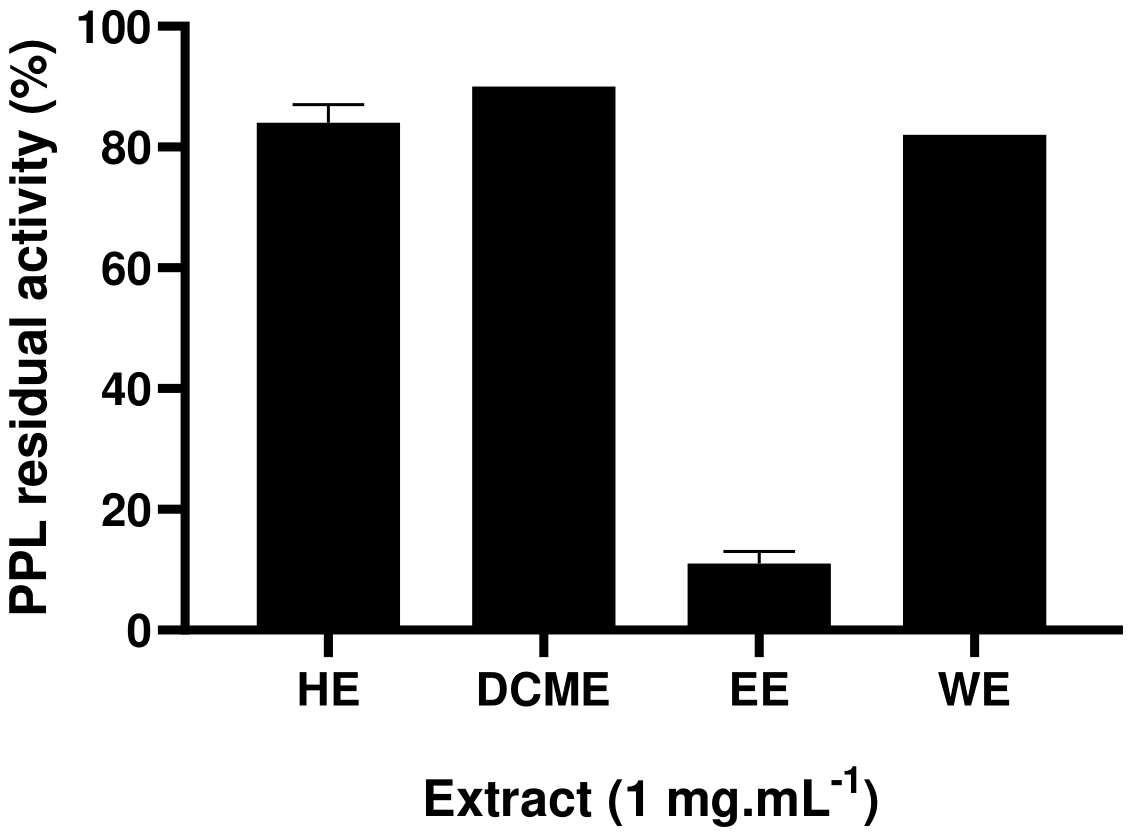
The inhibitory potential of the HE, DCME, EE and WE of C. cassia bark was determined. Using a final concentration of 1 mg.mL-1 of each of these extracts, the residual activity of PPL was shown to be 84 ±3%, 90%, 11 ±2% and 82%, respectively (Figure 2). These results clearly demonstrate that the inhibitory compounds were extracted by ethanol. It has been shown previously that at a concentration of 200 µg.mL-1, the EE of C. cassia had a stronger inhibitory activity compared with DCM: methanol extract with an PPL inhibitory activity of 80 ±10% and 30 ±6%, respectively (Abeysekera et al., 2017). However, it has been demonstrated that the HE of Cinnamomum did not display any inhibitory potential against turkey pancreatic lipase with a RA of 100% (Sellami et al., 2017). In addition, modern pharmacology study showed that the EE of C. cassia barks had hypolipedemic and anti-diabetic effects (Vijayakumar et al., 2023).

3.3. Bioactivity-Guided Fractionation

To further refine the bioactivity-guided fractionation of the EE and based on the results of inhibitory potential of the different C. cassia extracts, we evaluated the inhibitory activity of the initial C. cassia dried bark following a sequential extraction process: initial delipidation with hexane followed by extraction with ethanol. The results demonstrated a significant enhancement in inhibitory potency: the RA (%) of PPL dropped to 1 ± 2% with the delipidated extract, compared to 11 ± 2% for the crude EE (Figure S1A). This marked increase in efficacy can be attributed to the delipidation step, which likely concentrated the primary inhibitory compounds while simultaneously removing antagonistic lipophilic components that may have interfered with the inhibitory effect. The defatted EE was analyzed by NP-TLC. A mobile phase of ethyl acetate/petroleum ether (6/4 v:v) provided the most effective separation (Figure S1B). The chromatogram shows at least 6 compounds with decreasing polarity from the base line to the front of the solvent.
Based on these results, the defatted EE was fractionated using silica gel chromatography following the optimized protocol, yielding three distinct fractions: F1, F2 and F3 (Figure S2). Subsequent anti-lipase activity assay (Figure S2A) and NP-TLC analysis (Figure S2B) revealed that the original extract separated into three groups of compounds based on their polarity. The inhibitory assays showed that at a concentration of 0.5 µg.mL-1, the RA (%) activity of PPL was 80% ± 2, 49% ± 3, and 9% ± 5 for F1, F2, and F3, respectively (Figure S2A). Fraction F3 exhibited the highest inhibitory potency against PPL. NP-TLC analysis demonstrated that F3 possessed a highly polar character. Consequently, a second fractionation step was performed on reversed phase C18 column to further isolate the active compounds. Chromatographic separation of F3 yielded five distinct sub-fractions, designated F3-1 to F3-5 (Figure S3A). These were subsequently evaluated for their inhibitory activity against PPL and characterized by TLC. The results indicated that at a concentration of 100 µg.mL-1, the fraction F3-1 exhibited the most potent inhibitory potential with RA (%) of 18% ± 4. NP-TLC analysis revealed that fractions F3-1 and F3-2 exhibited minimal mobility, remaining largely retained by the stationary phase (Figure S3B). This behavior confirms the highly polar nature of these two fractions. Their polarity was further validated using RP-TLC with a highly polar mobile phase. Under these conditions, fraction F3-1 displayed a distinct, migrating spot, suggesting the presence of a specific inhibitory compound (Figure S3C). Based on these analytical results, we focused the subsequent characterization efforts on fraction F3-1 due to its relatively lower complexity.
To further isolate and enrich the active principle, fraction F3-1 was subjected to reversed phase C18 column chromatography, and two distinct subfractions were eluted, F3-1-1 and F3-1-2 (Figure S4A). At a concentration of 100 µg.mL-1, the RA (%) of PPL was shown to be 3% ± 2 for F3-1-1 and 98% ± 4 for F3-1-2 (Figure S4B). Notably, the specific inhibitory potency increased significantly throughout the purification process; the amount of fraction required to inhibit PPL decreased tenfold after three successive fractionation steps. Before bioactivity-guided fractionation, the RA (%) of PPL was 9% ± 2 in the presence of 1 mg.mL-1 of the crude EE (Figure S2A), while at the end of this process, the RA (%) of PPL, measured under the same conditions, was 3% ± 2 in the presence of 100 µg.mL-1 of the enriched fraction F3-1-1 (Figure S4B). One of the hallmarks of bioactivity-guided fractionation is the trend of increasing potency with fractionation. Furthermore, previous reports suggest that this phyto-fractionation process holds promise for an efficient natural drug discovery approach (Nothias et al., 2018). Based on these results, fraction F3-1-1 was selected as the most potent lead to evaluate its inhibitory spectrum against other key digestive enzymes, specifically rHPL and rDGL.

3.4. The Inhibitory Potential of Fraction F3-1-1 Against Digestive Lipases

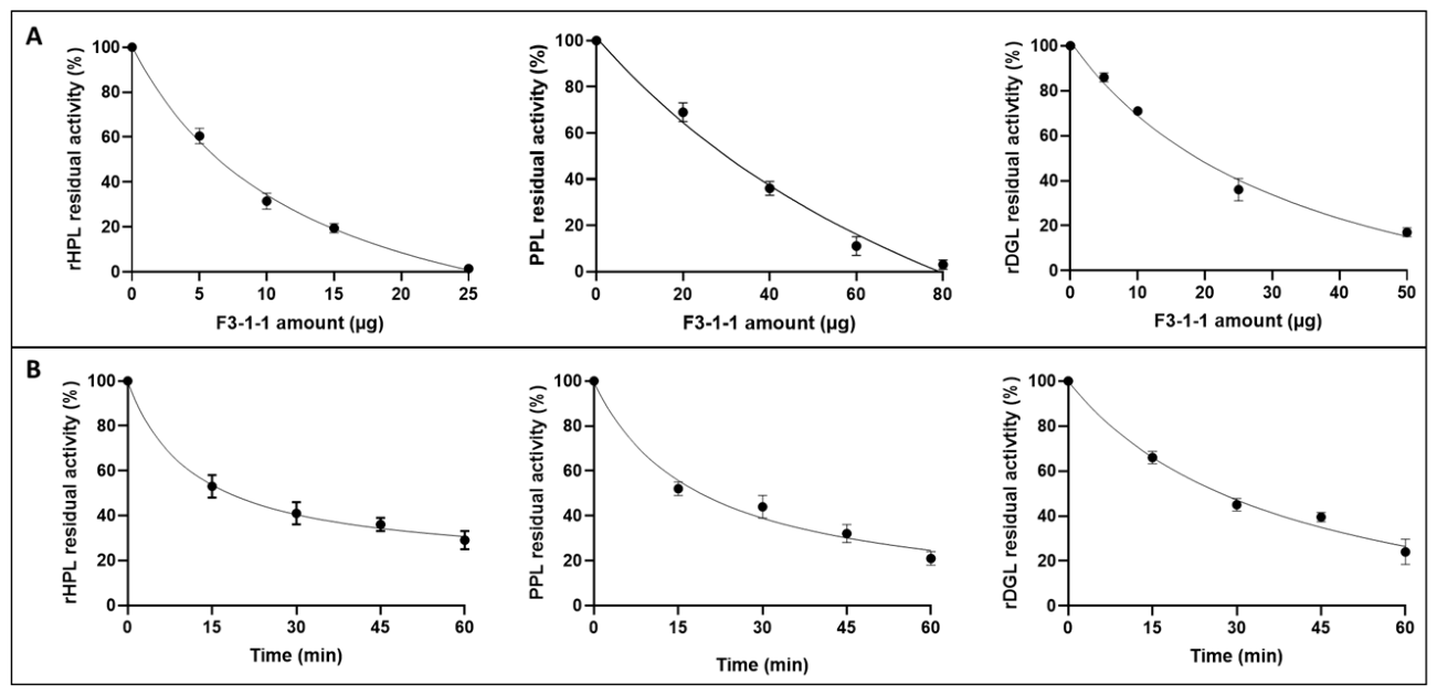
The current study aims to identify natural-derived digestive lipase inhibitors in order to reduce lipid absorption for the treatment of obesity. We therefore evaluated the inhibitory potential of an enriched fraction of C. cassia against digestive lipases. Initially, the inhibitory activity of the crude extract and different fractions of C. cassia was assessed against PPL, which was used as the initial target for inhibitor screening. Subsequently, the inhibitory activity of fraction F3-1-1 was then tested against purified rHPL and rDGL. PPL and rHPL share 85% identity (Lowe et al., 1989), and rDGL is an interesting model enzyme for human gastric lipase (HGL). Indeed, the amino acid sequence identity between these two lipases is 85.7% and their three-dimensional structures are superimposable (Roussel et al., 2002). The influence of the amount values of fraction F3-1-1 and pre-incubation times were evaluated using the enzyme and inhibitor pre-incubation method in the absence of the substrate as indicated in Material and Methods section. The inhibition profiles thus obtained revealed that this fraction possesses a dose-dependent inhibitory activity (Figure 3A). RA (%) of 1% ± 2, 11% ± 4%, or 15% ± 2% were obtained after a 30-min pre-incubation of rHPL, PPL, or rDGL (0.5 µM, final concentration) with 25 µg, 60 µg, or 50 µg of the F3-1-1 fraction, respectively. The results obtained indicate that F3-1-1 inhibits rHPL much more effectively than PPL and rDGL, with αI50 values of 7 µg, 32 µg, and 19.5 µg, respectively (Table 2). Furthermore, rHPL is completely inhibited at an αI of 25 µg after 30 minutes of pre-incubation, whereas PPL and rDGL retain 40% and 30% of their activity at an αI of 40 µg (Figure 3A).
The influence of the pre-incubation time of rHPL, PPL, or rDGL with F3-1-1 was further evaluated (Figure 3B). In all cases, the residual activity of these lipases decreased rapidly, reaching approximately 40% after 30 min of pre-incubation. This residual activity then decreased slightly, reaching 35% for rHPL, 25% for PPL, and 28% for DGL after 1 h of pre-incubation in the presence of 8 µg, 35 µg and 20 µg of F3-1-1, respectively (Figure 3B). From these inhibition graphs, the half-lives were then determined and found to be 17 min, 16 min, and 25 min for rHPL, PPL, and rDGL, respectively (Table 2). Such values of αI50 and t½ reflect a strongly inhibitory rate of these lipases by F3-1-1.
Based on αI50 and t½ values, the F3-1-1 had the ability to inhibit the catalytic activity of digestive lipases which are the main therapeutic target for obesity (Subramaniyan & Hanim, 2025). During the digestion phase of a test meal, HGL and HPL hydrolyze, respectively, 17.5% and 48.5% of the acyl chains of TGs, thus leading to the 66% hydrolysis necessary for the complete absorption of lipids from this same test meal (Aloulou & Carrière, 2008; Carriere et al., 1993). Consequently, specific inhibition of both gastric and pancreatic lipases impairs gastrointestinal digestion, significantly reduces intestinal absorption of lipolysis products from ingested lipids, and thus decreases caloric intake (Carrière et al., 2001). Gastric and pancreatic lipases are therefore a promising therapeutic target for the treatment of obesity.
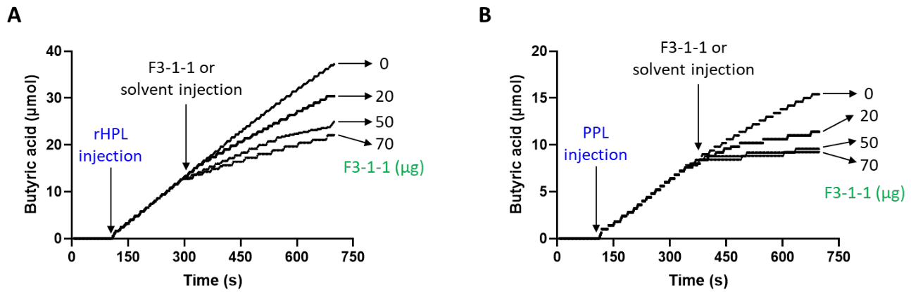
The influence of F3-1-1 on the rate of TC4 hydrolysis was evaluated for rHPL and PPL to test the possibility of inhibition of lipolytic activity during lipolysis. Four minutes after the addition of the enzyme, either solvent alone (ethanol/water 7/3, v:v) or F3-1-1 at variable amounts were injected. As shown from the kinetic curves in Figure 4, the rate of TC4 hydrolysis by the action of rHPL (Figure 4A) or PPL (Figure 4B) decreased after the addition of the inhibitor, but to different extents. The decrease in enzyme activity was more pronounced at higher F3-1-1 amounts. At a concentration of 70 µg of F3-1-1, approximately 36% and 12% inhibition of the enzyme activity of rHPL and PPL, respectively, was observed.
It is worth noting that inhibition of rHPL is very strong when the enzyme is pre-incubated with F3-1-1 in the presence of NaTDC compared to the method of injecting the inhibitor during lipolysis. This result shows that the inhibitor is primarily associated with the lipid phase, even in the presence of bile salts. This physicochemical property is probably a necessary condition for the effectiveness of the lipase inhibitor under physiological conditions. In the case of PPL, inhibition by F3-1-1 was more pronounced when the inhibitor was added during lipolysis than when the enzyme and inhibitor were pre-incubated in the absence of the substrate. This difference could be due either to the presence of other proteins in the pancreatic extract containing PPL, which exert a protective effect in the aqueous phase, or to the specific physicochemical properties of the inhibitor, or to the presence of a micellar concentration of NaTDC in the preincubation medium. Moreover, using the olive oil as substrate, the F3-1-1 against PPL was 52 µg (Figure S5) while it was 37 µg using TC4. A previous studies on lipase inhibitors showed that the rate of inhibition of dog pancreatic lipase and HPL differed depending on the method and substrate used respectively (Cudrey et al., 1993; Tiss et al., 2009).

3.5. UPLC-HRMS/MS-Based Chemical Characterization of Fraction F3-1-1

To characterize the main compounds, present in fraction F3-1-1, UPLC-HRMS/MS analysis was performed. Figure S6 shows the total ion chromatogram profile of this fraction. Five major compounds were tentatively annotated based on their measured mass-to-charge ratios (m/z), proposed molecular formulas, MS/MS fragmentation patterns, database searches, and comparison with literature data, including previously reported natural compounds from C. cassia. The corresponding MS/MS data are presented in Table S3.
Compound 1 (RT = 0.8 min) which has the highest intensity, was annotated as levoglucosan (1,6-Anhydro-beta-D-glucopyranose) a hexose with m/z 163.06 (C6H10O5) according to HMDB, Mass Bank and Food Database (Bergauff et al., 2010).
Compounds 2 (RT = 4 min) and 4 (RT = 7.4 min) showed peaks with [M+H]+ at m/z 155.03 (C7H6O4) and m/z 123.04 (C7H6O2), and were annotated as a 3,5-dihydroxybenzoic acid and benzoic acid, respectively (Gruz et al., 2008; Okabe & Kyoyama, 2001; Özokan et al., 2025).
Compound 3 (RT= 5,8 min) exhibiting a [M+H]+ ion at m/z 545.25 and consistent with the molecular formula C26H40O12, was annotated as cinncassiol A-19 glucoside based on database searches and literature data related to C. cassia (F. Wang et al., 2021). This molecule is a diterpenoid previously isolated and characterized from the bark of C. cassia (Pham et al., 2019). Diterpenoids from Lauraceae, and more specifically from C. cassia have been shown to possess numerous biological activities, particularly in the treatment of tumors and chronic inflammatory diseases, and represent a major area of research in pharmacology, organic chemistry and biosynthesis (Feng et al., 2022; C. Zhang et al., 2019).
Compound 5 (RT = 8 min) exhibit a [M+H-] at m/z 185.08 (C9H12O4), and its MS-MS fragmentation gave predominant ions at m/z 139.07, m/z 93.07 and m/z 121.06 which were found in MS-MS spectra of 3,4,5-trimethoxyphenol (Jia et al., 2012; MassBank of North America, n.d.). This compound was already found and characterized in the bark of C. cassia (Farag et al., 2022).

3.6. Peptide Mass Fingerprinting and Molecular Docking Analysis

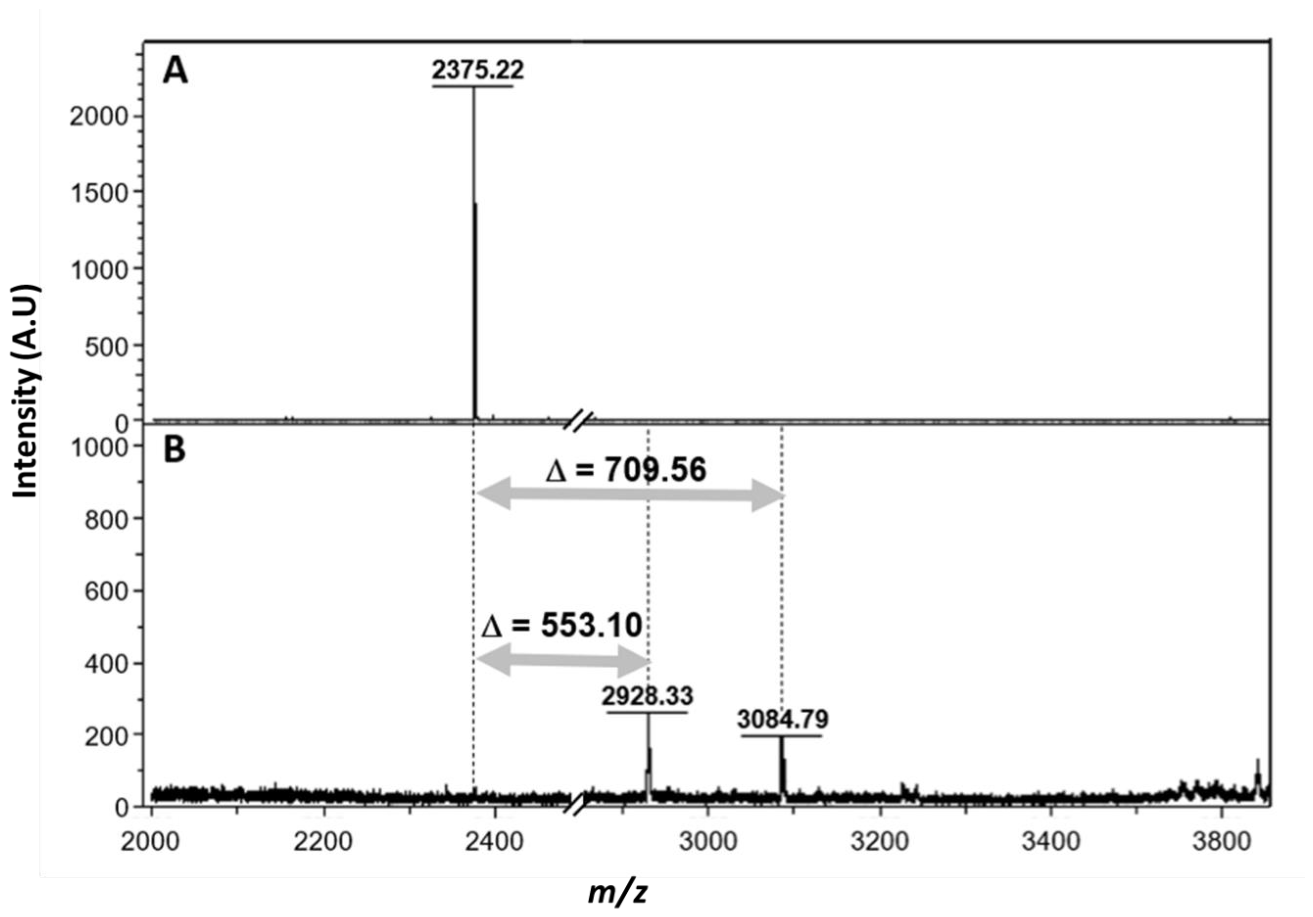
A proteomic approach combined with MALDI-TOF mass spectrometry was used to identify the inhibitor in the F3-1-1 fraction and to investigate the nature of the inhibition mechanism. After trypsin digestion, peptides from untreated and F3-1-1-treated rDGL were analysed by mass spectrometry. For the untreated enzyme, the catalytic peptide L147-K168 containing the catalytic S153 (LHYVGHS153QGTTIGFIAFSTNPK, theoretical mass 2374.21 Da) was detected at [M+H] + = 2375.25 Da (Figure 5A). In contrast, for rDGL pre-incubated with F3-1-1, no mass signal was detected at this m/z ratio, and two new mass signals were detected at m/z = 2928.33 Da and m/z = 3084.78 Da (Figure 5B). In the presence of F3-1-1, an increase in the mass of the catalytic peptide and two mass increments of + 553.10 Da and +709.56 Da were observed compared to the untreated enzyme (Figure 5B). These mass variations between F3-1-1 treated and untreated rDGL could be due to specific coupling between catalytic S153 and a component of F3-1-1. Based on the UPLC-HRMS and MS/MS results of this fraction shown previously, the mass increment of + 553.10 is consistent with the covalent attachment of cinncassiol-related species detected in F3-1-1. Furthermore, it is worth noticing that the F3-1-1-treated enzyme was completely inactivated. The second mass shift corresponding to +709.56 Da, observed in the enzyme treated with F3-1-1, could result from the fixation of cinncassiol A-19 itself linked to two levoglucosan residues previously detected in the fraction.
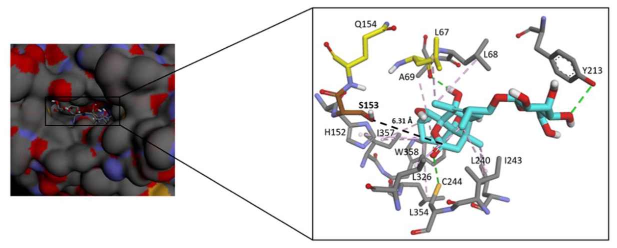
To investigate HGL-cinncassiol A 19 glucoside binding interactions, in silico molecular docking was performed using VINA software (Figure 6). The open conformation of HGL, in complex with THL covalently bound to its catalytic residue Ser 153, was used as the reference model to improve the reliability of covalently binding. The active site of HGL was found to be accessible thus enabling docking experiments. Automated docking resulted in several possible conformations of Cinncassiol A 19 glycoside within the HGL catalytic pocket, with favorable binding energy of −7.6 kcal mol−1. The binding affinity of HGL-Cinncassiol A 19 glycoside complex was maintained with the three hydrogen bonds and nine hydrophobic interactions. For the docking results of the complex THL-HGL, a favorable binding energy of −7.9 kcal mol−1 was found. For subsequent analysis, we retained the best matching conformation that directly exposes the lactone carbonyl residue of the Cinncassiol A 19 glycoside inhibitor towards the oxygen group of Ser 153, within a calculated distance of 6.31 Å which is sufficient to promote the nucleophilic attack. In the case of THL, the distance was found around 2.02 Å which explains the strong inhibitory effect of THL on DGL.
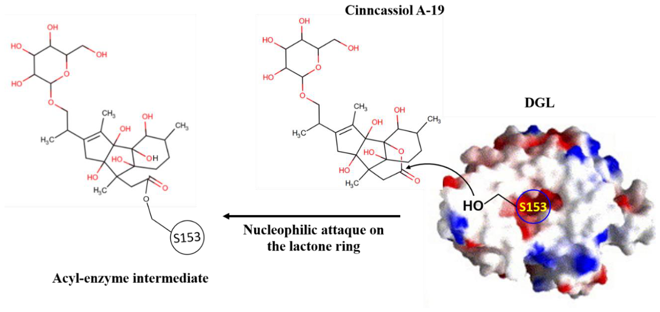
These results along with the observed mass shift in the treated enzyme, clearly confirm the formation of the DGL-inhibitor covalent complex, likely involving the catalytic Ser153 of the enzyme with cinncassiol A-19. Indeed, cinncassiol A-19 glucoside is a diterpene containing a lactone group that can be the target of nucleophilic attack by the catalytic S153 of the enzyme (Figure 7). Other similar interactions between lipase and the inhibitor have been reported in the literature. MALDI-TOF results have previously shown an increase of approximately 500 Da in the molecular mass of HPL, and this increase most likely reflects the covalent bonding of the S152 of the HPL active site and the β-lactone ring of Orlistat (MW=496 Da). Similarly, an enriched extract of star anise revealed the presence of a covalent inhibitor-enzyme complex by MALDI-TOF, with a mass increment of 489.52 Da (Tiss et al., 2009). This specific coupling is probably due to the binding of myricitrin-5-methyl ether (479.11 Da) with rDGL (Kamoun et al., 2019).

4. Conclusions and Perspectives

Using a rigorous bioactivity-guided fractionation workflow, we isolated fraction F3-1-1, which potently inhibits rHPL, PPL, and rDGL, with αI50 values of 7 µg, 32 µg, and 19.5 µg, respectively. UPLC-HRMS/MS analysis identified cinncassiol A-19 glucoside as the major bioactive constituent. For the first time, PMF directly demonstrated that this compound covalently binds to the catalytic Ser153 of gastric lipase, forming stable adducts characterized by mass shifts of +553.10 Da and +709.56 Da.
These findings are particularly significant from a nutritional perspective because gastric lipase plays the key, initiating role in dietary fat digestion. By hydrolyzing TGs, it releases the first free fatty acids, which are required for the subsequent activation of pancreatic lipase. Thus, by inhibiting gastric lipase – the upstream key enzyme – cinncassiol A-19 glucoside not only blocks gastric lipolysis but also prevents the activation of pancreatic lipase, thereby disrupting the entire synergistic action of the two major digestive lipases. This dual covalent inhibition makes cinncassiol A-19 glucoside a highly promising natural candidate for the development of nutraceuticals or pharmaceutical agents targeting dietary fat absorption in the management of obesity.
Further studies are required to assess selectivity, cytotoxicity, stability under simulated gastrointestinal conditions, and, most importantly, in vivo efficacy in animal models of obesity. Total synthesis of cinncassiol A-19 glucoside would be a significant advance to establish precise structure–activity relationships and define optimal dosing regimens.

Supplementary Materials

The following supporting information can be downloaded at the website of this paper posted on Preprints.org.

Funding

This work was funded by the Multilateral Project Partenariats Hubert Curien-Utique (PHC-Utique)-2024-2026 Program of the French Ministry of Higher Education and Research, code Campus France: 50314TG and supported by the Ministry of Higher Education and Scientific Research, Tunisia.

Acknowledgments

We would like to thank Régine Lebrun and Christophe Verthuyand (Aix-Marseille University, CNRS, Institut de Microbiologie de la Méditerranée, Plateforme Protéomique, Fédération de Recherche 3479, Marseille, France Aix-Marseille University, CNRS, Architecture et Fonction des Macromolécules) for PMF analysis. We would like to thank also Caroline Bourgeois and Antoine Vauchez (ICBMS-Lyon), Centre commun de spectrométrie de masse, for the mass spectrometry analysis.

Conflicts of Interest

All authors declare no conflicts of interest.

Abbreviations

BSA, bovine serum albumin; DCM, dichloromethane; DCME, dichloromethane extract; EE, ethanol extract; ESI, electrospray ionization; GA, gum Arabic; GNPS, global natural products social molecular networking; HE, hexane extract; HGL, human gastric lipase; HMDB, human metabolome database; HRMS, high-resolution mass spectrometry; MS/MS, tandem mass spectrometry; NaTDC, sodium taurodeoxycholate; NP-TLC, normal-phase thin-layer chromatography; PMF, peptide mass fingerprinting; PPL, porcine pancreatic lipase; Q-TOF, quadrupole time-of-flight; RA, residual activity; rDGL, recombinant dog gastric lipase; rHPL, recombinant HPL; RP-LC, reversed-phase liquid chromatography; RP-TLC, reversed-phase thin-layer chromatography; RT, retention time; TC4, glyceryl tributyrate; THL, tetrahydrolipstatin; UPLC, ultra-performance liquid chromatography; WE, water extract; αI50, amount of inhibitor required to reduce enzyme activity by 50%; t1/2, half-inactivation time

References

  1. Abdelaal, M.; Le Roux, C. W.; Docherty, N. G. Morbidity and mortality associated with obesity. Ann. Transl. Med. 2017, 5(7), 161–161. [Google Scholar] [CrossRef]
  2. Abdelmottaleb Moussa, S. A.; Ibrahim, F. A. A.; Abd Elbaset, M.; Aziz, S. W.; Abd El-Latif, N. A.; Afifi, S. M.; Esatbeyoglu, T.; El Toumy, S. A.; Salib, J. Y.; Ae Bashandy, S. The potency of Thymus vulgaris seed extract in alleviating obesity complications and iron overload in obese rats. J. Funct. Foods 2024, 121, 106416. [Google Scholar] [CrossRef]
  3. Abeysekera, W. P. K. M.; Arachchige, S. P. G.; Ratnasooriya, W. D. Bark Extracts of Ceylon Cinnamon Possess Antilipidemic Activities and Bind Bile Acids In Vitro. Evid.-Based Complement. Altern. Med. 2017, 2017(1), 7347219. [Google Scholar] [CrossRef]
  4. Aboura, I.; Nani, A.; Belarbi, M.; Murtaza, B.; Fluckiger, A.; Dumont, A.; Benammar, C.; Tounsi, M. S.; Ghiringhelli, F.; Rialland, M.; Khan, N. A.; Hichami, A. Protective effects of polyphenol-rich infusions from carob (Ceratonia siliqua) leaves and cladodes of Opuntia ficus-indica against inflammation associated with diet-induced obesity and DSS-induced colitis in Swiss mice. Biomed. Pharmacother. 2017, 96, 1022–1035. [Google Scholar] [CrossRef]
  5. Aloulou, A.; Carrière, F. Gastric lipase: An extremophilic interfacial enzyme with medical applications. Cell. Mol. Life Sci. 2008, 65(6), 851–854. [Google Scholar] [CrossRef]
  6. Ansari, R.; Zarshenas, M. M.; Dadbakhsh, A. H. A Review on Pharmacological and Clinical Aspects of Linum usitatissimum L. Curr. Drug Discov. Technol. 2019, 16(2), 148–158. [Google Scholar] [CrossRef]
  7. Bahmani, M.; Eftekhari, Z.; Saki, K.; Fazeli-Moghadam, E.; Jelodari, M.; Rafieian-Kopaei, M. Obesity Phytotherapy: Review of Native Herbs Used in Traditional Medicine for Obesity. J. Evid.-Based Complement. Altern. Med. 2016, 21(3), 228–234. [Google Scholar] [CrossRef]
  8. Bergauff, M. A.; Ward, T. J.; Noonan, C. W.; Migliaccio, C. T.; Simpson, C. D.; Evanoski, A. R.; Palmer, C. P. Urinary levoglucosan as a biomarker of wood smoke: Results of human exposure studies. J. Expo. Sci. Environ. Epidemiol. 2010, 20(4), 385–392. [Google Scholar] [CrossRef]
  9. Błaszczyk, N.; Rosiak, A.; Kałużna-Czaplińska, J. The Potential Role of Cinnamon in Human Health. Forests 2021, 12(5), 648. [Google Scholar] [CrossRef]
  10. Blüher, M. Obesity: Global epidemiology and pathogenesis. Nat. Rev. Endocrinol. 2019, 15(5), 288–298. [Google Scholar] [CrossRef]
  11. Borgström, B. Mode of action of tetrahydrolipstatin: A derivative of the naturally occurring lipase inhibitor lipstatin. Biochim. Et. Biophys. Acta (BBA) -Lipids Lipid Metab. 1988, 962(3), 308–316. [Google Scholar] [CrossRef]
  12. Carriere, F.; Barrowman, J. A.; Verger, R.; René, L. Secretion and contribution to lipolysis of gastric and pancreatic lipases during a test meal in humans. Gastroenterology 1993, 105(3), 876–888. [Google Scholar] [CrossRef]
  13. Carrière, F.; Renou, C.; Ransac, S.; Lopez, V.; De Caro, J.; Ferrato, F.; De Caro, A.; Fleury, A.; Sanwald-Ducray, P.; Lengsfeld, H.; Beglinger, C.; Hadvary, P.; Verger, R.; Laugier, R. Inhibition of gastrointestinal lipolysis by Orlistat during digestion of test meals in healthy volunteers. Am. J. Physiol.-Gastrointest. Liver Physiol. 2001, 281(1), G16–G28. [Google Scholar] [CrossRef]
  14. Chao, A. M.; Tronieri, J. S.; Amaro, A.; Wadden, T. A. Semaglutide for the treatment of obesity. Trends Cardiovasc. Med. 2023, 33(3), 159–166. [Google Scholar] [CrossRef]
  15. Cudrey, C.; Van Tilbeurgh, H.; Gargouri, Y.; Verger, R. Inactivation of pancreatic lipases by amphiphilic reagents 5-(dodecyldithio)-2-nitrobenzoic acid and tetrahydrolipstatin. Dependence upon partitioning between micellar and oil phases. Biochemistry 1993, 32(50), 13800–13808. [Google Scholar] [CrossRef]
  16. Farag, M. A.; Kabbash, E. M.; Mediani, A.; Döll, S.; Esatbeyoglu, T.; Afifi, S. M. Comparative Metabolite Fingerprinting of Four Different Cinnamon Species Analyzed via UPLC–MS and GC–MS and Chemometric Tools. Molecules 2022, 27(9), 2935. [Google Scholar] [CrossRef]
  17. Feng, H.; Jiang, Y.; Cao, H.; Shu, Y.; Yang, X.; Zhu, D.; Shao, M. Chemical characteristics of the sesquiterpenes and diterpenes from Lauraceae family and their multifaceted health benefits: A review. Heliyon 2022, 8(12), e12013. [Google Scholar] [CrossRef]
  18. Fernandez, S.; Jannin, V.; Rodier, J.-D.; Ritter, N.; Mahler, B.; Carrière, F. Comparative study on digestive lipase activities on the self emulsifying excipient Labrasol®, medium chain glycerides and PEG esters. Biochim. Et. Biophys. Acta (BBA) -Mol. Cell Biol. Lipids 2007, 1771(5), 633–640. [Google Scholar] [CrossRef]
  19. Gómez-Ambrosi, J.; Catalán, V.; Frühbeck, G. The evolution of the understanding of obesity over the last 100 years. Int. J. Obes. 2025, 49(2), 168–176. [Google Scholar] [CrossRef]
  20. Gruz, J.; Novák, O.; Strnad, M. Rapid analysis of phenolic acids in beverages by UPLC–MS/MS. Food Chem. 2008, 111(3), 789–794. [Google Scholar] [CrossRef]
  21. Jakkawanpitak, C.; Wunnoo, S.; Sermwittayawong, D.; Runsaeng, P.; Amnuaikit, T.; Voravuthikunchai, S. P. Quercus infectoria gall extract modulates adipocyte differentiation and lipid metabolism through dual regulation of adipogenesis and lipolysis in 3T3-L1 adipocytes. Anim. Cells Syst. 2026, 30(1), 161–182. [Google Scholar] [CrossRef]
  22. Jaradat, N.; Abualhasan, M.; Hawash, M.; Qadi, M.; Al-Maharik, N.; Abdallah, S.; Mousa, A.; Zarour, A.; Arar, M.; Sobuh, S.; Hussein, F.; Issa, L.; Jaber, A.; Hamduni, H.; Alshahatit, S. Chromatography analysis, in light of vitro antioxidant, antidiabetic, antiobesity, anti-inflammatory, antimicrobial, anticancer, and three-dimensional cancer spheroids’ formation blocking activities of Laurus nobilis aromatic oil from Palestine. Chem. Biol. Technol. Agric. 2023, 10(1), 25. [Google Scholar] [CrossRef]
  23. Jia, X.-C.; Li, J.; Yu, Z.-R.; Zhang, H.; Zhou, L. 3,4,5-Trimeth oxy phenol. Acta Crystallogr. Sect. E Struct. Rep. Online 2012, 68(11), o3160–o3160. [Google Scholar] [CrossRef]
  24. Kamoun, J.; Rahier, R.; Sellami, M.; Koubaa, I.; Mansuelle, P.; Lebrun, R.; Berlioz-Barbier, A.; Fiore, M.; Alvarez, K.; Abousalham, A.; Carrière, F.; Aloulou, A. Identification of a new natural gastric lipase inhibitor from star anise. Food Funct. 2019, 10(1), 469–478. [Google Scholar] [CrossRef]
  25. Khaleghi, A. A.; Salari, N.; Darvishi, N.; Bokaee, S.; Jafari, S.; Hemmati, M.; Mohammadi, M. Global prevalence of obesity in the older adults: A meta-analysis. Public Health Pract. 2025, 9, 100585. [Google Scholar] [CrossRef] [PubMed]
  26. Kim, M.; Van, J. Y.; Park, S.; Hwang, J. Y.; Seong, Y.; Park, Y. J.; Lee, S.; Ryu, B.; Jun, J. Y.; Jung, W.; Lee, B. Upcycling Agricultural By-Product: Therapeutic Potential of Oriental Melon Stem ( Cucumis melo var. makuwa ) in Treating Obesity and Fatty Liver via Enhanced Thermogenesis. Food Front. 2026, 7(2), e70246. [Google Scholar] [CrossRef]
  27. Lengsfeld, H.; Beaumier-Gallon, G.; Chahinian, H.; De Caro, A.; Verger, R.; Laugier, R.; Carrière, F. Physiology of Gastrointestinal Lipolysis and Therapeutical Use of Lipases and Digestive Lipase Inhibitors. In Lipases and Phospholipases in Drug Development, 1st ed.; Müller, G., Petry, S., Eds.; Wiley, 2004; pp. 195–229. [Google Scholar] [CrossRef]
  28. Liu, T.-T.; Liu, X.-T.; Chen, Q.-X.; Shi, Y. Lipase Inhibitors for Obesity: A Review. Biomed. Pharmacother. 2020, 128, 110314. [Google Scholar] [CrossRef]
  29. Lorenzi, M.; Puppo, C.; Lebrun, R.; Lignon, S.; Roubaud, V.; Martinho, M.; Mileo, E.; Tordo, P.; Marque, S. R. A.; Gontero, B.; Guigliarelli, B.; Belle, V. Tyrosine-Targeted Spin Labeling and EPR Spectroscopy: An Alternative Strategy for Studying Structural Transitions in Proteins. Angew. Chem. Int. Ed. 2011, 50(39), 9108–9111. [Google Scholar] [CrossRef]
  30. Lowe, M. E.; Rosenblum, J. L.; Strauss, A. W. Cloning and Characterization of Human Pancreatic Lipase cDNA. J. Biol. Chem. 1989, 264(33), 20042–20048. [Google Scholar] [CrossRef]
  31. MassBank of North America. (n.d.). Retrieved April 12, 2026. Available online: https://mona.fiehnlab.ucdavis.edu/spectra/display/EAD000067.
  32. Müller, G.; Petry, S. Lipases and Phospholipases in Drug Development: From Biochemistry to Molecular Pharmacology; John Wiley & Sons.Google-Books-ID: S9XCjRJznxIC, 2006. [Google Scholar]
  33. Nothias, L.-F.; Nothias-Esposito, M.; Da Silva, R.; Wang, M.; Protsyuk, I.; Zhang, Z.; Sarvepalli, A.; Leyssen, P.; Touboul, D.; Costa, J.; Paolini, J.; Alexandrov, T.; Litaudon, M.; Dorrestein, P. C. Bioactivity-Based Molecular Networking for the Discovery of Drug Leads in Natural Product Bioassay-Guided Fractionation. J. Nat. Prod. 2018, 81(4), 758–767. [Google Scholar] [CrossRef]
  34. Okabe, N.; Kyoyama, H. 2,3-Dihydroxybenzoic acid. Acta Crystallogr. Sect. E Struct. Rep. Online 2001, 57(12), o1224–o1226. [Google Scholar] [CrossRef]
  35. Oketch-Rabah, H. A.; Marles, R. J.; Brinckmann, J. A. Cinnamon and Cassia Nomenclature Confusion: A Challenge to the Applicability of Clinical Data. Clin. Pharmacol. Ther. 2018, 104(3), 435–445. [Google Scholar] [CrossRef]
  36. Omonkhua, A. A.; Otitolaiye, C.; Aguebor-Ogie, B.; Elekofehinti, O.; Okello, E.; Onoagbe, I.; Okonofua, F. Anti-diabetic, anti-pancreatic lipase, and anti-protein glycation potential of Irvingia gabonensis stem bark extracts: In vitro and in silico studies. Silico Pharmacol. 2024, 12(1), 43. [Google Scholar] [CrossRef]
  37. Özokan, G.; Bilginer, A.; Gümüş, M. K. Synthesis of Natural Carboxylic Acids and Alcohols from Cinnamon cassia Oil via Green Chemistry. Processes 2025, 13(7), 2002. [Google Scholar] [CrossRef]
  38. Peddio, S.; Padiglia, A.; Cannea, F. B.; Crnjar, R.; Zam, W.; Sharifi-Rad, J.; Rescigno, A.; Zucca, P. Common bean (Phaseolus vulgaris L.) α-amylase inhibitors as safe nutraceutical strategy against diabetes and obesity: An update review. Phyther. Res. 2022, 36(7), 2803–2823. [Google Scholar] [CrossRef]
  39. Pham, V. C.; Nguyen, T. T. A.; Vu, T. O.; Cao, T. Q.; Min, B. S.; Kim, J. A. Five new diterpenoids from the barks of Cinnamomum cassia (L.) J. Presl. Phytochem. Lett. 2019, 32, 23–28. [Google Scholar] [CrossRef]
  40. Phelps, N. H.; Singleton, R. K.; Zhou, B.; Heap, R. A.; Mishra, A.; Bennett, J. E.; Paciorek, C. J.; Lhoste, V. P.; Carrillo-Larco, R. M.; Stevens, G. A.; Rodriguez-Martinez, A.; Bixby, H.; Bentham, J.; Di Cesare, M.; Danaei, G.; Rayner, A. W.; Barradas-Pires, A.; Cowan, M. J.; Savin, S.; …; Ezzati, M. Worldwide trends in underweight and obesity from 1990 to 2022: A pooled analysis of 3663 population-representative studies with 222 million children, adolescents, and adults. The Lancet 2024, 403(10431), 1027–1050. [Google Scholar] [CrossRef]
  41. Phillips, S. D.; Roberts, W. C. Comparison of Body Mass Index Among Patients With Versus Without Angiographic Coronary Artery Disease. Am. J. Cardiol. 2007, 100(1), 18–22. [Google Scholar] [CrossRef]
  42. Roussel, A.; Miled, N.; Berti-Dupuis, L.; Rivière, M.; Spinelli, S.; Berna, P.; Gruber, V.; Verger, R.; Cambillau, C. Crystal Structure of the Open Form of Dog Gastric Lipase in Complex with a Phosphonate Inhibitor *. J. Biol. Chem. 2002, 277(3), 2266–2274. [Google Scholar] [CrossRef]
  43. Ruiz, P. L.; Karlstad, Ø.; Nøkleby, K.; Slåtsve, K.; Gulseth, H. L.; Meyer, H. E.; Sveen, K. A.; Qvigstad, E.; Furu, K. Pharmacological treatment of obesity in adults in Norway 2004–2022. Diabetes Obes. Metab. 2024, 26(6), 2102–2110. [Google Scholar] [CrossRef]
  44. Saleem, H.; Yaqub, A.; Rafique, R.; Ali Chohan, T.; Malik, D.-S.; Tousif, M. I.; Khurshid, U.; Ahemad, N.; Ramasubburayan, R.; Rengasamy, K. R. Nutritional and medicinal plants as potential sources of enzyme inhibitors toward the bioactive functional foods: An updated review. Crit. Rev. Food Sci. Nutr. 2024, 64(27), 9805–9828. [Google Scholar] [CrossRef]
  45. Sall, D.; Wang, J.; Rashkin, M.; Welch, M.; Droege, C.; Schauer, D. Orlistat-induced fulminant hepatic failure. Clin. Obes. 2014, 4(6), 342–347. [Google Scholar] [CrossRef] [PubMed]
  46. Sankpal, S.; Pathade, K. S.; Kondawar, M. Coffea arabica: Herbal drug on Global health issue - Obesity. Asian J. Res. Pharm. Sci. 2021, 11(1), 29–34. [Google Scholar] [CrossRef]
  47. Scheen, A. J. Cardiovascular Risk-Benefit Profile of Sibutramine. Am. J. Cardiovasc. Drugs 2010, 10(5), 321–334. [Google Scholar] [CrossRef]
  48. Sellami, M.; Louati, H.; Kamoun, J.; Kchaou, A.; Damak, M.; Gargouri, Y. Inhibition of pancreatic lipase and amylase by extracts of different spices and plants. Int. J. Food Sci. Nutr. 2017, 68(3), 313–320. [Google Scholar] [CrossRef]
  49. Song, M. Y.; Kang, S. Y.; Kang, A.; Hwang, J. H.; Park, Y.-K.; Jung, H. W. Cinnamomum cassia Prevents High-Fat Diet-Induced Obesity in Mice through the Increase of Muscle Energy. Am. J. Chin. Med. 2017, 45(05), 1017–1031. [Google Scholar] [CrossRef]
  50. Spieker, E. A.; Pyzocha, N. Economic Impact of Obesity. Prim. Care Clin. Off. Pract. 2016, 43(1), 83–95. [Google Scholar] [CrossRef]
  51. Subramaniyan, V.; Hanim, Y. U. Role of pancreatic lipase inhibition in obesity treatment: Mechanisms and challenges towards current insights and future directions. Int. J. Obes. 2025, 49(3), 492–506. [Google Scholar] [CrossRef]
  52. The GBD 2015 Obesity Collaborators. Health Effects of Overweight and Obesity in 195 Countries over 25 Years. N. Engl. J. Med. 2017, 377(1), 13–27. [Google Scholar] [CrossRef]
  53. Tiss, A.; Carrière, F.; Verger, R. Effects of Gum Arabic on Lipase Interfacial Binding and Activity. Anal. Biochem. 2001, 294(1), 36–43. [Google Scholar] [CrossRef]
  54. Tiss, A.; Lengsfeld, H.; Carrière, F.; Verger, R. Inhibition of human pancreatic lipase by tetrahydrolipstatin: Further kinetic studies showing its reversibility. J. Mol. Catal. B Enzym. 2009, 58(1), 41–47. [Google Scholar] [CrossRef]
  55. Tiss, A.; Lengsfeld, H.; Verger, R. Étude cinétique comparative des lipases pancréatiques humaines et de Thermomyces lanuginosa: Effets inhibiteurs de la tétrahydrolipstatine en présence de substrats lipidiques. J. Mol. Catal. B Enzym. 2010, 62(1), 19–26. [Google Scholar] [CrossRef]
  56. Vijayakumar, K.; Prasanna, B.; Rengarajan, R. L.; Rathinam, A.; Velayuthaprabhu, S.; Vijaya Anand, A. Anti-diabetic and hypolipidemic effects of Cinnamon cassia bark extracts: An in vitro, in vivo, and in silico approach. Arch. Physiol. Biochem. 2023, 129(2), 338–348. [Google Scholar] [CrossRef]
  57. Wang, F.; Liigand, J.; Tian, S.; Arndt, D.; Greiner, R.; Wishart, D. S. CFM-ID 4.0: More Accurate ESI-MS/MS Spectral Prediction and Compound Identification. Anal. Chem. 2021, 93(34), 11692–11700. [Google Scholar] [CrossRef]
  58. Wang, J.; Ke, W.; Bao, R.; Hu, X.; Chen, F. Beneficial effects of ginger Zingiber officinale Roscoe on obesity and metabolic syndrome: A review. Ann. N. Y. Acad. Sci. 2017, 1398(1), 83–98. [Google Scholar] [CrossRef]
  59. Wang, W.; Barkat, M. Q.; Amin, S.; Alshammari, A.; Albekairi, N. A.; Manzoor, H.; Chauhdary, Z.; Bazmi, R. R.; Hussain, L.; Chen, Q. Pharmacological Appraisal of Aqueous Ethanolic Extract of Cuminum cyminum (Cumin Seeds) in Obesity and Endocrinopathies Associated With Polycystic Ovarian Syndrome in Animal Models. J. Food Biochem. 2024, 2024(1), 1184854. [Google Scholar] [CrossRef]
  60. Watanabe, M.; Laviano, A.; Balena, A.; Vitaterna, A.; Angeloni, E.; Toscano, R.; Natoli, G.; Lubrano, C.; Gnessi, L. Effect on Weight Loss of an Oral Supplement Containing Cinnamon Bark (Cinnamomum cassia) and Withania somnifera in Adult Patients with Overweight and Obesity: A Pilot Study. J 2023, 6(3), 508–516. [Google Scholar] [CrossRef]
  61. Watcharachaisoponsiri, T.; Thiyajai, P.; Charoenkiatkul, S.; Temviriyanukul, P.; Chupeerach, C.; Suttisansanee, U. The Screening of Anti-lipase Activity in Thai Local Chili Peppers. Thai J. Toxicol. 2017, 32(1), 35–52. Available online: https://li01.tci-thaijo.org/index.php/ThaiJToxicol/article/view/244133.
  62. Weill, A.; Païta, M.; Tuppin, P.; Fagot, J.; Neumann, A.; Simon, D.; Ricordeau, P.; Montastruc, J.; Allemand, H. Benfluorex and valvular heart disease: A cohort study of a million people with diabetes mellitus. Pharmacoepidemiol. Drug Saf. 2010, 19(12), 1256–1262. [Google Scholar] [CrossRef]
  63. Wilding, J. P. H.; Batterham, R. L.; Calanna, S.; Davies, M.; Van Gaal, L. F.; Lingvay, I.; McGowan, B. M.; Rosenstock, J.; Tran, M. T. D.; Wadden, T. A.; Wharton, S.; Yokote, K.; Zeuthen, N.; Kushner, R. F. Once-Weekly Semaglutide in Adults with Overweight or Obesity. N. Engl. J. Med. 2021, 384(11), 989–1002. [Google Scholar] [CrossRef] [PubMed]
  64. Zehl, M.; Leščić, I.; Abramić, M.; Rizzi, A.; Kojić-Prodić, B.; Allmaier, G. Characterization of covalently inhibited extracellular lipase from Streptomyces rimosus by matrix-assisted laser desorption/ionization time-of-flight and matrix-assisted laser desorption/ionization quadrupole ion trap reflectron time-of-flight mass spectrometry: Localization of the active site serine. J. Mass Spectrom. 2004, 39(12), 1474–1483. [Google Scholar] [CrossRef]
  65. Zhang, C.; Fan, L.; Fan, S.; Wang, J.; Luo, T.; Tang, Y.; Chen, Z.; Yu, L. Cinnamomum cassia Presl: A Review of Its Traditional Uses, Phytochemistry, Pharmacology and Toxicology. Molecules 2019, 24(19), 3473. [Google Scholar] [CrossRef] [PubMed]
  66. Zhang, X.; Liu, J.; Ni, Y.; Yi, C.; Fang, Y.; Ning, Q.; Shen, B.; Zhang, K.; Liu, Y.; Yang, L.; Li, K.; Liu, Y.; Huang, R.; Li, Z. Global Prevalence of Overweight and Obesity in Children and Adolescents: A Systematic Review and Meta-Analysis. JAMA Pediatr. 2024, 178(8), 800. [Google Scholar] [CrossRef]
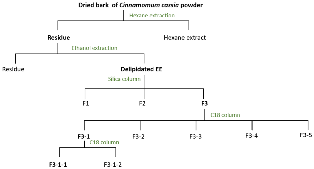
Figure 1. Bioactivity-guided fractionation scheme of the crude ethanolic extract of Cinnamomum cassia bark. The workflow illustrates the sequential extraction, defatting, silica gel column chromatography, reversed-phase C18 column chromatography steps, and the corresponding inhibitory activity against PPL measured at each stage. The most active fraction (F3-1-1) is highlighted.
Preprints 229588 g001
Figure 2. Inhibitory activities of four extracts of C. cassia against PPL using TC4 as substrate. HE, DCME, EE, and WE were tested at a final concentration of 1 mg mL-1. RA (%) is expressed relative to a control without inhibitor (set to 100%). Data represent mean ± SD (n = 3).
Preprints 229588 g002
Figure 3. Evaluation of the inhibitory effect of fraction F3-1-1 against digestive lipases: rDGL, rHPL, and PPL. (A) Dose-dependent inhibition. Each lipase was pre-incubated with various amounts of F3-1-1 for 30 min at 25 °C in the presence of 4 mM NaTDC. RA (%) was measured at 37 °C using TC4 as substrate. (B) Time-dependent inhibition. Each lipase was pre-incubated with a constant amount of F3-1-1 (rHPL: 3 µg; PPL: 35 µg; rDGL: 20 µg) at 25 °C for increasing time periods (0 to 60 min). RA (%) was then measured as above. Data are means ± SD (n = 3).
Preprints 229588 g003
Figure 4. Effect of fraction F3-1-1 on the rate of TC4 hydrolysis by rHPL (A) and PPL (B) during ongoing lipolysis. After addition of the enzyme (0.5 µM, final concentration) to the reaction medium, fraction F3-1-1 at variable amounts (10–70 µg) was injected 4 minutes later (arrow). Assays were performed at 37 °C in a thermostated vessel containing 0.2 mL TC4, 2.5 mM Tris-HCl buffer (pH 8), 2 mM CaCl2, and 0.5 mM NaTDC. The rate of fatty acid release was monitored continuously by pH-stat. Each kinetic trace is representative of at least three independent experiments.
Preprints 229588 g004
Figure 5. PMF spectra of untreated (A) and treated (B) rDGL with F3-1-1. A 30 min incubation of 2 µM of rDGL was performed at 25 °C in the presence or absence of a F3-1-1 (100 µg). The region of the unmodified isotopic catalytic peptide LHYVGHS153QGTTIGFIAFSTNPK containing the catalytic Ser153 and detected at [M−H] + m/z 2375.22 Da (A). This peak disappears in the treated enzyme, where two peptides are detected at 2928.33 Da and 3084.78 Da (B), resulting from the covalent binding of the inhibitor of interest in F3-1-1 to the catalytic S153. Mass shift was calculated as the difference between the experimental m/z of the modified peptide and of the unmodified catalytic peptide.
Preprints 229588 g005
Figure 6. Visualization of the HGL-cinncassiol A-19 glucoside binding interaction by molecular docking. (A) Molecular surface representation of the HGL active site crevice with the bound cinncassiol A-19 glycoside and stabilized by hydrogen bonding and hydrophobic contacts. O(seryl)-C(carbonyl) bond shown as a white dotted line. Residues of the oxyanion hole (L67, Q254) shown in yellow and the catalytic serine residue (S153) shown in brown. Hydrophobic interactions shown as pink dotted lines, hydrogen bonds shown as green dotted lines.
Preprints 229588 g006
Figure 7. Proposed mechanism of covalent inhibition of gastric lipase by cinncassiol A-19 glucoside. The hydroxyl group of the catalytic Ser153 performs a nucleophilic attack on the carbonyl carbon of the lactone ring present in cinncassiol A-19 glucoside. This leads to the formation of a stable acyl-enzyme covalent intermediate, which inactivates the enzyme. The same mechanism is proposed for the di-glycosylated form (cinncassiol A-19 diglucoside). R represents the glucosyl or diglucosyl moiety. This mechanism is analogous to that of Orlistat.
Preprints 229588 g007
Table 1. Inhibitory effect of different medicinal plant extract against PPL.
Plant PPL Residual Activity (%)
Cinnamonum cassia 11
Ceratonia siliqua 38
Quercus macrocarpa 74
Coffea arabica 51
Cuminum cyminum 62
Zingiber officinale 81
Linum usitatissimum 53
Cucumis melo 96
Thymus vulgaris 86
Table 2. αI50 and t½ values of F3-1-1 on rHPL, PPL and rDGL. t½ was determined by incubating 8 µg, 35 µg and 20 µg of F3-1-1 with rHPL, PPL and rDGL, respectively. Results were expressed as mean values of at least three independent experiments.
Enzyme rHPL PPL rDGL
αI50 (µg) 7 32 19.5
t1/2 (min) 17 16 25
Disclaimer/Publisher’s Note: The statements, opinions and data contained in all publications are solely those of the individual author(s) and contributor(s) and not of MDPI and/or the editor(s). MDPI and/or the editor(s) disclaim responsibility for any injury to people or property resulting from any ideas, methods, instructions or products referred to in the content.
Copyright: This open access article is published under a Creative Commons CC BY 4.0 license, which permit the free download, distribution, and reuse, provided that the author and preprint are cited in any reuse.