

Submitted:
01 November 2026
Posted:
03 November 2026
You are already at the latest version
Abstract
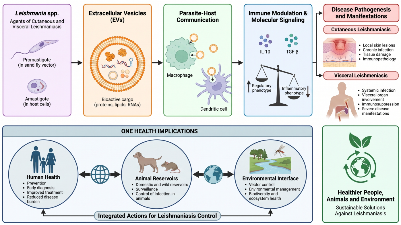
Urban visceral leishmaniasis is increasingly reported across Amazonian municipalities, where diagnostic complexity and parasite biology challenge effective surveillance and control. This study combined serological, parasitological, molecular, and sequence-based analyses of canine samples from four municipalities in the Brazilian Amazon with ultrastructural analysis of a Leishmania (L.) amazonensis reference strain. Diagnostic agreement between the Dual Path Platform rapid test (TR DPP®) and parasitological examination olymph-node aspirates was evaluated in 1,499 dogs using Cohen's kappa coefficient. Agreement was substantial (κ = 0.641; 95% CI: 0.604–0.677), although 275 dogs showed discordant results, consistent with distinct biological stages and diagnostic windows of infection. In a separate molecular subset of 130 dogs, polymerase chain reaction (PCR) targeting kinetoplast DNA (kDNA) and small subunit ribosomal DNA (SSU rDNA) revealed different detection patterns in blood and conjunctival swab samples. SSU rDNA sequencing identified a small number of nucleotide polymorphisms consistent with limited sequence variation, and sequence comparison and distance-based analyses indicated high overall similarity among the obtained sequences. Transmission electron microscopy showed lipid-associated inclusions, vacuolar compartments, and vesicle-like structures, which were described qualitatively on the basis of morphological criteria only. Because these datasets derive from complementary but distinct sources, they are presented as an integrative analytical framework rather than as evidence of mechanistic links within a single sample set. Together, the findings indicate that canine visceral leishmaniasis in Amazonian urban settings involves diagnostic discordance, matrix-dependent molecular detection, and limited SSU rDNA sequence variation, complemented by ultrastructural parasite features consistent with previous descriptions, with implications for surveillance and diagnostic strategies in endemic regions.
Keywords:
Leishmania infantum
; canine reservoir
; serodiagnosis
; kinetoplast DNA
; SSU rDNA
; transmission electron microscopy
; Amazon basin
1. Introduction
Visceral leishmaniasis (VL) remains one of the most important vector-borne zoonoses worldwide and continues to represent a major public health challenge in endemic regions [1,31]. In the Brazilian Amazon, environmental change, urban expansion, deforestation, and the adaptation of transmission cycles to anthropized settings have been associated with the emergence and persistence of VL in urban and peri-urban areas [5,6,7,26]. The epidemiology of leishmaniasis is shaped by complex interactions involving parasite species, vertebrate reservoirs, sand fly vectors, environmental changes, and human activities [1,5,31]. Domestic dogs constitute the principal domestic reservoir of Leishmania infantum and play a central role in maintaining transmission within endemic communities [29,35,36,41]. Despite advances in surveillance and control programmes, substantial clinical heterogeneity and diagnostic uncertainty continue to limit the identification of infected animals and complicate disease management strategies [9,10,16].
The genus Leishmania comprises a diverse group of protozoan parasites with distinct geographical distributions, transmission cycles, and clinical manifestations [34]. Parasite development within the sand fly vector involves complex differentiation processes culminating in the emergence of infective forms capable of transmission to mammalian hosts [13,14]. Understanding these biological processes is essential for improving surveillance strategies and control programmes in endemic settings.
Diagnostic approaches interrogate distinct biological compartments of infection and therefore provide complementary information. Serological assays primarily detect host humoral responses and may remain positive despite fluctuating or low parasite burdens, whereas parasitological examination provides direct evidence of infection through the microscopic detection of amastigote forms [9,10,16]. Molecular methods have further improved diagnostic sensitivity and specificity, particularly in samples with low parasite loads [10]. Nevertheless, discrepancies among serological, parasitological, and molecular findings remain frequent, highlighting the biological complexity of canine visceral leishmaniasis and the limitations associated with individual diagnostic approaches [9,10,16].
Recent advances in molecular biology, population genetics, and ultrastructural analysis have expanded current understanding of Leishmania biology and host-parasite interactions. Molecular studies have demonstrated that Leishmania populations may exhibit detectable levels of genetic variation despite an overall conserved phylogenetic structure [23,24,25,28]. Such variation may result from genomic plasticity, population structuring, or local evolutionary processes acting under different ecological conditions [23,24,25,28]. Although the biological implications of these patterns are not yet fully understood, their investigation contributes to a broader understanding of parasite diversity, adaptation, and transmission dynamics.
At the cellular level, ultrastructural studies have identified lipid droplets, membrane-bound vesicles, vacuolar compartments, flagellar-pocket-associated structures, and lysosome-related organelles as recurring features of Leishmania physiology [4,12,15,18]. These structures have been associated with cellular metabolism, differentiation, intracellular survival, stress adaptation, and parasite development within distinct host environments [4,12,15,18]. Vesicle-like structures associated with the flagellar pocket have also attracted increasing attention because of their potential role in cellular communication and membrane trafficking pathways [2,3,8,20,21,27,32,37,38,39].
Although epidemiological investigations [1,5,6,7,26,29,35,36], diagnostic studies [9,10,16], molecular surveys [10,17,23,24,25,28], ultrastructural analyses [4,12,15,18], and studies addressing vesicle-associated processes in Leishmania biology [2,3,8,20,21,27,32,37,38,39] have generated important insights into canine visceral leishmaniasis, these dimensions are commonly investigated independently. Consequently, relatively little information is available regarding how epidemiological patterns, diagnostic concordance, molecular detection profiles, sequence variation, and ultrastructural characteristics collectively contribute to the understanding of parasite persistence in endemic Amazonian settings.
The present study integrates epidemiological, diagnostic, molecular, and sequence-based datasets derived from canine populations in municipalities of the Brazilian Amazon, complemented by ultrastructural observations of a Leishmania (L.) amazonensis reference strain. Because these datasets originated from complementary but analytically distinct sources, associations among findings should be interpreted as an integrative analytical framework rather than direct within-sample mechanistic relationships. The sampling and diagnostic workflow of the study is summarized in the sampling flowchart presented in Supplementary Figure S1.
A subset of samples included in the molecular component of this investigation was previously analysed for molecular detection purposes by our group [17]. To ensure transparency regarding dataset provenance, this overlap is explicitly acknowledged. However, the scientific objectives, analytical scope, and biological interpretation of the present study differ substantially from those of the previous publication. Whereas Gabriel et al. [17] focused primarily on pathogen detection and preliminary molecular characterization, the current investigation incorporates diagnostic concordance analyses, tissue-specific molecular detection, SSU rDNA sequence comparison, genetic variation assessment, and ultrastructural characterization. Importantly, the overlap concerns sample origin and does not involve duplication of scientific objectives, analytical outputs, biological interpretations, or study conclusions. The relationship between both investigations is detailed in Supplementary Table S1.
Accordingly, the objective of this study was to evaluate diagnostic concordance, molecular detection patterns, and SSU rDNA sequence variation of L. infantum circulating in the Brazilian Amazon, complemented by the ultrastructural characterization of Leishmania developmental forms. By integrating evidence generated at epidemiological, molecular, cellular, and subcellular levels, this investigation seeks to provide a broader understanding of canine visceral leishmaniasis and factors associated with parasite persistence in endemic urban settings.
2. Materials and Methods
2.1. Study Areas and Sampling
We conducted a cross-sectional study in four municipalities of the Brazilian Amazon (Figure 1): Parauapebas (06°04′03″ S, 49°54′08″ W; 18 m above sea level; 7,077.2 km²), Belém (01°27′21″ S, 48°30′16″ W; 10 m; 1,070.1 km²), Marabá (05°22′07″ S, 49°07′04″ W; 84 m; 15,157.9 km²), and Colares (00°56′12″ S, 48°16′54″ W; 15 m; 612.5 km²). These municipalities represent distinct urban and epidemiological settings within the eastern Brazilian Amazon and have previously reported canine and human visceral leishmaniasis cases [1,7].
The study incorporated two complementary but analytically distinct datasets. The first dataset comprised 1,499 domestic dogs examined in Parauapebas between 2019 and 2020 during routine municipal surveillance activities. Animals were georeferenced according to administrative zones and clinically evaluated following the updated LeishVet guidelines for canine leishmaniasis [41]. This cohort was used for epidemiological, clinical, serological, parasitological, and diagnostic concordance analyses.
The second dataset comprised 130 dogs sampled between 2015 and 2018 in Belém, Marabá, and Colares. Samples from this cohort were used for molecular detection, sequencing, sequence comparison analyses, and parasite characterization. Ultrastructural investigations, including the morphological evaluation of vesicle-like structures, were conducted separately using a reference strain of Leishmania (L.) amazonensis (Section 2.5) and were not derived from either canine cohort. Consequently, not all analytical components presented in this study originated from the same animals or the same experimental cohort. Rather, the epidemiological, molecular, and ultrastructural datasets should be interpreted as complementary sources of evidence integrated within a common analytical framework. The sampling and diagnostic workflow is summarized in the sampling flowchart presented in Supplementary Figure S1, while a detailed comparison of datasets and analytical components is provided in Supplementary Table S1.
All procedures were conducted in accordance with Brazilian regulations governing animal research and municipal surveillance programmes (UFPA CEUA 8044181219; SISBIO 39285; UFRA CEUA 034/2014).
A subset of samples included in the molecular dataset was previously used for molecular detection analyses reported by Gabriel et al. [17]. To ensure transparency regarding dataset provenance, this overlap is explicitly acknowledged. However, the present investigation was designed to address different scientific objectives and incorporates analytical components that were not included in the previous publication, including diagnostic concordance analyses, tissue-specific molecular detection, SSU rDNA sequence comparison and variation analyses, and ultrastructural characterization, including the morphological evaluation of vesicle-like structures. Accordingly, sample reuse was restricted to the generation of new analytical outputs and biological interpretations beyond the pathogen detection and preliminary molecular characterization previously reported [17]. Importantly, the overlap between studies concerns sample origin and does not involve duplication of scientific objectives, analytical outputs, biological interpretations, or study conclusions. A detailed comparison between both investigations is presented in Supplementary Table S1.
2.2. Serological and Parasitological Diagnostics
Serological screening was performed using the TR DPP® rapid test according to the manufacturer’s instructions and previously validated protocols for canine visceral leishmaniasis [9,16]. Parasitological examination was conducted using lymph node aspirates. Lymph-node smears were air-dried, methanol-fixed, stained with Giemsa, and examined under light microscopy following standard parasitological procedures. Samples were considered positive when at least one intracellular amastigote was visualized, providing direct parasitological evidence of infection [16].
Diagnostic agreement between TR DPP® and parasitological examination was evaluated using a 2 × 2 contingency table in which animals were classified as DPP-positive/parasitology-positive, DPP-positive/parasitology-negative, DPP-negative/parasitology-positive, or DPP-negative/parasitology-negative. Concordance between diagnostic methods was quantified using Cohen’s kappa coefficient (κ) and interpreted according to established criteria [42].
2.3. Molecular Detection (kDNA and SSU rDNA)
Peripheral blood samples (approximately 4 mL, collected by cephalic venipuncture into EDTA tubes) and bilateral conjunctival swabs were obtained from the dogs included in the molecular subset (n = 130; Belém, n = 49; Marabá, n = 50; Colares, n = 31). Blood DNA extraction was performed using the AxyPrep™ Blood Genomic DNA Miniprep Kit (Axygen Biosciences, Union City, CA, USA) according to the manufacturer’s instructions. Conjunctival swab DNA was extracted using the NaCl extraction method described by Abrão et al. (2005). Extracted DNA was stored at −20 °C until further analysis.
Molecular detection of Leishmania infantum was performed using two complementary molecular targets: kinetoplast DNA (kDNA), selected because of its high analytical sensitivity, and small subunit ribosomal DNA (SSU rDNA), a conserved nuclear ribosomal gene selected for sequence-based characterization.
2.3.1. kDNA PCR
Amplification of Leishmania minicircle kinetoplast DNA was performed using primers Leish1 (5′-AACTTTTCTGGTCCTCCGGGTAG-3′) and Leish2 (5′-ACCCCCAGTTTCCCGCC-3′).
PCR reactions were performed in a final volume of 25 μL containing 10-20 ng of template DNA, 1.0 mM MgCl₂, 1.0 mM dNTPs, 10 mM Tris-HCl, 50 mM KCl, 1 μM of each primer, and 0.2 U Taq DNA polymerase (Invitrogen, Carlsbad, CA, USA).
Thermal cycling consisted of an initial denaturation step at 94 °C for 3 min, followed by 40 cycles of 94 °C for 30 s, 58 °C for 30 s, and 72 °C for 30 s, with a final extension at 72 °C for 5 min.
2.3.2. SSU rDNA PCR
SSU rDNA amplification was performed using nested PCR strategies.
The first amplification employed primers R221 (5′-GGTTCCTTTCCTGATTTACG-3′) and R332 (5′-GGCCGTAAAGGCCGAATAG-3′). Reactions were performed in a final volume of 25 μL containing 10-20 ng of DNA, 1.0 mM MgCl₂, 2.0 mM dNTPs, 10 mM Tris-HCl, 50 mM KCl, 2.0 μM of each primer, and 0.2 U Taq DNA polymerase (Invitrogen).
Thermal cycling conditions consisted of 94 °C for 3 min followed by 40 cycles of 94 °C for 30 s, 60 °C for 30 s, and 72 °C for 45 s, with a final extension at 72 °C for 10 min.
Nested amplification employed primers R223 (5′-TCCCATCGCAACCTCGGTT-3′) and R333 (5′-AAAGCGGGCGCGGTGCTG-3′) using 1.5 μL of the primary PCR product as template. Cycling conditions consisted of 40 cycles of 94 °C for 30 s, 65 °C for 30 s, and 72 °C for 30 s, followed by a final extension step at 72 °C for 10 min.
A second SSU rDNA amplification strategy employed primers S4 (5′-GATCCAGCTGCAGGTTCACC-3′) and S12 (5′-GGTTGATTCCGTCAACGGAC-3′). PCR reactions contained approximately 20 ng of template DNA. Cycling conditions consisted of 94 °C for 3 min followed by 35 cycles of 94 °C for 1 min, 50 °C for 1 min, and 72 °C for 1 min, with a final extension at 72 °C for 7 min.
Nested amplification employed primers S17 (5′-CCAAGCTGCCCAGTAGAAT-3′) and S18 (5′-TCGGGCGGATAAAACACC-3′), using 1 μL of the primary amplification product. Cycling conditions consisted of 94 °C for 4 min followed by 30 cycles of 94 °C for 1 min, 55 °C for 1 min, and 72 °C for 30 s, with a final extension at 72 °C for 7 min.
These protocols generated SSU rDNA amplicons of approximately 300 bp and 600 bp suitable for downstream sequence analysis (Supplementary Figure S8). Positive and negative controls were included in each PCR run, consisting of Leishmania infantum reference DNA and nuclease-free water, respectively. Amplification products were evaluated by agarose gel electrophoresis and visualized under ultraviolet illumination to verify successful amplification of fragments of the expected molecular size.
2.4. Sequencing and Sequence Analysis
SSU rDNA amplicons selected for molecular characterization were purified using ExoSAP PCR DNA Purification Reagent (GE Healthcare, Chicago, IL, USA) and subjected to bidirectional Sanger sequencing using BigDye Terminator v3.1 chemistry (Applied Biosystems, Foster City, CA, USA).
Sequencing reactions were analysed on an ABI Prism 3500XL Genetic Analyzer (Applied Biosystems). Forward and reverse chromatograms were manually inspected, edited, and assembled using BioEdit software. Sequence alignments were generated using CLUSTAL W and manually refined where necessary.
Comparative sequence analyses focused on the identification of nucleotide substitutions and ambiguous positions detected in SSU rDNA sequences. Pairwise genetic distances were calculated using MEGA version 6.0 under the Kimura two-parameter model, which was selected because it is appropriate for closely related sequences with low nucleotide divergence and accounts for differences between transition and transversion rates, consistent with the transition-type changes observed (C→T, Y = C/T, and R = A/G). Neighbor-joining clustering, with branch support evaluated using 1,000 bootstrap replicates, was used only as a complementary assessment of overall similarity among the obtained sequences and not as a formal phylogenetic reconstruction, given the limited resolution of the SSU rDNA marker.
2.5. Ultrastructural Analysis
Ultrastructural analyses were performed using a reference strain of Leishmania (Leishmania) amazonensis (MHOM/BR/26361), originally obtained from the stocks of the Leishmaniasis Program of the Instituto Evandro Chagas (Belém, Pará, Brazil), rather than isolates derived from the canine cohorts. This strain was used as a standardized model of Leishmania promastigote and amastigote morphology. Morphological evaluation was directed toward cellular structures commonly reported in Leishmania spp., including lipid droplets, flagellar pocket-associated vesicles, vacuolar inclusions and lysosome-related organelles, as previously described in the literature [4,12,15,18]. The ultrastructural findings were used as complementary evidence on parasite cell biology and were not linked to individual dogs or to the molecular findings.
Promastigotes were maintained in RPMI 1640 medium supplemented with 10% fetal bovine serum (FBS) at 27 °C in a Biological Oxygen Demand (B.O.D.) incubator and collected at the exponential (day 4) and stationary (day 7) phases of growth. To obtain intracellular amastigotes, peritoneal macrophages from male BALB/c mice (6–8 weeks old) were seeded at 2 × 10⁶ cells/mL in 25 cm² culture flasks and infected with stationary-phase promastigotes at a 10:1 parasite-to-cell ratio for 3 h at 35 °C in 5% CO₂. Non-internalized parasites were removed by washing with phosphate-buffered saline (PBS, pH 7.2), and infected cultures were maintained in DMEM supplemented with 10% FBS for 72 h before fixation. For transmission electron microscopy (TEM), promastigotes (2 × 10⁶ cells/mL) and infected macrophages were fixed in 2.5% glutaraldehyde, 4% paraformaldehyde, and 2.5% sucrose in 0.1 M sodium cacodylate buffer (pH 7.2), post-fixed in 1% osmium tetroxide containing 0.8% potassium ferrocyanide, dehydrated in a graded acetone series, and embedded in Epon® resin. Ultrathin sections were obtained using a Leica EM UC6 ultramicrotome, contrasted with uranyl acetate and lead citrate, and examined using a LEO 906E transmission electron microscope. Observations were recorded qualitatively.
2.6. Ultrastructural Evaluation of Vesicle-Like Structures
Potential extracellular vesicle (EV)-associated structures were evaluated by transmission electron microscopy (TEM) based on ultrastructural criteria previously described for Leishmania spp. [20,21]. Particular attention was given to membrane protrusions associated with the flagellar pocket, vesicle-like structures observed in the parasite surroundings, and intracellular multivesicular compartments consistent with previously reported EV biogenesis pathways.
The ultrastructural observations were interpreted qualitatively and compared with published descriptions of vesicle formation and release in Leishmania parasites [20,21]. Because specific EV isolation, purification, quantification, or molecular characterization procedures were not performed, the structures identified were conservatively described as EV-associated or vesicle-like structures based solely on morphological criteria.
Information regarding the protein, lipid, and RNA cargo of Leishmania extracellular vesicles was considered only for biological interpretation and discussion purposes, based on previously published studies [2,3,8,20,21,27,32,37,38,39]. No direct characterization of EV cargo was performed in the present study.
2.7. Statistical Analysis
Statistical analyses were conducted using BioEstat version 5.0 (Instituto Mamirauá, Belém, Brazil). Diagnostic agreement between serological and parasitological findings was assessed using Cohen’s kappa coefficient (κ) [42]. Descriptive analyses included the calculation of positivity frequencies for the evaluated diagnostic methods. Differences in positivity among the administrative zones of Parauapebas, among the municipalities of the molecular subset, and according to clinical status were assessed using the chi-square test of independence. Because the epidemiological and molecular datasets derive from distinct cohorts, statistical comparisons were performed within each dataset and not between them. The association between kDNA and SSU rDNA detection across blood and conjunctival swab samples was explored using the phi coefficient (Supplementary Figure S6). Statistical significance was established at p < 0.05.
3. Results
3.1. Spatial Heterogeneity and Diagnostic Concordance
A total of 1,499 dogs were evaluated using both TR DPP® and parasitological examination. TR DPP® yielded positive results in 911 animals (60.8%; 95% CI: 58.3–63.2%), whereas parasitological evidence of infection was detected in 676 animals (45.1%; 95% CI: 42.6–47.6%) (Table 1). Concordant positive results were observed in 656 dogs, while 568 animals were negative by both methods. Discordant results were identified in 275 dogs, comprising 255 TR DPP®-positive/parasitology-negative animals and 20 TR DPP®-negative/parasitology-positive animals. Diagnostic agreement between the two methods was substantial (Cohen’s κ = 0.641; 95% CI: 0.604–0.677). The same cross-classification for a subset of 366 dogs (331 TR DPP®-positive and 35 TR DPP®-negative animals) is presented in Supplementary Table. This subset is provided for descriptive purposes only; all agreement estimates reported here refer to the full cohort of 1,499 dogs (Table 1).
Positivity differed significantly among the administrative zones of Parauapebas for both TR DPP® (χ² = 17.6; df = 5; p = 0.004) and parasitological examination (χ² = 28.6; df = 5; p < 0.001) (Supplementary Figures S4 and S5; Supplementary Table S4). Parasitological positivity ranged from 25.7% (9/35) in the Rural zone to 55.3% (233/421) in the North zone, whereas TR DPP® positivity ranged from 51.2% (44/86) in the Southeast zone to 71.4% (25/35) in the Rural zone. Variations were also observed between symptomatic and asymptomatic dogs (Supplementary Figure S3). The distribution of diagnostic and clinical indicators according to administrative zone is summarized in Supplementary Table S4.
3.2. Molecular Detection in Blood and Conjunctival Swab Samples
Molecular detection results for blood and conjunctival swab samples are presented in Figure 2. Leishmania infantum DNA was detected in both sample types, with positive results observed in blood, conjunctival swabs, or both matrices. While a subset of dogs was positive in both samples, additional animals were detected exclusively through one of the two sampling approaches, highlighting the complementary diagnostic value of blood and conjunctival swab specimens for molecular detection.
For the integrated analysis presented in Figure 2, molecular positivity was defined as amplification obtained with either kDNA PCR or SSU rDNA PCR. Because kDNA and SSU rDNA differ in copy number and analytical sensitivity, marker-specific detection frequencies by sample type are reported separately in Supplementary Figures S6 and S7 and Supplementary Table S3.
SSU rDNA amplification generated two fragments (~600 bp and ~300 bp), both suitable for sequencing and subsequent sequence comparison analyses. Detection frequencies varied among biological matrices (Supplementary Figures S6 and S7). The association between kDNA detection in blood and SSU rDNA detection in conjunctival swabs did not reach statistical significance (phi coefficient; p = 0.059; Supplementary Figure S6). Within the molecular subset, Leishmania DNA was detected in 38/50 dogs from Marabá (76.0%), 26/49 from Belém (53.1%), and 11/31 from Colares (35.5%), and positivity differed significantly among these three municipalities (χ² = 13.6; df = 2; p = 0.001) (Supplementary Figure S2). The Parauapebas estimate shown in Supplementary Figure S2 derives from the epidemiological cohort and was therefore not included in this comparison.
3.3. SSU rDNA Sequence Variation
Bidirectional SSU rDNA sequencing identified a limited number of sequence variations, comprising one C→T nucleotide substitution and two ambiguous nucleotide positions (Y and R). The C→T substitution was detected at position 210 in the sample from Colares (S20.16), an ambiguous C/T position (Y) at position 455 in the sample from Belém (S22.3), and an ambiguous A/G position (R) at position 781 in one sample from Marabá (S22.13). No sequence variation relative to the reference sequence was detected in the second sample from Marabá (S22.31). These identifiers correspond to laboratory sample codes assigned during molecular characterization. No SSU rDNA sequences were available from the Parauapebas cohort.
The observed sequence variations were therefore restricted to three nucleotide positions in three of the four sequenced samples (Figure 3).
Detailed polymorphism data for all sequenced samples are provided in Supplementary Table S5.
Analysis of SSU rDNA sequences revealed limited nucleotide variation among the analysed Leishmania infantum isolates, with only a small number of substitutions and ambiguous positions detected across the dataset (Supplementary Table S5), consistent with the highly conserved nature of this genetic marker.
3.4. Ultrastructural Features of Promastigotes and Amastigotes
Transmission electron microscopy of L. (L.) amazonensis revealed lipid droplets and vesicle-like structures adjacent to the flagellar pocket in promastigotes, whereas intracellular amastigotes exhibited vacuolar lipid inclusions and lysosome-related organelles (Figure 4). The observed ultrastructural features were consistent with previous descriptions of Leishmania spp. developmental stages and contribute to the morphological characterization of the parasite.
3.5. Vesicle-Like Structures Observed by Transmission Electron Microscopy
Transmission electron microscopy revealed vesicle-like structures associated with the flagellar pocket and intracellular membranous compartments in L. (L.) amazonensis developmental forms (Figure 4). These ultrastructural features are consistent with previously reported morphological patterns associated with vesicle formation and release in Leishmania spp. The observations were qualitative and based exclusively on ultrastructural criteria. As no extracellular vesicle isolation, purification, quantification, or molecular characterization procedures were performed, the observed structures were conservatively interpreted as vesicle-like or EV-associated structures.
4. Discussion
The present study provides an integrated assessment of canine visceral leishmaniasis in municipalities of the Brazilian Amazon, combining clinical evaluation, serological screening, parasitological examination, molecular diagnosis and ultrastructural observations. The results reinforce the continued circulation of Leishmania infantum in the study area and highlight the diagnostic challenges associated with naturally infected dogs, particularly in regions where environmental and epidemiological conditions favour sustained parasite transmission.
Differences observed among diagnostic approaches underscore the complexity of canine leishmaniasis diagnosis. Serological, parasitological and molecular methods evaluate distinct biological aspects of infection and therefore may not identify the same animals as positive. While serological tests remain fundamental for epidemiological surveillance and control programmes, molecular detection contributes complementary evidence through the direct identification of parasite DNA. The detection of positive animals through both blood and conjunctival swab samples further supports the usefulness of combining biological matrices to improve diagnostic sensitivity in naturally infected dogs [9,10,16].
The occurrence of infection among both symptomatic and asymptomatic animals is epidemiologically relevant. Clinical manifestations observed in infected dogs were consistent with those commonly reported for canine visceral leishmaniasis, although the presence of clinically healthy infected animals reinforces their potential importance in maintaining parasite circulation. This finding highlights the limitations of relying exclusively on clinical examination for case identification and supports the continued use of laboratory-based diagnostic methods in surveillance programmes [29,35,36,41].
The pathogenesis of canine leishmaniasis involves a complex interplay among parasite persistence, host immune responses and tissue-specific inflammatory processes, resulting in marked clinical and pathological heterogeneity [17]. Disease progression is strongly influenced by the balance between parasite virulence factors and host immunological mechanisms, which ultimately determine infection outcome and clinical expression [22].
Molecular analyses demonstrated a high degree of similarity among the analysed SSU rDNA sequences, with limited nucleotide variation detected among the isolates. This observation is consistent with the highly conserved nature of the SSU rDNA marker and agrees with previous studies reporting low intraspecific variability in L. infantum when this genomic region is used for molecular characterization [23,24]. Although highly useful for species identification, SSU rDNA provides limited resolution for investigating fine-scale genetic diversity within parasite populations. Higher-resolution approaches, including multilocus microsatellite typing and whole-genome sequencing, have demonstrated their utility for population structure analyses and epidemiological investigations in Leishmania spp. [25,28]. Similar approaches have also contributed to the understanding of genetic diversity and evolutionary relationships within different Leishmania lineages [19].
Ultrastructural examination revealed characteristic features of Leishmania developmental forms, including lipid droplets, vesicle-like structures associated with the flagellar pocket, vacuolar lipid inclusions and lysosome-related organelles. Similar structures have been described in previous studies and are considered important components of parasite physiology, differentiation and cellular adaptation [4,12,15]. The presence of lipid droplets may reflect mechanisms related to lipid metabolism, cellular homeostasis and membrane biogenesis, which are increasingly recognized as essential for parasite survival and adaptation [4,18].
The vesicle-like structures observed in association with the flagellar pocket and intracellular membranous compartments are consistent with ultrastructural features previously reported in Leishmania spp. and may reflect cellular pathways involved in vesicle formation and release [2,8,20,21]. Environmental changes experienced throughout the parasite life cycle, including those encountered during transmission between insect vectors and mammalian hosts, have been associated with alterations in secretion pathways and parasite-host communication mechanisms [14,20].
Previous studies have demonstrated that Leishmania extracellular vesicles may contain a diverse repertoire of biomolecules, including proteins, lipids, RNAs, glycoconjugates and metabolites involved in parasite signalling, host-cell modulation and immune evasion [2,3,8,21,27,32]. Among the most frequently reported components are GP63, heat-shock proteins, phospholipids, lipophosphoglycan, regulatory RNAs and other molecules capable of influencing macrophage activation, inflammatory responses and parasite persistence [3,27,30,32,37,38,39].
Increasing evidence suggests that extracellular vesicles contribute to host-parasite communication by modulating innate and adaptive immune responses, influencing cellular signalling pathways and promoting parasite adaptation to diverse microenvironments [8,27,37,38,39]. The growing recognition of these biological functions has stimulated efforts toward methodological standardization and harmonization of extracellular vesicle research in parasitology [11,33].
Nevertheless, extracellular vesicle cargo composition was not investigated in the present study. Therefore, the ultrastructural observations reported herein should be interpreted exclusively as morphological findings compatible with vesicle biogenesis pathways and not as direct evidence of extracellular vesicle composition, secretion dynamics or biological function. Future studies integrating ultrastructural, proteomic, lipidomic, transcriptomic and genomic approaches will be necessary to clarify the biological significance of these structures in naturally occurring canine Leishmania infantum infection [11,23,28,30,32].
Overall, the present findings contribute to the understanding of canine visceral leishmaniasis in the Brazilian Amazon by demonstrating the value of combining conventional and molecular diagnostic approaches and by providing complementary ultrastructural observations of parasite developmental forms. These results reinforce the importance of continued surveillance of canine reservoirs and support further investigations into the biological mechanisms underlying parasite persistence, transmission and host-pathogen interactions in endemic regions.
4.1. Study Limitations
Several limitations should be considered when interpreting the findings of this study.
First, the investigation integrated complementary datasets generated from different canine populations and study components. Consequently, the epidemiological, diagnostic, molecular, and ultrastructural findings should be interpreted within an integrated analytical framework rather than as evidence of direct mechanistic relationships. Although this multidisciplinary approach enabled the incorporation of information from multiple biological levels, it does not allow causal associations between epidemiological patterns, molecular findings, and ultrastructural characteristics to be established.
Second, the two study datasets differed substantially in size, geographical origin, and sampling period. The diagnostic component included 1,499 dogs from Parauapebas, whereas the molecular analyses were performed on a subset of 130 dogs from Belém, Marabá, and Colares. Therefore, diagnostic agreement estimates are specific to the Parauapebas population and cannot be directly extrapolated to the other municipalities. Furthermore, the smaller molecular sample size limits the precision of detection frequencies and reduces the statistical power for comparisons among study areas. Differences in sampling periods also preclude direct temporal comparisons between datasets.
Third, the molecular analyses were based primarily on SSU rDNA, a highly conserved genetic marker that provides limited resolution for evaluating fine-scale population structure and intraspecific diversity. Although a small number of nucleotide polymorphisms were identified among the analysed sequences, additional genetic markers and higher-resolution approaches, such as multilocus sequence typing or whole-genome sequencing, would be required to determine their epidemiological and evolutionary significance.
Fourth, the ultrastructural evaluation was qualitative in nature and was performed on a laboratory reference strain of Leishmania (L.) amazonensis rather than on L. infantum isolates obtained from the canine cohorts. Although the observed features were consistent with ultrastructural characteristics previously described for Leishmania spp., they cannot be directly extrapolated to L. infantum circulating in naturally infected dogs, and their frequency, biological significance, and epidemiological implications could not be determined within the scope of the present study. Likewise, vesicle-like structures identified by transmission electron microscopy were interpreted exclusively on the basis of morphological criteria and should not be considered definitive evidence of extracellular vesicle production or function.
Finally, the study focused exclusively on canine hosts and parasite samples obtained from naturally infected dogs. Human infections, sand fly vectors [40], and environmental reservoirs were not investigated. Consequently, interpretations regarding transmission dynamics should be restricted to the canine populations examined and should not be extrapolated directly to other components of the transmission cycle.
Despite these limitations, the study integrates epidemiological, clinical, diagnostic, molecular, and ultrastructural evidence, providing a comprehensive overview of canine visceral leishmaniasis in municipalities of the Brazilian Amazon. The combined use of complementary diagnostic approaches and parasite characterization methods contributes valuable information to the understanding of Leishmania infantum infection in endemic settings and provides a foundation for future investigations addressing parasite diversity, transmission dynamics, and host-pathogen interactions.
5. Conclusions
This study demonstrated substantial diagnostic concordance between the TR DPP® test and parasitological evidence of infection while also identifying discordant outcomes, highlighting the biological complexity of canine visceral leishmaniasis and the limitations of relying on a single diagnostic approach.
The combined application of kDNA and SSU rDNA molecular markers revealed complementary patterns of Leishmania infantum detection across different biological matrices and identified limited genetic variability within a generally conserved SSU rDNA profile. The use of blood and conjunctival swab samples contributed additional evidence for molecular diagnosis and reinforced the value of multimatrix approaches for the detection of naturally infected dogs.
Ultrastructural analyses identified characteristic morphological features of Leishmania developmental forms, including lipid droplets, vesicle-like structures associated with the flagellar pocket, vacuolar lipid inclusions and lysosome-related organelles. These findings are consistent with previously reported ultrastructural characteristics of Leishmania spp.; because they were obtained from a reference strain of L. (L.) amazonensis, they should be regarded as complementary morphological evidence and not extrapolated directly to L. infantum circulating in canine populations.
The results support the combined use of serological, parasitological and molecular diagnostic approaches in canine surveillance programmes and reinforce the importance of integrating complementary methodologies for a more comprehensive assessment of infection. Future studies incorporating larger sample sets, longitudinal designs, high-resolution genotyping approaches and detailed parasite phenotypic characterization will be important for improving our understanding of parasite diversity, transmission dynamics and host-pathogen interactions in endemic regions.
Supplementary Materials
The following supporting information can be downloaded at the website of this paper posted on Preprints.org, Figure S1, Sampling flowchart for canine leishmaniasis diagnostics; Figure S2, Comparative prevalence of canine visceral leishmaniasis across municipalities in Pará; Figure S3, Sex, breed, and clinical status distribution of dogs tested for canine visceral leishmaniasis; Figure S4, Prevalence of canine Leishmania infantum infection across administrative zones of Parauapebas (2019–2020); Figure S5, Parasitological prevalence of canine visceral leishmaniasis across municipal zones of Parauapebas; Figure S6, Integrated molecular detection matrix for Leishmania spp. (kDNA × SSU rDNA × blood × conjunctival swab); Figure S7, Molecular detection of Leishmania spp. by marker (kDNA, SSU rDNA) and sample type (blood, conjunctival swab); Figure S8, Comparative schematic representation of SSU rDNA amplicons generated by the R223/R333 and S17/S18 primer sets; Table S1, Comparison between the present study and Gabriel et al. [17]; Table S2, Diagnostic concordance between TR DPP® and parasitological evidence of infection; Table S3, Molecular detection frequencies obtained using kDNA and SSU rDNA markers in the molecular subset of 130 dogs from Belém, Marabá, and Colares; Table S4, Distribution of diagnostic and clinical indicators across administrative zones of Parauapebas; and Table S5, SSU rDNA sequence variants identified among Leishmania infantum isolates.
Author Contributions
Conceptualization, Á.M.G. and F.A.C.B.; methodology, Á.M.G.; software, R.R.V.; validation, J.M.L., G.R.G. and A.G-P.; formal analysis, Á.M.G.; investigation, W.L.A.P.; resources, E.C.G.; data curation, M.S.C.; writing—original draft preparation, Á.M.G.; writing—review and editing, K.W.P.; visualization, R.R.V.; supervision, D.C.F.A.; project administration, E.O.S.; funding acquisition, F.A.C.B. All authors have read and agreed to the published version of the manuscript.
Funding
This research was supported by the Coordination for the Improvement of Higher Education Personnel (CAPES) and by the Portuguese Foundation for Science and Technology (FCT), through the research project GHTM–UID/Multi/04413/2013 and the Portugal–Brazil research project PTDC/SAU-PAR/28459/2017 EXOTRYPANO IHMT-NOVA/FMV-ULisboa/UFRN.
Institutional Review Board Statement
The animal study protocol was approved by the Animal Use Ethics Committee of the Federal University of Pará (UFPA, Brazil) (CEUA 8044181219; approved on 30 April 2020). Field activities were also authorized through the Biodiversity Information and Authorization System (SISBIO), authorization No. 39285, and by the Animal Use Committee of Universidade Federal Rural da Amazônia (UFRA), authorization No. 034/2014.
Informed Consent Statement
Not applicable.
Data Availability Statement
The datasets generated and analysed during the current study, including SSU rDNA sequence data, chromatograms, and sequence alignments, are available from the corresponding author upon reasonable request.
Acknowledgments
All authors acknowledge their respective laboratories, institutes, and universities, and thank the anonymous reviewers for their thoughtful comments and efforts to improve the manuscript. Á.M.G. gratefully acknowledges the Laboratory of Gabriela Santos-Gomes at IHMT-UNL, Portugal, for permanent support, scientific guidance, and thesis supervision.
Conflicts of Interest
The authors declare no conflict of interest. Ketil Winther Pedersen, affiliation: Thermo Fisher Scientific Declaration: The author declares that there are no conflicts of interest related to the work under consideration. The remaining authors declare that the research was conducted in the absence of any commercial or financial relationships that could be construed as a potential conflict of interest.
Abbreviations
The following abbreviations are used in this manuscript:
| Abbreviation | Definition |
| CI | Confidence interval |
| EDTA | Ethylenediaminetetraacetic acid |
| EV(s) | Extracellular vesicle(s) |
| FP | Flagellar pocket |
| IUPAC | International Union of Pure and Applied Chemistry |
| kDNA | Kinetoplast DNA |
| LD | Lipid droplet |
| PCR | Polymerase chain reaction |
| SSU rDNA | Small subunit ribosomal DNA |
| TEM | Transmission electron microscopy |
| TR DPP® | Dual Path Platform rapid test |
| VL | Visceral leishmaniasis |
| Y/R | Pyrimidine/purine ambiguity code |
References
- World Health Organization (WHO). Leishmaniasis: Epidemiology and Burden. Available online: https://www.who.int/health-topics/leishmaniasis (accessed on 23 July 2026).
- Atayde, V.D.; Aslan, H.; Townsend, S.; Hassani, K.; Kamhawi, S.; Olivier, M. Exosome secretion by the parasitic protozoan Leishmania within the sand fly midgut. Cell Rep. 2015, 13, 957–967. [Google Scholar] [CrossRef] [PubMed]
- Barbosa, F.M.C.; Dupin, T.V.; Toledo, M.S.; Reis, N.F.C.; Ribeiro, K.; Cronemberger-Andrade, A.; Rugani, J.N.; De Lorenzo, B.H.P.; Novaes-Brito, R.R.; Soares, R.P.; Torrecilhas, A.C.; Xander, P. Extracellular vesicles released by Leishmania (Leishmania) amazonensis promote disease progression and induce the production of different cytokines in macrophages and B-1 cells. Front. Microbiol. 2018, 9, 3056. [Google Scholar] [CrossRef] [PubMed]
- Veras, P.S.; de Menezes, J.P.; Santos, T.; Muxel, S.M.; Laranjeira-Silva, M.F.; Floeter-Winter, L.M. Lipid metabolism in Leishmania. Mol. Biochem. Parasitol. 2017, 212, 1–10. [Google Scholar] [CrossRef] [PubMed]
- Ready, P. D. Epidemiology of visceral leishmaniasis. Clin. Epidemiol. 2014, 6, 147–154. [Google Scholar] [CrossRef] [PubMed]
- França-Silva, J.C.; Barata, R.A.; Costa, R.T.; Monteiro, E.M.; Vieira, E.P.; Prata, A.; Mayrink, W.; Nascimento, E. Urbanization and vector ecology. Mem. Inst. Oswaldo Cruz 2005, 100, 603–609. [Google Scholar] [CrossRef] [PubMed]
- Filho, A.S.A.; Alencar, E.S.; Siqueira, G.R.S.; Fernandes, M.E.B.; Oliveira-Filho, A.B.; Simeone, D. Deforestation and visceral leishmaniasis: linking landscape change and health in the Amazon. Acta Trop. 2025, 271, 107888. [Google Scholar] [CrossRef] [PubMed]
- Szempruch, A.J.; Dennison, L.; Kieft, R.; Harrington, J.M.; Hajduk, S.L. Sending a message: extracellular vesicles of pathogenic protozoan parasites. Nat. Rev. Microbiol. 2016, 14, 669–675. [Google Scholar] [CrossRef] [PubMed]
- Laurenti, M.D.; de Santana Leandro, M.V., Jr.; Tomokane, T.Y.; De Lucca, H.R.L.; Aschar, M.; Souza, C.S.F.; Silva, R.M.; Marcondes, M.; da Matta, V.L.R. Comparative evaluation of the DPP® CVL rapid test for canine serodiagnosis in an area of visceral leishmaniasis. Vet. Parasitol. 2014, 205, 444–450. [Google Scholar] [CrossRef] [PubMed]
- Laurenti, M.D.; da Matta, V.L.; Tomokane, T.Y.; Marcondes, M.; Corbett, C.E. kDNA sensitivity in canine leishmaniasis. Vet. Parasitol. 2012, 187, 70–74. [Google Scholar] [CrossRef] [PubMed]
- Gabriel, Á.M.; Galué-Parra, A.; Pereira, W.L.A.; Pedersen, K.W.; da Silva, E.O. Leishmania 360°: Guidelines for Exosomal Research. Microorganisms 2021, 9, 2081. [Google Scholar] [CrossRef] [PubMed]
- Mesquita-Rodrigues, C.; Menna-Barreto, R.F.S.; Sabóia-Vahia, L.; Da-Silva, S.A.G.; de Souza, E.M.; Waghabi, M.C.; Cuervo, P.; De Jesus, J.B. Cellular Growth and Mitochondrial Ultrastructure of Leishmania (Viannia) braziliensis Promastigotes Are Affected by the Iron Chelator 2,2-Dipyridyl. PLoS Negl. Trop. Dis. 2013, 7, e2481. [Google Scholar] [CrossRef]
- Nzelu, C.O.; Bahrami, S.; Lawyer, P.G.; Peters, N.C. Detection of Leishmania Metacyclogenesis within the Sand Fly Vector Employing a Real-Time PCR for sherp Gene Expression: A Tool for Leishmania Surveillance and Transmission Potential. PLoS Negl. Trop. Dis. 2025, 19, e0012915. [Google Scholar] [CrossRef] [PubMed]
- Telleria, E.L.; Fonseca, V.W.; Tempone, A.J.; et al. Decoding the Sand Fly–Leishmania Interaction: From Biological Insights to Disease Control. Parasites Vectors 2026, 19, 76. [Google Scholar] [CrossRef]
- Rodrigues, J.C.; et al. Lysosomal related organelles. Traffic 2008, 9, 138–158. [Google Scholar] [CrossRef] [PubMed]
- Laranjeira, D.F.; da Matta, V.L.R.; Tomokane, T.Y.; Marcondes, M.; Corbett, C.E.P.; Laurenti, M.D. Serological and infection statuses of dogs from a visceral leishmaniasis-endemic area. Rev. Saúde Pública 2014, 48, 563–570. [Google Scholar] [CrossRef] [PubMed]
- Gabriel, Á.M.; Baía, F.A.C.; Pereira, W.L.A.; et al. Pathogenesis of canine leishmaniasis: An integrative review. Acad. Biol. 2025, 7, 491. [Google Scholar] [CrossRef]
- Mathiowetz, A.J.; Olzmann, J.A. Lipid Droplets and Cellular Lipid Flux. Nat. Cell Biol. 2024, 26, 331–345. [Google Scholar] [CrossRef] [PubMed]
- Gonzalez-Garcia, L.N.; Rodriguez, M.P.; Parra-Muñoz, M.; et al. Genetic Diversity and Comparative Genomics across Leishmania (Viannia) Species. Commun. Biol. 2025, 8, 925. [Google Scholar] [CrossRef] [PubMed]
- Hassani, K.; Antoniak, E.; Jardim, A.; Olivier, M. Temperature-induced protein secretion by Leishmania mexicana modulates macrophage signalling and function. PLoS ONE 2011, 6, e18724. [Google Scholar] [CrossRef] [PubMed]
- Silverman, J.M.; Clos, J.; de’Oliveira, C.C.; Shirvani, O.; Fang, Y.; Wang, C.; Foster, L.J.; Reiner, N.E. An exosome-based secretion pathway is responsible for protein export from Leishmania and communication with macrophages. J. Cell Sci. 2010, 123, 842–852. [Google Scholar] [CrossRef] [PubMed]
- Gabriel, Á.; Valério-Bolas, A.; Palma-Marques, J.; Mourata-Gonçalves, P.; Ruas, P.; Dias-Guerreiro, T.; Santos-Gomes, G. Cutaneous Leishmaniasis: The Complexity of Host’s Effective Immune Response against a Polymorphic Parasitic Disease. J. Immunol. Res. 2019, 2019, 2603730. [Google Scholar] [CrossRef] [PubMed]
- Rougeron, V.; De Meeûs, T.; Bañuls, A.-L. A primer for Leishmania population genetic studies. Trends Parasitol. 2015, 31, 52–59. [Google Scholar] [CrossRef] [PubMed]
- Kuhls, K.; Alam, M.Z.; Cupolillo, E.; Ferreira, G.E.M.; Mauricio, I.L.; Oddone, R.; Feliciangeli, M.D.; Wirth, T.; Miles, M.A.; Schonian, G. Comparative microsatellite typing of Leishmania infantum reveals strong population structure and low genetic diversity in the Mediterranean region. Int. J. Parasitol. 2011, 41, 131–142. [Google Scholar] [CrossRef] [PubMed]
- Alam, M.Z.; Kuhls, K.; Schweynoch, C.; Sundar, S.; Rijal, S.; Shamsuzzaman, A.K.M.; Raju, B.V.S.; Salotra, P.; Dujardin, J.-C.; Schönian, G. Multilocus microsatellite typing (MLMT) reveals genetic homogeneity of Leishmania donovani strains in the Indian subcontinent. Infect. Genet. Evol. 2009, 9, 351–357. [Google Scholar] [CrossRef] [PubMed]
- Lainson, R.; Shaw, J.J.; Silveira, F.T.; de Souza, A.A.; Braga, R.R.; Ishikawa, E.A. Amazonian leishmaniasis ecology. Mem. Inst. Oswaldo Cruz 1994, 89, 435–443. [Google Scholar] [CrossRef] [PubMed]
- Sharma, M.; Lozano-Amado, D.; Chowdhury, D.; Singh, U. Extracellular Vesicles and Their Impact on the Biology of Protozoan Parasites. Trop. Med. Infect. Dis. 2023, 8, 448. [Google Scholar] [CrossRef] [PubMed]
- Downing, T.; Imamura, H.; Decuypere, S.; Clark, T.G.; Coombs, G.H.; Cotton, J.A.; Hilley, J.D.; de Doncker, S.; Maes, I.; Mottram, J.C.; Quail, M.A.; Rijal, S.; Sanders, M.; Schönian, G.; Stark, O.; Sundar, S.; Vanaerschot, M.; Hertz-Fowler, C.; Dujardin, J.-C.; Berriman, M. Whole genome sequencing of multiple Leishmania donovani clinical isolates provides insights into population structure and mechanisms of drug resistance. Genome Res. 2011, 21, 2143–2156. [Google Scholar] [CrossRef] [PubMed]
- Campino, L.; Maia, C. The Role of Reservoirs: Canine Leishmaniasis. In Drug Resistance in Leishmania Parasites; Springer, 2018. [Google Scholar] [CrossRef]
- Kim, S.; Ibarra-Meneses, A.V.; Fernandez-Prada, C.; Huan, T. Global lipidomics reveals the lipid composition heterogeneity of extracellular vesicles from drug-resistant Leishmania. Metabolites 2024, 14, 658. [Google Scholar] [CrossRef] [PubMed]
- Burza, S.; Croft, S.L.; Boelaert, M. Leishmaniasis. Lancet 2018, 392, 951–970. [Google Scholar] [CrossRef] [PubMed]
- Dong, George; Wagner, Victoria; Minguez-Menendez, Aida; Fernandez-Prada, Christopher; Olivier, Martin. Extracellular vesicles and leishmaniasis: Current knowledge and promising avenues for future development. Mol. Immunol. 2021, 135, 73–83. [Google Scholar] [CrossRef] [PubMed]
- Théry, C.; Witwer, K.W.; Aikawa, E.; et al. Minimal information for studies of extracellular vesicles 2023 (MISEV2023): Guidelines for EV research. J. Extracell. Vesicles 2023, 12, e12345. [Google Scholar] [CrossRef] [PubMed]
- Lainson, R.; Shaw, J.J. Evolution, classification and geographical distribution of Leishmania. In The Leishmaniases in Biology and Medicine; Peters, W., Killick-Kendrick, R., Eds.; Academic Press: London, UK, 1987; Volume 1, pp. 1–120. [Google Scholar]
- Costa, C.H.; et al. Reservoir dynamics in Brazil. Emerg. Infect. Dis. 2005, 11, 1953–1956. [Google Scholar]
- Baneth, G.; Koutinas, A.F.; Solano-Gallego, L.; Bourdeau, P.; Ferrer, L. Canine leishmaniosis – new concepts and insights on an expanding zoonosis: part one. Trends Parasitol. 2008, 24, 324–330. [Google Scholar] [CrossRef] [PubMed]
- Silverman, J.M.; Clos, J.; Horakova, E.; Wang, A.Y.; Wiesgigl, M.; Kelly, I.; Lynn, M.A.; McMaster, W.R.; Foster, L.J.; Levings, M.K.; Reiner, N.E. Leishmania exosomes modulate innate and adaptive immune responses through effects on monocytes and dendritic cells. J. Immunol. 2010, 185, 5011–5022. [Google Scholar] [CrossRef] [PubMed]
- Marcilla, A.; et al. EVs in host–parasite communication. J. Extracell. Vesicles 2014, 3, 23242. [Google Scholar]
- Zamboni, D.S.; et al. Host–parasite signaling via EVs. J. Biol. Chem. 2004, 279, 34190–34197. [Google Scholar] [CrossRef] [PubMed]
- Fiocruz. Manual de Vigilância Entomológica em Leishmaniose. Fundação Oswaldo Cruz; Ministério da Saúde, Secretaria de Vigilância em Saúde: Brasília, Brasil, 2014; ISBN 978-85-334-2050-8. [Google Scholar]
- LeishVet Group. LEISHVET 2024: Practical Management of Canine Leishmaniosis. LeishVet. 2024. Available online: https://www.leishvet.org (accessed on 25 July 2026).
- Cohen, J. A Coefficient of Agreement for Nominal Scales. Educ. Psychol. Meas. 1960, 20, 37–46. [Google Scholar] [CrossRef]
Figure 1.
Geographic location of the study areas. (A) Location of the four study municipalities within the Brazilian Amazon and their position within the broader South American region. (B) Satellite view showing the geographical distribution of Belém, Colares, Marabá, and Parauapebas. Red markers indicate municipalities included in the study. Scale bars indicate approximate distances in kilometres. Belém, Colares, and Marabá comprised the molecular subset (n = 130), whereas Parauapebas constituted the epidemiological cohort (n = 1,499). These datasets were used for distinct but complementary analytical components as detailed in Supplementary Figure S1 and Supplementary Table S1.
Figure 1.
Geographic location of the study areas. (A) Location of the four study municipalities within the Brazilian Amazon and their position within the broader South American region. (B) Satellite view showing the geographical distribution of Belém, Colares, Marabá, and Parauapebas. Red markers indicate municipalities included in the study. Scale bars indicate approximate distances in kilometres. Belém, Colares, and Marabá comprised the molecular subset (n = 130), whereas Parauapebas constituted the epidemiological cohort (n = 1,499). These datasets were used for distinct but complementary analytical components as detailed in Supplementary Figure S1 and Supplementary Table S1.
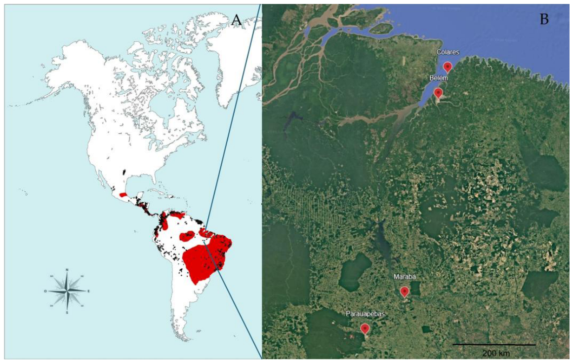
Figure 2.
Molecular detection of Leishmania infantum in blood and conjunctival swab samples from dogs. Molecular positivity was defined as amplification obtained with either kDNA PCR or SSU rDNA PCR. The figure shows the number of dogs positive in each biological matrix and in both matrices. Marker-specific results for kDNA and SSU rDNA are presented separately in Supplementary Figures S6 and S7 and Supplementary Table S3.
Figure 2.
Molecular detection of Leishmania infantum in blood and conjunctival swab samples from dogs. Molecular positivity was defined as amplification obtained with either kDNA PCR or SSU rDNA PCR. The figure shows the number of dogs positive in each biological matrix and in both matrices. Marker-specific results for kDNA and SSU rDNA are presented separately in Supplementary Figures S6 and S7 and Supplementary Table S3.
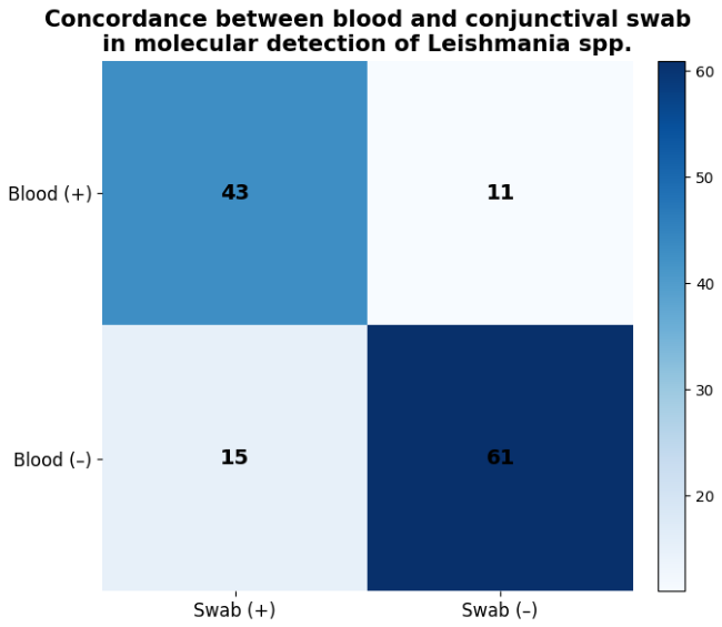
Figure 3.
SSU rDNA sequence variation identified in Leishmania infantum sequences obtained from canine samples from Colares, Belém, and Marabá. Variable nucleotide positions detected by bidirectional Sanger sequencing are shown relative to the reference sequence. Highlighted cells indicate nucleotide substitutions or ambiguous base calls. Ambiguous nucleotides are represented according to IUPAC nomenclature (Y = C/T; R = A/G). Sequence identifiers (S20.16, S22.3, S22.13, and S22.31) correspond to laboratory sample codes assigned during molecular characterization; no variation relative to the reference sequence was detected in S22.31. Detailed polymorphism data are provided in Supplementary Table S5.
Figure 3.
SSU rDNA sequence variation identified in Leishmania infantum sequences obtained from canine samples from Colares, Belém, and Marabá. Variable nucleotide positions detected by bidirectional Sanger sequencing are shown relative to the reference sequence. Highlighted cells indicate nucleotide substitutions or ambiguous base calls. Ambiguous nucleotides are represented according to IUPAC nomenclature (Y = C/T; R = A/G). Sequence identifiers (S20.16, S22.3, S22.13, and S22.31) correspond to laboratory sample codes assigned during molecular characterization; no variation relative to the reference sequence was detected in S22.31. Detailed polymorphism data are provided in Supplementary Table S5.
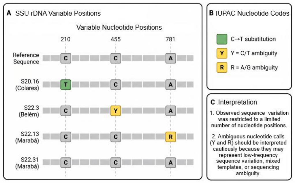
Figure 4.
Transmission electron micrographs of Leishmania (L.) amazonensis (MHOM/BR/26361) developmental forms. (A) Promastigote showing lipid droplets (LD) and vesicle-like structures associated with the flagellar pocket (FP). (B) Intracellular amastigote within a murine peritoneal macrophage showing vacuolar lipid inclusions (VLI) and lysosome-related organelles (LRO). Arrows indicate representative ultrastructural features discussed in the text. Scale bars as indicated in each panel.
Figure 4.
Transmission electron micrographs of Leishmania (L.) amazonensis (MHOM/BR/26361) developmental forms. (A) Promastigote showing lipid droplets (LD) and vesicle-like structures associated with the flagellar pocket (FP). (B) Intracellular amastigote within a murine peritoneal macrophage showing vacuolar lipid inclusions (VLI) and lysosome-related organelles (LRO). Arrows indicate representative ultrastructural features discussed in the text. Scale bars as indicated in each panel.
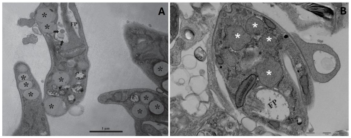

Table 1.
Concordance between TR DPP® and parasitological evidence of infection among 1,499 dogs from Parauapebas, Brazilian Amazon.
Table 1.
Concordance between TR DPP® and parasitological evidence of infection among 1,499 dogs from Parauapebas, Brazilian Amazon.
| TR DPP® Result | Parasitology Positive | Parasitology Negative | Total |
|---|---|---|---|
| Positive | 656 | 255 | 911 |
| Negative | 20 | 568 | 588 |
| Total | 676 | 823 | 1499 |
Agreement between diagnostic methods was evaluated using Cohen’s kappa coefficient (κ = 0.641; 95% CI: 0.604–0.677). Parasitological positivity was defined by the microscopic visualization of intracellular amastigote forms.
Disclaimer/Publisher’s Note: The statements, opinions and data contained in all publications are solely those of the individual author(s) and contributor(s) and not of MDPI and/or the editor(s). MDPI and/or the editor(s) disclaim responsibility for any injury to people or property resulting from any ideas, methods, instructions or products referred to in the content. |
© 2026 by the authors. Licensee MDPI, Basel, Switzerland. This article is an open access article distributed under the terms and conditions of the Creative Commons Attribution (CC BY) license (http://creativecommons.org/licenses/by/4.0/).
Copyright: This open access article is published under a Creative Commons CC BY 4.0 license, which permit the free download, distribution, and reuse, provided that the author and preprint are cited in any reuse.