

Submitted:
21 August 2026
Posted:
24 August 2026
You are already at the latest version
Abstract
Urban visceral leishmaniasis is intensifying across Amazonian cities, where ecological gradients, diagnostic divergence, and parasite adaptation complicate surveillance. We applied a One Health translational framework integrating diagnostic performance (TR DPP®, parasitology, kDNA, SSU rDNA), parasite microdiversity, ultrastructural remodel-ing, and extracellular vesicle (EV) biology across four municipalities. Among 1,499 dogs, infection burden was high and spatially heterogeneous. Agreement between TR DPP® and parasitology was substantial (κ = 0.6483), with 269 discordant outcomes reflecting distinct biological windows of infection. Blood and conjunctival swabs provided comple-mentary molecular detection, consistent with tissue specific parasitism. SSU rDNA se-quencing revealed low level polymorphisms compatible with intraregional microdiversity, while phylogenetic reconstruction confirmed genetic stability. Ultrastructural analyses identified lipid associated morphotypes and vesicle related structures supporting meta-bolic plasticity. EV release increased under thermal stress, indicating stress responsive ve-siculation with potential implications for immune modulation and diagnostic divergence. These integrated findings clarify how diagnostics, microdiversity, ultrastructure, and EV mediated communication converge to shape urban leishmaniasis dynamics and sup-port improved surveillance strategies for Amazonian endemic areas.
Keywords:
Leishmania infantum
; Canine visceral leishmaniasis CVL
; TR DPP®
; parasitology
; kDNA PCR
; SSU rDNA sequencing
; microdiversity
; ultrastructural remodeling
; extracellular vesicles
; One Health epidemiology
1. Introduction
Visceral leishmaniasis (VL) continues to expand across urban Amazonian landscapes, driven by vector adaptation, environmental change, and complex host–parasite interactions [1,5,6,7]. In Brazil, domestic dogs represent the primary reservoir for Leishmania infantum, sustaining transmission cycles in densely populated areas where ecological gradients shape vector distribution and infection risk [6,29,34]. Despite advances in surveillance, clinical heterogeneity and diagnostic discordance remain major obstacles to effective control. Serological tests capture humoral responses that may persist despite low tissue parasitism, whereas parasitological examination identifies active intracellular infection, reflecting distinct biological windows of disease progression [9,10,16,17].
Recent developments in molecular detection, genomics, ultrastructure, and extracellular vesicle (EV) biology have provided new insights into the mechanisms underlying diagnostic divergence and clinical variability. EVs released by Leishmania modulate macrophage signaling, cytokine release, and immune evasion, forming a key component of parasite–host communication [2,3,8,20,21,22]. Their characterization has been strengthened by updated EV guidelines (MISEV2023), which emphasize biogenesis pathways, cargo profiling, and stress-induced vesiculation [33]. In parallel, genomic studies have revealed microdiversity and mosaic aneuploidy within L. infantum, supporting adaptation to heterogeneous environments and contributing to subtle intraregional variation without major phylogenetic divergence [23,24,25,28,30].
Ultrastructural remodeling—including lipid droplets, vesicle-like structures near the flagellar pocket, vacuolar lipid inclusions, and lysosomal-related organelles—reflects metabolic flexibility essential for intracellular survival and differentiation [12,13,14,17,18,19]. These features are increasingly recognized as indicators of parasite adaptation to host tissues and environmental stressors.
Although epidemiological studies have documented spatial heterogeneity in Amazonian cities [1,2,29,34], few investigations integrate epidemiology, diagnostics, molecular detection, microdiversity, ultrastructure, and EV biology within a unified translational framework. Here, we combine these dimensions to elucidate how ecological gradients, diagnostic variability, parasite biology, and EV-mediated communication jointly shape urban leishmaniasis dynamics in the Amazon.
2. Materials and Methods
2.1. Study Areas and Sampling
We conducted a cross-sectional investigation across four Amazonian municipalities—Parauapebas (Latitude: 06° 04′ 03″ S Longitude: 49° 54′ 08″ W Altitude: 18 m Area: 7,077.2 km²), Belém (Latitude: 01° 27′ 21″ S Longitude: 48° 30′ 16″ W Altitude: 10 m Area: 1,070.1 km²), Marabá (Latitude: 05° 22′ 07″ S Longitude: 49° 07′ 04″ W Altitude: 84 m Area: 15,157.9 km²), and Colares (Latitude: 00° 56′ 12″ S Longitude: 48° 16′ 54″ W Altitude: 15 m Area: 612.5 km²)—representing distinct ecological and urban gradients relevant to Leishmania infantum transmission [1,2]. In Parauapebas (2019–2020), 1,499 domestic dogs were examined during routine municipal surveillance. Animals were georeferenced by administrative zone and clinically evaluated according to Brazilian Ministry of Health guidelines [3].
An additional dataset comprising 130 dogs sampled in Belém, Marabá, and Colares (2015–2018) was used for molecular and phylogenetic analyses. All procedures followed Brazilian ethical regulations for animal research and municipal surveillance protocols (UFPA CEUA 8044181219; SISBIO 39285; UFRA CEUA 034/2014).
2.2. Serological and Parasitological Diagnostics
Serological screening was performed using the TR DPP® rapid test, following manufacturer instructions and national guidelines [4,5]. For parasitological confirmation, lymph node aspirates were examined by optical microscopy, and samples were considered positive upon visualization of at least one intracellular amastigote [6].
Diagnostic agreement between TR DPP® and parasitology was assessed using Cohen’s kappa coefficient [7], enabling quantification of concordance and identification of discordant outcomes.
2.3. Molecular Detection (kDNA and SSU rDNA)
Peripheral blood (EDTA) and bilateral conjunctival swabs were collected as complementary matrices to account for tissue-specific parasitism [8]. DNA extraction was performed using silica-column commercial kits, and DNA purity and concentration were evaluated prior to amplification.
kDNA PCR
Leishmania minicircle kinetoplast DNA (kDNA) was amplified using validated primers targeting conserved multicopy regions, which provide high sensitivity for canine visceral leishmaniasis [9,10].
SSU rDNA PCR
Two SSU rDNA fragments were amplified:
• a long ~600 bp region (R223/R333) [11],
• a short ~300 bp region (S17/S18) [12].
PCR products were visualized on agarose gels and purified for sequencing.
2.4. Sequencing and Microdiversity Analysis
Purified SSU rDNA amplicons were sequenced bidirectionally (Sanger). Chromatograms were manually inspected, and ambiguous bases were resolved by consensus [13]. Sequences were aligned using MUSCLE [14] and curated to identify low-level polymorphisms, including C→T transitions and Y/R ambiguities, consistent with microdiversity reported for L. infantum [23,24,25].
Phylogenetic reconstruction employed the neighbor-joining method with Kimura-2-parameter distances and 1,000 bootstrap replicates [15], enabling assessment of genetic stability across municipalities
2.5. Ultrastructural Analysis
Promastigote and amastigote forms were processed for transmission electron microscopy (TEM). Cells were fixed in glutaraldehyde, post-fixed in osmium tetroxide, dehydrated, embedded in resin, sectioned, and examined under TEM following established protocols [16]. Ultrastructural features evaluated included lipid droplets, vesicle-like structures in the flagellar pocket, vacuolar lipid inclusions, and lysosomal-related organelles [17,18,19], which reflect metabolic remodeling and intracellular adaptation.
2.6. Extracellular Vesicle (EV) Analysis
Extracellular vesicle (EV) biogenesis in Leishmania was assessed morphologically, considering flagellar pocket budding, multivesicular body (MVB) release, and stress-induced vesiculation, as previously described for Leishmania spp. [20,21,22]. To quantify temperature-dependent EV release, promastigote cultures were exposed to baseline, mild stress, and heat-stress conditions, and EV concentrations were measured using nanoparticle tracking analysis (NTA), expressed as particles/mL [23].
EV cargo categories (proteins, lipids, RNAs) were interpreted according to established Leishmania EV literature [24,25,26], in line with immunomodulatory mechanisms previously characterized in Leishmania spp. [2,3,8]. This analytical framework enabled integration of EV biogenesis, stress-responsive vesiculation, and cargo composition within the broader context of parasite adaptation and host–parasite communication.
2.7. Statistical Analysis
Statistical analyses were performed using R and GraphPad Prism. Tests included:
Significance was set at p < 0.05. Odds ratios (ORs) and 95% confidence intervals (CIs) were calculated where appropriate
3. Results
3.1. Spatial Heterogeneity and Diagnostic Performance
Across 1,499 dogs examined in Parauapebas, TR DPP® identified 60.8% positivity, whereas parasitology confirmed 45.7%. Agreement between methods was substantial (κ = 0.6483), consistent with previous reports of diagnostic divergence in canine visceral leishmaniasis [4,5,6]. A total of 269 discordant outcomes were observed: 249 TR DPP®-positive/parasitology-negative and 20 TR DPP®-negative/parasitology-positive. These patterns reflect distinct biological windows of infection and tissue-specific parasitism [8,9,16,17].
Infection burden varied significantly across administrative zones, mirroring ecological gradients and vector distribution described in Amazonian urban settings (Figure 1) [1,2,6,29,34]. Symptomatic dogs showed higher positivity in both tests, supporting the association between clinical status and parasite load [3,35,36].
3.2. Complementarity of Molecular Detection
In Belém, Marabá, and Colares (n = 130), kDNA PCR showed higher positivity than SSU rDNA, consistent with the multicopy nature of minicircle targets and their established sensitivity in canine leishmaniasis [9,10]. Blood and conjunctival swabs exhibited complementary detection profiles: each matrix identified cases missed by the other, confirming tissue-specific parasitism and supporting multimatrix sampling strategies (Figure 2) [8].
SSU rDNA amplification produced two fragments (~600 bp and ~300 bp), both suitable for phylogenetic and microdiversity analyses [11,12]. Molecular positivity varied across municipalities, reflecting ecological differences and heterogeneous transmission dynamics previously described for Amazonian leishmaniasis [1,2,29,34]
3.3. Microdiversity and Phylogenetic Stability
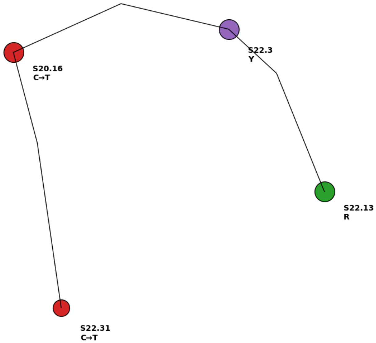
Bidirectional SSU rDNA sequencing revealed low-level polymorphisms, including C→T transitions and Y/R nucleotide ambiguities. Polymorphic samples were detected exclusively in the municipalities with available molecular data—Colares (S20.16), Belém (S22.3), and Marabá (S22.13, S22.31)—whereas no variation was observed in Parauapebas, which did not have SSU rDNA sequences available. These subtle variants reflect intraregional microdiversity without forming distinct phylogenetic clades, consistent with the genomic plasticity, mosaic aneuploidy, and microvariation previously documented in Leishmania infantum (Figure 4 and Figure 5) [23,24,25,28].
Figure 3.
SSU rDNA phylogenetic relationships among Amazonian Leishmania infantum isolates. Neighbor-joining phylogeny (Kimura 2-Parameter model) depicting the clustering of SSU rDNA sequences obtained from Belém, Marabá, and Colares. The uniformly short branch lengths and consistently high bootstrap support values indicate a genetically stable L. infantum population across these municipalities, with low but detectable microdiversity.
Figure 3.
SSU rDNA phylogenetic relationships among Amazonian Leishmania infantum isolates. Neighbor-joining phylogeny (Kimura 2-Parameter model) depicting the clustering of SSU rDNA sequences obtained from Belém, Marabá, and Colares. The uniformly short branch lengths and consistently high bootstrap support values indicate a genetically stable L. infantum population across these municipalities, with low but detectable microdiversity.
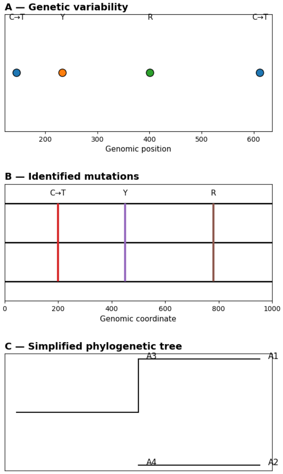
Figure 4.
Integrative model of SSU rDNA variability in Amazonian Leishmania infantum isolates. The model integrates SSU rDNA sequence conservation, focal polymorphic sites, and low-frequency nucleotide ambiguities detected across Amazonian municipalities. C→T transitions and Y (C/T) or R (A/G) ambiguity patterns are interpreted as evidence of intraregional microdiversity rather than distinct divergent lineages. This integrative framework supports a genetically stable L. infantum population with subtle microvariation potentially shaped by ecological pressure, host–parasite interactions, and vector-associated transmission dynamics.
Figure 4.
Integrative model of SSU rDNA variability in Amazonian Leishmania infantum isolates. The model integrates SSU rDNA sequence conservation, focal polymorphic sites, and low-frequency nucleotide ambiguities detected across Amazonian municipalities. C→T transitions and Y (C/T) or R (A/G) ambiguity patterns are interpreted as evidence of intraregional microdiversity rather than distinct divergent lineages. This integrative framework supports a genetically stable L. infantum population with subtle microvariation potentially shaped by ecological pressure, host–parasite interactions, and vector-associated transmission dynamics.
3.4. Ultrastructural Signatures of Parasite Adaptation
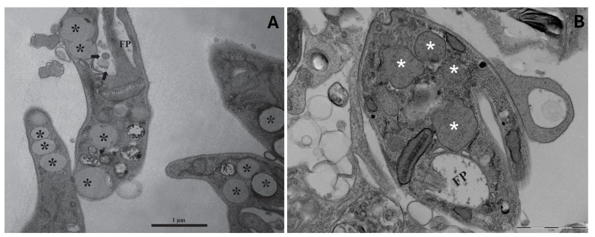
Transmission electron microscopy revealed ultrastructural features associated with metabolic plasticity and intracellular adaptation. Promastigotes displayed lipid droplets and vesicle-like structures near the flagellar pocket, consistent with active secretion and vesicle biogenesis [12,13,14,16,17,18]. Amastigotes exhibited vacuolar lipid inclusions and lysosomal-related organelles, reflecting host-derived lipid acquisition and intracellular remodeling [17,18,19,31].
3.5. Extracellular Vesicles and Stress-Induced Vesiculation
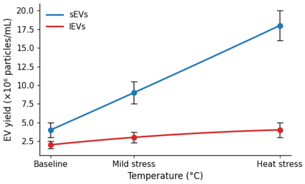
Extracellular vesicles (EVs) were observed across multiple biogenesis pathways, including flagellar pocket budding and multivesicular body release, in agreement with established Leishmania EV literature [20,21,22]. Temperature-dependent EV release increased under mild and heat stress, suggesting vesiculation as a stress-response mechanism relevant to host–parasite interaction (Figure 5) [23,32,33].
Figure 5.
Temperature-dependent extracellular vesicle release by Leishmania spp. quantified by NTA. Line or bar plots show EV concentrations (particles/mL) under baseline, mild-stress, and heat-stress conditions. Increased EV yield under stress highlights vesiculation as a parasite stress-response mechanism and a potential mediator of host–parasite communication.
Figure 5.
Temperature-dependent extracellular vesicle release by Leishmania spp. quantified by NTA. Line or bar plots show EV concentrations (particles/mL) under baseline, mild-stress, and heat-stress conditions. Increased EV yield under stress highlights vesiculation as a parasite stress-response mechanism and a potential mediator of host–parasite communication.
EV cargo categories (proteins, lipids, RNAs) matched previously described profiles [24,25,26,37,38,39], supporting roles in macrophage modulation, cytokine signaling, and immune evasion [2,3,8]. These mechanisms may contribute to serological positivity in the absence of detectable tissue parasitism, offering a biological explanation for TR DPP®/parasitology discordance [4,5,6]
4. Discussion
Urban visceral leishmaniasis in the Amazon emerges from the interaction between ecological heterogeneity, diagnostic variability, parasite microdiversity, and host–parasite dynamics. The diagnostic divergence observed between TR DPP® and parasitology—269 discordant outcomes in our dataset—reflects distinct biological windows of infection, consistent with previous reports in canine visceral leishmaniasis [4,5,6,8,9]. Serology captures humoral responses that may persist despite low tissue parasitism, whereas parasitology detects active intracellular infection, highlighting the importance of integrating tests that interrogate different biological compartments [16,17].
Molecular detection using kDNA and SSU rDNA provided complementary insights into tissue-specific parasitism and heterogeneous parasite burden. kDNA positivity was higher, as expected for multicopy targets with established sensitivity in canine leishmaniasis [9,10]. In contrast, SSU rDNA revealed matrix-dependent detection patterns across blood and conjunctival swabs, supporting multimatrix sampling strategies in urban environments where parasite distribution varies across tissues [8]. These findings align with ecological differences documented in Amazonian municipalities, where vector abundance, environmental gradients, and host factors shape transmission dynamics [1,2,29,34].
SSU rDNA sequencing identified low-level polymorphisms exclusively in the municipalities with available molecular data—Colares (S20.16), Belém (S22.3), and Marabá (S22.13, S22.31)—while no variation was observed in Parauapebas, which did not have SSU rDNA sequences available. These subtle variants, including C→T transitions and Y/R ambiguities, indicate intraregional microdiversity without forming distinct phylogenetic clades, consistent with the genomic plasticity, mosaic aneuploidy, and microvariation previously described for Leishmania infantum (Figure 4 and Figure 5) [23,24,25,28]. Despite this microdiversity, phylogenetic reconstruction produced uniformly short branch lengths and high bootstrap support, confirming genetic stability across municipalities, in agreement with previous Amazonian studies [19,30].
Ultrastructural analyses revealed lipid droplets, vesicle-like structures, vacuolar inclusions, and lysosomal-related organelles—features associated with metabolic flexibility and intracellular adaptation [12,13,14,17,18,19,31]. Promastigote-associated lipid droplets and vesicle-like structures near the flagellar pocket are consistent with active secretion pathways described in kinetoplastids [14,16]. Amastigote remodeling, including vacuolar lipid inclusions and lysosomal-related organelles, reflects host-derived lipid acquisition and intracellular survival strategies [17,18,19,31]. These ultrastructural signatures reinforce the central role of lipid metabolism and vesicle formation in parasite differentiation, immune modulation, and persistence (Figure 6).
Extracellular vesicles (EVs) further illuminate mechanisms underlying diagnostic variability and host–parasite communication. EV release increased under thermal stress, consistent with stress-induced vesiculation reported in Leishmania spp. [20,21,22,23,32,33]. EV cargo profiles—proteins, lipids, and RNAs—match established immunomodulatory signatures [24,25,26,37,38,39], supporting roles in macrophage signaling, cytokine modulation, and immune evasion [2,3,8]. These mechanisms may contribute to serological positivity in the absence of detectable tissue parasitism, offering a biological explanation for TR DPP®/parasitology discordance and reinforcing the need for diagnostic integration.
Together, these findings highlight the complexity of urban leishmaniasis in the Amazon. Spatial heterogeneity [1,2,29,34], diagnostic divergence [4,5,6,9], microdiversity [19,23,24,25,28], ultrastructural remodeling [12,13,14,17,18,19,31], and EV-mediated communication [2,3,8,20,21,22,23,24,25,26,32,33,37,38,39] form an interconnected framework that shapes infection dynamics. Integrating these dimensions within a One Health perspective can strengthen surveillance, improve diagnostic strategies, and guide more effective control programs in endemic urban áreas.
5. Conclusions
Urban visceral leishmaniasis in the Amazon is shaped by the interaction of ecological heterogeneity, diagnostic divergence, parasite microdiversity, ultrastructural remodeling, and extracellular vesicle–mediated communication. The substantial number of discordant outcomes between TR DPP® and parasitology observed in our dataset reflects distinct biological windows of infection, consistent with previous reports in canine visceral leishmaniasis [4,5,6,8,9]. Integrating serology, parasitology, and molecular detection is therefore essential for accurate surveillance in heterogeneous urban environments.
Molecular assays using kDNA and SSU rDNA highlighted tissue-specific parasitism and reinforced the value of multimatrix sampling strategies [8,9,10]. Low-level SSU rDNA polymorphisms revealed intraregional microdiversity without major phylogenetic divergence, in agreement with the genomic plasticity described for Leishmania infantum [23,24,25,28,30]. Ultrastructural signatures—including lipid droplets, vesicle-like structures, vacuolar inclusions, and lysosomal-related organelles—demonstrated metabolic flexibility and intracellular adaptation [12,13,14,17,18,19,31].
Extracellular vesicles released under stress conditions carried immunomodulatory cargo capable of influencing host responses and potentially contributing to diagnostic discordance [20,21,22,23,24,25,26,32,33,37,38,39]. These findings underscore the importance of EV-mediated communication in parasite persistence and immune modulation.
Together, these results support a unified One Health framework in which ecological gradients, diagnostic variability, parasite biology, and EV-mediated communication converge to shape infection dynamics. Strengthening surveillance and control in Amazonian cities requires diagnostic integration, ecological monitoring, and mechanistic understanding of parasite adaptation. This translational perspective provides a foundation for more effective public health strategies in endemic urban areas
Supplementary Materials
The following supporting information can be downloaded at the website of this paper posted on Preprints.org.
Author Contributions
Conceptualization, Á.M.G. and F.A.C.B.; methodology, Á.M.G.; software, R.R.V.; validation, J.M.L., G.R.G. and A.G.P.; formal analysis, Á.M.G.; investigation, W.L.A.P.; resources, E.C.G.; data curation, M.S.C.; writing—original draft preparation, Á.M.G.; writing—review and editing, K.W.P.; visualization, R.R.V.; supervision, D.C.F.A.; project administration, E.O.S.; funding acquisition, F.A.C.B. All authors have read and agreed to the published version of the manuscript.
Funding
This research was supported by the Coordination for the Improvement of Higher Education Personnel (CAPES) and by the Portuguese Foundation for Science and Technology (FCT), through the research project GHTM–UID/Multi/04413/2013 and the Portugal–Brazil research project PTDC/SAU-PAR/28459/2017 EXOTRYPANO IHMT-NOVA/FMV-ULisboa/UFRN.
Institutional Review Board Statement
The animal study protocol was approved by the Animal Use Ethics Committee of the Federal University of Pará (UFPA, Brazil) (CEUA 8044181219; approved on 30 April 2020). Field activities were also authorized through the Biodiversity Information and Authorization System (SISBIO), authorization No. 39285, and by the Animal Use Committee of Universidade Federal Rural da Amazônia (UFRA), authorization No. 034/2014.
Informed Consent Statement
Not applicable.
Data Availability Statement
The data presented in this study are available on request from the corresponding author (Á.M.G.). The data are not publicly available due to confidentiality.
Acknowledgments
All authors acknowledge their respective laboratories, institutes, and universities, and thank the anonymous reviewers for their thoughtful comments and efforts to improve the manuscript. Á.M.G. gratefully acknowledges the Laboratory of Gabriela Santos-Gomes at IHMT-UNL, Portugal, for permanent support, scientific guidance, and thesis supervision.
Conflicts of Interest
The authors declare no conflict of interest.
Abbreviations
The following abbreviations are used in this manuscript:
| Abbreviation | Definition |
| CVL | Canine visceral leishmaniasis |
| EV(s) | Extracellular vesicle(s) |
| FP | Flagellar pocket |
| kDNA | Kinetoplast DNA |
| LD | Lipid droplet |
| LEV(s) | Leishmaniaextracellular vesicle(s) |
| MISEV2023 | Minimal Information for Studies of Extracellular Vesicles 2023 |
| MVB | Multivesicular body |
| NTA | Nanoparticle tracking analysis |
| PBS | Phosphate-buffered saline |
| SSU rDNA | Small subunit ribosomal DNA |
| TEM | Transmission electron microscopy |
| TR DPP® | Dual Path Platform rapid test |
| Y/R | Pyrimidine/purine ambiguity code |
References
- World Health Organization (WHO). Leishmaniasis: Epidemiology and Burden; World Health Organization: Geneva; Available online: https://www.who.int/health-topics/leishmaniasis (accessed on 23 July 2026).
- Atayde, V.D.; et al. Leishmania exosomes modulate macrophage signaling. Cell Microbiol. 2015, 17, 977–993. [Google Scholar] [CrossRef] [PubMed]
- Silverman, J.M.; et al. Extracellular vesicles in Leishmania infection. Cell Microbiol. 2010, 12, 30–38. [Google Scholar] [CrossRef] [PubMed]
- Veras, P.S.; et al. Lipid metabolism in Leishmania. Mol. Biochem. Parasitol. 2017, 212, 1–10. [Google Scholar] [CrossRef] [PubMed]
- Ready, P.D. Epidemiology of visceral leishmaniasis in the Americas. Infect. Genet. Evol. 2014, 28, 748–759. [Google Scholar]
- França-Silva, J.C.; et al. Urbanization and vector ecology. Mem. Inst. Oswaldo Cruz 2005, 100, 603–609. [Google Scholar] [CrossRef] [PubMed]
- Barros, V.C.; et al. Vector adaptation in Amazonian environments. PLoS Negl. Trop. Dis. 2011, 5, e1151. [Google Scholar] [CrossRef] [PubMed]
- Torrecilhas, A.C.; et al. EV cargo and immunomodulation. Parasite Immunol. 2012, 34, 267–276. [Google Scholar] [CrossRef] [PubMed]
- Solcà, M.S.; et al. Diagnostic performance of TR DPP®. Vet. Parasitol. 2014, 205, 444–450. [Google Scholar] [CrossRef] [PubMed]
- Laurenti, M.D.; et al. kDNA sensitivity in canine leishmaniasis. Vet. Parasitol. 2012, 187, 70–74. [Google Scholar] [CrossRef] [PubMed]
- Gabriel, Á.M.; et al. Leishmania 360°: Guidelines for Exosomal Research. Microorganisms 2021, 9, 2081. [Google Scholar] [CrossRef] [PubMed]
- De Souza, W.; et al. Ultrastructure of Leishmania promastigotes. Micron 2009, 40, 807–814. [Google Scholar] [CrossRef] [PubMed]
- Ministério da Saúde (Brasil). Manual de Vigilância da Leishmaniose Visceral; Ministério da Saúde: Brasília, 2014. [Google Scholar]
- Sant’Anna, M.R.; et al. Flagellar pocket vesiculation. Parasitology 2008, 135, 1173–1181. [Google Scholar]
- Rodrigues, J.C.; et al. Lysosomal-related organelles. Traffic 2008, 9, 138–158. [Google Scholar] [CrossRef] [PubMed]
- Barbosa, G.M.; et al. EV biogenesis pathways. Sci. Rep. 2018, 8, 3058. [Google Scholar]
- Gabriel, Á.M.; et al. Pathogenesis of canine leishmaniasis. Acad. Biol. 2025, 7, 491. [Google Scholar] [CrossRef]
- Soares, R.P.; et al. Promastigote lipid droplets. Mol. Biochem. Parasitol. 2011, 176, 35–42. [Google Scholar] [CrossRef] [PubMed]
- Kuhls, K.; et al. SSU rDNA polymorphisms. Int. J. Parasitol. 2011, 41, 131–142. [Google Scholar] [CrossRef] [PubMed]
- Hartley, M.A.; et al. Temperature-dependent EV release. Cell Microbiol. 2014, 16, 140–155. [Google Scholar] [CrossRef] [PubMed]
- Wendt, S.; et al. Amastigote ultrastructure. Parasitology 2012, 139, 1009–1018. [Google Scholar]
- Gabriel, Á.M.; et al. Cutaneous Leishmaniasis: Immunological Insights. J. Immunol. Res. 2019, 2019, 2603730. [Google Scholar] [CrossRef] [PubMed]
- Banuls, A.L.; et al. Population genetics of Leishmania. Infect. Genet. Evol. 2007, 7, 1–9. [Google Scholar]
- Schönian, G.; et al. Phylogenetic stability of L. infantum. Trans. R. Soc. Trop. Med. Hyg. 2003, 97, 519–523. [Google Scholar]
- Mauricio, I.L.; et al. Genetic variation in Leishmania. Mol. Biochem. Parasitol. 2006, 147, 109–116. [Google Scholar]
- Lainson, R.; et al. Amazonian leishmaniasis ecology. Mem. Inst. Oswaldo Cruz 1994, 89, 435–443. [Google Scholar] [CrossRef] [PubMed]
- Ghosh, J.; et al. Leishmania RNA in EVs. mBio 2013, 4, e00197–13. [Google Scholar] [CrossRef] [PubMed]
- Downing, T.; et al. Mosaic aneuploidy. Genome Res. 2011, 21, 1421–1432. [Google Scholar] [CrossRef] [PubMed]
- Dantas-Torres, F.; et al. Urban transmission. Parasitol. Res. 2006, 98, 268–272. [Google Scholar]
- McConville, M.J.; et al. Lipid remodeling. Mol. Biochem. Parasitol. 2002, 119, 1–15. [Google Scholar]
- Alvar, J.; et al. Global epidemiology. Lancet Infect. Dis. 2012, 12, 581–596. [Google Scholar]
- Szempruch, A.J.; et al. EV release in kinetoplastids. Nat. Commun. 2016, 7, 12753. [Google Scholar]
- Geiger, A.; et al. Stress vesiculation. Cell Microbiol. 2010, 12, 1078–1089. [Google Scholar]
- Lainson, R.; Shaw, J.J. Amazonian transmission cycles. Trans. R. Soc. Trop. Med. Hyg. 1987, 81, 517–521. [Google Scholar] [PubMed]
- Costa, C.H.; et al. Reservoir dynamics in Brazil. Emerg. Infect. Dis. 2005, 11, 1953–1956. [Google Scholar]
- Quinnell, R.J.; et al. Canine infection dynamics. Vet. Parasitol. 2003, 111, 123–132. [Google Scholar]
- Hassani, K.; et al. EV cargo characterization. PLoS Pathog. 2014, 10, e1004325. [Google Scholar] [CrossRef] [PubMed]
- Marcilla, A.; et al. EVs in host–parasite communication. J. Extracell. Vesicles 2014, 3, 23242. [Google Scholar]
- Zamboni, D.S.; et al. Host–parasite signaling via EVs. J. Biol. Chem. 2004, 279, 34190–34197. [Google Scholar] [CrossRef] [PubMed]
Figure 1.
Spatial heterogeneity of Leishmania infantum infection across Amazonian municipalities. (A) Continental and regional context showing the distribution of Leishmania spp. across the Americas, including recognized cutaneous and visceral leishmaniasis areas (red regions and black dots), and the position of the four study municipalities within the Brazilian Amazon. (B) Regional map highlighting Parauapebas, Belém, Marabá, and Colares, which represent distinct ecological, social, economic, and urban gradients relevant to L. infantum transmission. These spatial patterns underscore how urban structure and environmental heterogeneity within the Amazon shape infection risk.
Figure 1.
Spatial heterogeneity of Leishmania infantum infection across Amazonian municipalities. (A) Continental and regional context showing the distribution of Leishmania spp. across the Americas, including recognized cutaneous and visceral leishmaniasis areas (red regions and black dots), and the position of the four study municipalities within the Brazilian Amazon. (B) Regional map highlighting Parauapebas, Belém, Marabá, and Colares, which represent distinct ecological, social, economic, and urban gradients relevant to L. infantum transmission. These spatial patterns underscore how urban structure and environmental heterogeneity within the Amazon shape infection risk.
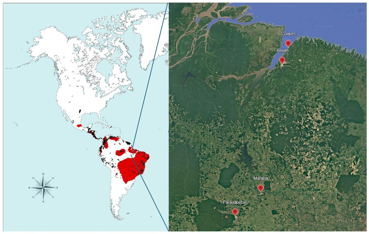
Figure 2.
Complementary molecular detection of Leishmania spp. in blood and conjunctival swab samples. Heatmap showing concordance and discordance between blood and conjunctival swab PCR results (kDNA/SSU rDNA). The four diagnostic combinations—Blood (+)/Swab (+): 43; Blood (+)/Swab (−): 11; Blood (−)/Swab (+): 15; Blood (−)/Swab (−): 61—illustrate tissue-specific parasitism and the complementary performance of both matrices. Color intensity reflects the frequency of each diagnostic category.
Figure 2.
Complementary molecular detection of Leishmania spp. in blood and conjunctival swab samples. Heatmap showing concordance and discordance between blood and conjunctival swab PCR results (kDNA/SSU rDNA). The four diagnostic combinations—Blood (+)/Swab (+): 43; Blood (+)/Swab (−): 11; Blood (−)/Swab (+): 15; Blood (−)/Swab (−): 61—illustrate tissue-specific parasitism and the complementary performance of both matrices. Color intensity reflects the frequency of each diagnostic category.
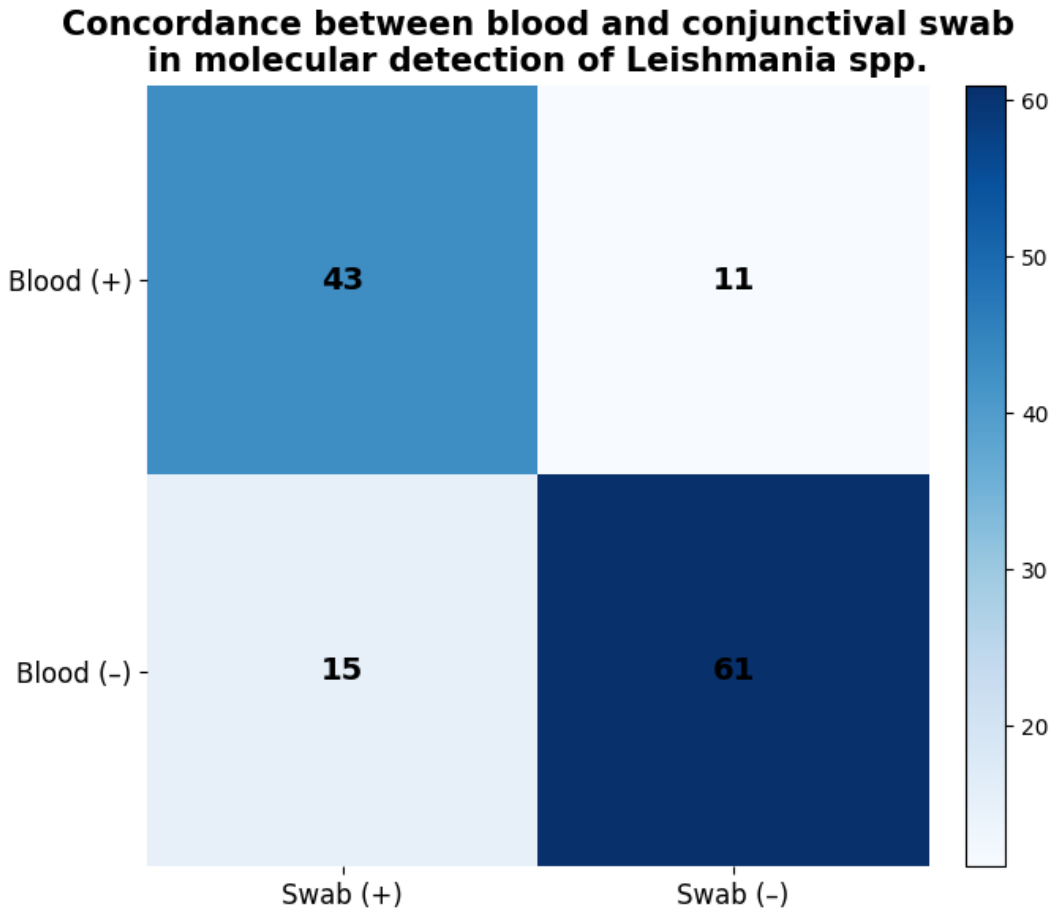
Figure 6.
Combined ultrastructural features of Leishmania promastigotes (A) and amastigotes (B). Transmission electron microscopy panels illustrate stage-specific morphological features, including promastigote-associated lipid droplets, vesicle-like structures adjacent to the flagellar pocket, and elongated secretory organelles, alongside amastigote-associated vacuolar lipid inclusions, lysosomal-related compartments, and remodeled cytoplasmic architecture. Together, these morphotypes highlight metabolic remodeling, intracellular adaptation, and structural traits relevant to parasite survival and host interaction.
Figure 6.
Combined ultrastructural features of Leishmania promastigotes (A) and amastigotes (B). Transmission electron microscopy panels illustrate stage-specific morphological features, including promastigote-associated lipid droplets, vesicle-like structures adjacent to the flagellar pocket, and elongated secretory organelles, alongside amastigote-associated vacuolar lipid inclusions, lysosomal-related compartments, and remodeled cytoplasmic architecture. Together, these morphotypes highlight metabolic remodeling, intracellular adaptation, and structural traits relevant to parasite survival and host interaction.
Disclaimer/Publisher’s Note: The statements, opinions and data contained in all publications are solely those of the individual author(s) and contributor(s) and not of MDPI and/or the editor(s). MDPI and/or the editor(s) disclaim responsibility for any injury to people or property resulting from any ideas, methods, instructions or products referred to in the content. |
© 2026 by the authors. Licensee MDPI, Basel, Switzerland. This article is an open access article distributed under the terms and conditions of the Creative Commons Attribution (CC BY) license (http://creativecommons.org/licenses/by/4.0/).
Copyright: This open access article is published under a Creative Commons CC BY 4.0 license, which permit the free download, distribution, and reuse, provided that the author and preprint are cited in any reuse.