

Submitted:
20 August 2026
Posted:
21 August 2026
You are already at the latest version
Abstract
Adaptive radiotherapy (ART) has emerged as one of the most significant advances in modern radiation oncology, enabling increasingly precise and individualized treatment through adaptation to patient-specific changes occurring during therapy. While ART encompasses both anatomical and biological adaptation, most clinical implementation and technological development to date have focused on managing geometric variation through advanced imaging, motion management, and online replanning. Biological adaptation, however, remains a largely underexplored frontier with the potential to further personalize radiation delivery according to the evolving characteristics of both tumor and host. In this review, we revisit the classical and emerging 6 R's of radiobiology (repair, redistribution, repopulation, reoxygenation, radiosensitivity, and immune reactivation) as a biological framework for adaptive decision-making during radiotherapy. We summarize advances in molecular, imaging, and liquid-biopsy biomarkers that increasingly permit longitudinal assessment of tumor and host biology, including functional MRI, PET-based hypoxia and proliferation imaging, circulating tumor DNA, radiomics, radiogenomics, and immune profiling. We further examine emerging paradigms of biology-guided adaptation, including response-guided dose escalation and de-escalation, hypoxia-guided dose painting, metabolism-guided adaptation, functional tissue avoidance, adaptive immunologic modulation, and liquid-biopsy-driven treatment personalization. In parallel, we discuss enabling technologies such as PET-guided radiotherapy, MR-guided adaptive platforms, and artificial intelligence. Finally, we highlight key scientific, technical, and workflow challenges, including biomarker validation, quantitative imaging standardization, real-time biological monitoring, and prospective clinical trial development. Collectively, these advances support the evolution of ART from a paradigm centered primarily on geometric adaptation toward a more comprehensive framework that integrates both anatomical and biological information to guide treatment personalization. Biology-guided ART represents an important next step in precision oncology, enabling dynamic adaptation based on the evolving biology of tumors and their microenvironment throughout the course of therapy.
Keywords:
adaptive radiotherapy
; quantitative imaging
; radiobiology
; biomarker
; personalized medicine
; precision medicine
; hypoxia
; dose escalation
; functional avoidance
; immunotherapy
Introduction
Biology has been integral to the concept of adaptive radiotherapy (ART) since its earliest formulation. Adaptive radiotherapy, as a concept introduced twenty years ago, “intends to improve radiation treatment by systematically monitoring treatment variations and incorporating them to re-optimize the treatment plan early on during the course of treatment”[1]. The authors presciently envisioned routine customization of “field margin and treatment dose” guided by ongoing measurements. This original definition was inherently broad, encompassing not only geometric changes but also biological dynamics.
Over the past decade, ART has gained substantial momentum, driven by the clinical adoption of streamlined platforms equipped with advanced imaging capabilities that enable efficient adaptation to anatomical and geometric variation[2]. To date, most developments have focused on anatomy- or geometry-guided ART, which represent the current mainstay and near-term trajectory of the field. While this represents a critical and practical first step, it captures only part of the adaptive paradigm originally envisioned.
In contrast, this narrative review centers on the comparatively underexplored domain of biology-guided ART. Table 1 provides a brief comparison between anatomy-guided and biology-guided ART. Although still in its early stages, characterized by emerging research protocols, limited clinical trials, and evolving enabling technologies, the biology-guided ART paradigm opens the door to truly adaptive dose modulation and deeper multidisciplinary integration. In doing so, it extends the scope of ART beyond structural adaptation and moves toward a more comprehensive, biologically informed framework.
Individual cells within a tumor are genetically diverse making the tumor microenvironment intrinsically heterogeneous, both spatially and temporally. Consequently, solid tumors exist within a complex environment and consist of diverse populations of cell subclones shaped by the response of the normal tissue parenchymal cells, resident and recruited immune cells and other microenvironmental factors. Leveraging such biological information could enable truly adaptive dose painting and treatment strategies tailored to tumor biology. We will first revisit the 6 R’s of radiobiology (Repair, Redistribution, Repopulation, Reoxygenation, Radiosensitivity, and immune Reactivation) and consider how each R suggests potential adaptive interventions[3]. Concurrently, we review measurable biomarkers and imaging techniques that probe the corresponding processes, such as hypoxia and proliferation[4,5]. We then examine examples of “bio-adaptive” strategies from functional imaging-guided boosts and avoidance, biomarker-triggered dose adaptation, to temporal optimization of dose fractionation and survey enabling technologies (onboard PET/MR, rapid assays, AI planning) and workflow/logistical considerations. Finally, we highlight key challenges and a research/development roadmap as the field moves toward a comprehensive systems-based adaptive RT.
The “6 R’s” of Radiobiology and Biomarkers for Adaptive Decision-Making
The classical radiation biology framework to delineate the therapeutic ratio after conventional fractionated radiotherapy involves the 4 R’s: Repair of DNA damage, Repopulation of cells between fractions, Redistribution (cell cycle progression), and Reoxygenation of the tumor[6]. Later, Intrinsic Radiosensitivity (governed by tumor genetics) was recognized as a fifth R, and more recently immune Reactivation has been proposed as a sixth[3,7]. The influence of the 4 R’s on radiation response of tumors has been repeatedly demonstrated in preclinical and clinical studies, most notably, tumor repopulation and repair in a comparison of conventional and split-course radiotherapy by Overgaard et al[8]. They demonstrated that protracting overall treatment time narrowed the therapeutic window, a clinical observation attributed to the 4 Rs. Likewise, Withers and colleagues demonstrated the repopulation and repair of normal jejunal crypt stem cells during fractionated 60Co γ-radiation validating the applicability of the 4 R’s to normal tissues radiation responses[9]. Collectively, these biological processes emphasize that radiotherapy response depends not only on static anatomy, but also on dynamic molecular, cellular, and microenvironmental changes occurring throughout treatment, which is reflective of the functionality and impact of the 6 R’s on overall radiation response. Importantly, many of these processes are increasingly measurable through molecular and imaging biomarkers, creating opportunities for biology-guided adaptive radiotherapy[4,5]. Figure 1 summarizes the evolving framework linking the 6 R’s of radiobiology with emerging molecular and imaging biomarkers that may enable biologically informed ART.
Repair: Repair reflects the ability of tumor cells to recover from radiation-induced DNA damage. Biomarkers such as γH2AX foci, ATM, RAD51, and DNA-PKcs expression provide insight into DNA repair activity, while multigene expression signatures including the radiosensitivity index and genomic-adjusted radiation dose (GARD) attempt to quantify intrinsic repair capacity and genomic diversity and predict optimal dose response[10,11,12,13,14]. Although no standard clinical imaging directly measures DNA repair, investigational approaches incorporating PET tracers and serial blood-based assays may eventually allow longitudinal assessment of repair kinetics during treatment[15,16].
Redistribution: Redistribution describes the relative movement of surviving radiation-resistant cells through the cell cycle, during the fractional interval between conventional radiotherapy fractions, into more radiation-sensitive cell cycle phases, thereby increasing overall population radiation sensitivity. For example, surviving radiation-resistant S phase cells would progress into the more radiation sensitive G2/M phase during the inter-fraction interval. Biomarkers such as Ki-67 labeling and S-phase fraction measurements provide indicators of proliferative activity and cell-cycle dynamics[17,18,]. Functional imaging with 18F-FLT PET can quantify thymidine incorporation and cellular proliferation, while diffusion-weighted MRI and apparent diffusion coefficient (ADC) mapping provide indirect markers of cellular density and early treatment response[19,20]. Persistent FLT uptake or restricted diffusion during treatment may identify resistant tumor subvolumes suitable for adaptive intensification.
Repopulation: Repopulation refers to accelerated tumor cell proliferation during the course of radiotherapy, particularly during prolonged treatment schedules. Classical studies demonstrated that clonogenic repopulation may accelerate several weeks into treatment, potentially reducing tumor control probability; likewise, shortening the duration of the treatment schedule can prevent tumor repopulation[21,22]. Serial measurements of proliferation markers, FDG-PET metabolic activity, and circulating tumor DNA (ctDNA) and cell free (cfDNA) provide potential methods for monitoring repopulation dynamics[23,24,25,26]. Persistent metabolic activity or rising ctDNA during therapy may indicate inadequate treatment response and support adaptive escalation or salvage strategies.
Reoxygenation: Among the 6 R’s, reoxygenation has perhaps the most direct relevance for biologically adaptive dose painting. Hypoxic cells within a tumor are more resistant to killing by ionizing radiation than oxygenated cells, and during the inter-fraction interval acute and chronic hypoxic areas of the tumor become reoxygenated and therefore more radiation sensitive. Hypoxia is a major driver of radiation resistance and exhibits substantial spatial and temporal heterogeneity during treatment[27,28]. Tissue biomarkers including hypoxia-inducible factor-1 (HIF-1), carbonic anhydrase IX (CAIX), and vascular endothelial growth factor (VEGF) reflect hypoxic signaling, while PET tracers such as 18F-FMISO and 18F-FAZA allow noninvasive mapping of hypoxic subvolumes [29,30,31,32]. Serial hypoxia imaging during treatment has demonstrated prognostic significance and provides a rationale for adaptive boosting of resistant regions or incorporation of hypoxia-targeting agents[33]. Complementary MRI techniques, including BOLD, oxygen-enhanced, and dynamic contrast-enhanced MRI, provide additional physiological information regarding oxygen delivery and vascular perfusion[34,35]. Also, hypoxic cells within tumors can provide a tumor-specific targeting strategy. For example, use of the hypoxia-activated prodrug Tirapazamine or hypoxic radiosensitizers such as mitomycin C[36].
Radiosensitivity: Intrinsic radiosensitivity reflects broader genetic and microenvironmental determinants of treatment response, and are indicative of genomic diversity within tumors [37,38]. Tumor mutational status, DNA repair proficiency, proliferative activity, FDG-PET avidity, and imaging heterogeneity have all been associated with radiation resistance[39,40,41,42]. Radiomics and radiogenomic approaches attempt to correlate imaging phenotypes with underlying molecular signatures, enabling noninvasive characterization of aggressive tumor biology[43,44]. While these approaches remain investigational, they may ultimately facilitate biologically individualized dose prescription and adaptive treatment selection.
Immune Reactivation: Immune reactivation represents a more recent extension of the radiobiologic framework[3]. Radiation therapy induces anti-tumor immune responses that can be augmented using immunotherapies to increase the clinical therapeutic ratio by facilitating tumor regression. Radiation can stimulate antitumor immunity through antigen release and dendritic-cell activation because radiation damage increases tumor inflammation activating the NF-κB and the Type I interferon response pathways, while also inducing immunosuppressive pathways including PD-L1 and TGF-β signaling[45,46,47,48]. Biomarkers such as CD8+ tumor-infiltrating lymphocytes, interferon-related gene signatures, circulating cytokines, and peripheral immune-cell dynamics may provide insight into evolving immune response during therapy[49,50,51]. Emerging immune-PET tracers targeting CD8 or PD-1 further illustrate the potential for noninvasive immune monitoring [50,52]. These developments have motivated adaptive strategies integrating radiotherapy with immunotherapy and biologically optimized fractionation schedules[2,5,49,50,53].
Together, the 6 R’s provide a biological framework for understanding how tumors evolve throughout radiotherapy and how adaptive interventions may be guided by measurable biomarkers rather than anatomy alone. Advances in functional imaging, liquid biopsy technologies, radiomics/delta-radiomics, and genomic profiling increasingly allow these biological processes to be quantified longitudinally during treatment[5]. Although many approaches remain investigational, they collectively support the transition from geometry-guided replanning toward biologically informed ART based on dynamic assessment of tumor response, hypoxia, proliferation, radiosensitivity, and immune modulation.
Emerging Paradigms of Biology-Guided Adaptation
Clinical translation of biology-guided ART is still nascent, but several exemplary strategies have been reported.
Response-Guided Dose Escalation and De-escalation: Response-guided adaptation represents one of the most clinically translatable forms of biology-guided ART, where treatment dose is modified according to early biological response rather than baseline staging alone. Functional imaging studies have shown that biological changes frequently precede visible anatomical regression[54]. In head and neck cancer, diffusion-weighted MRI studies demonstrated that early increases in apparent diffusion coefficient during chemoradiation correlate with improved locoregional control, reflecting reduced tumor cellularity and favorable response and making them candidates for dose de-escalation[55]. Conversely, persistent restricted diffusion may identify resistant disease suitable for treatment intensification. Clinical trials such as ECOG-ACRIN Cancer Research Group 3311 have evaluated response-guided adaptation strategies with encouraging early outcomes[56,57].
Beyond conventional imaging, radiomics and delta-radiomics provide quantitative approaches for longitudinal response assessment[58,59,60]. For example, MRI and cone-beam CT radiomic analyses in rectal cancer have shown promise in predicting pathological complete response following chemoradiotherapy, and CT- and PET-based delta-radiomics in lung cancer and pancreatic cancer have demonstrated associations with treatment response and survival[58,59,61,62,63]. Additional biological imaging approaches, such as FLT-PET and amino-acid PET tracers for proliferation imaging, dynamic contrast-enhanced MRI for perfusion assessment, and DOTATATE PET/MRI-guided radiosurgery, further support the emerging paradigm of iterative response-adaptive radiotherapy guided by longitudinal biological imaging rather than static baseline anatomy alone [64,65,66,67].
Hypoxia-Guided Boosts: A paradigm case is boosting hypoxic tumor regions. In head-neck cancer, Zips et al. used FMISO-PET at baseline and early during chemoradiotherapy (at week 1 or week 2). They found that patients whose tumors remained hypoxic at these early time points had much worse control compared to those whose hypoxia resolved[68]. This led to trials to boost RT dose to the FMISO-positive subvolumes[69,70,71]. Biological modeling suggested that hypoxia-guided dose painting may enable dose escalation up to 84 Gy, substantially improving tumor control probability while maintaining acceptable normal tissue constraints[72]. Similar investigations have also been carried out in other disease sites such as lung cancer and cervical cancer[73,74]. And additional imaging biomarker techniques have been shown useful such as oxygen-enhanced MRI, blood oxygenation level-dependent MRI, and other PET tracers[34,35,75].
Metabolism-Guided Dose Adaptation: FDG-PET reveals metabolism and can hence aid in identifying metabolically active tumor regions and occult nodal metastases that structural imaging alone might miss[76]. For example, in lung cancer, FDG-PET is routinely used to delineate more precise targets that encompass PET-avid disease while sparing atelectatic but non-tumoral lung tissue[77]. During the treatment course, uptake value changes have been shown to correlate with pathologic response and survival, forming the biological basis for adaptive dose adaptation. Landmark clinical trials include NRG-RTOG1106 for lung cancer, and a phase II trial for head and neck cancer[78,79]. Notably, while feasible and safe, NRG-RTOG1106 showed no significant benefit in locoregional control with PET-based adaptive dose escalation, suggesting a need for better patient selection based on other biological characteristics like PD-L1 status or hypoxia[78].
Dose Painting by Biological Imaging: Outside formal trials, many centers practice a form of adaptation: they use PET or MRI to modulate dose distribution at planning. For example, in prostate cancer, some groups boost the MRI or PSMA-defined dominant intraprostatic lesion in brachytherapy or external beam radiation[80,81,82]. In breast cancer, there is interest in boosting triple-negative subregions based on PET or hypoxia markers[83,84]. In head-neck cancer, ongoing studies target HPV-negative or EGFR-high subareas differently than the rest of the tumor[56,57,85,86]. These are not typically called “ART” because they are done at baseline, but they reflect the same principle of heterogeneous dose according to biology. The unanswered question is how these spatial plans should change over time. For instance, if a boosted subregion shrinks significantly, can we de-escalate subsequent fractions there to spare toxicity? Or if a new hypoxic area emerges mid-treatment, should we adapt to cover it? Such dynamic, “response-adaptive” dose painting is conceptually appealing but not yet standard.
In one example study, Lee et al. developed and evaluated functional lung avoidance and response-adaptive escalation radiation therapy (FLARE RT), a novel biology-guided radiotherapy strategy for lung cancer that integrates dual functional imaging biomarkers into treatment planning[87]. The approach combines 99mTc-MAA SPECT/CT–based avoidance of highly perfused lung tissue with 18F-FDG PET/CT–guided dose painting to metabolically active tumor subregions at increased risk of local failure. In the planning study, FLARE RT successfully achieved biologically guided dose redistribution while maintaining target coverage and standard OAR constraints. Compared with conventional plans, it increased radiation dose to biologically aggressive tumor regions while substantially reducing dose to functional lung. The study demonstrated the feasibility of simultaneously optimizing tumor control and normal tissue preservation using multimodality functional imaging, establishing an important proof-of-concept for biology-guided ART.
Functional Avoidance and Adaptation: Another emerging paradigm of biology-guided adaptation is functional avoidance radiotherapy and adaptation, in which functional imaging is used to selectively spare critical normal tissue subregions with preserved physiologic function and apply ongoing adaptation [88]. This approach recognizes that organs are themselves spatially heterogeneous, with regional variation in ventilation, perfusion, metabolism, and neural connectivity. Functional imaging modalities, such as 4D-CT ventilation imaging, perfusion SPECT, DWI, DTI, and functional MRI, can generate patient-specific maps of regional organ function that may be incorporated into treatment planning. In lung cancer, parametric response mapping and 4D-CT ventilation imaging have been used to identify highly functional lung regions for preferential sparing during IMRT planning[89,90]. A recent multi-institutional phase II trial demonstrated the feasibility of 4DCT ventilation-guided functional avoidance radiotherapy, achieving a low rate of grade ≥2 pneumonitis of approximately 15%, below historical control benchmarks[91].
Functional avoidance strategies are also being explored in other disease sites. In liver radiotherapy, Tc-99m MAA SPECT and gadoxetate-enhanced MRI have been used to characterize regional hepatic perfusion and function, enabling selective sparing of well-functioning liver parenchyma during dose optimization[92,93]. Yorke et al. investigated the clinical utility of functional liver avoidance planning (FLAP) for hepatocellular carcinoma using Tc-99m sulfur colloid SPECT/CT imaging to identify and spare highly functioning liver subregions during radiotherapy[94]. Functional liver volumes derived from SPECT imaging were incorporated into photon SBRT and proton therapy optimization, enabling biologically informed dose redistribution while maintaining tumor coverage. FLAP was particularly beneficial in patients with impaired baseline liver function, where conventional plans frequently exceeded functional liver dose constraints. Compared with standard plans, FLAP reduced mean functional liver dose by approximately 13% and functional liver V20 by 4%, demonstrating the feasibility of preserving functional hepatic reserve through image-guided treatment adaptation. This study represents an important example of biology-guided radiotherapy, showing how spatially resolved functional imaging can be integrated into treatment planning to potentially reduce hepatotoxicity and improve the therapeutic ratio in liver cancer. Similarly, DWI and DTI biomarkers have been investigated for brain radiotherapy to identify critical white matter tracts and functionally eloquent regions for avoidance[95,96]. These approaches broaden the concept of biology-guided ART beyond tumor-focused adaptation alone, emphasizing preservation of organ function and reduction of treatment-related toxicity through biologically informed spatial optimization.
Adaptive Immunologic Modulation: An emerging extension of biology-guided adaptive radiotherapy is the Personalized Ultra-Fractionated Stereotactic Adaptive Radiotherapy (PULSAR) paradigm[53]. Unlike conventional daily fractionation, PULSAR delivers a few high-dose stereotactic fractions separated by much longer intervals, allowing reassessment of tumor and host biology between treatments[53,97]. This approach leverages adaptive replanning not only for anatomical changes, but also for evolving biological and immunologic responses. Preclinical studies demonstrated improved immune priming, stronger synergy with immune checkpoint blockade, such as PD-L1 blockade, and improved tumor control, suggesting that treatment timing itself may function as an adaptive biological variable. Early clinical experience, on both MR-guided and CBCT-guided adaptive platforms, has demonstrated the feasibility of pulse-by-pulse replanning[97,98]. Repeated imaging and rapid replanning provide opportunities to adapt treatment between pulses based on evolving anatomy and potentially functional response. By introducing prolonged intervals between radiation pulses, PULSAR creates opportunities to reassess treatment response, reoxygenation, and immune activation prior to subsequent fractions, thereby transforming radiation delivery into a temporally adaptive biological process rather than a fixed geometric prescription.
This represents an early example extending adaptive radiotherapy beyond geometry-guided replanning toward an iterative biologically guided framework integrating tumor response, immune modulation, and treatment timing. In such paradigms, adaptation is not limited to anatomical change or dose redistribution, but incorporates the evolving biological state of both the tumor and host.
Liquid-Biopsy-Guided Adaptation: The EP-STAR trial with liquid biopsy-guided adaptation represents a recent landmark study transitioning from static, "one-size-fits-all" treatment to dynamic, liquid-biopsy-guided cancer treatment adaptation[99]. In nasopharyngeal carcinoma, baseline plasma Epstein–Barr virus DNA (cfEBV DNA) is a known prognostic factor[100]. Utilizing the clearance kinetics of cfEBV DNA during the "window of opportunity" provided by induction chemotherapy, the EP-STAR trial successfully stratified patients into precise risk cohorts and improved outcome for high-risk patients with intensified chemoradiation therapy[101]. This trial exemplifies adaptive therapy in the molecular domain: by “closing the loop” on lipid biopsy of the circulating tumor DNA (ctDNA), clinicians modified treatment for those showing inadequate response. While the trial primarily adapted systemic therapy, leaving the radiotherapy component standardized, its results establish a crucial framework for future strategies to adapt radiation dose and volume based on individual molecular response.
Emerging Integrated Platforms Supporting Biology-Guided Adaptive Radiotherapy: Beyond individual biological adaptation strategies, several emerging radiation delivery platforms are being developed to streamline the integration of biological/functional imaging, real-time monitoring, and adaptive treatment workflows within a unified treatment environment. Such applications on the systems remain largely investigational and forward-looking, but collectively illustrate the broader technological trajectory toward biologically integrated ART.
Among emerging platforms leveraging biology-guided ART, the commercially available RefleXion X1 SCINTIX® biology-guided radiotherapy system represents a pioneering clinical and research platform. By integrating a linear accelerator and PET detectors within a single gantry ring, it is designed to incorporate many of the paradigms described above. Using real-time radiotracer emissions, most commonly FDG-PET, to guide radiation beam delivery, the SCINTIX system defines and continuously updates a biological target volume (BTV), in which radiation dose is modulated according to tumor biology rather than solely anatomical boundaries. This enables metabolism-guided targeting and adaptive dose modulation within a unified workflow, allowing dynamic dose intensification to biologically active or resistant regions while potentially permitting de-escalation in less active areas. Importantly, the platform represents an early step toward true real-time biology-guided ART, where imaging, biological response assessment, motion management, and treatment delivery are integrated continuously during irradiation rather than performed sequentially.
In addition, PET-guided platforms provide a valuable framework for future multimodal biological adaptation. The development of novel radiotracers targeting hypoxia, proliferation, immune cell infiltration, fibroblast activation protein, or PD-L1 expression may further refine BTV definition and enable increasingly specific biology-guided ART strategies. Such approaches could facilitate adaptive dose painting based on evolving tumor hypoxia, metabolic response, or immune microenvironment characteristics during treatment. In combination with immunotherapy and other systemic agents, these advances hold substantial potential for personalized adaptive radiotherapy. Nevertheless, important technical and practical challenges remain, including limited temporal resolution, signal-to-noise constraints, motion management complexity, high implementation costs, and the need for continued maturation of software and hardware capabilities before broad clinical adoption.
At the same time, MR-guided radiotherapy platforms are increasingly incorporating advanced functional MRI techniques, including diffusion-weighted imaging, dynamic contrast-enhanced MRI, perfusion imaging, spectroscopy, and potentially real-time functional assessment during treatment delivery. The combination of superior soft tissue visualization, online adaptation, and continuous intrafraction tracking on MR-Linac systems creates opportunities for future biologically guided adaptation based not only on anatomy, but also evolving tumor physiology and treatment response. In parallel, CT-based adaptive platforms continue to evolve beyond purely geometric guidance. Emerging approaches incorporating 4D-CT ventilation imaging, perfusion CT, dual-energy CT, and radiomics or delta-radiomics analyses from serial onboard imaging may enable biologically informed adaptation using more widely available treatment platforms. Collectively, these developments suggest a future convergence of multimodal biological imaging, real-time motion management, radiomics, and adaptive treatment delivery into increasingly integrated biology-guided radiotherapy ecosystems, although substantial technical, logistical, and validation challenges remain before widespread clinical adoption.
Enabling Technologies, Workflow, and Challenges for Biology-Guided ART
The implementation of biology-guided ART requires integration of advanced imaging, molecular assays, rapid replanning infrastructure, and multidisciplinary clinical workflows. Modern adaptive platforms enable daily onboard imaging and support online replanning based on evolving anatomy[2]. Beyond geometric adaptation, these systems create opportunities to incorporate functional imaging such as diffusion-weighted MRI, perfusion imaging, oxygen-enhanced MRI, and PET-based biologic assessment into adaptive decision-making[5]. Parallel advances in circulating biomarkers, including ctDNA, cytokines, and genomic assays, further expand the potential for real-time biologic monitoring during treatment[4]. At the planning level, modern treatment planning systems increasingly support biologically informed dose optimization, while AI-based auto-segmentation, radiomics/delta radiomics, and machine-learning models may help automate response assessment and adaptive replanning workflows[5,102,103,104].
Despite these advances, major challenges continue to limit widespread implementation of biology-guided ART. Many biomarkers lack standardized thresholds or validated clinical endpoints, and prospective evidence supporting biologically adapted interventions remains limited[4,5]. Another major barrier preventing widespread clinical implementation is the lack of standardization and reproducibility in quantitative imaging. Substantial variability remains across imaging platforms, acquisition protocols, reconstruction methods, postprocessing pipelines, and quantitative analysis algorithms. Differences in scanner hardware, phantom calibration, and vendor-specific processing approaches can lead to inconsistent biomarker measurements both longitudinally within the same patient and across institutions. Such variability undermines the reliability of quantitative biomarkers for treatment adaptation, response assessment, and biologically guided dose escalation. Consequently, the development of biologically guided adaptive radiotherapy will require robust standardization of imaging acquisition, harmonization of postprocessing methodologies, validation of quantitative imaging phantoms, and establishment of reproducible multicenter quality assurance frameworks. Integration of biological adaptation into radiotherapy workflows also requires biologically-guided (auto-)contouring, uncertainty-aware biological model-based optimization, rigorous quality assurance, and close multidisciplinary coordination within clinically practical timeframes. Tumor heterogeneity and temporal evolution further impose substantial technological, logistical, and resource challenges that require careful workflow optimization, as biologic characteristics such as hypoxia, proliferation, and immune response can change dynamically throughout the course of treatment[105,106]. In addition, integration with systemic therapies, particularly immunotherapy and targeted agents, introduces additional biological complexity that remains poorly understood in adaptive settings. Collectively, these challenges highlight that while the technological foundation for biology-guided ART is rapidly emerging, substantial work remains in biomarker validation, clinical trial design, workflow development and optimization, and systems-level integration before biologically adaptive radiotherapy can become routine clinical practice.
Future Directions
Despite substantial scientific, technological, and workflow challenges, the long-term promise of biology-guided ART is transformative. The field is progressively shifting from static, anatomy-centered treatment paradigms toward iterative, biologically informed, and dynamically adaptive radiotherapy ecosystems. Rather than relying solely on geometric changes, future ART frameworks may continuously integrate functional imaging, liquid biopsy, genomic profiling, immune monitoring, and AI-driven prediction models to guide individualized adaptation throughout the treatment course.
Figure 2 outlines a conceptual roadmap for the evolution of biology-guided ART, spanning biomarker discovery and validation, multimodal biological integration, real-time adaptive monitoring, biologically individualized dose modulation, and ultimately comprehensive systems-level adaptive oncology.
At present, one of the most important priorities remains prospective clinical validation. While numerous studies have demonstrated correlations between molecular or imaging biomarkers and treatment outcomes, relatively few have shown that modifying radiotherapy based on these biomarkers improves survival or reduces toxicity[4,5]. Definitive randomized trials remain essential. Examples include hypoxia-guided dose painting trials using FMISO-PET in head and neck cancer, genomic-guided dose personalization based on radiosensitivity signatures such as GARD, and biomarker-triggered adaptive escalation or de-escalation strategies[12,107,108,109]. Future prospective trials will likely require integrated correlative imaging, molecular profiling, and longitudinal biospecimen collection to refine biomarker thresholds and establish clinically actionable decision frameworks.
Another major direction involves multi-modal biomarker integration. Individual biomarkers often provide incomplete or context-dependent information, whereas combining orthogonal biological measurements may substantially improve predictive power and robustness. For example, persistent hypoxia on PET imaging combined with high proliferative gene signatures, adverse radiomic phenotypes, and rising ctDNA levels may more reliably identify resistant disease than any single modality alone[110,111]. Advances in machine learning and AI will likely play an increasingly important role in integrating imaging, molecular, histopathologic, and blood-based data into unified predictive models capable of generating adaptive treatment recommendations in near real time[5,44].
Real-time biological monitoring also represents a critical enabling frontier. Current biologic assessments are often limited by delayed turnaround times, logistical complexity, and incomplete temporal sampling. Emerging technologies including rapid ctDNA assays, same-day molecular diagnostics, wearable physiological sensors, rapid-sequence MRI, and next-generation PET tracers with improved pharmacokinetics may enable substantially more responsive adaptive workflows [4,5,112]. In parallel, advances in onboard imaging systems and integrated PET/MR-guided radiotherapy platforms may eventually permit longitudinal biologic characterization during routine treatment delivery[2].
Importantly, future biological adaptation will likely involve not only dose escalation, but also biologically informed de-escalation. Much of the current literature focuses on intensifying treatment to resistant tumor subvolumes; however, adaptive de-intensification may ultimately prove equally valuable for reducing toxicity and improving quality of life. Biological adaptation and biologically informed de-escalation must be implemented with the clinical framework of improving the therapeutic ratio. Tumors demonstrating rapid metabolic response, early ctDNA clearance, favorable diffusion-weighted MRI changes, or robust immune activation may represent candidates for reduced dose, smaller target volumes, or abbreviated treatment courses[113,114,115]. Such response-adaptive de-escalation strategies remain relatively underexplored but align closely with the broader goals of precision oncology.
Integration with immunotherapy and systemic biological agents may further expand the adaptive paradigm. Increasing evidence suggests that radiation response depends not only on tumor intrinsic radiosensitivity, but also on evolving host immune dynamics and treatment sequencing[116]. Future adaptive strategies may therefore incorporate temporally optimized radiation scheduling, biologically guided pulse spacing, selective irradiation of immunologically resistant subvolumes, or dynamic adaptation based on immune biomarkers and immune-PET imaging. Paradigms such as PULSAR illustrate how treatment timing itself may become an adaptive biological variable rather than a fixed geometric prescription[53].
Finally, widespread implementation of biology-guided ART will require substantial progress in workflow standardization, quality assurance, and systems-level coordination. Professional societies and cooperative groups will likely play an important role in establishing consensus guidelines for biological imaging acquisition, quantitative biomarker interpretation, adaptive decision thresholds, and biologically informed treatment planning methodologies[117,118]. Multi-institutional collaborations and shared databases will also be essential for developing robust AI models, validating biomarkers across heterogeneous populations, and accelerating clinical translation. Deeper multidisciplinary collaboration across radiation oncology, medical oncology, radiology, nuclear medicine, pathology, immunology, and data science will also be critical to support integrated biological decision-making. Future adaptive paradigms will increasingly require a systems-level approach that considers the dynamic interactions among radiotherapy, systemic therapies, tumor biology, and the host environment over time. Successful implementation will additionally depend on training experts who can bridge quantitative imaging, computational science, biology, and clinical oncology across traditional disciplinary boundaries.
Ultimately, biology-guided adaptive radiotherapy represents a broader conceptual transition from static radiation prescription toward continuously learning, biologically responsive cancer therapy. While many components remain investigational, the convergence of advanced imaging, molecular diagnostics, AI-driven analytics, and adaptive treatment platforms is steadily laying the foundation for a future in which radiotherapy is dynamically individualized according to the evolving biology of both tumor and host.
Conclusion
The vision of biology-guided ART is still emerging. The technology is rapidly advancing to make this practical. The classical 6 R’s of radiobiology remain a useful framework, but ART must extend beyond them into the broader tumor-host system. By revisiting radiobiology in the context of modern science, and by thoughtfully developing and rigorously testing biological adaptation strategies, the field can realize the original vision of ART: customizing radiation dose and its spatial/temporal distribution not just to anatomy, but to each tumor’s evolving biology.
Author Contributions
Conceptualization, DZ; literature search and data curation, DZ, BM, BC; writing—original draft preparation, DZ; writing—review and editing, DZ, BM, BC, MC, HX and AP. All authors have read and agreed to the published version of the manuscript.
Competing Interests
The authors declare no competing interests.
References
- Yan, D.; Vicini, F.; Wong, J.; Martinez, A. Adaptive radiation therapy. Phys. Med. Biol. 1997, 42(1), 123–32. [Google Scholar]
- Dona Lemus, O.M.; Cao, M.; Cai, B.; Cummings, M.; Zheng, D. Adaptive radiotherapy: next-generation radiotherapy. Cancers 2024, 16(6), 1206. [Google Scholar]
- Boustani, J.; Grapin, M.; Laurent, P.-A.; Apetoh, L.; Mirjolet, C. The 6th R of radiobiology: reactivation of anti-tumor immune response. Cancers 2019, 11(6), 860. [Google Scholar]
- Sarhadi, V.K.; Armengol, G. Molecular biomarkers in cancer. Biomolecules 2022, 12(8), 1021. [Google Scholar]
- Zheng, D.; El Naqa, I.; Qi, X.S.; Sethi, A.; Alongi, F. Imaging Biomarkers in Radiotherapy. Cancers 2026, 18(8), 1232. [Google Scholar]
- Withers, H.R. The four R's of radiotherapy. In Advances in radiation biology; Elsevier, 1975; Volume 5, pp. 241–71. [Google Scholar]
- Steel, G.G.; McMillan, T.J.; Peacock, J. The 5Rs of radiobiology. Int. J. Radiat. Biol. 1989, 56(6), 1045–8. [Google Scholar]
- Overgaard, J.; Hjelm-Hansen, M.; Johansen, L.V.; Andersen, A.P. Comparison of conventional and split-course radiotherapy as primary treatment in carcinoma of the larynx. Acta Oncol. 1988, 27(2), 147–52. [Google Scholar]
- Rodney Withers, H.; Mason, K.; Reid, B.; Dubravsky, N.; Barkley, H., Jr.; Brown, B.; et al. Response of mouse intestine to neutrons and gamma rays in relation to dose fractionation and division cycle. Cancer 1974, 34(1), 39–47. [Google Scholar]
- An, J.; Huang, Y.-C.; Xu, Q.-Z.; Zhou, L.-J.; Shang, Z.-F.; Huang, B.; et al. DNA-PKcs plays a dominant role in the regulation of H2AX phosphorylation in response to DNA damage and cell cycle progression. BMC Mol. Biol. 2010, 11(1), 18. [Google Scholar]
- Bright, S.J.; Flint, D.B.; Martinus, D.K.; Turner, B.X.; Manandhar, M.; Kacem, M.B.; et al. Targeted inhibition of DNA-PKcs, ATM, ATR, PARP, and Rad51 modulate response to X rays and protons. Radiat. Res. 2022, 198(4), 336–46. [Google Scholar]
- Scott, J.G.; Berglund, A.; Schell, M.J.; Mihaylov, I.; Fulp, W.J.; Yue, B.; et al. A genome-based model for adjusting radiotherapy dose (GARD): a retrospective, cohort-based study. Lancet Oncol. 2017, 18(2), 202–11. [Google Scholar]
- Scott, J.G.; Sedor, G.; Ellsworth, P.; Scarborough, J.A.; Ahmed, K.A.; Oliver, D.E.; et al. Pan-cancer prediction of radiotherapy benefit using genomic-adjusted radiation dose (GARD): a cohort-based pooled analysis. Lancet Oncol. 2021, 22(9), 1221–9. [Google Scholar]
- Dai, Y.-H.; Wang, Y.-F.; Shen, P.-C.; Lo, C.-H.; Yang, J.-F.; Lin, C.-S.; et al. Radiosensitivity index emerges as a potential biomarker for combined radiotherapy and immunotherapy. npj Genom. Med. 2021, 6(1), 40. [Google Scholar]
- Knight, J.C.; Koustoulidou, S.; Cornelissen, B. Imaging the DNA damage response with PET and SPECT. Eur. J. Nucl. Med. Mol. Imaging 2017, 44(6), 1065–78. [Google Scholar]
- Li, M-j; Wang, W-w; Chen, S-w; Shen, Q.; Min, R. Radiation dose effect of DNA repair-related gene expression in mouse white blood cells. Med. Sci. Monit. Int. Med. J. Exp. Clin. Res. 2011, 17(10), BR290. [Google Scholar]
- Bruno, S.; Darzynkiewicz, Z. Cell cycle dependent expression and stability of the nuclear protein detected by Ki-67 antibody in HL-60 cells. Cell Prolif. 1992, 25(1), 31–40. [Google Scholar]
- Pinto, A.; Andre, S.; Pereira, T.; Nóbrega, S.; Soares, J. Prognostic comparative study of S-phase fraction and Ki-67 index in breast carcinoma. J. Clin. Pathol. 2001, 54(7), 543–9. [Google Scholar]
- Chen, W.; Cloughesy, T.; Kamdar, N.; Satyamurthy, N.; Bergsneider, M.; Liau, L.; et al. Imaging proliferation in brain tumors with 18F-FLT PET: comparison with 18F-FDG. J. Nucl. Med. 2005, 46(6), 945–52. [Google Scholar]
- Horvat, J.V.; Bernard-Davila, B.; Helbich, T.H.; Zhang, M.; Morris, E.A.; Thakur, S.B.; et al. Diffusion-weighted imaging (DWI) with apparent diffusion coefficient (ADC) mapping as a quantitative imaging biomarker for prediction of immunohistochemical receptor status, proliferation rate, and molecular subtypes of breast cancer. J. Magn. Reson. Imaging 2019, 50(3), 836–46. [Google Scholar]
- Kim, J.J.; Tannock, I.F. Repopulation of cancer cells during therapy: an important cause of treatment failure. Nat. Rev. Cancer 2005, 5(7), 516–25. [Google Scholar]
- Withers, H.; Taylor, J.; Maciejewski, B. The hazard of accelerated tumor clonogen repopulation during radiotherapy. Acta Oncol. 1988, 27(2), 131–46. [Google Scholar]
- Whitfield, M.L.; George, L.K.; Grant, G.D.; Perou, C.M. Common markers of proliferation. Nat. Rev. Cancer 2006, 6(2), 99–106. [Google Scholar]
- Buck, A.K.; Halter, G.; Schirrmeister, H.; Kotzerke, J.; Wurziger, I.; Glatting, G.; et al. Imaging proliferation in lung tumors with PET: 18F-FLT versus 18F-FDG. J. Nucl. Med. 2003, 44(9), 1426–31. [Google Scholar]
- Alix-Panabières, C.; Schwarzenbach, H.; Pantel, K. Circulating tumor cells and circulating tumor DNA. Annu. Rev. Med. 2012, 63(1), 199–215. [Google Scholar]
- Bettegowda, C.; Sausen, M.; Leary, R.J.; Kinde, I.; Wang, Y.; Agrawal, N.; et al. Detection of circulating tumor DNA in early-and late-stage human malignancies. Sci. Transl. Med. 2014, 6(224), 224ra24–ra24. [Google Scholar]
- Harrison, L.B.; Chadha, M.; Hill, R.J.; Hu, K.; Shasha, D. Impact of tumor hypoxia and anemia on radiation therapy outcomes. The oncologist 2002, 7(6), 492–508. [Google Scholar]
- Horsman, M.R.; Overgaard, J. The impact of hypoxia and its modification of the outcome of radiotherapy. J. Radiat. Res. 2016, 57(S1), i90–i8. [Google Scholar]
- Nordal, R.A.; Nagy, A.; Pintilie, M.; Wong, C.S. Hypoxia and hypoxia-inducible factor-1 target genes in central nervous system radiation injury: a role for vascular endothelial growth factor. Clin. Cancer Res. 2004, 10(10), 3342–53. [Google Scholar]
- Ward, C.; Meehan, J.; Gray, M.; Kunkler, I.H.; Langdon, S.P.; Argyle, D.J. Carbonic anhydrase IX (CAIX), cancer, and radiation responsiveness. Metabolites 2018, 8(1), 13. [Google Scholar]
- Thorwarth, D.; Eschmann, S.M.; Paulsen, F.; Alber, M. A kinetic model for dynamic [18F]-Fmiso PET data to analyse tumour hypoxia. Phys. Med. Biol. 2005, 50(10), 2209–24. [Google Scholar]
- Grosu, A.-L.; Souvatzoglou, M.; Röper, B.; Dobritz, M.; Wiedenmann, N.; Jacob, V.; et al. Hypoxia imaging with FAZA-PET and theoretical considerations with regard to dose painting for individualization of radiotherapy in patients with head and neck cancer. Int. J. Radiat. Oncol.* Biol.* Phys. 2007, 69(2), 541–51. [Google Scholar]
- Carlin, S.; Humm, J.L. PET of hypoxia: current and future perspectives. J. Nucl. Med. 2012, 53(8), 1171–4. [Google Scholar]
- McCabe, A.; Martin, S.; Rowe, S.; Shah, J.; Morgan, P.S.; Borys, D.; et al. Oxygen-enhanced MRI assessment of tumour hypoxia in head and neck cancer is feasible and well tolerated in the clinical setting. Eur. Radiol. Exp. 2024, 8(1), 27. [Google Scholar]
- Salem, A.; Little, R.A.; Latif, A.; Featherstone, A.K.; Babur, M.; Peset, I.; et al. Oxygen-enhanced MRI is feasible, repeatable, and detects radiotherapy-induced change in hypoxia in xenograft models and in patients with non–small cell lung cancer. Clin. Cancer Res. 2019, 25(13), 3818–29. [Google Scholar]
- Brown, J.M.; Wilson, W.R. Exploiting tumour hypoxia in cancer treatment. Nat. Rev. Cancer 2004, 4(6), 437–47. [Google Scholar]
- Fertil, B.; Malaise, E. Intrinsic radiosensitivity of human cell lines is correlated with radioresponsiveness of human tumors: analysis of 101 published survival curves. Int. J. Radiat. Oncol.* Biol.* Phys. 1985, 11(9), 1699–707. [Google Scholar]
- Fertil, B.; Malaise, E.-P. Inherent cellular radiosensitivity as a basic concept for human tumor radiotherapy. Int. J. Radiat. Oncol.* Biol.* Phys. 1981, 7(5), 621–9. [Google Scholar]
- Tang, L.; Wei, F.; Wu, Y.; He, Y.; Shi, L.; Xiong, F.; et al. Role of metabolism in cancer cell radioresistance and radiosensitization methods. J. Exp. Clin. Cancer Res. 2018, 37(1), 87. [Google Scholar]
- Suwa, T.; Kobayashi, M.; Nam, J.-M.; Harada, H. Tumor microenvironment and radioresistance. Exp. Mol. Med. 2021, 53(6), 1029–35. [Google Scholar]
- Baumann, M.; Krause, M.; Hill, R. Exploring the role of cancer stem cells in radioresistance. Nat. Rev. Cancer 2008, 8(7), 545–54. [Google Scholar]
- Dewhirst, M.W.; Chi, J.-T. (Eds.) Understanding the tumor microenvironment and radioresistance by combining functional imaging with global gene expression. In Seminars in radiation oncology; Elsevier, 2013. [Google Scholar]
- Bodalal, Z.; Trebeschi, S.; Nguyen-Kim, T.D.L.; Schats, W.; Beets-Tan, R. Radiogenomics: bridging imaging and genomics. Abdom. Radiol. 2019, 44(6), 1960–84. [Google Scholar]
- Zheng, D.; Grandgenett, P.M.; Zhang, Q.; Baine, M.; Shi, Y.; Du, Q.; et al. radioGWAS links radiome to genome to discover driver genes with somatic mutations for heterogeneous tumor image phenotype in pancreatic cancer. Sci. Rep. 2024, 14(1), 12316. [Google Scholar]
- Rajani, K.R.; Carlstrom, L.P.; Parney, I.F.; Johnson, A.J.; Warrington, A.E.; Burns, T.C. Harnessing radiation biology to augment immunotherapy for glioblastoma. Front. Oncol. 2019, 8, 656. [Google Scholar]
- Vaes, R.D.; Hendriks, L.E.; Vooijs, M.; De Ruysscher, D. Biomarkers of radiotherapy-induced immunogenic cell death. Cells 2021, 10(4), 930. [Google Scholar]
- Yi, M.; Li, T.; Niu, M.; Wu, Y.; Zhao, Z.; Wu, K. TGF-β: A novel predictor and target for anti-PD-1/PD-L1 therapy. Front. Immunol. 2022, 13, 1061394. [Google Scholar]
- Spiotto, M.; Fu, Y.-X.; Weichselbaum, R.R. The intersection of radiotherapy and immunotherapy: mechanisms and clinical implications. Sci. Immunol. 2016, 1(3), eaag1266–eaag. [Google Scholar]
- Chen, X.; Liu, W.; Wang, Y.; Yue, Z.; Wang, J.; Liu, Y.; et al. Synergistic effects of radiotherapy and immunotherapy: improving oncological outcomes. Front. Immunol. 2026, 17, 1771355. [Google Scholar]
- Zhou, L.; Liu, Q.; He, Y.; Tang, Y.; Liu, S.; Wang, X.; et al. Combining advanced radiotherapy techniques and immunotherapy: immunomodulatory mechanisms and clinical prospects. Front. Immunol. 2026, 17, 1737661. [Google Scholar]
- Kaczmarek, F.; Marcinkowska-Gapińska, A.; Bartkowiak-Wieczorek, J.; Nowak, M.; Kmiecik, M.; Brzezińska, K.; et al. Blood-based biomarkers as predictive and prognostic factors in immunotherapy-treated patients with solid tumors—currents and perspectives. Cancers 2025, 17(12), 2001. [Google Scholar]
- Abousaway, O.; Rakhshandehroo, T.; Van den Abbeele, A.D.; Kircher, M.F.; Rashidian, M. Noninvasive imaging of cancer immunotherapy. Nanotheranostics 2021, 5(1), 90. [Google Scholar]
- Moore, C.; Hsu, C.-C.; Chen, W.-M.; Chen, B.P.; Han, C.; Story, M.; et al. Personalized ultrafractionated stereotactic adaptive radiotherapy (PULSAR) in preclinical models enhances single-agent immune checkpoint blockade. Int. J. Radiat. Oncol.* Biol.* Phys. 2021, 110(5), 1306–16. [Google Scholar]
- Histed, S.N.; Lindenberg, M.L.; Mena, E.; Turkbey, B.; Choyke, P.L.; Kurdziel, K.A. Review of functional/anatomical imaging in oncology. Nucl. Med. Commun. 2012, 33(4), 349–61. [Google Scholar]
- Cao, Y.; Popovtzer, A.; Li, D.; Chepeha, D.B.; Moyer, J.S.; Prince, M.E.; et al. Early prediction of outcome in advanced head-and-neck cancer based on tumor blood volume alterations during therapy: a prospective study. Int. J. Radiat. Oncol.* Biol.* Phys. 2008, 72(5), 1287–90. [Google Scholar]
- Ferris, R.L.; Flamand, Y.; Weinstein, G.S.; Li, S.; Quon, H.; Mehra, R.; et al. Transoral robotic surgical resection followed by randomization to low-or standard-dose IMRT in resectable p16+ locally advanced oropharynx cancer: a trial of the ECOG-ACRIN Cancer Research Group (E3311); American Society of Clinical Oncology, 2020. [Google Scholar]
- Mierzwa, M.L.; Aryal, M.; Lee, C.; Schipper, M.; VanTil, M.; Morales, K.; et al. Randomized phase II study of physiologic MRI-directed adaptive radiation boost in poor prognosis head and neck cancer. Clin. Cancer Res. 2022, 28(23), 5049–57. [Google Scholar]
- Chetan, M.R.; Gleeson, F.V. Radiomics in predicting treatment response in non-small-cell lung cancer: current status, challenges and future perspectives. Eur. Radiol. 2021, 31(2), 1049–58. [Google Scholar]
- Nasief, H.; Zheng, C.; Schott, D.; Hall, W.; Tsai, S.; Erickson, B.; et al. A machine learning based delta-radiomics process for early prediction of treatment response of pancreatic cancer. npj Precis. Oncol. 2019, 3(1), 25. [Google Scholar]
- Cousin, F.; Louis, T.; Dheur, S.; Aboubakar, F.; Ghaye, B.; Occhipinti, M.; et al. Radiomics and delta-radiomics signatures to predict response and survival in patients with non-small-cell lung cancer treated with immune checkpoint inhibitors. Cancers 2023, 15(7), 1968. [Google Scholar]
- Liu, Z.; Zhang, X.-Y.; Shi, Y.-J.; Wang, L.; Zhu, H.-T.; Tang, Z.; et al. Radiomics analysis for evaluation of pathological complete response to neoadjuvant chemoradiotherapy in locally advanced rectal cancer. Clin. Cancer Res. 2017, 23(23), 7253–62. [Google Scholar]
- Horvat, N.; Veeraraghavan, H.; Khan, M.; Blazic, I.; Zheng, J.; Capanu, M.; et al. MR imaging of rectal cancer: radiomics analysis to assess treatment response after neoadjuvant therapy. Radiology 2018, 287(3), 833–43. [Google Scholar]
- Li, Z.; Raldow, A.C.; Weidhaas, J.B.; Zhou, Q.; Qi, X.S. Prediction of radiation treatment response for locally advanced rectal cancer via a longitudinal trend analysis framework on cone-beam CT. Cancers 2023, 15(21), 5142. [Google Scholar]
- Troost, E.G.; Bussink, J.; Hoffmann, A.L.; Boerman, O.C.; Oyen, W.J.; Kaanders, J.H. 18F-FLT PET/CT for early response monitoring and dose escalation in oropharyngeal tumors. J. Nucl. Med. 2010, 51(6), 866–74. [Google Scholar]
- Ivanidze, J.; Chang, S.J.; Haghdel, A.; Kim, J.T.; Roy Choudhury, A.; Wu, A.; et al. [Ga68] DOTATATE PET/MRI-guided radiosurgical treatment planning and response assessment in meningiomas. Neuro-oncology 2024, 26(8), 1526–35. [Google Scholar]
- Zahra, M.A.; Hollingsworth, K.G.; Sala, E.; Lomas, D.J.; Tan, L.T. Dynamic contrast-enhanced MRI as a predictor of tumour response to radiotherapy. Lancet Oncol. 2007, 8(1), 63–74. [Google Scholar]
- Grosu, A.L.; Weber, W.A.; Franz, M.; Stärk, S.; Piert, M.; Thamm, R.; et al. Reirradiation of recurrent high-grade gliomas using amino acid PET (SPECT)/CT/MRI image fusion to determine gross tumor volume for stereotactic fractionated radiotherapy. Int. J. Radiat. Oncol.* Biol.* Phys. 2005, 63(2), 511–9. [Google Scholar]
- Zips, D.; Zöphel, K.; Abolmaali, N.; Perrin, R.; Abramyuk, A.; Haase, R.; et al. Exploratory prospective trial of hypoxia-specific PET imaging during radiochemotherapy in patients with locally advanced head-and-neck cancer. Radiother. Oncol. 2012, 105(1), 21–8. [Google Scholar]
- Dolezel, M.; Slavik, M.; Blazek, T.; Kazda, T.; Koranda, P.; Veverkova, L.; et al. FMISO-based adaptive radiotherapy in head and neck cancer. J. Pers. Med. 2022, 12(8), 1245. [Google Scholar]
- Hendrickson, K.; Phillips, M.; Smith, W.; Peterson, L.; Krohn, K.; Rajendran, J. Hypoxia imaging with [F-18] FMISO-PET in head and neck cancer: potential for guiding intensity modulated radiation therapy in overcoming hypoxia-induced treatment resistance. Radiother. Oncol. 2011, 101(3), 369–75. [Google Scholar]
- Welz, S.; Paulsen, F.; Pfannenberg, C.; Reimold, M.; Reischl, G.; Nikolaou, K.; et al. Dose escalation to hypoxic subvolumes in head and neck cancer: A randomized phase II study using dynamic [18F] FMISO PET/CT. Radiother. Oncol. 2022, 171, 30–6. [Google Scholar]
- Chang, J.H.; Wada, M.; Anderson, N.J.; Lim Joon, D.; Lee, S.T.; Gong, S.J.; et al. Hypoxia-targeted radiotherapy dose painting for head and neck cancer using 18F-FMISO PET: a biological modeling study. Acta Oncol. 2013, 52(8), 1723–9. [Google Scholar]
- Vera, P.; Thureau, S.; Chaumet-Riffaud, P.; Modzelewski, R.; Bohn, P.; Vermandel, M.; et al. Phase II study of a radiotherapy total dose increase in hypoxic lesions identified by 18F-misonidazole PET/CT in patients with non–small cell lung carcinoma (RTEP5 study). J. Nucl. Med. 2017, 58(7), 1045–53. [Google Scholar]
- Lim, K.; Chan, P.; Dinniwell, R.; Fyles, A.; Haider, M.; Cho, Y.-B.; et al. Cervical cancer regression measured using weekly magnetic resonance imaging during fractionated radiotherapy: radiobiologic modeling and correlation with tumor hypoxia. Int. J. Radiat. Oncol.* Biol.* Phys. 2008, 70(1), 126–33. [Google Scholar]
- Carlin, S.; Zhang, H.; Reese, M.; Ramos, N.N.; Chen, Q.; Ricketts, S.-A. A comparison of the imaging characteristics and microregional distribution of 4 hypoxia PET tracers. J. Nucl. Med. 2014, 55(3), 515–21. [Google Scholar]
- Kostakoglu, L.; Agress, Jr H; Goldsmith, S.J. Clinical role of FDG PET in evaluation of cancer patients. Radiographics 2003, 23(2), 315–40. [Google Scholar]
- Bradley, J.; Thorstad, W.L.; Mutic, S.; Miller, T.R.; Dehdashti, F.; Siegel, B.A.; et al. Impact of FDG-PET on radiation therapy volume delineation in non–small-cell lung cancer. Int. J. Radiat. Oncol.* Biol.* Phys. 2004, 59(1), 78–86. [Google Scholar]
- Kong, F.-M.S.; Hu, C.; Ten Haken, R.; Xiao, Y.; Matuszak, M.; Hirsh, V.; et al. NRG-RTOG 1106/ACRIN 6697: A phase IIR trial of standard versus adaptive (mid-treatment PET-based) chemoradiotherapy for stage III NSCLC—Results and comparison to NRG-RTOG 0617 (non-personalized RT dose escalation); Wolters Kluwer Health, 2021. [Google Scholar]
- Regan, S.N.; Rosen, B.S.; Suresh, K.; Cao, Y.; Aryal, M.P.; Allen, S.G.; et al. FDG-PET-based Selective De-escalation of Radiotherapy for HPV-Related Oropharynx Cancer: Results from a Phase II Trial. Int. J. Radiat. Oncol. Biol. Phys. 2024, 118(5), e11. [Google Scholar]
- von Eyben, F.E.; Kiljunen, T.; Kangasmaki, A.; Kairemo, K.; von Eyben, R.; Joensuu, T. Radiotherapy boost for the dominant intraprostatic cancer lesion—a systematic review and meta-analysis. Clin. Genitourin. Cancer 2016, 14(3), 189–97. [Google Scholar]
- Zamboglou, C.; Thomann, B.; Koubar, K.; Bronsert, P.; Krauss, T.; Rischke, H.C.; et al. Focal dose escalation for prostate cancer using 68Ga-HBED-CC PSMA PET/CT and MRI: a planning study based on histology reference. Radiat. Oncol. 2018, 13(1), 81. [Google Scholar]
- Zamboglou, C.; Sachpazidis, I.; Koubar, K.; Drendel, V.; Wiehle, R.; Kirste, S.; et al. Evaluation of intensity modulated radiation therapy dose painting for localized prostate cancer using 68Ga-HBED-CC PSMA-PET/CT: a planning study based on histopathology reference. Radiother. Oncol. 2017, 123(3), 472–7. [Google Scholar]
- Sun, X.; Luo, H.; Han, C.; Zhang, Y.; Yan, C. Identification of a hypoxia-related molecular classification and hypoxic tumor microenvironment signature for predicting the prognosis of patients with triple-negative breast cancer. Front. Oncol. 2021, 11, 700062. [Google Scholar]
- Zheng, S.; Zou, Y.; Jy, Liang; Xiao, W.; Yang, A.; Meng, T.; et al. Identification and validation of a combined hypoxia and immune index for triple-negative breast cancer. Mol. Oncol. 2020, 14(11), 2814–33. [Google Scholar]
- Fasano, M.; Della Corte, C.M.; Viscardi, G.; Di Liello, R.; Paragliola, F.; Sparano, F.; et al. Head and neck cancer: the role of anti-EGFR agents in the era of immunotherapy. Ther. Adv. Med. Oncol. 2021, 13, 1758835920949418. [Google Scholar]
- Sturgis, E.M.; Ang, K.K. The epidemic of HPV-associated oropharyngeal cancer is here: is it time to change our treatment paradigms? J. Natl. Compr. Cancer Netw. 2011, 9(6), 665–73. [Google Scholar]
- Lee, E.; Zeng, J.; Miyaoka, R.S.; Saini, J.; Kinahan, P.E.; Sandison, G.A.; et al. Functional lung avoidance and response-adaptive escalation (FLARE) RT: multimodality plan dosimetry of a precision radiation oncology strategy. Med. Phys. 2017, 44(7), 3418–29. [Google Scholar]
- Matuszak, M.M.; Kashani, R.; Green, M.; Lee, C.; Cao, Y.; Owen, D.; et al. (Eds.) Functional adaptation in radiation therapy. In Seminars in radiation oncology; Elsevier, 2019. [Google Scholar]
- Baschnagel, A.M.; Flakus, M.J.; Wallat, E.M.; Wuschner, A.E.; Chappell, R.J.; Bayliss, R.A.; et al. A phase 2 randomized clinical trial evaluating 4-dimensional computed tomography ventilation-based functional lung avoidance radiation therapy for non-small cell lung cancer. Int. J. Radiat. Oncol.* Biol.* Phys. 2024, 119(5), 1393–402. [Google Scholar]
- Yamamoto, T.; Kabus, S.; Bal, M.; Keall, P.J.; Moran, A.; Wright, C.; et al. Four-dimensional computed tomography ventilation image-guided lung functional avoidance radiation therapy: A single-arm prospective pilot clinical trial. Int. J. Radiat. Oncol.* Biol.* Phys. 2023, 115(5), 1144–54. [Google Scholar]
- Lombardo, J.; Castillo, E.; Castillo, R.; Miller, R.; Jones, B.; Miften, M.; et al. Prospective trial of functional lung avoidance radiation therapy for lung cancer: quality of life report. Int. J. Radiat. Oncol.* Biol.* Phys. 2024, 120(2), 593–602. [Google Scholar]
- Bowen, S.R.; Saini, J.; Chapman, T.R.; Miyaoka, R.S.; Kinahan, P.E.; Sandison, G.A.; et al. Differential hepatic avoidance radiation therapy: Proof of concept in hepatocellular carcinoma patients. Radiother. Oncol. 2015, 115(2), 203–10. [Google Scholar]
- Zhou, P.-X.; Zhang, Y.; Zhang, Q.-B.; Zhang, G.-Q.; Yu, H.; Zhang, S.-X. Functional liver imaging in radiotherapy for liver cancer: a systematic review and meta-analysis. Front. Oncol. 2022, 12, 898435. [Google Scholar]
- Yorke, A.A.; Nyflot, M.J.; Apisarnthanarax, S.; Sponseller, P.A.; Bowen, S.R. Precision Radiation Therapy for Hepatocellular Carcinoma: Exploring the Clinical Utility of Functional Liver Avoidance Planning. Pract. Radiat. Oncol. 2026. [Google Scholar]
- Yahya, N.; Manan, H.A. Utilisation of diffusion tensor imaging in intracranial radiotherapy and radiosurgery planning for white matter dose optimization: a systematic review. World Neurosurg. 2019, 130, e188–e98. [Google Scholar]
- Haisraely, O.; Mayer, A.; Jaffe, M.; Ben-Ayun, M.; Dubinsky, S.; Taliansky, A.; et al. Memory tract sparing using diffusion tensor imaging in radiation planning of primary brain tumors. Pract. Radiat. Oncol. 2025, 15(1), e3–e9. [Google Scholar]
- Leipold, V.; Jakšić, B.; Avdičević, A.; Kosmina, D.; Kaučić, H.; Alerić, I.; et al. Personalized ultra-fractionated stereotactic adaptive radiotherapy for non-small cell lung cancer using Varian ethos therapy system. Curr. Oncol. 2024, 31(12), 7625–30. [Google Scholar]
- Yariv, O.; Elamir, A.; Al Mutar, S.; Kazmi, S.; Beg, S.M.; Porembka, M.; et al. Personalized Ultrafractionated Stereotactic Adaptive Radiotherapy (PULSAR) for the Treatment of Gastrointestinal Cancers. Int. J. Radiat. Oncol. 2024, 120((2) Supplement, e502–e3. [Google Scholar]
- Sun, Y.; Ma, J.; Lyu, J.-W.; Xu, X.; Zhou, G.-Q.; Lin, L.; et al. Dynamic circulating tumor DNA-driven, risk-adapted systematic therapy in nasopharyngeal carcinoma: the EP-STAR trial; American Society of Clinical Oncology, 2025. [Google Scholar]
- Zhang, J.; Shu, C.; Song, Y.; Li, Q.; Huang, J.; Ma, X. Epstein–Barr virus DNA level as a novel prognostic factor in nasopharyngeal carcinoma: a meta-analysis. Medicine 2016, 95(40), e5130. [Google Scholar]
- Lv, J.; Zheng, D.-X.; Liang, J.-H.; Zhang, N.; Ye, Z.-L.; Xu, X.-D.; et al. Risk-adaptive therapy guided by dynamic ctDNA in nasopharyngeal carcinoma. Nature 2026, 1–9. [Google Scholar]
- Brahme, A. Individualizing cancer treatment: biological optimization models in treatment planning and delivery. Int. J. Radiat. Oncol.* Biol.* Phys. 2001, 49(2), 327–37. [Google Scholar]
- Unkelbach, J.; Paganetti, H. (Eds.) Robust proton treatment planning: physical and biological optimization. In Seminars in radiation oncology; Elsevier, 2018. [Google Scholar]
- Wang, C.; Zhu, X.; Hong, J.C.; Zheng, D. Artificial intelligence in radiotherapy treatment planning: present and future. Technol. Cancer Res. Treat. 2019, 18, 1533033819873922. [Google Scholar]
- Kempf, H.; Bleicher, M.; Meyer-Hermann, M. Spatio-temporal dynamics of hypoxia during radiotherapy. PLoS ONE 2015, 10(8), e0133357. [Google Scholar]
- Zhang, A.; Miao, K.; Sun, H.; Deng, C.-X. Tumor heterogeneity reshapes the tumor microenvironment to influence drug resistance. Int. J. Biol. Sci. 2022, 18(7), 3019. [Google Scholar]
- Abdel-Wahab, M.; Giammarile, F. PET-guided therapy: synergies, strategies, and future directions in external beam radiation therapy and radiopharmaceutical therapy. Front. Med. 2026, 13, 1794722. [Google Scholar]
- Gouel, P.; Decazes, P.; Vera, P.; Gardin, I.; Thureau, S.; Bohn, P. Advances in PET and MRI imaging of tumor hypoxia. Front. Med. 2023, 10, 1055062. [Google Scholar]
- Kabacik, S.; Mackay, A.; Tamber, N.; Manning, G.; Finnon, P.; Paillier, F.; et al. Gene expression following ionising radiation: identification of biomarkers for dose estimation and prediction of individual response. Int. J. Radiat. Biol. 2011, 87(2), 115–29. [Google Scholar]
- Christodoulou, R.C.; Papageorgiou, P.S.; Eller, D.; Vassiliou, E.; Papageorgiou, S.G.; Solomou, E.E.; et al. Multimodal artificial intelligence in glioma management: integrating neuroimaging and hematologic biomarkers for precision oncology. Front. Oncol. 2026, 16, 1812518. [Google Scholar]
- Wei, L.; Aryal, M.P.; Lee, C.; Shah, J.L.; Mierzwa, M.L.; Cao, Y. Interpretable survival network for progression risk analysis of multimodality imaging biomarkers in poor-prognosis head and neck cancers. Sci. Rep. 2024, 14(1), 30004. [Google Scholar]
- Ahmed, H.M.; Al-Hussainy, A.F.; Mustafa, W.W.; Jyothi, S.R.; Nayak, P.P.; Janney, J.B.; et al. Circulating tumor DNA dynamics predict pathological response and guide therapy personalization in the neoadjuvant setting. Discov. Oncol. 2026. [Google Scholar]
- Gruber, G. Escalation and de-escalation of adjuvant radiotherapy in early breast cancer: strategies for risk-adapted optimization. Cancers 2024, 16(17), 2946. [Google Scholar]
- Matsuo, K.; Matsuzaki, S.; Nagase, Y.; Keymeulen, S.; Karunaratne, M.S.; Lee, A.J.; et al. Identifying possible candidate population for adjuvant radiotherapy de-escalation for intermediate-risk cervical cancer. Int. J. Gynecol. Cancer 2025, 102674. [Google Scholar]
- Rühle, A.; Grosu, A.-L.; Nicolay, N.H. De-escalation strategies of (chemo) radiation for head-and-neck squamous cell cancers—HPV and beyond. Cancers 2021, 13(9), 2204. [Google Scholar]
- Gregucci, F.; Beal, K.; Knisely, J.P.; Pagnini, P.; Fiorentino, A.; Bonzano, E.; et al. Biological insights and radiation–immuno–oncology developments in primary and secondary brain tumors. Cancers 2024, 16(11), 2047. [Google Scholar]
- Haga, A.; Takahashi, W.; Aoki, S.; Nawa, K.; Yamashita, H.; Abe, O.; et al. Standardization of imaging features for radiomics analysis. J. Med. Investig. 2019, 66(1.2), 35–7. [Google Scholar]
- Sullivan, D.C.; Obuchowski, N.A.; Kessler, L.G.; Raunig, D.L.; Gatsonis, C.; Huang, E.P.; et al. Metrology standards for quantitative imaging biomarkers. Radiology 2015, 277(3), 813–25. [Google Scholar]
Figure 1.
The classical and emerging 6 R’s of radiobiology provide a biological framework for ART. Advances in tissue, imaging, and liquid-biopsy biomarkers increasingly enable longitudinal assessment of tumor biology during treatment, supporting dynamic biologically informed adaptation beyond anatomy alone. Potential adaptive strategies include response-guided dose escalation/de-escalation, hypoxia-guided boosting, biological dose painting, and adaptive immunologic modulation etc.
Figure 1.
The classical and emerging 6 R’s of radiobiology provide a biological framework for ART. Advances in tissue, imaging, and liquid-biopsy biomarkers increasingly enable longitudinal assessment of tumor biology during treatment, supporting dynamic biologically informed adaptation beyond anatomy alone. Potential adaptive strategies include response-guided dose escalation/de-escalation, hypoxia-guided boosting, biological dose painting, and adaptive immunologic modulation etc.
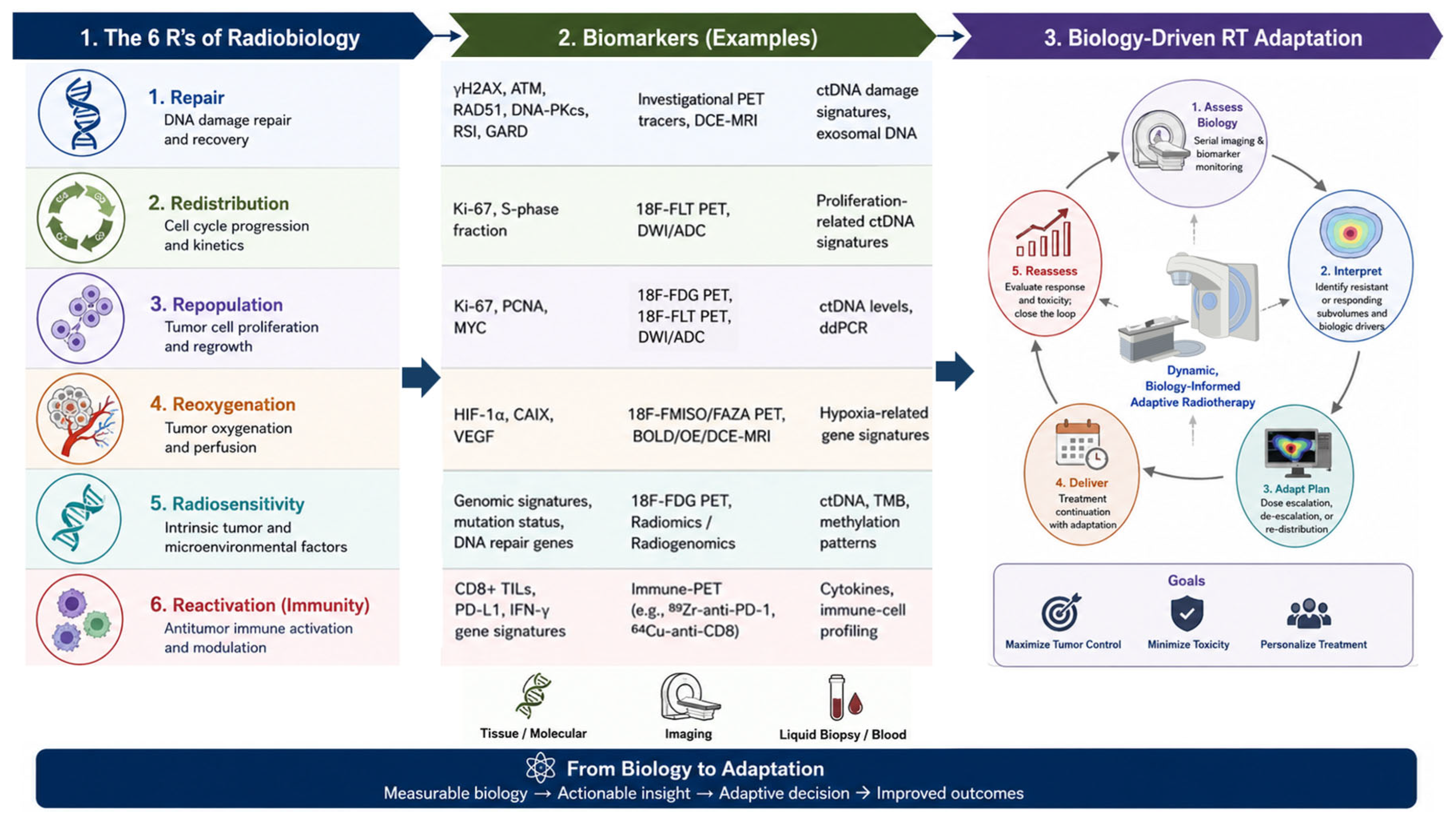
Figure 2.
A conceptual roadmap for the evolution of biological-guided ART. The field is progressively transitioning from biomarker discovery and validation, and multimodal biological integration toward real-time monitoring, biologically guided adaptive intervention, and ultimately systems-level adaptive oncology. Enabling infrastructure, including standardization, quality assurance, clinical validation, shared databases, multidisciplinary collaboration, and workforce development, will be essential for clinical implementation.
Figure 2.
A conceptual roadmap for the evolution of biological-guided ART. The field is progressively transitioning from biomarker discovery and validation, and multimodal biological integration toward real-time monitoring, biologically guided adaptive intervention, and ultimately systems-level adaptive oncology. Enabling infrastructure, including standardization, quality assurance, clinical validation, shared databases, multidisciplinary collaboration, and workforce development, will be essential for clinical implementation.
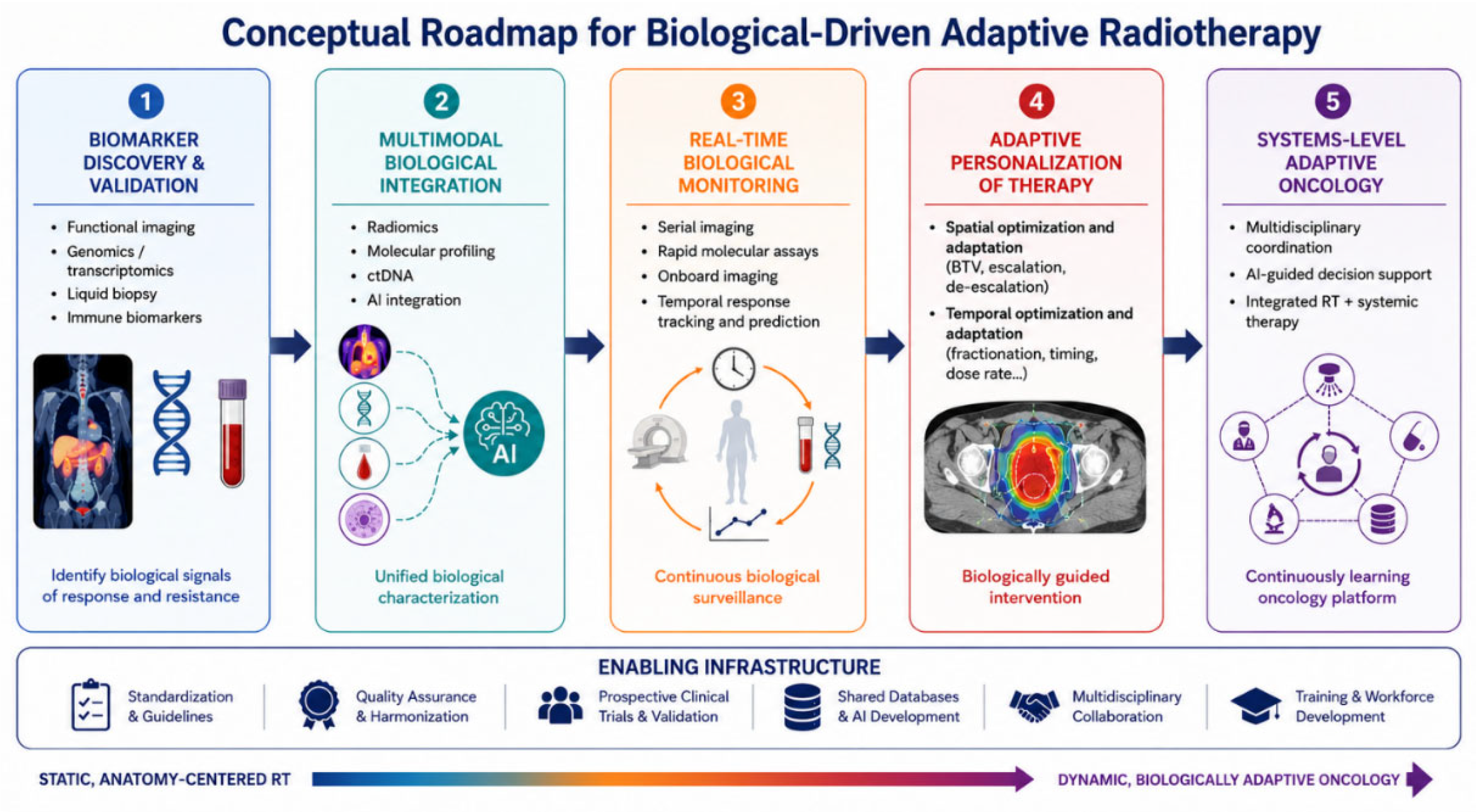

Table 1.
A brief comparison of anatomy-guided ART and biology-guided ART.
| Feature | Anatomy-Guided ART | Biology-Guided ART |
|---|---|---|
| Primary Drivers | Geometric variations, tumor shrinkage, weight loss, and organ filling, deformation and motion | Physiological dynamics, metabolic activity, radiosensitivity, hypoxia, proliferation, receptor expression, immune reactivation, and microenvironment |
| Typical Measurement Tools | CT/CBCT, and structural MRI | PET (FDG, FMISO, FLT...), physiological and functional MRI (DWI, PWI, BOLD...), ctDNA and cfDNA liquid biopsies, gene expression profiles |
| Margin Baseline | Population-based margins (e.g., van Herk formula) are replaced by patient-specific adjustments | Biological target volume (BTV) delineation and adaptive dose painting |
| Clinical Objectives | Dose fidelity, margin reduction, and sparing of OARs | Overcoming radiation resistance, selective dose escalation/deescalation, dynamic dose modulation, and multidisciplinary synergy |
| Workflow Action | Re-contouring, margin reduction, dose re-optimization | Dose painting, escalation/de-escalation, fractionation changes |
Disclaimer/Publisher’s Note: The statements, opinions and data contained in all publications are solely those of the individual author(s) and contributor(s) and not of MDPI and/or the editor(s). MDPI and/or the editor(s) disclaim responsibility for any injury to people or property resulting from any ideas, methods, instructions or products referred to in the content. |
© 2026 by the authors. Licensee MDPI, Basel, Switzerland. This article is an open access article distributed under the terms and conditions of the Creative Commons Attribution (CC BY) license (http://creativecommons.org/licenses/by/4.0/).
Copyright: This open access article is published under a Creative Commons CC BY 4.0 license, which permit the free download, distribution, and reuse, provided that the author and preprint are cited in any reuse.