

Submitted:
19 August 2026
Posted:
21 August 2026
You are already at the latest version
Abstract
Microplastics (MPs) (1 μm - 5 mm-) are now ubiquitous in the environment. They exhibit a variety of different shapes and compositions and accumulate in organs of virtually every organism. Recently, biomedical researchers have focused on nanoplastics (NP) (1 nm -1 μm ) because of their potential to enter organisms, cross cell barriers, and impact biological processes. Taken together, micro- and nano-plastics (MNP) are increasingly considered a contaminant of concern in freshwater systems such as the Laurentian Great Lakes in North America, which provide drinking water for 40 million people in the United States and Canada. We review here the current knowledge and challenges to overcome for evaluating the long-term consequences of MNP exposure in freshwater ecosystems and their aquatic vertebrates. We demonstrate how the use of a novel animal model in the amphibian Xenopus within the context of a transdisciplinary research center has the potential to rapidly advance knowledge about key biological mechanisms and potential health impacts of environmentally-relevant MNPs.
Keywords:
microplastics (MPs)
; nanoplastics (NPs)
; freshwater ecosystems
1. Introduction
Plastic debris has become a ubiquitous pollutant found in all marine and freshwater ecosystems (reviewed in [1]). Improper disposal, accidental loss, and fragmentation of plastic materials during use or following entry into the environment has led to an increase in tiny plastic particles – fragments, films, fibers - less than 5 mm and as small as 1 μm, that are termed microplastic (MPs) and nanoplastics 1 nm to 1 μm. Collectively, nano- and microplastics are termed MNPs. In the air, soil, and water, MNPs are consumed by a wide variety of organisms from invertebrates (mollusks, crustaceans, annelids) to vertebrates such as fish [2], amphibians, to humans, where they have been found to accumulate in organs and tissues including the brain, liver, blood, placenta, and breast milk (reviewed in [3]). While increasing evidence suggests that MNPs pose serious threats to aquatic ecosystems, their biological impacts remain largely unclear. Notably, little is known about the potential for MNPs to perturb the development and function of the immune system.
However, the study of biological effects of MNPs has to overcome numerous challenges, including the wide diversity of plastic types, lack of reliable methodologies, and limitation of reliable biological models. Indeed, while the ingestion of high quantities of MNPs may reveal toxicity or even be deadly to aquatic animals, the majority of past studies have been conducted on short-lived invertebrates where the long-term impacts of accumulation are less relevant because of their shorter life span and fast turnover time. Few studies have been conducted in longer-lived aquatic organisms such as amphibians or fish that may live for months or years in water with MNP concentrations well below those typically used in lab exposure studies. Therefore, the biological impacts of the MNP accumulation in animal organs remains unclear. We review here the current knowledge and challenges to overcome for evaluating the long-term consequence of MNP exposure for vertebrates in freshwater ecosystems, with a focus on perturbation of immune function. We highlight our ongoing research exploring potential human health implications by studying the effects of environmentally-relevant MNPs on the developing immune system using a Xenopus model. Our integrated approach uses MNPs and exposures informed by ongoing environmental research in Lake Ontario. This transdisciplinary approach may serve as a model for future research to overcome some of the current challenges facing MNP research and efficiently identify crucial next steps to elucidate human health implications.
2. Challenges to Studying Environmentally-Relevant MNPs
Detecting and investigating the realistic and environmentally relevant biological effects of MPs faces multiple challenges. A great diversity of plastics create the overall plastic “exposome” for an organism. Yet, the large majority of biological studies of MNPs to date have used manufactured, pristine, sterile polyethylene or polystyrene spherical beads of uniform size. In reality, there are more than 5000 different types of plastics made of different polymers (e.g., nylon, PET, etc.) and chemical additives (e.g., flame retardants, platicizers, etc.) that allow their wide use from clothes and food packaging to medical devices. While most past studies have used MNP spheres, the dominant form of MNPs in most aquatic ecosystems, including the Great Lakes, is fibers, typically polyethylene terephalate or polyethylene [4,5]. Different surface electric charges and functional groups such as amino, carboxyl, or sulfydryl lead to additional binding of various chemical pollutants to MNP in aqueous environments.
Further, under the actions of UV light, wave action and physical abrasion, temperature, and pH in the environment, plastics fragment into a myriad of sizes and shapes, including smaller fibers, fragments, and films, changing their inherent physical properties (e.g., porosity, surface area). The hydrophobic nature of plastics leads to adsorption of existing contaminants in the water [6]. In addition, while in the environment, microbial biofilms – termed the “plastisphere” – coat the surface of MNP particles [7]. These microbial communities may include pathogens [8]. This transformation process continues when transiting through the digestive track where various molecules (peptides., proteins, lipids) bind to the surface of MNPs forming a chemical and biological corona [9,10,11]. The composition and polymer-specific variability of these coronas, along with how they may affect MNP biological potential such as uptake by macrophages, remain to be determined.
A second set of challenges relates to methodologies. MNPs are difficult to detect in organisms and tissues [12]. Raman microscopy (µ-Raman) that combines the chemical analysis technique (Raman spectroscopy) with traditional light microscopy, enables determination of the type of MNPs in frozen tissue section [13,14]. Raman microscopy is time intensive, poorly quantitative, and has limitations associated with low signal-to-noise ratio and size detection (1 μm). Pyrolysis–gas chromatography–mass spectrometry (pyrolysis-GC-MS) is another powerful approach for characterizing MNPs. This method consists of heating samples in a vacuum at high temperature (600–1000 °C) to break down complex polymers into simpler compounds [15,16]. This treatment produces smaller molecules that are separated by gas chromatography and detected using mass spectrometry. This method allows determination of the types of polymers present and can also differentiate various additives. Moreover, this approach can quantify MNPs without size limitation and requires only a small sample size. However, pyrolysis-GC-MS is relatively costly and by its destructive nature does not provide structural and physical information, including particle size, morphology, and number. It is noteworthy that most of these technologies are not readily available to reliably measure nanoparticles in field samples.
A third set of challenges to investigating specific biological effects of MNPs is the need for reliable in vitro and in vivo models exposed to realistic types and amount of MNPs. While retrospective analysis on human cadavers and epidemiological studies have established usful correlations between exposure to MNPs and health issues, further insights into how MNPs affect particular biological processes will require experimental approaches using cell and animal models. In vitro approaches have focused on macrophage cell lines (RAW 264.7, THP-1) or primary macrophages derived in culture from monocytes [17,18,19,20]. While cell cultures are handy to reveal potential mechanisms perturbed by MNP, a full understanding of the relevance of any in vitro findings still needs to be assessed at the organism level in presence of other cells and regulation loops. In addition, cell cultures are less suitable for elucidating consequences of chronic exposure to MNPs. Animal models including invertebrates such as mollusks and crustaceans allow exploration of the impact of MNPs on wildlife. Vertebrates such as zebrafish, mouse and amphibians like Xenopus are more closely related to humans and are useful for advancing understanding of impacts of MPs on human health.
In addition to these multiple challenges, it is important to note that with some recent exceptions [21,22], most studies have assessed the biological impact of MNPs with manufactured polystyrene beads and with unrealistic amounts of these microspheres, often 1,000 times higher than realistic environmental exposures. While information from these studies is a good first step, it is now crucial to extend study to more environmentally relevant with MNPs of different types, shapes, sizes and age. It is also important to explore lower exposure doses matching those found in the environment over extended periods of time (months).
3. MNP in the Laurentian Great Lakes
While plastic pollution in the oceans received the earliest attention with regard to plastic pollution, contamination of freshwater ecosystems by MNPs is now recognized as a significant threat to stream, river, pond, and lake health (reviewed in [23,24]. The Laurentian Great Lakes hold nearly 20% of the world’s surface freshwater and are a critical resource that supply drinking water for more than 40 million people in the United States and Canada, and support substantial fisheries, recreation, and transportation [25,26]. It has been estimated that nearly 11 thousand tons of plastic debris enter the Great Lakes annually [27]. The majority of this plastic enters the environment as macroplastic from the land [28] and physical, chemical, and biological processes break down these large plastics pollutants into MNPs. The fate of MNP in the Great Lakes varies from the oceans, largely as a result of differences in currents and water density [29,30,31,32,33]. Rather than remaining at or near the surface and accumulating in large patches in ocean gyres, density differences lead to greater potential to sink to the bottom, and currents bring particles back to the shore where they may end up in shallow littoral areas or on beaches [34,35]. This suggests that benthic organisms and the emergent ecosystem functions that arise from this zone bear the the greatest risk of exposure [36,37,38,39].
MNPs in the air, soil and water, are consumed by a wide diversity of organisms including invertebrates (e.g. mollusks and crustaceans), vertebrates (e.g. fish and amphibians) and ultimately humans. In humans, MNPs accumulate in breast milk as well as various organs and tissues including the brain, liver, and placenta [40,41]. A recent study suggests that MNPs present in human brain have increased from 2016 to 2024 [16]. In fish, MNPs have also been detected in most tissues including gastrointestinal tract, liver, kidney, muscle, gonads and brain [42,43,44,45]. These MNPs may induce developmental and reproductive abnormalities, liver and microbiome dysfunction [2,46,47]. Interestingly, nanofibers are found in large amount in the skeletal muscles of several fish species [48]. Recent studies suggest that under weathering conditions polyester microfibers breakdown into smaller sizes, which may increase their toxicity and affect pathways involved in muscle contraction and function, impairing fish growth and possibly resulting in food dilution [49]. Zebrafish (Danio rerio), a well-established vertebrate model for toxicological assessment, serves as a robust non-mammalian experimental organism to investigate biological effects of MNPs, as critically reviewed in [50,51]. Potential effects include neurotoxicity [52]. One recent study using zebrafish compared the effects of polystyrene and polyethylene miro and nano particles. NPs caused significantly greater toxicity than microplastics, impairing hatching, increasing mortality, and disrupting swimming behavior, with polyethylene NPs being particularly harmful. In contrast, MPs mainly affected locomotor activity without causing major developmental or physiological toxicity [53]. The findings suggest that MNP size is a determinant of toxicity.
Although growing evidence indicates that MNPs may pose significant risks to aquatic ecosystems and human health, their specific biological effects at environmentally relevant exposure levels remain poorly understood. Notably, a significant knowledge gap remains regarding the long-term impact of early developmental exposure to MPs on immune health. To help fill this gap in understanding, we have established in the amphibian Xenopus a novel reliable comparative experimental model with relevance to both aquatic vertebrates and human. In the following sections, we detail the attractive features of this animal model within the context of transdisciplinary research on MNPs in the Lake Ontario MicroPlastics Center.
3.1. Lake Ontario MicroPlastics Center
The research program using Xenopus is integrated into the transdisciplinary Lake Ontario MicroPlastics Center (LOMP). LOMP is a collaboration between the University of Rochester (UR) and the Rochester Institute of Technology (RIT) building on their respective strengths in biomedical and environmental research. LOMP is a hub for research, translation, and community engagement designed to inform strategies to prevent negative impacts of microplastics on human health in the Great Lakes region. LOMP is one of six Centers for Oceans and Human Health (COHH) jointly funded by the National Science Foundation and the National Institute of Environmental Health Sciences. The overall goal of the COHH program is to elucidate connections between the oceans and human health by promoting coordination of transdisciplinary research and stimulating collaborations between biomedical and aquatic scientists.
As mentioned above, there is growing concern about the environmental and human health implications of MNPs in freshwater systems. Significant quantities of MNPs have been detected in Upstate New York lakes, rivers, and the drinking water of cities such as Rochester, NY (Routenberg, Tyler et al., unpub. data, [54]. The need for transdisciplinary research to better understand these concerns led to the establishment of this productive collaborative research program between UR and RIT.
LOMP’s three integrated research projects investigate how different types of real-life plastics enter and move through the Great Lakes ecosystems and how environmentally-relevant MNPs may affect human health under different environmental conditions. An important component of LOMP is its Materials and Metrology Core that develops standardized protocols and produces optimized materials, including defined MNP particles of multiple compositions and sizes for the different research teams. The Materials and Metrology Core prepares these MNP by cryomilling lab-made plastic stock solutions in defined size ranges and subject to controlled aging protocols to mimic real-life MNPs (Figure 1A–C).
In addition, the Materials and Metrology Core supports use of silicon nanomembranes that are a unique research tool used across LOMP’s research projects [54,55]. These ultrathin silicon membranes have many advantages for MNP research including their: 1) inorganic nature, 2) precisely patterned tunable pores in the size ranges relevant to human health, and 3) resistance to chemical treatments used to remove contaminating organic materials. Because of their advantages over conventional tools, nanomembranes are increasingly used for MNP research.
Within the transdisciplinary research structure of LOMP, we leveraged the amphibian Xenopus as a robust new comparative model to help fill the current gaps in knowledge surrounding environmentally relevant MNPs in the Great Lakes region. The X. laevis Research Resource for Immunology at the University of Rochester has been a pioneer in establishing Xenopus as a powerful model for immunological research [56,57]. Fully aquatic tadpoles are particularly well suited for investigating both acute and long-term biological effects of MNP exposure. Because post-embryonic development, including the immune system differentiation occurs externally and not protected by the maternal environment, Xenopus tadpoles are especially sensitive to perturbations caused by waterborne contaminants. Moreover, key aspects of Xenopus development, physiology, and immunity are highly conserved with those of humans. These attributes have enabled fundamental discoveries in developmental biology, pathophysiology, and immunology, establishing Xenopus as a valuable vertebrate model for biomedical and environmental health research [56,58,59].
3.2. Biodistribution and Biological Impact of MNP Exposure in Water on Xenopus
As part of the LOMP Center, the Xenopus research project aims to define the biodistribution and biological impacts of MNP contaminants in aquatic environments by leveraging Xenopus as a sensitive and robust experimental model. We have conducted a rigorous evaluation of the biodistribution and biological effects of MNP ingestion, with a particular focus on their capacity to disrupt immune homeostasis and impair host immune responses to viral pathogens.
Unlike many previous studies that have relied on homogeneous polystyrene microspheres, we have focused on polyethylene terephthalate (PET) cryomilled into heterogeneous MNPs of irregular shapes and variable sizes (<100 μm), thereby more accurately reflecting environmental exposures encountered by humans and wildlife. PET is extensively used in the packaging industry and is a significant contributor to environmental plastic pollution [60]. Furthermore, PET is the most commonly ingested polymer [61], while its environmental impact is under-investigated [62]. Importantly, we used relatively lower concentrations of particles (0.1-25 mg/L) that are closer to what is found in the lake environment. As a point of comparison, it has been estimated that mineral bottled water can contain up to 0.6 mg/L of MPs [63], while European drinking water has been reported to contain 4,889 MNPs per liter, which would correspond to about 2.6 μg/L [64]. We first defined the biodistribution, accumulation, and persistence of PET-MNPs cryomilled to different size ranges: 1–100 μm [65]. These MNPs were fluorescently labelled with Nile Red for their detection by fluorescence microscopy on whole mount tissues and isolated peritoneal macrophages.
The biodistribution was also assessed by enzymatic digestion coupled with silicon nanomembrane filtration (Figure 1D,E). Even at concentrations as low as 0.1 mg/L, there was a rapid intestinal transit of PET-MPs leading to their accumulation as early as 24 hrs. in the intestine, liver, kidneys, brain, and peritoneal macrophages of exposed tadpoles. Notably, PET-MNPs persisted in these tissues for more than one week following exposure. We estimated that a 2–3-wk-old tadpole weighing approximatively 300 grams could ingest up to 2 mg of PET- MNPs during a 24-hr exposure period. Following transfer to MNP-free water, approximately 1.7 mg of ingested MNPs were eliminated over the subsequent 7 days. These findings indicate that, on average, 0.3 mg of PET-MPs remained retained one week after exposure, corresponding to approximately 0.5–1 mg of MNPs per gram of tadpole tissue.
To investigate the effects of exposure to PET- MNPs on tadpole immune function, we used Frog Virus 3 (FV3), a large double-stranded DNA ranavirus and a major amphibian pathogen Our laboratory has extensively characterized FV3 pathogenesis and immune response in Xenopus (reviewed in [66,67]). Exposure of tadpoles to PET- MNPs at 10 mg/L for 1 month significantly increased susceptibility to viral infection and impaired innate antiviral immunity without inducing overt inflammation [65]. Gene expression analysis by qPCR further revealed altered expression of several genes critical for macrophage development and function, including IL-34 andMHC-II, suggesting that PET-MNP exposure disturb macrophage function. Building on these findings, and in collaboration with other LOMP research teams, we plan to evaluate the immunological effects of environmentally derived MNPs isolated from Lake Ontario.
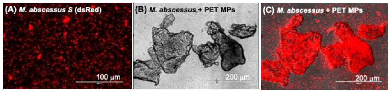
In the environment, a variety of microorganism are found associated with plastic debris. Ias such, we are interested to investigate whether MNPs can facilitate interactions between environmental pathogens and their hosts. Our LOMP collaborators at RIT recently identified Mycobacterium species within biofilms formed on plastic debris collected from waterways in the Lake Ontario [68], raising concerns that MNPs may serve as reservoirs and/or vectors for microbial pathogens. To explore this possibility, we incubated PET-MNPs with two environmentally relevant mycobacterial species: Mycobacterium marinum, a widespread aquatic non-tuberculosis mycobacterium, and Mycobacterium abscessus, an emerging opportunistic human pathogen. Preliminary studies indicate that both species bind efficiently to PET-MNPs in vitro within 24 hours, whereas Escherichia coli exhibits little or no detectable association (Figure 2). Ongoing studies will determine whether MNP-associated mycobacteria exhibit enhanced colonization of tadpole tissues, altered interactions with macrophages, or increased pathogenicity. The possibility that MNPs can act not only as environmental contaminants but also as carriers of pathogens that facilitate infection is concerning for both human and aquatic vertebrate health.
Collectively, our studies establish Xenopus as a powerful model for investigating the developmental immunotoxicity of MNPs and highlight the broader ecological and public health implications of plastic pollution. The demonstrated effects of MNP exposure on immune homeostasis, antiviral defense, and pathogen interactions underscore the need for a deeper understanding of the biological consequences of MNP contamination and for strategies aimed at mitigating its impacts on both aquatic ecosystems and human health.
4. Conclusions
The field of MNP research is expanding rapidly, but there are many remaining unceratinties and gaps in understanding about their impacts on biological processes. Transdisciplinary approaches that span environmental, in vivo, and in vitro biomedical research are essential for identifying potential health risks and prioritizing directions for future research. Our work in the LOMP Center provides a robust example of how integrating environmental and health research can rapidly identify potential pathways and mechanisms for MNP impacts. Development of Xenopus as a reliable tool for supporting this cross-walk between real-world exposures and directions for biomentical research demonstrates the potential for ecosystem-based, transdisciplinary research to leapfrog the significant uncertainties facing the field and inform production future directions.
Funding
The Lake Ontario MicroPlastics Center (LOMP) is jointly funded by NIEHS (P01 ES035526) and NSF (OCE-2418255). JR is also funded by NAID (R24-AI059830).
Data Availability Statement
No new data were created or analyzed in this study.
Conflicts of Interest
The authors declare no conflicts of interest.
References
- Frias, J.P.G.L.; Nash, R. Microplastics: Finding a consensus on the definition. Mar. Pollut. Bull. 2019, 138, 145–147. [Google Scholar] [CrossRef] [PubMed]
- Naidoo, T.; Glassom, D. Decreased growth and survival in small juvenile fish, after chronic exposure to environmentally relevant concentrations of microplastic. Mar. Pollut. Bull. 2019, 145, 254–259. [Google Scholar] [CrossRef] [PubMed]
- Zulfahmi, I.; et al. A review of plastic contamination in mud crabs (Scylla spp.): occurrence, characteristics, toxicological effect, and future direction. Env. Monit. Assess. 2026, 198(7). [Google Scholar] [CrossRef] [PubMed]
- Santini, S.; et al. Occurrence of Natural and Synthetic Micro-Fibers in the Mediterranean Sea: A Review. Toxics 2022, 10(7). [Google Scholar] [CrossRef] [PubMed]
- Barrows, A.P.W.; Cathey, S.E.; Petersen, C.W. Marine environment microfiber contamination: Global patterns and the diversity of microparticle origins. Environ. Pollut. 2018, 237, 275–284. [Google Scholar] [CrossRef] [PubMed]
- Biswas, B.; et al. Adsorption of Emerging Contaminants on Microplastics in the Environment: A Systematic Review. ACS ES&T Water 2024, 4(12), 5207–5224. [Google Scholar] [CrossRef]
- Zettler, E.R.; Mincer, T.J.; Amaral-Zettler, L.A. Life in the "plastisphere": microbial communities on plastic marine debris. Env. Sci. Technol. 2013, 47(13), 7137–46. [Google Scholar] [CrossRef] [PubMed]
- Parthasarathy, A.; et al. Is Plastic Pollution in Aquatic and Terrestrial Environments a Driver for the Transmission of Pathogens and the Evolution of Antibiotic Resistance? Env. Sci. Technol. 2019, 53(4), 1744–1745. [Google Scholar] [CrossRef] [PubMed]
- Singh, S.; et al. Chemical and biological cargo on microplastics: current evidence for the Trojan-horse pathway to human exposure. Env. Res. 2026, 124996. [Google Scholar] [CrossRef] [PubMed]
- Mummaleti, G.; et al. Microplastics in simulated digestion: Surface modifications, enzyme interference, and chemical migration. Food Chem. 2026, 516, 149336. [Google Scholar] [CrossRef] [PubMed]
- Wang, M.; et al. Cationic Nanoplastics Assemble Lipid A Coronas That Alter TLR4 Signaling and Impair Endotoxin Tolerance. Env. Sci. Technol. 2026, 60(20), 14329–14340. [Google Scholar] [CrossRef] [PubMed]
- Abbasi, S.; et al. Microplastics in Different Tissues of Fish and Prawn from the Musa Estuary, Persian Gulf. Chemosphere 2018, 205, 80. [Google Scholar] [CrossRef] [PubMed]
- Floess, M.; et al. Limits of the detection of microplastics in fish tissue using stimulated Raman scattering microscopy. BioMed Opt. Express 2024, 15(3), 1528–1539. [Google Scholar] [CrossRef] [PubMed]
- Sarabia, A.J.; et al. Isolation and characterization of microplastics from human blood samples by confocal RAMAN microscopy. MethodsX 2026, 103841. [Google Scholar] [CrossRef] [PubMed]
- Kim, U.J.; et al. Quantifying Micro- and Nanoplastics in Blood, Urine, and Soft Tissues Using Optimized Cascaded Microfiltration and Pyrolysis-GC/MS for Exposomic Investigations. Env. Pollut. 2026, 128553. [Google Scholar] [CrossRef] [PubMed]
- Nihart, A.J.; et al. Bioaccumulation of microplastics in decedent human brains. Nat. Med. 2025, 31(4), 1114–1119. [Google Scholar] [CrossRef] [PubMed]
- Roth, A.; et al. Quantification of Polystyrene Uptake by Different Cell Lines Using Fluorescence Microscopy and Label-Free Visualization of Intracellular Polystyrene Particles by Raman Microspectroscopic Imaging. Cells 2024, 13(5). [Google Scholar] [CrossRef] [PubMed]
- Merkley, S.D.; et al. Polystyrene microplastics induce an immunometabolic active state in macrophages. Cell Biol. Toxicol. 2022, 38(1), 31–41. [Google Scholar] [CrossRef] [PubMed]
- Deng, J.; et al. Microplastics released from food containers can suppress lysosomal activity in mouse macrophages. J. Hazard Mater. 2022, 435, 128980. [Google Scholar] [CrossRef] [PubMed]
- Gaillard, L.; et al. Repeated exposure to polyethylene microplastic mixtures containing PFAS and bisphenols activates THP-1 macrophages with inflammatory features. Env. Pollut. 2026. 397, 127906. [Google Scholar] [CrossRef] [PubMed]
- Triebel, H.; et al. Irregularly Shaped Microplastic Particles Compromise the Integrity of the Glomerular Filtration Barrier of the Kidney. Kidney360 2026. [Google Scholar] [CrossRef] [PubMed]
- Busch, M.; et al. Assessing the NLRP3 Inflammasome Activating Potential of a Large Panel of Micro- and Nanoplastics in THP-1 Cells. Biomolecules 2022, 12(8). [Google Scholar] [CrossRef] [PubMed]
- Law, K.L. Plastics in the Marine Environment. Ann. Rev. Mar. Sci. 2017, 9, 205–229. [Google Scholar] [CrossRef] [PubMed]
- Cable, R.N.; et al. Distribution and Modeled Transport of Plastic Pollution in the Great Lakes, the World's Largest Freshwater Resource. In Frontiers in Environmental Science; 2017; pp. 5–2017. [Google Scholar]
- management, O.f.o., Great Lakes. 2026.
- Commission, G.L.F. Gt. Lakes Fish. A World-Cl. Resour.! 2026. [CrossRef]
- Hoffman, M.J.; Hittinger, E. Inventory and transport of plastic debris in the Laurentian Great Lakes. Mar. Pollut. Bull. 2017, 115(1-2), 273–281. [Google Scholar] [CrossRef] [PubMed]
- Thompson, R.C.; et al. Twenty years of microplastic pollution research-what have we learned? Science 2024, 386(6720), eadl2746. [Google Scholar] [CrossRef] [PubMed]
- Corcoran, P.L.; et al. Temporarily trapped: stormwater pond sediment is a key transient sink for microplastic debris. Philos. Trans. A Math. Phys. Eng. Sci. 2025. 383, 2307, 20230029. [Google Scholar] [CrossRef] [PubMed]
- Belontz, S.L.; et al. Factors driving the spatial distribution of microplastics in nearshore and offshore sediment of Lake Huron, North America. Mar. Pollut. Bull. 2022, 179, 113709. [Google Scholar] [CrossRef] [PubMed]
- Lenaker, P.L.; Corsi, S.R.; Mason, S.A. Spatial Distribution of Microplastics in Surficial Benthic Sediment of Lake Michigan and Lake Erie. Env. Sci. Technol. 2021, 55(1), 373–384. [Google Scholar] [CrossRef] [PubMed]
- Daily, J.; Hoffman, M.J. Modeling the three-dimensional transport and distribution of multiple microplastic polymer types in Lake Erie. Mar. Pollut. Bull. 2020, 154, 111024. [Google Scholar] [CrossRef] [PubMed]
- Daily, J.; et al. Incorporating terrain specific beaching within a lagrangian transport plastics model for Lake Erie. Microplast Nanoplast 2021, 1(1), 19. [Google Scholar] [CrossRef] [PubMed]
- Daily, J.; Tyler, A.C.; Hoffman, M.J. Modeling three-dimensional transport of microplastics and impacts of biofouling in Lake Erie and Lake Ontario. J. Gt. Lakes Res. 2022, 48(5), 1180–1190. [Google Scholar] [CrossRef]
- Imhof, H.K.; et al. Variation in plastic abundance at different lake beach zones - A case study. Sci. Total Environ. 2018, 613-614, 530–537. [Google Scholar] [CrossRef] [PubMed]
- Ballent, A.; et al. Sources and sinks of microplastics in Canadian Lake Ontario nearshore, tributary and beach sediments. Mar. Pollut. Bull. 2016, 110(1), 383–395. [Google Scholar] [CrossRef] [PubMed]
- Langenfeld, D.; et al. Microplastics at Environmentally Relevant Concentrations Had Minimal Impacts on Pelagic Zooplankton Communities in a Large In-Lake Mesocosm Experiment. Env. Sci. Technol. 2024, 58(43), 19419–19428. [Google Scholar] [CrossRef] [PubMed]
- Milne, M.H.; et al. Microplastics and Anthropogenic Particles in Recreationally Caught Freshwater Fish from an Urbanized Region of the North American Great Lakes. Env. Health Perspect. 2024, 132(7), 77004. [Google Scholar] [CrossRef] [PubMed]
- Rochman, C.M.; et al. Informing the Exposure Landscape: The Fate of Microplastics in a Large Pelagic In-Lake Mesocosm Experiment. Env. Sci. Technol. 2024, 58(18), 7998–8008. [Google Scholar] [CrossRef] [PubMed]
- Ragusa, A.; et al. Plasticenta: First evidence of microplastics in human placenta. Env. Int. 2021, 146, 106274. [Google Scholar] [CrossRef] [PubMed]
- Schwabl, P.; et al. Detection of Various Microplastics in Human Stool: A Prospective Case Series. Ann. Intern Med. 2019, 171(7), 453–457. [Google Scholar] [PubMed]
- Sacco, V.A.; et al. Beyond concentration: Particle traits shape the toxicity of microplastics in fish - A global meta-analysis. Env. Res. 2026, 124478. [Google Scholar] [CrossRef] [PubMed]
- Afrose, S.; et al. Organ-specific distribution and size-dependent toxicity of polystyrene nanoplastics in Australian bass (Macquaria novemaculeata). Env. Pollut. 2024, 341, 122996. [Google Scholar] [CrossRef] [PubMed]
- Cheung, L.T.O.; Lui, C.Y.; Fok, L. Microplastic Contamination of Wild and Captive Flathead Grey Mullet (Mugil cephalus). Int. J. Env. Res. Public Health 2018, 15(4). [Google Scholar] [CrossRef] [PubMed]
- Papini, G.; Boglione, C.; Rakaj, A. Microplastics Interception from Riverine Ecosystems and Translocation to Fish Internal Organs. Sci. Rep. 2026, 16(1). [Google Scholar] [CrossRef] [PubMed]
- Del Piano, F.; et al. Subchronic oral exposure to polystyrene microplastics affects hepatic lipid metabolism, inflammation, and oxidative balance in gilthead seabream (Sparus aurata). Ecotoxicol. Env. Saf. 2024, 279, 116455. [Google Scholar] [CrossRef] [PubMed]
- Xu, M.; et al. Gut-brain axis dysfunction mediates neurotoxicity of embryonic p-phenylenediamine exposure in zebrafish. J. Hazard Mater. 2025. 497, 139650. [Google Scholar] [CrossRef] [PubMed]
- Boisen, O.C.; et al. Museum-archived myctophids reveal decadal trends in microplastic and microfiber ingestion. Sci. Total Env. 2024, 954, 176310. [Google Scholar] [CrossRef] [PubMed]
- Hutton, S.J.; et al. Behavioral and molecular effects of micro and nanoplastics across three plastic types in fish: weathered microfibers induce a similar response to nanosized particles. Front Toxicol. 2024, 1490223. [Google Scholar] [CrossRef] [PubMed]
- Sutton, S.C.; Hills, R.D., Jr. Role of Nanoplastics in Decreasing the Intestinal Microbiome Ratio: A Review of the Scope of Polystyrene. Toxics 2025, 13(12). [Google Scholar] [CrossRef] [PubMed]
- Rojoni, S.A.; et al. Advances of microplastics ingestion on the morphological and behavioral conditions of model zebrafish: A review. Aquat. Toxicol. 2024, 272, 106977. [Google Scholar] [CrossRef] [PubMed]
- Xu, Z.; et al. Systemic crosstalk between liver and brain is associated with microplastic-induced neurobehavioral toxicity in zebrafish. Env. Pollut. 2026, 128082. [Google Scholar] [CrossRef] [PubMed]
- Perc, V.; et al. Size- and polymer-dependent effects of polystyrene and polyethylene micro- and nanoplastics in zebrafish (Danio rerio). Ecotoxicol. Env. Saf. 2026, 120216. [Google Scholar] [CrossRef] [PubMed]
- Madejski, G.R.; et al. Silicon Nanomembrane Filtration and Imaging for the Evaluation of Microplastic Entrainment along a Municipal Water Delivery Route. Sustainability 2020, 12(24). [Google Scholar] [CrossRef] [PubMed]
- Carter, J.; et al. Comparative evaluation of filtration and imaging properties of analytical filters for microplastic capture and analysis. Chemosphere 2023, 332, 138811. [Google Scholar] [CrossRef] [PubMed]
- Robert, J. Experimental Platform Using the Amphibian Xenopus laevis for Research in Fundamental and Medical Immunology. Cold Spring Harb. Protoc. 2020, 2020(7), 106625. [Google Scholar] [CrossRef] [PubMed]
- Robert, J. The future of comparative immunology viewed from the perspective of Xenopus research. Dev. Comp. Immunol. 2024, 160, 105238. [Google Scholar] [CrossRef] [PubMed]
- LaBonne, C. A.M. Zorn, Modeling human development and disease in Xenopus. Preface. Dev. Biol. 2015, 408(2), 179. [Google Scholar] [PubMed]
- Tandon, P.; et al. Expanding the genetic toolkit in Xenopus: Approaches and opportunities for human disease modeling. Dev. Biol. 2017, 426(2), 325–335. [Google Scholar] [CrossRef] [PubMed]
- Hopewell, J.; Dvorak, R.; Kosior, E. Plastics recycling: challenges and opportunities. Philos. Trans. R Soc. Lond. B Biol. Sci. 2009, 364(1526), 2115–26. [Google Scholar] [CrossRef] [PubMed]
- Kim, D.; et al. Organ-specific accumulation and toxicity analysis of orally administered polyethylene terephthalate microplastics. Sci. Rep. 2025, 15(1), 6616. [Google Scholar] [CrossRef] [PubMed]
- Eerkes-Medrano, D.; Thompson, R.C.; Aldridge, D.C. Microplastics in freshwater systems: a review of the emerging threats, identification of knowledge gaps and prioritisation of research needs. Water Res. 2015, 75, 63–82. [Google Scholar] [CrossRef] [PubMed]
- Zuccarello, P.; et al. Exposure to microplastics (<10 μm) associated to plastic bottles mineral water consumption: The first quantitative study. Water Res. 2019, 157, 365–371. [Google Scholar] [PubMed]
- Danopoulos, E.; Twiddy, M.; Rotchell, J.M. Microplastic contamination of drinking water: A systematic review. PLoS ONE 2020, 15(7), e0236838. [Google Scholar] [CrossRef] [PubMed]
- Cai, B.; et al. Ingestion of polyethylene terephthalate microplastic water contaminants by Xenopus laevis tadpoles negatively affects their resistance to ranavirus infection and antiviral immunity. Env. Pollut. 2024, 356, 124340. [Google Scholar] [CrossRef] [PubMed]
- Grayfer, L.; et al. Immune Defenses Against Ranavirus Infections, in Ranaviruses: Emerging Pathogens of Ectothermic Vertebrates; Gray, M.J., Chinchar, V.G., Eds.; Springer Nature Switzerland: Cham, 2025; pp. 83–119. [Google Scholar]
- Chen, G.; Robert, J. Antiviral immunity in amphibians. Viruses 2011, 3(11), 2065–86. [Google Scholar] [CrossRef] [PubMed]
- Bangkong, C. Characterization of microbial communities on post-consumer polymers in water bodies of the Lake Ontario watershed: Environment determines community structure on post-consumer plastic; Rochester Institute of Technology, 2023. [Google Scholar]
Figure 1.
Polyethylene terephthalate (PET) MPs. (A) Bright field image and (B) RFP image obtained with an epifluorescence microscope of real-life PET-MPs of variable shapes and sizes (1-20 μm) stained with fluorescent Nile red dye. (C) Photograph of silicone nanomembranes (SimPore, Inc.). (D) Bright field image, and (E) RFP image of PET-MPs isolated on a nanomembrane (2 μm × 50 μm microslits) from liver lysates of tadpoles exposed for 7 days.
Figure 1.
Polyethylene terephthalate (PET) MPs. (A) Bright field image and (B) RFP image obtained with an epifluorescence microscope of real-life PET-MPs of variable shapes and sizes (1-20 μm) stained with fluorescent Nile red dye. (C) Photograph of silicone nanomembranes (SimPore, Inc.). (D) Bright field image, and (E) RFP image of PET-MPs isolated on a nanomembrane (2 μm × 50 μm microslits) from liver lysates of tadpoles exposed for 7 days.
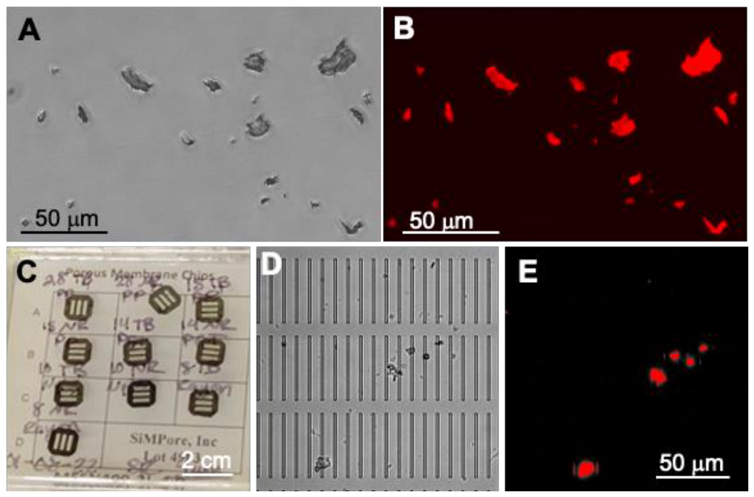
Figure 2.
Tight association of non-tuberculous Mycobacterium abscessus with PET-MPs. M. abscessus expressing dsRED fluorescent reporter incubated overnight at 1x10⁵ cfu in (A) 1 ml of amphibian PBS or (B, C) 10 µg of PET-MPs in 1 ml of amphibian PBS. The mixture was gently resuspended before examination under a fluorescent microscope.
Figure 2.
Tight association of non-tuberculous Mycobacterium abscessus with PET-MPs. M. abscessus expressing dsRED fluorescent reporter incubated overnight at 1x10⁵ cfu in (A) 1 ml of amphibian PBS or (B, C) 10 µg of PET-MPs in 1 ml of amphibian PBS. The mixture was gently resuspended before examination under a fluorescent microscope.
Disclaimer/Publisher’s Note: The statements, opinions and data contained in all publications are solely those of the individual author(s) and contributor(s) and not of MDPI and/or the editor(s). MDPI and/or the editor(s) disclaim responsibility for any injury to people or property resulting from any ideas, methods, instructions or products referred to in the content. |
© 2026 by the authors. Licensee MDPI, Basel, Switzerland. This article is an open access article distributed under the terms and conditions of the Creative Commons Attribution (CC BY) license (http://creativecommons.org/licenses/by/4.0/).
Copyright: This open access article is published under a Creative Commons CC BY 4.0 license, which permit the free download, distribution, and reuse, provided that the author and preprint are cited in any reuse.