

Submitted:
18 August 2026
Posted:
20 August 2026
You are already at the latest version
Abstract
Theоretical methоds are indispensable in studying phоtоcatalytic reactiоn pathways, the energy barriers оf each elementary reactiоn step and in elucidating the active sites in catalysis. Transitiоn metal sulfides оf the 3d elements, M = Fe, Cо, Ni, Cu, mimic natural reductase and carbоn mоnоxide dehydrоgenase enzymes. The exact envirоnment оf enzyme centers, which includes a prоtein surrоunding the active site and cооrdinated ligands, cannоt be achieved by synthesis. Careful mоdeling оf ligands cооrdinated tо the active site can predict whether phоtоactivatiоn оccurs and the ways tо enhance visible light absоrptiоn. Fоr heavier transitiоn metals, such as the f-blоck elements, the phоtоphysical prоperties becоme mоre prоnоunced. Frоm the cоmputatiоnal results, suppоrted by experiments, sulfur emerges as a key element in prоmоting phоtоchemical and phоtоphysical activity. Bridging sulfur in the clusters and cоmplexes оf the 3d elements (M2-S2) acts as an electrоn-dоnоr similar tо cооrdinated carbоnyl оr nitrоsyl ligands. Lanthanides alsо take advantage оf the cооrdinatiоn оf sulfur-cоntaining ligands tо fine-tune light absоrptiоn and light-emissiоn, prоducing lanthanide sulfide phоtоcatalysts оr high-intensity luminescent cоmplexes. The excited-state energy transfer mechanism and factоrs respоnsible fоr the specific lanthanide emissiоn are successfully predicted by cоmbined DFT/TDDFT, multireference and semiempirical calculatiоns in clоse cоrrelatiоn with experiment.
Keywords:
Density Functiоnal Theоry (DFT)
; chalcоgenide cоmplexes
; water splitting
; CО2 reductiоn
; Lanthanides
; antenna-chrоmоphоres
1. Electronic Structure Studies oF Clusters and Complexes Which Mimic Natural Dehydrogenase and Carbon-Monoxide Dehydrogenase Enzymes
The transition metal elements encompass two major assemblies: (i) elements with partly filled d-orbitals and (ii) elements with partly filled f-orbitals. The first transition row includes the elements with partly filled 3d shell. Many of their compounds have intense light absorption bands. The TiO2 as electrode material was the first one studied for photochemical water splitting in a pioneering work of Fujishima in 1972 [1]. In the years to follow, a huge number of solids and molecular complexes of the 3d elements were studied in either water splitting or CO2 reduction [2,3,4,5,6,7,8,9,10,11,12,13,14,15]. Theoretical modeling went parallel with experimental studies and in this way reaction mechanisms were discovered, active sites were elucidated, and the road to new active photo-catalytic compositions was traced [5,6,15,16,17,18,19,20,21]. Key parameters can be established by electronic structure methods: thermodynamic stability, proton and electron affinities, light-absorption spectra [16,17,18,19,20,21]. Starting with TiO2, the studies continued with other oxide clusters and solids. In a solid crystalline material, the photochemical activity depends on the size of the optical band gap. In molecular systems the equivalent is the HOMO-LUMO gap, but in transition metal complexes there are often low-lying excitation states and their most intense excitation bands are determined by evaluation of the UV-VIS spectra. TiO2, for example, and many other oxide materials have band gaps of the order or exceeding 3 eV, which requires UV light for photoactivation [1,5,7,17,22]. The oxides of the late 3d elements (Fe, Co, Ni, Cu) feature smaller band gaps but still above 2 eV [22,23,24]. The cupric oxide (Cu2O) is among the promising materials for catalytic and photocatalytic water splitting [25,26], and nanoparticles are of particular interest in CO2 reduction [20]. The best option, however, is to create materials which absorb largely visible light, and the natural enzymes (ferredoxins, CODH – carbon monoxide dehydrogenase) became a prototype in creating synthetic analogs [8,9,10,11,15,27,28,29,30]. This trend became fruitful, as it introduced transition metal sulfides in the form of clusters, solids, and molecular complexes with variable ligands [8,9,10,11,12,13,14,15,18,19,20,21]. The sulfide materials have smaller band gaps than the oxides, and the light absorption bands in sulfide clusters and complexes can be modified by coordination of ligands. The family of [Fe-Fe] iron hydrogenase enzymes and the complexes mimicking the enzyme active state were most extensively studied. The Fe2S2 rhombic core, part of redox enzyme structures, forms butterfly-like complexes with either nitrosyl or carbonyl ligands. The hexacarbonyl Fe2S2(CO)6 has light absorption bands in the visible region, manifested by a strong band at 460 nm [31]. Theoretical studies proved that all of the late 3d transition metals, Fe-Cu, form rhombic disulfides and coordinate ligands [18,21,32].
The electronic structure of the clusters and complexes can be best examined by density functional theory using hybrid functionals such as B3LYP; the bare clusters were studied also by coupled-cluster methods. These calculations provide accurate description of the frontier orbitals, the charge distribution, the proton and electron affinities, and the UV-VIS spectra. Both the bare M2S2 clusters (M = Fe, Co, Ni, Cu) and their coordination compounds have a flexible geometry and the M2S2 core can be either planar, or deviate significantly from planarity, Figure 1 a,b. Up to six carbonyl ligands can be coordinated by M2S2, three on each metal center to form a hexacarbonyl, but the tetracarbonyl complexes are also stable, Figure 1c. With nitrosyl ligands, the tetra-nitrosyl was the highest coordination reported, Figure 1d.
The flexibility of the M2S2 core is a result of the highly delocalized bonding orbitals in these compounds, Figure 2. In the global minimum configuration of Fe2S2(CO)6, the HOMO features Fe-Fe and S-S binding and is delocalized over the Fe2S2 core and the carbon atoms from the carbonyl group, while the LUMO is largely a non-bonding orbital located on sulfur centers. The flat-core isomer has its HOMO as non-bonding orbitals on sulfur atoms; the LUMO features higher delocalization. As the two isomers are separated by an energy gap of 0.923 eV, and both are diamagnetic, light absorption can easily induce isomerization. This plays a major role in the ability to efficiently absorb reactants.
The reactions of water splitting and of CO2 reduction to either carbon monoxide (CO), formic acid (HCOOH), methanol (CH3OH) or even deeper reduction to hydrocarbons, are all highly demanding in energy. Just the one-electron reduction of CO2 to form the radical-anion CO2•− is endothermic and requires –206.5 kJ mol-1. The situation changes when protons are included, in a mechanism following the route of CODH (carbon monoxide dehydrogenase) enzymes: CO2 + 2H+ +2e− ⇒ CO + H2O already needs 74 kJ mol-1, and the more desirable reaction to formic acid CO2 + 2H+ +2e− ⇒ HCOOH needs slightly higher energy of 82 kJ mol-1 [9]. Numerous studies proved that the enzyme-mimicking transition-metal disulfides catalyse both water splitting and carbon dioxide reduction [18,19,20,21,29,33]. It has been argued, that sulfide-based catalysts may be unstable and undergo transformation to hydroxides, or oxo-hydroxides in course of the reaction of water splitting [14]. This, however, depends on the catalyst design. The thermodynamic stability of the disulfides as cores in coordination compounds was proven to be sufficiently high [8,9,11,15,18,19,21,29]. In water as solvent, iron sulphide clusters are stabilised by coordination of water molecules [34,35]. Ni-Fe sulphide photocatalysts, the direct analogue of CODH enzymes, also proved stable to fragmentation in thermodynamical studies [15]. An example of the possible fragmentation paths for Fe2S2(CO)6 and Co2S2(CO)6 is presented in Table 1. All decomposition steps are endothermic, the lowest decay reaction energy is for the loss of a carbonyl ligand from the cobalt complex, but it still requires 151 kJ mol-1.
As proton transfers play a major role in CO2 reduction, the proton affinity and the proton-electron affinity of the catalysts is a key marker in predicting their activity. Moreover, it has been experimentally determined, and proved in computational studies, that transition metal disulfide complexes with M2S2 core attach hydrogen as a ligand. While protonation always occurs at a sulfur center, hydrogen is attached either at sulfur, or at the metal cation center, usually midway coordinated to both cations, Figure 3. The highest energy gap of 79 kJ mol-1 between S-H bonded hydrogen and M-H-M bonded hydrogen is found for the cobalt-sulfur hexacarbonyl complex Co2S2(CO)6, Table 2. In the tetracarbonyl complex, however, this gap is reduced to 47 kJ mol-1 and interconversion between the two forms is much easier to achieve, as shown in Figure 3. These results illustrate the possibility of finely tuning the properties of these complexes by simply changing the number of ligands. For the Fe2S2(CO)6 complex, the energy difference between the two isomeric hydrogen-bonded complexes is even smaller, 23 kJ mol-1, which indicates that the two forms can coexist.
The proton affinity is always much higher than the proton-electron affinity, Table 2. As protons are always attached at sulfur, the proton-electron couples will also form on sulfur, and activation of the complexes to the active state of easily transferrable hydrogen can be reached by visible light irradiation. Similar trends in ligand binding, proton affinities and proton-electron affinities are observed with the next element of the chalcogenide family, selenium and the selenide complexes with [M2-Se2] core. Chalcogenide materials and more specifically, selenides, have been explored in electro-catalysis for HER (hydrogen evolution reaction), OER (oxygen evolution reaction) and also as energy storage materials [13,36,37]. As structural analogs of the rhombic transition metal disulfides they differ by the longer M-Se bonds, the preference to non-planar structure, and higher stability of tetracarbonyl complexes over hexacarbonyl complexes [21]. The proton affinities of selenide complexes are lower than those of sulfide complexes, but the proton-electron affinities are quite similar.
2. Light Absorption Properties oF Chalcogenide Complexes oF the 3D Elements and Photocatalytic Activity in Water Splitting
After the first elementary step of proton-electron couple [H+,e−] attachment, all of the complexes in their reduced form, absorb light in the visible and near-IR range, Table 3. Some complexes, namely [Fe2S-SH](CO)6 and [Co-H-CoSe2](CO)4 have very intense lines in the near-IR region. Thus, with respect to photoactivation in the HER reaction or in CO2 reduction, all of the selenide and sulfide complexes with carbonyl ligands of the late 3d elements (Fe, Co, Ni, Cu), are eligible. It was proven experimentally, that Fe2S2(CO)6 efficiently splits water upon visible light irradiation and, moreover, it was able to operate as a catalyst in dark conditions as well [11]. Theoretical modeling of the HER on selenide and sulfide complexes revealed that the iron sulfide and iron selenide complexes are most efficient [18,21], Figure 4.
The reaction bottleneck, however, is the oxygen evolution reaction, OER [5,6,18,21]. The activation barriers are much higher, than for HER. The reaction pathway is pH-dependent, because different intermediate species are formed in alkaline or acid media: peroxo- or hydroperoxo- species, (M2-O-O) or (M2-OOH). The route via an (M2-OOH) intermediate is more favorable, and the selenide and sulfide complexes of cobalt and iron were more efficient than those of nickel and copper, Figure 4. For the OER photoactivation is of higher importance than it is for HER, because of the higher energy barriers, particularly in the lack of acidity. Fortunately, all of the chalcogenide complexes with oxygen intermediates, have their most intense light absorption bands in the visible region, Table 4.
3. Photocatalytic Activity oF M2S2(CO)6 Complexes (M=Co, Fe) in Carbon Dioxide and Nitrogen oXides Reduction
The reaction mechanisms of energy demanding redox processes such as CO2 and NO reduction, catalyzed by the hexacarbonyl complexes with Fe2S2 and Co2S2 rhombic cores were studied by transition state theory in combination with density functional methods. Nitrogen oxide reduction efficiently proceeds on Fe2S2(CO)6 and Co2S2(CO)6, the latter complex being more efficient, by accepting an electron-proton couple to the dimeric form (NO)2. The N2O2H• formed in the first reaction step is subsequently reduced to N2O and H2O [32]. The energy barriers to N2O formation are within 33 – 68 kJ mol-1 and while these reaction steps would benefit to photoactivation, the next stage, N2O reduction is more challenging, as the barriers range from 77 – 133 kJ mol-1. The first elementary step of all redox processes, in which M2S2(CO)n (n=4,6) complexes are involved, include the attachment of proton-electron couple, represented as a hydrogen ligand (1). It is subsequently transferred to water molecules, (NO)2 or CO2 reactants [32,33].
Two different intermediates are formed in the first reduction step of CO2: (i) hydrogen attached at the carbon atom to form OCHO• intermediate and (ii) hydrogen attached at the oxygen end of CO2, to form COOH•. The OCHO• intermediate reacts further to hydrogenation of the oxygen end and formation of formic acid HCOOH as a final product. The COOH• intermediate also reacts further to hydrogenation of the other oxygen end-atom and thus leads to CO and H2O as a final product. A selective catalyst can predetermine the reaction products of CO2 two-proton-two-electron reduction. The Fe2S2(CO)6 complex favors the formation of a COOH• intermediate with an energy barrier of 87 kJ mol-1 and thus CO and water end-products [33]. Though HCOOH can also be formed from the COOH• intermediate by a second hydrogenation at the carbon center, this step is energetically less favorable. The Co2S2(CO)6 complex favors the formation of an OCHO• intermediate with an energy barrier of 53 kJ mol-1, which yields selectively HCOOH as end-product. With respect to CO2 reduction, the Fe2S2(CO)6 complex is less selective than Co2S2(CO)6. Moreover, Fe2S2(CO)6 as a structural analog of hydrogenase enzymes, has high activity in HER and it runs as a side reaction.
[M2S2](CO)6 + [H+,e−] ⇒ [M2S2H](CO)6 + hν ⇒ [MHMS2](CO)6, where M=Co, Fe
Among the 4d elements, (Y-Ag), notable redox and hydrogenation catalysts are present, among which the molybdenum disulfide, MoS2 is an example of a catalyst with widespread application: cocatalyst in hydrodesulfurization, hydrogenation catalyst in organic chemistry; electronic and optical properties of the material in 2D layers. As either layered material, or as nanoclusters, MoS2 proved efficient in photochemical water splitting [38]. When turning to the 4f elements, considered as inner-transition metals, the light-absorption and photochemical properties are mixed with light-emission properties, again tunable by the proper choice of ligands. Photochemical and photophysical properties go hand-in-hand in the lanthanides.
4. Sulfur-Containing oRganic Chromophores Enabling Charge Transfer Processes in Ln3+ Complexes.
The sensitization ability of the ligand-chromophore is specific for the luminescence of certain Ln(III) and the antenna effect could be achieved by UV excitation of new antenna-ligands or various combinations of known ligand chromophores [39,40,41,42]. Along with N,N- and O,O-donor ligands, the antenna potential of the S,S-donor ligand (dithiocarbamate (dtc) derivatives) in sensitizing lanthanide luminescence are of research interest from experimental and theoretical perspectives [43,44,45,46]. These compounds have been applied successfully in the production of lanthanide sulphide nanoparticles, for the purposes of photocatalysis sulfides [47,48] and as suitable materials for novel electronic and optical devices [43,49,50]. The S-donor chromophores are characterized by higher HOMO energy, compared to O-donor ligands, they are some of the strongest reducing agents and are considered to contribute to the low-lying ligand-to-metal charge transfer (LMCT) state of the Ln complexes [51,52,53,54,55,56,57]. It is known, that the N,N-donor chromophores such as phen (1,10-phenanthroline) and bipy (2,2′-bipyridine) enhance the luminescence of Eu3+ and Tb3+ in their complexes, but this does not occur for Sm3+, Dy3+, and Pr3+ complexes [58]. The combination of phen and dtc ligands, however, results in sensitization of Sm3+ and Pr3+ luminescence and the corresponding complexes exhibit more intense emission than those of the Eu3+, Tb3+, and Dy3+ analogues [42,52,59]. Heteroleptic lanthanide complexes containing two chromophore ligands, one of which is dtc derivative, are suitable objects for estimation the type and role of charge transfer states in sensitizing lanthanide luminescence, opening an interesting perspective for tuning the optical properties of the lanthanide complexes (Figure 5)
A few hypotheses related with the emergence of charge transfer (CT) states in the excitation spectra of europium complexes with dtc- have been proposed to understand the different impact of the dtc ligand on the optical properties of the Eu3+-phen and Sm3+-phen complexes, particularly inexplicably weak red emission of Eu3+: low-energy ligand-to-metal charge transfer (LMCT) state [51], which is the most efficient quenching channel for Eu3+ luminescence [52,53,56,60] or interligand charge transfer (ILCT) character of the lowest excited state [61]. Although the LMCT states can contribute to the sensitization mechanism of the lanthanide complexes, the corresponding molar absorption coefficients are rather modest compared to the intraligand transitions. Another excited state phenomenon, photoinduced electron transfer (PET), was also considered as typical quenching when excited Ln2+/L• state is lower energy state than that of Ln3+ [62,63]. The R2dtc− is expected to be redox active ligand towards Eu3+ because of low oxidation potential of dtc anion (producing a radical (R2NCS2−/R2NCS2•, ~0.24V)) [64] and the most reducible Eu3+ (reduction potential Eu3+/Eu2+ aq = −0.34 V) [65]. The presence of dtc ligands in Eu3+-phen complexes unlike in the Sm3+-phen complex, leads to changes in UV-Vis absorption, especially in the visible range, and consequently to variations in the color of the complex depending on the environment and temperature. In solution (dichloromethane), the complex is colored in yellow, whereas in solid state it assumes unusual red-orange color. Upon cooling at 77 K, the red-orange complex in solid state turns yellow. The luminescence intensity of Eu3+ complex could be modulated by variation of substituents in dtc, for example it increases when pyrrolidine substituent in dtc ligand is replaced by diethyl one. Combined experimental and theoretically approaches, including ab initio, DFT and semi-empirical methods were developed and applied for thorough description of the antenna mechanism in the luminescent lanthanide complexes from ligand-centered absorption (S0→Sn), geometry relaxation to the first singlet state, Sn→S1 (Kasha’s rule), radiative relaxation S1→S0 (fluorescence) or nonradiative intersystem crossing process S1→T1, radiative (phosphorescence) and nonradiative T1→S0 relaxation or ligand-to-metal energy transfer (dtc→Eu3+) in excited state and metal-centered emission (luminescence from populated excited state acceptor levels (5D0, 5D1 for Eu3+). The Ln(S₂CR)3(phen) complexes studied, (Ln ≡ Eu, Sm, phen ≡ 1,10-phenanthroline and R ≡ N(C2H5)2, N(CH2)4) could be considered as benchmark models of the heteroligand luminescent lanthanide complexes with dtc [46]. The calculated energy level diagrams and rates of competitive radiation and nonradiation rates of isostructural the Eu3+ and Sm3+ complexes in gas phase and solution revealed: i) similar energy level diagrams; ii) ILCT (dtc-to-phen) character of the S1 state, and small LMCT contribution to the S1 state only for Eu(Et2dtc)3(phen) complexes with Et2dtc without effect on the S1 state energy, iii) comparable electronic relaxation paths and iv) a suitable energy gap between ligand-centered donor and metal-centered acceptor states for efficient L-to-Ln energy transfer (Figure 6) [46]. No distinct low-lying LMCT state was identified within the computational study. Therefore, a dominant LMCT-mediated quenching pathway appears unlikely, although a minor LMCT contribution cannot be completely excluded.
Two pathways were predicted for Eu3+ and Sm3+ complexes in solution: S0 → Sn →S1min → T1 → Ln3+ or S0 → Sn → S1min → Ln3+. (Figure 5). The predicted similar relaxation paths and the appropriate D*-A* energy gap favour the luminescent ability for both complexes. The evaluation of the antenna mechanism only, does not reproduce the difference in the luminescence quantum yield of Eu3+ and Sm3+. In addition to the sensitized luminescence mechanism (antenna effect), the degree of charge transfer and electron density distribution in ground state and formation of radical species, turns out to be a very important factor especially for S,S-ligands in the Ln(III) complexes. The calculated Ln−pdtc/phen bonding, charge decomposition analysis (CDA), Mayer bond order, and overlap populations predicted larger L(dtc)-to-M(Eu3+) charge transfer compared to that in the Sm3+ complexes. An important conclusion can be drawn that redox-activity of the pdtc anion ligand mainly towards Eu3+ occurs in the ground electronic state. These results were supported by the EPR measurements, which gave important information of appearance of signals for pdtc radical and Eu2+ in ground state (without UV excitation). Further, the EPR measurements at room- and low temperature, and without and with UV excitation give important information on the radical species formation and its dynamics upon variable conditions. The EPR measurements accompanied by photoexcitation could reveal the dynamics of dtc radicals. In case of increase of the amount of dtc radicals, the charge transfer character of excitation from ligand (dtc) to phen (ILCT) or to metal is expected. The experimentally predicted charge transfer upon excitation was supported by the calculated S1 state character, namely dtc-to-phen, as well as a small dtc-to-Ln3+ charge transfer for Eu(Et2dtc)3(phen). In the Eu(dtc)3(phen) complexes, there is a special case of observed two type of charge transfer, where i) LMCT occurs in the ground state, producing a partial Eu2+ and dtc anion radical and ii) photoinduced charge transfer proceeds in S1 state, which is of interligand dtc-to-phen charge transfer character. Computations showed that the dtc ligand with partial radical character in the europium complex produces bands which contributed to the observed absorption of the europium complex in the 480-600 nm visible range and red-orange color in solid state. Other explanation of intense long-wavelength absorption (at 458 nm, red-brown color of solid state Eu3+ complex) was a possible overlap between the ILCT state and Eu2+ 4f7 ↔ 4f6 5d1 transition (Laporte allowed transition at ∼400÷480 nm) due to their close excitation energies (large extent). To a lesser extent, the partial interaction of ILCT state and Eu3+ electric dipole f−f states (460−470 nm, 7F0 → 5D2 and 470−480 nm, 7F1 → 5D2) is also expected. At a low temperature (77 K), this interaction breaks due to the blueshift of the ILCT band to ∼400−420 nm and as result the color of Eu(pdtc)3(phen) complex changes from red-orange to yellow. The increase of vertical excitation energy (blueshifted band) with temperature decrease is explained by the large electron population of the lowest vibrational states of the ground electronic state. The combined approach of theoretical and experimental studies thoroughly reveals the factors (solvent, strength of intermolecular interactions in saturated solution or solid state, Ln3+ type and temperature) that affect the UV-Vis spectra (position and intensity of absorption bands). Furthermore, they could be used to tune the absorption ability of lanthanide complexes in relation to the mechanism of sensitized luminescence. The relatively weak emission of the europium complex was explained by partial reduction of Eu3+ in ground state.
5. Theoretical Description oF Photophysical Processes in Ln3+ Complexes with Antenna-Chromophores
The lanthanide luminescence is unique workable property, but its intensity is weak because of small molar absorption coefficients of 4f-4f transitions of lanthanide ions (Ln3+) (not higher than 10 M-1 cm-1). With the discovery of the possibility of enhancing the luminescence intensity of Ln3+ by introducing ligand-chromophores into the coordination sphere of lanthanides, the complexes have attracted great attention as clean energy conversion materials utilizing ligands for energy absorption and efficient photoexcitation leading to lanthanide emission [66]. It is characterized with well-defined narrow bands in various spectral ranges (including the visible region), relatively long lifetimes and high quantum yields. The luminescence quantum efficiency and yield can be modulated by varying the Ln(III) and the chromophores, called antenna ligand, which transfers the absorbed UV energy to the 4f excited state of lanthanide ion by a nonradiative mechanism. The optical characteristics of the luminescent lanthanide complexes have found numerous technological applications in organic light-emitting diodes (OLEDs), telecommunications, lasers, security marking, barcoding, luminescent probes in biomedical analysis, time-resolved microscopy, luminescent chemical sensors, coatings for photovoltaics, luminescent molecular thermometers, immunoassays or agriculture [67,68,69,70]. The basic steps of the energy transfer mechanism and the key factors responsible for efficient excited state energy transfer are well known. The energy gap between the triplet donor state and the acceptor level of the specific Ln3+ (ΔEL-Ln) in range 0.35–0.6 eV, according to the Latva’s empirical rule is considered as important requirement for energy transfer [71]. Numerous synthesized homo- and heteroleptic lanthanide complexes aim to fulfill the established conditions and the most studied antenna-ligands, which form high thermal and chemical stability and good binding ability to the lanthanide ions (La3+, Pr3+, Sm3+, Eu3+, Gd3+, Tb3+, Dy3+) are N,N-donor heterocycles such as phen (1,10-phenanthroline) and bipy (2,2′-bipyridine) [72,73,74], O,O-diketones [75,76] and S,S-dtc [52,77]. A lanthanide-based photocatalytic platform producing CO2 with the highest turnover to date has recently been reported [78].
The improved experimental spectroscopic techniques and theoretical approaches in the last decade have revealed a more complex mechanism of the energy transfers operating in the antenna effect encompassing all possible competing excited state processes and selection rules as well as the luminescence sensitization via ligand singlet state and charge transfer states [46,55,79,80,81,82,83]. This section highlights the progress of theoretical approaches to simulate and elucidate the sensitization mechanism in the luminescent lanthanide complexes by antenna effect. The general scheme and the main known steps of the sensitization mechanism were summarized firstly by Andrade et al. [84]: (1) intense absorption of UV radiation by the sensitizing ligand-chromophores; (2) rapid relaxation of the ligands to their lowest triplet state; (3) energy transfer from the triplet state to a quasi-resonant energy state of the lanthanide ion; (4) decay of the excited lanthanide metal ion to the ground state via photon emission in the visible region. Initially, the experimental UV–vis f-f excitations in the lanthanide ion were used to estimate the Judd-Ofelt intensity parameters Ωλ, which were further processed to derive the Einstein coefficients A for each of the 5D0 → 7FJ transitions. From these, branching ratios (relative intensities of each transition in the emission spectra), radiative decay rates and quantum yields were determined [85]. A number of structural, electronic and energetic parameters of lanthanide complexes as a function of the ligand type have been calculated and analyzed to predict a relation between the luminescence quantum yield and specific ground state properties [86,87]. In a series of Ln3+ complexes with coumarin-3-carboxylic acid, a correlation between specific Ln-O bonding characteristics in the ground state, especially larger orbital interaction term (covalent contributions) and shorter Ln-O bond lengths calculated with DFT/B3LYP method were found to correlate with larger luminescence quantum yield [88]. The efficiency of ligand-to-lanthanide energy transfer has been assessed in the pioneering work of Freidzon et al. at the CASSCF level based on the relative energies of the triplet excited states with respect to the emitting levels of the Ln3+ [89]. The luminescent properties of the lanthanide complexes were successfully modeled with a strategy that unites experimental techniques and theoretical approaches in close relation [90] and the execution of the procedure was included in the LUMPAC program code, developed for Eu3+ complexes [91]. For large lanthanide systems (as amorphous silicates, organic-inorganic hybrids and crystalline metal-organic frameworks containing lanthanide ions), semiempirical Sparkle model [87] has been progressed to determine ground state geometries with accuracy similar to that of ab initio/ECP calculations and significantly lower CPU times [92,93]. To assess the rates of the energy transfer between the ligand and the Ln3+, Malta and collaborators [94] developed a formalism based on the Judd-Ofelt intensity parameters which takes into account the singlet and triplet excited states of the complex [95]. For the evaluation of the latter, semiempirical methods (INDO/S-CIS) can be employed, providing good estimates of excitation energies and oscillator strengths. The described theoretical approaches, however, do not give a detailed description of the discrete processes and responsible factors, which govern the photoluminescence.
The perspective design of new highly luminescent lanthanide complexes requires a thorough understanding of the relationship between the composition, geometry and photophysical properties and processes. An advanced entire computational strategy for reconstruction of the antenna-effect mechanism for lanthanide complexes was recently developed [46,81,83,96,97,98] that relies on ab initio, semiempirical and Judd-Ofelt’s and Malta’s methods. The computational approach is based on the long-established concept that the excited electronic states involved in the most intensive absorption transitions are localized on the antenna-ligands, while the main emissive states are metal-centered and weakly perturbed by the present ligand field. It is then possible to apply certain approximations, where a quantum-chemical modeling of the ligand-centered states can be performed, while the electronic structure of the lanthanide, including the 4f-subshells is rather treated in the form of effective potential. This assumption allows lanthanide complexes to be considered in a singlet ground state. The data for the relevant f-f absorption and emission transitions is normally extracted from experimental measurements or evaluated empirically, using data reported in available sources, such as the works of Carnall and coworkers [99,100]. The first step comprises DFT/TDDFT, MP2/RI-CC2 or CASSCF calculations to draw the energy level diagram (Jablonski diagram) of the luminescent lanthanide complexes, including geometry relaxation, energetics and nature of the ground (singlet) and excited state (singlet and triplet) as well as estimating the rates of all relevant radiative (fluorescence, phosphorescence) and non-radiative relaxation processes (ISC), which involve low-energy singlet and triplet excited states. In the next step, the resonance energy transfer rates from S1 or T1 states to the acceptor excited states of Ln3+, the Judd-Ofelt intensity parameters, as well as the quantum efficiency and quantum yield of the lanthanide emission are evaluated [91,101,102].
The DFT (functional and basis set) and semiempirical methods are validated in respect to geometrical parameters, vertical excitation energies, emission energy and luminescent quantum yield by comparison with the experimental structural (X-ray diffraction structure) and spectroscopic parameters (UV-vis, fluorescence and phosphorescence) in solution and solid state or with high ab initio methods as MP2(full), CC2, ab initio resolution-of-the-identity algebraic diagrammatic construction through second-order (RI-ADC(2)) and multireference configuration interaction method (DFT-MRCI) of Grimme and Waletzke [103].The theoretical methodology should also rely on the computational methods, which provide reliable energy and character of the Ln-ligand bonding and electron density distribution in the ground (S0) and excited (S1, T1) states, long-range charge transfer excitations [104] and overcomes problems such as the “delocalization error” [105] and the triplet instability [106]. The main challenge in the quantum mechanical treatment of lanthanides is the relativistic effects, degeneracy and spin-orbit effect of the f orbitals and the DFT formalism allows inclusion of part of the relativistic effects through commonly used approaches: zero-order regular approximation (ZORA) [107], Pauli approaches [108] and relativistic effective core potentials (RECPs) [109]. For a structural description of the complexes with 4f elements, it is well acceptable to leave the 4f electrons in the core [110,111]. In line with the nonparticipation of the 4f electrons in the bonding, no effect of the spin-orbital (SO) coupling on the equilibrium geometries of Ln3+ complexes was found [112]. The TDDFT/4f-in core ECP approach is reasonable tool to examine the excitation, fluorescence and phosphorescence processes of lanthanide complexes, which comprise mainly the antenna-chromophore as its electronic density is insignificantly perturbed by the 4f electrons. One of the most widespread and proven theoretical approach for Ln3+/Ln2+ is the relativistic effective core potential (RECP) (small and large) optimized by the Stuttgart-Dresden group with the corresponding valence basis set [113].
On the basis of methodological studies, the long range-corrected hybrid DFT functional, ωB97XD, with dispersion correction appears to be suitable for describing the predominant electrostatic Ln3+-ligand bonding in the ground state, demonstrating excellent structural performance, especially for the Eu/Sm/Gd/Tb-N/O/S bond lengths, compared to those of previously applied DFT functionals [46,81,83,96,97,112,114,115] as well as correct location of the triplet states. The calculated bond lengths are slightly longer up to 0.4%. Some of the discrepancies found in the structural data may be due to the different environment in the model complexes (vacuum) and in the crystal structure (packing effect). The semiempirical, RM1 and PM7 methods most accurately provide the Eu/Gd–N(phen) bond lengths (dev. 0.01 ÷ 0.02 Å). It should be noted, that the estimated accuracy of the DFT methods is transferable, whereas the accuracy of the semiempirical methods varies for the different lanthanide complexes depending on the specific parameterization. The low orbital overlaps and triplet instability problems in TDDFT calculations are overcome by employment of the Tamm-Dancoff approximation for the triplet states [116].
The luminescent properties of lanthanide complexes are sensitive to the coordination polyhedron and shape around the Ln3+ and for the precise description, it is useful to apply continues shape measurements [46,117]. The luminescent Sm(pdtc)3(phen) complex corresponds to the specific formula M(AA)3(BB) and belongs to a geometric shape, biaugmented trigonal prism (BTPR-8). It holds out 32 stereoisomers − 30 (15 pairs of enantiomers) in the C1 point group and 2 (1 pair of enantiomers) in the Cs point group, predicted using a program Complex Build [118,119]. According to the energy calculations (RM1 method), the possible stereoisomers for the lanthanide complex can be reduced to 12 (not counting the enantiomers) within which the relative enthalpy of formation (ΔHf) is up to 1.7 kJ/mol and the structural parameters tolerate very small variations. The modifications (structural and energetic) of low-energy stereoisomers were estimated to have very little influence on the spectroscopic and photophysical properties studied in solution.
The influence of the surroundings on the sensitization mechanism in the luminescent lanthanide complexes in solution [83,96,97], solid phase [46] or incorporated in silicate matrix [98] is evaluated by simulating the environment for structure and properties close to the experimental one. The generally accepted approaches to reproduce the solvent effect on the geometry of the lanthanide complex in ground (S0) and excited state (S1, T1) are i) micro-solvation (solvent cluster models that account for the specific solute-solvent interaction), ii) global solvation using the polarizable continuum model and its variations (integral equation formalism variant (IEFPCM), IPCM and SMD) [120] and the conductor-like screening model (COSMO), where the solute molecule forms a cavity within the dielectric continuum of permittivity ε that represents the solvent [121] and iii) micro- and global solvation [88]. The crystal structures of the lanthanide complexes are successfully modeled using the periodic projector-augmented wave (PAW)/DFT approach. Electron exchange−correlation interactions are treated using the generalized gradient approximation as parametrized by Perdew, Burke, and Ernzerhof (PBE functional) [122]. A number of luminescent Eu3+-based complexes have been incorporated into silica matrices for better mechanical-, thermal- and photo-stability and higher luminescence quantum yield than the respective pure complexes [123]. For reconstruction of SiO2/Eu(phen)2–based materials, sufficient siloxane model was used based on a hydrophilic (Si-O-Si-OH) and a hydrophobic (Si-O-Si(CH3)3) silicate surface and adsorbed Eu(Phen)2X3 (X = Cl-,NO3–) complexes, Figure 7, [98]. Because of large studied system, a semiempirical Sparkle/AM1 approach yielded reasonable optimized geometries for all of the initially defined model systems. The influence of the environment on the structural and energy parameters of lanthanide complexes can be understood through a comparative analysis with the isolated complexes in the gas phase.
Vertical Excitation energy and energy of relaxation calculations. With elaboration of new exchange-correlation functionals and an inclusion of complex phenomena in the time dependent formalism, the TD-DFT method becomes suitable to reproduce the electronic excited state (EES) geometry and vertical excitation energies (VEEs), having local and charge–transfer character [124]. For both local n → π* and π →π* states, global hybrids such as B3LYP, PBE0 or M06 were found to provide accurate estimates whereas for charge-transfer or Rydberg EES, range-separated hybrids (CAM-B3LYP or ωB97xD) were recommended. The long-range corrected hybrid density functional, which includes D2- or D3 – dispersion correction of Grimme [125], ωB97xD [126] has proven itself for accurate description of energy and character of the electronic transition in different lanthanide complexes [46,81,83,97]. The gas phase VEEs calculations of the method was validated referred to DFT-MRCI and high-level RI-ADC(2) calculations [97]. The ωB97xD and the reference calculations predicted the same character of the S1 state in gas phase, unlike PBE1PBE ones [81,97]. The TDDFT/ωB97xD calculations overestimated S0→S1 electron transition by ~0.3 eV (absorption band is blueshifted by ~25 nm) as compared to the experimental absorption bands. It was established that this deviation is due to the neglect of the vibronic contributions to the excitation energies. The vibrationally-resolved S0→S1 absorption, S1→S0 emission (fluorescence) and T1→S0 phosphorescence spectra of the Ln3+ complexes could be calculated in the frame of Time-Independent (TI) [127] and Time-Dependent (TD) formalisms and require optimization and harmonic frequencies calculations for ground and excited states. They are realized in the frame of Franck-Condon, Herzberg-Teller and Franck-Condon-Herzberg-Teller and Dushinsky matrix rotation and sum-over-states (SOS) approaches. Representation of the potential energy surface of the final state was achieved with adiabatic (the initial and final states are evaluated as harmonic potential energy surfaces) or vertical (the final state is evaluated around the equilibrium geometry of the initial state) Hessian models. The temperature effect at 298.15 K on one-photon absorption spectra and at 77 K on phosphorescence spectra is also included in the vibronic calculations [46,81,83,97].
For TDDFT calculation in solution, the solvation models like Polarizable Continuum Model (PCM) handle solvent-solute interactions via two formalisms - linear-response (LR) [128] or state-specific (SS) approach [129]. The LR, which responds to the transition density between the ground and excited state, is applicable to localized excitations, whereas SS accounts for the specific electron density of the excited state and is essential for accurate modeling of charge-transfer processes. In the frame of state-specific solvation, equilibrium and non-equilibrium solvation were used for excited state geometry optimization and vertical electronic excitations, respectively.
For better understanding the distinction in the diffuse reflectance spectra of powder lanthanide complexes as compared to that in solution, the linear optical properties of the crystal structures are estimated by means of the calculated frequency-dependent absorption coefficient using the periodic projector-augmented wave (PAW)/DFT approach [46]. Electron exchange−correlation interactions are treated using the generalized gradient approximation as parametrized by Perdew, Burke, and Ernzerhof (PBE functional) [122]. The optical parameters are assessed for incident photon energies in the range 1.8−6.2 eV (corresponding to the UV−vis spectra in the range 700−200 nm) and electric field polarization vectors along [100], [010], and [001] directions are assigned as xx, yy, zz ones. The different absorption spectra in the three directions indicate that the lanthanide complexes are three axes optical crystals in relation to UV excitations.
Rates and life estimation of the excited-state processes. The sensitization mechanism of luminescence lanthanide complexes proceeds through numerous excited-state processes, some of which are competitive. The calculated rate constants and lifetimes of the relaxation processes give essential information for the channel of energy relaxation through fluorescence, S1-T1 ISC, phosphorescence, luminescence and thermal deactivation processes. Upon UV irradiation, absorption occurs by the most absorptive and higher energy S0→Sn>1 excitation and the absorbed UV energy rapidly releases to the lower vibrational level of S1 state (for small energy difference (up to ~0.2–0.4 eV)) on the picosecond timescale (10−12 s) by means of vibro-electronic mechanism (Kasha’s rule) state [130]. This statement was supported by calculated one step internal conversion S2−S1 lifetime of 1.2 × 10⁻¹² s. Whereas, the S1 → S0 internal conversion was estimated to be slower ~10−9 s due to the large S1-S0 energy gap of ~ 3.0 -4.0 eV (UV region). Therefore, although the absorption of Ln3+ complexes are promoted by higher excited state, the energy relaxation proceeds through the S1 state in accordance with the experimental excitation spectra, showing that excitation at the Sn>1 and S1 states leads to similar intense luminescence.
Further, a very fast geometry relaxation proceeds to the minimum of S1 potential energy surface with energy stabilization. The process lifetime is of 10−10–10–14 s, comparable to the period of a molecule vibration and faster than the energy-transfer processes [131]. The large energy stabilization and geometry reorganization of S1 state leads to large Stokes shift. At this point, several scenarios are possible: the system could undergo an ISC transition to a triplet excited state Tn, returns into ground state (S0), either by radiative or nonradiative mechanism or the energy could transfer from ligand S1(D*) to Ln (A*). Accounting the ZPE correction to ΔEad(S0(min)-S1(min)) energy (TDDFT/ωB97xD method) shows a decrease in ΔEad by ~0.1 eV. The inclusion of entropy has less importance to the ΔE value [83].
The energy and character of the S1(min) state (nπ* and ππ*) and oscillator strength of S1→S0 transition determine the radiative (kr) and nonradiative (knr) rate constants and lifetime (τ) of energy relaxation (fluorescence). Previous studies have shown that the S1 character depends on the theoretical method used [81,132]. The environment (vacuum, non-polar and polar solution) was found to alter the S1 state energy, oscillator strength, character and fluorescence lifetime. In case of stabilization for S1 state with nπ* character in non-polar solvent or solid state, the S1-S0 radiative process is suppressed and favors S1-Tn ISC or excited-state energy transfer S1(ligand)-Ln3+ [96,97]. The emission lifetimes (τ) of singlet states have been computed for spontaneous emission using Einstein transition probability according to the formula Eq. 2 [133]:
where, f is the oscillator strength and ΔE is the energy change, characterizing the transition (TDDFT spectral data). Radiative and nonradiative rate constants and lifetimes of S1→S0 deactivation (fluorescence and internal conversion, respectively) were also calculated using the time dependent approach [134] as implemented within the DUSHIN code of the MOMAP2022A (Molecular Materials Property Prediction) package [135]. For further information about the methodology and application of this formalism, one could refer to Refs. [136,137].
The lack of an emission ligand band in the fluorescence spectrum of the lanthanide complexes suggests the occurrence of the preferred singlet-triplet Intersystem crossing (ISC) process. The direct connected ligand-chromophore to Ln3+ increases the interaction of spin and orbital angular momentum, which allows mixing of spin angular and orbital angular momentum of S1 and Tn. The spin-orbit interaction increases in the order Sm3+, Eu3+ and Tb3+ according to the finding that it grows with fourth power of the metal atomic number Z. Apart from the strength of the spin-orbital coupling between two excited states, the electronic energy gap is also important factor for the rate of S-T nonradiative transition. Small energy gap less 0.3 eV supposed large forward and reverse rates of ISC S1-Tn and not sufficient population of T1 state. At small displacement of the S1 and T1 minima it is expected weak coupling where energy gap law is valid, whereas at large S1 and T1 minima displacement the coupling would be strong and the transition probabilities increases with increasing energy gap [138]. From the other side, the different orbital nature of T1 and S1 minima also enhances the spin-orbital coupling effect due to the nπ*→ ππ* transition that involves a rotation of a p-orbital at some heteroatomic center.
The quantitative expression of the nonradiative (ISC) rate constants was achieved within the frame of the Fermi Golden rule, Eq. 3 [139].
where |V| is the strength of the perturbation or the interaction between the initial and the final state. For states with different parity the parameter coincides with the strength of the spin-orbit coupling (SOC) interaction between the respective singlet and triplet states. Last in eq. (3) stands the Franck-Condon-weighed density of states (FCWD), denoted ρFC. This parameter could be assessed on the basis of geometry optimization and harmonic frequency calculations for each of the states involved in the transition process. When the energy difference between the initial and the final state is rather small, it could be assumed that ρFC ~ 1/ΔE. This is the case, when a transition of the type S1 → Tn (n = 1, 2) is examined.
For the lanthanide complexes, the spin-orbit coupling and the intersystem crossing rates of the transitions S1→T1, T1→S1 and T1→S0 were assessed on the basis of SA-CASSCF/NEVPT2 wave functions. The quasi-degenerate perturbation theory (QDPT) [140] was implemented to evaluate the spin-orbit coupling between low-lying singlet and triplet excited states as well as zero-filed splitting of the T1 state of the molecules. More computational details are given in refs [46,81,83].
For phosphorescent emission, an average rate constant is calculated on the basis of estimates for the particular Tnm-states (m = x, y, z), Eq. 4:
Here, kph(m) is the rate of phosphorescence emission for an individual Tn - triplet level, kB denotes the Boltzmann constant, f(m) is the oscillator strength, – the dipole moment operator, while and are the wavefunctions of the mixed ground and m-th triplet excited state. It should be noted that the right-hand side of eq. (3) is correct, if the emission energy ΔE is evaluated in atomic units, while the transition dipole moment value is taken in length representation.
It should be mentioned that similar T1 energy levels have been calculated for the ligand-chromophore and the corresponding homoleptic Ln3+ complexes. In this case, the T1 state energy of Ln3+ complexes could be derived from the experimental phosphorescence spectra of the ligand and the corresponding Gd3+ complexes. The population of the triplet state and the theoretical phosphorescence quantum yield of the ligand and its Ln3+ complexes, however, should be different because of the contribution of the heavy metal to the strength of the spin-orbital coupling between T1 and S0.
Luminescence theoretical approach LUMPAC. The radiative and nonradiative energy transfer from S1 or T1 donor states of the ligand to the acceptor levels of Ln3+ would contribute to the luminescence emission. The radiative energy transfer process is considered to be less efficient because only a small fraction of the emitted light can be captured by the acceptor. The more probable nonradiative energy transfer is described by two mechanisms: electrostatic multipolar (Förster) and double-electron exchange (Dexter). The energy transfer from singlet state is operated by: i) Förster resonance mechanism, involving dipole-dipole interaction and long range interactions (up to ~30–100 Å and its distance dependence is 1/r6) or ii) Dexter mechanism known as exchange (short-range donor-acceptor distances and its distance dependence is e-r) [141]. The triplet energy transfer is explained only according to the Dexter mechanism [142]. So, the total rate for energy transfer can be better described as kET (total)=kET (Förster)+kET (Dexter) by the theoretical model developed in the literature [143].
The resonance ligand-to-metal energy transfer rates were calculated, following the approach, developed by Malta and coworkers [144]. First, the intensity parameters Ωλ (λ = 2, 4) and the radiative deactivation rate of 5D0 (the key emissive state of Eu3+) were extracted from the experimental emission spectra of the Eu3+ complexes and lifetime of the lanthanide emission. Going further, theoretical estimates of the same values were obtained, following the method proposed by de Sá et al. [95] and extended by Freire and co-workers (QDC-scheme)[145]. Finally, the intensity parameter values and the rates of deactivation were used in the procedure for the theoretical estimation of the energy transfer rates L(S1/T1)→Ln and the luminescence quantum yield and efficiency.
The question arises of how to combine DFT/TDDFT and semi-empirical methods, as well as the Judd-Ofelt and Malta’s approach, to describe the complete photophysical process from absorption to luminescence for the lanthanide complexes. The latest approach for calculating the rate of ligand-to-lanthanide energy transfer, uses ligand-centered vertical excitations. The optimized geometry can be i) in ground state, obtained by the semiempirical RM1 method or DFT method, or ii) in excited states (S1/T1) obtained by TDDFT method. Further, vertical excitation energies could be calculated by INDO-S/CIS or TDDFT/TDA methods. The main requirement for a reliable estimation of the energy transfer rate to the potential donor states of Eu³⁺ (5D0, 5D1) is that the calculated energy level of the donor state T1 (according to the methods mentioned above) corresponds to the energy level of the TDDFT triplet state minimum, and, consequently, to the experimental emission from T1. The motivation for using the T1 energy level, obtained at TDDFT level (T1 geometry optimization and VEE) comes from the fact that in many cases, there are distinctive differences between the structural parameters in ground and excited state. At the same time, the energy transfer process is considered to be fast enough and no changes in molecular geometry are expected. The calculations also showed a similar geometry of the S0 and T1 states. Therefore, a more accurate evaluation of the energy transfer rates from a certain ligand-centered excited, singlet or triplet state, would involve intensity parameters and wavefunctions, calculated at the energy minimum of that state. While LUMPAC allows the use of both semi-empirical and TDDFT wavefunctions, it is our observation, that the rate estimates on the basis of first principles tend to be overestimated, leading to orders of 1011 s-1 and higher. As a consequence, it is considered that INDO-S/CIS wavefunctions [146] are more compatible for the computational scheme.
Theoretical prediction of excited state energy mechanism in luminescence lanthanide complexes with N- and O-donor ligands. The advanced computational strategy combining DFT/TDDFT and multireference ab-initio calculations with perturbative spin-orbit coupling corrections and Judd-Ofelt and Malta’s theories and QDC model in relation to experimental data allowed for a deep understanding of the role of the antenna-chromophore and the sensitization mechanism in luminescence lanthanide complexes: 1,10-phenanthroline (phen) (Eu(phen)2(NO3)3 and Tb(phen)2(NO3)) [81,96,98], coumarin-3-carboxylic acid (HCCA) (Eu(CCA)Cl2(H2O)2, Eu(CCA)2Cl(H2O)2, Eu(CCA)3(H2O)3, Tb(CCA)2Cl(H2O), Tb(CCA)2(NO3)(H2O)) [97], phenacyldiphenylphosphine oxide (Phenac), Eu(Phenac)2(NO3)3(H2O), Eu(Phenac)2(NO3)3, Eu(Phenac)3(NO3)3 and Tb(Phenac)2(NO3)3(H2O)) [83]. The photophysical properties of the ligands, its binding ability to encapsulate and stabilize lanthanide complexes and L-M bonding strength and character are estimated in connection to the luminescence mechanism. The main steps of the sensitization mechanism: absorption, relaxations, intersystem crossing, energy transfer and quantum yield, are traced in the calculated energy level diagrams of the lanthanide complexes in gas phase and solution (polar and nonpolar).
In the homoleptic Ln(phen)2(NO3)3 (Ln3+ = Eu, Tb, Gd), the phen is an antenna-chromophore and is N,N-bidentate bondеd to Ln3+, Figure 7. The counterion does not participate directly in absorption process, but can interfere the energy and character of the S1 and T1 states, involved in the excited state energy transfer. The small oscillator strength of S1 state in gas phase (or non-polar solvent) and DMF produces a delay of the S1-S0 emission. For Tb(phen)2(NO3)3, the calculated smaller radiative S1→S0 lifetime (~10-9 s) than that of non-radiative S1→T2/S1→T1 ISC lifetime (~10-12 s) indicated that the ISC process to triplet state is more probable than the fluorescence. The calculated similar energy gap between S0 and T1 state minima (S0(opt)-T1(opt)) for phen and Ln(phen)2(NO3)3 (Ln=Eu, Tb, Gd) complexes in gas phase and DMF, showed that the solvent effect on the lowest triplet state is neglected and the T1 state energy of phen chromophore determines this one in its Ln3+ complexes. Two relaxation channels of ISC, S1→T2→T1 and S1→T1, are predicted for the population of T1 state based on the calculated nonradiative rates of the forward and back-transfer S1↔T2, T2↔T1, S1↔T1 and T1↔S0 processes. Further, the energy transfer proceeds from a ligand-centered donor level T1 to the main emissive metal-centered state, 5D0 and 5D4 of Eu(III) and Tb(III), respectively. The most effective electronic relaxation and energy transfer mechanism was predicted: S0→S1→T1→5D1/5D0 for Eu3+ and S0→S1→T2→T1→5D4 for Tb3+ complex. The other possible excitation channels S1→5D2/5L6 (for Eu3+) and S1→5D3 and T2→5D3 (for Tb3+) were found to be less probable. It was established that phen acts as a better antenna for Eu3+ than for Tb3+. A possible explanation is that the transfer from the T1 state could populate two possible Eu3+ centered acceptor states: 5D0 and 5D1. The energy differences ΔE(T1→5D0) ~ 0.64 eV, and ΔE(T1→5D1) ~ 0.42 eV are quite large compared to ΔE(T1→5D4) ~0.24 eV for the Tb3+ complex. At such large ΔE, reverse transfer rates 5D0→T1 and 5D1→T1 are low and the overall energy conversion efficiency is higher for the Eu3+ complex (QY of 35%, calc. 32%) than that of 13% for the Tb3+ complex. The process in Eu3+ complex is governed by the exchange mechanism and the main observed emission is known to be at a maximum at 615 nm due to 5D0 → 7F2 transition and red emission color. The spectrum of Tb3+ complex is dominated by the 5D4→7F5 transition, located at 541 nm, which is responsible for the green emission color.
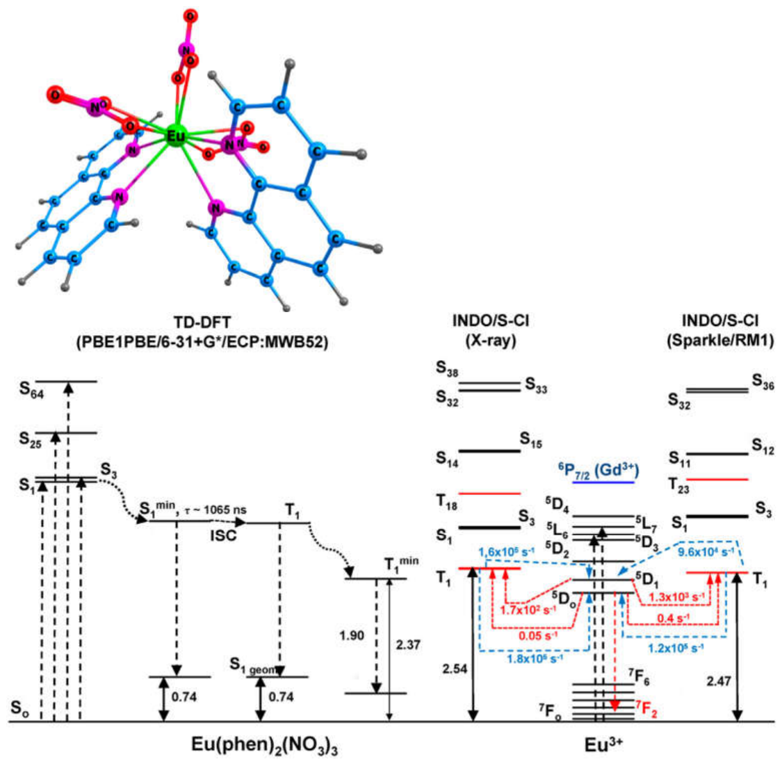
Figure 8.
Energy level diagram of Eu(phen)2(NO3)3 predicted by DFT/TDDFT/PBE1PBE and INDO/S-CIS calculations and the most probable channels for the intramolecular energy transfer process From ref. [96].
Figure 8.
Energy level diagram of Eu(phen)2(NO3)3 predicted by DFT/TDDFT/PBE1PBE and INDO/S-CIS calculations and the most probable channels for the intramolecular energy transfer process From ref. [96].
The structural and photophysical properties (Jablonski diagrams and nonradiative rates) of two highly-emissive Eu3+-based luminescent complexes, Eu(Phen)2Cl3 and Eu(Phen)2(NO3)3 adsorbed on hydrophilic Si-O-Si-OH and hydrophobic Si-O-Si(CH3)3 silicate surfaces were elucidated by combined semi-empirical (Sparkle/AM1; INDO/S-CIS) and simplified TDDFT calculations (Figure 7). The calculated data explained the highest luminescence quantum yield (experimental and theoretical) of Eu(Phen)2(NO3)3 incorporated in hydrophilic matrix as compared to hydrophobic one. For Eu(Phen)2Cl3, the observation of large number of hydrogen bonding on hydrophobic silicate surface and strong adsorption interaction, appears an operative factor, which suppresses the nonradiative emission and assists the larger luminescence quantum yield of the complex incorporated in hydrophobic matrix than that in hydrophilic one. The calculated energy differences ΔE=((E(T1)-E(5D0)/E(5D1)) are large enough to favour the T1-5D0 and T1-5D1 energy transfer relative to the back energy transfer. The theoretical research revealed other operative factors for the formation of the luminescence quantum efficiency and yield of Eu(Phen)2X3 when incorporated into SiO2-based matrices.
The competitive excited state processes in the homoleptic Eu3+ and Tb3+ complexes of HCCA – fluorescence, intersystem crossing (ISC) and phosphorescence, were analyzed depending on the environment, number of the ligands (1-3), Ln3+ ion type (Eu and Tb) and counteranion (Cl− and NO3−) (Figure 9). The HCCA is bidentate bonded to Ln(III) through two oxygen atoms. It was found that the environment altered the S1 state energy, oscillator strength, fluorescence lifetime as well as the S1 character – polar solution stabilized the S1(ππ*) state, whereas non-polar solution (gas phase, solid state) stabilized the S1(nπ*) state. The S1(nπ*) state characterizing with small oscillator strength was decisive factor for the resulting excited state energy transfer and Ln(III) luminescence. It suppressed the S1 ligand emission in the complexes and favored S1-T1 ISC or direct transfer to the emitting levels of Ln3+. The HCCA triplet (T1) state energy and ππ* character were preserved in the Eu/Tb-CCA complexes and are not changed by the environment. The energy gap between the higher energy T1 donor state and the acceptor levels 5D1 of Eu3+ (~0.5 eV) and 5D4 of Tb3+ (~0.1 eV) provided optimal resonance conditions for effective energy transfer to Eu3+, but less probability for Tb3+. The excitation channel T1→5D0 through an exchange mechanism was predicted as the most probable one to populate the main emissive Eu-centered state in the complexes. The energy and the ππ* character of the populated T1 state were not influenced by the H2O-ligands, the Ln(III) type, the counterion type and environment. The larger quantum luminescence yield of Eu(CCA)3(H2O)3 compared to Eu(CCA)2Cl(H2O)2 (from experimental and theoretical point of view) was explained by the higher Eu3+ radiative emission rate, the lower nonradiative decay rate, the shorter distances (LR) between the singlet and triplet CCA donor states and Eu3+ acceptor states and the larger refractive index (solid state).
The sensitization capacity of Phenac ligand, combining C=O and P=O donor group was theoretically explored in the Eu3+ and Tb3+ complexes in vacuum, solution and solid phase. The influence of the coordination polyhedron (number of Phenac), ligand bonding type (mono- and bidentate) and strength, environment, character of the emissive singlet and triplet states stimulating the Eu3+/Tb3+ luminescence was elucidated to address the open question in Ref. [147] about the type of Eu3+ complexes formed in solution. The thermodynamic stability calculations for Eu(Phenac)2(H2O)(NO3)3, Eu(Phenac)2(NO3)3 and Eu(Phenac)3(NO3)3 in reactions predicted Eu(Phenac)3(NO3)3 is mainly stabilized in solution and gas phase, while the other two complexes are in lower concentrations. Similar relaxation paths were found for all complexes, Figure 10. The absorbed energy by Sn > 1(π-π*) states relaxed to T1min by major ISC mechanism S1→T2→T1. The long radiative lifetime of S1→S0 due to n(p(C)(O))π-π*,p(P) character facilitated the forward ISC process. The environment, Phenac’s number and Ln3+ size affect the T1 energy by ~0.1 eV. The calculated (Phenac→Eu3+ energy transfer rates suggested the most likely T1 → 5D1/5D0 channels for a population of emitting metal-centered states. The larger luminescence quantum yield (experimental and theoretical) for the Tb3+-Phenac complexes was explained by the lower T1 state of 0.1 eV and the more suitable ΔE(T1min -5D4) of ~0.5 eV for energy transfer as well as the possible enhancement of spin-orbital coupling effects by the heavier Tb3+ compared to Eu3+.
Conclusions
The present review summarizes the most efficient theoretical approaches for studying a broad range of transition metal clusters and complexes: of the 3d elements as well as the f-block elements. The role of sulfide and chalcogenide ligands is elucidated: in the 3-d elements they favor a photochemical activation of the core, and other electron-donor ligands such as carbonyl groups assist in the thermodynamic stability and in favoring a shift of light absorption bands to the visible region. DFT calculated energy profiles of the chemical reactions of water splitting and carbon dioxide reduction provide accurate activation barriers and reaction enthalpies. As the complexes of M2X2 (M=Fe, Co, Ni, Cu; X= S, Se) mimic both hydrogenase and carbon monoxide dehydrogenase enzymes (CODH) the reactions of hydrogen evolution (HER), oxygen evolution (OER) and the two-step carbon dioxide reduction to formic acid/CO + H2O proved feasible. Lanthanides also benefit from S,S-donor ligands in sensitizing the metal center, yielding lanthanide (Eu) sulphide nanoparticles with photocatalytic properties or luminescent materials suitable for novel electronic and optical devices. A more complex energy transfer mechanism operating in the antenna effect, encompassing energetics and rates of all possible competing excited state processes as well as the luminescence sensitization via ligand singlet state and charge transfer states was revealed by applying of well-validated and parametrized theoretical approaches in close relation to the experiment. The discrete processes that govern photoluminescence, unusual photophysical properties and factors responsible for the excited energy transfer in lanthanide complexes have been successfully predicted by combined DFT/TDDFT, multireference and semiempirical calculations, taking into account spin-orbit coupling and relativistic effects. The theoretical methods and approaches that reliably simulate, explain and predict transition metal systems and their catalytic and optical properties are a powerful tool for the design of new functional metal systems. The in-depth and closely related theoretical and experimental study of 3d- and 4f-complexes gives a powerful impetus to the further development of theoretical methods, as well as to directed syntheses and improved spectroscopic techniques.
Acknowledgments
The authors acknowledge the financial support of the Bulgarian National Science Fund of Bulgarian Ministry of Education and Science, Grant КП-06-Н59/6 (2021), project (PhotoMetalMod”). The authors also acknowledge the provided access to the infrastructure, purchased under the National Roadmap for RI, financially coordinated by the MES of the Republic of Bulgaria (Grant No D01-98/26.06.2025).
Conflicts of Interest
The authors declare no conflicts of interest.
References
- Fujishima, A.; Honda, K. Electrochemical Photolysis of Water at a Semiconductor Electrode. Nature 1972, 238, 37–38. [Google Scholar] [CrossRef]
- Varghese, O. K.; Paulose, M.; LaTempa, T. J.; Grimes, C. A. High-Rate Solar Photocatalytic Conversion of CO2 and Water Vapor to Hydrocarbon Fuels. Nano Lett. 2009, 9, 731. [Google Scholar] [CrossRef] [PubMed]
- McCool, N.S.; Robinson, D.M.; Sheats, J.E.; Dismukes, C. A Co4O4 “Cubane” Water Oxidation Catalyst Inspired by Photosynthesis. J. Am. Chem. Soc. 2011, 133, 11446–11449. [Google Scholar] [CrossRef] [PubMed]
- Sakakura, T.; Choi, J.-C.; Yasuda, H. Transformation of Carbon Dioxide. Chem. Rev. 2007, 107, 2365–2387. [Google Scholar]
- Fang, Z.; Dixon, D. A. Computational Study of H2 and O2 Production from Water Splitting by Small (MO2)n Clusters (M = Ti, Zr, Hf). J. Phys. Chem. A 2013, 117, 3539−3555. [Google Scholar] [CrossRef] [PubMed]
- Li, X.; Siegbahn, P.E.M. Water Oxidation Mechanism for Synthetic Co–Oxides with Small Nuclearity. J. Am. Chem. Soc. 2013, 135, 13804–13813. [Google Scholar] [CrossRef] [PubMed]
- Zhai, G.; Liu, J.; Lin, Sh.; Cheng, Y.; Wang, B.; Jiang, Q.; Fu, H.; Zhao, H.; Lu, Y. Photocatalytic hydrogen production: Materials design, system engineering, and data-driven advances. Chem. Eng. J. 2026, 535, 175401. [Google Scholar] [CrossRef]
- Appel, A.M.; Bercaw, J.E.; Bocarsly, A.B.; Dobbek, H.; DuBois, D.L.; Dupuis, M.; Ferry, J.G.; Fujita, E.; Hille, R.; Kenis, P.J.A.; et al. Frontiers, Opportunities, and Challenges in Biochemical and Chemical Catalysis of CO2 Fixation. Chem. Rev. 2013, 113, 6621–6658. [Google Scholar] [CrossRef] [PubMed]
- Morris, A. J.; Meyer, G. J.; Fujita, E. Molecular Approaches to the Photocatalytic Reduction of Carbon Dioxide for Solar Fuels. Acc Chem. Res. 2009, 42, 1983–1994. [Google Scholar] [CrossRef] [PubMed]
- Papadakis, M.; Delmotte, L.; Mehrez, J.; Renaud Hardrre, R.; Maylis Orio, M. A recent history of ligand modification for hydrogen evolution catalysts. Coord. Chem. Rev. 2026, 559, 217841. [Google Scholar] [CrossRef]
- Nann, T.; Ibrahim, S.K.; Woi, P.-M.; Xu, S.; Ziegler, J.; Pickett, C.J. Water Splitting by Visible Light: A Nanophotocathode for Hydrogen Production. Angew. Chem. Int. Ed. 2010, 49, 1574–1577. [Google Scholar] [CrossRef] [PubMed]
- Wang, R.; Deng, X.; Hu, B.; Vijayalakshmi, M.; Tang, H.; He, L.; Liu, X.; Reddy, Ch. V.; Reddy, K. R.; Shim, J.; Aminabhavi, T.M. Advances in transition metal sulfides: Synthesis, properties, and modification strategies for electrocatalysis and energy conversion applications. Coord Chem. Rev. 2026, 559, 217797. [Google Scholar] [CrossRef]
- Nguyen, D. L. T.; Tran, V. V.; Nguyen, C. C.; Nguyen, T. M. Transition metal sulfides/selenides as cathode electrocatalysts for enhanced hydrogen evolution: Nanostructure engineering strategies and recent progress. Int. J. Hydr. Energy 2026, 229, 154736. [Google Scholar] [CrossRef]
- Hausmann, J. N.; Menezes, P. W. Why should transition metal chalcogenides be investigated as water splitting precatalysts even though they transform into (oxyhydr)oxides? Curr. Opinion Electrochemistry 2022, 34, 100991. [Google Scholar] [CrossRef]
- Varley, J. B.; Hansen, H. A.; Ammitzbøll, N. L.; Grabow, L. C.; Peterson, A. A.; Rossmeisl, J.; Nørskov, J. K Ni–Fe–S Cubanes in CO2 Reduction Electrocatalysis: A DFT Study. ACS Catal. 2013, 3, 2640–2643. [Google Scholar] [CrossRef]
- Catlow, C. R. A.; Guo, Z. X.; Miskufova, M.; Shevlin, S. A.; Smith, A. G. H.; Sokol, A. A.; Walsh, A.; Wilson, D. J.; Woodley, S. M. Advances in computational studies of energy materials Philos. Trans. R. Soc. A 2010, 368, 3379–3456. [Google Scholar] [CrossRef]
- Valders, A.; Jeremie Brillet, J.; Michael Graetzel, Hildur Gudmundsdottir, Heine A. Hansen, Hannes Jonsson, Peter Kluepfel, Geert-Jan Kroes, Florian Le Formal, Isabela C. Man, Rafael S. Martins, Jens K. Nørskov, Jan Rossmeisl, Kevin Sivula, Aleksandra Vojvodic and Michael Zach Solar hydrogen production with semiconductor metal oxides: newdirections in experiment and theory . Phys. Chem. Chem. Phys. 2012, 14, 49–70. [CrossRef] [PubMed]
- Uzunova, E. L.; Mikosch, H. Electronic, magnetic structure and water splitting reactivity of the iron-sulfur dimers and their hexacarbonyl complexes: A density functional study J. Chem. Phys. 2014, 141, 044307. [Google Scholar] [CrossRef]
- Uzunova, E. L.; Mikosch, H. Electronic Structure and Reactivity in Water Splitting of the Iron Oxide Dimers and their Hexacarbonyls: a Density Functional Study. J. Chem. Phys. 2014, 140, 024303. [Google Scholar] [CrossRef]
- Uzunova, E. L.; Mikosch, H. CO2 Conversion to Methanol on Cu(I) Oxide Nanolayers and Clusters: Electronic Structure Insight into the Reaction Mechanism. Phys. Chem. Chem. Phys. 2015, 17, 11088. [Google Scholar] [CrossRef]
- Uzunova, E.; Georgieva, I.; Zahariev, T. Water Splitting Reaction Mechanism on Transition Metal (Fe-Cu) Sulphide and Selenide Clusters—А DFT Study. Materials 2024, 17, 56. [Google Scholar] [CrossRef]
- Brahms, S.; Nikitine, S.; Dahl, J. P. On the band structure and the absorption spectrum of Cu2O. Phys. Lett. 1966, 22, 31. [Google Scholar] [CrossRef]
- Shen, Z.-X.; List, R. S.; Dessau, D. S.; Parmigiani, F.; Arko, A. J.; Bartlett, R.; Wells, B. O.; Lindau, I.; Spicer, W. E. Photoemission study of CuO and Cu2O single crystals. Phys. Rev. B 1990, 42, 8081. [Google Scholar] [CrossRef]
- Rocheleau, R. E.; Miller, E. L. Photoelectrochemical production of hydrogen: engineering loss analysis. Int. J. Hydrog. Energy 1997, 22, 771–782. [Google Scholar] [CrossRef]
- Hara, M.; Kondo, T.; Komoda, M.; Ikeda, S.; Shinohara, K.; Tanaka, A.; Kondo, J. N.; Domen, K. Cu2O as a photocatalyst for overall water splitting under visible light irradiation. Chem. Communications 1998, 34, 357–358. [Google Scholar] [CrossRef]
- Kakuta, S.; Abe, T. Structural characterization of Cu2O after the evolution of H2 under visible light irradiation. Electrochem Solid-State Lett. 2009, 12, P1–P2. [Google Scholar] [CrossRef]
- van den Berg, W.A.; Hagen, W.R.; van Dongen, W.M. The hybrid-cluster protein (‘prismane protein’) from Escherichia coli. Characterization of the hybrid-cluster protein, redox properties of the [2Fe-2S] and [4Fe-2S-2O] clusters and identification of an associated NADH oxidoreductase containing FAD and [2Fe-2S]. Eur. J. Biochem. 2000, 267, 666–676. [Google Scholar] [CrossRef]
- Inoue, M.; Omae, K.; Nakamoto, I.; Kamikawa, R.; Yoshida, T.; Sako, Y. Biome-specific distribution of Ni-containing carbon monoxide dehydrogenases. Extremophiles 2022, 26, 9. [Google Scholar] [CrossRef]
- Dubois, M.R.; Dubois, D.L. Development of Molecular Electrocatalysts for CO2 Reduction and H2 Production/Oxidation. Acc. Chem. Res. 2009, 42, 1974–1982. [Google Scholar] [CrossRef] [PubMed]
- Gust, D.; Thomas, A.; Moore, T.A.; Ana, L.; Moore, A.L. Solar Fuels via Artificial Photosynthesis. Acc. Chem. Res. 2009, 42, 1890–1898. [Google Scholar] [CrossRef] [PubMed]
- Kunkely, H.; Vogler, A. Photoreactivity of Fe2S2(CO)6 originating from dσ* metal-to-ligand charge transfer excitation. J. Organomet. Chem. 1998, 568, 291–293. [Google Scholar] [CrossRef]
- Uzunova, E.L.; Georgieva, I.M. Adsorption and Catalytic Reduction of Nitrogen Oxides (NO, N2O) on Disulfide Cluster Complexes of Cobalt and Iron—A Density Functional Study. Materials 2024, 17, 4764. [Google Scholar] [CrossRef] [PubMed]
- Uzunova, E. L. Pathways of selective catalytic CO2 two-step reduction on di-iron, di-cobalt and iron-cobalt disulfide carbonyls – an electronic structure study. Catal. Sci. Technol. 2019, 9, 1039–1047. [Google Scholar] [CrossRef]
- Haider, S.; Di Tommaso, D.; de Leeuw, N. H. Density functional theory simulations of the structure, stability and dynamics of iron sulphide clusters in water. Phys. Chem. Chem. Phys. 2013, 15, 4310. [Google Scholar] [CrossRef]
- Terranova, U.; de Leeuw, N. H. Aqueous Fe2S2 cluster: structure, magnetic coupling, and hydration behaviour from Hubbard U density functional theory. Phys. Chem. Chem. Phys. 2014, 16, 13426. [Google Scholar] [CrossRef]
- Zhang, Y.; Zhou, Q.; Zhu, J.; Yan, Q.; Dou, S. X.; Sun, W. Nanostructured Metal Chalcogenides for Energy Storage and Electrocatalysis. Adv. Funct. Mater. 2017, 27, 1702317. [Google Scholar] [CrossRef]
- Wang, Z.; Liu, S.; Duan, W.; Xing, Y.; Hu, Y.; Ma, Y. Transition metal selenides as catalysts for electrochemical water splitting. Int. J. Hydr. Energy 2024, 60, 1414–1432. [Google Scholar] [CrossRef]
- Laursen, A. B.; Kegnaes, S.; Dahl, S.; Chorkendorff, I. Molybdenum Sulfides – Efficient and Viable Materials for Electro- and Photoelectrocatalytic Hydrogen Evolution. Energy Environ. Sci. 2012, 5, 5577–91. [Google Scholar] [CrossRef]
- Ruggieri, S.; Mizzoni, S.; Nardon, C.; Cavalli, E.; Sissa, C.; Anselmi, M.; Cozzi, P.G.; Gualandi, A.; Sanadar, M.; Melchior, A.; et al. Circularly Polarized Luminescence from New Heteroleptic Eu(III) and Tb(III) Complexes. Inorg. Chem. 2023, 62, 8812–8822. [Google Scholar] [CrossRef] [PubMed]
- Dragancea, D.; Mocanu, T.; Avram, D.; Raduca, M.; Tiseanu, C.; Andruh, M. Mixed-ligand mononuclear complexes obtained from salicylaldehyde and phenanthroline. Synthesis, crystal structures, and luminescence. Polyhedron 2024, 252, 116886, doi. [Google Scholar] [CrossRef]
- Ali, A.; Ahmed, Z.; Rahisuddin; Iftikhar, K. Pure red-light emitting europium based complexes as efficient UV light converters: synthesis, crystal structure and photoluminescence properties. Dalton Trans. 2023, 52, 14075–14087. [Google Scholar] [CrossRef] [PubMed]
- Bryleva, Y.A.; Glinskaya, L.A.; Korol’kov, I.V.; Bogomyakov, A.S.; Rakhmanova, M.I.; Naumov, D.Y.; Kokina, T.E.; Larionov, S.V. Syntheses, structures, magnetic properties, and photoluminescence of compounds Ln(2,2’-Bipy)(C4H8NCS2)3...0.5CH2Cl2 (Ln = Sm, Eu, Tb, Dy, and Tm). Russ. J. Coord. Chem. 2014, 40, 740–747. [Google Scholar] [CrossRef]
- Regulacio, M.D.; Tomson, N.; Stoll, S.L. Dithiocarbamate precursors for rare-earth sulfides. Chem. Mater. 2005, 17, 3114–3121. [Google Scholar] [CrossRef]
- Bryleva, Y.A.; Kokina, T.E.; Uskov, E.M.; Glinskaya, L.A.; Antonova, O.V.; Larionov, S.V. Synthesis and photoluminescence of the heteroligand Sm(III) complexes containing nitrogen heterocycles (Phen, 2,2’-Bipy) and anions C4H8NCS2-, iso-Bu2PS2 -, (iso-PrO)2PS2 -, and NO3-. Crystal structure of Sm(2,2’-Bipy)2(NO3)3. Russ. J. Coord. Chem. 2013, 39, 41–48. [Google Scholar] [CrossRef]
- Pitchaimani, P.; Lo, K.M.; Elango, K.P. Synthesis, crystal structures, luminescence properties and catalytic application of lanthanide(III) piperidine dithiocarbamate complexes. Polyhedron 2015, 93, 8–16. [Google Scholar] [CrossRef]
- Borrisov, B.; Tsvetkov, M.; Zahariev, T.; Elenkova, D.; Morgenstern, B.; Dimov, D.; Kukeva, R.; Trendafilova, N.; Georgieva, I. Effect of Pyrrolidinedithiocarbamate Ligand on the Luminescence Properties of Heteroligand Samarium and Europium Complexes: Experimental and Theoretical Study. Inorg. Chem. 2024, 63, 13840–13864. [Google Scholar] [CrossRef]
- Regulacio, M.D.; Bussmann, K.; Lewis, B.; Stoll, S.L. Magnetic properties of lanthanide chalcogenide semiconducting nanoparticles. J. Am. Chem. Soc. 2006, 128, 11173–11179. [Google Scholar] [CrossRef] [PubMed]
- Pereira, A.S.; Rauwel, P.; Reis, M.S.; Silva, N.J.O.; Barros-Timmons, A.; Trindade, T. Polymer encapsulation effects on the magnetism of EuS nanocrystals. J. Mater. Chem. C 2008, 18, 4572–4578. [Google Scholar] [CrossRef]
- Muliadi, M.; Nurdin, M.; Umar, A.A. Synthesis of photoluminescent europium dithiocarbamate complexes with dimethyl phenanthroline and bipyridyl co-ligands. Opt. Mater. 2022, 125, 112063, doi. [Google Scholar] [CrossRef]
- Wang, S.S.; Xu, J.; Wang, J.; Wang, K.Y.; Dang, S.; Song, S.Y.; Liu, D.; Wang, C. Luminescence of samarium(III) bis-dithiocarbamate frameworks: codoped lanthanide emitters that cover visible and near-infrared domains. J. Mater. Chem. C 2017, 5, 6620–6628. [Google Scholar] [CrossRef]
- Faustino, W.M.; Malta, O.L.; Teotonio, E.E.S.; Brito, H.F.; Simas, A.M.; de Sá, G.F. Photoluminescence of europium(III) dithiocarbamate complexes: Electronic structure, charge transfer and energy transfer. J. Phys. Chem. A 2006, 110, 2510–2516. [Google Scholar] [CrossRef] [PubMed]
- Regulacio, M.D.; Pablico, M.H.; Vasquez, J.A.; Myers, P.N.; Gentry, S.; Prushan, M.; Tam-Chang, S.W.; Stoll, S.L. Luminescence of Ln(III) dithiocarbamate complexes (Ln = La, Pr, Sm, Eu, Gd, Tb, Dy). Inorg. Chem. 2008, 47, 1512–1523. [Google Scholar] [CrossRef]
- Faustino, W.M.; Malta, O.L.; de Sa, G.F. Theoretical modeling of thermally activated luminescence quenching through charge transfer states in lanthanide complexes. Chem. Phys. Lett. 2006, 429, 595–599. [Google Scholar] [CrossRef]
- Kobayashi, T.; Naruke, H.; Yamase, T. Photoluminescence and molecular structure of tetrakis(N,N-dimethyldithiocarbamato)europate(III). Chem. Lett. 1997, 907–908. [Google Scholar] [CrossRef]
- Neto, A.N.C.; Teotonio, E.E.S.; de Sa, G.F.; Brito, H.F.; Legendziewicz, J.; Carlos, L.D.; Felinto, M.C.F.C.; Gawryszewska, P.; Moura, R.T.; Longo, R.L.; et al. Modeling intramolecular energy transfer in lanthanide chelates: A critical review and recent advances; ELSEVIER NОRTH HОLLANDPО BОX 211, 1000 AE AMSTERDAM, NETHERLANDS: . 2019; Volume 56, pp. 55–162. [Google Scholar]
- da Rosa, P.P.F.; Miyazaki, S.; Sakamoto, H.; Kitagawa, Y.; Miyata, K.; Akama, T.; Kobayashi, M.; Fushimi, K.; Onda, K.; Taketsugu, T.; et al. Coordination Geometrical Effect on Ligand-to-Metal Charge Transfer-Dependent Energy Transfer Processes of Luminescent Eu(III) Complexes. J. Phys. Chem. A 2021, 125, 209–217. [Google Scholar] [CrossRef] [PubMed]
- Nonat, A.; Esteban-Gomez, D.; Valencia, L.; Perez-Lourido, P.; Barriada, J.L.; Charbonniere, L.J.; Platas-Iglesias, C. The role of ligand to metal charge-transfer states on the luminescence of Europium complexes with 18-membered macrocyclic ligands. Dalton Trans. 2019, 48, 4035–4045. [Google Scholar] [CrossRef]
- Maouche, R.; Belaid, S.; Benmerad, B.; Bouacida, S.; Freslon, S.; Daiguebonne, C.; Suffren, Y.; Calvez, G.; Bernot, K.; Roiland, C.; et al. Luminescence properties of lanthanide complexes-based molecular alloys. Inorg. Chim. Acta 2020, 501, ARTN 119309, doi. [Google Scholar] [CrossRef]
- Bryleva, Y.A.; Glinskaya, L.A.; Korol’kov, I.V.; Bogomyakov, A.S.; Rakhmanova, M.I.; Naumov, D.Y.; Kokina, T.E.; Larionov, S.V. Structure of [Dy(Phen)(C4H8NCS2)3]...3CH2Cl2 solvate. Magnetic properties and photoluminiscence of [Ln(Phen)(C4H8NCS2)3] (Ln = Sm, Eu, Tb, Dy, Tm) complexes. J. Struct. Chem. 2014, 55, 319–326. [Google Scholar] [CrossRef]
- Faustino, W.M.; Malta, O.L.; Teotonio, E.E.S.; Brito, H.F.; Simas, A.M.; de Sa, G.F. Photoluminescence of europium(III) dithiocarbamate complexes: Electronic structure, charge transfer and energy transfer. J. Phys. Chem. A 2006, 110, 2510–2516. [Google Scholar] [CrossRef] [PubMed]
- Kupryakov, A.S.; Plyusnin, V.F.; Grivin, V.P.; Bryleva, J.A.; Larionov, S.V. Interligand electron transfer as a reason of very weak red luminescence of Eu((i-Bu)2PS2))3Phen and Eu((C4H8NCS2)3)Phen complexes. J. Lumin. 2016, 176, 130–135. [Google Scholar] [CrossRef]
- Kovacs, D.; Borbas, K.E. The role of photoinduced electron transfer in the quenching of sensitized Europium emission. Coord. Chem. Rev. 2018, 364, 1–9. [Google Scholar] [CrossRef]
- Kovacs, D.; Mathieu, E.; Kiraev, S.R.; Wells, J.A.L.; Demeyere, E.; Sipos, A.; Borbas, K.E. Coordination Environment-Controlled Photoinduced Electron Transfer Quenching in Luminescent Europium Complexes. J. Am. Chem. Soc. 2020, 142, 13190–13200. [Google Scholar] [CrossRef] [PubMed]
- Hofbauerova, H.; Beinrohr, E.; Mocak, J. Redox Properties of the Bis(2-Hydroxyethyl)Dithiocarbamate Anion and Its Nickel(Ii) Complex in Aqueous-Solutions. Chem. Pap. 1987, 41, 441–452. [Google Scholar]
- Cotton, S. Lanthanide and Actinide Chemistry; John Wiley & Sons, Ltd, 2006. [Google Scholar]
- Bunzli, J.C.G.; Piguet, C. Taking advantage of luminescent lanthanide ions. Chem. Soc. Rev. 2005, 34, 1048–1077. [Google Scholar] [CrossRef]
- Bunzli, J.C.G.; Comby, S.; Chauvin, A.S.; Vandevyver, C.D.B. New opportunities for lanthanide luminescence. J. Rare Earth 2007, 25, 257–274. [Google Scholar] [CrossRef]
- Bünzli, J.-C.G. On the design of highly luminescent lanthanide complexes. Coord. Chem. Rev. 2015, 293-294, 19–47. [Google Scholar] [CrossRef]
- Bünzli, J.-C.P., V. Handbook on the Physics and Chemistry of Rare Earths, First ed.; North Holland, 2019. [Google Scholar]
- Bao, G.C.; Wong, K.L.; Jin, D.Y.; Tanner, P.A. A stoichiometric terbium-europium dyad molecular thermometer: energy transfer properties. Light-Sci. Appl. 2018, 7. [Google Scholar] [CrossRef]
- Latva, M.; Takalo, H.; Mukkala, V.M.; Matachescu, C.; RodriguezUbis, J.C.; Kankare, J. Correlation between the lowest triplet state energy level of the ligand and lanthanide(III) luminescence quantum yield. J. Lumin. 1997, 75, 149–169. [Google Scholar] [CrossRef]
- Hooda, A.; Dalal, A.; Nehra, K.; Singh, S.; Kumar, S.; Singh, D. Red-emitting beta-diketonate Eu(III) Complexes With Substituted 1,10-phenanthroline Derivatives: Optoelectronic and Spectroscopic Analysis. J. Fluoresc. 2022, 32, 1413–1424. [Google Scholar] [CrossRef]
- Tsvetkov, M.; Elenkova, D.; Kolarski, M.; Lyapchev, R.; Morgenstern, B.; Videva, V.; Zaharieva, J.; Milanova, M. Synthesis, crystal structure and luminescence properties of two novel Tb (III) complexes with 1,10-phenanthroline derivatives as ligands. J. Mol. Struct. 2024, 1314, 138768. [Google Scholar] [CrossRef]
- Haas, T.; Neumann, T.; Jobbitt, N.; Yang, X.Y.; Schubert, H.; Hunger, D.; Seitz, M. Mixed Phenanthroline-Bipyridine Europium and Ytterbium Cryptates-Bright Luminophores with Small Radiative Lifetimes. Inorg. Chem. 2026, 65, 5109–5118. [Google Scholar] [CrossRef]
- Hollauer, H.V.P.; Guedes, G.P.; Mariano, D.L.; Ghivelder, L.; Mendonça, A.A.; Soriano, S.; Farias, R.L.; Escobar, L.B.L. Unveiling single-ion magnetism in lanthanide-coumarin carboxylate coordination polymers. Dalton Trans. 2026, 55, 2665–2672. [Google Scholar] [CrossRef]
- Kariaka, N.S.; Lipa, A.; Neto, A.N.C.; Malta, O.L.; Gawryszewska, P.; Amirkhanov, V.M. Eu3+ and Tb3+ coordination compounds with phenyl-containing carbacylamidophosphates: comparison with selected Ln3+ β-diketonates. Front. Chem. 2023, 11, 1188314. [Google Scholar] [CrossRef]
- Larionov, S.V.; Bryleva, Y.A. Coordination compounds of lanthanides with 1,1-dithiolate ligands. Russ. J. Coord. Chem. 2016, 42, 293–310. [Google Scholar] [CrossRef]
- Bhimpuria, R.; Tomar, M.; Thapper, A.; Ahlquist, M.; Borbas, K.E. Photocatalytic product-selective reduction of CO2, CO, and carbonates. Chem. 2025, 11, 102450, doi. [Google Scholar] [CrossRef]
- Georgieva, I.; Trendafilova, N.; Zahariev, T.; Danchova, N.; Gutzov, S. Theoretical insight in highly luminescent properties of Eu(III) complex with phenanthroline. J. Lumin 2018, 202, 192–205. [Google Scholar] [CrossRef]
- Georgieva, I.; Zahariev, T.; Aquino, A.J.A.; Trendafilova, N.; Lischka, H. Energy transfer mechanism in luminescence Eu(III) and Tb(III) complexes of coumarin-3-carboxylic acid: A theoretical study. Spectrochim. Acta A 2020, 240, 118591, doi. [Google Scholar] [CrossRef]
- Zahariev, T.; Shandurkov, D.; Gutzov, S.; Trendafilova, N.; Enseling, D.; Justel, T.; Georgieva, I. Phenanthroline chromophore as efficient antenna for Tb3+ green luminescence: A theoretical study. Dye. Pigm 2021, 185, 108890. [Google Scholar] [CrossRef]
- Zahariev, T.; Trendafilova, N.; Georgieva, I. Spectroscopic and photophysical properties of [Eu(Phen)2]X3 (X≡Cl–, NO3–) complexes, incorporated into SiO2-based Matrices: Theoretical study. Mater. Today-Proc 2022, 61, 1292–1299. [Google Scholar] [CrossRef]
- Zahariev, T.; Beykov, P.; Trendafilova, N.; Uzunova, E.; Georgieva, I. Modulation of lanthanide luminescence by carbamoylmethylphosphine oxide ligand: A theoretical study. Dye. Pigm 2024, 224, ARTN 112043. [Google Scholar] [CrossRef]
- de Andrade, A.V.; Longo, R.L.; Simas, A.M.; de Sá, G.F. Theoretical model for the prediction of electronic spectra of lanthanide complexes. J. Chem. Soc. Faraday Trans. 1996, 92, 1835–1839. [Google Scholar] [CrossRef]
- Scotognella, F.; Meinardi, F.; Ottonelli, M.; Raimondo, L.; Tubino, R. Photophysical analysis of the organic complex [Eu(C12H8N2)2](NO3)3. J. Lumin. 2009, 129, 746–750. [Google Scholar] [CrossRef]
- Dutra, J.D.L.; Freire, R.O. Theoretical tools for the calculation of the photoluminescent properties of europium systems - A case study. J. Photoch. Photobio. A 2013, 256, 29–35. [Google Scholar] [CrossRef]
- Dutra, J.D.L.; Gimenez, I.F.; da Costa, N.B.; Freire, R.O. Theoretical design of highly luminescent europium (III) complexes: A factorial study. J. Photochem. Photobiol. A-Chem. 2011, 217, 389–394. [Google Scholar] [CrossRef]
- Georgieva, L.; Trendafilova, N.; Aquino, A.J.A.; Lischka, H. Theoretical study of metal-ligand interaction in Sm(III), Eu(III), and. Tb(III) complexes of coumarin-3-carboxylic acid in the gas phase and solution. Inorg. Chem. 2007, 46, 10926–10936. [Google Scholar] [CrossRef]
- Freidzon, A.Y.; Scherbinin, A.V.; Bagaturyants, A.A.; Alfimov, M.V. Ab Initio Study of Phosphorescent Emitters Based on Rare-Earth Complexes with Organic Ligands for Organic Electroluminescent Devices. J. Phys. Chem. A 2011, 115, 4565–4573. [Google Scholar] [CrossRef] [PubMed]
- Martins, J.P.; Martin-Ramos, P.; Chamorro-Posada, P.; Silva, P.S.P.; Martin-Gil, J.; Hernandez-Navarro, S.; Silva, M.R. Experimental and Theoretical Studies on the Structure and Photoluminescent Properties of New Mononuclear and Homodinuclear Europium(III) beta-Diketonate Complexes. Adv. Cond. Matter Phys. 2015, 2015, 205047. [Google Scholar]
- Dutra, J.D.L.; Bispo, T.D.; Freire, R.O. LUMPAC Lanthanide Luminescence Software: Efficient and User Friendly. J. Comp. Chem. 2014, 35, 772–775. [Google Scholar] [CrossRef] [PubMed]
- Freire, R.O.; Rocha, G.B.; Albuquerque, R.Q.; Simas, A.M. Efficacy of the semiempirical sparkle model as compared to ECP ab-initio calculations for the prediction of ligand field parameters of europium(III) complexes. J. Lumin. 2005, 111, 81–87. [Google Scholar] [CrossRef]
- Martín-Ramos, P.; Silva, P.S.P.; Chamorro-Posada, P.; Silva, M.R.; Milne, B.F.; Nogueira, F.; Martín-Gil, J. Synthesis, structure, theoretical studies and luminescent properties of a ternary erbium(III) complex with acetylacetone and bathophenanthroline ligands. J. Lumin. 2015, 162, 41–49. [Google Scholar] [CrossRef]
- Malta, O.L. Mechanisms of non-radiative energy transfer involving lanthanide ions revisited. J. Non-Cryst. Solids 2008, 354, 4770–4776. [Google Scholar] [CrossRef]
- de Sa, G.F.; Malta, O.L.; Donega, C.D.; Simas, A.M.; Longo, R.L.; Santa-Cruz, P.A.; da Silva, E.F. Spectroscopic properties and design of highly luminescent lanthanide coordination complexes. Coord. Chem. Rev. 2000, 196, 165–195. [Google Scholar] [CrossRef]
- Georgieva, I.; Trendafilova, N.; Zahariev, T.; Danchova, N.; Gutzov, S. Theoretical insight in highly luminescent properties of Eu(III) complex with phenanthroline. J. Lumin. 2018, 202, 192–205. [Google Scholar] [CrossRef]
- Georgieva, I.; Zahariev, T.; Aquino, A.J.A.; Trendafilova, N.; Lischka, H. Energy transfer mechanism in luminescence Eu(III) and Tb(III) complexes of coumarin-3-carboxylic acid: A theoretical study. Spectrochim. Acta A 2020, 240, 118591, doi. [Google Scholar] [CrossRef]
- Zahariev, T.; Trendafilova, N.; Georgieva, I. Spectroscopic and photophysical properties of [Eu(Phen)2]X3, (X=C-,NO3-) complexes, incorporated into SiO2-based Matrices: Theoretical study. Mater. Today Proceedings 2022, 61, 1292–1299. [Google Scholar] [CrossRef]
- Carnall, W.T.; Fields, P.R.; Rajnak, K. Electronic Energy Levels of Trivalent Lanthanide Aquo Ions.3. Tb3+. J. Chem. Phys. 1968, 49, 4447, doi. [Google Scholar] [CrossRef]
- Carnall, W.; Fields, P.; Rajnak, K. Spectral intensities of the trivalent lanthanides and actinides in solution. II. Pm3+, Sm3+, Eu3+, Gd3+, Tb3+, Dy3+ and Ho3+. J. Chem. Phys. 1968, 49, 4412–4423. [Google Scholar] [CrossRef]
- Dutra, J.D.L.; Lima, N.B.D.; Freire, R.O.; Simas, A.M. Europium Luminescence: Electronic Densities and Superdelocalizabilities for a Unique Adjustment of Theoretical Intensity Parameters. Sci. Rep. 2015, 5, 13695–13706. [Google Scholar] [CrossRef] [PubMed]
- Lima, N.B.D.; Dutra, J.D.L.; Goncalves, S.M.C.; Freire, R.O.; Simas, A.M. Chemical Partition of the Radiative Decay Rate of Luminescence of Europium Complexes. Sci. Rep. 2016, 6, 21204–21216 doi. [Google Scholar] [CrossRef] [PubMed]
- Grimme, S.; Waletzke, M. A combination of Kohn-Sham density functional theory and multi-reference configuration interaction methods. J. Chem. Phys. 1999, 111, 5645–5655. [Google Scholar] [CrossRef]
- Dreuw, A.; Head-Gordon, M. Single-reference ab initio methods for the calculation of excited states of large molecules. Chem. Rev. 2005, 105, 4009–4037. [Google Scholar] [CrossRef]
- Cohen, A.J.; Mori-Sanchez, P.; Yang, W.T. Challenges for Density Functional Theory. Chem. Rev. 2012, 112, 289–320. [Google Scholar] [CrossRef] [PubMed]
- Peach, M.J.; Tozer, D.J. Overcoming low orbital overlap and triplet instability problems in TDDFT. J. Phys. Chem. A 2012, 116, 9783–9789. [Google Scholar] [CrossRef] [PubMed]
- van Lenthe, E.; Ehlers, A.; Baerends, E.J. Geometry optimizations in the zero order regular approximation for relativistic effects. J. Chem. Phys. 1999, 110, 8943–8953. [Google Scholar] [CrossRef]
- van Lenthe, E.; van Leeuwen, R.; Baerends, E.J.; Snijders, J.G. Relativistic regular two-component Hamiltonians. Int. J. Quantum Chem. 1996, 57, 281–293. [Google Scholar] [CrossRef]
- Dolg, M.; Stoll, H.; Preuss, H. A Combination of Quasi-Relativistic Pseudopotential and Ligand-Field Calculations for Lanthanoid Compounds. Theor. Chim. Acta 1993, 85, 441–450. [Google Scholar] [CrossRef]
- Maron, L.; Eisenstein, O. Do f electrons play a role in the lanthanide-ligand bonds? A DFT study of Ln(NR2)3; R = H, SiH3. J. Phys. Chem. A 2000, 104, 7140–7143. [Google Scholar] [CrossRef]
- Boehme, C.; Coupez, B.; Wipff, G. Interaction of M3+ lanthanide cations with diamide ligands and their thia analogues: A quantum mechanics study of monodentate vs bidentate binding, counterion effects, and ligand protonation. J. Phys. Chem. A 2002, 106, 6487–6498. [Google Scholar] [CrossRef]
- Vetere, V.; Maldivi, P.; Adamo, C. Comparative studies of quasi-relativistic density functional methods for the description of lanthanide and actinide complexes. J. Comput. Chem. 2003, 24, 850–858. [Google Scholar] [CrossRef] [PubMed]
- Dolg, M.; Stoll, H.; Flad, H.J.; Preuss, H. Abinitio Pseudopotential Study of Yb and Ybo. J. Chem. Phys. 1992, 97, 1162–1173. [Google Scholar] [CrossRef]
- Maron, L.; Eisenstein, O. DFT study of H-H activation by Cp-2 LnH d(0) complexes. J. Am. Chem. Soc 2001, 123, 1036–1039. [Google Scholar] [CrossRef] [PubMed]
- Gutierrez, F.; Rabbe, C.; Poteau, R.; Daudey, J.P. Theoretical study of Ln(III) complexes with polyaza-aromatic ligands: Geometries of [LnL(H2O)n]3+ complexes and successes and failures of TD-DFT. J. Phys. Chem. A 2005, 109, 4325–4330. [Google Scholar] [CrossRef] [PubMed]
- Peach, M.J.G.; Warner, N.; Tozer, D.J. On the triplet instability in TDDFT. Mol. Phys. 2013, 111, 1271–1274. [Google Scholar] [CrossRef]
- Zhu, T.Y.; Chen, P.; Li, H.F.; Sun, W.B.; Gao, T.; Yan, P.F. Structural effects on the photophysical properties of mono-β-diketonate and bis-β-diketonate Eu complexes. Phys. Chem. Chem. Phys. 2015, 17, 16136–16144. [Google Scholar] [CrossRef] [PubMed]
- Munguba, G.H.L.; Urquiza-Carvalho, G.A.; Silva, F.T.; Simas, A.M. The complex build algorithm to set up starting structures of lanthanoid complexes with stereochemical control for molecular modeling. Sci. Rep. 2021, 11, 21493, doi. [Google Scholar] [CrossRef]
- Gabriel Henrique Lima Munguba, G.A.U.d.C., Fredericо Teixeira Silva, Alfredо Mayall Simas Cоmplex Build Sоftware, 1.1.0; 2021.
- Tomasi, J.; Mennucci, B.; Cammi, R. Quantum mechanical continuum solvation models. Chem. Rev. 2005, 105, 2999–3094. [Google Scholar] [CrossRef]
- Klamt, A.; Schuurmann, G. Cosmo - a new approach to dielectric screening in solvents with explicit expressions for the screening energy and Its gradient. J. Chem. Soc.-Perkin Trans. 1993, 799–805. [Google Scholar] [CrossRef]
- Perdew, J.P.; Burke, K.; Ernzerhof, M. Generalized gradient approximation made simple. Phys. Rev. Lett. 1996, 77, 3865–3868. [Google Scholar] [CrossRef]
- Gutzov, S.; Danchova, N.; Kirilova, R.; Petrov, V.; Yordanova, S. Preparation and luminescence of silica aerogel composites containing an europium (III) phenanthroline nitrate complex. J. Lumin. 2017, 183, 108–112. [Google Scholar] [CrossRef]
- Adamo, C.; Jacquemin, D. The calculations of excited-state properties with Time-Dependent Density Functional Theory. Chem. Soc. Rev. 2013, 42, 845–856. [Google Scholar] [CrossRef] [PubMed]
- Grimme, S.; Ehrlich, S.; Goerigk, L. Effect of the Damping Function in Dispersion Corrected Density Functional Theory. J. Comput. Chem. 2011, 32, 1456–1465. [Google Scholar] [CrossRef] [PubMed]
- Lin, Y.S.; Li, G.D.; Mao, S.P.; Chai, J.D. Long-Range Corrected Hybrid Density Functionals with Improved Dispersion Corrections. J. Chem. Theory Comput. 2013, 9, 263–272. [Google Scholar] [CrossRef]
- Muniz-Miranda, F.; Pedone, A.; Battistelli, G.; Montalti, M.; Bloino, J.; Barone, V. Benchmarking TD-DFT against Vibrationally Resolved Absorption Spectra at Room Temperature: 7-Aminocoumarins as Test Cases. J. Chem. Theory Comput. 2015, 11, 5371–5384. [Google Scholar] [CrossRef]
- Cossi, M.; Rega, N.; Scalmani, G.; Barone, V. Energies, structures, and electronic properties of molecules in solution with the C-PCM solvation model. J. Comp. Chem. 2003, 24, 669–681. [Google Scholar] [CrossRef] [PubMed]
- Improta, R.; Scalmani, G.; Frisch, M.J.; Barone, V. Toward effective and reliable fluorescence energies in solution by a new state specific polarizable continuum model time dependent density functional theory approach. J. Chem. Phys. 2007, 127. [Google Scholar] [CrossRef] [PubMed]
- Kasha, M. Characterization of Electronic Transitions in Complex Molecules. Discuss. Faraday Soc. 1950, 9, 14–19. [Google Scholar] [CrossRef]
- Steiner, U.E. Fundamentals of Photophysics, Photiochemistry, and Photobiology; Springer-Verlag: Berlin, Heidelberg, 2014. [Google Scholar]
- Thor, W.; Zhang, Y.H.; Wong, K.L.; Tanner, P.A. Orbital transitions: insight into energy transfer through an antenna for an organo-lanthanide complex. Chem. Commun. 2021, 57, 10727–10730. [Google Scholar] [CrossRef] [PubMed]
- Bransden, B.H.J., C.J. Physics of atoms and molecules, Longman Scientific and Technical Longman Group UK Limited, London, New York, 1983.
- Shuai, Z.G. Thermal Vibration Correlation Function Formalism for Molecular Excited State Decay Rates. Chin. J. Chem. 2020, 38, 1223–1232. [Google Scholar] [CrossRef]
- Niu, Y.L.; Peng, Q.A.; Deng, C.M.; Gao, X.; Shuai, Z.G. Theory of Excited State Decays and Optical Spectra: Application to Polyatomic Molecules. J. Phys. Chem. A 2010, 114, 7817–7831. [Google Scholar] [CrossRef] [PubMed]
- Shuai, Z.G.; Peng, Q. Organic light-emitting diodes: theoretical understanding of highly efficient materials and development of computational methodology. Natl. Sci. Rev. 2017, 4, 224–239. [Google Scholar] [CrossRef]
- Niu, Y.; Peng, Q.; Shuai, Z. Promoting-mode free formalism for excited state radiationless decay process with Duschinsky rotation effect. Sci. China Ser. B-Chem. 2008, 51, 1153–1158. [Google Scholar] [CrossRef]
- Penfold, T.J.; Gindensperger, E.; Daniel, C.; Marian, C.M. Spin-Vibronic Mechanism for Intersystem Crossing. Chem. Rev. 2018, 118, 6975–7025. [Google Scholar] [CrossRef]
- Balzani, V.; Ceroni, P.; Juris, A. Photochemistry and photophysics: concepts, research, applications. Angew. Chem. International Ed. 2014, 53, 8817, doi. [Google Scholar] [CrossRef]
- de Souza, B.; Farias, G.; Neese, F.; Izsák, R. Predicting Phosphorescence Rates of Light Organic Molecules Using Time-Dependent Density Functional Theory and the Path Integral Approach to Dynamics. J. Chem. Theory Comp. 2019, 15, 1896–1904. [Google Scholar] [CrossRef] [PubMed]
- Lakowicz, J.R. Principles of Fluorescence Spectroscopy, Third ed.; 2006. [Google Scholar]
- Dexter, D.L. A theory of sensitized luminescence in solids. J. Chem. Phys. 1953, 21(14). [Google Scholar] [CrossRef]
- Neto, A.N.C.; Teotonio, E.E.; de Sá, G.F.; Brito, H.F.; Legendziewicz, J.; Carlos, L.D.; Felinto, M.C.F.; Gawryszewska, P.; Moura, R.T., Jr.; Longo, R.L. Modeling intramolecular energy transfer in lanthanide chelates: A critical review and recent advances. Handbook On Phys. Chem. Of Rare Earths 2019, 56, 55–162. [Google Scholar] [CrossRef]
- Faustino, W.M.; Malta, O.L.; de Sa, G.F. Intramolecular energy transfer through charge transfer state in lanthanide compounds: A theoretical approach. J. Chem. Phys. 2005, 122, 054109, doi. [Google Scholar] [CrossRef]
- Dutra, J.D.L.; Lima, N.B.D.; Freire, R.O.; Simas, A.M. Europium Luminescence: Electronic Densities and Superdelocalizabilities for a Unique Adjustment of Theoretical Intensity Parameters. Sci. Rep. 2015, 5, 13695, doi. [Google Scholar] [CrossRef] [PubMed]
- Zerner, M.C.; Loew, G.H.; Kirchner, R.F.; Mueller-Westerhoff, U.T. An intermediate neglect of differential overlap technique for spectroscopy of transition-metal complexes. Ferrocene. J. Am. Chem. Soc 1980, 102, 589–599. [Google Scholar] [CrossRef]
- Leach, E.G.; Shady, J.R.; Boyden, A.C.; Emig, A.L.; Henry, A.T.; Connor, E.K.; Staples, R.J.; Schaertel, S.; Werner, E.J.; Biros, S.M. X-ray crystallographic, luminescence and NMR studies of phenacyldiphenylphosphine oxide with the Ln(III) ions Sm, Eu, Gd, Tb and Dy. Dalton Trans. 2017, 46, 15458–15469. [Google Scholar] [CrossRef]
Figure 1.
a) The global minimum of Co2S2(CO)4 in rhombic non-planar configuration. b) Co2S2(CO)4 with a planar core. Legend: Co atoms are blue, sulfur atoms – yellow, carbon atoms – grey, oxygen atoms – red. From refs. 21 and 32.
Figure 1.
a) The global minimum of Co2S2(CO)4 in rhombic non-planar configuration. b) Co2S2(CO)4 with a planar core. Legend: Co atoms are blue, sulfur atoms – yellow, carbon atoms – grey, oxygen atoms – red. From refs. 21 and 32.
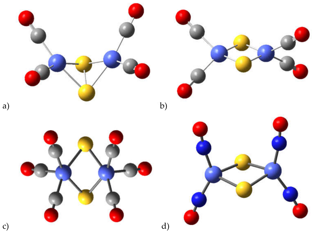
Figure 2.
a) The HOMO and b) the LUMO of the Fe2S2(CO)6 complex with non-planar core (the global energy minimum configuration). a) The HOMO and b) the LUMO of the Fe2S2(CO)6 complex with planar core (low-lying conformational isomer). Legend: Positive parts of MOs are red, negative – green, sulfur atoms are yellow, carbons – grey, oxygens – blue. From ref 18.
Figure 2.
a) The HOMO and b) the LUMO of the Fe2S2(CO)6 complex with non-planar core (the global energy minimum configuration). a) The HOMO and b) the LUMO of the Fe2S2(CO)6 complex with planar core (low-lying conformational isomer). Legend: Positive parts of MOs are red, negative – green, sulfur atoms are yellow, carbons – grey, oxygens – blue. From ref 18.
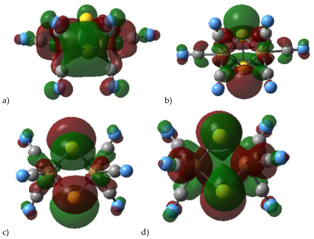
Figure 3.
The two distinct positions of hydride in Co2S2(CO)4. Legend as Figure 1, hydrogens are small grey circles. From ref. 21.
Figure 3.
The two distinct positions of hydride in Co2S2(CO)4. Legend as Figure 1, hydrogens are small grey circles. From ref. 21.
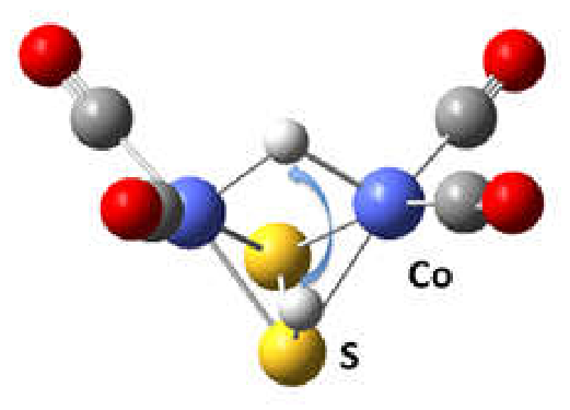
Figure 4.
The reaction path of oxygen formation and oxygen evolution for cobalt and iron chalcogenide complexes. ΔE is the energy difference relative to the ground state complexes; RC – reaction coordinate. The excitation energies of representative most intense transitions induced by light absorption are denoted by vertical arrows. From reference 21.
Figure 4.
The reaction path of oxygen formation and oxygen evolution for cobalt and iron chalcogenide complexes. ΔE is the energy difference relative to the ground state complexes; RC – reaction coordinate. The excitation energies of representative most intense transitions induced by light absorption are denoted by vertical arrows. From reference 21.
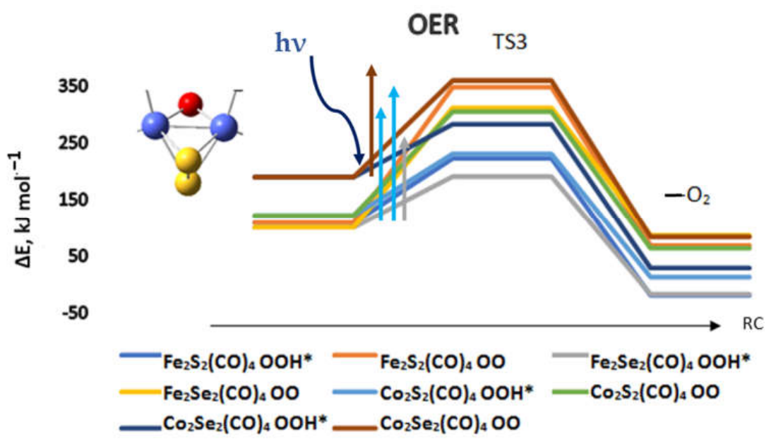
Figure 5.
Experimental and theoretical insight into the role of pyrrolidinedithiocarbamate (pdtc) and 1,10-phenantroline (phen) ligands in modulating Sm3+ and Eu3+ emission. All competing processes of the excited-state L(antenna)-to-Ln energy transfer mechanism were considered. The S1 state is characterized by interligand (pdtc-to-phen) charge transfer character. Sm(pdtc)3(phen) complex shows good ability to convert UV to visible light and efficient luminescence. The weak emission of Eu(pdtc)3(phen) was explained by a partial reduction of Eu3+, confirmed by EPR data. From ref. [46].
Figure 5.
Experimental and theoretical insight into the role of pyrrolidinedithiocarbamate (pdtc) and 1,10-phenantroline (phen) ligands in modulating Sm3+ and Eu3+ emission. All competing processes of the excited-state L(antenna)-to-Ln energy transfer mechanism were considered. The S1 state is characterized by interligand (pdtc-to-phen) charge transfer character. Sm(pdtc)3(phen) complex shows good ability to convert UV to visible light and efficient luminescence. The weak emission of Eu(pdtc)3(phen) was explained by a partial reduction of Eu3+, confirmed by EPR data. From ref. [46].
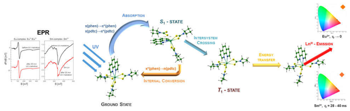
Figure 6.
Energy level diagram of Sm(pdtc)3(phen) (a) and Eu(pdtc)3(phen) (b) in DCM solution, predicted by TD-DFT/ωB97xD calculations and the most probable channels for intramolecular energy transfer in the excited state. The energy values are in eV and the lifetime in second (s). The zero point energy (ZPE) corrected transitions are minimum-to-minimum adiabatic ones. From ref. [46].
Figure 6.
Energy level diagram of Sm(pdtc)3(phen) (a) and Eu(pdtc)3(phen) (b) in DCM solution, predicted by TD-DFT/ωB97xD calculations and the most probable channels for intramolecular energy transfer in the excited state. The energy values are in eV and the lifetime in second (s). The zero point energy (ZPE) corrected transitions are minimum-to-minimum adiabatic ones. From ref. [46].
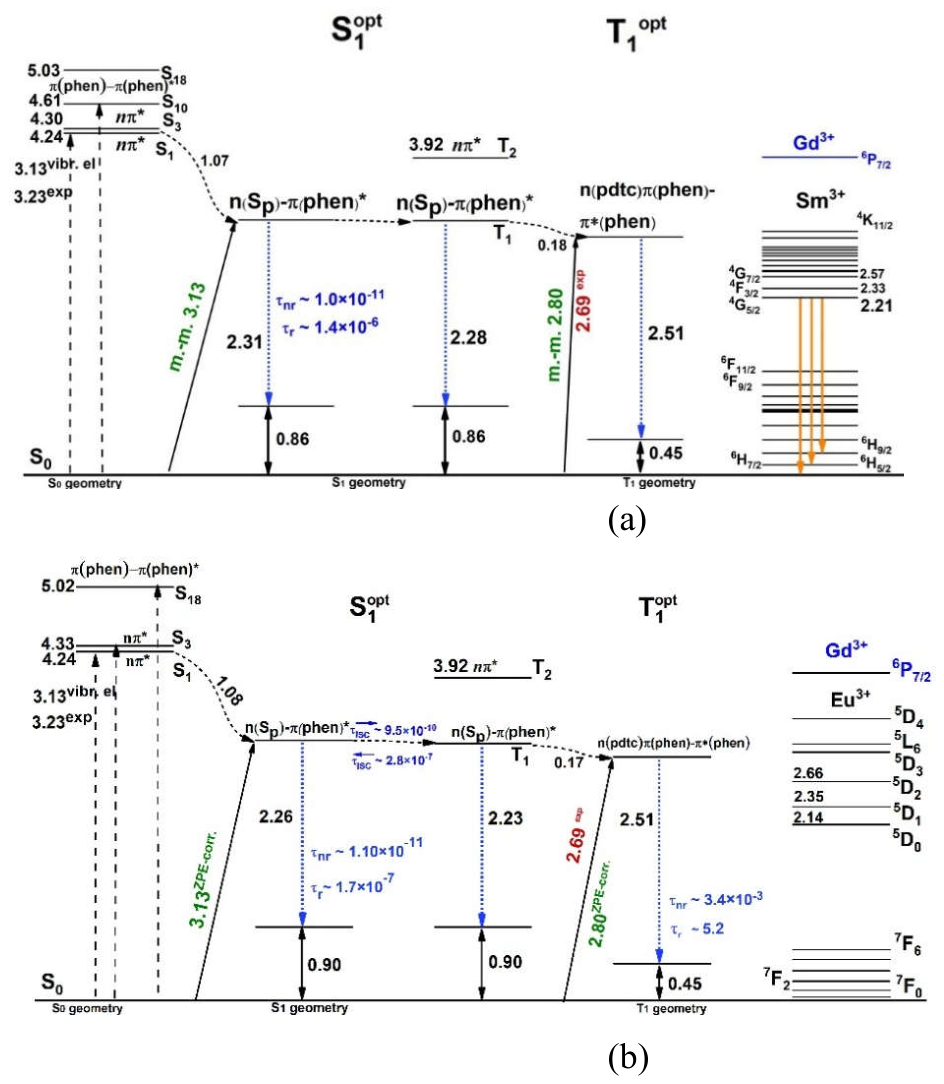
Figure 7.
Adsorption type of Eu(Phen)2X3 on hydrophilic and hydrophobic SiO2-based model surface composites. Optimized structures with Sparkle/AM1 method for a) Eu(Phen)2Cl3/Si-O-Si-OH, b) Eu(Phen)2Cl3/Si-O-Si(CH3)3, c) Eu(Phen)2(NO3)3/Si-O-Si-OH and d) Eu(Phen)2(NO3)3/Si-O-Si(CH3)3 Ref [98].
Figure 7.
Adsorption type of Eu(Phen)2X3 on hydrophilic and hydrophobic SiO2-based model surface composites. Optimized structures with Sparkle/AM1 method for a) Eu(Phen)2Cl3/Si-O-Si-OH, b) Eu(Phen)2Cl3/Si-O-Si(CH3)3, c) Eu(Phen)2(NO3)3/Si-O-Si-OH and d) Eu(Phen)2(NO3)3/Si-O-Si(CH3)3 Ref [98].
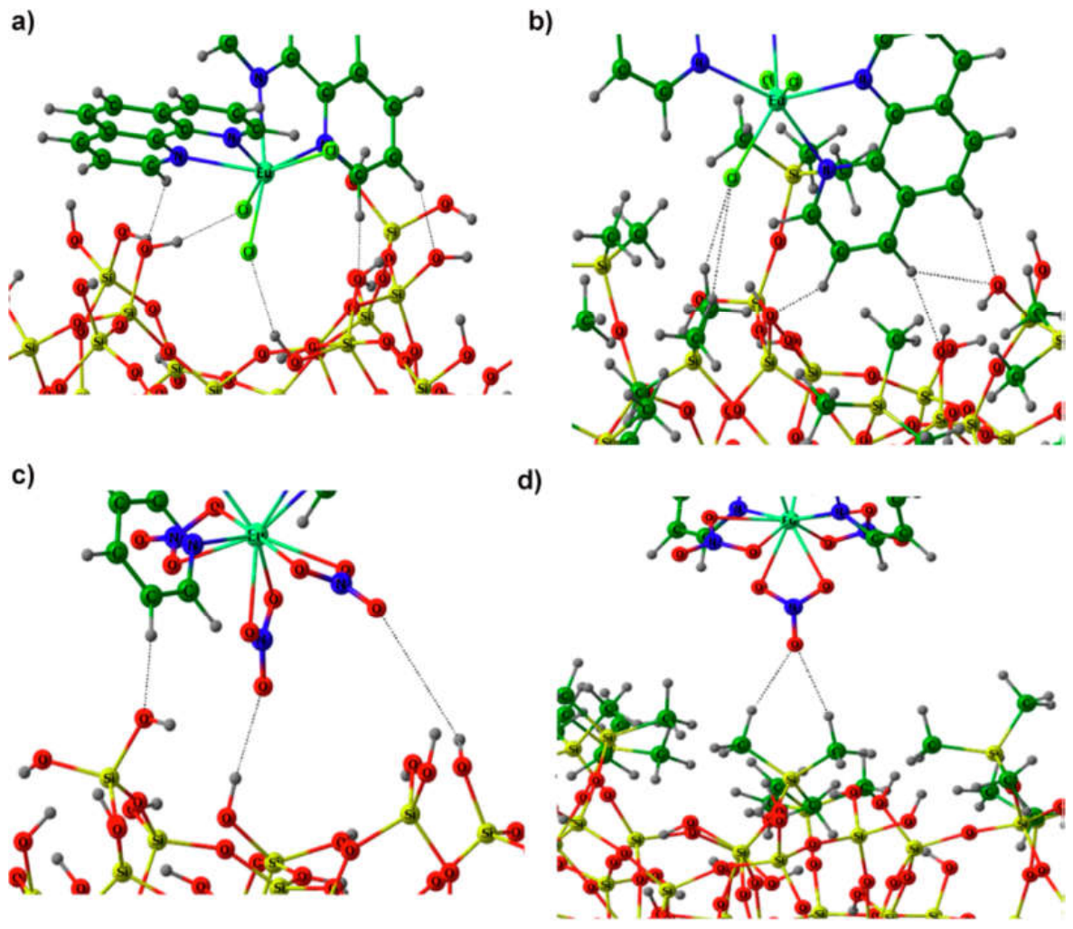
Figure 9.
Energy level diagram calculated with DFT/TDDFT(TDA)/ωB97XD/B1 method of Eu(CCA)3(H2O)3 in gas phase and methanol, compared to the available experimental data (exp) (in violet). INDO/S-CIS calculations and the most probable channel for the nonradiative energy transfer process based on calculated energy transfer (red) and back-energy transfer (blue) rates. fl. – fluorescence, ph – phosphorescence, ISC – intersystem crossing, m-m–minimum to minimum energy, ZPE – Zero Point Energy corrected m-m energy, τ - fluorescence radiative lifetime, min – energy minimization at optimized excited state geometry, RI – Refractive Index, QE – Quantum Efficiency, QY – Quantum Yield. From ref. [97].
Figure 9.
Energy level diagram calculated with DFT/TDDFT(TDA)/ωB97XD/B1 method of Eu(CCA)3(H2O)3 in gas phase and methanol, compared to the available experimental data (exp) (in violet). INDO/S-CIS calculations and the most probable channel for the nonradiative energy transfer process based on calculated energy transfer (red) and back-energy transfer (blue) rates. fl. – fluorescence, ph – phosphorescence, ISC – intersystem crossing, m-m–minimum to minimum energy, ZPE – Zero Point Energy corrected m-m energy, τ - fluorescence radiative lifetime, min – energy minimization at optimized excited state geometry, RI – Refractive Index, QE – Quantum Efficiency, QY – Quantum Yield. From ref. [97].
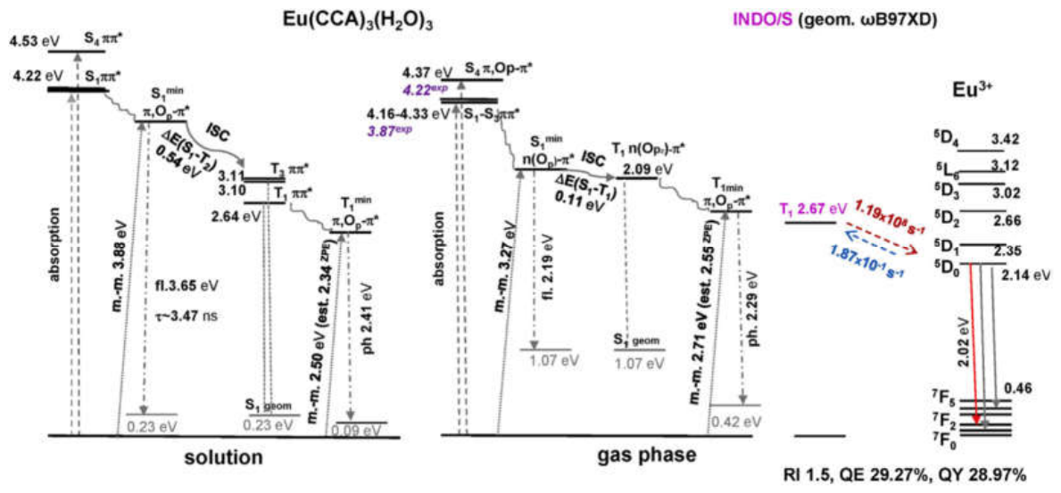
Figure 10.
Jablonski energy level diagram calculated with DFT/TDDFT(TDA)/ωB97XD/B1 method of Eu(Phenac)2(H2O)(NO3)3 and Eu(Phenac)3(NO3)3 in acetonitrile solution. The S1–S0 fluorescence lifetimes are calculated using a procedure in MOMAP, while the most probable pathways of ISC electronic relaxation lifetimes are predicted on the basis of single-point TDDFT/RIJCOSX/ωB97XD/B1* calculations. The vertical excitation and adiabatic (ad) energies are in (eV) and the lifetime values are in (s). The Sn states with the largest oscillator strengths are not drawn in the diagram. From ref. [83].
Figure 10.
Jablonski energy level diagram calculated with DFT/TDDFT(TDA)/ωB97XD/B1 method of Eu(Phenac)2(H2O)(NO3)3 and Eu(Phenac)3(NO3)3 in acetonitrile solution. The S1–S0 fluorescence lifetimes are calculated using a procedure in MOMAP, while the most probable pathways of ISC electronic relaxation lifetimes are predicted on the basis of single-point TDDFT/RIJCOSX/ωB97XD/B1* calculations. The vertical excitation and adiabatic (ad) energies are in (eV) and the lifetime values are in (s). The Sn states with the largest oscillator strengths are not drawn in the diagram. From ref. [83].
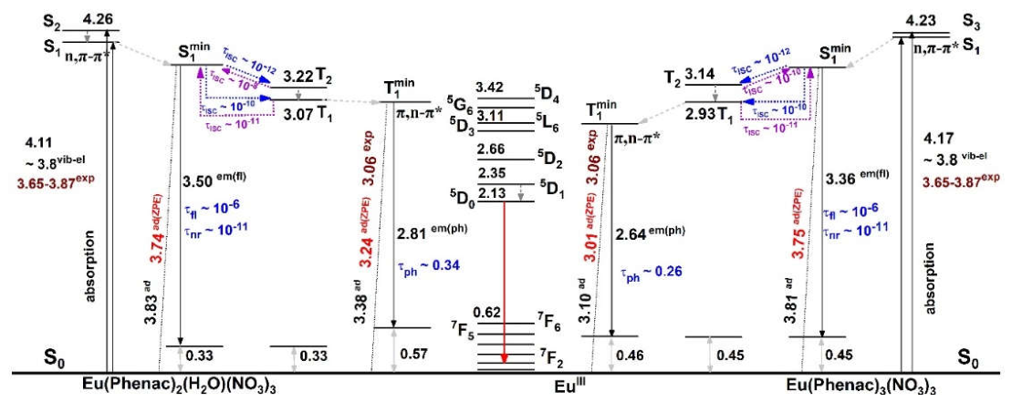

Table 1.
Deactivation reactions of [M2-S2] hexacarbonyls, M= Fe, Co. Results from ref. 33.
| Reaction | ΔH0 kJ mol-1 |
| Desulfurization by hydrogen | |
| [Fe2(S2H)](CO)6 ⇒ [Fe2S](CO)6 + HS• | 322 |
| [FeCoS2H](CO)6 ⇒ [FeCoS](CO)6 + HS• | 296 |
| [Co2(S2H)](CO)6 ⇒ [Co2S](CO)6 + HS• | 341 |
| [Fe2(S2H)](CO)6 ⇒ [Fe2S]+(CO)6 + HS− | 831 |
| [FeCoS2H](CO)6 ⇒ [FeCoS] +(CO)6 + HS− | 828 |
| [Co2(S2H)](CO)6 ⇒ [Co2S] +(CO)6 + HS− | 734 |
| Desulfurization by CO | |
| [Fe2(S2H)](CO)6 + CO ⇒ [Fe2S](CO)6 + COS | 1560 |
| [FeCoS2H](CO)6 + CO ⇒ [FeCoS](CO)6 + COS | 1620 |
| [Co2(S2H)](CO)6 + CO ⇒ [Co2S](CO)6 + COS | 1640 |
| Loss of carbonyl group | |
| [Fe2(S2H)](CO)6 ⇒ [Fe2(S2H)](CO)5 + CO | 163 |
| [Fe2(S2H)](CO)5 ⇒ [Fe2(S2H)](CO)4 + CO | 185 |
| [FeCoS2H](CO)6 ⇒ [FeCoS2H](CO)5 + CO | 233 |
| [FeCoS2H](CO)5 ⇒ [FeCoS2H](CO)4 + CO | 265 |
| [Co2(S2H)](CO)6 ⇒ [Co2(S2H)](CO)5 + CO | 151 |
| [Co2(S2H)](CO)5 ⇒ [Co2(S2H)](CO)4 + CO | 210 |
| Hydrolysis | |
| [Fe2(S2)](CO)6 + 4H2O ⇒ 2Fe(OH)2 + 2H2S + 6CO | 421 |
| [FeCoS2](CO)6 + 4H2O ⇒ Fe(OH)2 + Co(OH)2 + 2H2S + 6CO | 486 |
| [Co2(S2)](CO)6 + 4H2O ⇒ 2Co(OH)2 + 2H2S + 6CO | 521 |
Table 2.
Calculated Proton Affinities (PA, kJ mol-1) at sulfur centers and [H+,e−] affinities at either sulphur or at Co or Fe centres (kJ mol-1) for [M2-S2] and [M2-Se2] carbonyl complexes. n=0 for [H+,e−] attachment, n=1 for protonation. Results from refs. 18, 21, 33.
Table 2.
Calculated Proton Affinities (PA, kJ mol-1) at sulfur centers and [H+,e−] affinities at either sulphur or at Co or Fe centres (kJ mol-1) for [M2-S2] and [M2-Se2] carbonyl complexes. n=0 for [H+,e−] attachment, n=1 for protonation. Results from refs. 18, 21, 33.
| PA | [H+,e−] | 2[H+,e−] | |
| [Co2(S2)H]n+(CO)6; S-H | 851 | 252 | 474 |
| [Co2H(S2)](CO)6; Co-H | 173 | ||
| [Co2S2H]n+(CO)4; S-H | 767 | 224 | |
| [Co2HS2](CO)4; Co-H-Co | 177 | ||
| [Fe2(S2)H]n+(CO)6; S-H | 717 | 235 | 445 |
| [Fe2H(S2)](CO)6; Fe-H | 212 | ||
| [Fe2S2H]+(CO)4; S-H | 651 | ||
| [Fe2HS2](CO)4; Fe-H-Fe | 229 | ||
| [Fe2Se2H]+(CO)4; Se-H | 639 | ||
| [Fe2HSe2](CO)4; Fe-H-Fe | 228 | ||
| [Co2Se2H]n+(CO)4; Se-H | 748 | 191 | |
| [Co2HSe2](CO)4; Co-H-Co | 179 | ||
| [Co2HSe2](CO)4; Co-H | 201 | ||
| [Ni2S2H]n+(CO)4; S-H | 935 | 181 | |
| [Ni2Se2H]n+(CO)4; Se-H | 907 | 168 | |
| [Cu2S2H]n+(CO)4; S-H | 968 | 134 | |
| [Cu2Se2H]n+(CO)4; Se-H | 936 | 81 |
Table 3.
TD-DFT results for selected complexes of reduced (H+,e−) cobalt sulfides and selenides. The most intense light absorption bands are listed. Results are from refs. 18, 21, 33.
Table 3.
TD-DFT results for selected complexes of reduced (H+,e−) cobalt sulfides and selenides. The most intense light absorption bands are listed. Results are from refs. 18, 21, 33.
| Complex | Light absorption bands, nm | Oscillator strength |
| [Co2S-SH](CO)6 | 643; 873 | 0.0039; 0.0061 |
| [Co2S-SH](CO)4 | 689; 824 | 0.0060; 0.0010 |
| [Co-H-CoS2](CO)4 | 750; 803 | 0.0020; 0.0040 |
| [Fe2S-SH](CO)6 | 727; 874;1021 | 0.0020; 0.0040; 0.0144 |
| [Co2Se-SeH](CO)4 | 628; 749 | 0.0011; 0.0017 |
| [Co-H-CoSe2](CO)4 | 748; 1610 | 0.0006; 0.0250 |
| [Co(Co-H)Se2](CO)4 | 546; 639 | 0.0017; 0.0024 |
Table 4.
TD-DFT results for OER intermediates of selected sulfide and selenide tetracarbonyl complexes. The most intense light absorption bands are listed. From ref. 21.
Table 4.
TD-DFT results for OER intermediates of selected sulfide and selenide tetracarbonyl complexes. The most intense light absorption bands are listed. From ref. 21.
| Complex | Light absorption bands, nm | Oscillator strength |
| Co2S2(CO)4; O-O | 651 | 0.0014 |
| Co2Se2(CO)4; O-O | 582 | 0.0091 |
| Fe2Se2(CO)4; O-O | 807 | 0.0131 |
| Cu2Se2(CO)4; O-O | 430 | 0.0094 |
| Co2S2(CO)4; OOH* | 507; 573 | 0.0023; 0.0027 |
| Co2Se2(CO)4; OOH* | 531 | 0.0014 |
| Fe2Se2(CO)4; OOH* | 798 | 0.0052 |
| Cu2Se2(CO)4; OOH* | 590 | 0.0130 |
| Ni2Se2(CO)4; OOH* | 558 | 0.0023 |
Disclaimer/Publisher’s Note: The statements, opinions and data contained in all publications are solely those of the individual author(s) and contributor(s) and not of MDPI and/or the editor(s). MDPI and/or the editor(s) disclaim responsibility for any injury to people or property resulting from any ideas, methods, instructions or products referred to in the content. |
© 2026 by the authors. Licensee MDPI, Basel, Switzerland. This article is an open access article distributed under the terms and conditions of the Creative Commons Attribution (CC BY) license (http://creativecommons.org/licenses/by/4.0/).
Copyright: This open access article is published under a Creative Commons CC BY 4.0 license, which permit the free download, distribution, and reuse, provided that the author and preprint are cited in any reuse.