

Submitted:
18 August 2026
Posted:
20 August 2026
You are already at the latest version
Abstract
Deltamethrin (DLT), a type-II pyrethroid insecticide, can interfere with voltage-gated Na+ currents (INa) in excitable cells; however, its effects on INa gating and neuronal discharge remain unclear. Using Neuro-2a motor neuron-like cells and whole-cell patch-clamp recordings, we found that DLT (10 μM) mildly increased transient INa (INa(T)) but markedly enhanced late INa (INa(L)) and tail INa (INa(Tail)). In the continued presence of DLT, dapagliflozin (Dapa) counteracted the DLT-induced increases in INa(L) and INa(Tail). We further incorporated modified INa(T), INa(L), and INa(Tail) kinetics mimicking DLT effects into a computational model of respiratory neurons. The simulations generated two distinct action potential (AP) bursting patterns characterized by either slow or fast intraburst firing. Both patterns exhibited progressively shortened AP intervals within bursts, indicating reverse spike-frequency adaptation. Molecular docking further predicted hydrophobic interactions between DLT and the NaV1.8 channel. Collectively, these findings demonstrate that DLT preferentially enhances INa(L) and INa(Tail), which may substantially alter bursting behavior in modeled respiratory neurons.
Keywords:
deltamethrin
; dapagliflozin
; voltage-gated Na+ current
; neuronal bursting
; reverse spike-frequency adaptation
; theoretical simulation
1. Introduction
Deltamethrin (DLT, also known as decamethrin, see Figure 1) is a cyclopropanecarboxylate ester resulting from formal condensation of 3-(2,2-dibromovinyl)-2,2-dimethylcyclopanecarboxylic acid with cyano (3-phenoxyphenyl)methanol. It serves as the active insecticidal component of the proinsecticide tralomethrin [1]. Pyrethroids, such as DLT and tefluthrin, have been shown to alter the gating properties of voltage-gated Na+ (NaV) channels [2,3,4,5]. Recent studies indicate that DLT and other structurally related pyrethroids can exert significant toxic effects across various species, including fish, laying hens, and rats [3,5,6,7,8,9,10,11,12,13]. Notably, DLT—a neurotoxic type II pyrethroid ester insecticide—has been associated with a reversible sequence of motor symptoms in rats, characterized by hind limb rigidity and choreoathetosis. Additionally, it has been implicated as a potential contributor to Cockayne syndrome [6,13,14]. Cockayne syndrome is a rare, inherited genetic disorder characterized by growth failure, neurological degeneration, photosensitivity, and premature aging [13,14].
Voltage-gated Na+ (NaV) channels are essential for the excitability of mammalian tissues, including the central and peripheral nervous systems as well as endocrine and neuroendocrine cells, To date, nine isoforms (NaV1.1 to 1.9), encoded by the SCN1A-SCN5A and SCN8A-SCN11A genes, have been identified across these systems [15]. Structurally, eukaryotic NaV channels consist of a single α-subunit organized into four homologous domains (I-IV). Each domain contains six-transmembrane segments (S1-S6), which together form the functional architecture responsible for channel gating and ion conduction [15].
Following rapid membrane depolarization, NaV channels undergo a sequence of conformational changes, transitioning from the closed (resting) state to an activated open state and subsequently to an inactivated state. These transitions generate the macroscopic Na+ current (INa) [15]. Repeated short-duration depolarizing stimuli can progressively enhance current inactivation, leading to a cumulative reduction in INa and thereby producing use-dependent changes of NaV channel availability [16,17,18,19]. In addition to transient INa, increasing attention has been directed toward the persistent or late component of INa (INa(L)), whose magnitude appears to be dynamically regulated and may represent an important pharmacological target [20,21,22,23]. Activation of NaV channels increases inward Na+ influx, further depolarizing the membrane and establishing a positive feedback loop that accelerates the rapid upstroke of action potentials (APs). Through this mechanism, NaV channel activity is therefore a critical determinant of AP amplitude, firing frequency, and discharge patterns, and thereby contributing to the regulation of neuronal signal propagation and hormonal secretion across a variety of excitable cell types [20].
The Neuro-2a (N2a) cell line, derived from murine neuroblastoma, exhibits characteristics reminiscent of motor neurons and is widely utilized in NaV 1.7 neuroscience research. Under specific conditions—such as treatment with retinoic acid—N2a can undergo differentiation, extending neuritis and acquiring neuron-like properties. This makes them a valuable in vitro model for investigating neuronal differentiation, neurotoxicity, and neurodegenerative disorders [18,21,22,23,24]. Additionally, N2a cells have been reported to express several NaV channel isoforms, including NaV1.2, 1.3, and 1.7 channels [23]. NaV1.2 and 1.3 channels are predominantly expressed in the central nervous system and are implicated in the pathophysiology of epilepsy, while NaV1.7 channel is primarily found in the peripheral nervous system and plays a key role in mediating neuropathic pain.
In this study, we aimed to explore the effects of DLT, a type II pyrethroid, and functionally related compounds such as dapagliflozin (Dapa), on transmembrane ionic currents, with a particular emphasis on the current density of INa. To assess how DLT influences INa gating behavior, we employed a theoretical approach to simulate INa dynamics. Furthermore, using a computational model of respiratory neurons located in the pre-Bötzinger complex (pre-BötC) of the mammalian brainstem [25], we investigated how exposure to DLT may alter the bursting firing patterns of these neurons.
2. Materials and Methods
2.1. Chemicals, Compounds, Drugs, Reagents, and Solutions
Deltamethrin (DLT, also referred to as decamethrin; molecular formula C22H19Br2NO3; IUPAC name: (S)-α-cyano-3-phenoxybenzyl (1R,3R)-3-(2,2-dibromovinyl)-2,2-dimethylcyclopropane-1-carboxylate, Figure 1) was purchased from MedChemExpress through Asia Biomed Inc., (Taipei, Taiwan). Dapagliflozin (Dapa, marketed under the trade names Farxiga® and Forxiga®) was retrieved from Cayman Chemical (Ann Arbor, MI). Atropine, tetraethylammonium chloride (TEA), and tetrodotoxin (TTX) were acquired from Sigma-Aldrich (Genechain, Kaohsiung, Taiwan). Because deltamethrin is susceptible to both photodegradation and microbial decomposition [1,26,27], a 10 mM stock solution was freshly prepared in dimethyl sulfoxide (DMSO). The stock solution was protected from light by wrapping the container with aluminum foil and stored at −20 °C until used to preserve its stability.
Cell culture preparations, including fetal bovine serum, L-glutamine, trypsin–EDTA, culture media, were purchased from HyCloneTM (Thermo Fisher Scientific, Tainan, Taiwan). Additional chemicals, such as CsCl, CsOH, CdCl2, and other reagents and solvents used throughout the experiments, were of analytical grade and obtained from Sigma-Aldrich through Genechain (Kaohsiung, Taiwan).
Whole-cell recordings were performed using a standard HEPES-buffered Tyrode’s solution as the extracellular bathing medium. The solution contained (in mM): 136.5 NaCl, 5.4 KCl, 1.8 CaCl2, 0.53 MgCl2, 5.5 glucose, and 5.5 HEPES, with the pH adjusted to 7.4 using NaOH. For the measurement of K+ currents, recording pipettes were digitally subtracted were filled with an intracellular solution composed of (mM): 130 K-aspartate, 20 KCl, 1 MgCl2, 1 KH2PO4, 3 Na2ATP, 0.1 Na2GTP, 0.1 EGTA, and 5 HEPES, with the pH adjusted to 7.2 using KOH. To isolate voltage-gated Na+ current (INa), K+ in the intracellular solution were completely substituted with an equimolar concentration of Cs+ to suppress endogenous K+ conductances. The pH of the Cs+-based internal solution was adjusted to 7.2 with CsOH before use.
2.2. Cell Preparation
Neuro-2a (N2a) cells, a mouse neuroblastoma-derived clonal cell line originating from a spontaneous tumor in strain A albino mice (Mus musculus), were obtained from the Bioresources Collection and Research Center (BCRC-60026, Hsinchu, Taiwan). The cell line was originally established by the American Type Culture Collection (ATCC® CCL-131TM; Manassas, VA) and has been extensively employed as an electrically excitable neuronal model for electrophysiological and pharmacological investigations [18,22].
N2a cells were cultured in Dulbecco’s Modified Eagle Medium (DMEM) supplemented with 10% (v/v) heat-inactivated fetal bovine serum, 2 mM L-glutamine, 1.5 g/L sodium bicarbonate, 0.1 mM non-essential amino acids, and 1.0 mM sodium pyruvate. Cell cultures were maintained at 37 °C in a humidified incubator with an atmosphere of 5% CO2 and 95% air [18]. Routine passaging was performed using 0.025% trypsin–EDTA (HyCloneTM) containing 0.01% sodium N,N-diethyldithiocarbamate. Electrophysiological experiments were conducted on cultures that had reached approximately 60–70% confluence, generally within 5–7 days after plating [18,19].
2.3. Electrophysiological Recordings Using the Patch-Clamp Technique
Following enzymatic dissociation, several drops of the Neuro-2a cell suspension were placed into a custom-designed recording chamber positioned on the stage of an inverted microscope. The cells were superfused with normal Tyrode’s solution of the ionic composition described above and maintained at room temperature (20-25 °C). Before each measurement were initiated, sufficient time was allowed for the cells to settle and firmly attach to the bottom surface of the chamber.
Patch pipettes were fabricated from Kimax®-51 borosilicate glass capillaries (catalog no. DWK34500-99; Kimble®, Merck, Tainan, Taiwan) and fire-polished to obtain a final tip resistance of 2–4 MΩ when filled with the internal solution. Each pipette was secured in a sealed electrode holder fitted with a lateral suction port to facilitate seal formation. Electrical continuity between the amplifier and the pipette solution was established using an Ag/AgCl wire. Membrane currents were measured in the whole-cell configuration of the patch-clamp technique with an RK-400 patch-clamp amplifier (Bio-Logic, Claix, France), operated under a modified recording protocol, as described previously [2,28].
A liquid junction potential arose from the difference in ionic composition between the extracellular bathing solution and the pipette internal solution. This potential was compensated immediately before establishing the gigaohm (GΩ) seal and was further corrected during subsequent analysis of whole-cell recordings. To reduce capacitive currents associated with the activation of INa during depolarizing voltage steps, the currents elicited by an isoelectric hyperpolarizing pulse were provided previously digitally subtracted from the recorded traces.
2.4. Statistical Analyses
Results are presented as the mean ± standard error of the mean (SEM), and n denotes the number of individual cells included in each analysis. Linear and nonlinear models were fitted by minimizing the least-squares error using either Microsoft® Excel® for Microsoft 365 MSO (64-bit; version 16.017628.20006) with the Solver add-in (Microsoft Corporation, Redmond, WA) or OriginPro® 2024b (OriginLab Corporation, Northampton, MA). Differences between two groups were evaluated using paired or unpaired two-tailed Student’s t-tests, as appropriate. For comparisons involving multiple groups, one-way analysis of variance (ANOVA) followed by Dunnett’s multiple-comparison test was applied. Unless otherwise specified, statistical analyses were conducted with SPSS statistics, version 23.0 (IBM Corporation, Armonk, NY). A P-value below 0.05 was considered indicative of statistical significance.
2.5. Computer Simulations
To simulate the DLT-induced alterations in the amplitude and gating kinetics of INa, a modified model, originally developed by Abdulla et al., 2022 [25], was mathematically constructed for this study. Below is a description of the kinetic scheme of INa that accounts for the experimentally observed results.
The expressions of INa(T), INa(L), and INa(Tail) are derived from the voltage- and time-dependent characteristics of the NaV channels. The INa(Tail) is given by:
The currents INa(T) and INa(L) are expressed as the product of their maximal conductances, voltage- and time-dependent gating variables, and the term (V - ENa), which represents the difference between the membrane potential and the Na+ reversal potential. Each of the Na+ gating variables x ϵ {mNa, hNa, mNa(L), hNa(L)} satisfies the following equation
where
The parameter values used for these equations, including the equilibrium potential for Na+ ions (ENa), were provided previously [28].
In this model, the membrane potential (V) is governed by the current balance equations:
The membrane currents in (I) include the transient Na+ current (INa(T)), the late (or persistent) Na+ current (Na(L)), the delayed rectifier K+ current (IK(DR)), the leakage current (IL), and the synaptic current (ISyn) [28]. This respiratory model neuron exhibits bursting behavior by coupling membrane potential dynamics with the intrinsic mechanisms of Na+ and K+ currents.
3. Results
3.1. Effect of DLT on Voltage-Gated Na+ Current (INa) Present in Neuro-2a Neuronal Cells
In the initial experiment, we investigated the potential effects of DLT on the INa elicited by step depolarization in these cells. To isolate INa, the cells were bathed in a Ca2+-free Tyrode’s solution supplemented with 0.5 mM CdCl2 and 10 mM tetraethylammonium chloride (TEA). CdCl2 was used to block nonspecific Ca2+ currents, while TEA was included to suppress the majority of nonspecific K+ currents. The ionic composition of the Tyrode’s solution is described in detail in the Materials and Methods section. Whole-cell voltage-clamp recordings of INa were performed using a Cs+-containing pipette solution.
As illustrated in Figure 2A, Neuro-2a cells were initially held at a membrane potential of −80 mV. To ensure complete inactivation of INa, the cells were hyperpolarized to −100 mV for 40 msec. This was followed by a depolarizing step to −10 mV for 40 msec to activate INa, which produced a downward deflection in the current trace. To assess INa(Tail), the membrane potential was then repolarized to −50 mV for another 40 msec. Finally, the voltage was returned to the initial holding potential of −80 mV. The corresponding voltage protocol is shown in the upper panel of Figure 2A.
In these recordings, the cell was depolarized from −100 to −10 mV for 40 msec, which reliably evoked a robust, transient inward INa (INa(T))—a rapidly activating component known to be sensitive to blockade by 1 μM tetrodotoxin (TTX). Upon exposure to 10 μM DLT, both the peak density of INa(T) and the late or persistent component (INa(L)) of INa density were enhanced. Furthermore, as the membrane potential was repolarized to more hyperpolarized levels following the depolarizing step, the current density of INa(Tail) progressively increased. Despite these changes, the kinetics of fast and slow inactivation course of INa evoked by the short depolarizing pulse remained unaffected by 10 μM DLT. Notably, the DLT-induced increase in INa(L) density was significantly greater than that observed for INa(T). In addition, the density of tail component (INa(Tail)) measured at the level of −50 mV or −80 mV was markedly elevated in the presence of 10 μM DLT.
Figure 2B and 2C summarize the effects of DLT alone and in combination with dapagliflozin (Dapa) on the densities of INa(T) and INa(L) in Neuro-2a neuronal cells. Dapa, a well-characterized inhibitor of the Na+-glucose co-transporter, has recently been reported to selectively suppress INa(L) [2,24,31]. In this study, treatment with DLT significantly increased overall INa density in Neuro-2a cells, despite having no observable effect on the fast or slow inactivation kinetics of the current. Interestingly, the DLT-induced enhancement appeared to be more prominent in the INa(L) and INa(Tail) components than in INa(T) component, suggesting a degree of selectivity in its modulatory action. Furthermore, the addition of Dapa in the continued presence of DLT partially reversed the DLT-mediated increase in INa density, particularly affecting the INa(L) and INa(Tail) components. These findings highlight a potential interaction between DLT and Dapa in modulating INa dynamics in neuronal cells.
Simulation studies on INa(T), INa(L), and INa(Tail)
The activity of INa, including its persistent component INa(L), has been implicated in the generation of respiratory rhythms within the pre-BötC [32]. To explore the potential effects of DLT on INa, we developed a computational model that incorporates INa(T), INa(L), and INa(Tail) components. The simulated INa model was adapted from the work of Abdulla et al. [25], with a detailed description of the kinetic scheme provided in the Materials and Methods section. As shown in Figure 3, following appropriate parameter adjustments, we successfully reproduced the INa in Neuro-2a cells, clearly distinguishing the three components: INa(T), INa(L), and INa(Tail). The total INa (INa(Tot)) represents the sum of these three components. The voltage-clamp protocol applied in these simulations is depicted at the top of Figure 3 and Figure 4. To ensure comparability across experiments, current densities were normalized to membrane capacitance.
3.2. Simulation of Increases in INa(T), INa(L), and INa(Tail) to Mimic DLT Effects on INa
To simulate the effects of DLT on INa, we adjusted specific parameters (GNa(T), GNa(L), and GNa(Tai)) to generate simulated INa with increased INa(T), INa(L), and INa(Tail) components. The simulation results, shown in Figure 4, illustrate the different forms of INa, namely INa(T), INa(L), and INa(Tail). Notably, the simulated densities of INa(L) and INa(Tail) exhibited a greater increase compared to INa(T), closely mirroring the experimental observations (INa) shown in Figure 2. Although the densities of INa(L) and INa(Tail) were significantly elevated relative to INa(T), the inactivation time course of INa elicited by short depolarizing pulse remained unchanged.
Neuronal burst firing of action potentials (APs) as simulated INa was incorporated to the modeled respiratory neuron
We next incorporated the simulated INa into the bursting pattern of a modeled respiratory neuron. The upper part of Figure 5 illustrates this neuron’s distinct bursting pattern, which aligns with earlier observations [25]. The interburst rate was approximately 0.33 Hz, and a progressive shortening in intraburst intervals from 92 to 38 msec was observed. The AP amplitude within the bursts progressively decreased, along with a gradual shortening of the intraburst AP intervals. The lower part of Figure 5 depicts the corresponding INa(Tot), showing a progressive decline in its density. This decline reflects the frequency dependence of INa, consistent with previous studies [16,17,18,19].
Figure 6 illustrates the AP bursting patterns resulting from the modification of INa to mimic the effects of DLT on INa(T), INa(L), and INa(Tail). Figure 7 provides an enlarged view of the dashed box in the uppermost part of Figure 6. Notably, two distinct patterns of AP bursting, categorized as slow and fast types, were clearly observed, as indicated with asterisk (*) and double asterisks (**), in the potential trace at the top of Figure 6.
The slow bursting pattern exhibited an intraburst rate that progressively shortened from 271 to 128 msec across an individual burst, while the fast bursting pattern demonstrated a quicker intraburst rate, with intervals decreasing from 90 to 39 msec. One bursting pattern exhibited a slow intraburst rate with a progressive shortening of intraburst AP intervals from 271 to 128 msec, while the other was noted to display fast intraburst rate with a progressive shortening of intraburst intervals from 90 to 39 msec. Both patterns showed reverse spike-frequency adaptation during their respective slow or fast bursting firing [33]. The simulated interburst rate during both fast and slow bursts was approximately 0.24 Hz.
During slow-type bursts, the resting membrane potential depolarized from −54.8 to −52.9 mV. This depolarization was more pronounced during fast-type bursts, where the resting potential shifted from −53,4 to −50.2 mV. Additionally, the amplitude of APs during fast-type bursts progressively decreased from 26.7 to 15.6 mV, while that in slow-type bursts, the amplitude declined from 28.7 to 23.6 mV. The post-burst hyperpolarizing potential was −55.1 mV during slow- versus fast-type bursts and −56.1 mV during fast-type bursts.
Moreover, during initial generation of INa(T) became progressively declined; however, the density of INa(L) and INa(Tail) was gradually and evidently increased. Such simulations show that DLT exerts a significant impact on burst patterns of this modeled respiratory neuron through its stimulation of marked under the trade name channels. Consequently, this could lead to substantial disturbances in the respiratory movement of organisms [34,35].
3.3. Molecular Docking on Interaction Between NaV Channel and DLT
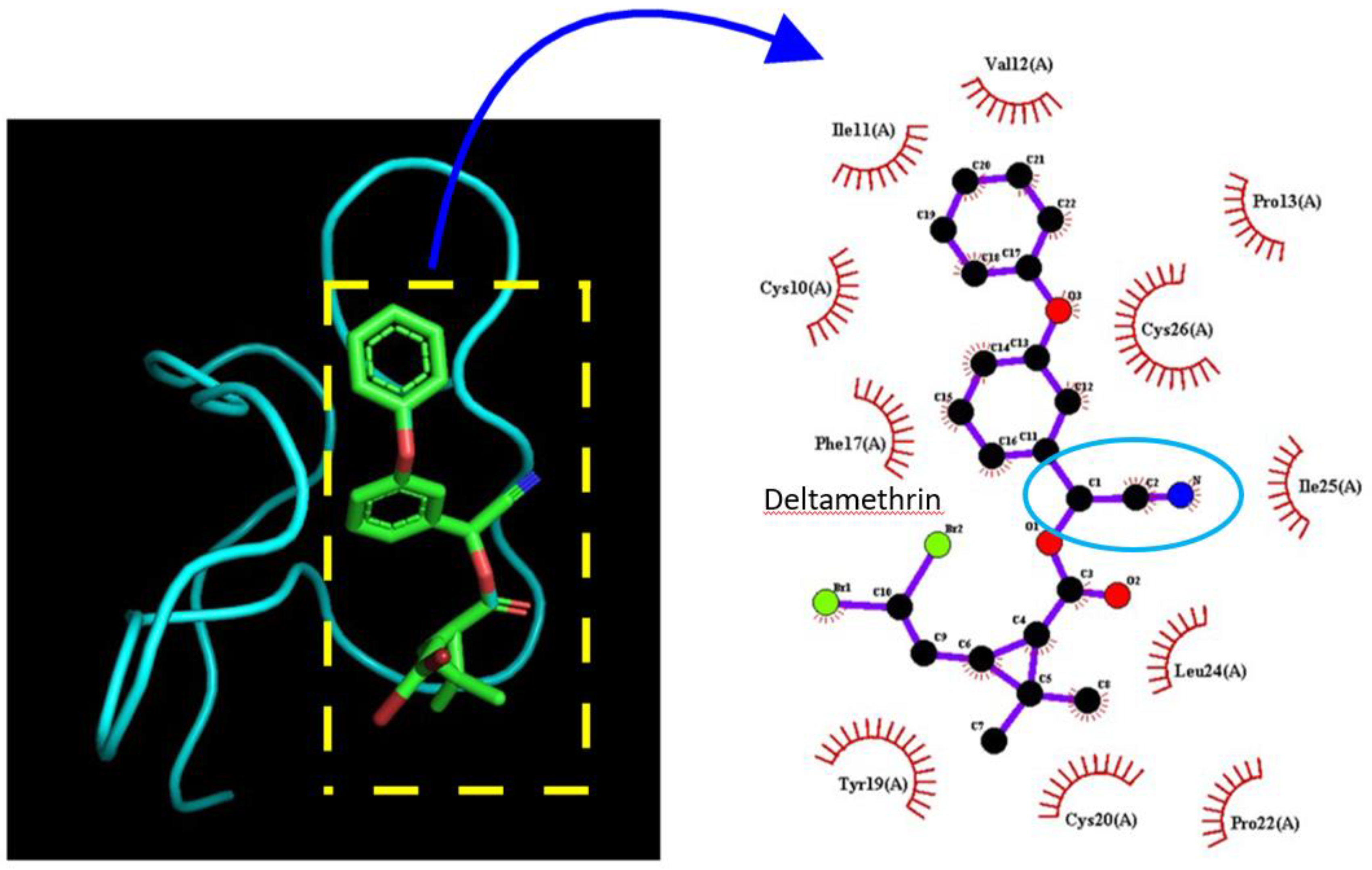
Since our results have shown that DLT has stimulatory effects on the current density and gating kinetics of INa, we also conducted further investigations to examine the direct binding of DLT on the NaV protein. The docking of DLT with the NaV1.8-channel protein was performed using PyRx software, and the resulting predicted docking sites of the compound are depicted in Figure 8. Of note, DLT can form hydrophobic contacts with eleven amino-acid residues Cys10(A), Ile11(A), Val12(A), Pro13(A), Phe17(A), Tyr19(A), Cys20(A), Pro22(A), Leu24(A), Ile25(A), and Cys26(A). The docking site appears to be located near the Ile-Phe-Met motif and the N-terminal helix, as noted by Chen et al., 2024 [34]. Based on the results from in-silico docking studies, it suggests that the DLT molecule has the potential to bind to the specific region of the NaV1.8 channel with a binding affinity of −6.3 kcal/mol.
4. Discussion
This study reports six key findings: (1) In Neuro-2a neuroblastoma cells, deltamethrin (DLT), a known gating modifier of voltage-gated Na+ current (INa), significantly enhanced the densities of INa(T), INa(L), and INa(Tail); (2) Among these, the increase in the density of transient INa (INa(T)) induced by DLT was less pronounced compared to the more substantial enhancements observed in late INa (INa(L)) and tail INa (INa(Tail)); (3) Co-application of dapagliflozin (Dapa) effectively reversed the DLT-induced increase in all three forms of INa (INa(T), INa(L), and INa(Tail)), even in the continued presence of DLT; (4) A computational model was constructed to replicate DLT’s stimulatory effects on these distinct components of INa; (5) Numerical simulations were performed using this model to assess how DLT might influence the bursting pattern of respiratory neurons; and (6) Molecular docking analysis predicted potential docking sites for DLT on specific amino acid residues within the NaV1.8 channel. Together, these findings demonstrate that DLT-induced alterations on INa amplitude and gating properties—particularly in INa(T), INa(L), and INa(Tail)—can significantly disrupt neuronal bursting patterns [32].
Previous studies have suggested that the neurotoxic effects of DLT might involve its interaction with muscarinic receptors [36,37,38]. Neuro-2a cells are known to express muscarinic receptors on their membranes [21]. However, in our study, treatment with atropine—a muscarinic receptor antagonist—did not reverse the DLT-induced alterations in INa. This finding indicates that DLT likely exerts a direct effect on NaV channels, rather than acting indirectly through muscarinic receptor pathways. Additionally, it appears unlikely that the DLT-induced changes in INa are primarily mediated by effects on voltage-gated anion channels or Ca2+-activated Cl- channels, as recently proposed in other studies [24,39].
Dapagliflozin (Dapa), a known inhibitor of Na+-glucose co-transporters (SGLT), is primarily used to manage type 2 diabetes by lowering blood glucose levels [40]. In the present study, however, Dapa was found to inhibit DLT-induced alterations in the amplitude and gating properties of the INa. Although SGLT activity has been observed in some endocrine cells [43,44], it has not been reported in Neuro-2a cells. This suggests that Dapa may exert a direct effect on NaV channels in Neuro-2a cells, consistent with previous findings [2,31,41,42,43].
Previous studies have shown that type-I pyrethroids, such as tefluthrin, increase the amplitude of INa(T) and concurrently slow its inactivation time course [28,44]. In contrast, type II pyrethroids, including DLT, also enhance INa(T) amplitude but do not significantly affect the inactivation time constant of either the fast or slow components of the current. Notably, both the current study and earlier reports [2] have demonstrated that DLT additionally augments INa(L) and INa(Tail). Structurally, type II pyrethroids are distinguished from type I pyrethroids by the presence of an α-cyano group on the benzyl moiety of the compound (Figure 1 and Figure 8), which is absent in type-1 pyrethroids. These findings suggest that the α-cyano group in the pyrethroid molecule may play a role in the differential modulation of NaV channel gating by various pyrethroids.
An important consideration from our experimental results is that while Neuro-2a cells can display motor neuron-like properties, they are not identical to primary motor neurons. As a transformed cell line with cancerous origins, Neuro-2a cells can exhibit significant differences in behavior and characteristics compared to primary neurons [22,23,24]. Researchers frequently use Neuro-2a cells due to their ease of culture and manipulation. However, it is crucial to validate findings obtained from Neuro-2a cells using primary neurons or in vivo models to ensure their relevance and accuracy.
In simulations where DLT was present, perturbations to INa resulted in two distinct burst patterns in the modeled respiratory neuron (Figure 6 and Figure 7). This model, originally developed to simulate pre-Bötzinger complex (pre-BötC) neuronal activity [25], revealed one pattern characterized by a slowing of intraburst AP firing, while the other exhibited a faster intraburst firing rate. Both patterns also demonstrated a progressive shortening of intraburst intervals over time. This phenomenon is indicative of reverse spike-frequency adaptation where a neuron exhibits in increase in its firing rate during intraburst activity, as previously described in subthalamic nucleus neurons [33]. This is in contrast to the more common spike-frequency adaptation, where the firing rate typically decreases over time during intraburst activity. These findings suggest that the activity of INa(L) and INa(Tail), particularly under conditions involving DLT exposure, may be responsible for the reverse spike-frequency adaptation observed in the intraburst firing of this modeled respiratory neuron.
In these simulations, the AP amplitude exhibited a significant and progressive decrease during fast-type bursts, while the reduction during slow-type bursts was less pronounced. This observation suggests that the cumulative inhibition of INa, as previously described [17,19,45], coupled with reverse spike-frequency adaptation, is more prominent during fast-type bursts compared to slow-type bursts. Additionally, the post-burst hyperpolarizing potential was notably larger during fast-type bursts, and the extent of resting membrane potential depolarization was also more pronounced during fast-type bursts compared to slow-type bursts.
Bursting patterns in different modeled neurons are significant because they play a crucial role in understanding the complex dynamics of neural activity, such as neural coding and information processing, synchronization and network dynamics, and pathophysiological insights [46]. Collectively, these findings suggest that exposure to DLT is capable of inducing changes in the density and gating properties of INa [2], significantly influencing the burst patterns of respiratory neurons and potentially impacting respiratory function, as previously reported [6,10,20,25,32,35].
In our docking prediction of the DLT molecule on NaV channel (Figure 8), we found that DLT forms hydrophobic contacts with several amino-acid residues in the NaV1.8 channel, indicating that these regions may have a specific role in modulating DLT’s binding affinity to NaV1.8. NaV1.8 channels, known as a NaV channel α subunit 8 (SCN10A), are a type of NaV channel primarily expressed in peripheral sensory neurons, particularly in dorsal root ganglia neurons [47,48]. If this region contains these amino-acid residues, performing knockdown or knockout maneuvers is likely to result in these treated cells showing increased resistance to DLT or other type II pyrethroids’ modifications of INa [1,4,37,52]. However, whether DLT produces similar docking sites for the amino acids in NaV1.2, NaV1.3, and NaV1.7 channels in Neuro-2a cells [23] remains to be further investigated. Additionally, further investigation is needed to determine whether DLT or other type II pyrethroids interact with the auxiliary β subunits of NaV channels.
Conclusions
Our experimental observations and in silico studies demonstrate that DLT modulates INa, specifically INa(L) and INa(Tail). These changes in INa dynamics significantly influence the bursting behavior of modeled respiratory neurons, highlighting a potential role for DLT in modulating respiratory rhythmogenesis.
Funding
This work was supported by grants from the Ministry of Science and Technology, Taiwan (NSTC-113-2923-B-906-001), An Nan Hospital (ANHRF114-43 and ANHRF114-49), and Chang Bing Show-Chwan Memorial Hospital (BRD112026), Taiwan. The sponsors were not involved in the design of the study, data collection, analysis, or interpretation of the results.
Data Availability Statement
The original datasets and associated programming codes are available from the corresponding author upon reasonable request.
Declaration of Competing Interest
The authors declare no known competing financial interests or personal relationships that could have influenced the work presented in this paper.
Ethics and Consent to Participate Declarations
Not applicable.
Abbreviations
AP, action potential; Dapa, dapagliflozin; DLT, deltamethrin; INa, voltage-gated Na+ current; INa(L), late (or persistent) Na+ current; INa(T), transient Na+ current (peak component of INa); INa(Tail), tail Na+ current; INa(Tot), total Na+ current (including INa(T), INa(L), and INa(Tail)); NaV channel, voltage-gated Na+ channel.
References
- Laskowski, D.A. Physical and chemical properties of pyrethroids. Rev. Env. Contam. Toxicol. 2002, 174, 49–170. [Google Scholar] [CrossRef] [PubMed]
- Lin, M.H.; Lin, J.F.; Yu, M.C.; Wu, S.N.; Wu, C.L.; Cho, H.Y. Characterization in Potent Modulation on Voltage-Gated Na(+) Current Exerted by Deltamethrin, a Pyrethroid Insecticide. Int. J. Mol. Sci. 2022, 23. [Google Scholar] [CrossRef] [PubMed]
- Raposo-Garcia, S.; Costas, C.; Louzao, M.C.; Vale, C.; Botana, L.M. Synergistic effect of environmental food pollutants: Pesticides and marine biotoxins. Sci. Total Environ. 2023, 858, 160111. [Google Scholar] [CrossRef] [PubMed]
- Symington, S.B.; Toltin, A.C.; Murenzi, E.; Lansky, D.; Clark, J.M. Determination of potential toxicodynamic differences of pyrethroid insecticides on native voltage-sensitive sodium channels in juvenile versus adult rat brain. Pestic. Biochem Physiol. 2023, 189, 105296. [Google Scholar] [CrossRef] [PubMed]
- Vineetha, V.P.; Tejaswi, H.N.; Sooraj, N.S.; Das, S.; Pillai, D. Implications of deltamethrin on hematology, cardiac pathology, and gene expression in Nile tilapia (Oreochromis niloticus) and its possible amelioration with Shatavari (Asparagus racemosus). Vet. Res. Commun. 2024, 48, 811–826. [Google Scholar] [CrossRef] [PubMed]
- Spencer, J.; O'Malley, M. Pyrethroid illnesses in California, 1996-2002. Rev. Env. Contam. Toxicol. 2006, 186, 57–72. [Google Scholar] [CrossRef]
- Elbanna, R.; Osman, K.A.; Salama, M.S. Biomarkers of oral subacute toxicity of deltamethrin in exposed male Albino rats. Toxicol. Ind. Health 2023, 39, 735–753. [Google Scholar] [CrossRef] [PubMed]
- Liu, Y.; Liu, C.; Han, M.; Yu, N.; Pan, W.; Wang, J.; Fan, Z.; Wang, W.; Li, X.; Gu, X. Distribution and Elimination of Deltamethrin Toxicity in Laying Hens. Foods 2023, 12, 4385. [Google Scholar] [CrossRef] [PubMed]
- Otçu, S.; Deveci, E.; Özgökçe, Ç.; Gürsoy, G.T.; Tuncer, M.C. Protective effect of nebivolol on rat ovary exposed to deltamethrin toxicity. Acta Cir. Bras. 2023, 38, e385423. [Google Scholar] [CrossRef] [PubMed]
- Buzenchi Proca, T.M.; Solcan, C.; Solcan, G. Neurotoxicity of Some Environmental Pollutants to Zebrafish. Life 2024, 14. [Google Scholar] [CrossRef] [PubMed]
- Ma, R.; Wang, X.; Ren, K.; Ma, Y.; Min, T.; Yang, Y.; Xie, X.; Li, K.; Zhu, K.; Yuan, D.; Mo, C.; Deng, X.; Zhang, Y.; Dang, C.; Zhang, H.; Sun, T. Chronic low-dose deltamethrin exposure induces colon injury and aggravates DSS-induced colitis via promoting cellular senescence. Ecotoxicol. Env. Saf. 2024, 274, 116214. [Google Scholar] [CrossRef] [PubMed]
- Nashed, M.S.; Hassanen, E.I.; Issa, M.Y.; Tohamy, A.F.; Prince, A.M.; Hussien, A.M.; Soliman, M.M. The mollifying effect of Sambucus nigra extract on StAR gene expression, oxidative stress, and apoptosis induced by fenpropathrin in male rats. Food Chem. Toxicol. 2024, 189, 114744. [Google Scholar] [CrossRef] [PubMed]
- Shi, T.; Zhang, Q.; Chen, X.; Mao, G.; Feng, W.; Yang, L.; Zhao, T.; Wu, X.; Chen, Y. Overview of deltamethrin residues and toxic effects in the global environment. Env. Geochem Health 2024, 46, 271. [Google Scholar] [CrossRef] [PubMed]
- Scheepers, L.D.; Freercks, R.; Merwe, E.V. Acute cypermethrin and other pyrethroid poisoning - An organophosphate-like poisoning: A case report and review. Toxicol. Rep. 2023, 11, 107–110. [Google Scholar] [CrossRef] [PubMed]
- Catterall, W.A.; Lenaeus, M.J.; Gamal El-Din, T.M. Structure and Pharmacology of Voltage-Gated Sodium and Calcium Channels. Annu Rev. Pharmacol. Toxicol. 2020, 60, 133–154. [Google Scholar] [CrossRef] [PubMed]
- Gaudioso, C.; Hao, J.; Martin-Eauclaire, M.F.; Gabriac, M.; Delmas, P. Menthol pain relief through cumulative inactivation of voltage-gated sodium channels. Pain 2012, 153, 473–484. [Google Scholar] [CrossRef] [PubMed]
- Navarro, M.A.; Salari, A.; Lin, J.L.; Cowan, L.M.; Penington, N.J.; Milescu, M.; Milescu, L.S. Sodium channels implement a molecular leaky integrator that detects action potentials and regulates neuronal firing. Elife 2020, 9. [Google Scholar] [CrossRef] [PubMed]
- Wu, P.M.; Lai, P.C.; Cho, H.Y.; Chuang, T.H.; Wu, S.N.; Tu, Y.F. Effective Perturbations by Phenobarbital on I(Na), I(K(erg)), I(K(M)) and I(K(DR)) during Pulse Train Stimulation in Neuroblastoma Neuro-2a Cells. Biomedicines 2022, 10. [Google Scholar] [CrossRef] [PubMed]
- Wu, P.M.; Lin, Y.C.; Chiang, C.W.; Cho, H.Y.; Chuang, T.H.; Yu, M.C.; Wu, S.N.; Tu, Y.F. Effective Modulation by Lacosamide on Cumulative Inhibition of I(Na) during High-Frequency Stimulation and Recovery of I(Na) Block during Conditioning Pulse Train. Int. J. Mol. Sci. 2022, 23. [Google Scholar] [CrossRef] [PubMed]
- Müller, P.; Draguhn, A.; Egorov, A.V. Persistent sodium currents in neurons: potential mechanisms and pharmacological blockers. Pflügers Arch.-Eur. J. Physiol. 2024, 476, 1445–1473. [Google Scholar] [CrossRef] [PubMed]
- Edwards; Gillard, M.; Merler, E. Characterization of muscarinic receptors: type M2 (subtype B) on neuro-2A neuroblastoma cells. J. Recept Res. 1989, 9, 259–270. [Google Scholar] [CrossRef] [PubMed]
- LePage, K.T.; Dickey, R.W.; Gerwick, W.H.; Jester, E.L.; Murray, T.F. On the use of neuro-2a neuroblastoma cells versus intact neurons in primary culture for neurotoxicity studies. Crit. Rev. Neurobiol. 2005, 17, 27–50. [Google Scholar] [CrossRef] [PubMed]
- Lou, J.Y.; Laezza, F.; Gerber, B.R.; Xiao, M.; Yamada, K.A.; Hartmann, H.; Craig, A.M.; Nerbonne, J.M.; Ornitz, D.M. Fibroblast growth factor 14 is an intracellular modulator of voltage-gated sodium channels. J. Physiol. 2005, 569, 179–193. [Google Scholar] [CrossRef] [PubMed]
- Chen, M.; Ma, Z.; Hou, J.; Zhang, L.; Li-Byarlay, H.; He, B. The effects of Cl− channel inhibitors and pyrethroid insecticides on calcium-activated chloride channels in neurons of Helicoverpa armigera. Comp. Biochem. Physiol. Part C Toxicol. Pharmacol. 2024, 285, 109999. [Google Scholar] [CrossRef] [PubMed]
- Abdulla, M.U.; Phillips, R.S.; Rubin, J.E. Dynamics of ramping bursts in a respiratory neuron model. J. Comput Neurosci. 2022, 50, 161–180. [Google Scholar] [CrossRef] [PubMed]
- Li, Z.; Chen, L.; Dou, Y.; Wang, H.; Chen, C.; Wang, X. Innovative detection mechanism for deltamethrin based on a dual-emitting Fluoroprobe and its application in a smartphone-based photoelectric conversion device. Food Chem. 2024, 449, 139231. [Google Scholar] [CrossRef] [PubMed]
- Zhang, Y.; Tang, J.; Wang, S.; Zhou, X.; Peng, C.; Zhou, H.; Wang, D.; Lin, H.; Xiang, W.; Zhang, Q.; Cai, T.; Yu, X. Mechanism of deltamethrin biodegradation by Brevibacillus parabrevis BCP-09 with proteomic methods. Chemosphere 2024, 350, 141100. [Google Scholar] [CrossRef] [PubMed]
- So, E.C.; Wu, S.N.; Lo, Y.C.; Su, K. Differential regulation of tefluthrin and telmisartan on the gating charges of I(Na) activation and inactivation as well as on resurgent and persistent I(Na) in a pituitary cell line (GH(3)). Toxicol. Lett. 2018, 285, 104–112. [Google Scholar] [CrossRef] [PubMed]
- Wu, S.N. Simulations of the cardiac action potential based on the Hodgkin-Huxley kinetics with the use of Microsoft Excel spreadsheets. Chin. J. Physiol. 2004, 47, 15–22. [Google Scholar] [PubMed]
- Wong, S.L.; Shih, C.L.; Cho, H.Y.; Wu, S.N. Effective suppression of I(h) and I(Na) caused by capsazepine, known to be a blocker of TRPV1 receptor. Brain Res. 2024, 1839, 149008. [Google Scholar] [CrossRef] [PubMed]
- Wu, S.N. Ion Channels as a Potential Target in Pharmaceutical Designs. Int. J. Mol. Sci. 2023, 24. [Google Scholar] [CrossRef] [PubMed]
- Koizumi, H.; Smith, J.C. Persistent Na+ and K+-dominated leak currents contribute to respiratory rhythm generation in the pre-Bötzinger complex in vitro. J. Neurosci. 2008, 28, 1773–1785. [Google Scholar] [CrossRef] [PubMed]
- Wilson, C.J.; Weyrick, A.; Terman, D.; Hallworth, N.E.; Bevan, M.D. A model of reverse spike frequency adaptation and repetitive firing of subthalamic nucleus neurons. J. Neurophysiol. 2004, 91, 1963–1980. [Google Scholar] [CrossRef] [PubMed]
- Martinez-Larrañaga, M.R.; Anadón, A.; Martínez, M.A.; Martínez, M.; Castellano, V.J.; Díaz, M.J. 5-HT loss in rat brain by type II pyrethroid insecticides. Toxicol. Ind. Health 2003, 19, 147–155. [Google Scholar] [CrossRef] [PubMed]
- Vinha, G.L.; Plata-Rueda, A.; Soares, M.A.; Zanuncio, J.C.; Serrão, J.E.; Martínez, L.C. Deltamethrin-Mediated Effects on Locomotion, Respiration, Feeding, and Histological Changes in the Midgut of Spodoptera frugiperda Caterpillars. Insects 2021, 12, 483. [Google Scholar] [CrossRef] [PubMed]
- Eriksson, P.; Nordberg, A. Effects of two pyrethroids, bioallethrin and deltamethrin, on subpopulations of muscarinic and nicotinic receptors in the neonatal mouse brain. Toxicol. Appl. Pharmacol. 1990, 102, 456–463. [Google Scholar] [CrossRef] [PubMed]
- Pauluhn, J.; Schmuck, G. Critical analysis of potential body temperature confounders on neurochemical endpoints caused by direct dosing and maternal separation in neonatal mice: a study of bioallethrin and deltamethrin interactions with temperature on brain muscarinic receptors. J. Appl. Toxicol. 2003, 23, 9–18. [Google Scholar] [CrossRef] [PubMed]
- Luo, H.Y.; Masika, J.; Guan, X.W.; Nie, L.; Ao, D.H.; Qi, Y.; Shi, R.; Hescheler, J.; Zeng, Y. Long Term Perinatal Deltamethrin Exposure Alters Electrophysiological Properties of Embryonic Ventricular Cardiomyocyte. Curr. Med. Sci. 2019, 39, 21–27. [Google Scholar] [CrossRef] [PubMed]
- Zhang, R.; Wang, X.X.; Xie, J.F.; Yao, T.T.; Guo, Q.W.; Wang, Q.; Ding, Z.; Zhang, J.P.; Zhang, M.R.; Xu, L.C. Cypermethrin induces Sertoli cell apoptosis through endoplasmic reticulum-mitochondrial coupling involving IP3R1-GRP75-VDAC1. Reprod. Toxicol. 2024, 124, 108552. [Google Scholar] [CrossRef] [PubMed]
- Lunati, M.E.; Cimino, V.; Bernasconi, D.; Gandolfi, A.; Morpurgo, P.S.; Tinari, C.; Lazzaroni, E.; Baruffaldi, L.; Muratori, M.; Montefusco, L.; Pastore, I.; Rossi, A.; Franzetti, I.G.; Muratori, F.; Manfrini, R.; Disoteo, O.E.; Terranova, R.; Desenzani, P.; Girelli, A.; Ghelardi, R.; D'Addio, F.; Ben Nasr, M.; Berra, C.; Folli, F.; Bucciarelli, L.; Fiorina, P. Type 2 diabetes mellitus pharmacological remission with dapagliflozin plus oral semaglutide. Pharmacol. Res. 2024, 199, 107040. [Google Scholar] [CrossRef] [PubMed]
- Philippaert, K.; Kalyaanamoorthy, S.; Fatehi, M.; Long, W.; Soni, S.; Byrne, N.J.; Barr, A.; Singh, J.; Wong, J.; Palechuk, T.; Schneider, C.; Darwesh, A.M.; Maayah, Z.H.; Seubert, J.M.; Barakat, K.; Dyck, J.R.B.; Light, P.E. Cardiac Late Sodium Channel Current Is a Molecular Target for the Sodium/Glucose Cotransporter 2 Inhibitor Empagliflozin. Circulation 2021, 143, 2188–2204. [Google Scholar] [CrossRef] [PubMed]
- Chen, H.; Xia, Z.; Dong, J.; Huang, B.; Zhang, J.; Zhou, F.; Yan, R.; Shi, Y.; Gong, J.; Jiang, J.; Huang, Z.; Jiang, D. Structural mechanism of voltage-gated sodium channel slow inactivation. Nat. Commun. 2024, 15, 3691. [Google Scholar] [CrossRef] [PubMed]
- Mani, S.; Balasubramanian, A.; Veluswami, K.; Rao, S.; Aggarwal, S. Sodium-Glucose Cotransporter-2 (SGLT2) Inhibitors and Cardiovascular Outcomes: A Review of Literature. Cureus 2024, 16, e63796. [Google Scholar] [CrossRef] [PubMed]
- Wu, S.N.; Wu, Y.H.; Chen, B.S.; Lo, Y.C.; Liu, Y.C. Underlying mechanism of actions of tefluthrin, a pyrethroid insecticide, on voltage-gated ion currents and on action currents in pituitary tumor (GH3) cells and GnRH-secreting (GT1-7) neurons. Toxicology 2009, 258, 70–77. [Google Scholar] [CrossRef] [PubMed]
- Huang, C.W.; Hung, T.Y.; Wu, S.N. The inhibitory actions by lacosamide, a functionalized amino acid, on voltage-gated Na+ currents. Neuroscience 2015, 287, 125–136. [Google Scholar] [CrossRef] [PubMed]
- Loffler, H.; Gupta, D.S.; Bahmer, A. Neural coding of space by time. Biol. Cybern. 2024, 118, 215–227. [Google Scholar] [CrossRef] [PubMed]
- Deuis, J.R.; Dekan, Z.; Inserra, M.C.; Lee, T.-H.; Aguilar, M.-I.; Craik, D.J.; Lewis, R.J.; Alewood, P.F.; Mobli, M.; Schroeder, C.I.; Henriques, S.T.; Vetter, I. Development of a μO-Conotoxin Analogue with Improved Lipid Membrane Interactions and Potency for the Analgesic Sodium Channel NaV1.8. J. Biol. Chem. 2016, 291, 11829–11842. [Google Scholar] [CrossRef] [PubMed]
- Kan, P.; Zhu, Y.F.; Ma, J.; Singh, G. Computational modeling to study the impact of changes in Nav1.8 sodium channel on neuropathic pain. Front Comput Neurosci. 2024, 18, 1327986. [Google Scholar] [CrossRef] [PubMed]
Figure 1.
Chemical structure of deltamethrin (DLT, (S)-alpha-cyano-3-phenoxybenzyl (1R,3R)-3-(2,2-dibromovinyl)-2,2-dimethylcyclopropane-1-carboxylate). In this 3-dimensional structure, the ellipsoidal light blue figure indicates the α-cyano group portion.
Figure 1.
Chemical structure of deltamethrin (DLT, (S)-alpha-cyano-3-phenoxybenzyl (1R,3R)-3-(2,2-dibromovinyl)-2,2-dimethylcyclopropane-1-carboxylate). In this 3-dimensional structure, the ellipsoidal light blue figure indicates the α-cyano group portion.
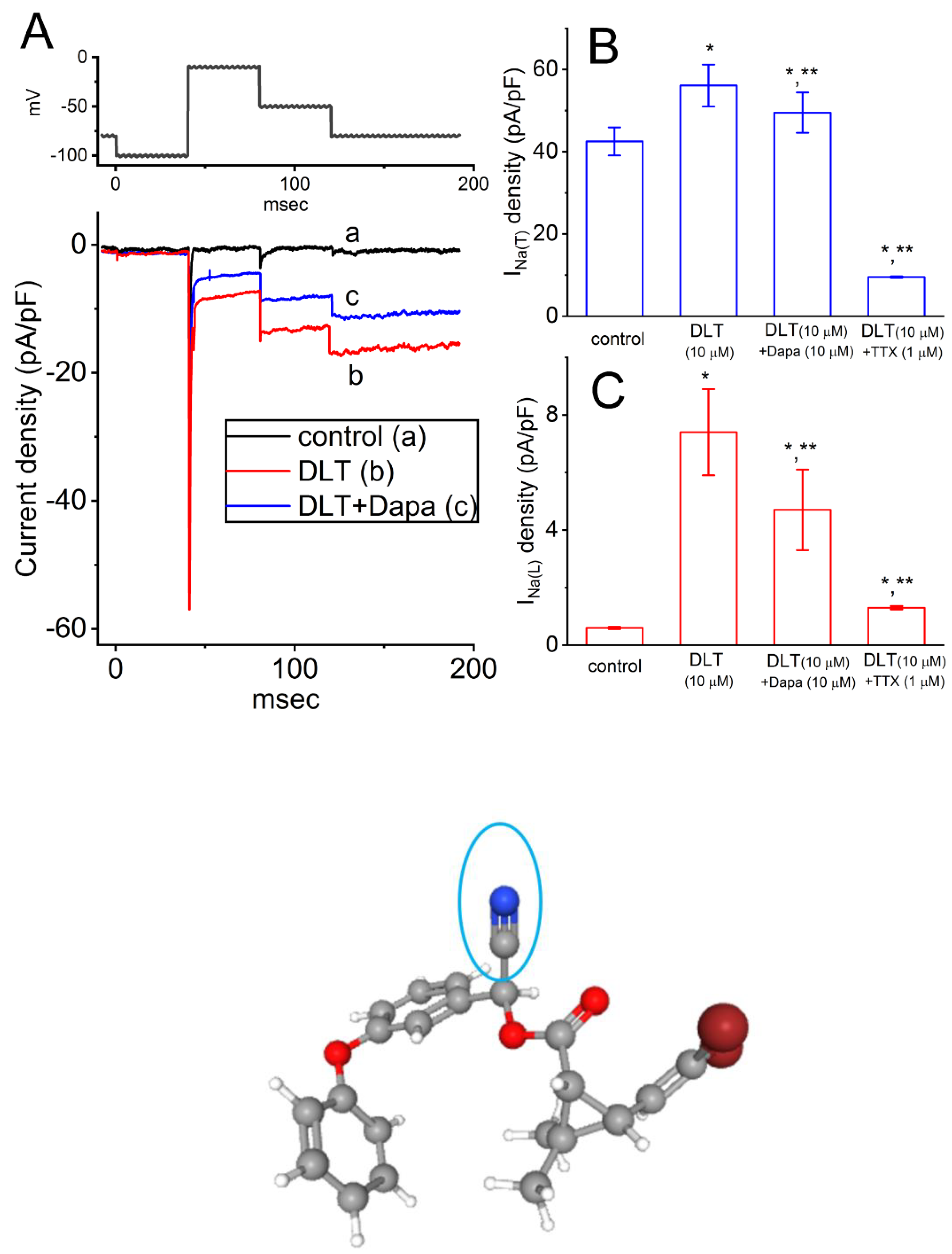
Figure 2.
Stimulatory effect of deltamethrin (DLT) and DLT plus dapagliflozin (Dapa) on the current density of voltage-gated Na+ current (INa) in Neuro-2a neuronal cells. In these measurements, we placed cells in Ca2+-free Tyrode’s solution containing 0.5 mM CdCl2 and 10 mM TEA, while the recording pipettes were filled up with Cs+-containing solution. (A) Superimposed INa traces obtained without DLT (a, black), with 10 μM DLT (b, red), and with 10 μM DLT plus 10 μM Dapa (c, blue). The upper section of (A) illustrates the voltage protocol applied to cells at a frequency of 0.1 Hz. The capacitive transients were compensated by applying a hyperpolarizing pulse of equal magnitude. The asterisk (*) and double asterisk (**) near the current trace (b) indicate the INa(Tail) at the level of -50 and -100 mV, respectively. Note that the presence of DLT enhances INa(Tail). Furthermore, the increase at a voltage of -100 mV is greater than the increase at -50 mV. (B) and (C) summarize the data on the effects of DLT and DLT plus Dapa on the current densities of INa(T) and INa(L), respectively. The density of INa(T) or INa(L) was measured at the onset or endpoint of the depolarizing step command from -100 to -10 mV, lasting 40 msec, respectively. In the experiments with DLT plus Dapa, Dapa was subsequently added to the bath in the presence of DLT. In (B) and (C), asterisks (*) denote significant differences from control values (P < 0.05), while double asterisks (**) indicate differences from the DLT (10 μM) alone group (P < 0.05). Each vertical bar represents the mean ± SEM (n = 9).
Figure 2.
Stimulatory effect of deltamethrin (DLT) and DLT plus dapagliflozin (Dapa) on the current density of voltage-gated Na+ current (INa) in Neuro-2a neuronal cells. In these measurements, we placed cells in Ca2+-free Tyrode’s solution containing 0.5 mM CdCl2 and 10 mM TEA, while the recording pipettes were filled up with Cs+-containing solution. (A) Superimposed INa traces obtained without DLT (a, black), with 10 μM DLT (b, red), and with 10 μM DLT plus 10 μM Dapa (c, blue). The upper section of (A) illustrates the voltage protocol applied to cells at a frequency of 0.1 Hz. The capacitive transients were compensated by applying a hyperpolarizing pulse of equal magnitude. The asterisk (*) and double asterisk (**) near the current trace (b) indicate the INa(Tail) at the level of -50 and -100 mV, respectively. Note that the presence of DLT enhances INa(Tail). Furthermore, the increase at a voltage of -100 mV is greater than the increase at -50 mV. (B) and (C) summarize the data on the effects of DLT and DLT plus Dapa on the current densities of INa(T) and INa(L), respectively. The density of INa(T) or INa(L) was measured at the onset or endpoint of the depolarizing step command from -100 to -10 mV, lasting 40 msec, respectively. In the experiments with DLT plus Dapa, Dapa was subsequently added to the bath in the presence of DLT. In (B) and (C), asterisks (*) denote significant differences from control values (P < 0.05), while double asterisks (**) indicate differences from the DLT (10 μM) alone group (P < 0.05). Each vertical bar represents the mean ± SEM (n = 9).
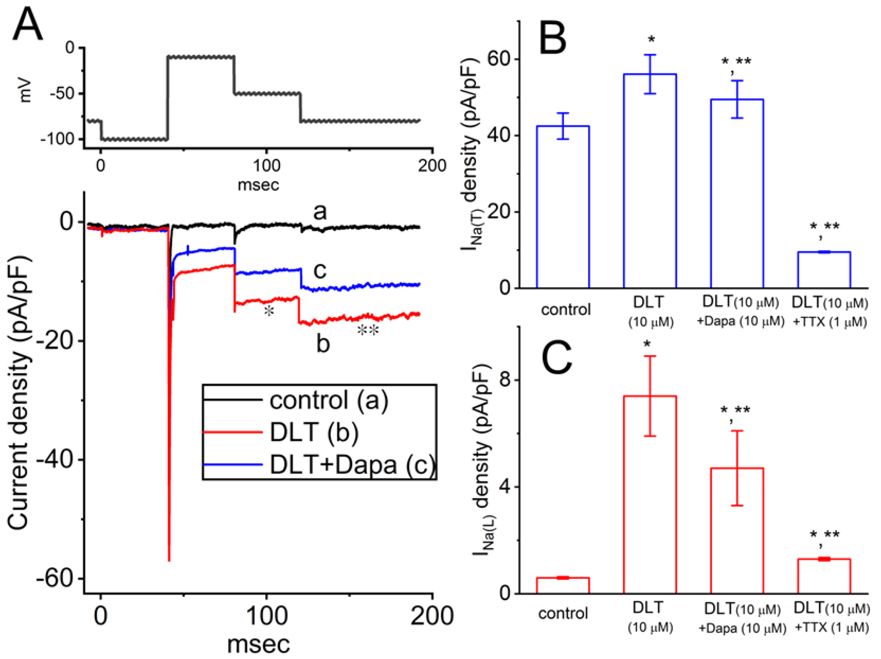
Figure 3.
Simulated INa traces elicited by step depolarization. This modeled respiratory neuron with INa simulations was adopted from a previous paper [25], with detailed formulations provided in the paper as well as in the Materials and Methods section of this article. The uppermost part indicates the voltage protocol applied. The values of GNa(T), GNa(L), and GNa(Tail) were arbitrarily set at 50, 100, and 0.5 μS, respectively. According to this model, simulated INa(Tot) (d, red) was divided into three parts. That is, INa(T) (a, purple), INa(L) (b, green), and INa(Tail) (c, red). INa(Tot) (d) thus refers to the sum of INa(T) (a), INa(L) (b), and INa(Tail) (c), whilc trace labeled (e) indicates the enlarged view from the dashed box of trace (d).
Figure 3.
Simulated INa traces elicited by step depolarization. This modeled respiratory neuron with INa simulations was adopted from a previous paper [25], with detailed formulations provided in the paper as well as in the Materials and Methods section of this article. The uppermost part indicates the voltage protocol applied. The values of GNa(T), GNa(L), and GNa(Tail) were arbitrarily set at 50, 100, and 0.5 μS, respectively. According to this model, simulated INa(Tot) (d, red) was divided into three parts. That is, INa(T) (a, purple), INa(L) (b, green), and INa(Tail) (c, red). INa(Tot) (d) thus refers to the sum of INa(T) (a), INa(L) (b), and INa(Tail) (c), whilc trace labeled (e) indicates the enlarged view from the dashed box of trace (d).
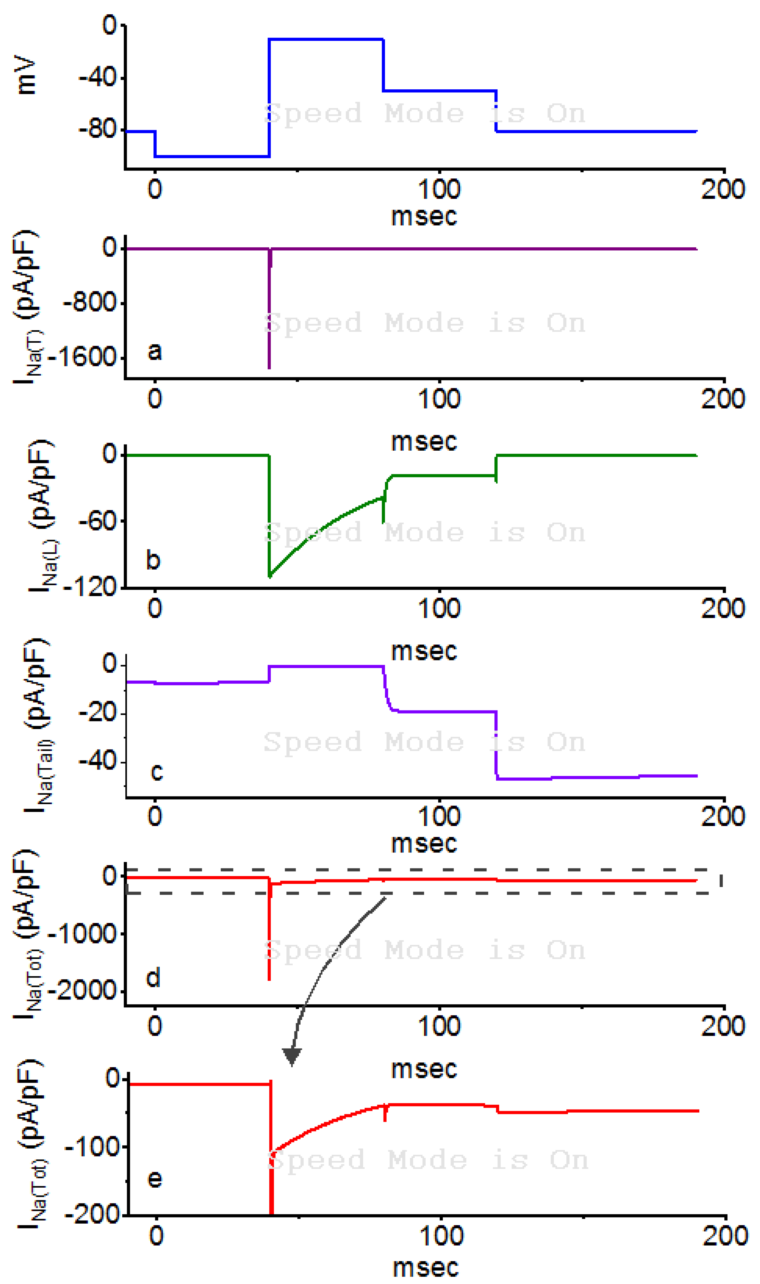
Figure 4.
Theoretical simulation of INa created to mimic the effect of 10 μM DLT. The voltage-clamp protocol shown in the uppermost part was the same as that in Figure 3. The values of GNa(T), GNa(L), and GNa(Tail) were arbitrarily set at 90, 40, and 10 nS, respectively. INa(T), INa(L), INa(Tail), and INa(Tot) were labeled ‘a’, ‘b’, ‘c’, and ‘d’, respectively. The INa(Tot)’s labeled d (red) in Figure 3 and Figure 4 are noted to closely match the experimentally observed results in Figure 2. Of note, in conditions used to mimic the effect of 10 μM DLT on INa, the current density of simulated INa(T) was slightly increased, along with the enhanced densities of INa(L) and INa(Tail). The asterisk (*) and double asterisk (**) indicate the simulated INa(L) and INa(Tail) (at the level of -50 and -100 mV), respectively.
Figure 4.
Theoretical simulation of INa created to mimic the effect of 10 μM DLT. The voltage-clamp protocol shown in the uppermost part was the same as that in Figure 3. The values of GNa(T), GNa(L), and GNa(Tail) were arbitrarily set at 90, 40, and 10 nS, respectively. INa(T), INa(L), INa(Tail), and INa(Tot) were labeled ‘a’, ‘b’, ‘c’, and ‘d’, respectively. The INa(Tot)’s labeled d (red) in Figure 3 and Figure 4 are noted to closely match the experimentally observed results in Figure 2. Of note, in conditions used to mimic the effect of 10 μM DLT on INa, the current density of simulated INa(T) was slightly increased, along with the enhanced densities of INa(L) and INa(Tail). The asterisk (*) and double asterisk (**) indicate the simulated INa(L) and INa(Tail) (at the level of -50 and -100 mV), respectively.
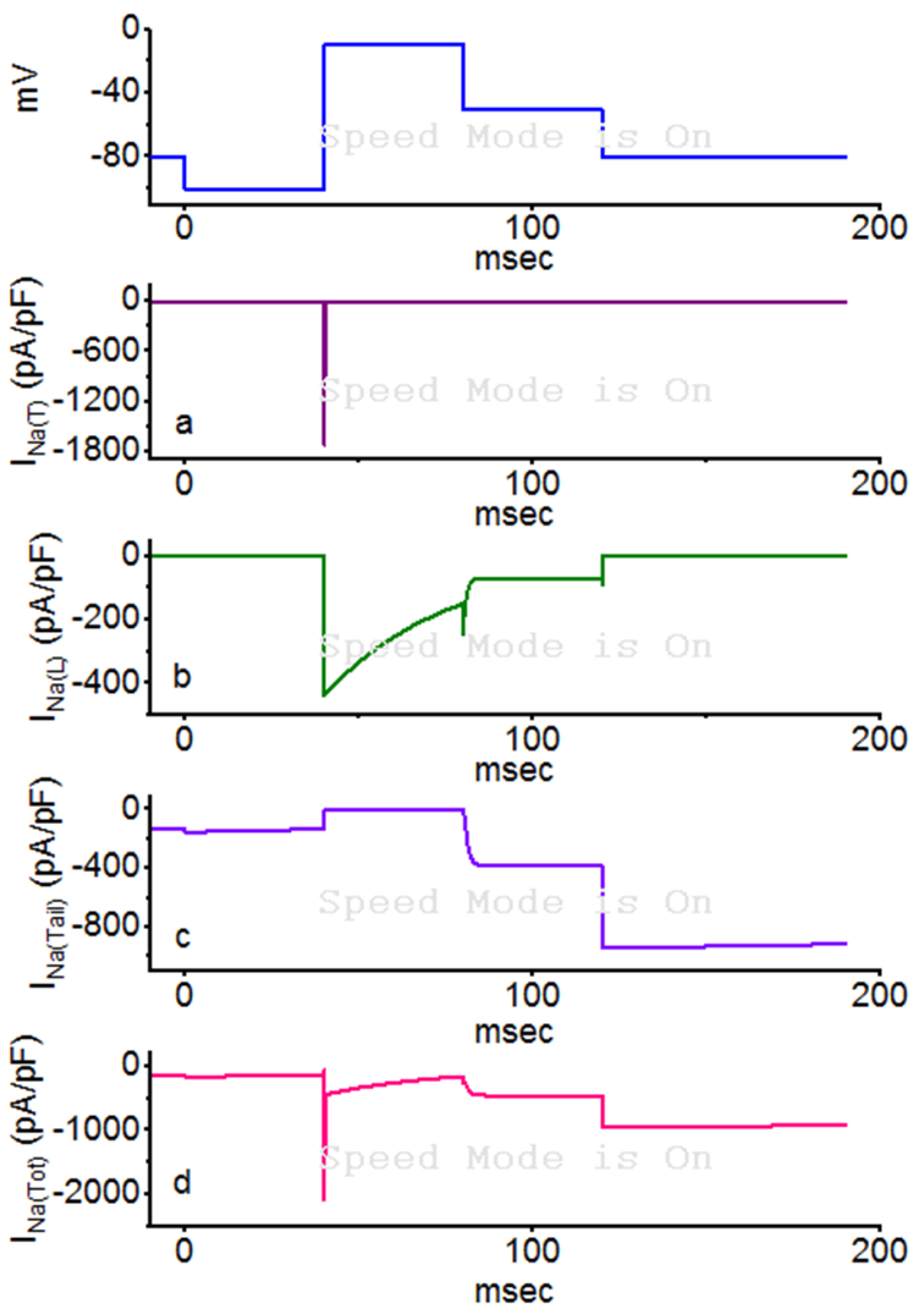
Figure 5.
The simulated action potentials (a, blue) of a modeled respiratory neuron, and the corresponding simulated INa trace (b, red). The asterisk (*) in the upper part (a) indicates specific ramping burst, while the curved arrow in the lower pat (b) illustrates the cumulative inhibition of INa during high-frequency firing in this model neuron.
Figure 5.
The simulated action potentials (a, blue) of a modeled respiratory neuron, and the corresponding simulated INa trace (b, red). The asterisk (*) in the upper part (a) indicates specific ramping burst, while the curved arrow in the lower pat (b) illustrates the cumulative inhibition of INa during high-frequency firing in this model neuron.
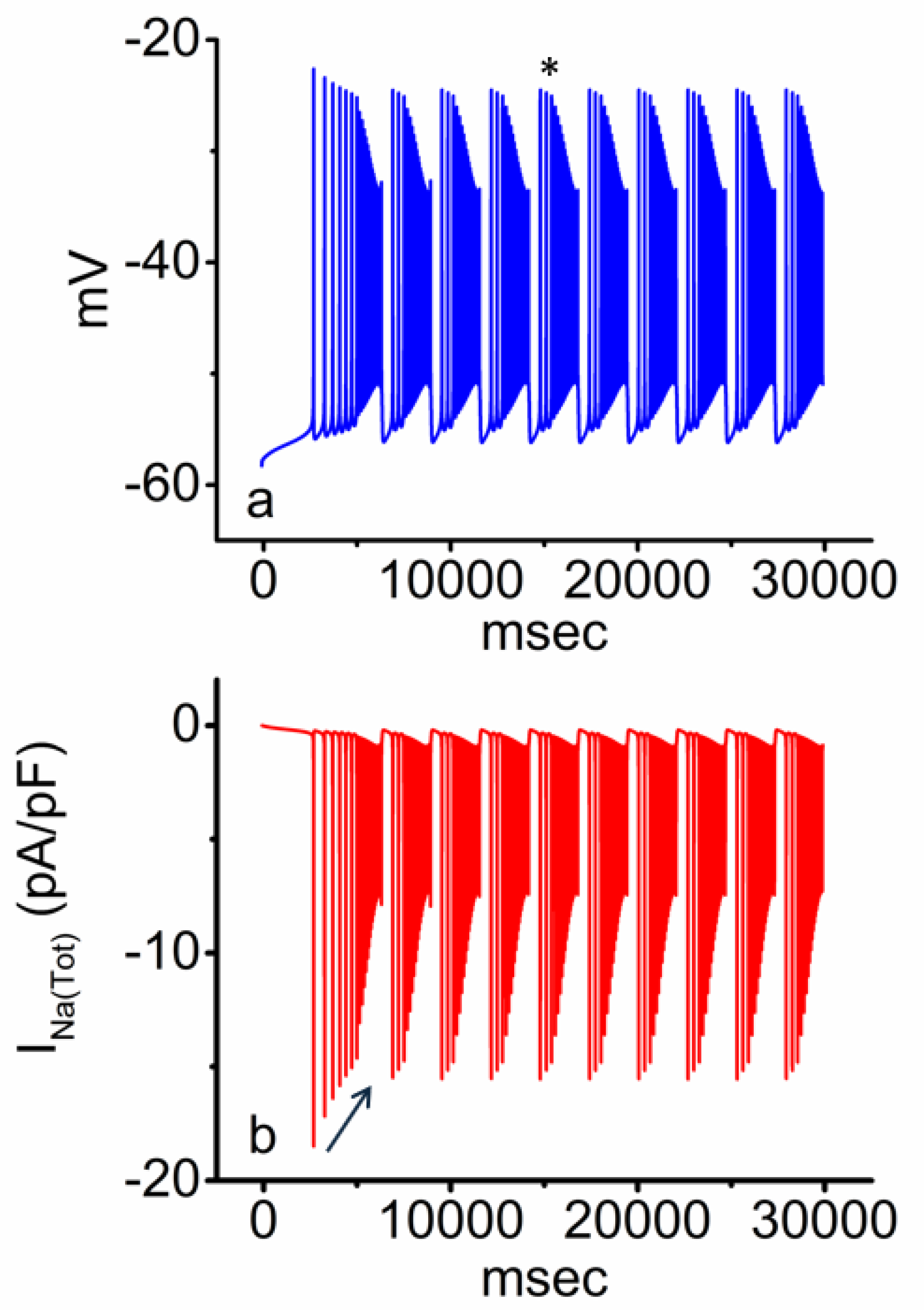
Figure 6.
Simulation of the potential burst firing induced by DLT (10 μM) in respiratory modeled neurons (a) and the corresponding different types of INa (b-e). The area within the red dashed box in panel ‘a’ is enlarged and depicted in Figure 7. The asterisk (*) and double asterisk (**) indicate two different burst patterns, slow and fast types, respectively. The burst firing marked by an asterisk (slow type) exhibits more pronounced and progressive shortening of intraburst intervals with a lower bursting firing, compared to the burst firing marked by a double asterisk (fast type). Panel breakdown: b, INa(T); c, INa(L); d, INa(Tail); and e, INa(Tot). The dashed curve arrow in b indicates cumulative inhibition of INa(T), while that in d shows a progressive increase in the density of INa(Tail).
Figure 6.
Simulation of the potential burst firing induced by DLT (10 μM) in respiratory modeled neurons (a) and the corresponding different types of INa (b-e). The area within the red dashed box in panel ‘a’ is enlarged and depicted in Figure 7. The asterisk (*) and double asterisk (**) indicate two different burst patterns, slow and fast types, respectively. The burst firing marked by an asterisk (slow type) exhibits more pronounced and progressive shortening of intraburst intervals with a lower bursting firing, compared to the burst firing marked by a double asterisk (fast type). Panel breakdown: b, INa(T); c, INa(L); d, INa(Tail); and e, INa(Tot). The dashed curve arrow in b indicates cumulative inhibition of INa(T), while that in d shows a progressive increase in the density of INa(Tail).
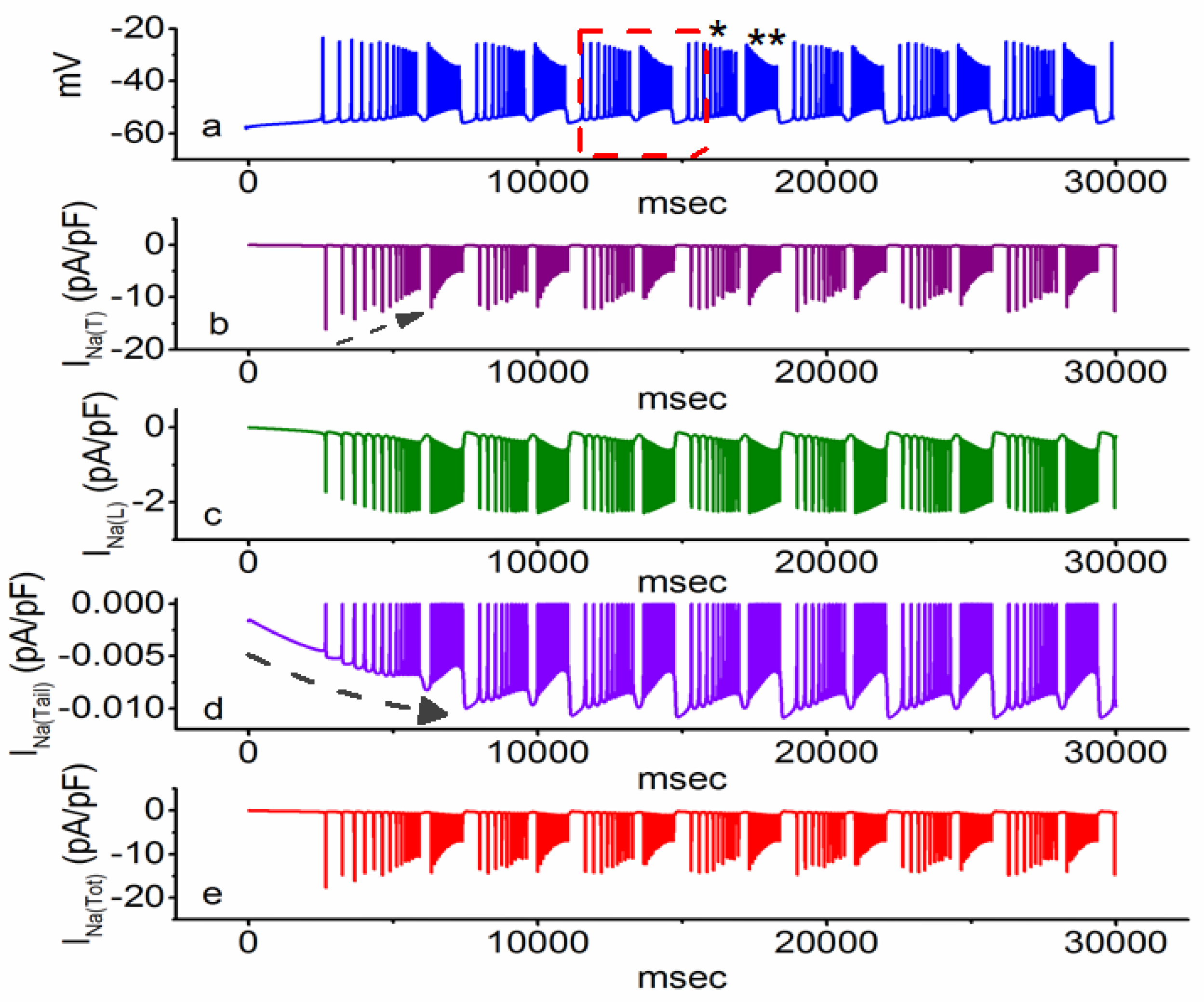
Figure 7.
Two distinct patterns of burst firing, classified as slow and fast types, observed in respiratory modeled neuron after incorporating INa densities that mimic the presence of DLT. This figure presents an enlarged view of the region highlighted within the red dashed box in Figure 5a. Notably, both types of bursting, which mimic exposure to DLT, exhibit progressive shortening of intraburst intervals. However, the magnitude of the post-burst hyperpolarizing potential (indicated by red arrow) observed during fast-type bursting is significantly greater than that in slow-type bursting.
Figure 7.
Two distinct patterns of burst firing, classified as slow and fast types, observed in respiratory modeled neuron after incorporating INa densities that mimic the presence of DLT. This figure presents an enlarged view of the region highlighted within the red dashed box in Figure 5a. Notably, both types of bursting, which mimic exposure to DLT, exhibit progressive shortening of intraburst intervals. However, the magnitude of the post-burst hyperpolarizing potential (indicated by red arrow) observed during fast-type bursting is significantly greater than that in slow-type bursting.
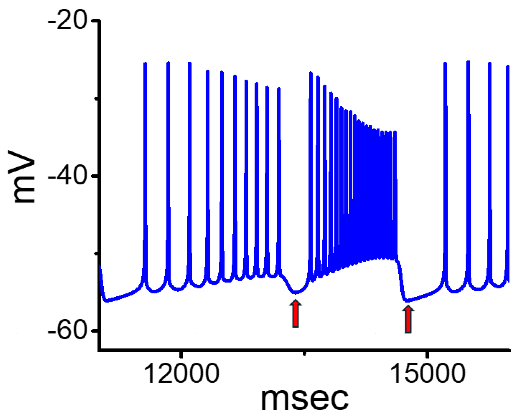
Figure 8.
Molecular docking analysis of DLT binding to the NaV1.8 channel. (A) The docking results demonstrating a potential interaction between the NaV1.8 channel and the DLT molecule. The protein structure of the NaV1.8 channel was obtained from the Protein Data Bank (PDB ID: 2N7F, accessible at http://doi.org/10.2210/pdb2n7f/pdf, accessed on 13 Aug 2024) (Deuis et al., 2016), while the chemical structure of DLT was sourced from PubChem (Compound CID: 40585). The right image provides an enlarged view of the region highlighted by the yellow dashed box in the left panel, with the ellipsoidal light blue figure indicating the α-cyano group. In the right panel generated using LigPlot+ (accessible at https://www.ebi.ac.uk/thornton-srv/software/LigPlus/, accessed on 13 Aug 2024, hydrophobic contacts are depicted by red arcs with spokes directed towards the ligand (DLT) at the center.
Figure 8.
Molecular docking analysis of DLT binding to the NaV1.8 channel. (A) The docking results demonstrating a potential interaction between the NaV1.8 channel and the DLT molecule. The protein structure of the NaV1.8 channel was obtained from the Protein Data Bank (PDB ID: 2N7F, accessible at http://doi.org/10.2210/pdb2n7f/pdf, accessed on 13 Aug 2024) (Deuis et al., 2016), while the chemical structure of DLT was sourced from PubChem (Compound CID: 40585). The right image provides an enlarged view of the region highlighted by the yellow dashed box in the left panel, with the ellipsoidal light blue figure indicating the α-cyano group. In the right panel generated using LigPlot+ (accessible at https://www.ebi.ac.uk/thornton-srv/software/LigPlus/, accessed on 13 Aug 2024, hydrophobic contacts are depicted by red arcs with spokes directed towards the ligand (DLT) at the center.
Disclaimer/Publisher’s Note: The statements, opinions and data contained in all publications are solely those of the individual author(s) and contributor(s) and not of MDPI and/or the editor(s). MDPI and/or the editor(s) disclaim responsibility for any injury to people or property resulting from any ideas, methods, instructions or products referred to in the content. |
© 2026 by the authors. Licensee MDPI, Basel, Switzerland. This article is an open access article distributed under the terms and conditions of the Creative Commons Attribution (CC BY) license (http://creativecommons.org/licenses/by/4.0/).
Copyright: This open access article is published under a Creative Commons CC BY 4.0 license, which permit the free download, distribution, and reuse, provided that the author and preprint are cited in any reuse.