
Submitted:
18 August 2026
Posted:
19 August 2026
You are already at the latest version
Abstract
This article introduces the concept of enosis – the ability of life forms to adapt by accessing each other’s molecules, structures, information and other contents – and discusses the roots and implications of this concept using cancer and cancer therapy as a case study.
Keywords:
cell theory
; cell-cell communication
; cancer
; intercellular bridges. intercellular exchange
; history of science
; basic properties of life
“The problem of malignant cells is still one of the greatest challenges in biology.”
The first sentence of an article published 101 years ago (A. Fischer, 1925).
The main approach of cancer therapy and underlying research has been to find and exploit differences between cancerous and normal cells (National Cancer Institute, 2015). For example, the discovery that some cancers are deficient in DNA repair proteins has been exploited to kill their cells selectively by causing DNA damage (Bryant et al., 2005; Farmer et al., 2005; Lord & Ashworth, 2017; Wicks et al., 2022), while the quest for “the holy grail of cancer: an antigenic target that is simultaneously abundant in cancerous cells and absent in normal tissues” has been waged to benefit cancer immunotherapy (Zamora et al., 2018).
As logical as targeting such differences is, this logic relies on the tacit assumption that cancer cells cannot acquire components that enable normal cells to survive therapy. For example, targeting a deficiency in DNA repair proteins would lose its premise if cancer cells could acquire these proteins, or genes that encode them, from their normal neighbors (Lazebnik, 2019). Likewise, the quest for “the holy grail of cancer” would make sense only if normal and cancerous cells do not exchange their antigens.
Yet, such acquisitions and exchanges have been documented by an increasing number of reports. For example, cancer cells can recover from irradiation (Sarkari & Lou, 2024; X. Wang & Gerdes, 2015), acquire multiple drug resistance (Del Vecchio et al., 2024), grow faster (Watson et al., 2023) and metastasize quicker (Hoover et al., 2025) by importing mitochondria and other components from normal and neoplastic neighbors (Berridge et al., 2025). Cancer cells can also overcome metabolic shortcomings by exchanging metabolites with normal cells (Monterisi et al., 2022; S. Shin et al., 2025; Swietach & Monterisi, 2019), can become more aggressive by acquiring components of signaling pathways from normal cells (Portela et al., 2019), can benefit from merging into networks with normal and neoplastic neighbors (Osswald et al., 2015, 2016; S. Shin et al., 2025; Venkataramani et al., 2022; Venkatesh & Lou, 2019), and can evade immune surveillance by depleting immune cells of mitochondria (Saha et al., 2022), by injecting these cells with “cancerous” mitochondria (Ikeda et al., 2025), by stealing their surface markers to disguise themselves as normal cells (Campana et al., 2015; Matkó & Tóth, 2021; J. H. Shin et al., 2021; Zaccard et al., 2016), and by fusing to each other to reshuffle their genomes and changing gene expression patterns (Duelli & Lazebnik, 2003, 2007; Lazebnik, 2014) while silencing gene expression in the process (Foidart et al., 2026)
These feats of adaptation have revealed an unwelcome paradox (from the Greek for contrary to expectations): the very cells that cancer therapy aims to save are saving the cells that this therapy aims to kill. To put it differently, future victims let a serial killer survive by becoming his live organ donors.
Given that even a single surviving cell can seed a new tumor (Furth et al., 1937; Landau et al., 2020; Quintana et al., 2008) and that relapses are a plague of cancer therapy, finding whether the killer paradox is relevant to human disease and learning how to deal with the sabotage it reveals is not a purely academic problem.
As a search for an answer is still in its early stages (Brestoff et al., 2025; Lou et al., 2024; Maurais et al., 2026; Venkataramani et al., 2025) and thus still faces the headwinds of disbelief and dismissal (Fleck, 1981), it may benefit from concepts that can reveal the nature of the observed phenomena, explain their relationships, and bridge the expertise of diverse fields for the common good.
To this end, I would like to propose that the phenomena that involve component transfer, including those underlying the killer paradox, are manifestations of a basic property of life defined as the ability of life forms to adapt by accessing each other’s contents and suggest calling this property enosis, from the Greek ένωση for union.
Let me overview the roots of the concept, outline the evidence that has prompted it, and discuss its implications. Since the terminology related to enosis is still incomplete and yet to be harmonized across fields (Brestoff et al., 2025), I will be introducing terms as needed and have them compiled for your convenience in a glossary at the end of this article.
ENOSIS – Roots and Background
Researching the roots of enosis made me realize that its manifestations had already been discussed in textbooks a century and a half ago, occupied the minds of researchers for decades, but then were forgotten well enough to be rediscovered decades later, a process that has accelerated within the last decade. I thought that outlining this evolution, even if as superficially as a non-historian can do, would introduce background and terminology required for our discussion and serve as a reminder that the observations and concepts now viewed as a novelty by some and dismissed as curiosities by others are rightful heirs of deep and illustrious ancestry.
Conveying Juices
The concept that cells can exchange their contents and properties was proposed a century and a half ago by Rudolf Virchow (Silver, 1987), “the father of modern pathology” (Rubin et al., 2008) who earned this title by aligning pathology with cell theory (Silver, 1987; Virchow & Chance, 1860), a revolutionary at the time idea that cells are the atoms of life (H. Harris, 2000; Schwann & Schleyden, 1847). Virchow has also been credited with popularizing the concept that each cell is a product of another (H. Harris, 2000) and for postulating that cells in an organism act as citizens of a “well-ordered state” (Reynolds, 2007; Virchow, 1958), that is as individual self-contained units which communicate through extracorporeal space. This doctrine still governs cancer research and its applications.
Yet, Virchow already concluded in his Cellular Pathology, published in 1858, that cells of some tissues “anastomose with one another” by “anastomosing processes” into “a reticular arrangement […] similar to that which we see in capillary vessels”. In these arrangements, or “anastomosing networks,” as he also called them, cells exchange their “juices” while retaining “a certain independence” (Virchow & Chance, 1860).
By anastomoses Virchow meant “[S]pots […] at which it is no longer possible to say where the one cell ends and the other begins” (Virchow and Chance, 1860). Let us define an anastomosis as a pore resulting from fusing the membranes of two cells in one spot, and an anastomotic bridge as any structure that uses membrane anastomosis to transfer cellular components (Figure 1, Glossary).
Figure 1.
Anastomotic bridge. This bridge, formed by local merger of cell membranes, can join cell bodies (shown), cell protrusions to cell body, or protrusions to each other. Note that anastomotic bridges enable the continuity of cell membranes, shown by the striped pattern to indicate intermixing of membrane components, and of cytoplasms. Please see Glossary for examples and more information.
Figure 1.
Anastomotic bridge. This bridge, formed by local merger of cell membranes, can join cell bodies (shown), cell protrusions to cell body, or protrusions to each other. Note that anastomotic bridges enable the continuity of cell membranes, shown by the striped pattern to indicate intermixing of membrane components, and of cytoplasms. Please see Glossary for examples and more information.
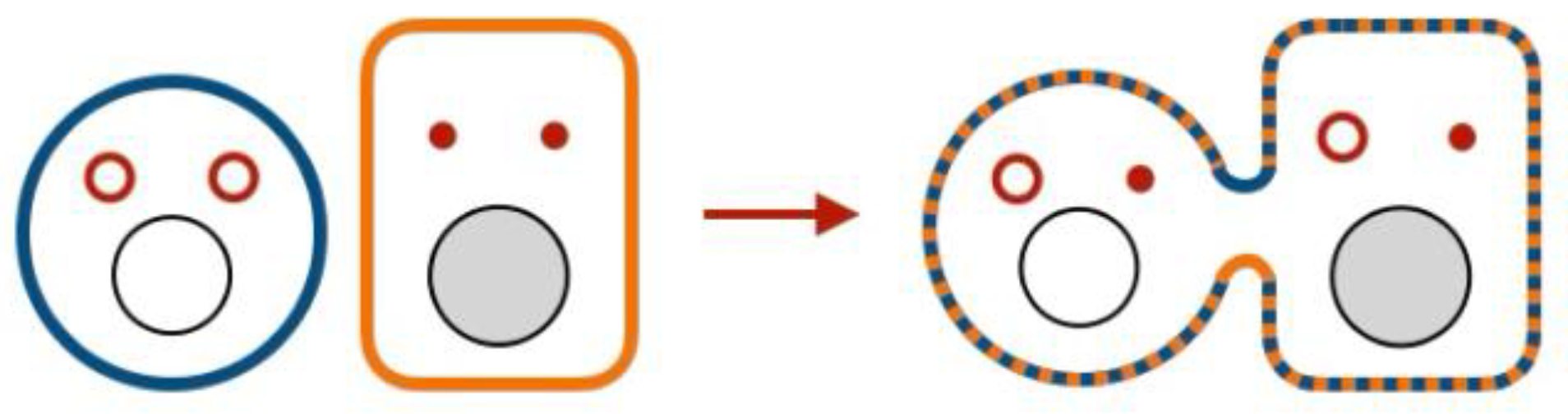
Figure 2.
Virchow: Cancerous “juices” make a normal cell cancerous. Please see text for explanation.
Figure 2.
Virchow: Cancerous “juices” make a normal cell cancerous. Please see text for explanation.
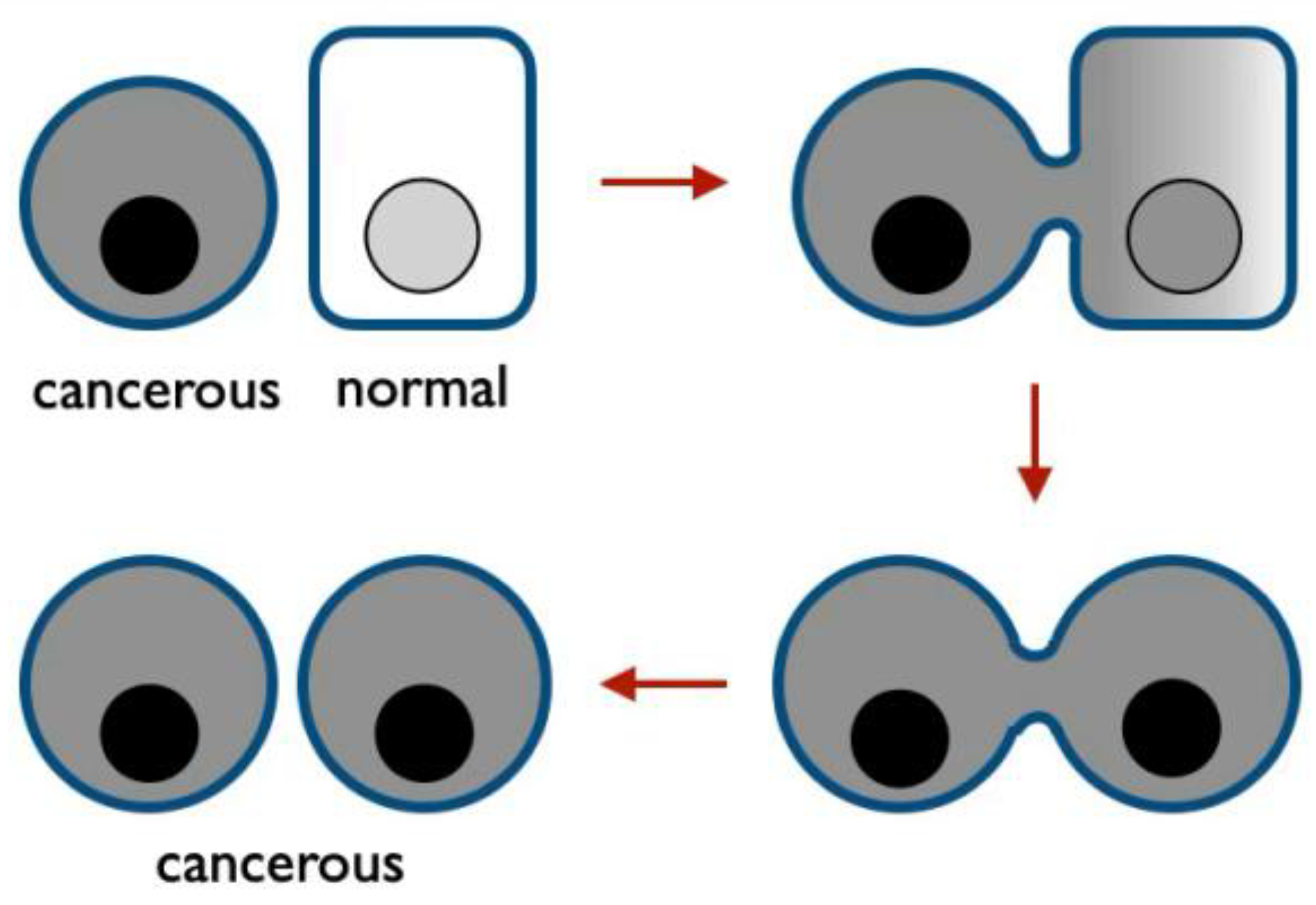
Of course, Virchow could not see cell membranes and verify their anastomoses in human tissues because the electron microscopy required to do so and the current understanding of what membranes look like were nearly a century away (Lombard, 2014; Robertson, 1960), but let us follow his logic nonetheless, as in essence he has been proven right.
A Distinct Way to Regulate
Virchow already recognized that a member of an “anastomosing network” can be regulated by “something which lay who knows how far off” (Virchow & Chance, 1860), or, to use modern parlance, that regulatory molecules, including genes (Maurais et al., 2026), can act non-cell-autonomously by traveling between cells.
Because Virchow thought that “there is manifestly a contagious matter formed in the tumour itself,” he proposed that “the cells, which are in its [tumor’s] immediate neighbourhood and are connected by anastomoses with the diseased cells, likewise enter upon the heterologous proliferation,” that is become neoplastic. This phenomenon would be reported a century later as horizontal oncogenesis and the cells it produces named adopted neoplastic cells more recently (García-Olmo et al., 2012; Goldenberg & Pavia, 1982; Lazebnik, 2023).
Overall, unlike some of our contemporaries (Baker, 2017), Virchow saw no contradiction between the notions that cells are the atoms of life and that they function not only as individuals. This opinion was shared and expanded by his fellow pathologists.
Higher Elementary Parts
In 1859, Alfred von Kölliker (JAMA, 1968), who discovered that spermatozoids are not animals but cells that come from testes (Birkhead & Montgomerie, 2009), published A manual of human microscopic anatomy (Kölliker, 1860) in which he classified “elementary parts” of tissues into simple and compound.
Simple parts are individual cells, which we will define as the smallest self-replicating parcels of life or, in practical terms, single mononucleated cells.
The compound, or “higher elementary parts,” as Kölliker also called them, include two types that differ in whether cells retain their identity upon entering a union (Figure 3). In some unions, cells “retain their cell-nature and in part also their independence”. We will call these unions symplasts (Glossary). If cells “coalesce,” or as we now say fuse (cell fusion, Glossary), “entirely loose their independence on union” to form syncytia (Glossary).
Kölliker’s inventory implied that understanding how a tissue or an organism functions one needs to know the properties, functions, and contributions not only of individual cells but also of symplasts and syncytia, or at least be aware whether they are present.
One pathologist proposed, however, that solid tissues have no individual cells at all.
Organism as a Symplast
In 1873, Carl Heitzmann, a pathologist who a year earlier had discovered the hematoblast and three years later co-founded the American Dermatology Association (Fatovic-Ferencic, 2000), concluded that “in no tissue whatsoever are “cells” present as isolated individuals. Every tissue […] represents a cell colony in which one “cell” stands in uninterrupted connection with all others, and all with one, a connection mediated by living matter. However, each cell colony is, in turn, connected without interruption to its neighboring colonies, such that the entire animal body may be regarded as a single cell colony.” (Heitzmann, 1873)
This concept, which raises some eyebrows even a century and a half later (Baker, 2017), already had what is now called supporting functional evidence, although the dots were connected only decades later (A. L. Harris, 2018).
Pinching the Ureter and Slicing the Heart
In 1869, Theodor Engelmann, a co-founder of the current model of how and why our heart beats (Kuhtz-Buschbeck et al., 2021), reported that “[i]f one presses or pinches the ureter of a dog, rabbit, cat, or rat, a contraction ensues that propagates from the stimulated site in both directions” (Engelmann, 1869). According to the then prevailing view, this contraction was mediated by the ganglion cells and nerve fibers. As Engelmann found none in the ureter, he concluded “that the stimulus propagates directly from one muscle cell to another” through contacts “so intimate that it amounts to a physiological continuity” and proposed that this continuity works in other organs, including the heart.
To test his hypothesis, Engelmann cut a ventricle of a frog’s heart to keep two or more fragments connected by “a very narrow bridge of muscular tissue”. As his hypothesis predicted, stimulating one fragment caused others to contract no matter where the cuts were made, as long as they remained connected and their cells alive (Engelmann, 1875).
Five years later, the “physiological continuity” Engelmann found in the ureter was discovered in plants.
Like Animals, Like Plants
Unlike animal cells, plant cells are surrounded by cell walls. That these walls should have some “valves, or diaphragms” to let “juices” circulate was already acknowledged by Robert Hooke in 1665, in the book in which he introduces the term cell. Hooke’s microscope, however, was still too simple to allow him to see the valves his mind envisioned (Peters, 2024).
A mere two centuries later, Eduard Tangl reported “a system of connecting channels, through which an open communication between neighboring cell elements and a continuous connection of their protoplasmic bodies is established” (Tangl, 1880). Although channels between plant cells had been noted previously (Carr, 1976a; Hanstein, 1864), Tangl is credited for recognizing that they “play an important role in the physiological processes” by enabling transfer of contents between cells and by joining individual cells in to “a unit of a higher order” (Carr, 1976a; Tangl, 1880).
Within two decades, Tangl’s channels, named plasmodesmata (Glossary) from the Greek for fluid bridges, were found to be ubiquitous in plants and to arise by two mechanisms: by incomplete cell division and by cell anastomosis (Carr, 1976a).
Let us call bridges formed by incomplete cell division familial bridges (Figure 4 and Glossary) because, unlike anastomotic bridges, they connect only daughter cells. We will call the union of cells interconnected by familial bridges familial symplast (Glossary).
The discovery of plasmodesmata prompted a hypothesis that all cells of a plant, as Heitzmann had envisioned for animals, are bridged into one symplast.
All Difficulties Vanish
In 1930, Ernst Münch, who described himself as a forest botanist, reported a solution to a problem that had been perplexing his predecessors – how nutrients made by photosynthesis in the leaves are distributed to the rest of a plant (Knoblauch & Peters, 2017). Münch realized that “all difficulties vanish” if one assumes that all cells in a plant function not as individual units but as “the symplast—the entirety of living cells interconnected by permeable plasmatic links,” that is by plasmodesmata (Münch, 1930)
Münch presciently concluded: “The fusion of all living cells into a symplast—effected through plasmatic connections—transforms them into a unified organism which, with regard to sap flow, functions as a single cell; enclosed by a common outer plasma membrane, it permits the translocation of nutrient solutions in all directions via unilateral positive pressure, yet offers sufficient resistance within the plasmodesmata to ensure that the individuality of the individual cells is not lost.”
A condescending review of Münch’s book, published in Nature the following year, mentions that his “distinctly unusual conceptions of solute movements are accompanied by calculations […] which involve so many assumptions that their value is doubtful” and concludes that “his monograph will probably not convey to most readers the impression that […] the phy sical mechanism of movement of substances in the phloem now placed beyond doubt.”(“Movement of Solutes within the Plant,” 1931)
In some way the reviewer was right, as after a few decades of neglect “[t]he Münch hypothesis has gained wide acceptance based to a large extent on its simplicity and plausibility, rather than on experimental evidence” (Knoblauch et al., 2016) and as such is still being tested (Peters and Knoblauch, 2022).
As if to illustrate again that plants and animals are more alike that it might appear, a contemporary of Münch made symplasts visible in an animal tissue by using a technique that would be reinvented for this purpose decades later.
Mapping Symplasts
In 1925, Martha Schmidtmann, who would go on to become the first woman in pathology in Germany, direct a pathology institute, and have a street named after her in the city were she graduated (“Martha Schmidtmann,” 2025), reported an accidental and presciently interpreted observation.
While injecting a dye to measure the acidity of the cytoplasm in squamous epithelial cells of human oral mucosa, she found that the dye injected into one cell “spreads not only within the cell into which the deposition has occurred, but also very rapidly extends to the protoplasm of the surrounding cells. […] The boundary between the stained and unstained regions is irregular, resulting in a map-like appearance”. As far as I am aware, this was the first map of a symplast in an animal tissue.
After ruling out alternative explanations, she concluded that “ [w]hile we are accustomed, from a histological standpoint, to viewing the cell as a self-contained unit, experiment here demonstrates that, for certain physiological functions, no such separation of cells exists” (Schmidtmann, 1925; Harris, 2018a).
While symplasts in animal tissues still remain invisible to light microscopy without approaches that Martha Schmidtmann introduced, cell fusion and the resulting syncytia could be observed even with relatively simple microscopes. These observations led to two concepts of carcinogenesis.
Coalescing Into Cancer
A commonly known example of cell fusion is fertilization, “one of the most dramatic and complex cell transformations in human biology: two highly differentiated gametes fuse, and the resulting cell, the zygote, is capable of dividing to generate all the cells of the human body” (Clift & Schuh, 2013). Fittingly, this process inspired two concepts of the most dramatic and complex disease today, cancer (Figure 5).
One concept (Figure 5, top) was articulated in 1914 by Theodor Boveri, a zoologist who discovered the centrosome as a coordinator of cell division and postulated that chromosomes carry genetic information (Ried, 2009). Bovery noticed that fertilization of sea urchin eggs is sometimes followed by abnormal distribution of chromosomes, and thus properties, between dividing cells, an observation that led him to propose that cancer is a result of an abnormal assortment of chromosomes (Boveri, 2008), a condition later named later aneuploidy by a plant biologist (Santaguida & Amon, 2015).
A contemporary review of Boveri’s monograph in Science concluded that “[f]rom the nature of the case the theory is difficult if not impossible to analyze by direct experiment, and for this reason, as well as for its impracticability, it is probable that the hypothesis will not be favorably received by the medical profession” (Calkins, 1914). This prediction held true for almost a century (Weinberg, 2008), but by the centennial of Boveri’s publication his concept became a part of “basic tenets of tumour biology” (Wright, 2014).
The second concept inspired by fertilization (Figure 5, bottom) is still a stranger outside of a group of enthusiasts but supporting evidence and the consequent awareness have been accumulating steadily (Cozzo et al., 2023; Platt & Cascalho, 2024; Shultes et al., 2024; Weiler & Dittmar, 2026; Whalen et al., 2024).
This concept, proposed by Otto Aichel in 1908 and detailed in 1911 (Aichel, 1908, 1911), was an outcome of a mental experiment to test what would happen if a lymphocyte, a cell which can migrate freely throughout the body, fuses with a cell which proliferates at will. Aichel concluded that “if we examine the properties and capabilities of the malignant cell on the one hand, and supplement the properties and capabilities of the somatic parent cell [Aichel points to “a persisting embryonic cell” as a suspect] with those of the leukocyte on the other, we arrive at the conclusion that the product of this fusion corresponds exactly to the malignant cell”. Additional variation in the properties of these hybrids, Aichel argued, would result from “various modes of chromosome distribution” as Boveri proposed (Aichel, 1911).
This “think out of a single cell” concept and the notion that somatic cells can fuse were forgotten accidental observations revealed that somatic cell fusion does happen (Barski et al., 1961; H. Harris, 1971), provided evidence in support of Aichel’s hypothesis (Pawelek & Chakraborty, 2008), and demonstrated that fusion between different cells can produce malignant progeny (Brito et al., 2021; Duelli & Lazebnik, 2007; Merle et al., 2021) for reasons that are intrinsic to how cells are organized as systems (Koulakov & Lazebnik, 2012; Lazebnik, 2014).
But this would happen nearly a century later, after the notion that cells act solely as atoms evolved into a doctrine which defined for decades how one should think about organisms.
Out of Fashion
Considering the observations and concepts that we have discussed, it is hardly surprising that by the beginning of the 20th century the notion that cells exchange components and form symplasts and syncytia was routinely kept in mind while investigating biological and medical phenomena.
For example, The Cell in Development and Heredity, a textbook published in 1925 and still considered “the founding document” in the field of cell biology (Maienschein et al., 2024), mentions that “it is probable that an important part in the coordination of the cell-activities is played by direct protoplasmic connections between cells (“cell-bridges,” “plasmo-desms”)” and that “[i]n animal tissues the existence of both cell-anastomoses and of intercellular bridges is now well established for many kinds of cells. […] The facts thus briefly reviewed have led some important modern writers to accept Heitzmann’s general conclusion almost in its entirety” (Wilson, 1925).
Then, for reasons which I leave to historians to explain (Carr, 1976a; H. Harris, 2000; Maienschein, 2017; Nicholson, 2010; Reynolds, 2007), the doctrine that cells act solely as atoms took over the minds of biologists, as if to demonstrate, as a plant biologist put it, “how strongly the consensus of opinion affects discovery and the retention of the corpus of discovered knowledge.”(Carr, 1976a) and to remind us that science, including cancer research (H. Harris, 2005), is as susceptible to changes in fashion as other human activities.
A revival began several decades later thanks to contrarians who kept the flame alive, to new technologies able to unambiguously visualize intercellular channels and detect component transfer, and to accidental observations that were reported despite being contrary to prevailing doctrines.
Accepting What Everyone Can See
The development of electron microscopy made the ubiquitous presence of plasmodesmata in plants difficult to ignore, but it took decades to acquiesce to their existence and to accept that these channels form symplasts (Carr, 1976a). “Still in 1967, [a plant biologist] vehemently rejected the [Münch’s] idea of mass flow on the grounds that “forest botanists” […] had ignored the fact that cell anatomy was “the alpha and omega of all physiological research”” (Knoblauch & Peters, 2017).
I get an impression that cancer biology is at this stage now, even though cell anatomy is studied by sophisticated single-cell analyses which overlook symplasts by design, but let us return to plant biology to see what advances it has made.
Today, the notions that plasmodesmata unite nearly all cells into symplasts (Kirk & Benitez-Alfonso, 2022), that these symplasts rather than individual cells are the primary operational units of a plant (Lucas et al., 1993; Lucas & Lee, 2004), and that plant cells exchange signals both through the extracellular space (apoplastic signaling) and plasmodesmata (symplastic signaling) (Li et al., 2021; Otero et al., 2016) have all returned to textbooks (Raven et al., 2013), with research in these areas ongoing actively (Bayer & Benitez-Alfonso, 2024; Tee & Faulkner, 2024).
As if to remind us again that plants and animals are not that different (Lucas & Lee, 2004; J. Pitts et al., 1987), the revival of plasmodesmata research was helped by discovering intercellular bridges in animals (Carr, 1976b) as we are about to discuss.
Germ Cells in Chains
Animal counterparts of primary plasmodesmata, which are a product of incomplete cell division, were reported in 1955 as a result of applying then still nascent electron microscopy to studying cat spermatogenesis (Burgos & Fawcett, 1955). The discoverers noticed that spermatids, the precursors of sperm, were often connected into pairs or groups of four by “protoplasmic bridges” about a micron wide. They considered this observation “one of the most puzzling problems” to interpret because such connections were not supposed to exist in the world of single cells, but correctly concluded that these connections were not an artifacts but a result of incomplete cell division and predicted “that future studies of other species with the electron microscope will reveal that intercellular connections between developing germ cells are a rather general phenomenon.” Within a few years these familial bridges were indeed found in species ranging from hydra to humans (Fawcett, 1961) and are now considered “an ancient feature that is fundamental for the development and function” of germ cells (Figure 6) (Brubacher, 2024; Chaigne & Brunet, 2022; Gerhold et al., 2022).
The discovery of the “intimate connections” predicted by Theodor Engelmann from observing the contraction of the ureter was next.
From the Ureter to Biological Computers
The connections Engelmann predicted in 1869 were discovered in 1969, again thanks to electron microscopy, and named gap junctions (Payton et al., 1969; Revel & Karnovsky, 1967). Over the subsequent decades they were found to be clusters of transmembrane channels which bridge “virtually all cells in solid tissues” (D. A. Goodenough & Paul, 2009) and transmit components ranging from ions to small nucleic acids and proteins (Cieniewicz & Woodruff, 2010).
We will call the structures that transmit components between cells across their plasma membranes transmembrane bridges (Figure 7 and Glossary), and symplasts formed by these bridges transmembrane symplasts (Glossary).
Among its other consequences, the discovery of gap junctions explained the phenomena of metabolic cooperation, a process by which cells acquire from their neighbors metabolites that they cannot make themselves (Friedmann et al., 1968; Subak-Sharpe et al., 1969) and ionic coupling, that is unimpeded transmission of electric current between adjacent cells (Gilula et al., 1972; Loewenstein et al., 1965), a phenomenon observed by Theodor Engelmann and then forgotten.
Mapping ionic coupling in various tissues revealed a “ cell system with fairly continuous interior, at least as far as much of its ion content is concerned, bounded by a diffusion barrier which extents and is continuous along the entire surface of the system. The system rather than individual cell components constitute the unit of ion environment [where] ions, metabolites, and, perhaps, hormonal and genetic information may conceivably flow from one cell to another, with the economy of external nervous and humoral controls” (Loewenstein et al., 1965). Some of these transmembrane symplasts involved few cells, others formed “tortuous channels” or comprised an entire tissue.
These symplasts may be related to recently discovered “bioelectric compartments” and the concept that tissues other than the nervous system can compute using gap junctions as “biological transistors” (Landau et al., 2020; Levin, 2021; Palacios-Prado & Bukauskas, 2009) because gap junctional channels, like transistors in computational devices, can both conduct electric current and be regulated by it.
Overall the discovery or gap junctions and their role in the human body has validated Virchow’s vision of a “juice-conveying system of tubes or canals” and Heitzmann’s hypothesis of an organism as a symplast, even if anastomoses they thought to enable these connections did not exist. However, anastomoses were also discovered in due course.
From Giant Axons to Nanotubes
In 1924, George Edwin Johnson reported “a strikingly unique alliance between neurons” in shrimp and crayfish (Johnson, 1924). This emphatic reaction, which was not as common then as it is now, was echoed fifteen years later by John Zachary Young (LaTourelle, 2016; Maxson Jones, 2022), who found in the nervous system of squid “an arrangement […] so strange that it requires careful description” (Young, 1939).
Both scientists were surprised because they found that neurons could communicate through anastomoses (Figure 8, A), which was “inconsistent with the orthodox neuron theory,” as Young put it, because according to this theory neurons are independent cells that communicate only through synapses.
The first case involved two neurons whose axons cross in the middle of the body, as in animals each side of the brain controls the opposite side of the body (Vulliemoz et al., 2005). These two axons anastomosed at the crossing, apparently to ensure that both sides of the body react to a sensory signal whether it comes from the right or left side. Young was able to demonstrate the continuity of the cytoplasms and membranes at the anastomosis “with great clarity” because these axons in the squid are unusually large, up to 150µ in diameter (Young, 1939).
Both researchers also discovered that the axons of multiple neurons can anastomose into one, thus creating an anastomotic symplast with multiple “heads” (Figure 8, B). In the squid, these symplasts were “formed by the fusion of many axons, several hundreds probably collaborating to make up the larger fibres [which] conduct like other single nerve fibres as units, although they arise as the processes of a number of separate nerve cell bodies.” The size of the giant axons, as they were named, made them an amenable model to study the propagation of nerve impulses, the research that earned a Nobel Prize in 1963 (Hodgkin, 1963).
I mention this prize because I find it remarkable that despite the publicity it gave to anastomosed axons, the notion that neurons act solely as independent cells remained so dominant that five years after the prize was awarded a group of researchers “were startled to see areas of cell contact between axons and sheath glial cells in which the two plasma membranes appeared to fuse forming clear images of membrane pores” (Eugenin et al., 2022). They “felt that the pores were probably a fixation artifact and decided not to publish these data” until a communication from another group prompted them to reconsider. As a result, a picture of the anastomosis was featured on the cover of Nature (Eugenin et al., 2022; Peracchia, 1981)
This publicity, however, also did not help. As the authors of the article recalled four decades later, “for several decades the possibility that some cells might be able under special conditions to directly communicate via membrane pores was by and large ignored” (Eugenin et al., 2022). Until the discovery of nanotubes broke the ice.
From Conveying Juices to Clinical Trials
In 2004, the long forgotten “anastomosing processes” Virchow envisioned were discovered, documented to transfer the “juices,” including organelles, and aptly named tunneling nanotubes (Rustom et al., 2004). These nanotubes were then found to correct cell deficiencies by transferring mitochondria (Spees et al., 2006), a discovery that led to clinical trials merely two decades later (Brestoff et al., 2025; Gomzikova et al., 2021) and to “potential therapeutic opportunities” for treating cancer (Berridge et al., 2025), and became the founding member of a growing family of structures which have been discovered across the species, from archaea to humans and have been explored in cancer therapy (Berridge et al., 2025; Sarkari & Lou, 2024).
Are These Phenomena Related?
The fact that organisms as different as plants, bacteria, mammals, and protists use intercellular bridges and these these bridges are functionally related revived a discussion which began a century and a half ago about whether direct transfer of components between cells is a rule rather than an exception.
An open question is whether the bridges independently across the tree of life and “different groups of organisms found different answers to this task” (Bloemendal & Kück, 2013), as is commonly held (Bloemendal & Kück, 2013; Demin et al., 2021; Matkó & Tóth, 2021; J. D. Pitts, 1990; Rustom, 2009, 2016), or these “answers” come from the same source, as the concept of enosis implies.
Let me outline the observations that prompted this hypothesis.
Enosis – A Case for Its Existence
The concept of enosis posits that the ability of life forms to access each other’s contents is as basic as is their ability to proliferate (Soto et al., 2016). This hypothesis implies that enosis should be a feature of life from its beginning, or at least since as early as we can reliably discern.
Enosis Is Primordial
To look back to the early days of life we will use cyanobacteria, as they have earned the title of “the ultimate in ‘living fossils’“ (Butterfield, 2007), and mitochondria, the descendants of a bacterium adopted by our ancestor when life was still young.
Cyanobacteria live as symplasts. Cyanobacteria are ubiquitous unicellular and multicellular organisms that emerged 3.5 billion years ago (Fournier et al., 2021; Garcia-Pichel et al., 2019; Schirrmeister et al., 2015) and over the subsequent billion and a half years invented photosynthesis, the process which packs solar energy into carbohydrates and releases oxygen (Herrero et al., 2016), filled with this oxygen the primordial oceans and the atmosphere (Schirrmeister et al., 2015), gave rise to plastids, the organelles responsible for photosynthesis in plants (W. Martin & Kowallik, 1999; Renna et al., 2025), were among the earliest to learn how to fix nitrogen, that is incorporate this otherwise inert gas into precursors of amino acids, nucleotides, and other molecules required for life (Chen et al., 2022; Latysheva et al., 2012; Pi et al., 2022; Rucker & Kaçar, 2024), and have been vital to life on this planet ever since.
This track record is relevant to our discussion because some groups of cyanobacteria evolved so little over the last two billion years that their “fossils are indistinguishable from members of extant cyanobacterial taxa, not only in their morphology and cellular structure but also in their life cycles and inferred processes of cell division as well as their ecological setting, the biotic structure of the communities in which they occur, and the types of stromatolites [sediments] they produce” (Schopf, 2012). In other words, some groups of modern cyanobacteria can tell us how their ancestors functioned at least two billions years ago.
One of these groups is known as heterocyst-forming cyanobacteria (Figure 9) because these filamentous multicellular organisms respond to a deficiency in fixed nitrogen by inducing every tenth or so cell in a filament to convert into heterocysts, the cells specialized in fixing nitrogen (Herrero et al., 2016). This conversion involves shutting down photosynthesis, to prevent oxygen it produces from inactivating the enzyme that fixes nitrogen, and building a thick capsule which isolates the heterocyst from external oxygen and makes these cells recognizable. As a result of creating heterocysts, the organism can fix nitrogen and use photosynthesis at the same time, both for its own benefit and for that of organisms up the food chain, including us.
Creating heterocysts, keeping them alive, and distributing their products to other cells is possible because each filament is a symplast whose cells are connected by transmembrane and familial bridges (Figure 9).
Adjacent cells in a filament are bridged by septal junctions (Flores, 2025; Kieninger & Maldener, 2021), which are transmembrane bridges “mechanistically similar” to gap junctional channels of metazoans (Weiss et al., 2019). Septal junctions are required to initiate the differentiation (Janović et al., 2024; Zeng & Zhang, 2022), to transport nitrogen-containing molecules from heterocysts to other cells, and to feed heterocysts with carbohydrates, as their own photosynthesis is shut down (Herrero, 2025; Herrero et al., 2016).
The regulatory circuit which ensures that only some cells are converted into heterocysts relies on transmitting a peptide through the periplasm (Figure 9) and on the continuity of periplasm along the filament (Flores et al., 2006; Mariscal et al., 2007; Xu et al., 2026). This continuity, in turn, results from specialized cell division which leaves the outer cell membranes of the daughter cells continuous, thus creating a diderm variation of a familial bridge (Figure 9, top) (Herrero, 2025). Besides their role in transporting cellular components, familial bridges and septal junctions enable the structural integrity of filament (Herrero, 2025; Janović et al., 2024).
The finding that genes required to build septal junctions are present across all filamentous cyanobacteria, of which heterocyst-formers are a part, led to a conclusion that “regulated intercellular exchange was a feature of filamentous cyanobacteria since they first evolved in the Archean” (Boden et al., 2025), that is 2.5 billion years ago or earlier. Filamentous species, however, are not the only group of cyanobacteria that form symplasts.
A recent discovery revealed that two genera of unicellular cyanobacteria which share a 3-billion year ancestry (Dvořák et al., 2014) and are the most abundant photosynthetic organisms in the ocean, also make symplasts (Angulo-Cánovas et al., 2024). They do so by connecting through nanotubes which merge the membranes, periplasmic spaces, and cytoplasms of the bridged cells (Figure 10). The nanotubes are wide enough to enable the exchange of most structural and regulatory components within and across species with the “implications for the evolution and ecology of microbial life in the open ocean” (Angulo-Cánovas et al., 2024).
Given that cyanobacteria and heterotrophic bacteria are thought to diverge more than three billion years ago and both use nanotubes, it is reasonable to assume that these structures are as old. This conclusion is consistent with the features of our next guest from primordial times, mitochondria.
Mitochondria are commonly viewed, albeit not by all (Harish & Kurland, 2017), as the descendants of a bacterium captured by an ancestor of modern eukaryotes about two billion years ago (Geiger et al., 2023; Sagan, 1967; Vosseberg et al., 2024). The descendants of this putative captive adapt to their environment by exchanging components through anastomotic bridges (mitochondrial kiss-and-run or transient fusion (Arimura et al., 2004; X. Liu et al., 2009) and nanotunnels (Tábara et al., 2025; Vincent et al., 2017)), by fusion (Carmichael et al., 2023; X. Liu et al., 2009; Møller et al., 2021; Ono et al., 2001; Youle & van der Bliek, 2012), by incomplete mitochondria fission which results in familial bridges known as mitochondria-on-a-string phenotype (Zhang et al., 2016) and a hexokinase-ring constriction (Pilic et al., 2024), and by bridging into networks (Mitra et al., 2009; Singh et al., 2025).
Given that modern free-living bacteria also respond to stress through the same mechanisms (Z. Liu et al., 2013; Meysman, 2018; Vassallo et al., 2015) despite living outside of bondage for the last two billion years, it is plausible that mitochondria and modern bacteria inherited enosis from their primordial ancestors rather than developed it independently.
If enosis is indeed primordial, it would be expected to manifest itself in all domains of life.
Enosis Across All Kingdoms of Life
Bacteria rely on enosis to survive starvation, sporulate (Morlot & Rodrigues, 2018), fix nitrogen, form multicellular organisms, acquire new properties (Virolle et al., 2020), including antibiotic resistance (Álvarez-Rodríguez et al., 2020), recombine their genomes (Choufa et al., 2024), build electric batteries to have unlimited source of energy (Meysman, 2018), rejuvenate their kin (Sah & Wall, 2020), kill other bacteria or us (Granato et al., 2019), establish symbiotic relationships (Z. Liu et al., 2013; Wanner et al., 2008), and adapt in many other ways.
Archaea, “the most recently discovered and the least understood of life’s domains” (Hedlund et al., 2021), have nonetheless already provided enough information to reveal their reliance on enosis (Comolli & Banfield, 2014; J. Liu et al., 2021). For example, a group of archaea, which “accounts for half of all the archaea diversity” (M. D. Johnson, Shepherd, et al., 2024) despite its inability to make many components required for life, survives by importing what they need from their archaean hosts via bridges they build together (Gaisin et al., 2024; M. D. Johnson, Sakai, et al., 2024). Some archaea bridge in response to stress or in its anticipation to exchange genomes and their fragments (Ajon et al., 2011; Rosenshine et al., 1989; Sivabalasarma et al., 2020; Wagner et al., 2017; Wu et al., 2025), while others form cellular networks, with potential advantages the resulting multicellularity brings (Rodrigues-Oliveira et al., 2023). These networks are reminiscent of that formed by filamentous fungi (Fricker et al., 2017).
In fungi, familial bridges known as septal pores enable the transport of components and nutrients throughout a mycelium of filamentous species while anastomoses between the mycelia help genomic and epigenetic diversification and expand areas accessible for foraging (Fricker et al., 2017). Spores of filamentous fungi, conidia, mate by establishing conidial anastomoses (Gabriela Roca et al., 2005), while spores of budding yeast mate by first building an anastomotic bridge which enables asymmetric distribution of some components (Cooperman & McMurray, 2024; Spiliotis & McMurray, 2020), a function accomplished during yeast budding by the mother-bud neck, a familial bridge (Cooperman & McMurray, 2024; Spiliotis & McMurray, 2020).
In plants, as we already discussed, most cells are connected to their neighbors by familial (primary plasmodesmata) or anastomotic (de novo secondary plasmodesmata) bridges, which link their cytoplasms, cell membranes, and endoplasmic reticu la into networks known as symplastic domains (K. Fischer et al., 2021; Kurotani & Notaguchi, 2021; Sager & Lee, 2018). Some cells also connect through other types of anastomotic bridges, such as cytomictic channels (Fuentes et al., 2014; Mursalimov et al., 2021) and anastomoses between laticifers, the cells that produce compounds ranging from opium to natural rubber (Hagel et al., 2008). The exchange of molecules and structures through all these bridges is a requisite part of plant development, functioning, and evolution (Bayer & Benitez-Alfonso, 2024; Brunkard & Zambryski, 2017).
In protists, manifestations of enosis have been known for more than a century as somatogamy, “the mainstream strategy of survival and biodiversity maintenance among unicellular organisms.” (Demin et al., 2021). Protists engage in somatogamy by building anastomotic bridges (incomplete somatogamy, or pseudoconjugation) in response to starvation (Gao et al., 2025), or, if the bridges expand, by fusing into syncytia (complete somatogamy) which then remain as such or break into multiple cells by fission (Demin et al., 2021). Some protists, as is common across eukaryotes, form so-called clonally multicellular organisms by keeping clonally related cells connected by familial bridges to enable exchange of components and regulatory factors, as happens in germ cysts in animals (Chaigne & Brunet, 2022).
In animals, as we also discussed, gap junctions connect virtually all cells in solid tissues to play roles ranging from exchanging metabolites to making our heart beat. Likewise, in “virtually every animal species investigated so far” germ cells – male, female, or both – are linked by familial bridges into groups while they mature into gametes, thus enabling them, among other adaptive benefits, to remain phenotypically diploid while having haploid genomes (Brubacher, 2024; Chaigne & Brunet, 2022). Finally, widespread exchange of genes and organelles that are too large to pass through gap junctions (Gutierrez et al., 2022; Korenkova et al., 2024) point to the existence of anastomotic symplasts whose role in tissue homeostasis and disease is yet to be understood. Many adaptive roles of cell fusion and syncytia it produces are already well known and range from fertilization to building muscles and bones, albeit its molecular mechanisms in animals are only beginning to emerge from obscurity (Shultes et al., 2024).
Such diversity of enotic phenomena, of which I have mentioned only a fraction, is consistent with the primordial origin of enosis and its retention during subsequent evolution. If, however, enosis indeed emerged independently multiple times during evolution, then the individuals in which this event would happen had to be able to procreate as otherwise the newly acquired feature would die with them.
Enosis Is Required for Life as We Know It
Since “[s]ex is a ubiquitous, ancient, and inherent attribute of eukaryotic life” (Speijer et al., 2015) and involves “reassorting and recombining chromosomes in a process that entails regulated fusions of haploid gametes” (U. Goodenough & Heitman, 2014), it follows that sex is a manifestation of enosis and that without enosis there would be no eukaryotes as we know them.
For animals, sex would be futile anyway because their germ cells would not mature properly outside of symplasts formed by familial bridges (Brubacher, 2024; Chaigne & Brunet, 2022) and their oocytes would not become ready for fertilization without components, nutrients, and signals they receive from other cells through gap junctions (Clarke, 2018).
An alternative to sex, known as somatic sex or parasex, is “a non-meiotic process of ploidy reduction that produces genetically diverse progeny” (Mishra et al., 2021) that was discovered in fungi (Pontecorvo, 1956) and then found in normal (G. M. Martin & Sprague, 1969) and cancerous human cells (Haas, 2021; Miroshnychenko et al., 2021). However, parasex also uses anastomotic bridges or cell fusion to increase ploidy before reducing it and thus also would not be available without enosis. The ploidy can be doubled by skipping cytokinesis, but this mechanism is not as consequential as that caused by joining different cells (Lazebnik, 2014).
Without gap junctions, which are ubiquitous in metazoans and have multiple regulatory and structural roles, multicellular animals would not exist in their present form, even if they were to learn how to procreate and diversify without sex or parasex. The cells of clonal multicellular organisms communicate through familial bridges (Chaigne & Brunet, 2022), but these bridges also would not be available.
Plants, as they are, would not exist without plasmodesmata, while filamentous cyanobacteria, along with nitrogen, carbohydrates, and oxygen they have contributed to life since its early days, would also vanish without septal junctions and familial bridges. How their unicellular brethren, which release more oxygen than all of the tropical rainforests combined (How Much Oxygen Comes from the Ocean?, 2025), would manage without exchanging their contents through nanotubes (Angulo-Cánovas et al., 2024) is too early to tell as their presence in these organisms was discovered only recently, but nanotubes do have a notable role in heterotrophic bacteria as does conjugation, the transmission of genes through specialized transmembrane bridges (Wachino, 2025).
This dystopian vision of life without enosis makes it difficult for me to envision how enosis would emerge anew multiple times, if only because the existence and procreation of individuals in which this emergence were to take place would be doubtful without enosis.
This conclusion prompted me to ask: What makes enosis so irreplaceable as an adaptive process?
Enotic Ways to Adapt
Enosis can enable otherwise difficult or impossible feats of adaptation thanks to its three features. First, cells can access and use each other’s molecules, structures, genomes, gene expression patterns, and other information. Second, this access can enable required changes nearly instantaneously or set to occur recurrently for a long time, say by inducing genomic instability. Third, enosis is a regulated process, which means it can be deployed when and where adaptation is needed to meet the needs now and then, rather than waiting for a lucky random mutation. These three features work in synergy to enable at least four types of adaptive processes (Figure 11): enotic complementation, creation of enotic structures, enotic reprogramming, and enotic regulation .
Enotic complementation (Figure 11, top left) is a regulated process of acquiring from others what cannot be made “in house” or obtained by other means, either in time required to adapt or at all. For a patient, this could be a heart sent in a special package or a body-to-body blood transfusion, for a bacterium, an essential amino acid (Pande et al., 2015) or an antibiotic resistance gene sent in a vesicle or injected through nanotubes or transmembrane channels (Wachino, 2025), for a fungus an additional foraging area obtained by anastomosis to a neighbor (Mela et al., 2020), for a plant a transferred nucleus to originate a new species (Fuentes et al., 2014), and for a cancer cell donated or “vampirized” (Portela et al., 2019) proteins, genes, mitochondria, nuclei, or whatever else that can enable their survival and predation (Berridge et al., 2025; Osswald et al., 2016).
Unlike organ transplantation in human patients, complementation in other settings can involve practically any number of cells or organisms, multiple components, mutual exchanges, and can provide a saving hand, a mortal strike, or both at the same time (Stempler et al., 2017).
For example, by temporarily merging their outer membranes, a swarm of clonally related predatory myxobacteria heal membrane wounds by exchanging membrane components, replenish missing or damaged proteins in ailing individuals, and kill interlopers by sharing a toxin to which the members of this clone are immune. This transient symplast then breaks into single rejuvenated bacteria whose reacquired rigor and uniformity help them to hunt and sporulate,(Cao et al., 2015; Sah & Wall, 2020).
Enotic reprogramming (Figure 11, top right) is a process of changing properties of individual cells by letting them access each other’s contents.
For example, two bacteria species placed into a medium that only one of them can consume react by forming an anastomotic symplast (Benomar et al., 2015; Charubin et al., 2020; Foster et al., 2021) whose metabolism is reprogrammed using “the same strategy that a biotechnologist would follow” (Benomar et al., 2015). This reprogramming changes the processing of nutrients, makes it more efficient, and enables the use of media in which neither of the species is able to grow by themselves. The symplast then breaks into individual cells which live their new life endowed with the acquired properties and transferring them to their progeny (Benomar et al., 2015; Charubin & Papoutsakis, 2019).
We previously suggested that such rewiring is an intrinsic outcome of how cells are organized as systems and what the consequences of merging such systems, known as attractor networks, are (Koulakov and Lazebnik, 2012; Lazebnik, 2014). This type adaptation can be achieved without altering the genomes of the bridged cells and thus would go undetected by phylogenetic analysis.
Enotic structures (Figure 11, bottom left) are those made by bridging or fusing cells or organisms, like the filaments of cyanobacteria or our heart, the examples we discussed. An example that surprised me by its innovation even more is that of cable bacteria.
Cable bacteria have this name because they form filaments which conduct electricity through wires sheathed by continuous periplasm and outer membrane (Cornelissen et al., 2018; Meysman, 2018). Like in cyanobacteria, this continuity is an outcome of incomplete cell division and familial bridges it leaves behind. By connecting two spatially distant minerals, sometimes thousands of cell lengths apart, these cables literally make an electric battery and use it as a practically unlimited supply of energy (Meysman, 2018).
What these example demonstrate is that bridging cells in certain way endows the resulting structures with emergent properties, that is those absent in the contributing members. This ability is fundamental to enosis and may extend even beyond life.
Let me use an analogy to explain why. If cells are atoms of life, then the compound parts, symplasts and syncytia, can be considered as molecules of life as they are made by joining the atoms (Figure 11, bottom left). This analogy is meaningful for several reasons. First, like atoms, which form a molecule by establishing chemical bonds as a result of donating or sharing electrons, cells form the compound parts by building bridges together to exchange components. Second, emergent properties of molecules – a molecule of water has properties absent in atoms of oxygen and hydrogen – are an outcome of merging atoms, which is also true for symplasts whose properties emerge from merging cells. Third, both molecules and symplasts can break into free members. Finally, by combinatorial association, both atoms and cells can create a diversity of building blocks that exceeds what single cells or single atoms can provide. An additional layer of diversity for the atoms and molecules compared to their non-italicized counterparts is in the ability to change their properties over time and to retain them after leaving a union.
Enotic regulation (Figure 11, bottom right) is a process by which regulatory elements – genes, proteins, metabolites, or electric currents – produced in one cell or organism are transmitted to others without being exposed to the extracellular milieu (Figure 10, bottom right). This mode of regulation protects the regulatory elements from destruction and unwanted modification, bypasses the need to create signal transduction pathways for each element, enables regulatory elements to act across cell types and species, and can direct a signal to a cell or a group of cells by building and disconnecting bridges.
What we have discussed so far is that enosis is primordial, that its manifestations are present across all kingdoms of life, that these manifestations are required for life as we know it, and that this requirement can be explained by the uniqueness of the adaptation mechanisms that bridging cells provides.
What also unites manifestations of enosis are the conduits they use. Let us look at them closer.
Three Puzzles of Enotic Conduits
The apparent diversity of enotic bridges can be reduced to three basic types which correspond to the three available ways to merge cell contents. Transmembrane bridges pierce cell membranes, anastomotic bridges merge them, and familial bridges keep the membranes of daughter cells continuous.
How and, perhaps, why these types are interrelated despite the distinct underlying mechanisms can be gleaned from the following three puzzles.
The first puzzle is that plasmodesmata, the bridges that join plant cells, can be made by two distinct mechanisms with produce indistinguishable outcomes (K. Fischer et al., 2021).
Primary plasmodesmata result from incomplete cell division and thus join only daughter cells. Secondary plasmodesmata, however, can join unrelated cells, such as that of a grafted plant and its host, and thus must be made by a different mechanism. This mechanism is yet to be discovered, but it is reasonable to assume that it involves anastomosis of plasma membranes or another process that makes them continious.
To emphasize, plasmodesmata are not merely holes between two cells but complex, regulated, and regulatory structures that establish the continuity of plasma membranes, endoplasmic reticula, and cytoplasms, control transport through these continuities and the channels they form, and are an integral part of plant tissues and the plant as a whole (Bayer & Benitez-Alfonso, 2024).
How could it happen that this complex structure can be produced by processes as distinct as incomplete cell division and cell anastomosis?
A similarly puzzling case is that of gap junctional channels. These channels are ubiquitous in metazoans and are “remarkably similar” both structurally and functionally between vertebrates and invertebrates (Skerrett & Williams, 2017; Welzel & Schuster, 2022). This similarity would hardly be surprising given that vertebrates evolved from invertebrates (Xu et al., 2019), had it not been for the fact that the primary sequence of connexins, the family of proteins that form gap junctional channels in vertebrates, is unrelated to that of innexins, the counterparts of connexins in invertebrates, and neither are the sequences and structures of their genes (Skerrett & Williams, 2017; Welzel & Schuster, 2022). Why vertebrates had to reinvent a family of proteins to build gap junctions and how exactly they achieved this feat is still unclear (Welzel & Schuster, 2022).
Reinventing the wheel, so to speak, is also the topic of the third puzzle, which relates to the procreation of humans and other animals who have a placenta, the organ required for embryonic development. A part of placenta is the syncytiotrophoblast, which in humans is formed by the fusion of about 60 billion cells to serve as an interface between the mother and the fetus (Renaud & Jeyarajah, 2022; Simpson et al., 1992). This fusion is executed by syncytins, the proteins of endogenous viruses, that is viruses that became part of mammalian genomes at some point during mammalian evolution (Lavialle et al., 2013; Shimode, 2023).
One would expect that given the common origin of placental animals, proteins required for their procreation would be as conserved as is the syncytiotrophoblast. However, the primary sequences of syncytins differ between species, which has been explained by a hypothesis that, say mice and humans, captured different viruses during their evolution and integrated them to enable their expression only in the placenta. This may be a naive question, but how had these species and their ancestors procreated before they captured their own virus?
Altogether, these three puzzles are commonly explained as a result of converging evolution driven by random genomic changes, be they mutations or viral integration, whose outcome is then selected by environmental pressure. While this concept can explain some observations, I feel that accepting it as an explanation for the puzzles we have just discussed would require such a leap of faith over counterintuitive assumptions that alternative explanations seem more attractive to a skeptical mind.
One alternative is offered by the concept that the selectable unit of evolution is a function and that functions emerge not only from random events but also, and perhaps mainly, as a product of natural laws that we are yet to learn (Wong et al., 2023). To quote the authors, “all evolving systems—including but not limited to life—are composed of diverse components that can combine into configurational states that are then selected for or against based on function. […] [w]e propose an approach based on identifying multiple examples of disparate, ostensibly evolving macroscopic phenomena that exhibit striking conceptual similarities, strongly suggesting the possibility of an underlying, law-governed conceptual equivalence” (Wong et al., 2023)”
I would like to suggest that the primordial origin of enosis, the conceptual and mechanistic similarities in its manifestations across species, and the otherwise puzzling relationships between bridges point to such yet to be discovered laws which would make the differences in genes and proteins that enable the primary function of the bridges – to join cell contents – secondary.
The recent discovery that an organism can synthesize DNA using a protein rather than a nucleic acid as a template (Deng et al., 2026), a process which had been considered impossible, gives a glimpse of how functions can be converted into genes and not only the other way around. This conceptual shift would not be without a relevant precedent, as the concept that retroviruses make DNA using RNA as a template was also revolutionary for some and blasphemous to most until the reverse transcriptase was identified (Coffin, 2021).
The notion that functions are the units by which life operates makes other instances of cooperation and complementation between intercellular bridges natural.
Conduits from the Same Toolbox
As we have discussed (Figure 9), the filaments of cyanobacteria are created by cooperation of transmembrane channels (septal junctions) and continuous periplasm and outer membrane (familial bridges). Likewise, brain tumor cells form symplasts in which some cells are bridged by gap junctions while others by anastomoses through a distinct type of bridging tubes (Venkataramani et al., 2022). Normal cortical glial cells in Drosophila form familial symplasts which can reversibly anastomose with each other to “generate connected areas from different origins, leading to exchange of subcellular compartments and associated signals at larger spatial scale” (Rujano et al., 2022), a strategy reminiscent of filamentous fungi, whose cells are joined by familial bridges known as septs. These fungi also anastomose with each other to acquire access to spatially larger areas of foraging, while functional gap junction channels, transmembrane bridges, are required for component transfer through anastomotic nanotubes in mammalian cells (Ariazi et al., 2017; Islam et al., 2012).
Bridges that involve membrane anastomoses can morph into each other. Anastomoses between cell bodies can expand to result in cell fusion or morph into bridging tubes (Rustom, 2009; Staufer et al., 2018). These tubes, in turn, can merge laterally to make thicker tubes (Soto et al., 2016) similarly to the squid axons we have discussed (Figure 8), resolve into cell fusion (Sivabalasarma et al., 2020; Takito & Nakamura, 2020), or break into extracellular vesicles (Gill et al., 2019; J. Liu et al., 2021). Extracellular vesicles, in turn, can merge their membranes to form bridging tubes in mammalian cells (Rustom, 2016), are thought to do the same in archaea (J. Liu et al., 2021) and bacteria (Gill et al., 2019), and can deliver their cargo by merging their membrane to that of the target cell (Gurung et al., 2021; O’Donoghue & Krachler, 2016).
Even such seemingly unrelated mechanisms as piercing membranes and merging them are not complete strangers, as they can be part of the same structure. For example, the proteins of a bacterial secretion system, a transmembrane bridge that delivers DNA and proteins to other cells by piercing a target membrane, also fuse plasma membranes of adjacent mammalian cells to enable bacterial spread (Kostow & Welch, 2022; Toesca et al., 2014). Likewise, components of another bacterial secretion system have been proposed to deliver DNA and proteins by facilitating the fusion between the membranes of the donor and recipient cells (Cabezón et al., 2015; Chang et al., 2018).
Overall, functional and structural similarities between enotic conduits are an illustration of the concept that “evolving systems—including but not limited to life—are composed of diverse components that can combine into configurational states that are then selected for or against based on function.” (Wong et al., 2023).
How are enotic conduits selected and coordinated to achieve a desired outcome?
A Systemic Process with Common Logistics
It is difficult not to conclude that manifestations of enosis rely on spatial and temporal coordination of multiple events and require integration with such basic features of cells and organisms as proliferation, survival, differentiation, and motility (Figure 12). Let us consider two examples of how involved this coordination is.
A bacterium deprived of an amino acid that it cannot make survives by sprouting a protrusion that taps this nutrient from a bacterium of another species (Pande et al., 2015; Shitut et al., 2019). This emergency “blood transfusion” is arranged if the amino acid is absent and a donor is present (Pande et al., 2015), which implies the existence of a process that senses the deficiency of the nutrient, determines that bridging to another cell as a solution to solve the problem, finds a donor, reaches an agreement to bridge, bridges the contents of the two cells, transfers the needed component, and disconnects when the transfer is accomplished.
Likewise, to execute the horizontal transfer of plastids between plant cells to create a plant with new properties (Hertle et al., 2021), the walls between the donor and acceptor cells are drilled to produce holes of a certain size, the cells insert their protrusions into the holes where they anastomose, and plastids differentiate into their smaller motile amoeboid versions which fit the size of the hole and can crawl through the bridge.
However different these and other manifestations of enosis are, we may notice that they involve a stereotypic set of steps which are similar to that of organ transplantation in medicine, perhaps because this medical procedure can be considered as a manifestation of enosis enabled by humans.
These steps are (Figure 12):
- Evaluate and integrate external and internal signals and translate them into a decision to request a transplant when needed.
- Find a donor who has the needed component or information.
- Discuss whether the donor is capable and willing to donate, a process known in fungi research as cell-to-cell dialogue (Wernet et al., 2023), a term which implies that the partners have a common “molecular language” (Haj Hammadeh et al., 2022).
- If an agreement is reached, choose a conduit – a bridge or a ferry – that can deliver the required component or enable access to it.
- If needed, drill tunnels in cell walls.
- If the donor is remote, reach out with a protrusion or use ferries.
- Prepare component(s) for transfer.
- Transfer or access what is required.
- Evaluate if the need is met.
- Once the effectiveness of the transfer and the consequent adaptation is confirmed, disconnect if needed.
- Throughout the process coordinate it with basic cell functions to avoid death or unauthorized change in properties.
This overview of logistics is as rudimentary as is specific knowledge about the involved processes, but it might serve as a map to plan future research and to understand potential complexities of manipulating enosis for therapeutic needs, including cancer therapy and prevention.
Enosis and Cancer
However convincing the prevailing view of cancer might be while listening to a keynote address or reading reviews and public releases, some doubts might creep in after one walks into a cancer center, this time as a patient, and realizes with the acuity that mortal danger tends to awaken that early detection and timely surgery are still the best bets in this still statistical game of survival.
Contemplating this reality as an outcome of the immense effort to find a cure prompts one to wonder if the problem lies in relying on wrong assumptions, as has happened many times in the history of science and medicine.
A recent example is the quest to cure peptic ulcer, a common, debilitating, and sometimes lethal condition. This quest was futile for decades because it relied on the assumption that the cause of the disease was stress and the so-called “peptic ulcer personality” (Roth, 1955) which was somewhat different from the real culprit – a bacterial infection. A particularly relevant part of this story is that bacteria had been considered as a culprit by researchers and doctors alike for a century (Kidd & Modlin, 1998), including a doctor was was indicted for treating thousands of his ulcer patients with antibiotics (“John Lykoudis,” 2026; Rigas et al., 1999), the standard of care today. This doctor died a “disappointed man” (Rigas et al., 1999) only four years before the discovery of Helicobacter pylori, the bacterium which causes peptic ulcer, was published by Barry Marshall and Robin Warren (B. Marshall & Warren, 1984).
Barry Marshall summarized his experience of convincing the medical establishment to consider Helicobacter with a quote: “The greatest obstacle to knowledge is not ignorance; it is the illusion of knowledge” (B. J. Marshall, 2005).
A key assumption underlying the prevailing view of cancer is that cells, and cancer cells in particular, function solely as Virchow’s self-reliant atoms which survive all assassination attempts by relying on random changes in their genomes and on communicating with their equally self-contained peers, enemies, and victims solely through the extracellular environment.
The concept of enosis and the evidence that we have reviewed imply that this view is an illusion and thus is bound to enable failure because having an adequate representation of reality is the minimal requirement for achieving one’s goal, whether it is to cross a street or to cure cancer.
As happens with illusions, however, they are pursued by disregarding even established facts. For example, the notion that cells act solely as atoms still guides the minds of cancer researchers even though it contradicts a decades-old and widely accepted fact that gap junctions bridge virtually all cells in solid tissues (D. A. Goodenough & Paul, 2009) and despite realizing “that some of the apparently successful connexin-based anticancer treatment modalities [...] are based on blocking connexin and gap junction activity in advanced disease, a concept inconsistent with the mainstream beliefs [that cancer cells are disconnected from each other and other cells] of the field over the past 50 years” (Aasen et al., 2017).
What, then, has been learned over these decades? We know the proteins which make gap junctions, connexins, down to atomic details, we know what happens if some of these proteins are deleted, mutated, or abnormally expressed. We also know that they have functions unrelated to gap junctions, and that they can transmit about 40 thousand metabolites as well as regulatory nucleic acids, peptides, and some proteins (Cieniewicz & Woodruff, 2010; Aasen et al., 2017; Mulkearns-Hubert et al., 2020).
I am not aware, however, of a systematic effort to find why gap junctions bridge cells in tumors and what are the entities that these bridges create.Indeed, if tumor cells act solely like atoms, then why are they bridged? If they are bridged, then why are single cells still considered as the operational units of tumors while the symplasts that these bridges create are overlooked (Monterisi et al., 2022)? What types of neoplastic and normal cells do these symplasts involve? Are they permanent or transient? How do they affect tissue structure and organization? Do these symplasts have anything to do with the phenomenon of field cancerization (Curtius et al., 2018) and the theory linking tissue organization to cancer (Soto & Sonnenschein, 2011)? Do these symplasts contribute to how malignant or drug-resistant a tumor is? Do symplasts also form in tissue culture, organoids, and other experimental models used in drug discovery? If they do, do they faithfully represent the symplasts formed in human cancers?
A century after Martha Schmidtmann visualized symplasts and their complexity in human tissue (Schmidtmann, 1925), is it still sensible to ask questions like “Connexins in Cancer: Jekyll or Hyde?” (Mulkearns-Hubert et al., 2020), especially given the concept that gap junctions function as transistors in the symplasts known as bioelectric compartments (Levin, 2023)? Can one understand computers by studying only the structure of transistors and by shutting them all down?
Given the conceptual confusion these unanswered or unasked questions reveal, the failure to understand whether or how gap junctions can be targeted is hardly unexpected. This confusion is not limited to one field, of course, but our discussion has already been going for a while, so let us now begin to summarize what we have learned this time.
If the concept of enosis is correct, then cancer relies for its survival on primordial forces which have helped life prosper by enabling otherwise impossible feats of adaptation for more than three billion years. Most opponents of this disease, however, are under the spell of a doctrine incompatible with facts. This is not a fair fight in any sense, and I hope that our discussion will help to wake them up. It is time to pick up where Virchow and other pioneers we have discussed left off, this time with technologies they could only dream about and, I hope, with the conceptual framework that our discussion has provided.
While I have used the failures of cancer therapy to illustrate the concept of enosis, I have also tried to convey its broader implications. To summarize what they are, let me finish our discussion with a quote, as I could not articulate the idea it expresses better.
“The prevailing model of life as a collection of well-defined individuals may need revision [...]. We anticipate a biological paradigm shift analogous to the leap between classical mechanics and quantum mechanics: just as we replaced localized individual particles and discrete electron orbitals with wavefunctions and electron clouds, we may one day replace biological individuals with a “fuzzier,” networked picture of life. Such a view might still permit the existence of individual units but would stress the relationality among them” (Wong et al., 2023).
I hope that the concept of enosis will accelerate this shift to bring to our lives all the benefits that an adequate representation of reality can provide.
Glossary
The concept of enosis integrates observations made in fields ranging from the biology of green algae to the biology of cancer, which have their own, often still evolving terminologies. As a result, the same term can be used to describe the same entity and an entity can have multiple names. To facilitate collaborations across the fields and to guide our discussion, I suggest to use the following definitions as a step to a systematic and unifying terminology.
Examples of symplasts are functional (Carmeliet, 2019) or gap junction syncytia (Smythies & Edelstein, 2014), bioelectric compartments (Levin, 2021), astrocyte syncytia (Kiyoshi & Zhou, 2019), astroglial networks (Giaume et al., 2010), neuro–glial syncytium (Liang et al., 2020) in mammals, and cortical glia units and connected areas in Drosophila (Rujano et al., 2022), syncytial groups or cysts formed by familial bridges between germ cells (Greenbaum et al., 2011), supracellular structures in fungi (Haj Hammadeh et al., 2022), multicellular tumor networks (Osswald et al., 2015; Venkataramani et al., 2022), and electrical or functional syncytia of neurons bridged by cell junctions (Bennett, 2002).
Syncytium (plural syncytia): “a inultinucleate cell formed by the fusion of previously separate cells,”(Poste, 1970) Examples are fertilized egg, osteoclasts, muscle fibers, and the syncytiotrophoblast, and cells produced by fusogenic viruses in the human body. Syncytia differ from symplasts in two functionally consequental ways: i) cells bridged into a symplasts can retain their identities (cell types) and divide independently (Caneparo et al., 2011), while cells fused into a syncytium have their identities (re)combined or extinguished; ii) cells of a symplast can disconnect from each other, while cells fused into a syncytium become one, albeit some exceptions do exist.
Intercellular bridge: a structure that enables content transfer between living cells by making their cytoplasms, membranes, or both continuous (Figure 13). Depending on how this continuity is established, intercellular bridges can be transmembrane, anastomotic, or familial, with each of these types including a family of diverse, regulated, and mostly still enigmatic structures (Wegner et al., 2023).
Transmembrane bridge: a structure that establishes cytoplasmic continuity by piercing adjacent cell membranes and transfers cytoplasmic components through its lumen or by other means (Fg. 13, left). The extent of this continuity varies with a bridge. Examples are gap junctional channels of animals (D. A. Goodenough & Paul, 2009; Laird & Lampe, 2018; Lampe & Laird, 2022), septal junctions of cyanobacteria (Kieninger & Maldener, 2021; Weiss et al., 2019), some bacterial (Choufa et al., 2024; E. R. Green & Mecsas, 2016) and archaeal (Wu et al., 2025) secretion systems, as well as archaeal proteinaceous nanotubes (M. D. Johnson, Shepherd, et al., 2024) and attachment organelle (Gaisin et al., 2024). Transmembrane bridges can transfer component from ions to proteins, nucleic acids, and chromosomes (Choufa et al., 2024; Cieniewicz & Woodruff, 2010; M. D. Johnson, Shepherd, et al., 2024; Peterson et al., 2020). Another group of ubiquitous archaea respond to DNA damage by chromosomal DNA exchange through a transmembrane bridge akin bacterial T4SS, with the subsequent DNA repair and recombination that this exchange enables (Ajon et al., 2011; Wagner et al., 2017; Wu et al., 2025).
Anastomotic bridge: a structure that establishes cytoplasmic and membrane continuity by merging adjacent membranes to create a regulated pore (Figure 13, center). The size (aperture) of the pore, its regulated flexibility, and membrane continuity enable the transfer of most cellular components, including nuclei (Fuentes et al., 2014; Mursalimov et al., 2021; Pennanen et al., 2017). Examples are secondary plasmodesmata (K. Fischer et al., 2021) and connective pores (Hertle et al., 2021) in plants, various bridges between animal cells reported as true protoplasmic bridges (Young, 1939) the nexus (Dewey & Barr, 1962), membrane pores (Peracchia, 1981), passageways in the membranes (Witkin et al., 1995), [transient pores, fusion events or kissing junctions (Carmichael et al., 2023), kiss-and-run fusion in plant (Arimura et al., 2004) and animal (X. Liu et al., 2009) mitochondria], partial fusion (Driesen et al., 2005), transient or atypical cell fusion (Rujano et al., 2022), epithelial cell bridges (Zani & Edelman, 2010), as well as nanotubes (Angulo-Cánovas et al., 2024; Baidya et al., 2018) in bacteria, synapse-like structures (Comolli & Banfield, 2014) and cell-cell bridges (Sivabalasarma et al., 2020) in archaea, fusion bridges in fungi (Mela et al., 2020, p. 202), and incomplete somatogamy in protists (Demin et al., 2021). Anastomotic bridges allow cells to exchange components without losing their identities, such as shape and cell type, which are 9recombined if cells a bridge too far to end up in cell fusion. retention of identities emphasized by (Arimura et al., 2004) who discovered kiss-and-run in mitochondria. cytomictic channels (Mursalimov et al., 2021) or or cytoplasmic channels (Hertle et al., 2021) in plants
Familial bridge: a structure that maintains cytoplasmic and membrane continuity between daughter cells by preventing the cells from separating completely at the end of mitosis, meiosis, or cell fission (Figure 13, right). Like anastomotic bridges, familial bridges can transfer components as large as cell nuclei (Mursalimov et al., 2021). Examples are primary plasmodesmata of plants and algae (Sager & Lee, 2018), septal pores of fungi (Fricker et al., 2017), bridges that link germ cells in practically all Metazoans (Gerhold et al., 2022) and known as ring canals (Haglund et al., 2011; Price et al., 2023), stable intercellular bridges (Haglund et al., 2011), true intercellular bridges (Fawcett, 1961), and cytoplasmic bridges (Chaigne & Brunet, 2022), and germline intercellular bridges (Korenkova et al., 2020). Familial bridges between somatic cells are also called cytoplasmic bridge system in green algae (K. J. Green et al., 1981; Iida et al., 2013), cell division remnants in C. elegans (Haglund et al., 2011), mitochondrial fission arrest (mitochondria-on-a-string) phenotype (Pilic et al., 2024; Zhang et al., 2016) and nanotunnels (Vincent et al., 2017) in mitochondria, cell junctions in cable bacteria (Geerlings et al., 2021), pit connections in red algae (Kim et al., 2022; Wetherbee & Quirk, 1982), cytokinetic bridges in zebrafish (Korenkova et al., 2024), and somatic intercellular bridges (Korenkova et al., 2020), as well as unnamed bridges between various cells (Haglund et al., 2011). Fission arrest in plastids resulting in “chlorophyll chains” which are chloroplasts connected by “isthmuses” (Schattat et al., 2015) . mother–bud neck in budding yeast (Spiliotis & McMurray, 2020). Trophic cord (in some insects) (Brubacher, 2024)
Bridging tube: a cellular protrusion bridged to a cell body or to another protrusion, thus connecting cells whose bodies are remote or (and) are separated by cell walls. Examples are conidial anastomosis tubes of fungi (Gabriela Roca et al., 2005), tunneling nanotubes (TNT) (Driscoll et al., 2022; Korenkova et al., 2024, p. 202; Rustom, 2016; Rustom et al., 2004), epithelial bridges (Zani et al., 2010; Zani & Edelman, 2010), tumor microtubes (Lou et al., 2024; Osswald et al., 2015; Venkataramani et al., 2022), airinemes (Eom, 2020), and micrometer-level-tubes (Pennanen et al., 2017) of animal cells, periplasmic tubules of bacteria (Z. Liu et al., 2013, p. 201; Wanner et al., 2008), nanotubes (J. Liu et al., 2021) of archaea and bacteria (Angulo-Cánovas et al., 2024; Baidya et al., 2018), tubular structures (Comolli & Banfield, 2014), membrane tubes (M. D. Johnson, Shepherd, et al., 2024), some cannula (Nickell et al., 2003), and cell-cell bridges (Sivabalasarma et al., 2020) in archaea, tubules (C. Wang et al., 2015), mitochondrial tubules (Tábara et al., 2025; C. Wang et al., 2015) and nanotunnels (Vincent et al., 2017) of mitochondria. cytokinetic bridges of zebrafish embryo (Caneparo et al., 2011; Korenkova et al., 2024). membranous tubes of bacteria (Chang et al., 2018). Bridging tubes also include organelle extensions such as nanotubes of mitochondria and, potentially, stromules of plastids (Mathur, 2021).
Symplast: This term (from the Greek sym/syn for together, and the term protoplast for a cell (SYMPLAST Definition and Meaning | Collins English Dictionary, 2025)) was introduced by Hanstein (Hanstein, 1879) and has been used in plant biology to describe groups of two or more cells bridged by plasmodesmata (Brunkard & Zambryski, 2017). A symplast formed by all cells of a plant is called the symplast. For the sake of uniformity and clarity and because bridged cells unrelated to plants have no term to describe them as a class I suggest to extend the term symplast to cells of any origin and regardless of the types of bridges used. Depending on the bridge involved, symplasts can be transmembrane, anastomotic, familial, or, if multiple bridge types involved, composite (Figure 13). The absence of a term for bridged cells has caused confusion by conflating symplasts with syncytia, which have distinct properties and origin. The term supracellular structures which is sometimes used to describe symplasts is ambiguous because it also applies to associations of individual cells that do not involve cell bridging.
Examples of symplasts are functional (Carmeliet, 2019) or gap junction syncytia (Smythies & Edelstein, 2014), bioelectric compartments (Levin, 2021), astrocyte syncytia (Kiyoshi & Zhou, 2019), astroglial networks (Giaume et al., 2010), neuro–glial syncytium (Liang et al., 2020) in mammals, and cortical glia units and connected areas in Drosophila (Rujano et al., 2022), syncytial groups or cysts formed by familial bridges between germ cells (Greenbaum et al., 2011), supracellular structures in fungi (Haj Hammadeh et al., 2022), multicellular tumor networks (Osswald et al., 2015; Venkataramani et al., 2022), and electrical or functional syncytia of neurons bridged by cell junctions (Bennett, 2002).
Symplasm: the cytoplasm of a symplast.
Symplastic connection: connection established by intercellular bridges.
Symplastic transport: the transfer of ions, molecules, and structures between living cells accomplished without releasing them beyond cellular membranes (Eugenin et al., 2022; Niu et al., 2009). This term comes from plant biology where transport through the extracellular space is called apoplastic.
Apoplastic transport: the transfer of cellular components through the extracellular space.
Symplastic domain: a symplast surrounded in a tissue by other symplasts, by single cells, or both.
Cell fusion: “[t]he merging of the plasma membranes of two independent cells with the subsequent formation of an individual cell containing both cytoplasmic contents mixed” (Brukman et al., 2019). This cell is known as syncytium.
Syncytium (plural syncytia): “a inultinucleate cell formed by the fusion of previously separate cells,”(Poste, 1970) Examples are fertilized egg, osteoclasts, muscle fibers, and the syncytiotrophoblast, and cells produced by fusogenic viruses in the human body. Syncytia differ from symplasts in two functionally consequental ways: i) cells bridged into a symplasts can retain their identities (cell types) and divide independently (Caneparo et al., 2011), while cells fused into a syncytium have their identities (re)combined or extinguished; ii) cells of a symplast can disconnect from each other, while cells fused into a syncytium become one, albeit some exceptions do exist.
Coenocyte: a multinuclear cell produced by mitosis or meiosis that are not followed by cytokinesis (Daubenmire, 1936). A coenocyte may be undistinguishable from a syncytium morphologically and thus unless the origin of a multinucleated cell is known it is called such.
Cell fission: the process by which multinuclear cell splits into cells with fewer nuclei without undergoing mitosis or meiosis (Jansen et al., 2012), Examples are cellularization, a process in which multinuclear cells, such as muscle fibers of animals, embryos of insects, endosperm in plants, or entire organisms in protists fission into mononuclear cells (Demin et al., 2021; McCartney & Dudin, 2023; Um et al., 2017), and mammalian myotube cleavage, which splits myotubes into cells with fewer nuclei (Hjiantoniou et al., 2008; McGann et al., 2001; Odelberg et al., 2000).
Cell-to-cell dialogue: signaling exchange between cells that negotiates their bridging. (Wernet et al., 2023).
Cell: As it happens with fundamental concepts, definitions of the cell are many and they are descriptive or not entirely satisfying. “A cell is a mass of cytoplasm that is bound externally by a cell membrane” Harvey Lodish (Lodish, n.d.) or “t]he cell is the basic structural and functional unit of all forms of life or organisms.” (“Cell (Biology),” 2026)” and “the smallest unit that can live on its own and that makes up all living organisms and the tissues of the body.” (Definition of Cell - NCI Dictionary of Cancer Terms - NCI, 2011). A suggest that a cell is a minimal self-replicating unit of life.
Ferry: a vesicle, a virus, or another package designated to transfer components between cells without establishing continuity between their membranes or cytoplasms.
Acknowledgments
I am grateful to Egor Lazebnik, Dina Spano, Jyoti Raychaudhuri, Andriy Marusyk, Carlos Sonnenschein, Jan Brábek, Frank Rösl, and Andy Koff for criticisms, suggestions, and encouragement.
Conflicts of Interest
I declare that I have no conflicts of interest.
References
- Aasen, T.; Mesnil, M.; Naus, C. C.; Lampe, P. D.; Laird, D. W. Gap junctions and cancer: Communicating for 50 years. Nature Reviews. Cancer 2017, 17(1), 74. [Google Scholar] [CrossRef] [PubMed]
- Aichel, O. Eine neue Hypothese über Ursachen und Wesen bösartiger Geschwülste; Universo, 1908; Available online: http://archive.org/details/b3061210x.
- Aichel, O. Über Zellverschmelzung mit qualitativ abnormer Chromosomenverteilung als Ursache der Geschwulstbildung. 1911. Available online: http://s2w.hbz-nrw.de/physische_anthropologie/554902.
- Ajon, M.; Fröls, S.; van Wolferen, M.; Stoecker, K.; Teichmann, D.; Driessen, A. J. M.; Grogan, D. W.; Albers, S.-V.; Schleper, C. UV-inducible DNA exchange in hyperthermophilic archaea mediated by type IV pili. Molecular Microbiology 2011, 82(4), 807–817. [Google Scholar] [CrossRef] [PubMed]
- Álvarez-Rodríguez, I.; Arana, L.; Ugarte-Uribe, B.; Gómez-Rubio, E.; Martín-Santamaría, S.; Garbisu, C.; Alkorta, I. Type IV Coupling Proteins as Potential Targets to Control the Dissemination of Antibiotic Resistance. Frontiers in Molecular Biosciences 2020, 7. Available online: https://www.frontiersin.org/article/10.3389/fmolb.2020.00201. [CrossRef] [PubMed]
- Angulo-Cánovas, E.; Bartual, A.; López-Igual, R.; Luque, I.; Radzinski, N. P.; Shilova, I.; Anjur-Dietrich, M.; García-Jurado, G.; Úbeda, B.; González-Reyes, J. A.; Díez, J.; Chisholm, S. W.; García-Fernández, J. M.; del Carmen Muñoz-Marín, M. Direct interaction between marine cyanobacteria mediated by nanotubes. Science Advances 2024, 10(21), eadj1539. [Google Scholar] [CrossRef] [PubMed]
- Ariazi, J.; Benowitz, A.; De Biasi, V.; Den Boer, M. L.; Cherqui, S.; Cui, H.; Douillet, N.; Eugenin, E. A.; Favre, D.; Goodman, S.; Gousset, K.; Hanein, D.; Israel, D. I.; Kimura, S.; Kirkpatrick, R. B.; Kuhn, N.; Jeong, C.; Lou, E.; Mailliard, R.; …; Zurzolo, C. Tunneling Nanotubes and Gap Junctions–Their Role in Long-Range Intercellular Communication during Development, Health, and Disease Conditions. Frontiers in Molecular Neuroscience 2017, 10, 333. [Google Scholar] [CrossRef] [PubMed]
- Arimura, S.; Yamamoto, J.; Aida, G. P.; Nakazono, M.; Tsutsumi, N. Frequent fusion and fission of plant mitochondria with unequal nucleoid distribution. Proceedings of the National Academy of Sciences (world). 2004, 101(20), 7805–7808. [Google Scholar] [CrossRef] [PubMed]
- Baidya, A. K.; Bhattacharya, S.; Dubey, G. P.; Mamou, G.; Ben-Yehuda, S. Bacterial nanotubes: A conduit for intercellular molecular trade. Current Opinion in Microbiology 2018, 42, 1–6. [Google Scholar] [CrossRef] [PubMed]
- Baker, M. How the Internet of cells has biologists buzzing. Nature 2017, 549(7672), 322–324. [Google Scholar] [CrossRef] [PubMed]
- Barski, G.; Sorieul, S.; Cornefert, F. “Hybrid” type cells in combined cultures of two different mammalian cell strains. Journal of the National Cancer Institute 1961, 26, 1269–1291. [Google Scholar] [CrossRef]
- Bayer, E. M.; Benitez-Alfonso, Y. Plasmodesmata: Channels Under Pressure. Annual Review of Plant Biology 2024, 75(1), 291–317. [Google Scholar] [CrossRef] [PubMed]
- Bennett, M. V. L. Neoreticularism and neuronal polarization. In Progress in Brain Research; Elsevier, 2002; Vol. 136, pp. 189–201. [Google Scholar] [CrossRef] [PubMed]
- Benomar, S.; Ranava, D.; Cárdenas, M. L.; Trably, E.; Rafrafi, Y.; Ducret, A.; Hamelin, J.; Lojou, E.; Steyer, J.-P.; Giudici-Orticoni, M.-T. Nutritional stress induces exchange of cell material and energetic coupling between bacterial species. Nature Communications 2015, 6, 6283. [Google Scholar] [CrossRef] [PubMed]
- Berridge, M. V.; Zobalova, R.; Boukalova, S.; Caicedo, A.; Rushworth, S. A.; Neuzil, J. Horizontal mitochondrial transfer in cancer biology: Potential clinical relevance. Cancer Cell. 2025, p. S1535610825000819. [CrossRef]
- Birkhead, T. R.; Montgomerie, R. Three centuries of sperm research. In Sperm Biology; Elsevier, 2009; pp. 1–42. [Google Scholar] [CrossRef]
- Bloemendal, S.; Kück, U. Cell-to-cell communication in plants, animals, and fungi: A comparative review. Die Naturwissenschaften 2013, 100(1), 3–19. [Google Scholar] [CrossRef] [PubMed]
- Boden, J. S.; Nieves-Morión, M.; Nürnberg, D.; Arévalo, S.; Flores, E.; Sánchez-Baracaldo, P. Evolution of multicellularity genes in Cyanobacteria in the lead up to the great oxidation event. Communications Biology 2025, 8(1), 1721. [Google Scholar] [CrossRef] [PubMed]
- Boveri, T. Concerning the Origin of Malignant Tumours by Theodor Boveri. Translated and annotated by Henry Harris 2008, 121 (Suppl. S1), 1–84. [CrossRef] [PubMed]
- Brestoff, J. R.; Singh, K. K.; Aquilano, K.; Becker, L. B.; Berridge, M. V.; Boilard, E.; Caicedo, A.; Crewe, C.; Enríquez, J. A.; Gao, J.; Gustafsson, Å. B.; Hayakawa, K.; Khoury, M.; Lee, Y.-S.; Lettieri-Barbato, D.; Luz-Crawford, P.; McBride, H. M.; McCully, J. D.; Nakai, R.; …; Zheng, M. Recommendations for mitochondria transfer and transplantation nomenclature and characterization. Nature Metabolism 2025. [Google Scholar] [CrossRef] [PubMed]
- Brito, A.; Merle, C.; Lagarde, P.; Faustin, B.; Devin, A.; Lartigue, L.; Chibon, F. Cell fusion enhances energy metabolism of mesenchymal tumor hybrid cells to sustain their proliferation and invasion. BMC Cancer 2021, 21(1), 863. [Google Scholar] [CrossRef] [PubMed]
- Brubacher, J. L. Female Germline Cysts in Animals: Evolution and Function. In Syncytia: Origin, Structure, and Functions; Kloc, M., Uosef, A., Eds.; Springer International Publishing, 2024; Vol. 71, pp. 23–46. [Google Scholar] [CrossRef] [PubMed]
- Brukman, N. G.; Uygur, B.; Podbilewicz, B.; Chernomordik, L. V. How cells fuse. Journal of Cell Biology 2019, 218(5), 1436–1451. [Google Scholar] [CrossRef] [PubMed]
- Brunkard, J. O.; Zambryski, P. C. Plasmodesmata enable multicellularity: New insights into their evolution, biogenesis, and functions in development and immunity. Current Opinion in Plant Biology, 35 Growth and Development 2017 2017, 35, 76–83. [Google Scholar] [CrossRef] [PubMed]
- Bryant, H. E.; Schultz, N.; Thomas, H. D.; Parker, K. M.; Flower, D.; Lopez, E.; Kyle, S.; Meuth, M.; Curtin, N. J.; Helleday, T. Specific killing of BRCA2-deficient tumours with inhibitors of poly(ADP-ribose) polymerase. Nature 2005, 434(7035), 913–917. [Google Scholar] [CrossRef] [PubMed]
- Burgos, M. H.; Fawcett, D. W. Studies on the fine structure of the mammalian testis. I. Differentiation of the spermatids in the cat (Felis domestica). The Journal of Biophysical and Biochemical Cytology 1955, 1(4), 287–300. [Google Scholar] [CrossRef] [PubMed]
- Butterfield, N. J. Macroevolution and macroecology through deep time. Palaeontology 2007, 50(1), 41–55. [Google Scholar] [CrossRef]
- Cabezón, E.; Ripoll-Rozada, J.; Peña, A.; de la Cruz, F.; Arechaga, I. Towards an integrated model of bacterial conjugation. FEMS Microbiology Reviews 2015, 39(1), 81–95. [Google Scholar] [CrossRef] [PubMed]
- Calkins, G. N. Zur Frage der Entstehung maligner Tumoren. By Th. Boveri. Jena, Gustav Fischer. 1914. 64 pages. Science 1914, 40(1041), 857–859. [Google Scholar] [CrossRef]
- Campana, S.; De Pasquale, C.; Carrega, P.; Ferlazzo, G.; Bonaccorsi, I. Cross-dressing: An alternative mechanism for antigen presentation. Immunology Letters 2015, 168(2), 349–354. [Google Scholar] [CrossRef] [PubMed]
- Caneparo, L.; Pantazis, P.; Dempsey, W.; Fraser, S. E. Intercellular bridges in vertebrate gastrulation. PloS One 2011, 6(5), e20230. [Google Scholar] [CrossRef] [PubMed]
- Cao, P.; Dey, A.; Vassallo, C. N.; Wall, D. How Myxobacteria Cooperate. Journal of Molecular Biology 2015, 427(23), 3709–3721. [Google Scholar] [CrossRef] [PubMed]
- Carmeliet, E. Conduction in cardiac tissue. Historical reflections. Physiological Reports 2019, 7(1), e13860. [Google Scholar] [CrossRef] [PubMed]
- Carmichael, R. E.; Richards, D. M.; Fahimi, H. D.; Schrader, M. Organelle Membrane Extensions in Mammalian Cells. Biology 2023, 12(5), 664. [Google Scholar] [CrossRef] [PubMed]
- Carr, D. J. Historical Perspectives on Plasmodesmata. In Intercellular Communication in Plants: Studies on Plasmodesmata; Gunning, B. E. S., Robards, A. W., Eds.; Springer Berlin Heidelberg, 1976a; pp. 291–295. [Google Scholar] [CrossRef]
- Carr, D. J. Plasmodesmata in Growth and Development. In Intercellular Communication in Plants: Studies on Plasmodesmata; Gunning, B. E. S., Robards, A. W., Eds.; Springer, 1976b; pp. 243–289. [Google Scholar] [CrossRef]
- Cell (biology). Wikipedia. 2026. Available online: https://en.wikipedia.org/w/index.php?title=Cell_(biology)&oldid=1349371405.
- Chaigne, A.; Brunet, T. Incomplete abscission and cytoplasmic bridges in the evolution of eukaryotic multicellularity. Current Biology: CB 2022, 32(8), R385–R397. [Google Scholar] [CrossRef] [PubMed]
- Chang, Y.-W.; Shaffer, C. L.; Rettberg, L. A.; Ghosal, D.; Jensen, G. J. In Vivo Structures of the Helicobacter pylori cag Type IV Secretion System. Cell Reports 2018, 23(3), 673–681. [Google Scholar] [CrossRef] [PubMed]
- Charubin, K.; Papoutsakis, E. T. Direct cell-to-cell exchange of matter in a synthetic Clostridium syntrophy enables CO2 fixation, superior metabolite yields, and an expanded metabolic space. Metabolic Engineering 2019, 52, 9–19. [Google Scholar] [CrossRef] [PubMed]
- Chen, M.-Y.; Teng, W.-K.; Zhao, L.; Han, B.-P.; Song, L.-R.; Shu, W.-S. Phylogenomics Uncovers Evolutionary Trajectory of Nitrogen Fixation in Cyanobacteria. Molecular Biology and Evolution 2022, 39(9), msac171. [Google Scholar] [CrossRef] [PubMed]
- Choufa, C.; Gascht, P.; Leblond, H.; Gauthier, A.; Vos, M.; Bontemps, C.; Leblond, P. Conjugation Mediates Large-Scale Chromosomal Transfer in Streptomyces Driving Diversification of Antibiotic Biosynthetic Gene Clusters. Molecular Biology and Evolution 2024, 41(11), msae236. [Google Scholar] [CrossRef] [PubMed]
- Cieniewicz, A. M.; Woodruff, R. I. Passage through vertebrate gap junctions of 17/18kDa molecules is primarily dependent upon molecular configuration. Tissue & Cell 2010, 42(1), 47–52. [Google Scholar] [CrossRef] [PubMed]
- Clarke, H. J. Regulation of germ cell development by intercellular signaling in the mammalian ovarian follicle. WIREs Developmental Biology 2018, 7(1), e294. [Google Scholar] [CrossRef] [PubMed]
- Clift, D.; Schuh, M. Restarting life: Fertilization and the transition from meiosis to mitosis. Nature Reviews Molecular Cell Biology 2013, 14(9), 549–562. [Google Scholar] [CrossRef] [PubMed]
- Coffin, J. M. 50th anniversary of the discovery of reverse transcriptase. Molecular Biology of the Cell 2021, 32(2), 91–97. [Google Scholar] [CrossRef] [PubMed]
- Comolli, L. R.; Banfield, J. F. Inter-species interconnections in acid mine drainage microbial communities. Frontiers in Microbiology 2014, 5, 367. [Google Scholar] [CrossRef] [PubMed]
- Cooperman, B.; McMurray, M. Roles for the canonical polarity machinery in the de novo establishment of polarity in budding yeast spores. bioRxiv: The Preprint Server for Biology 2024, 2024, 08.29.610423. [Google Scholar] [CrossRef] [PubMed]
- Cornelissen, R.; Bøggild, A.; Thiruvallur Eachambadi, R.; Koning, R. I.; Kremer, A.; Hidalgo-Martinez, S.; Zetsche, E.-M.; Damgaard, L. R.; Bonné, R.; Drijkoningen, J.; Geelhoed, J. S.; Boesen, T.; Boschker, H. T. S.; Valcke, R.; Nielsen, L. P.; D’Haen, J.; Manca, J. V.; Meysman, F. J. R. The Cell Envelope Structure of Cable Bacteria. Frontiers in Microbiology 2018, 9, 3044. [Google Scholar] [CrossRef] [PubMed]
- Cozzo, A. J.; Coleman, M. F.; Hursting, S. D. You complete me: Tumor cell-myeloid cell nuclear fusion as a facilitator of organ-specific metastasis. Frontiers in Oncology 2023, 13. Available online: https://www.frontiersin.org/articles/10.3389/fonc.2023.1191332. [CrossRef] [PubMed]
- Curtius, K.; Wright, N. A.; Graham, T. A. An evolutionary perspective on field cancerization. Nature Reviews Cancer 2018, 18(1), 19–32. [Google Scholar] [CrossRef] [PubMed]
- Daubenmire, R. F. The Use of the Terms Coenocyte and Syncytium in Biology. Science 1936, 84(2189), 533–533. [Google Scholar] [CrossRef] [PubMed]
- Definition of cell—NCI Dictionary of Cancer Terms—NCI;nciAppModulePage; (nciglobal, 2 February 2011. Available online: https://www.cancer.gov/publications/dictionaries/cancer-terms/def/cell.
- Del Vecchio, V.; Rehman, A.; Panda, S. K.; Torsiello, M.; Marigliano, M.; Nicoletti, M. M.; Ferraro, G. A.; De Falco, V.; Lappano, R.; Lieto, E.; Pagliuca, F.; Caputo, C.; La Noce, M.; Papaccio, G.; Tirino, V.; Robinson, N.; Desiderio, V.; Papaccio, F. Mitochondrial transfer from Adipose stem cells to breast cancer cells drives multi-drug resistance. Journal of Experimental & Clinical Cancer Research 2024, 43(1), 166. [Google Scholar] [CrossRef] [PubMed]
- Demin, S.; Berdieva, M.; Goodkov, A. Cell-cell fusions and cell-in-cell phenomena in healthy cells and cancer: Lessons from protists and invertebrates. Seminars in Cancer Biology. 2021, p. S1044579X21000547. [CrossRef]
- Deng, P.; Lee, H.; Armijo, C.; Wang, H.; Gao, A. Protein-templated synthesis of dinucleotide repeat DNA by an antiphage reverse transcriptase. Science 2026, 392(6804), 1274–1281. [Google Scholar] [CrossRef] [PubMed]
- Dewey, M. M.; Barr, L. Intercellular Connection between Smooth Muscle Cells: The Nexus. Science (New York, N.Y.) 1962, 137(3531), 670–672. [Google Scholar] [CrossRef] [PubMed]
- Driesen, R. B.; Dispersyn, G. D.; Verheyen, F. K.; van den Eijnde, S. M.; Hofstra, L.; Thoné, F.; Dijkstra, P.; Debie, W.; Borgers, M.; Ramaekers, F. C. S. Partial cell fusion: A newly recognized type of communication between dedifferentiating cardiomyocytes and fibroblasts. Cardiovascular Research 2005, 68(1), 37–46. [Google Scholar] [CrossRef] [PubMed]
- Driscoll, J.; Gondaliya, P.; Patel, T. Tunneling Nanotube-Mediated Communication: A Mechanism of Intercellular Nucleic Acid Transfer. International Journal of Molecular Sciences 2022, 23(10), 5487. [Google Scholar] [CrossRef] [PubMed]
- Duelli, D.; Lazebnik, Y. Cell fusion: A hidden enemy? Cancer Cell 2003, 3(5), 445–448. [Google Scholar] [CrossRef] [PubMed]
- Duelli, D.; Lazebnik, Y. Cell-to-cell fusion as a link between viruses and cancer. Nature Reviews. Cancer 2007, 7(12), 968–976. [Google Scholar] [CrossRef] [PubMed]
- Dvořák, P.; Casamatta, D. A.; Poulíčková, A.; Hašler, P.; Ondřej, V.; Sanges, R. Synechococcus: 3 billion years of global dominance. Molecular Ecology 2014, 23(22), 5538–5551. [Google Scholar] [CrossRef] [PubMed]
- Engelmann, Th. W. Zur Physiologie des Ureter. Archiv für die gesamte Physiologie des Menschen und der Tiere 1869, 2(1), 243–293. [Google Scholar] [CrossRef]
- Engelmann, Th. W. Ueber die Leitung der Erregung im Herzmuskel. Archiv für die gesamte Physiologie des Menschen und der Tiere 1875, 11(1), 465–480. [Google Scholar] [CrossRef]
- Eom, D. S. Airinemes: Thin cellular protrusions mediate long-distance signalling guided by macrophages. Open Biology 2020, 10(8), 200039. [Google Scholar] [CrossRef] [PubMed]
- Eugenin, E.; Camporesi, E.; Peracchia, C. Direct Cell-Cell Communication via Membrane Pores, Gap Junction Channels, and Tunneling Nanotubes: Medical Relevance of Mitochondrial Exchange. International Journal of Molecular Sciences 2022, 23(11), 11. [Google Scholar] [CrossRef] [PubMed]
- Farmer, H.; McCabe, N.; Lord, C. J.; Tutt, A. N. J.; Johnson, D. A.; Richardson, T. B.; Santarosa, M.; Dillon, K. J.; Hickson, I.; Knights, C.; Martin, N. M. B.; Jackson, S. P.; Smith, G. C. M.; Ashworth, A. Targeting the DNA repair defect in BRCA mutant cells as a therapeutic strategy. Nature 2005, 434(7035), 917–921. [Google Scholar] [CrossRef] [PubMed]
- Fatovic-Ferencic, S. The discovery of the hematoblast by Carl Heitzmann (1836–1896) in 1872. International Journal of Dermatology 2000, 39(8), 632–635. [Google Scholar] [CrossRef] [PubMed]
- Fawcett, D. W. Intercellular bridges. Experimental Cell Research 1961, Suppl 8, 174–187. [Google Scholar] [CrossRef] [PubMed]
- Fischer, A. Beitrag zur Biologie der Gewebezellen: Eine vergleichend-biologische Studie der normalen und malignen Gewebezellen in vitro. Archiv für Mikroskopische Anatomie und Entwicklungsmechanik 1925, 104(1–2), 210–261. [Google Scholar] [CrossRef]
- Fischer, K.; Lachner, L. A.-M.; Olsen, S.; Mulisch, M.; Krause, K. The Enigma of Interspecific Plasmodesmata: Insight From Parasitic Plants. Frontiers in Plant Science 2021, 12, 526. [Google Scholar] [CrossRef] [PubMed]
- Fleck, L.; Kuhn, T. S. Genesis and Development of a Scientific Fact, New edition; Trenn, T. J., Merton, R. K., Eds.; Bradley, F., Translator; University of Chicago Press, 1981. [Google Scholar]
- Flores, E. The periplasmic and cytoplasmic faces of septal protein SepJ from filamentous cyanobacteria. Journal of Bacteriology 2025, 207(5), e00488-24. [Google Scholar] [CrossRef] [PubMed]
- Flores, E.; Herrero, A.; Wolk, C. P.; Maldener, I. Is the periplasm continuous in filamentous multicellular cyanobacteria? Trends in Microbiology 2006, 14(10), 439–443. [Google Scholar] [CrossRef] [PubMed]
- Foidart, P.; Li, Z.; Cai, X.; Seehawer, M.; Brown, D. D.; Tawawalla, A.; Baldominos, P.; Parvin, S.; Nishida, J.; Rojas-Jimenez, E.; Bui, T. M.; Diciaccio, B.; Kumar, R.; Schlegel, B. T.; Goyette, M.-A.; Scales, T.; Yan, P.; Qiu, X.; Li, R.; Polyak, K. Whole-genome doubling drives immune evasion by silencing antigen presentation. Cancer Cell 2026, S1535610826002187. [CrossRef] [PubMed]
- Fournier, G. P.; Moore, K. R.; Rangel, L. T.; Payette, J. G.; Momper, L.; Bosak, T. The Archean origin of oxygenic photosynthesis and extant cyanobacterial lineages. Proceedings of the Royal Society B: Biological Sciences 2021, 288(1959), 20210675. [Google Scholar] [CrossRef] [PubMed]
- Fricker, M. D.; Heaton, L. L. M.; Jones, N. S.; Boddy, L. The Mycelium as a Network. Microbiology Spectrum 2017, 5(3). [Google Scholar] [CrossRef] [PubMed]
- Friedmann, T.; Seegmiller, J. E.; Subak-Sharpe, J. H. Metabolic Cooperation between Genetically Marked Human Fibroblasts in Tissue Culture. Nature 1968, 220(5164), 5164. [Google Scholar] [CrossRef]
- Fuentes, I.; Stegemann, S.; Golczyk, H.; Karcher, D.; Bock, R. Horizontal genome transfer as an asexual path to the formation of new species. Nature 2014, 511(7508), 232–235. [Google Scholar] [CrossRef] [PubMed]
- Furth, J.; Kahn, M. C.; Breedis, C. The Transmission of Leukemia of Mice with a Single Cell1. The American Journal of Cancer 1937, 31(2), 276–282. [Google Scholar] [CrossRef]
- Gabriela Roca, M.; Read, N. D.; Wheals, A. E. Conidial anastomosis tubes in filamentous fungi. FEMS Microbiology Letters 2005, 249(2), 191–198. [Google Scholar] [CrossRef] [PubMed]
- Gaisin, V. A.; van Wolferen, M.; Albers, S.-V.; Pilhofer, M. Distinct life cycle stages of an ectosymbiotic DPANN archaeon. The ISME Journal 2024, 18(1), wrae076. [Google Scholar] [CrossRef] [PubMed]
- Gao, S.; Solbach, M. D.; Bast, J.; Dumack, K. Meiosis-associated expression patterns during starvation-induced cell fusion in the protist Fisculla terrestris. BMC Biology 2025, 23(1), 140. [Google Scholar] [CrossRef] [PubMed]
- García-Olmo, D. C.; Picazo, M. G.; García-Olmo, D. Transformation of non-tumor host cells during tumor progression: Theories and evidence. Expert Opinion on Biological Therapy 2012, 12(sup1), S199–S207. [Google Scholar] [CrossRef] [PubMed]
- Garcia-Pichel, F.; Lombard, J.; Soule, T.; Dunaj, S.; Wu, S. H.; Wojciechowski, M. F. Timing the Evolutionary Advent of Cyanobacteria and the Later Great Oxidation Event Using Gene Phylogenies of a Sunscreen. mBio 2019, 10(3), e00561-19. [Google Scholar] [CrossRef] [PubMed]
- Geerlings, N. M. J.; Geelhoed, J. S.; Vasquez-Cardenas, D.; Kienhuis, M. V. M.; Hidalgo-Martinez, S.; Boschker, H. T. S.; Middelburg, J. J.; Meysman, F. J. R.; Polerecky, L. Cell Cycle, Filament Growth and Synchronized Cell Division in Multicellular Cable Bacteria. Frontiers in Microbiology 2021, 12, 620807. [Google Scholar] [CrossRef] [PubMed]
- Geiger, O.; Sanchez-Flores, A.; Padilla-Gomez, J.; Degli Esposti, M. Multiple approaches of cellular metabolism define the bacterial ancestry of mitochondria. Science Advances 2023, 9(32), eadh0066. [Google Scholar] [CrossRef] [PubMed]
- Gerhold, A. R.; Labbé, J.-C.; Singh, R. Uncoupling cell division and cytokinesis during germline development in metazoans. Frontiers in Cell and Developmental Biology 2022, 10, 1001689. [Google Scholar] [CrossRef] [PubMed]
- Giaume, C.; Koulakoff, A.; Roux, L.; Holcman, D.; Rouach, N. Astroglial networks: A step further in neuroglial and gliovascular interactions. Nature Reviews. Neuroscience 2010, 11(2), 87–99. [Google Scholar] [CrossRef] [PubMed]
- Gill, S.; Catchpole, R.; Forterre, P. Extracellular membrane vesicles in the three domains of life and beyond. FEMS Microbiology Reviews 2019, 43(3), 273–303. [Google Scholar] [CrossRef] [PubMed]
- Gilula, N. B.; Reeves, O. R.; Steinbach, A. Metabolic coupling, ionic coupling and cell contacts. Nature 1972, 235(5336), 262–265. [Google Scholar] [CrossRef] [PubMed]
- Goldenberg, D. M.; Pavia, R. A. In vivo horizontal oncogenesis by a human tumor in nude mice. Proceedings of the National Academy of Sciences of the United States of America 1982, 79(7), 2389–2392. [Google Scholar] [CrossRef] [PubMed]
- Gomzikova, M. O.; James, V.; Rizvanov, A. A. Mitochondria Donation by Mesenchymal Stem Cells: Current Understanding and Mitochondria Transplantation Strategies. Frontiers in Cell and Developmental Biology 2021, 9, 653322. [Google Scholar] [CrossRef] [PubMed]
- Goodenough, D. A.; Paul, D. L. Gap Junctions. Cold Spring Harbor Perspectives in Biology 2009, 1(1), a002576. [Google Scholar] [CrossRef] [PubMed]
- Goodenough, U.; Heitman, J. Origins of eukaryotic sexual reproduction. Cold Spring Harbor Perspectives in Biology 2014, 6(3), a016154. [Google Scholar] [CrossRef] [PubMed]
- Granato, E. T.; Meiller-Legrand, T. A.; Foster, K. R. The Evolution and Ecology of Bacterial Warfare. Current Biology: CB 2019, 29(11), R521–R537. [Google Scholar] [CrossRef] [PubMed]
- Green, E. R.; Mecsas, J. Bacterial Secretion Systems: An Overview. Microbiology Spectrum 2016, 4(1), 4.1.13. [Google Scholar] [CrossRef] [PubMed]
- Green, K. J.; Viamontes, G. I.; Kirk, D. L. Mechanism of formation, ultrastructure, and function of the cytoplasmic bridge system during morphogenesis in Volvox. Journal of Cell Biology 1981, 91(3), 756–769. [Google Scholar] [CrossRef] [PubMed]
- Greenbaum, M. P.; Iwamori, T.; Buchold, G. M.; Matzuk, M. M. Germ cell intercellular bridges. Cold Spring Harbor Perspectives in Biology 2011, 3(8), a005850. [Google Scholar] [CrossRef] [PubMed]
- Gurung, S.; Perocheau, D.; Touramanidou, L.; Baruteau, J. The exosome journey: From biogenesis to uptake and intracellular signalling. Cell Communication and Signaling 2021, 19(1), 47. [Google Scholar] [CrossRef] [PubMed]
- Gutierrez, N. M.; Mikhalchenko, A.; Ma, H.; Koski, A.; Li, Y.; Dyken, C. V.; Tippner-Hedges, R.; Yoon, D.; Liang, D.; Hayama, T.; Battaglia, D.; Kang, E.; Lee, Y.; Barnes, A. P.; Amato, P.; Mitalipov, S. Horizontal mtDNA transfer between cells is common during mouse development. iScience 2022, 25(3). [Google Scholar] [CrossRef] [PubMed]
- Haas, O. A. Somatic Sex: On the Origin of Neoplasms With Chromosome Counts in Uneven Ploidy Ranges. Frontiers in Cell and Developmental Biology 2021, 9, 631946. [Google Scholar] [CrossRef] [PubMed]
- Hagel, J. M.; Yeung, E. C.; Facchini, P. J. Got milk? The secret life of laticifers. Trends in Plant Science 2008, 13(12), 631–639. [Google Scholar] [CrossRef] [PubMed]
- Haglund, K.; Nezis, I. P.; Stenmark, H. Structure and functions of stable intercellular bridges formed by incomplete cytokinesis during development. Communicative & Integrative Biology 2011, 4(1), 1–9. [Google Scholar] [CrossRef] [PubMed]
- Haj Hammadeh, H.; Serrano, A.; Wernet, V.; Stomberg, N.; Hellmeier, D.; Weichert, M.; Brandt, U.; Sieg, B.; Kanofsky, K.; Hehl, R.; Fischer, R.; Fleißner, A. A dialogue-like cell communication mechanism is conserved in filamentous ascomycete fungi and mediates interspecies interactions. Proceedings of the National Academy of Sciences 2022, 119(12), e2112518119. [Google Scholar] [CrossRef] [PubMed]
- von Hanstein, J. L. E. R. Die milchsaftgefæsse und die verwandten organe der rinde Eine von der Kaiserlichen akademie der wissenschaften zu Paris gekrœnte preisschrift; Wiegandt und Hempel, 1864; Available online: http://archive.org/details/bub_gb__4wXAAAAYAAJ.
- von Hanstein, J. L. E. R. Botanische Abhandlungen aus dem Gebiet der Morphologie und Physiologie / hrsg. Von Johannes Hanstein: Bd.4:Heft 1-3 (1879-1882); A. Marcus, 1879; Available online: https://www.biodiversitylibrary.org/item/210973.
- Harish, A.; Kurland, C. G. Mitochondria are not captive bacteria. Journal of Theoretical Biology 2017, 434, 88–98. [Google Scholar] [CrossRef] [PubMed]
- Harris, A. L. Electrical coupling and its channels. Journal of General Physiology 2018, 150(12), 1606–1639. [Google Scholar] [CrossRef] [PubMed]
- Harris, H. Cell fusion and the analysis of malignancy. Proceedings of the Royal Society of London. Series B, Biological Sciences 1971, 179(1054), 1–20. [Google Scholar] [CrossRef] [PubMed]
- Harris, H. The birth of the cell; Yale University Press, 2000. [Google Scholar]
- Harris, H. A long view of fashions in cancer research. BioEssays: News and Reviews in Molecular, Cellular and Developmental Biology 2005, 27(8), 833–838. [Google Scholar] [CrossRef] [PubMed]
- Hedlund, B. P.; Zhang, C.; Wang, F.; Rinke, C.; Martin, W. F. Editorial: Ecology, Metabolism and Evolution of Archaea-Perspectives From Proceedings of the International Workshop on Geo-Omics of Archaea. Frontiers in Microbiology 2021, 12, 827229. [Google Scholar] [CrossRef] [PubMed]
- Heitzmann, C. Untersuchungen über das Protoplasma. II. Das Verhältnis zwischen Protoplasma und Grundsubstanz im Thierkörper. 1873. Available online: http://s2w.hbz-nrw.de/physische_anthropologie/3140607.
- Herrero, A. Distinct cell division features in Anabaena, a multicellular cyanobacterium. Journal of Bacteriology 2025, 207(7), e00032-25. [Google Scholar] [CrossRef] [PubMed]
- Herrero, A.; Stavans, J.; Flores, E. The multicellular nature of filamentous heterocyst-forming cyanobacteria. FEMS Microbiology Reviews 2016, 40(6), 831–854. [Google Scholar] [CrossRef] [PubMed]
- Hertle, A. P.; Haberl, B.; Bock, R. Horizontal genome transfer by cell-to-cell travel of whole organelles. Science Advances 2021, 7(1), eabd8215. [Google Scholar] [CrossRef] [PubMed]
- Hjiantoniou, E.; Anayasa, M.; Nicolaou, P.; Bantounas, I.; Saito, M.; Iseki, S.; Uney, J. B.; Phylactou, L. A. Twist induces reversal of myotube formation. Differentiation; Research in Biological Diversity 2008, 76(2), 182–192. [Google Scholar] [CrossRef] [PubMed]
- Hoover, G.; Gilbert, S.; Curley, O.; Obellianne, C.; Lin, M. T.; Hixson, W.; Pierce, T. W.; Andrews, J. F.; Alexeyev, M. F.; Ding, Y.; Bu, P.; Behbod, F.; Medina, D.; Chang, J. T.; Ayala, G.; Grelet, S. Nerve-to-cancer transfer of mitochondria during cancer metastasis. Nature 2025, 1–11. [Google Scholar] [CrossRef] [PubMed]
- How much oxygen comes from the ocean? 2025. Available online: https://oceanservice.noaa.gov/facts/ocean-oxygen.html.
- Iida, H.; Ota, S.; Inouye, I. Cleavage, incomplete inversion, and cytoplasmic bridges in Gonium pectorale (Volvocales, Chlorophyta). Journal of Plant Research 2013, 126(5), 699–707. [Google Scholar] [CrossRef] [PubMed]
- Ikeda, H.; Kawase, K.; Nishi, T.; Watanabe, T.; Takenaga, K.; Inozume, T.; Ishino, T.; Aki, S.; Lin, J.; Kawashima, S.; Nagasaki, J.; Ueda, Y.; Suzuki, S.; Makinoshima, H.; Itami, M.; Nakamura, Y.; Tatsumi, Y.; Suenaga, Y.; Morinaga, T.; …; Togashi, Y. Immune evasion through mitochondrial transfer in the tumour microenvironment. Nature 2025, 638(8049), 225–236. [Google Scholar] [CrossRef] [PubMed]
- Islam, M. N.; Das, S. R.; Emin, M. T.; Wei, M.; Sun, L.; Westphalen, K.; Rowlands, D. J.; Quadri, S. K.; Bhattacharya, S.; Bhattacharya, J. Mitochondrial transfer from bone-marrow–derived stromal cells to pulmonary alveoli protects against acute lung injury. Nature Medicine 2012, 18(5), 759–765. [Google Scholar] [CrossRef] [PubMed]
- JAMA, E. ALBERT VON KÖLLIKER (1817-1905) WÜRZBURGER HISTOLOGIST. JAMA: The Journal of the American Medical Association 1968, 206(9), 2111. [Google Scholar] [CrossRef]
- Janović, A.; Maldener, I.; Menzel, C.; Parrett, G. A.; Risser, D. D. The role of FraI in cell-cell communication and differentiation in the hormogonia-forming cyanobacterium <i>Nostoc punctiforme</i>. mSphere 2024, 9(8), e0051024. [Google Scholar] [CrossRef] [PubMed]
- Jansen, I. D. C.; Vermeer, J. A. F.; Bloemen, V.; Stap, J.; Everts, V. Osteoclast fusion and fission. Calcified Tissue International 2012, 90(6), 515–522. [Google Scholar] [CrossRef] [PubMed]
- Lykoudis, John. Wikipedia. 2026. Available online: https://en.wikipedia.org/w/index.php?title=John_Lykoudis&oldid=1355254655.
- Johnson, G. E. Giant nerve fibers in crustaceans with special reference to Cambarus and Palaemonetes. Journal of Comparative Neurology 1924, 36(4), 323–373. [Google Scholar] [CrossRef]
- Johnson, M. D.; Sakai, H. D.; Paul, B.; Nunorura, T.; Dalvi, S.; Mudaliyar, M.; Shepherd, D. C.; Shimizu, M.; Udupa, S.; Ohkuma, M.; Kurosawa, N.; Ghosal, D. A large attachment organelle mediates interaction between a novel Nanobdellota archaeon YN1 and its host (p. 2024.05.04.592509). bioRxiv 2024. [Google Scholar] [CrossRef]
- Johnson, M. D.; Shepherd, D. C.; Sakai, H. D.; Mudaliyar, M.; Pandurangan, A. P.; Short, F. L.; Veith, P. D.; Scott, N. E.; Kurosawa, N.; Ghosal, D. Cell-to-cell interactions revealed by cryo-tomography of a DPANN co-culture system. Nature Communications 2024, 15(1), 7066. [Google Scholar] [CrossRef] [PubMed]
- Kidd, M.; Modlin, I. M. A century of Helicobacter pylori: Paradigms lost-paradigms regained. Digestion 1998, 59(1), 1–15. [Google Scholar] [CrossRef] [PubMed]
- Kieninger, A.-K.; Maldener, I. Cell–cell communication through septal junctions in filamentous cyanobacteria. Current Opinion in Microbiology 2021, 61, 35–41. [Google Scholar] [CrossRef] [PubMed]
- Kim, G. H.; Nagasato, C.; Kwak, M.; Lee, J. W.; Hong, C. Y.; Klochkova, T. A.; Motomura, T.; Kim, G. H.; Nagasato, C.; Kwak, M.; Lee, J. W.; Hong, C. Y.; Klochkova, T. A.; Motomura, T. Intercellular transport across pit-connections in the filamentous red alga Griffithsia monilis. Algae 2022, 37(1), 75–84. [Google Scholar] [CrossRef]
- Kirk, P.; Benitez-Alfonso, Y. Plasmodesmata Structural Components and Their Role in Signaling and Plant Development. In Methods in Molecular Biology; 2022; Volume 2457, pp. 3–22. [Google Scholar] [CrossRef] [PubMed]
- Kiyoshi, C.; Zhou, M. Astrocyte syncytium: A functional reticular system in the brain. Neural Regeneration Research 2019, 14(4), 595. [Google Scholar] [CrossRef] [PubMed]
- Knoblauch, M.; Knoblauch, J.; Mullendore, D. L.; Savage, J. A.; Babst, B. A.; Beecher, S. D.; Dodgen, A. C.; Jensen, K. H.; Holbrook, N. M. Testing the Münch hypothesis of long distance phloem transport in plants. eLife 2016, 5, e15341. [Google Scholar] [CrossRef] [PubMed]
- Knoblauch, M.; Peters, W. S. What actually is the Münch hypothesis? A short history of assimilate transport by mass flow. Journal of Integrative Plant Biology 2017, 59(5), 292–310. [Google Scholar] [CrossRef] [PubMed]
- Kölliker, A. A manual of human microscopic anatomy; John W. Parker, 1860; Available online: http://archive.org/details/b20418450.
- Korenkova, O.; Liu, S.; Prlesi, I.; Pepe, A.; Albadri, S.; Del Bene, F.; Zurzolo, C. Tunneling nanotubes enable intercellular transfer in zebrafish embryos. Developmental Cell 2024, S1534-5807(24), 00635–X. [Google Scholar] [CrossRef] [PubMed]
- Korenkova, O.; Pepe, A.; Zurzolo, C. Fine intercellular connections in development: TNTs, cytonemes, or intercellular bridges? Cell Stress 2020, 4(2), 30–43. [Google Scholar] [CrossRef] [PubMed]
- Kostow, N.; Welch, M. D. Plasma membrane protrusions mediate host cell-cell fusion induced by Burkholderia thailandensis. Molecular Biology of the Cell 2022, mbcE22020056. [Google Scholar] [CrossRef] [PubMed]
- Koulakov, A. A.; Lazebnik, Y. The problem of colliding networks and its relation to cell fusion and cancer. Biophys J 2012, 103(9), 2011–2020. [Google Scholar] [CrossRef] [PubMed]
- Kuhtz-Buschbeck, J. P.; Schaefer, J.; Wilder, N.; Wolze, W. T. The origin of the heartbeat and theories of muscle contraction. Physiological concepts and conflicts in the 19th century. Progress in Biophysics and Molecular Biology 2021, 159, 3–9. [Google Scholar] [CrossRef] [PubMed]
- Kurotani, K.-I.; Notaguchi, M. Cell-to-Cell Connection in Plant Grafting-Molecular Insights into Symplasmic Reconstruction. Plant & Cell Physiology 2021, 62(9), 1362–1371. [Google Scholar] [CrossRef] [PubMed]
- Laird, D. W.; Lampe, P. D. Therapeutic strategies targeting connexins. Nature Reviews Drug Discovery 2018, 17(12), 905–921. [Google Scholar] [CrossRef] [PubMed]
- Lampe, P. D.; Laird, D. W. Recent advances in connexin gap junction biology. Faculty Reviews 2022, 11. [Google Scholar] [CrossRef] [PubMed]
- Landau, H. J.; Yellapantula, V.; Diamond, B. T.; Rustad, E. H.; Maclachlan, K. H.; Gundem, G.; Medina-Martinez, J.; Ossa, J. A.; Levine, M. F.; Zhou, Y.; Kappagantula, R.; Baez, P.; Attiyeh, M.; Makohon-Moore, A.; Zhang, L.; Boyle, E. M.; Ashby, C.; Blaney, P.; Patel, M.; …; Maura, F. Accelerated single cell seeding in relapsed multiple myeloma. Nature Communications 2020, 11(1), 3617. [Google Scholar] [CrossRef] [PubMed]
- LaTourelle, J. J. Squids, Axons, and Action Potentials: Stories of Neurobiological Discovery | History of the Marine Biological Laboratory; (p. Woods Hole), 2016; Available online: https://history.archives.mbl.edu/browse/exhibits/squids-axons-and-action-potentials-stories-neurobiological-discovery.
- Latysheva, N.; Junker, V. L.; Palmer, W. J.; Codd, G. A.; Barker, D. The evolution of nitrogen fixation in cyanobacteria. Bioinformatics 2012, 28(5), 603–606. [Google Scholar] [CrossRef] [PubMed]
- Lavialle, C.; Cornelis, G.; Dupressoir, A.; Esnault, C.; Heidmann, O.; Vernochet, C.; Heidmann, T. Paleovirology of ‘ syncytins ’, retroviral env genes exapted for a role in placentation. Philosophical Transactions of the Royal Society B: Biological Sciences 2013, 368(1626), 20120507. [Google Scholar] [CrossRef] [PubMed]
- Lazebnik, Y. The shock of being united and symphiliosis. Another lesson from plants? Cell Cycle (Georgetown, Tex.) 2014, 13(15), 2323–2329. [Google Scholar] [CrossRef] [PubMed]
- Lazebnik, Y. Gestational tumors as a model to probe reticulate evolution in human neoplasia. Oncotarget 2019, 10(3), 259–262. [Google Scholar] [CrossRef] [PubMed]
- Lazebnik, Y. Adopted neoplastic cells and the consequences of their existence. Oncotarget 2023, 14, 321–341. [Google Scholar] [CrossRef] [PubMed]
- Levin, M. Bioelectric signaling: Reprogrammable circuits underlying embryogenesis, regeneration, and cancer. Cell 2021, 184(8), 1971–1989. [Google Scholar] [CrossRef] [PubMed]
- Levin, M. Bioelectric networks: The cognitive glue enabling evolutionary scaling from physiology to mind. Animal Cognition 2023, 26(6), 1865–1891. [Google Scholar] [CrossRef] [PubMed]
- Li, Z. P.; Paterlini, A.; Glavier, M.; Bayer, E. M. Intercellular trafficking via plasmodesmata: Molecular layers of complexity. Cellular and Molecular Life Sciences: CMLS 2021, 78(3), 799–816. [Google Scholar] [CrossRef] [PubMed]
- Liang, Z.; Wang, X.; Hao, Y.; Qiu, L.; Lou, Y.; Zhang, Y.; Ma, D.; Feng, J. The Multifaceted Role of Astrocyte Connexin 43 in Ischemic Stroke Through Forming Hemichannels and Gap Junctions. Frontiers in Neurology 2020, 11. [Google Scholar] [CrossRef] [PubMed]
- Liu, J.; Soler, N.; Gorlas, A.; Cvirkaite-Krupovic, V.; Krupovic, M.; Forterre, P. Extracellular membrane vesicles and nanotubes in Archaea. microLife 2021, 2, uqab007. [Google Scholar] [CrossRef] [PubMed]
- Liu, X.; Weaver, D.; Shirihai, O.; Hajnóczky, G. Mitochondrial ‘kiss-and-run’: Interplay between mitochondrial motility and fusion–fission dynamics. The EMBO Journal 2009, 28(20), 3074–3089. [Google Scholar] [CrossRef] [PubMed]
- Liu, Z.; Müller, J.; Li, T.; Alvey, R. M.; Vogl, K.; Frigaard, N.-U.; Rockwell, N. C.; Boyd, E. S.; Tomsho, L. P.; Schuster, S. C.; Henke, P.; Rohde, M.; Overmann, J.; Bryant, D. A. Genomic analysis reveals key aspects of prokaryotic symbiosis in the phototrophic consortium “Chlorochromatium aggregatum.”. Genome Biology 2013, 14(11), R127. [Google Scholar] [CrossRef] [PubMed]
- Lodish, H. Cell | Definition, Types, Functions, Diagram, Division, Theory, & Facts | Britannica. n.d. Available online: https://www.britannica.com/science/cell-biology.
- Loewenstein, W. R.; Socolar, S. J.; Higashino, S.; Kanno, Y.; Davidson, N. Intercellular Communication: Renal, Urinary Bladder, Sensory, and Salivary Gland Cells. Science (New York, N.Y.) 1965, 149(3681), 295–298. [Google Scholar] [CrossRef] [PubMed]
- Lombard, J. Once upon a time the cell membranes: 175 years of cell boundary research. Biology Direct 2014, 9(1), 32. [Google Scholar] [CrossRef] [PubMed]
- Lord, C. J.; Ashworth, A. PARP inhibitors: Synthetic lethality in the clinic. Science (New York, N.Y.) 2017, 355(6330), 1152–1158. [Google Scholar] [CrossRef] [PubMed]
- Lou, E.; Vérollet, C.; Winkler, F.; Zurzolo, C.; Valdebenito-Silva, S.; Eugenin, E. Tunneling nanotubes and tumor microtubes-Emerging data on their roles in intercellular communication and pathophysiology: Summary of an International FASEB Catalyst Conference October 2023. FASEB Journal: Official Publication of the Federation of American Societies for Experimental Biology 2024, 38(5), e23514. [Google Scholar] [CrossRef] [PubMed]
- Lucas, W. J.; Ding, B.; VAN DER Schoot, C. Plasmodesmata and the supracellular nature of plants. The New Phytologist 1993, 125(3), 435–476. [Google Scholar] [CrossRef] [PubMed]
- Lucas, W. J.; Lee, J.-Y. Plasmodesmata as a supracellular control network in plants. Nature Reviews. Molecular Cell Biology 2004, 5(9), 712–726. [Google Scholar] [CrossRef] [PubMed]
- Maienschein, J. The First Century of Cell Theory: From Structural Units to Complex Living Systems. In Integrated History and Philosophy of Science; Stadler, F., Ed.; Springer International Publishing, 2017; Vol. 20, pp. 43–54. [Google Scholar] [CrossRef]
- Maienschein, J.; Chalfie, M.; Pederson, T. The centennial of E.B. Wilson’s The Cell in Development and Heredity. Molecular Biology of the Cell 2024, 35(12), rt1. [Google Scholar] [CrossRef] [PubMed]
- Mariscal, V.; Herrero, A.; Flores, E. Continuous periplasm in a filamentous, heterocyst-forming cyanobacterium. Molecular Microbiology 2007, 65(4), 1139–1145. [Google Scholar] [CrossRef] [PubMed]
- Marshall, B. J. Helicobacter connections, Nobel Prize Lecture. 2005. Available online: https://www.nobelprize.org/uploads/2018/06/marshall-lecture.pdf.
- Marshall, B.; Warren, J. R. UNIDENTIFIED CURVED BACILLI IN THE STOMACH OF PATIENTS WITH GASTRITIS AND PEPTIC ULCERATION. The Lancet 1984, 323(8390), 1311–1315. [Google Scholar] [CrossRef] [PubMed]
- Schmidtmann, Martha. Wikipedia. 2025. Available online: https://de.wikipedia.org/w/index.php?title=Martha_Schmidtmann&oldid=260828757.
- Martin, G. M.; Sprague, C. A. Parasexual cycle in cultivated human somatic cells. Science (New York, N.Y.) 1969, 166(3906), 761–763. [Google Scholar] [CrossRef] [PubMed]
- Martin, W.; Kowallik, K. Annotated English translation of Mereschkowsky’s 1905 paper ‘Über Natur und Ursprung der Chromatophoren imPflanzenreiche. European Journal of Phycology 1999, 34(3), 287–295. [Google Scholar] [CrossRef]
- Mathur, J. Organelle extensions in plant cells. Plant Physiology 2021, 185(3), 593–607. [Google Scholar] [CrossRef] [PubMed]
- Matkó, J.; Tóth, E. A. Membrane nanotubes are ancient machinery for cell-to-cell communication and transport. Their interference with the immune system. Biologia Futura 2021, 1–12. [Google Scholar] [CrossRef] [PubMed]
- Maurais, E. G.; Mazzagatti, A.; Lin, Y.-F.; Narozna, M.; Hu, Q.; Dahiya, R.; Santiago-Ferrer, D.; Herlihy, C. P.; Krebs, M.; Pateraki, N.; Parcharidou, E.; Papathanasiou, S.; Beliveau, B. J.; Gorbsky, G. J.; Cortés-Ciriano, I.; Ly, P. Genome instability triggers intercellular DNA transfer between human cells. Cell. 2026, p. S0092867426005088. [CrossRef]
- Maxson Jones, K. “You’ve Got to Work on This Axon”: J. Z. Young and Squid Giant Axon Preparations in 20th-Century Neurobiology. Berichte Zur Wissenschaftsgeschichte 2022, 45(3), 317–331. [Google Scholar] [CrossRef] [PubMed]
- McCartney, B.; Dudin, O. Cellularization across eukaryotes: Conserved mechanisms and novel strategies. Current Opinion in Cell Biology 2023, 80, 102157. [Google Scholar] [CrossRef] [PubMed]
- McGann, C. J.; Odelberg, S. J.; Keating, M. T. Mammalian myotube dedifferentiation induced by newt regeneration extract. Proceedings of the National Academy of Sciences of the United States of America 2001, 98(24), 13699–13704. [Google Scholar] [CrossRef] [PubMed]
- Mela, A. P.; Rico-Ramírez, A. M.; Glass, N. L. Syncytia in Fungi. Cells 2020, 9(10), 2255. [Google Scholar] [CrossRef] [PubMed]
- Merle, C.; Lagarde, P.; Lartigue, L.; Chibon, F. Acquisition of cancer stem cell capacities after spontaneous cell fusion. BMC Cancer 2021, 21(1), 241. [Google Scholar] [CrossRef] [PubMed]
- Meysman, F. J. R. Cable Bacteria Take a New Breath Using Long-Distance Electricity. Trends in Microbiology 2018, 26(5), 411–422. [Google Scholar] [CrossRef] [PubMed]
- Miroshnychenko, D.; Baratchart, E.; Ferrall-Fairbanks, M. C.; Velde, R. V.; Laurie, M. A.; Bui, M. M.; Tan, A. C.; Altrock, P. M.; Basanta, D.; Marusyk, A. Spontaneous cell fusions as a mechanism of parasexual recombination in tumour cell populations. Nature Ecology & Evolution 2021, 5(3), 379–391. [Google Scholar] [CrossRef] [PubMed]
- Mishra, A.; Forche, A.; Anderson, M. Z. Parasexuality of Candida Species. Frontiers in Cellular and Infection Microbiology 2021, 11. [Google Scholar] [CrossRef] [PubMed]
- Mitra, K.; Wunder, C.; Roysam, B.; Lin, G.; Lippincott-Schwartz, J. A hyperfused mitochondrial state achieved at G1-S regulates cyclin E buildup and entry into S phase. Proceedings of the National Academy of Sciences of the United States of America 2009, 106(29), 11960–11965. [Google Scholar] [CrossRef] [PubMed]
- Møller, I. M.; Rasmusson, A. G.; Van Aken, O. Plant mitochondria—Past, present and future. The Plant Journal: For Cell and Molecular Biology 2021, 108(4), 912–959. [Google Scholar] [CrossRef] [PubMed]
- Monterisi, S.; Michl, J.; Hulikova, A.; Koth, J.; Bridges, E. M.; Hill, A. E.; Abdullayeva, G.; Bodmer, W. F.; Swietach, P. Solute exchange through gap junctions lessens the adverse effects of inactivating mutations in metabolite-handling genes. eLife 2022, 11, e78425. [Google Scholar] [CrossRef] [PubMed]
- Morlot, C.; Rodrigues, C. D. A. The New Kid on the Block: A Specialized Secretion System during Bacterial Sporulation. Trends in Microbiology 2018, 26(8), 663–676. [Google Scholar] [CrossRef] [PubMed]
- Movement of Solutes within the Plant. (1931). Nature 127(3206), 3206. [CrossRef]
- Mulkearns-Hubert, E. E.; Reizes, O.; Lathia, J. D. Connexins in Cancer: Jekyll or Hyde? Biomolecules 2020, 10(12), 12. [Google Scholar] [CrossRef] [PubMed]
- Münch, E. Die stoffbewegungen in der pflanze; G. Fischer, 1930; Available online: https://www.google.com/books/edition/Die_stoffbewegungen_in_der_pflanze/MQxSAAAAMAAJ?hl=en.
- Mursalimov, S.; Ohno, N.; Matsumoto, M.; Bayborodin, S.; Deineko, E. Serial Block-Face Scanning Electron Microscopy Reveals That Intercellular Nuclear Migration Occurs in Most Normal Tobacco Male Meiocytes. Frontiers in Plant Science 2021, 12. Available online: https://www.frontiersin.org/articles/10.3389/fpls.2021.672642. [CrossRef] [PubMed]
- National Cancer Institute. Cancer Biology Research—NCI [cgvArticle]. (nciglobal,ncienterprise). 7 May 2015. Available online: https://www.cancer.gov/research/areas/biology.
- Nicholson, D. J. Biological atomism and cell theory. Studies in History and Philosophy of Science Part C: Studies in History and Philosophy of Biological and Biomedical Sciences 2010, 41(3), 202–211. [Google Scholar] [CrossRef] [PubMed]
- Nickell, S.; Hegerl, R.; Baumeister, W.; Rachel, R. Pyrodictium cannulae enter the periplasmic space but do not enter the cytoplasm, as revealed by cryo-electron tomography. Journal of Structural Biology 2003, 141(1), 34–42. [Google Scholar] [CrossRef] [PubMed]
- Niu, X.; Gupta, K.; Yang, J. T.; Shamblott, M. J.; Levchenko, A. Physical transfer of membrane and cytoplasmic components as a general mechanism of cell-cell communication. Journal of Cell Science 2009, 122(5), 600–610. [Google Scholar] [CrossRef] [PubMed]
- Odelberg, S. J.; Kollhoff, A.; Keating, M. T. Dedifferentiation of mammalian myotubes induced by msx1. Cell 2000, 103(7), 1099–1109. [Google Scholar] [CrossRef] [PubMed]
- O’Donoghue, E. J.; Krachler, A. M. Mechanisms of outer membrane vesicle entry into host cells. Cellular Microbiology 2016, 18(11), 1508–1517. [Google Scholar] [CrossRef] [PubMed]
- Ono, T.; Isobe, K.; Nakada, K.; Hayashi, J.-I. Human cells are protected from mitochondrial dysfunction by complementation of DNA products in fused mitochondria. Nature Genetics 2001, 28(3), 272–275. [Google Scholar] [CrossRef] [PubMed]
- Osswald, M.; Jung, E.; Sahm, F.; Solecki, G.; Venkataramani, V.; Blaes, J.; Weil, S.; Horstmann, H.; Wiestler, B.; Syed, M.; Huang, L.; Ratliff, M.; Karimian Jazi, K.; Kurz, F. T.; Schmenger, T.; Lemke, D.; Gömmel, M.; Pauli, M.; Liao, Y.; …; Winkler, F. Brain tumour cells interconnect to a functional and resistant network. Nature 2015, 528(7580), 93–98. [Google Scholar] [CrossRef] [PubMed]
- Osswald, M.; Solecki, G.; Wick, W.; Winkler, F. A malignant cellular network in gliomas: Potential clinical implications. Neuro-Oncology 2016, 18(4), 479–485. [Google Scholar] [CrossRef] [PubMed]
- Otero, S.; Helariutta, Y.; Benitez-Alfonso, Y. Symplastic communication in organ formation and tissue patterning. Current Opinion in Plant Biology, Growth and Development 2016, 29, 21–28. [Google Scholar] [CrossRef] [PubMed]
- Palacios-Prado, N.; Bukauskas, F. F. Heterotypic gap junction channels as voltage-sensitive valves for intercellular signaling. Proceedings of the National Academy of Sciences 2009, 106(35), 14855–14860. [Google Scholar] [CrossRef] [PubMed]
- Pande, S.; Shitut, S.; Freund, L.; Westermann, M.; Bertels, F.; Colesie, C.; Bischofs, I. B.; Kost, C. Metabolic cross-feeding via intercellular nanotubes among bacteria. Nature Communications 2015, 6(1), 6238. [Google Scholar] [CrossRef] [PubMed]
- Pawelek, J. M.; Chakraborty, A. K. Fusion of tumour cells with bone marrow-derived cells: A unifying explanation for metastasis. Nature Reviews Cancer 2008, 8(5), 377–386. [Google Scholar] [CrossRef] [PubMed]
- Payton, B. W.; Bennett, M. V.; Pappas, G. D. Permeability and structure of junctional membranes at an electrotonic synapse. Science (New York, N.Y.) 1969, 166(3913), 1641–1643. [Google Scholar] [CrossRef] [PubMed]
- Pennanen, P.; Alanne, M. H.; Fazeli, E.; Deguchi, T.; Näreoja, T.; Peltonen, S.; Peltonen, J. Diversity of actin architecture in human osteoclasts: Network of curved and branched actin supporting cell shape and intercellular micrometer-level tubes. Molecular and Cellular Biochemistry 2017, 432(1–2), 131–139. [Google Scholar] [CrossRef] [PubMed]
- Peracchia, C. Direct communication between axons and sheath glial cells in crayfish. Nature 1981, 290(5807), 597–598. [Google Scholar] [CrossRef] [PubMed]
- Peters, W. S. Will the real Robert Hooke please stand up? The Plant Cell 2024, 36(11), 4680–4682. [Google Scholar] [CrossRef] [PubMed]
- Peters, W. S.; Knoblauch, M. How Münch’s adaptation of Pfeffer’s circulating water flow became the pressure-flow theory, and the resulting problems—A historical perspective. Journal of Plant Physiology 2022, 272, 153672. [Google Scholar] [CrossRef] [PubMed]
- Peterson, N. G.; Stormo, B. M.; Schoenfelder, K. P.; King, J. S.; Lee, R. R.; Fox, D. T. Cytoplasmic sharing through apical membrane remodeling. eLife 2020, 9, e58107. [Google Scholar] [CrossRef] [PubMed]
- Pi, H.-W.; Lin, J.-J.; Chen, C.-A.; Wang, P.-H.; Chiang, Y.-R.; Huang, C.-C.; Young, C.-C.; Li, W.-H. Origin and Evolution of Nitrogen Fixation in Prokaryotes. Molecular Biology and Evolution 2022, 39(9), msac181. [Google Scholar] [CrossRef] [PubMed]
- Pilic, J.; Gottschalk, B.; Bourgeois, B.; Habisch, H.; Koshenov, Z.; Oflaz, F. E.; Erdogan, Y. C.; Miri, S. M.; Yiğit, E. N.; Aydın, M. Ş.; Öztürk, G.; Eroglu, E.; Shoshan-Barmatz, V.; Madl, T.; Graier, W. F.; Malli, R. Hexokinase 1 forms rings that regulate mitochondrial fission during energy stress. Molecular Cell 2024, 84(14), 2732–2746.e5. [Google Scholar] [CrossRef] [PubMed]
- Pitts, J. D. Junctional Communication: The Role of Communication Compartments in Complex Multicellular Organisms. In Parallels in Cell to Cell Junctions in Plants and Animals; Robards, A. W., Lucas, W. J., Pitts, J. D., Jongsma, H. J., Spray, D. C., Eds.; Springer, 1990; pp. 53–62. [Google Scholar] [CrossRef]
- Pitts, J.; Kam, E.; Melville, L.; Watt, F. M. Patterns of junctional communication in animal tissues. Ciba Foundation Symposium 1987, 125, 140–153. [Google Scholar] [CrossRef] [PubMed]
- Platt, J. L.; Cascalho, M. Somatic Cell Fusion in Host Defense and Adaptation. In Syncytia: Origin, Structure, and Functions; Kloc, M., Uosef, A., Eds.; Springer International Publishing, 2024; pp. 213–225. [Google Scholar] [CrossRef] [PubMed]
- Pontecorvo, G. THE PARASEXUAL CYCLE IN FUNGI. Annual Review of Microbiology 1956, 10(1), 393–400. [Google Scholar] [CrossRef] [PubMed]
- Portela, M.; Venkataramani, V.; Fahey-Lozano, N.; Seco, E.; Losada-Perez, M.; Winkler, F.; Casas-Tintó, S. Glioblastoma cells vampirize WNT from neurons and trigger a JNK/MMP signaling loop that enhances glioblastoma progression and neurodegeneration. PLoS Biology 2019, 17(12), e3000545. [Google Scholar] [CrossRef] [PubMed]
- Poste, G. Virus-Induced Polykaryocytosis And The Mechanism Of Cell Fusion. In Advances in Virus Research; Smith, K. M., Lauffer, M. A., Bang, F. B., Eds.; Academic Press, 1970; Vol. 16, pp. 303–356. [Google Scholar] [CrossRef] [PubMed]
- Price, K. L.; Tharakan, D. M.; Cooley, L. Evolutionarily conserved midbody remodeling precedes ring canal formation during gametogenesis. Developmental Cell 2023, 58(6), 474–488.e5. [Google Scholar] [CrossRef] [PubMed]
- Quintana, E.; Shackleton, M.; Sabel, M. S.; Fullen, D. R.; Johnson, T. M.; Morrison, S. J. Efficient tumour formation by single human melanoma cells. Nature 2008, 456(7222), 593–598. [Google Scholar] [CrossRef] [PubMed]
- Raven, P. H.; Evert, R. F.; Eichhorn, S. E. Biology of plants (Eighth edition); W.H. Freeman and Company Publishers, 2013. [Google Scholar]
- Renaud, S. J.; Jeyarajah, M. J. How trophoblasts fuse: An in-depth look into placental syncytiotrophoblast formation. Cellular and Molecular Life Sciences 2022, 79(8), 433. [Google Scholar] [CrossRef] [PubMed]
- Renna, L.; Papini, A.; Mancuso, S.; Brandizzi, F.; Stefano, G. Plant plastids: From evolutionary origins to functional specialization and organelle interactions. Journal of Experimental Botany 2025, 77(1), 63–85. [Google Scholar] [CrossRef] [PubMed]
- Revel, J. P.; Karnovsky, M. J. Hexagonal array of subunits in intercellular junctions of the mouse heart and liver. The Journal of Cell Biology 1967, 33(3), C7–C12. [Google Scholar] [CrossRef] [PubMed]
- Reynolds, A. The Theory of the Cell State and the Question of Cell Autonomy in Nineteenth and Early Twentieth-Century Biology. Science in Context 2007, 20(01), 71. [Google Scholar] [CrossRef] [PubMed]
- Ried, T. Homage to Theodor Boveri (1862–1915): Boveri’s theory of cancer as a disease of the chromosomes, and the landscape of genomic imbalances in human carcinomas. Environmental and Molecular Mutagenesis 2009, 50(8), 593–601. [Google Scholar] [CrossRef] [PubMed]
- Rigas, B.; Feretis, C.; Papavassiliou, E. D. John Lykoudis: An unappreciated discoverer of the cause and treatment of peptic ulcer disease. Lancet (London, England) 1999, 354(9190), 1634–1635. [Google Scholar] [CrossRef] [PubMed]
- Robertson, D. The Molecular Structure and Contact Relationships of Cell Membranes. Progress in Biophysics and Biophysical Chemistry 1960, 10, 343–418. [Google Scholar] [CrossRef]
- Rodrigues-Oliveira, T.; Wollweber, F.; Ponce-Toledo, R. I.; Xu, J.; Rittmann, S. K.-M. R.; Klingl, A.; Pilhofer, M.; Schleper, C. Actin cytoskeleton and complex cell architecture in an Asgard archaeon. Nature 2023, 613(7943), 7943. [Google Scholar] [CrossRef] [PubMed]
- Rosenshine, I.; Tchelet, R.; Mevarech, M. The mechanism of DNA transfer in the mating system of an archaebacterium. Science (New York, N.Y.) 1989, 245(4924), 1387–1389. [Google Scholar] [CrossRef] [PubMed]
- Roth, H. P. The Peptic Ulcer Personality. Archives of Internal Medicine 1955, 96(1), 32. [Google Scholar] [CrossRef] [PubMed]
- Rubin, R.; Strayer, D. S.; Rubin, E.; McDonald, J. M. Rubin’s Pathology: Clinicopathologic Foundations of Medicine; Lippincott Williams & Wilkins, 2008; Available online: https://books.google.com/books?id=kD9VZ267wDEC.
- Rucker, H. R.; Kaçar, B. Enigmatic evolution of microbial nitrogen fixation: Insights from Earth’s past. Trends in Microbiology 2024, 32(6), 554–564. [Google Scholar] [CrossRef] [PubMed]
- Rujano, M. A.; Briand, D.; Ðelić, B.; Marc, J.; Spéder, P. An interplay between cellular growth and atypical fusion defines morphogenesis of a modular glial niche in Drosophila. Nature Communications 2022, 13(1), 4999. [Google Scholar] [CrossRef] [PubMed]
- Rustom, A. Hen or Egg?: Some Thoughts on Tunneling Nanotubes. Annals of the New York Academy of Sciences 2009, 1178(1), 129–136. [Google Scholar] [CrossRef] [PubMed]
- Rustom, A. The missing link: Does tunnelling nanotube-based supercellularity provide a new understanding of chronic and lifestyle diseases? Open Biology 2016, 6(6), 160057. [Google Scholar] [CrossRef] [PubMed]
- Rustom, A.; Saffrich, R.; Markovic, I.; Walther, P.; Gerdes, H.-H. Nanotubular highways for intercellular organelle transport. Science (New York, N.Y.) 2004, 303(5660), 1007–1010. [Google Scholar] [CrossRef] [PubMed]
- Sagan, L. On the origin of mitosing cells. Journal of Theoretical Biology 1967, 14(3), 255–274. [Google Scholar] [CrossRef] [PubMed]
- Sager, R. E.; Lee, J.-Y. Plasmodesmata at a glance. Journal of Cell Science 2018, 131(11), jcs209346. [Google Scholar] [CrossRef] [PubMed]
- Sah, G. P.; Wall, D. Kin recognition and outer membrane exchange (OME) in myxobacteria. Current Opinion in Microbiology 2020, 56, 81–88. [Google Scholar] [CrossRef] [PubMed]
- Saha, T.; Dash, C.; Jayabalan, R.; Khiste, S.; Kulkarni, A.; Kurmi, K.; Mondal, J.; Majumder, P. K.; Bardia, A.; Jang, H. L.; Sengupta, S. Intercellular nanotubes mediate mitochondrial trafficking between cancer and immune cells. Nature Nanotechnology 2022, 17(1), 98–106. [Google Scholar] [CrossRef] [PubMed]
- Santaguida, S.; Amon, A. Short- and long-term effects of chromosome mis-segregation and aneuploidy. Nature Reviews Molecular Cell Biology 2015, 16(8), 473–485. [Google Scholar] [CrossRef] [PubMed]
- Sarkari, A.; Lou, E. Do tunneling nanotubes drive chemoresistance in solid tumors and other malignancies? Biochemical Society Transactions 2024, 52(4), 1757–1764. [Google Scholar] [CrossRef] [PubMed]
- Schattat, M. H.; Barton, K. A.; Mathur, J. The myth of interconnected plastids and related phenomena. Protoplasma 2015, 252(1), 359–371. [Google Scholar] [CrossRef] [PubMed]
- Schirrmeister, B. E.; Gugger, M.; Donoghue, P. C. J. Cyanobacteria and the Great Oxidation Event: Evidence from genes and fossils. Palaeontology 2015, 58(5), 769–785. [Google Scholar] [CrossRef] [PubMed]
- Schmidtmann, M. Über die intracelluläre Wasserstoffionenkonzentration unter physiologischen und einigen pathologischen Bedingungen. Zeitschrift für die gesamte experimentelle Medizin 1925, 45(1), 714–742. [Google Scholar] [CrossRef]
- Schopf, J. W. The Fossil Record of Cyanobacteria. In Ecology of Cyanobacteria II; Whitton, B. A., Ed.; Springer Netherlands, 2012; pp. 15–36. [Google Scholar] [CrossRef]
- Schwann, T.; Schleyden, M. J. Microscopical researches into the accordance in the structure and growth of animals and plants; (p. 268). Printed for the Sydenham Society, 1847; Available online: http://vlp.mpiwg-berlin.mpg.de/references?id=lit28715.
- Shimode, S. Acquisition and Exaptation of Endogenous Retroviruses in Mammalian Placenta. Biomolecules 2023, 13(10), 1482. [Google Scholar] [CrossRef] [PubMed]
- Shin, J. H.; Jeong, J.; Maher, S. E.; Lee, H.-W.; Lim, J.; Bothwell, A. L. M. Colon cancer cells acquire immune regulatory molecules from tumor-infiltrating lymphocytes by trogocytosis. Proceedings of the National Academy of Sciences 2021, 118(48), e2110241118. [Google Scholar] [CrossRef] [PubMed]
- Shin, S.; Myoung, S. Y.; Cho, H. J.; Kim, S.; Lee, N.; Park, S. J. Neural hijacking in cancer metabolism: From nutrients to organelles. Trends in Cancer 2025, 0(0). [Google Scholar] [CrossRef] [PubMed]
- Shitut, S.; Ahsendorf, T.; Pande, S.; Egbert, M.; Kost, C. Nanotube-mediated cross-feeding couples the metabolism of interacting bacterial cells. Environmental Microbiology 2019, 21(4), 1306–1320. [Google Scholar] [CrossRef] [PubMed]
- Shultes, P. V.; Weaver, D. T.; Tadele, D. S.; Barker-Clarke, R. J.; Scott, J. G. Cell-cell fusion in cancer: The next cancer hallmark? The International Journal of Biochemistry & Cell Biology 2024, 175, 106649. [Google Scholar] [CrossRef] [PubMed]
- Silver, G. A. Virchow, the heroic model in medicine: Health policy by accolade. American Journal of Public Health 1987, 77(1), 82–88. [Google Scholar] [CrossRef] [PubMed]
- Simpson, R. A.; Mayhew, T. M.; Barnes, P. R. From 13 weeks to term, the trophoblast of human placenta grows by the continuous recruitment of new proliferative units: A study of nuclear number using the disector. Placenta 1992, 13(5), 501–512. [Google Scholar] [CrossRef] [PubMed]
- Singh, G.; Vengayil, V.; Khanna, A.; Adhikary, S.; Laxman, S. Active control of mitochondrial network morphology by metabolism-driven redox state. Proceedings of the National Academy of Sciences 2025, 122(16), e2421953122. [Google Scholar] [CrossRef] [PubMed]
- Sivabalasarma, S.; Wetzel, H.; Nußbaum, P.; van der Does, C.; Beeby, M.; Albers, S.-V. Analysis of Cell-Cell Bridges in Haloferax volcanii Using Electron Cryo-Tomography Reveal a Continuous Cytoplasm and S-Layer. Frontiers in Microbiology 2020, 11, 612239. [Google Scholar] [CrossRef] [PubMed]
- Skerrett, I. M.; Williams, J. B. A structural and functional comparison of gap junction channels composed of connexins and innexins. Developmental Neurobiology 2017, 77(5), 522–547. [Google Scholar] [CrossRef] [PubMed]
- Smythies, J.; Edelstein, L. Telocytes, exosomes, gap junctions and the cytoskeleton: The makings of a primitive nervous system? Frontiers in Cellular Neuroscience 2014, 7. [Google Scholar] [CrossRef] [PubMed]
- Soto, A. M.; Longo, G.; Montévil, M.; Sonnenschein, C. The biological default state of cell proliferation with variation and motility, a fundamental principle for a theory of organisms. Progress in Biophysics and Molecular Biology 2016, 122(1), 16–23. [Google Scholar] [CrossRef] [PubMed]
- Soto, A. M.; Sonnenschein, C. The tissue organization field theory of cancer: A testable replacement for the somatic mutation theory. BioEssays 2011, 33(5), 332–340. [Google Scholar] [CrossRef] [PubMed]
- Spees, J. L.; Olson, S. D.; Whitney, M. J.; Prockop, D. J. Mitochondrial transfer between cells can rescue aerobic respiration. Proceedings of the National Academy of Sciences 2006, 103(5), 1283–1288. [Google Scholar] [CrossRef] [PubMed]
- Speijer, D.; Lukeš, J.; Eliáš, M. Sex is a ubiquitous, ancient, and inherent attribute of eukaryotic life. Proceedings of the National Academy of Sciences of the United States of America 2015, 112(29), 8827–8834. [Google Scholar] [CrossRef] [PubMed]
- Spiliotis, E. T.; McMurray, M. A. Masters of asymmetry—Lessons and perspectives from 50 years of septins. Molecular Biology of the Cell 2020, 31(21), 2289–2297. [Google Scholar] [CrossRef] [PubMed]
- Staufer, O.; Hernandez B, J. E.; Rustom, A. Protease-resistant cell meshworks: An indication of membrane nanotube-based syncytia formation. Experimental Cell Research 2018, 372(2), 85–91. [Google Scholar] [CrossRef] [PubMed]
- Stempler, O.; Baidya, A. K.; Bhattacharya, S.; Malli Mohan, G. B.; Tzipilevich, E.; Sinai, L.; Mamou, G.; Ben-Yehuda, S. Interspecies nutrient extraction and toxin delivery between bacteria. Nature Communications 2017, 8(1), 315. [Google Scholar] [CrossRef] [PubMed]
- Subak-Sharpe, H.; Bürk, R. R.; Pitts, J. D. Metabolic co-operation between biochemically marked mammalian cells in tissue culture. Journal of Cell Science 1969, 4(2), 353–367. [Google Scholar] [CrossRef] [PubMed]
- Swietach, P.; Monterisi, S. A Barter Economy in Tumors: Exchanging Metabolites through Gap Junctions. Cancers 2019, 11(1), E117. [Google Scholar] [CrossRef] [PubMed]
- SYMPLAST definition and meaning | Collins English Dictionary. 27 January 2025. Available online: https://www.collinsdictionary.com/dictionary/english/symplast.
- Tábara, L.-C.; Segawa, M.; Prudent, J. Molecular mechanisms of mitochondrial dynamics. Nature Reviews. Molecular Cell Biology 2025, 26(2), 123–146. [Google Scholar] [CrossRef] [PubMed]
- Takito, J.; Nakamura, M. Heterogeneity and Actin Cytoskeleton in Osteoclast and Macrophage Multinucleation. International Journal of Molecular Sciences 2020, 21(18), 6629. [Google Scholar] [CrossRef] [PubMed]
- Tangl, E. Ueber offene Communicationen zwischen den Zellen des Endosperms einiger Samen. In Pringsheim’s Jahrbüchern für wissenschaftliche Botanik; Band XII., 1880; pp. 170–190. Available online: https://phaidra.univie.ac.at/detail/o:303285.
- Tee, E. E.; Faulkner, C. Plasmodesmata and intercellular molecular traffic control. The New Phytologist 2024, 243(1), 32–47. [Google Scholar] [CrossRef] [PubMed]
- Toesca, I. J.; French, C. T.; Miller, J. F. The Type VI Secretion System Spike Protein VgrG5 Mediates Membrane Fusion during Intercellular Spread by Pseudomallei Group Burkholderia Species. Infection and Immunity 2014, 82(4), 1436–1444. [Google Scholar] [CrossRef] [PubMed]
- Um, J.; Jung, D.-W.; Williams, D. R. Lessons from the swamp: Developing small molecules that confer salamander muscle cellularization in mammals. Clinical and Translational Medicine 2017, 6(1), 13. [Google Scholar] [CrossRef] [PubMed]
- Vassallo, C.; Pathak, D. T.; Cao, P.; Zuckerman, D. M.; Hoiczyk, E.; Wall, D. Cell rejuvenation and social behaviors promoted by LPS exchange in myxobacteria. Proceedings of the National Academy of Sciences 2015, 112(22), E2939–E2946. [Google Scholar] [CrossRef] [PubMed]
- Venkataramani, V.; Schneider, M.; Giordano, F. A.; Kuner, T.; Wick, W.; Herrlinger, U.; Winkler, F. Disconnecting multicellular networks in brain tumours. Nature Reviews Cancer 2022, 22(8), 481–491. [Google Scholar] [CrossRef] [PubMed]
- Venkataramani, V.; Yang, Y.; Ille, S.; Suchorska, B.; Loges, S.; Tost, H.; Sahm, F.; Pfister, S. M.; Trumpp, A.; Krieg, S. M.; Kuner, T.; Wick, W.; Winkler, F. Cancer Neuroscience of Brain Tumors: From Multicellular Networks to Neuroscience-Instructed Cancer Therapies. Cancer Discovery 2025, 15(1), 39–51. [Google Scholar] [CrossRef] [PubMed]
- Venkatesh, V. S.; Lou, E. Tunneling nanotubes: A bridge for heterogeneity in glioblastoma and a new therapeutic target? CANCER REPORTS 2019, 2(6). [Google Scholar] [CrossRef] [PubMed]
- Vincent, A. E.; Turnbull, D. M.; Eisner, V.; Hajnóczky, G.; Picard, M. Mitochondrial Nanotunnels. Trends in Cell Biology 2017, 27(11), 787–799. [Google Scholar] [CrossRef] [PubMed]
- Virchow, R. Disease, life, and man: Selected essays; Rather, L. J., Translator; Stanford Univ. Press, 1958. [Google Scholar]
- Virchow, R.; Chance, F. Cellular Pathology: As Based Upon Physiological and Pathological Histology. Twenty Lectures Delivered in the Pathological Institute of Berlin During the Months of February, March and April, 1858.; R. M. De Witt, 1860; Available online: https://books.google.com/books?id=JUth7ntb0_AC.
- Virolle, C.; Goldlust, K.; Djermoun, S.; Bigot, S.; Lesterlin, C. Plasmid Transfer by Conjugation in Gram-Negative Bacteria: From the Cellular to the Community Level. Genes 2020, 11(11), 1239. [Google Scholar] [CrossRef] [PubMed]
- Vosseberg, J.; van Hooff, J. J. E.; Köstlbacher, S.; Panagiotou, K.; Tamarit, D.; Ettema, T. J. G. The emerging view on the origin and early evolution of eukaryotic cells. Nature 2024, 633(8029), 295–305. [Google Scholar] [CrossRef] [PubMed]
- Vulliemoz, S.; Raineteau, O.; Jabaudon, D. Reaching beyond the midline: Why are human brains cross wired? The Lancet Neurology 2005, 4(2), 87–99. [Google Scholar] [CrossRef] [PubMed]
- Wachino, J. Horizontal Gene Transfer Systems for Spread of Antibiotic Resistance in Gram-Negative Bacteria. Microbiology and Immunology 2025, 69(7), 367–376. [Google Scholar] [CrossRef] [PubMed]
- Wagner, A.; Whitaker, R. J.; Krause, D. J.; Heilers, J.-H.; van Wolferen, M.; van der Does, C.; Albers, S.-V. Mechanisms of gene flow in archaea. Nature Reviews Microbiology 2017, 15(8), 492–501. [Google Scholar] [CrossRef] [PubMed]
- Wang, C.; Du, W.; Su, Q. P.; Zhu, M.; Feng, P.; Li, Y.; Zhou, Y.; Mi, N.; Zhu, Y.; Jiang, D.; Zhang, S.; Zhang, Z.; Sun, Y.; Yu, L. Dynamic tubulation of mitochondria drives mitochondrial network formation. Cell Research 2015, 25(10), 1108–1120. [Google Scholar] [CrossRef] [PubMed]
- Wang, X.; Gerdes, H.-H. Transfer of mitochondria via tunneling nanotubes rescues apoptotic PC12 cells. Cell Death & Differentiation 2015, 22(7), 1181–1191. [Google Scholar] [CrossRef] [PubMed]
- Wanner, G.; Vogl, K.; Overmann, J. Ultrastructural characterization of the prokaryotic symbiosis in “Chlorochromatium aggregatum.”. Journal of Bacteriology 2008, 190(10), 3721–3730. [Google Scholar] [CrossRef] [PubMed]
- Watson, D. C.; Bayik, D.; Storevik, S.; Moreino, S. S.; Sprowls, S. A.; Han, J.; Augustsson, M. T.; Lauko, A.; Sravya, P.; Røsland, G. V.; Troike, K.; Tronstad, K. J.; Wang, S.; Sarnow, K.; Kay, K.; Lunavat, T. R.; Silver, D. J.; Dayal, S.; Joseph, J. V.; …; Lathia, J. D. GAP43-dependent mitochondria transfer from astrocytes enhances glioblastoma tumorigenicity. Nature Cancer 2023, 4(5), 648–664. [Google Scholar] [CrossRef] [PubMed]
- Wegner, L.; Porth, M. L.; Ehlers, K. Multicellularity and the Need for Communication-A Systematic Overview on (Algal) Plasmodesmata and Other Types of Symplasmic Cell Connections. Plants (Basel, Switzerland) 2023, 12(18), 3342. [Google Scholar] [CrossRef] [PubMed]
- Weiler, J.; Dittmar, T. Genomic Reshuffling After Cell Fusion: Catalyst for Cancer Evolution and Progression. In Cell Fusion; Weiler, J., Sieler, M., Dittmar, T., Eds.; Springer Nature Switzerland, 2026; Vol. 1509, pp. 319–354. [Google Scholar] [CrossRef] [PubMed]
- Weinberg, R. A. In Retrospect: The chromosome trail. Nature 2008, 453(7196), 725–725. [Google Scholar] [CrossRef]
- Weiss, G. L.; Kieninger, A.-K.; Maldener, I.; Forchhammer, K.; Pilhofer, M. Structure and Function of a Bacterial Gap Junction Analog. Cell 2019, 178(2), 374–384.e15. [Google Scholar] [CrossRef] [PubMed]
- Welzel, G.; Schuster, S. Connexins evolved after early chordates lost innexin diversity. eLife 2022, 11, e74422. [Google Scholar] [CrossRef] [PubMed]
- Wernet, V.; Kriegler, M.; Kumpost, V.; Mikut, R.; Hilbert, L.; Fischer, R. Synchronization of oscillatory growth prepares fungal hyphae for fusion. eLife 2023, 12, e83310. [Google Scholar] [CrossRef] [PubMed]
- Wetherbee, R.; Quirk, H. M. The fine structure of secondary pit connection formation between the red algal alloparasiteHolmsella australis and its red algal hostGracilaria furcellata. Protoplasma 1982, 110(3), 166–176. [Google Scholar] [CrossRef]
- Whalen, R. M.; Anderson, A. N.; Jones, J. A.; Sims, Z.; Chang, Y. H.; Nederlof, M. A.; Wong, M. H.; Gibbs, S. L. Ultra high content analyses of circulating and tumor associated hybrid cells reveal phenotypic heterogeneity. Scientific Reports 2024, 14(1), 7350. [Google Scholar] [CrossRef] [PubMed]
- Wicks, A. J.; Krastev, D. B.; Pettitt, S. J.; Tutt, A. N. J.; Lord, C. J. Opinion: PARP inhibitors in cancer—what do we still need to know? In Open Biology; (world), 2022. [Google Scholar] [CrossRef] [PubMed]
- Witkin, J. W.; O’Sullivan, H.; Silverman, A. J. Novel associations among gonadotropin-releasing hormone neurons. Endocrinology 1995, 136(10), 4323–4330. [Google Scholar] [CrossRef] [PubMed]
- Wong, M. L.; Cleland, C. E.; Arend, D.; Bartlett, S.; Cleaves, H. J.; Demarest, H.; Prabhu, A.; Lunine, J. I.; Hazen, R. M. On the roles of function and selection in evolving systems. Proceedings of the National Academy of Sciences 2023, 120(43), e2310223120. [Google Scholar] [CrossRef] [PubMed]
- Wright, N. A. Boveri at 100: Cancer evolution, from preneoplasia to malignancy. The Journal of Pathology 2014, 234(2), 146–151. [Google Scholar] [CrossRef] [PubMed]
- Wu, P.; Zhang, M.; Kou, Y.; Liang, S.; Ni, J.; Huang, Q.; Shen, Y. Identification of novel components of the Ced and Ups systems in Saccharolobus islandicus REY15A. mLife 2025, 4(1), 17–28. [Google Scholar] [CrossRef] [PubMed]
- Xu, X.; Li, G.; Li, C.; Zhang, J.; Wang, Q.; Simmons, D. K.; Chen, X.; Wijesena, N.; Zhu, W.; Wang, Z.; Wang, Z.; Ju, B.; Ci, W.; Lu, X.; Yu, D.; Wang, Q.; Aluru, N.; Oliveri, P.; Zhang, Y. E.; …; Liu, J. Evolutionary transition between invertebrates and vertebrates via methylation reprogramming in embryogenesis. National Science Review 2019, 6(5), 993–1003. [Google Scholar] [CrossRef] [PubMed]
- Xu, X.; Scholivet, A.; Champ, S.; Bergé, M.; Yeerkenjiang, Z.; Desjardins, J.; Denis, Y.; Douzi, B.; Byrne, D.; Talla, E.; Latifi, A. Co-option of an ancestral peptidase controls developmental patterning in multicellular cyanobacteria. iScience 2026, 29(1), 114265. [Google Scholar] [CrossRef] [PubMed]
- Youle, R. J.; van der Bliek, A. M. Mitochondrial fission, fusion, and stress. Science (New York, N.Y.) 2012, 337(6098), 1062–1065. [Google Scholar] [CrossRef] [PubMed]
- Young, J. Fused neurons and synaptic contacts in the giant nerve fibres of cephalopods. Philosophical Transactions of the Royal Society of London. Series B, Biological Sciences 1939, 229(564), 465–503. [Google Scholar] [CrossRef]
- Zaccard, C. R.; Rinaldo, C. R.; Mailliard, R. B. Linked in: Immunologic membrane nanotube networks. Journal of Leukocyte Biology 2016, 100(1), 81–94. [Google Scholar] [CrossRef] [PubMed]
- Zamora, A. E.; Crawford, J. C.; Thomas, P. G. Hitting the Target: How T Cells Detect and Eliminate Tumors. The Journal of Immunology 2018, 200(2), 392–399. [Google Scholar] [CrossRef] [PubMed]
- Zani, B. G.; Edelman, E. R. Cellular bridges: Routes for intercellular communication and cell migration. Communicative & Integrative Biology 2010, 3(3), 215–220. [Google Scholar] [CrossRef] [PubMed]
- Zani, B. G.; Indolfi, L.; Edelman, E. R. Tubular bridges for bronchial epithelial cell migration and communication. PloS One 2010, 5(1), e8930. [Google Scholar] [CrossRef] [PubMed]
- Zeng, X.; Zhang, C.-C. The Making of a Heterocyst in Cyanobacteria. Annual Review of Microbiology 2022, 76(76), 597–618. [Google Scholar] [CrossRef] [PubMed]
- Zhang, L.; Trushin, S.; Christensen, T. A.; Bachmeier, B. V.; Gateno, B.; Schroeder, A.; Yao, J.; Itoh, K.; Sesaki, H.; Poon, W. W.; Gylys, K. H.; Patterson, E. R.; Parisi, J. E.; Diaz Brinton, R.; Salisbury, J. L.; Trushina, E. Altered brain energetics induces mitochondrial fission arrest in Alzheimer’s Disease. Scientific Reports 2016, 6, 18725. [Google Scholar] [CrossRef] [PubMed]
Figure 3.
Elementary parts of tissues according to Alfred von Kölliker. Please see text for explanation.
Figure 3.
Elementary parts of tissues according to Alfred von Kölliker. Please see text for explanation.
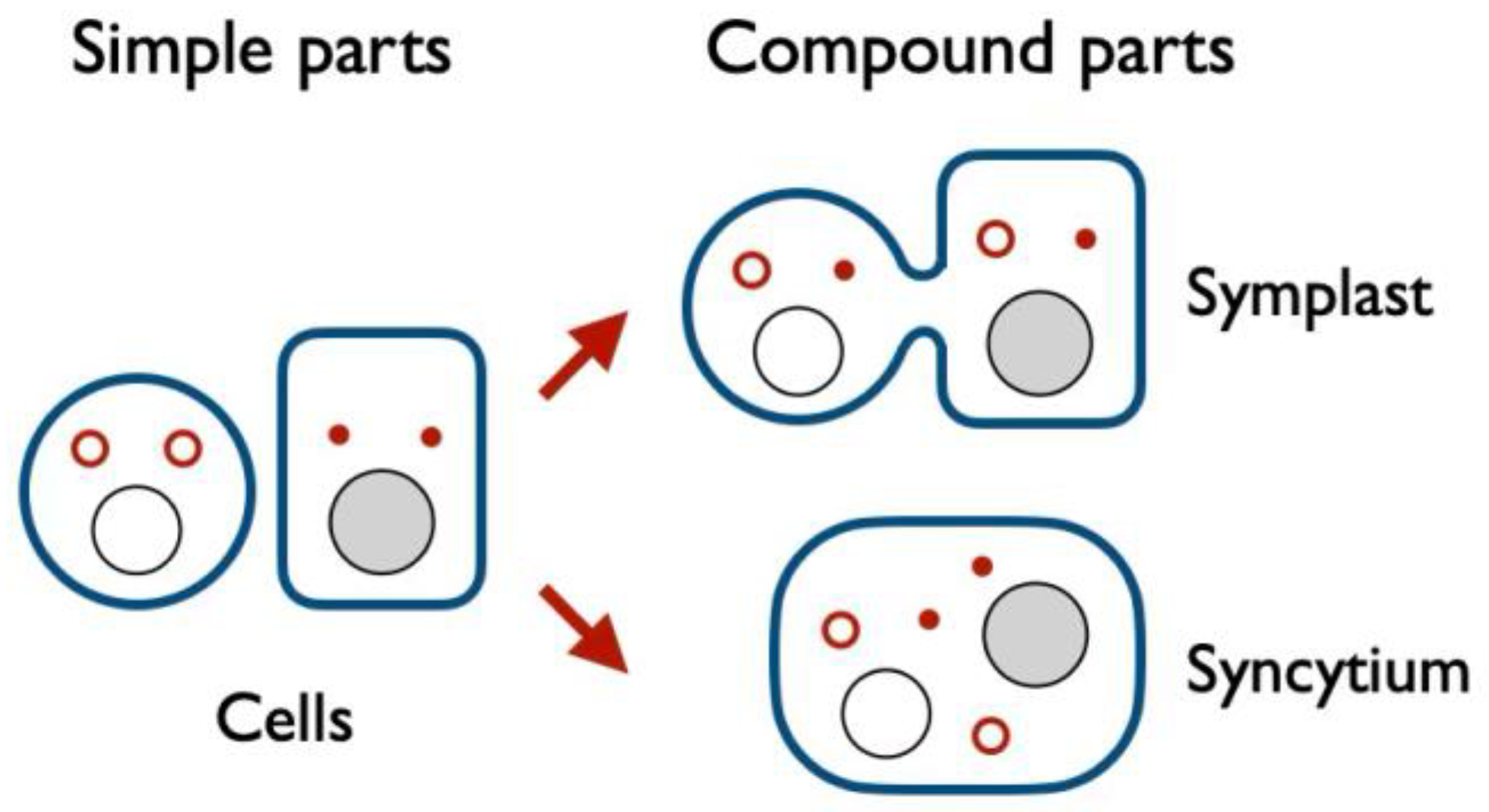
Figure 4.
Familial bridge. Familial bridges result from incomplete cell devision or incomplete cell fission (division without prior cell duplication). Like anastomotic bridges, familial bridges result in continuity of cell membranes and cytoplasms but can connect only related cells. Please see Glossary for examples.
Figure 4.
Familial bridge. Familial bridges result from incomplete cell devision or incomplete cell fission (division without prior cell duplication). Like anastomotic bridges, familial bridges result in continuity of cell membranes and cytoplasms but can connect only related cells. Please see Glossary for examples.
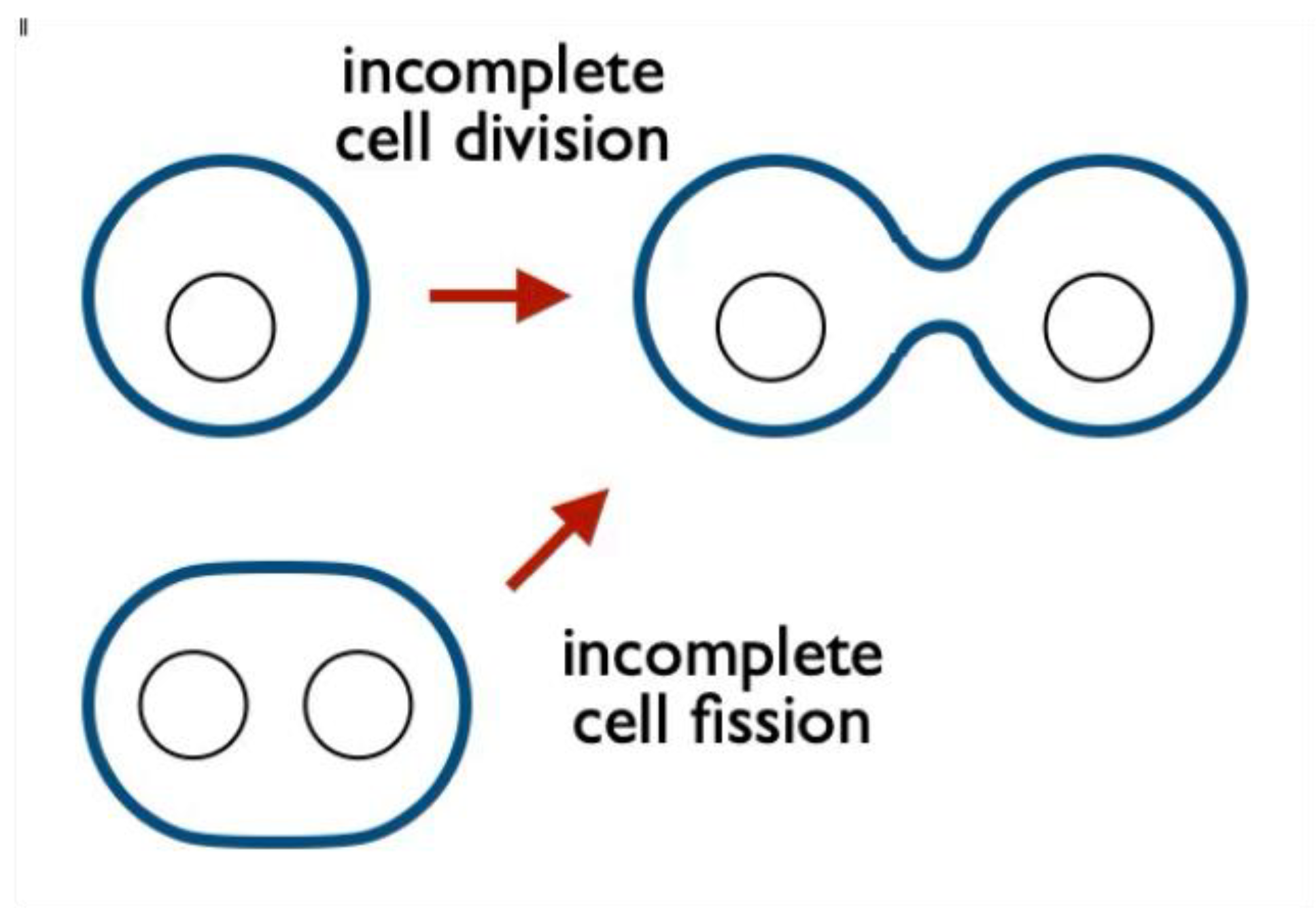
Figure 5.
Fertilization inspires two models of carcinogenesis.
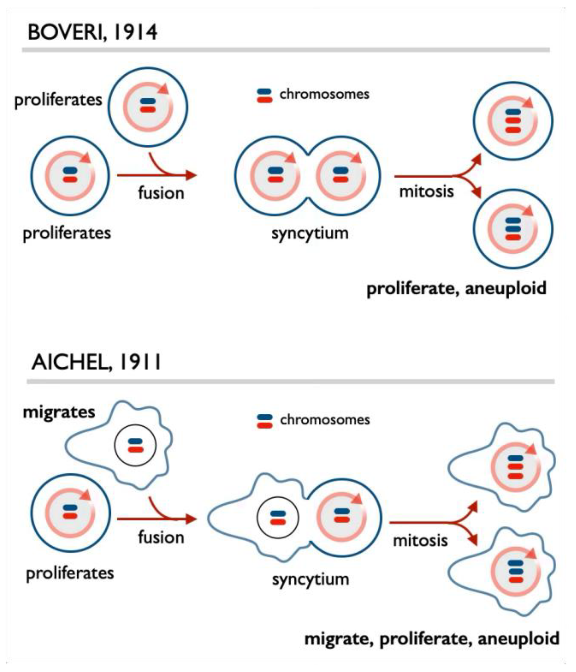
Figure 6.
An example of a familial symplast: oocyte cyst in Drosophila. This structure results from subsequent mitoses followed by incomplete cytokinesis, thus producing a symplast in which cells are connected by familial bridges known in this organism as ring canals. Only one of 16 cells in this symplast, known as a cyst, becomes an oocyte (indicated by a dark nucleus), while other 15 become nurse cells which feed the growing oocytes their contents and then die. All these activities are apparently regulated by a specialized organelle, the fusome (shown in orange).
Figure 6.
An example of a familial symplast: oocyte cyst in Drosophila. This structure results from subsequent mitoses followed by incomplete cytokinesis, thus producing a symplast in which cells are connected by familial bridges known in this organism as ring canals. Only one of 16 cells in this symplast, known as a cyst, becomes an oocyte (indicated by a dark nucleus), while other 15 become nurse cells which feed the growing oocytes their contents and then die. All these activities are apparently regulated by a specialized organelle, the fusome (shown in orange).
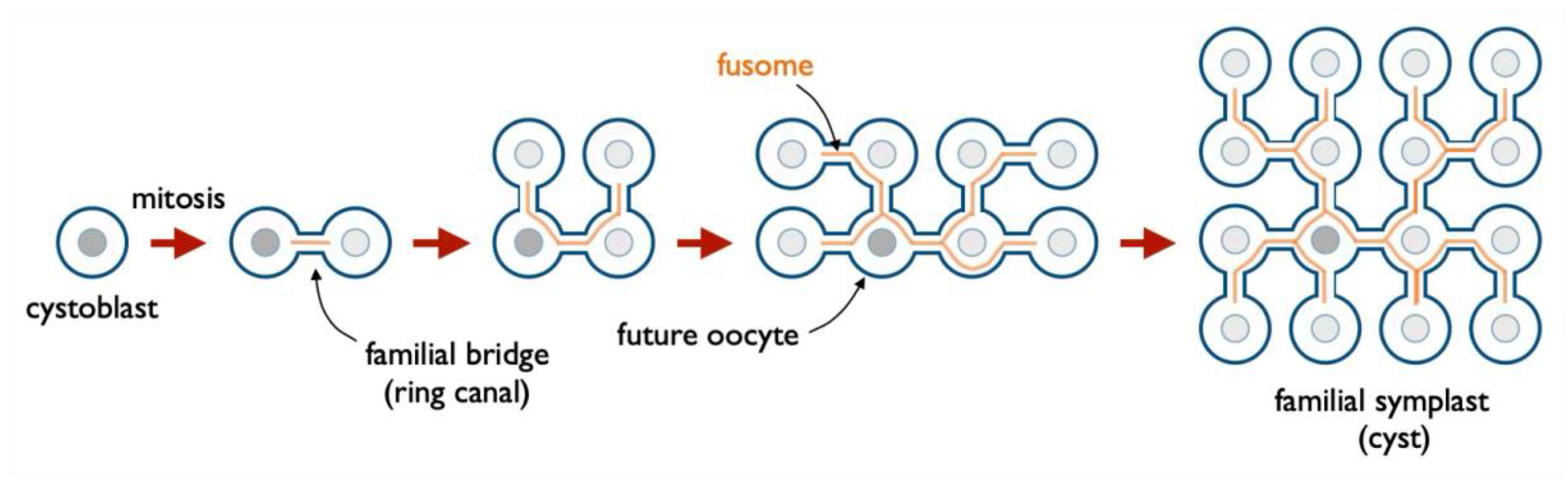
Figure 7.
Transmembrane bridge, a bridge that joins the cytoplasms but keeps the membranes discontinuous by piercing them. Please see Glossary for examples.
Figure 7.
Transmembrane bridge, a bridge that joins the cytoplasms but keeps the membranes discontinuous by piercing them. Please see Glossary for examples.
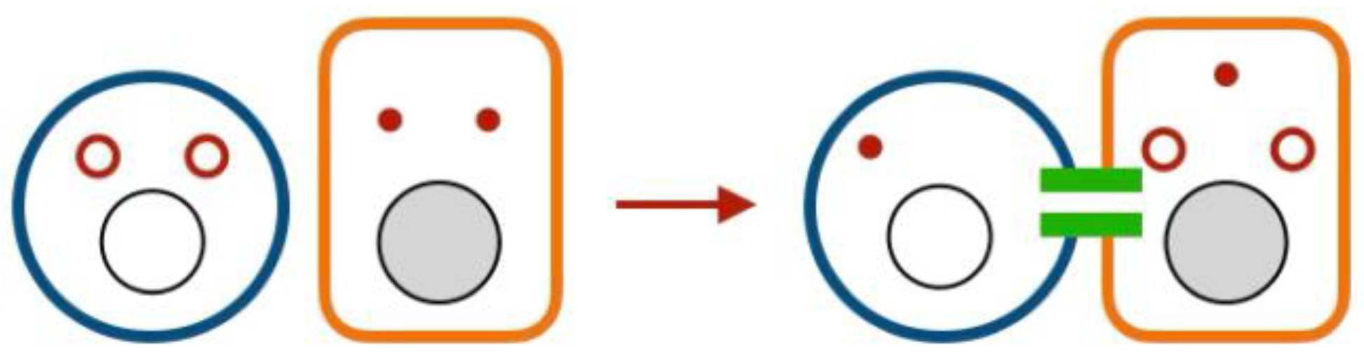
Figure 8.
Examples of anastomotic symplasts in the nervous system.
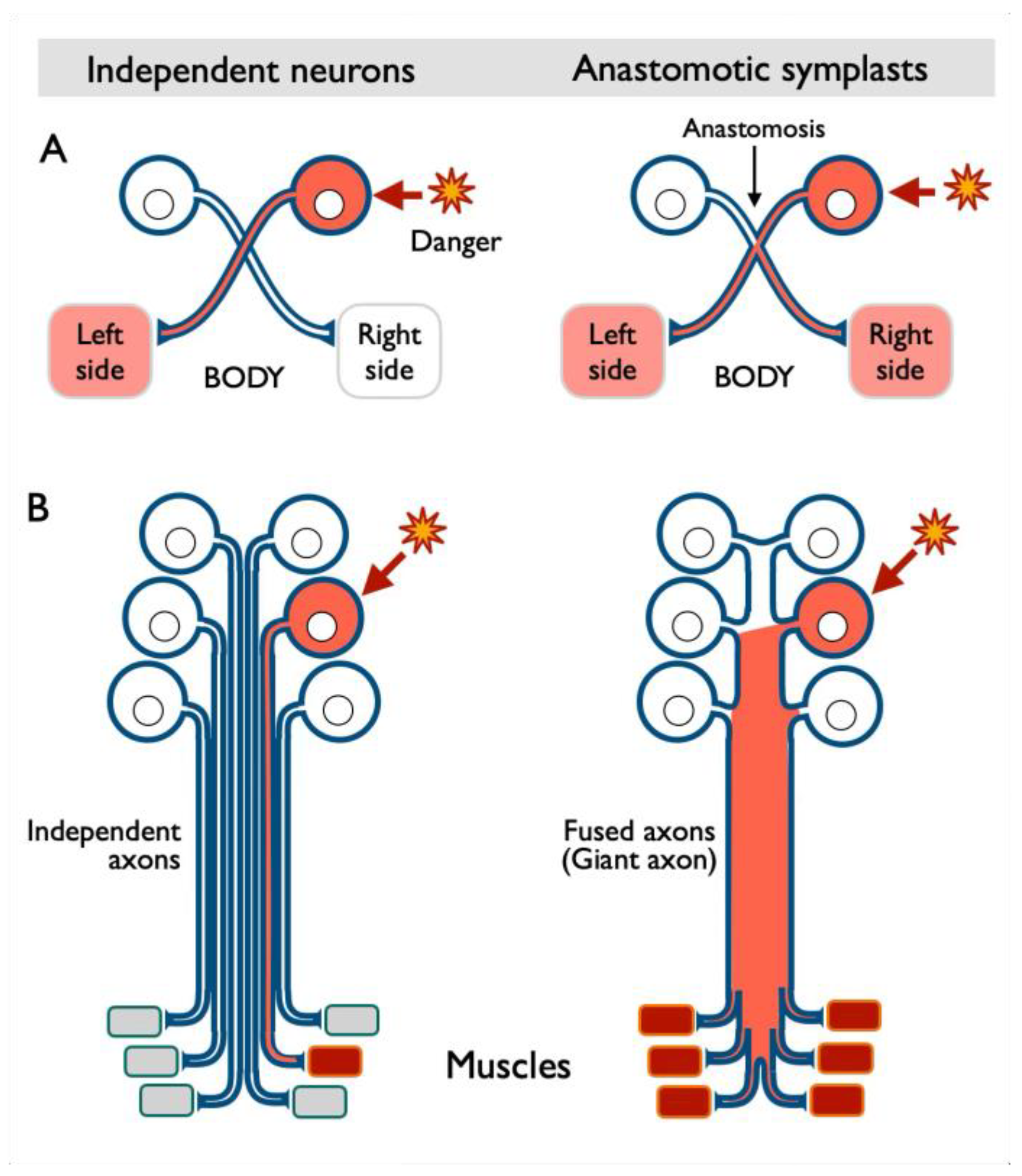
Figure 9.
Cyanobacteria filament is a symplast which enables cell specialization. Please see text for explanation.
Figure 9.
Cyanobacteria filament is a symplast which enables cell specialization. Please see text for explanation.
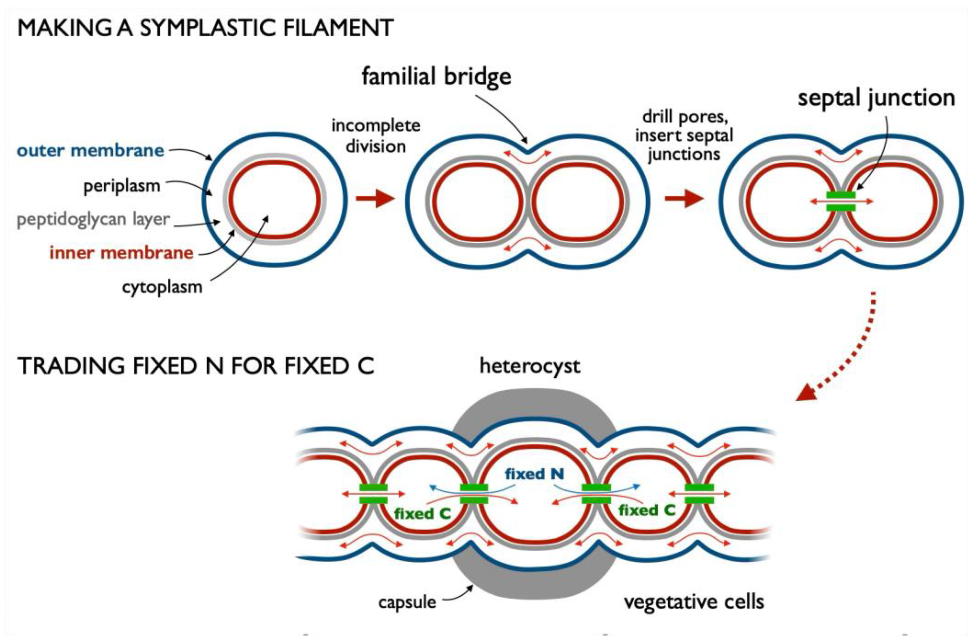
Figure 10.
The complexities of anastomosing cyanobacteria, mitochondria, and other diderms, that is cells, organisms, and organelles with two membranes. The details of this process are practically unknown, but the complexity of the surgery to merge inner and outer membranes, periplasmic space with its proteoglycan layer, and cytoplasms without killing the cells by spilling their guts, so to speak, is apparent.
Figure 10.
The complexities of anastomosing cyanobacteria, mitochondria, and other diderms, that is cells, organisms, and organelles with two membranes. The details of this process are practically unknown, but the complexity of the surgery to merge inner and outer membranes, periplasmic space with its proteoglycan layer, and cytoplasms without killing the cells by spilling their guts, so to speak, is apparent.
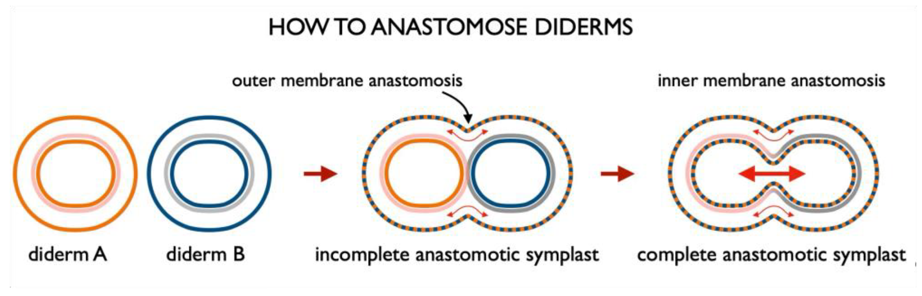
Figure 11.
Adaptation mechanisms enabled by enosis. Enotic complementation (top left) is a process of acquiring a component, a function, or information from another cell or organism without exposing the required items to the extracellular space, as shown for transfer of antibiotic resistance between bacteria through various conduits. Enotic reprogramming (bottom left) is reprogramming consequence to cell bridging or fusion. Enotic structures (bottom left) are structures created by cell bridging or fusion that have emergent properties. Please see text for examples and how this feature of enosis is related to atoms and molecules. Enotic regulation (bottom right) is that mediated through bridges or ferries, thus enabling regulatory molecules made in one cell affect others without the need for signal transduction across cellular membranes.
Figure 11.
Adaptation mechanisms enabled by enosis. Enotic complementation (top left) is a process of acquiring a component, a function, or information from another cell or organism without exposing the required items to the extracellular space, as shown for transfer of antibiotic resistance between bacteria through various conduits. Enotic reprogramming (bottom left) is reprogramming consequence to cell bridging or fusion. Enotic structures (bottom left) are structures created by cell bridging or fusion that have emergent properties. Please see text for examples and how this feature of enosis is related to atoms and molecules. Enotic regulation (bottom right) is that mediated through bridges or ferries, thus enabling regulatory molecules made in one cell affect others without the need for signal transduction across cellular membranes.
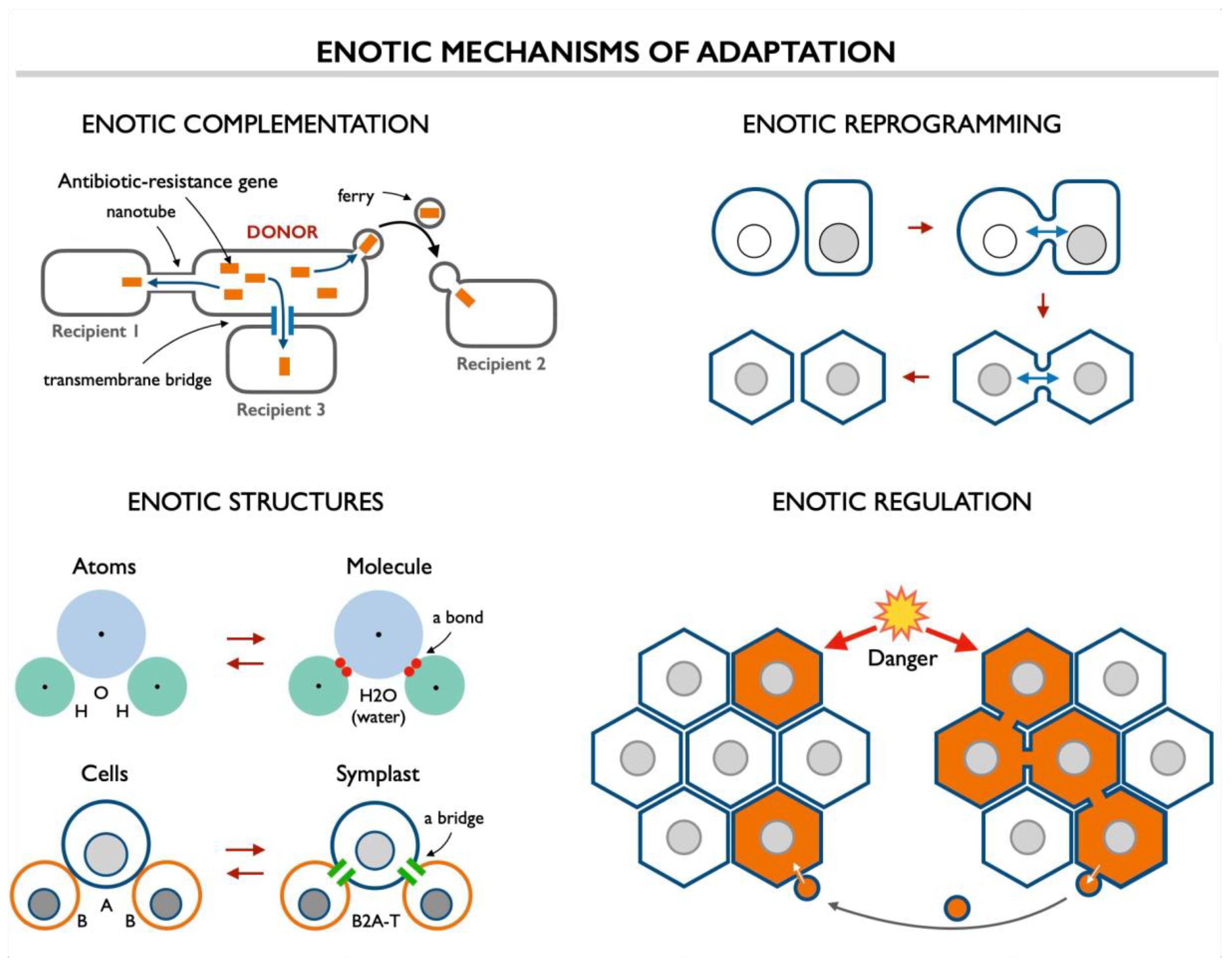
Figure 12.
The logistics of enosis as a system.
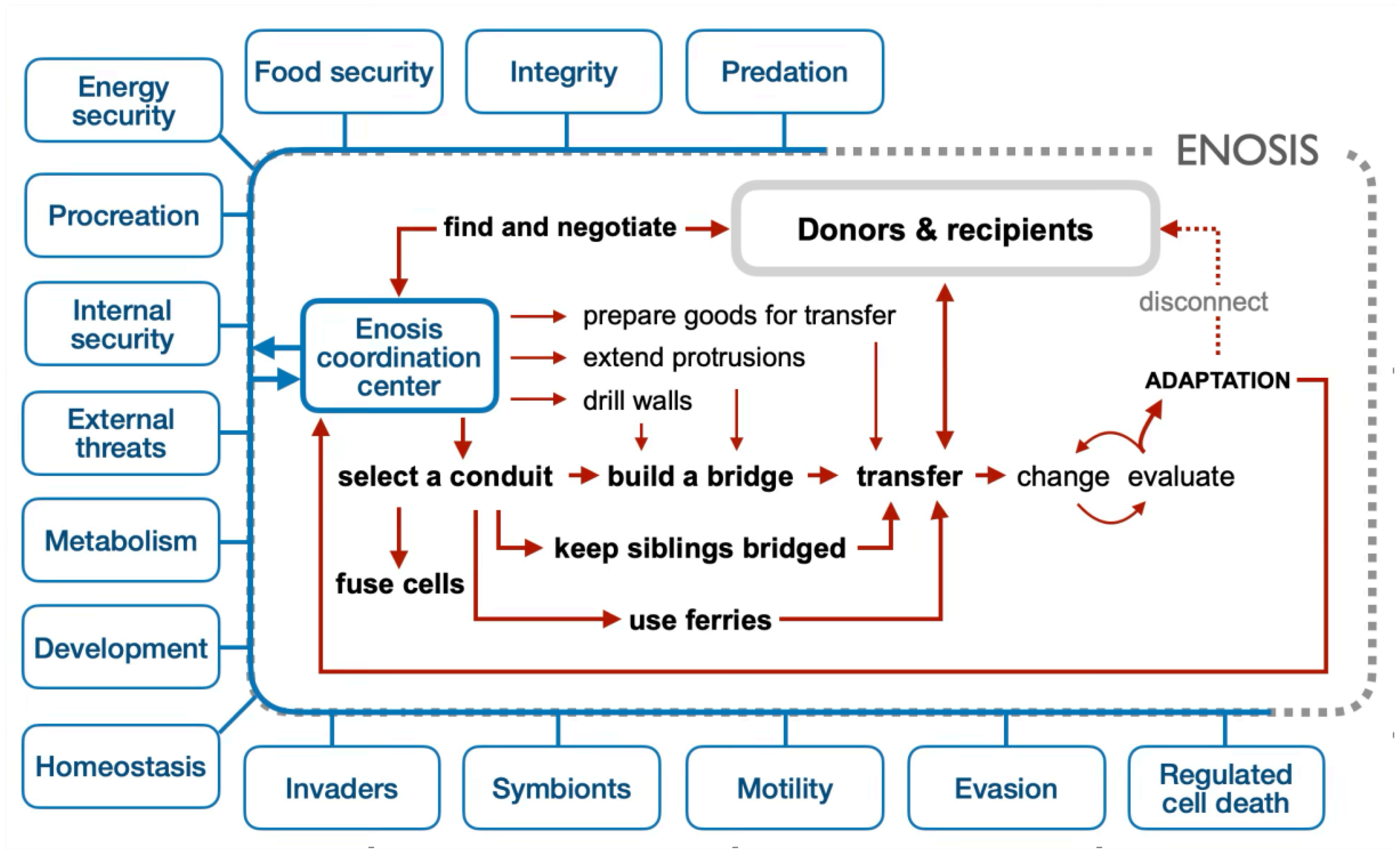
Figure 13.
A visual guide to enotic bridges, ferries, unions, and their relationships mentioned in the Glossary.
Figure 13.
A visual guide to enotic bridges, ferries, unions, and their relationships mentioned in the Glossary.
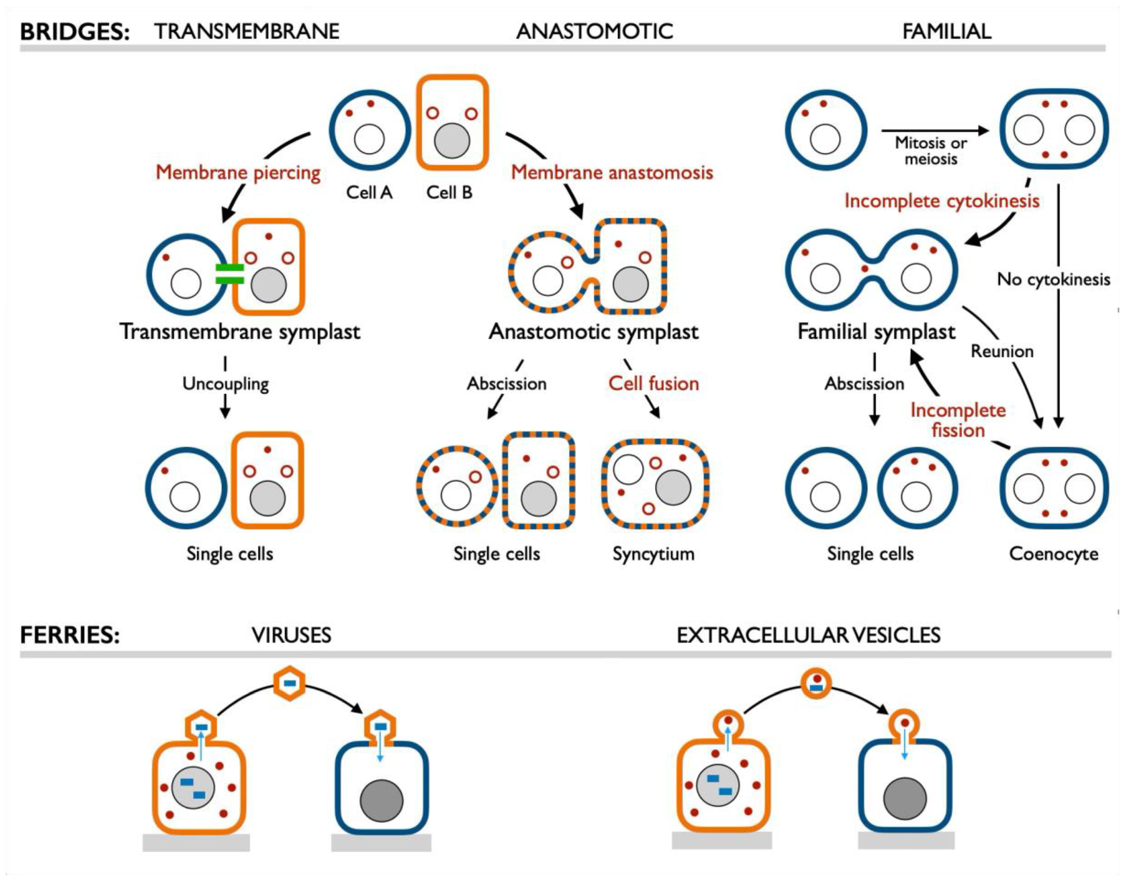
Disclaimer/Publisher’s Note: The statements, opinions and data contained in all publications are solely those of the individual author(s) and contributor(s) and not of MDPI and/or the editor(s). MDPI and/or the editor(s) disclaim responsibility for any injury to people or property resulting from any ideas, methods, instructions or products referred to in the content. |
© 2026 by the authors. Licensee MDPI, Basel, Switzerland. This article is an open access article distributed under the terms and conditions of the Creative Commons Attribution (CC BY) license (http://creativecommons.org/licenses/by/4.0/).
Copyright: This open access article is published under a Creative Commons CC BY 4.0 license, which permit the free download, distribution, and reuse, provided that the author and preprint are cited in any reuse.