

Submitted:
18 August 2026
Posted:
19 August 2026
You are already at the latest version
Abstract
Five new cembrane-type diterpenes (1–5), one new casbane-type diterpene (7), along with ten known related ones (6, 8–16), were isolated from the South China Sea soft coral Sinularia nanolobata. The structures of new compounds were elucidated through comprehensive spectroscopic analysis, NMR calculation with DP4+ probability analysis, time dependent density functional theory-electronic circular dichroism (TDDFT-ECD) calculations, and comparison with the reported spectroscopic data of known analogues. Structurally, compounds 2–5 featured rare five- to seven-membered oxygen-containing heterocyclic rings with diverse joints, reflecting remarkable chemical diversity of secondary metabolites. In bioassays, all isolates have been evaluated for their cytotoxic and anti-inflammatory effects.
Keywords:
soft coral
; Sinularia nanolobata
; cembrane
; casbane
; structural elucidation
1. Introduction
Marine organisms, especially invertebrates, are shaped by their complex and competitive environments, leading them to biosynthesize an enormous diversity of secondary metabolites with unique chemical scaffolds and potent biological activities [1]. Soft corals of the genus Sinularia (phylum Cnidaria, class Anthozoa, subclass Octocorallia, order Alcyonacea, family Alcyoniidae) stand out as particularly rich source of such marine natural products, and serves as an amazing treasure trove of novel compounds ranging from sesquiterpenes, diterpenes, steroids, alkaloids to other miscellaneous compounds [2,3,4]. Numerous chemical investigations of the genus Sinularia revealed a vast array of structurally diverse secondary metabolites displayed highly promising biological activities including antibacterial, osteoclast inhibitory, anti-inflammatory, antithrombotic, antiviral, cytotoxic, and immunosuppressive activities [4,5].
Our team has been continuously dedicated to the search for bioactive secondary metabolites with novel structures from marine invertebrates, particularly soft corals from the South China Sea, and a large quantity of secondary metabolites with structural diversity and interesting bioactivities have been isolated and characterized [6,7,8]. Previously, an unpredented dieterpene with a tricyclo [10.3.0.0¹,²]pentadecane carbon skeleton, along with multiple polyoxygenated and unusual endoperoxide-bridged casbane-type diterpenes, were found from S. nanolobata collected off Ximao Island [9].
Inspired by our previous work, a new collection of the title species off the Ximao Island, South China Sea, were studied, which led to the discovery of 16 diterpenes. Among them, five previously unreported cembrane-type diterpenes and one previously described casbane-type diterpene were characterized. All isolated compounds were evaluated for their cytotoxicity against several human tumor cell lines and anti-inflammatory effects against murine macrophage cell line RAW264.7. Herein, the isolation, structural characterization, and bioactivity evaluation of these compounds were reported.
2. Results
Samples of S. nanolobata were frozen immediately to –20 ℃ after collection and stored at that temperature until exhaustive extraction with dichloromethane and methanol (1:1, v/v). The EtOAc-soluble portion of the organic extract was subjected to repeated column chromatography (CC) (silica gel, Sephadex LH-20, and reversed-phase HPLC), yielding five new cembrane-type diterpenes (1–5), one new casbane-type diterpene (7), along with ten known related compounds (6, 8–16). Those known compounds were rapidly characterized as 12α-methyl-1E,3E,7E-cembratrien-10-one (6) [10], nanolobatone B (8) [9], 10-oxo-11,12-dihydrodepressin (9) [11], 1-epi-10-oxo-11,12-dihydrodepressin (10) [11], 10-hydroxydepressin (11) [11], 1-epi-10-hydroxydepressin (12) [11], 2-epi-10-hydroxydepressin (13) [12], sinularcasbane A (14) [13], casbene (15) [14], and 5-oxo-casbene (16) [11], respectively, by comparison of their NMR data and optical rotation [α]D values with those reported in the literature.
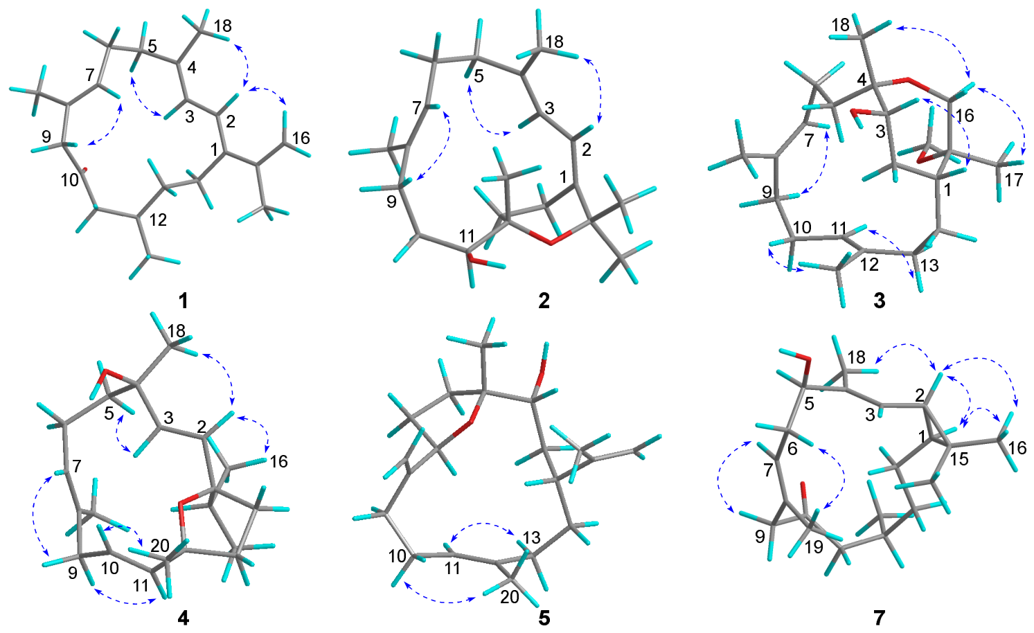
Compound 1 was isolated as a colorless oil with the molecular formula of C20H30O, which was established by the protonated molecular ion peak observed at m/z 287.2364 [M + H]+ (calcd. for C20H31O+, 287.2369) in the HRESIMS spectrum, implying six degrees of unsaturation. The 1H NMR spectroscopic data of 1 (Table 1) revealed three vinyl methyls at δH 1.92 (3H, s), 1.78 (3H, s) and 1.60 (3H, s), one doublet methyl at δH 0.90 (3H, d, J = 6.7 Hz), and five olefinic protons at δH 6.31 (1H, d, J = 11.2 Hz), 6.02 (1H, d, J = 11.2 Hz), 5.25 (1H, t, J = 6.8 Hz), 5.03 (1H, s) and 4.95 (1H, s). The 13C NMR spectroscopic data of 1 (Table 2) showed 20 carbon signals, identified with the aid of HSQC spectrum, including four methyls (δC 16.6, 16.9, 21.1, 21.4), eight olefinic carbons (δC 112.4, 122.7, 123.0, 129.2, 130.3, 138.4, 139.2, 143.5), one carbonyl carbon (δC 209.8), and seven methylene carbons (δC 24.0, 25.6, 27.7, 35.9, 39.9, 47.1, 54.3). The above-mentioned four double bonds and one carbonyl accounted for five of the six degrees of unsaturation, indicating a monocyclic carbon framework for 1. The NMR data of 1 were almost identical to those of previously reported compound, 12α-methyl-1E,3E,7E-cembratrien-10-one (6) [10], except that the isopropyl with a methoxy group at C-15 in 6 was replaced by an isopropenyl group in 1. This replacement was corroborated by the HMBC correlations from H2-16 to C-15 and C-17. The planar structure of 1 was finally determined as depicted in Figure 2 through analysis of 1H–1H COSY and HMBC spectra. The geometries of double bonds Δ1, Δ3 and Δ7 were assigned as “E” based on the shielded carbon resonances of two vinyl methyls at δC 16.9 (C-18), 16.6 (C-19), as well as the observed NOESY correlations of H-2/Me-18, H-3/H2-5, H-7/H2-9 (Figure 3). The absolute configuration of compound 1 { -2.2 (c 0.18, CHCl3)} was determined as same 12R as compound 6 { -13.5 (c 0.25, CHCl3)} based on comparison of optical rotation values recorded under the same conditions. Therefore, the structure of 1 was established as shown in Figure 1 and named 12α-methyl-1E,3E,7E,15-cembratetraen-10-one.
Figure 1.
Chemical structures of compounds 1–16.
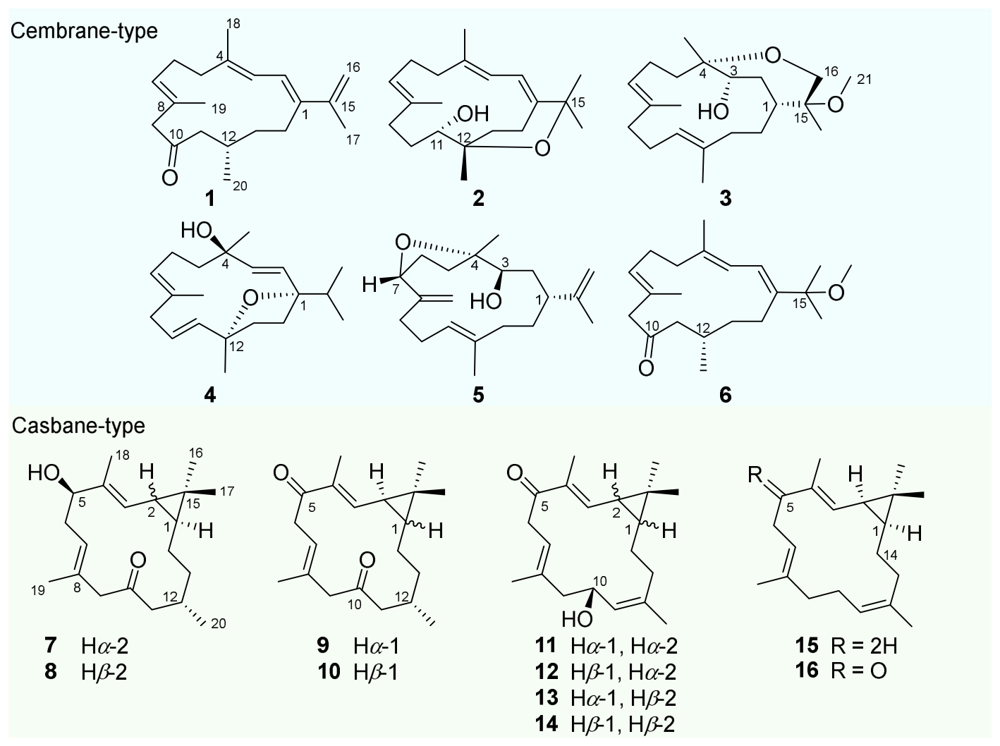
Figure 2.
1H–1H COSY and selected key HMBC correlations of compounds 1–5 and 7.
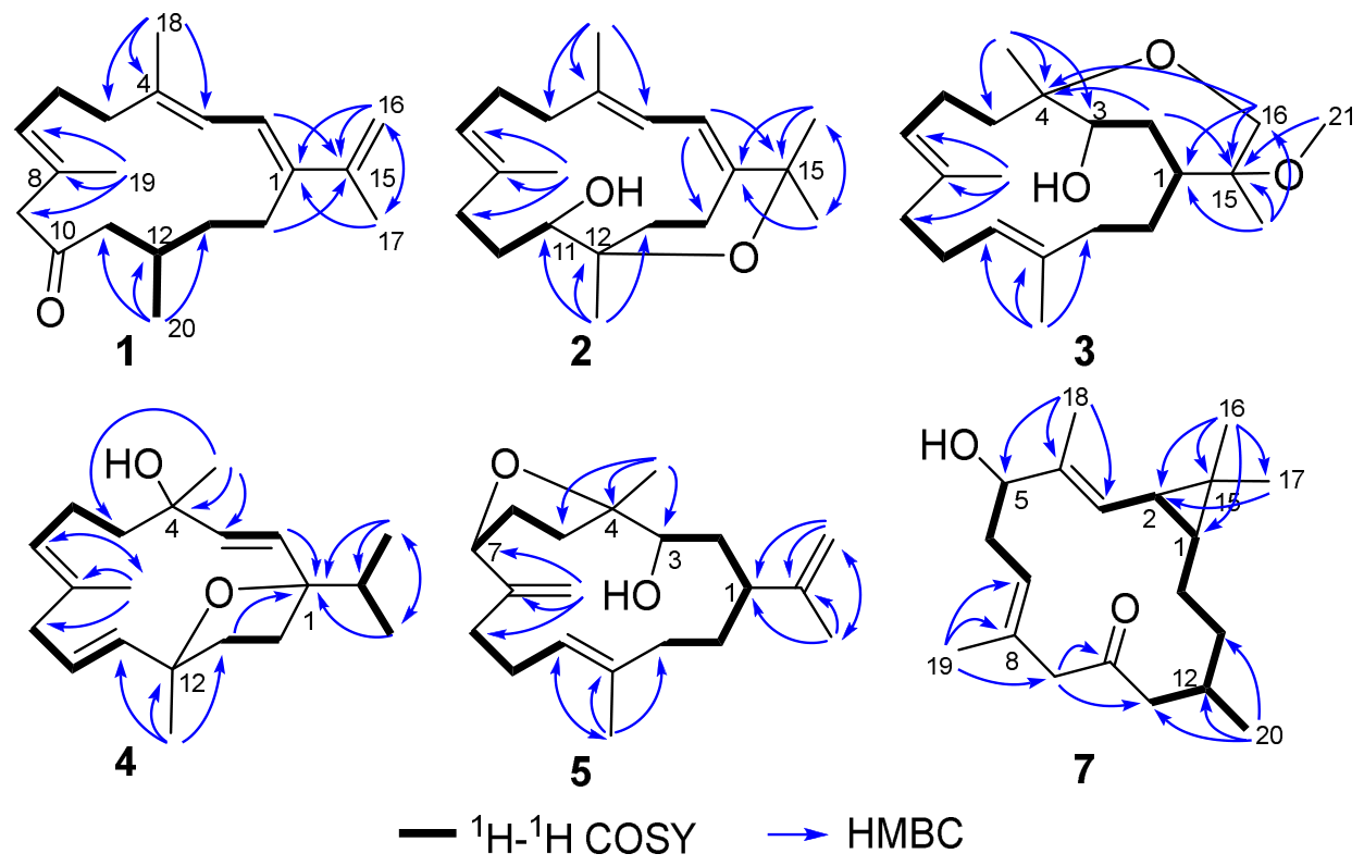
Figure 3.
The NOESY correlations of compounds 1–5 and 7.
Compound 2 was isolated as a colorless oil, and its molecular formula C20H32O2 was established by the protonated molecular ion peak observed at m/z 305.2477 [M + H]+ (calcd. for C20H33O2+, 305.2475) in the HRESIMS spectrum, which was 18 mass units more than that of 1, corresponding to five degrees of unsaturation. A comparison of the 1D NMR data of 2 with those of 1 suggested that they were structural analogues, both being cembrane-type diterpenes. The 1H NMR spectrum displayed three olefinic protons at δH 6.19 (1H, d, J = 10.0 Hz), 6.00 (1H, d, J = 10.0 Hz) and 5.06 (1H, t, J = 7.8 Hz), which were attributed to three trisubstituted double bonds. These moieties accounted for three of the five degrees of unsaturation, suggesting that 2 possessed a bicyclic system in its molecule structure. Moreover, the 13C NMR signals at δC 70.1 (CH), 74.7 (qC) and 76.0 (qC) confirmed the presence of a hydroxyl group and an additional oxygen-containing ring in 2, accounting for the remaining two oxygen atoms. The location of hydroxyl group at C-11 and the formation of the oxygen ring via the linkage of C-12 and C-15, respectively, as evidenced by the HMBC correlations from Me-16/17 to C-1 (δC 138.4), C-15 (δC 74.7) and C-17 (δC 29.9)/C-16 (δC 28.9), and from Me-20 to C-11 (δC 70.1), C-12 (δC 76.0), C-13 (δC 30.2). Thus, the planar structure of compound 2 was established on the basis of collective spectroscopic evidences (Figure 2).
All the geometries of the double bonds Δ1, Δ3 and Δ7 were assigned to be “E” based on the NOESY correlations of H-2/Me-18, H-3/H2-5, H-7/H2-9 (Figure 3). The relative configuration of C-11 and C-12 was determined to be 11S*,12S* by the QM-NMR calculation and DP4+ probability analysis, where the experimentally observed NMR data of 2 gave the best match of 100% to those of the (11S*,12S*)-isomer (Figure S58). Subsequently, the absolute configuration of 2 was further elucidated to be 11S,12S via TDDFT-ECD calculation, showing that Boltzmann-averaged ECD curve of (11S,12S)-2 highly matched to the experimental ECD spectrum of 2, whereas the enantiomer exhibited a completely opposite curve (Figure 4). Accordingly, the structure of 2 was characterized as shown in Figure 1, and the compound was designated with the trivial name nanolobatol A.
Compound 3 was also isolated as a colorless oil, with the molecular formula C21H36O3 established by HRESIMS from the molecular ion peak observed at m/z 337.2737 [M + H]+ (calcd. for C21H37O3+, 337.2737), suggesting that 3 possessed four degrees of unsaturation. The 1H, 13C NMR data and HSQC spectrum of 3 displayed 21 distinct carbon signals, including five singlet methyl groups (δH/δC 0.85/19.4, 1.07/19.2, 1.51/15.1, 1.60/15.0, 3.26/50.8), eight methylene groups (δC 22.4, 24.9, 25.3, 32.0, 36.8, 38.4, 39.5, 70.0), four methine groups (δC 30.9, 73.3, 127.7, 127.8), and four quaternary carbons (δC 79.0, 80.3, 133.4, 134.0). Based on the above data, the presence of two trisubstituted double bonds accounted for two of the four degrees of unsaturation, indicating that compound 3 was a bicyclic molecule. Furthermore, comprehensive analysis of the 1H–1H COSY spectrum revealed three distinct spin systems, as evidenced by the observed cross-peaks for H-3/H2-2/H-1/H2-14/H2-13, H2-5/H2-6/H-7, H2-9/H2-10/H-11. Additionally, the key HMBC correlations from Me-18 to C-3, C-4, and C-5 revealed the substitution of the hydroxyl at C-3. The HMBC cross-peaks of H2-16/C-4 were diagnostic for the ring linkage of C-4 and C-16. The methoxyl group at C-15 was indicated by the characteristic HMBC correlations from Me-21 to C-15 (Figure 2). Consequently, the planar structure of compound 3 was collectively established.
The geometries of the double bonds Δ7 and Δ11 were both assigned as “E” based on the shielded carbon resonances of the two vinyl methyl groups at δC 15.1 (C-19) and 15.0 (C-20). This assignment was further supported by the NOESY correlations (Figure 3) of H-7/H-9 and of H-11/H-13. The relative configurations of C-1/C-3 and C-4/C-15 in compound 3 were determined to be 1R*,3S* and 4S*,15R*, respectively, based on the diagnostic NOESY correlations of H-1 (δH 1.96)/H-3 (δH 4.26) and Me-17 (δH 0.85)/H-16 (δH 3.64)/Me-18 (δH 1.07). However, the relationships of the relative configurations of two chiral domains could not be unambiguously determined by NOESY analysis. Assisted by the QM-NMR calculation and DP4+ probability analysis, the full relative configuration of 3 was established as 1R*,3S*,4S*,15R*, because the experimental NMR data of 3 showed the best match (100%) with those of the (1R*,3S*,4S*,15R*)-isomer (Figure S60). As shown in Figure 5, the experimental ECD spectrum of 3 was in good agreement with the calculated curve for (1R,3S,4S,15R)-3, while being completely opposite to that of its enantiomer. Consequently, the absolute configuration of 3 was determined to be 1R,3S,4S,15R by TDDFT-ECD calculation. Herein, the structure of 3 (the trivial name nanolobatol B) was proposed as depicted.
Compound 4 was obtained as a colorless oil. The molecular formula of 4 was established as C20H32O2 based on the protonated molecular ion peak observed at m/z 305.2471 [M + H]+ (calcd. for C20H33O2+, 305.2475) in its HRESIMS spectrum, implying five degrees of unsaturation. The 1H NMR data displayed signals corresponding to one vinyl methyl at δH 1.61 (3H, s), two tertiary methyls at δH 1.26 (3H, s) and 1.33 (3H, s), two methyls appearing as doublets at δH 0.88 (6H, d, J = 6.8 Hz), and five olefinic protons at δH 5.24 (1H, t, J = 7.5 Hz), 5.51 (1H, d, J = 16 Hz), 5.52 (1H, dd, J = 16, 6 Hz), 5.64 (1H, d, J = 15.8 Hz) and 5.71 (1H, d, J = 15.8 Hz), which were attributed to two disubstituted and one trisubstituted double bonds. Based on the above, three double bonds accounted for three of the five degrees of unsaturation, indicating that a bicyclic ring system belonged to the molecular structure of 4. The HMBC correlations from Me-18 to C-2, C-3 and C-4 and from H-2 to C-1 corroborated the location of the hydroxyl group at C-4 and the settlement of double bond Δ2. The characteristic HMBC cross-peaks of Me-20/C-12, H2-13/C-1, along with the 1H–1H COSY cross-peaks for H2-13/H2-14, revealed the linkage of C-1 and C-12, forming the tetrahydrofuran ring. Finally, the planar structure of 4 was unequivocally established as shown in Figure 2.
The large coupling constants (J2,3 = 15.8 Hz, J10,11 = 16 Hz) established the “E” geometries of double bonds Δ2 and Δ10. Additionally, the shielded carbon resonance of the vinyl methyl at δC 17.6 (C-19) assigned the “E” geometry to the double bond Δ7. These assignments were further confirmed by analysis of the NOESY correlations (Figure 3). The results of the QM-NMR/DP4+ calculations (Figure S62) confirmed the relative configuration of compound 4 to be 1S*,4R*,12S*. Moreover, the absolute configuration of 4 was also determined by comparing its experimental ECD spectrum with the TDDFT-ECD calculated curves of its two enantiomers, showing excellent agreement with the one of the (1S,4R,12S)-4 isomer (Figure 5). Thus, the structure of 4 (the trivial name nanolobatol C) was assigned as depicted.
Compound 5 was isolated as a colorless oil, possessing the same molecular formula of C20H32O2 as 4 by HRESIMS ion peak at m/z 305.2472 [M + H]+ (calcd. for C20H33O2+, 305.2475). The 1H NMR data revealed the presence of two vinyl methyls at δH 1.60 (3H, s) and 1.70 (3H, s), one tertiary methyl at δH 1.19 (3H, s), and five olefinic protons at δH 4.77 (2H, s), 4.86 (1H, s), 4.91 (1H, s) and 5.30 (1H, t, J = 7.6 Hz), assigned to one trisubstituted and two terminal double bonds. These structural fragments accounted for three of the five degrees of unsaturation, indicating that a bicyclic ring system was incorporated in the molecular structure of 5. Subsequently, comprehensive analysis of the key ¹H–¹H COSY cross-peaks and long-range HMBC correlations collectively established the connectivity of the spin systems and the assembly of all structural fragments. In detail, the ¹H–¹H COSY cross-peaks of H-3/H₂-2/H-1/H₂-14/H₂-13, H₂-5/H₂-6/H-7, and H₂-9/H₂-10/H-11 revealed three distinct spin systems. The HMBC correlations from Me-17 to C-1 (δC 41.4), C-15 (δC 150.2), and C-16 (δC 110.9); from Me-18 to C-3 (δC 73.6), C-4 (δC 84.9), and C-5 (δC 37.1); from H₂-19 to C-7 (δC 80.3), C-8 (δC 151.9), and C-9 (δC 34.3); and from Me-20 to C-11 (δC 126.3), C-12 (δC 134.5), and C-13 (δC 36.5) revealed the linkage between C-4 and C-7, forming the tetrahydrofuran ring. In addition, the hydroxy group was established at C-3, and the two terminal double bonds were assigned to C-8 and C-15, respectively, thereby unambiguously determining the planar structure of 5 (Figure 2).
The “E” geometry of the Δ11,12 double bond was assigned based on the shielded carbon resonances of the vinyl methyl at δC 17.2 (C-20). The four stereogenic centers (C-1, C-3, C-4, and C-7) in compound 5 were all located on a flexible macrocyclic ring and remote from each other, which prevented definitive configurational assignment by NOESY analysis and thus required QM-NMR/DP4+ calculations. Comparison of the calculated NMR data with the experimental values showed excellent agreement (99.99%) for the 1R*,3R*,4S*,7R* configuration (Figure S64). Following the same TDDFT-ECD calculation protocol as applied to the other compounds in this study, the absolute configuration of 5 was finally established as 1R,3R,4S,7R (Figure 5). Accordingly, the structure of 5 (the trivial name nanolobatol D) was characterized as depicted.
Compound 7 was isolated as a colorless oil. Its molecular formula was determined to be C20H32O2, based on the HRESIMS ion peak observed at m/z 305.2475 [M + H]+ (calcd. for C20H33O2+, 305.2475). The 1H and 13C NMR data of 7 were highly similar to those of the co-occurring known casbane-type diterpenoid, nanolobatone B (8) [9], with the main differences observed at C-1, C-2 and their adjacent carbons, suggesting that they were structural analogues and epimers at either the C-1 or C-2 position. The large ΔδC value (more than 10 ppm) of the gem-dimethyls C-16 (δC 29.1) and C-17 (δC 15.9) indicated a 1,2-cis configuration for 7, which was further supported by the obvious NOESY correlations of H-1 (δH 0.61)/Me-16 (δH 1.06) and H-2 (δH 1.24)/H-1 (Figure 3). Careful comparison of the NMR data of 7 with those of the reported synthetic product S5 [15], particularly the chemical shifts of H-1/C-1 and H-2/C-2, indicated that 7 shared the same 1S*,2R* relative configuration as S5. Furthermore, the relative configuration of 5R*,12S* in 7 was confirmed to be identical to that of 8 based on the similarity of their NMR data. Therefore, the relative configuration of 7 was assigned as 1S*,2R*,5R*,12S*. Finally, the absolute configuration of 7 was determined to be 1S,2R,5R,12S by TDDFT-ECD calculations, as the experimental ECD spectrum matched well with the calculated curve for (1S,2R,5R,12S)-7 (Figure 5). Consequently, compound 7 was identified as the C-2 epimer of compound 8, named 2-epi-nanolobatone B.
In the in vitro bioassay, all isolated compounds were evaluated for their cytotoxic activities against MV-4-11, A-549, and 293T cell lines, as well as for their anti-inflammatory effects in RAW264.7 macrophages. Unfortunately, none of the compounds exhibited obvious bioactivity (IC50 > 50 µM). Comparatively, previous studies have reported that certain cembrane- and casbane-type diterpenes from Sinularia species exhibit moderate cytotoxic and/or anti-inflammatory effects, with IC50 values typically ranging from 10 to 50 µM. Despite the observed weak or negligible activities of the compounds in this study, evaluating these activities remains a routine and essential step for the full characterization of new natural products, whose potency may vary depending on structural substitution patterns. Notably, the lack of activity observed in the present study does not preclude the possibility of other biological effects, such as antimicrobial or neuroprotective activities, and additional bioassays to explore these possibilities are currently in progress.
3. Materials and Methods
3.1. General Experimental Procedures
Optical rotations were measured on a Perkin-Elmer 241MC polarimeter (PerkinElner, Fremont, CA, USA). NMR spectra were measured in CDCl3 with a Bruker Advance 600 MHz spectrometer (Bruker Biospin AG, Fällanden, Germany). Chemical shifts are reported in parts per million (δ) in CDCl3 (δH reported referred to CHCl3 at 7.26 ppm; δC reported referred to CDCl3 at 77.2 ppm), and coupling constants (J) are expressed in Hz. HRESIMS spectra were recorded on an Agilent 1290-6545 UHPLC-QTOF mass spectrometer (Agilent Technologies, CA, USA). Commercial silica gel (Qingdao Haiyang Chemical Group Co., Ltd., Qingdao, China, 200–300 and 300–400 mesh), Sephadex LH-20 gel (Amersham Biosciences, Uppsala, Sweden) were used for column chromatography, and precoated silica gel plates (Yan Tai Zi Fu Chemical Group Co., Yantai, China, G60 F-254) were used for analytical TLC. Reversed-phase (RP) HPLC was performed on an Agilent 1260 series liquid chromatography equipped with a DAD G1315D detector at 210 and 254 nm. A semi-preparative ODS-HG-5 column [5 µm, 250 × 9.4 mm] was employed for the purifications. All solvents used for CC and HPLC were of analytical grade (Shanghai Chemical Reagents Co., Ltd., Shanghai, China) and chromatographic grade (Dikma Technologies Inc., Beijing, China), respectively.
3.2. Animal Materials
Specimens of S. nanolobata were collected by scuba diving along the coast off Ximao Island (18°13.8′N, 109°22.1′E), Hainan Province, China, in May 2019, at a depth of 20 meters. A voucher specimen (19-XD-12) is deposited and available for inspection at the School of Medicine, Shanghai University.
3.3. Extraction and Isolation
The frozen animals (260.5 g, dry weight after extraction) were cut into pieces and extracted exhaustively with CH2Cl2/MeOH (1:1, v/v) at room temperature (2.0 L × 4). The organic extract was evaporated to give a brown residue, which was then partitioned between EtOAc and H2O. The EtOAc solution was evaporated to give a dark brown residue (15.7 g). The obtained residue was subjected to gradient silica gel (200–300 mesh) column chromatography (CC) [EtOAc/petroleum ether (PE) 0→100%] to yield five fractions (Fr. A-E).
Fraction A (127.4 mg) was directly purified by semi-preparative RP-HPLC (CH₃CN/H₂O, 98:2, 3.0 mL/min) to afford compound 15 (6.0 mg, tR = 27.5 min).
Fraction B (5.38 g) was separated by Sephadex LH-20 column chromatography eluted with PE/CH2Cl2/MeOH (2:1:1) to give four sub-fractions (B1–B4). Sub-fraction B4 (66.5 mg) was further purified by silica gel column chromatography (300–400 mesh) eluted with PE/EtOAc (40:1 and 15:1), followed by semi-preparative RP-HPLC (CH₃CN/H₂O, 87:13, 3.0 mL/min) to yield compound 16 (4.2 mg, tR = 21.3 min).
Fraction C (1.26 g) was first separated by Sephadex LH-20 column chromatography eluted with PE/CH2Cl2/MeOH (2:1:1) to give three sub-fractions (C1–C3). Sub-fraction C2 was separated by silica gel column chromatography (300–400 mesh) eluted with PE/EtOAc (30:1, 15:1, 5:1, and 1:1) to afford five secondary fractions (C2a–C2e). Fraction C2a was further separated by RP-HPLC (CH3CN/H2O, 80:20, 3.0 mL/min) to obtain compounds 4 (2.1 mg, tR = 16.6 min) and 5 (1.6 mg, tR = 19.2 min). Sub-fraction C2d was purified by semi-preparative RP-HPLC (CH₃CN/H₂O, 80:20, 3.0 mL/min) to yield compound 3 (2.6 mg, tR = 18.7 min). Sub-fraction C2e was purified by semi-preparative RP-HPLC (CH₃CN/H₂O, 80:20, 3.0 mL/min) to give compounds 9 (12.3 mg, tR = 10.8 min) and 10 (9.1 mg, tR = 11.3 min).
Fraction D (1.36 g) was first separated by Sephadex LH-20 column chromatography eluted with PE/CH2Cl2/MeOH (2:1:1) to obtain four sub-fractions (D1–D4). Sub-fraction D2 (528.4 mg) was subjected to silica gel column chromatography (300–400 mesh) eluted with PE/EtOAc (30:1, 20:1, 10:1, 5:1, and 1:1) to afford compound 14 (2.6 mg) and five secondary fractions (D2a–D2e). Sub-fraction D2e was purified by semi-preparative RP-HPLC (MeOH/H₂O, 83:17, 3.0 mL/min) to yield compound 2 (1.8 mg, tR = 16.4 min) and a tertiary fraction (10.2 mg, tR = 24.8 min). Sub-fraction D3 (554.2 mg) was separated by silica gel column chromatography (300–400 mesh) eluted with PE/EtOAc (30:1, 20:1, 10:1, 5:1, 1:1) to yield compound 6 (3.9 mg) and three secondary fractions (D3a–D3c). Sub-fraction D3c was purified by semi-preparative RP-HPLC (CH₃CN/H₂O, 67:33, 3.0 mL/min) to afford compounds 7 (3.2 mg, tR = 12.5 min) and 8 (2.8 mg, tR = 13.3 min).
Fraction E (1.06 g) was initially separated by Sephadex LH-20 column chromatography eluted with PE/CH2Cl2/MeOH (2:1:1) to give three sub-fractions (E1–E3). Sub-fraction E3 was separated by silica gel column chromatography (300–400 mesh) eluted with PE/EtOAc (30:1, 20:1, 10:1, 5:1, 1:1) to afford compound 1 (2.4 mg) and three secondary fractions (E3a–E3c). Sub-fraction E3c was purified by semi-preparative RP-HPLC (CH₃CN/H₂O, 55:45, 3.0 mL/min) to afford compounds 11 (11.4 mg, tR = 17.3 min), 12 (2.1 mg, tR = 18.7 min), and 13 (4.1 mg, tR = 19.3 min).
3.3.1. 12α-. Methyl-1E,3E,7E,15-Cembratetraen-10-One (1)
3.3.2. Nanolobatol A (2)
3.3.3. Nanolobatol B (3)
3.3.4. Nanolobatol C (4)
3.3.5. Nanolobatol D (5)
3.3.6. 2-Epi-Nanolobatone B (7)
3.4. Calculation Section
Conformational searches were performed using the torsional sampling (MCMM) approach and OPLS_2005 force field within an energy window of 21 kJ/mol. Conformers with Boltzmann populations above 1% were re-optimized at the B3LYP/6-311G(d,p) level using the IEFPCM solvent model for chloroform. Frequency analysis was also carried out to confirm that the re-optimized geometries were at the energy minima. Subsequently, NMR calculations were performed at the PCM/mPW1PW91/6-31G(d) level, as recommended for DP4+ [16]. NMR shielding constants were calculated using the GIAO method. Finally, shielding constants were averaged over the Boltzmann distribution obtained for each stereoisomer and correlated with the experimental data. ECD spectra were obtained by TDDFT calculations with the B3LYP/6-311G(d,p) level with the IEFPCM solvent model for CH3CN. Finally, the Boltzmann-averaged ECD spectra of the compounds were obtained using SpecDis 1.62.
3.5. Cytotoxicity Assays
The in vitro cytotoxic activities of compounds 1–16 were evaluated against MV-4-11, A-549, and 293T cell lines using the CCK-8 assay, with doxorubicin (DOX) serving as the positive control. The human non-small cell lung carcinoma cell line (A-549) was obtained from National Collection of Authenticated Cell Cultures of China, while the human myeloid monocytic leukemia (MV-4-11) and human embryonic kidney (HEK) 293T cell lines were purchased from Zhejiang Meisen Cell Technology Co., Ltd. The growth inhibitory effects of compounds on cancer cells from different tissue sources were tested at five concentration gradients. The maximum tested concentration was 50 µM, diluted fivefold, and the cells were treated with the five concentration gradients for 72 h. Compounds showing an inhibition rate greater than 60% at 50 µM were selected for further screening, and their IC50 values were calculated.
3.6. Anti-Inflammatory Assays
The murine macrophage cell line RAW264.7 was obtained from the American Type Culture Collection (ATCC, Manassas, VA, USA). The detailed experimental procedures were consistent with those described in our previous work [17]. The half maximal cytotoxic concentration (CC50) and IC50 values were estimated using the log (inhibitor) vs. normalized response nonlinear fit (GraphPad Prism 6.0). Dexamethasone was used as the positive control.
4. Conclusion
In this study, a total of 16 compounds were isolated and identified from the South China Sea soft coral S. nanolobata, which possessed the cembrane- and casbane-type carbon skeletons. Among them, compounds 1–5 were five new cembrane-type diterpenes and compound 7 was a new casbane-type diterpene. Interestingly, the new compounds 2–5 featured rare five- to seven-membered oxygen-containing heterocyclic rings as their structural highlights. In bioassays, all isolated compounds showed no significant cytotoxic or anti-inflammatory activities (IC50 > 50 µM), suggesting that these novel structures may not be primarily responsible for such effects.
Given the structural novelty and the lack of cytotoxicity or anti-inflammatory activity observed, future investigations should explore alternative biological evaluations. For instance, antimicrobial, antiviral, neuroprotective, or enzyme inhibitory assays may reveal other potential bioactivities of these rare oxacyclic ring systems. Furthermore, chemical derivatization or semi-synthesis of these novel scaffolds might improve their bioactivity profiles by introducing pharmacophoric groups. Overall, the discovery of these structurally unique diterpenoids extended the chemical diversity of S. nanolobata and provided valuable leads for future marine natural product-based drug discovery efforts, particularly in the search for novel mechanisms of action beyond conventional cytotoxicity and anti-inflammatory pathways.
Supplementary Materials
The following supporting information can be downloaded at the website of this paper posted on Preprints.org.
Author Contributions
Conceptualization, Y.-W.G.; methodology, L.-F. L.; validation, S.-W.L.; formal analysis, X.X.; investigation, Y.D.; resources, L.-G.Y.; writing—original draft preparation, Y.D.; writing—review and editing, L.-F. L., S.-W.L., and Y.-W.G.; supervision, Y.-W.G.; project administration, Y.-W.G.; funding acquisition, S.-W.L. and Y.-W.G. All authors have read and agreed to the published version of the manuscript.
Funding
This work was financially supported by the National Natural Science Foundation of China (NSFC) (No. 8230475), the Key R&D Program of Shandong Province, China (2024CXPT029 and 2025CXPT012).
Institutional Review Board Statement
Not applicable.
Data Availability Statement
Data are contained within the article or Supplementary Materials.
Acknowledgments
Not applicable.
Conflicts of Interest
The authors declare no conflict of interest.
References
- Liu, J.; Gu, Y.-C.; Su, M.-Z.; Guo, Y.-W. Chemistry and bioactivity of secondary metabolites from South China Sea marine fauna and flora: recent research advances and perspective. Acta Pharma. Sin. 2022, 43, 3062. [Google Scholar] [CrossRef] [PubMed]
- Chen, W.-T.; Li, Y.; Guo, Y.-W. Terpenoids of sinularia soft corals: chemistry and bioactivity. Acta Pharma. Sin. B. 2012, 2, 227. [Google Scholar] [CrossRef]
- Rodrigues, I.G.; Miguel, M.G.; Mnif, W. A brief review on new naturally occurring cembranoid diterpene derivatives from the soft corals of the genera Sarcophyton, Sinularia, and Lobophytum since 2016. Molecules 2019, 24, 781. [Google Scholar] [CrossRef] [PubMed]
- Yan, X.; Liu, J.; Leng, X.; Ouyang, H. Chemical diversity and biological activity of secondary metabolites from soft coral genus Sinularia since 2013. Mar. Drugs. 2021, 19, 335. [Google Scholar] [CrossRef] [PubMed]
- Liang, L.-F.; Li, Y.-F.; Liu, H.-L.; Guo, Y.-W. Chemistry and bioactivity of secondary metabolites from the soft corals of the genus Sinularia: research advances. J. Int. Pharm. Res. 2013, 40, 643–649. [Google Scholar]
- Zhu, S.-H.; Yu, D.-D.; Su, M.-Z.; Luo, H.; Yao, L.-G.; Liang, L.-F.; Wang, H.; Guo, Y.-W. Oxygenated cembrane diterpenes from the South China Sea soft coral Sinularia tumulosa. Chem. Biodivers. 2023, 20, 5. [Google Scholar] [CrossRef] [PubMed]
- Zhu, S.-H.; Chang, Y.-M.; Su, M.-Z.; Yao, L.-G.; Li, S.-W.; Wang, H.; Guo, Y.-W. Nine new antibacterial diterpenes and steroids from the South China Sea soft coral Lobophytum catalai Tixier-Durivault. Mar. Drugs. 2024, 22, 50. [Google Scholar] [CrossRef] [PubMed]
- Li, S.-W.; Cuadrado, C.; Liu, J.; Li, H.; Tang, W.; Daranas, A.H.; Guo, Y.-W. Macrocyclic anti-inflammatory diterpenes from the South China Sea soft coral Sinularia erecta and their isolation by combined column chromatography. J. Chromatogr. A. 2024, 1736, 465416. [Google Scholar] [CrossRef] [PubMed]
- Liu, J.; Li, S.-W.; Zhao, Q.-M.; Zhang, Z.-Y.; Yao, L.-G.; Gu, Y.-C.; Lan, L.-F.; Guo, Y.-W. Nanolobatone A, an unprecedented diterpenoid and related new casbanoids from the Hainan soft coral Sinularia nanolobata. Chem. Eur. J. 2023, 29, e202300055. [Google Scholar] [CrossRef] [PubMed]
- Yu, D.-D.; Ke, L.-M.; Liu, J.; Li, S.-W.; Su, M.-Z.; Yao, L.-G.; Luo, H.; Guo, Y.-W. Four new diterpenoids from the South China Sea soft coral Sinularia nanolobata and DFT-based structure elucidation. Molecules 2023, 28, 6892. [Google Scholar] [CrossRef] [PubMed]
- Li, Y.; Carbone, M.; Vitale, R.M.; Amodeo, P.; Castelluccio, F.; Sicilia, G.; Mollo, E.; Nappo, M.; Cimino, G.; Guo, Y.-W.; Gavagnin, M. Rare casbane diterpenoids from the Hainan soft coral Sinularia depressa. J. Nat. Prod. 2010, 73, 133–138. [Google Scholar] [CrossRef] [PubMed]
- Jiang, C.-S.; Li, Y.; Han, G.-Y.; Guo, Y.-W. Further casbane-type diterpenes from the soft coral Sinularia depressa. Chin. J. Nat. Med. 2014, 12, 853–6. [Google Scholar] [CrossRef] [PubMed]
- Yin, J.; Zhao, M.; Ma, M.; Xu, Y.; Xiang, Z.; Cai, Y.; Dong, J.; Lei, X.; Huang, K.; Yan, P. New casbane diterpenoids from a South China Sea soft coral, Sinularia sp. Mar Drugs 2013, 11, 455–465. [Google Scholar] [CrossRef] [PubMed]
- Zhao, L.; Gao, K.; Bu, L.; Lin, P.; Zhu, J.; Zi, J. Mining cytochrome P450 enzymes involved in casbene oxidation from Euphorbia fischeriana Steud. and clarifying their substrate specificity. J. Nat. Prod. 2026, 89, 1532–1543. [Google Scholar] [CrossRef] [PubMed]
- Smith, S.G.; Goodman, J.M. Assigning stereochemistry to single diastereoisomers by GIAO NMR calculation: the DP4 probability. J. Am. Chem. Soc. 2010, 132, 12946–12959. [Google Scholar] [CrossRef] [PubMed]
- Liu, J.; Wu, M.-J.; Li, H.; Wang, H.; Tang, W.; Gu, Y.-C.; Li, X.-W.; Guo, Y.-W. Unusual polyoxygenated casbane diterpenoids from the South China Sea soft coral Sinularia erecta. Bioorg. Chem. 2021, 114, 105028. [Google Scholar] [CrossRef] [PubMed]
Figure 4.
Experimental ECD spectrum of compound 2 (black line) and calculated ECD curves of (11S, 12S)-2 (red dashed line) and its enantiomer (blue dashed line).
Figure 4.
Experimental ECD spectrum of compound 2 (black line) and calculated ECD curves of (11S, 12S)-2 (red dashed line) and its enantiomer (blue dashed line).
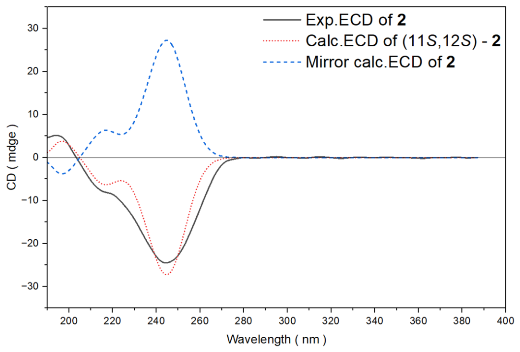
Figure 5.
Experimental ECD spectra and calculated ECD curves of compounds 3–5 and 7.
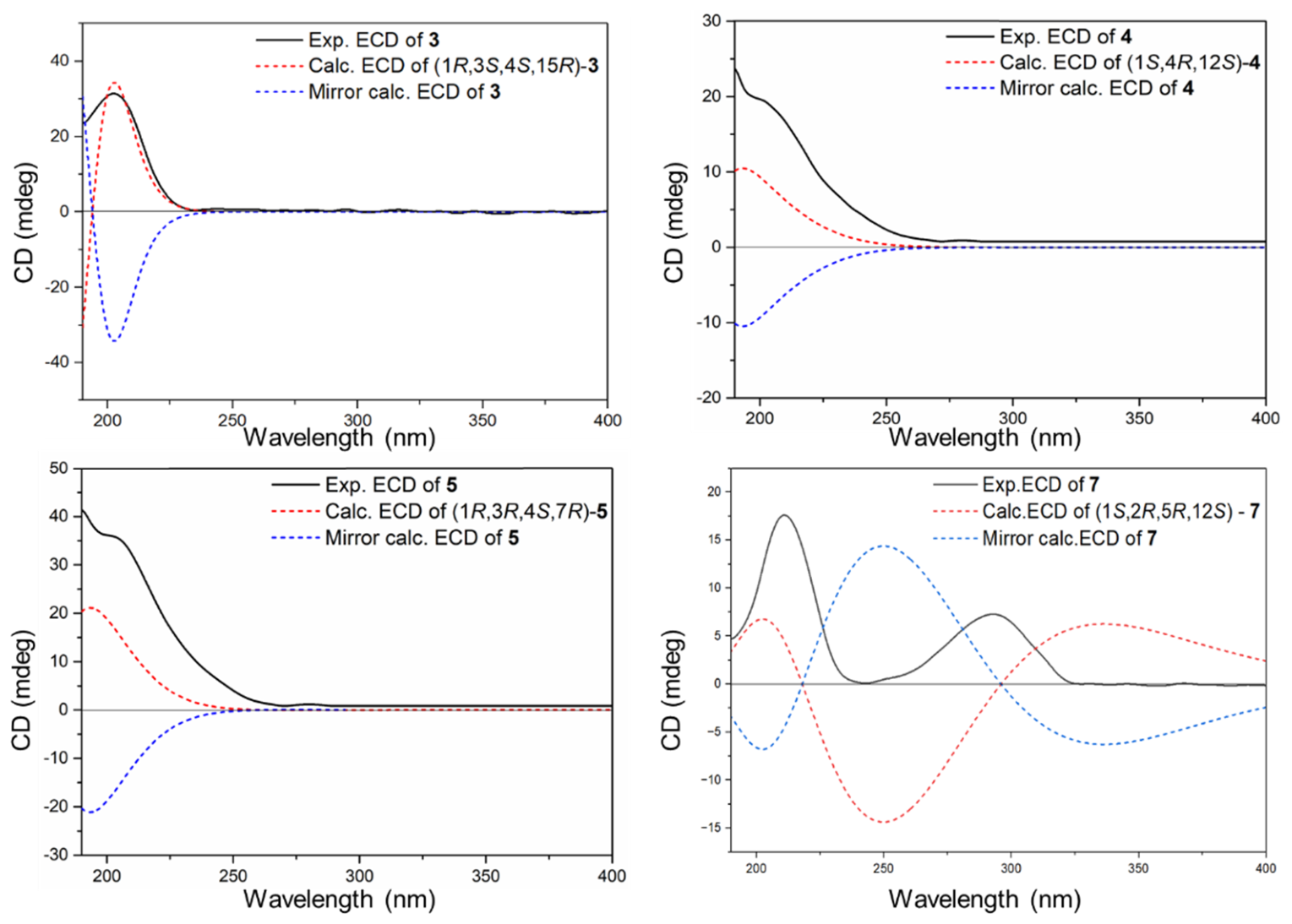

Table 1.
1H (600 MHz) NMR data of compounds 1–5 and 7 in CDCl3.a.
| No. | δH mult. (J in Hz) | |||||
|---|---|---|---|---|---|---|
| 1 | 2 | 3 | 4 | 5 | 7 | |
| 1 | / | / | 1.96 m | / | 2.78 t (6.8) | 0.61 td (9.3, 2.8) |
| 2 | 6.31 d (11.2) | 6.20 d (10.2) | 1.44 m | 5.64 d (15.8) | 1.74 m | 1.24 t (9.3) |
| / | / | 1.30 d (2.6) | / | 1.77 m | / | |
| 3 | 6.02 d (11.2) | 6.00 d (10.2) | 4.26 dd (11.5, 3.5) | 5.71 d (15.8) | 3.55 t (6.6) | 5.04 d (9.3) |
| 4 | / | / | / | / | / | / |
| 5 | 2.20 m | 2.75 td (12.6, 3.9) | 1.93 s | 1.68 m | 1.76 m | 4.05 dd (9.5, 6.1) |
| 2.20 m | 1.87 d (2.9) | 1.57 m | 1.78 m | 2.01 m | / | |
| 6 | 2.23 m | 2.30 td (9.1, 5.2) | 2.68 m | 2.14 m | 2.03 m | 2.36 s |
| 2.23 m | 2.08 m | 1.91 m | 2.15 m | 2.15 m | 2.37 d (8.3) | |
| 7 | 5.25 t (6.8) | 5.06 t (7.8) | 5.23 d (10.2) | 5.23 t (7.5) | 4.20 dd (10.1, 4.2) | 4.98 t (7.8) |
| 8 | / | / | / | / | / | / |
| 9 | 3.02 br s | 2.12 m | 2.14 m | 2.53 d (5.6) | 2.17 m | 2.89 d (13.1) |
| 2.93 d (2.9) | 1.98 m | 2.21 m | 2.53 d (5.6) | 2.37 m | 3.00 d (13.1) | |
| 10 | / | 1.89 dd (3.4, 1.4) | 1.70 dq (16.7, 7.6) | 5.52 dd (16, 6) | 2.05 m | / |
| / | 1.19 br s | 0.96 dd (16.7, 3.5) | / | 2.30 m | / | |
| 11 | 2.91 d (12.2, 4.6) | 3.56 d (11.3) | 4.96 d (7.6) | 5.51 d (15.8) | 5.30 t (7.6) | 2.64 dd (16.8, 7.0) |
| 1.97 m | / | / | / | / | 1.96 dd (16.8,6.1) | |
| 12 | 1.88 m | / | / | / | / | 2.17 m |
| 13 | 1.56 m | 2.21 dd (14.2, 9.9) | 2.13 m | 1.71 m | 1.88 m | 1.32 m |
| 1.28 m | 1.92 br s | 2.13 m | 2.08 dd (12.0, 7.6) | 2.12 m | 1.32 m | |
| 14 | 2.50 m | 2.69 m | 2.50 m | 1.93 m | 1.53 m | 1.44 m |
| 2.29 m | 2.28 m | 2.00 m | 1.99 dd (12.0, 8.1) | 1.59 m | 0.93 m | |
| 15 | / | / | / | 1.66 m | / | / |
| 16 | 5.03 s | 1.31 s | 3.64 d (14.8) | 0.88 d (6.8) | 4.77 s | 1.06 s |
| 4.95 s | / | 3.62 d (14.8) | / | 4.77 s | / | |
| 17 | 1.92 s | 1.36 s | 0.86 s | 0.88 d (6.8) | 1.70 s | 0.97 s |
| / | / | / | / | / | / | |
| 18 | 1.78 s | 1.82 s | 1.07 s | 1.26 s | 1.19 s | 1.67 s |
| 19 | 1.60 s | 1.63 s | 1.51 s | 1.61 s | 4.86 s | 1.69 s |
| / | / | / | / | 4.91 s | / | |
| 20 | 0.90 d (6.7) | 1.04 s | 1.60 s | 1.33 s | 1.60 s | 0.92 d (6.9) |
| -OMe | / | / | 3.26 s | / | / | / |
a 1H NMR (600 MHz) chemical shifts (δH) are reported in ppm referenced to CHCl3 (δH 7.26). HSQC and HMBC experiments aided signal assignments. Multiplicity abbreviations: “s” singlet, “d” doublet, “t” triplet, “m” multiplet, “q” quadruplet, and “br” broad.
Table 2.
13C (125 MHz) NMR data of compounds 1–5 and 7 in CDCl3.a.
| No. | δC, type | |||||
|---|---|---|---|---|---|---|
| 1 | 2 | 3 | 4 | 5 | 7 | |
| 1 | 143.5, C | 138.4, C | 30.9, CH | 88.1, C | 41.4, CH | 30.5, C |
| 2 | 122.7, CH | 119.9, CH | 32.0, CH2 | 130.9, CH | 36.5, CH2 | 25.3, CH |
| 3 | 123.0, CH | 121.5, CH | 73.3, CH | 136.6, CH | 73.6, CH | 125.8, CH |
| 4 | 138.4, C | 139.6, C | 79.0, C | 73.3, C | 84.9, C | 136.7, C |
| 5 | 39.9, CH2 | 31.4, CH2 | 38.4, CH2 | 43.3, CH2 | 37.1, CH2 | 79.2, CH2 |
| 6 | 25.6, CH2 | 25.0, CH2 | 22.4, CH2 | 23.1, CH2 | 31.5, CH2 | 33.0, CH2 |
| 7 | 129.2, CH | 124.2, CH | 127.8, CH | 126.1, CH | 80.3, CH | 123.8, CH |
| 8 | 130.3, C | 135.8, C | 133.4, C | 133.9, C | 151.9, C | 131.6, C |
| 9 | 54.3, CH2 | 34.9, CH2 | 39.5, CH2 | 39.5, CH2 | 34.4, CH2 | 53.3, CH2 |
| 10 | 209.8, C | 27.3, CH2 | 24.9, CH2 | 124.2, CH | 29.4, CH2 | 209.4, C |
| 11 | 47.1, CH2 | 70.1, CH | 127.7, CH | 138.9, CH | 126.3, CH | 47.7, CH2 |
| 12 | 27.7, CH2 | 76.0, C | 134.0, C | 82.8, C | 134.5, C | 27.0, CH |
| 13 | 35.9, CH2 | 30.2, CH2 | 36.8, CH2 | 35.7, CH2 | 36.5, CH2 | 36.1, CH2 |
| 14 | 24.0, CH2 | 20.1, CH2 | 25.3, CH2 | 34.2, CH2 | 29.5, CH2 | 19.3, CH2 |
| 15 | 139.2, C | 74.7, C | 80.3, C | 38.3, CH | 150.2, C | 20.6, C |
| 16 | 112.4, CH2 | 28.9, CH3 | 70.0, CH2 | 17.8, CH3 | 110.9, CH2 | 29.1, CH3 |
| 17 | 21.4, CH3 | 29.9, CH3 | 19.4, CH3 | 18.6, CH3 | 19.6, CH3 | 15.9, CH3 |
| 18 | 16.9, CH3 | 23.4, CH3 | 19.2, CH3 | 30.1, CH3 | 22.2, CH3 | 10.3, CH3 |
| 19 | 16.6, CH3 | 16.2, CH3 | 15.1, CH3 | 17.6, CH3 | 110.2, CH2 | 17.3, CH3 |
| 20 | 21.1, CH3 | 22.2, CH3 | 15.0, CH3 | 28.0, CH3 | 17.2, CH3 | 20.5, CH3 |
| 21 | 50.8, CH3 | |||||
a 13C NMR (150 MHz) chemical shifts (δC) are reported in ppm referenced to CDCl3 (δC 77.16).
Disclaimer/Publisher’s Note: The statements, opinions and data contained in all publications are solely those of the individual author(s) and contributor(s) and not of MDPI and/or the editor(s). MDPI and/or the editor(s) disclaim responsibility for any injury to people or property resulting from any ideas, methods, instructions or products referred to in the content. |
© 2026 by the authors. Licensee MDPI, Basel, Switzerland. This article is an open access article distributed under the terms and conditions of the Creative Commons Attribution (CC BY) license (http://creativecommons.org/licenses/by/4.0/).
Copyright: This open access article is published under a Creative Commons CC BY 4.0 license, which permit the free download, distribution, and reuse, provided that the author and preprint are cited in any reuse.