

Submitted:
15 August 2026
Posted:
18 August 2026
You are already at the latest version
Abstract
Urinary tract infections (UTIs) are major public health issues that are mostly caused by bacterial pathogens and less frequently by viral agents, especially in immunocompromised people, where they are more frequently linked to lower UTIs. Despite the long held belief that urine is sterile in healthy people, the urinary system is known to retain a range of viruses even in healthy populations. While bacterial pathogens continue to be the main cause of UTIs, mounting research indicates that viral infections are becoming more important. Six viruses were the subject of this narrative review: adenoviruses, CMV, West Nile virus, BK virus (human polyomavirus), Zika virus, and Severe Acute Respiratory Syndrome Coronavirus 2 (SARS-CoV-2). With the exception of West Nile virus, this has been found in urine but does not seem to cause.
Keywords:
urinary tract infection
; common viruses causing UTIs
; pathogenicity
; infectious disease management
1. Introduction
Urinary tract infections (UTIs) are common public health problems that are primarily caused by bacterial pathogens and occasionally by viruses, typically in individuals with weakened immune systems [1,2,3]. However, viruses are increasingly acknowledged as the source of lower urinary tract infections, particularly in individuals with weakened immune systems [1,3,4]. Although urine has traditionally been believed to be sterile in healthy individuals, the urinary system includes a variety of viruses in both populations [5,6,7]. Recent studies employing 16S rRNA sequencing have challenged the notion of urinary sterility by showing that a complex microbial community exists in the urine of a healthy human bladder [5,8].
These studies demonstrated that several bacterial species identified by 16S rRNA sequencing could in fact be cultured in the lab, providing strong evidence for the presence of bacterial communities in the human urinary system [9,10]. Given that both bacteria and viruses can be found in many human organs with an established microbiome, such as the skin, respiratory system, oral cavity, and gut, the existence, composition, and role of viral communities in a healthy human urinary system are far less understood [10]. Additionally, an investigation that combined an epifluorescence microscopy study with a metagenomic approach to search for viral communities in urine revealed the presence of herpes virus, human papillomavirus, polyomavirus, adenovirus, human polyomavirus (BK virus), cytomegalovirus, and herpes simplex virus type-1 and type-2 in patients without UTIs [1,2,10]. Further research revealed viruses in individuals experiencing symptoms related to the urinary tract, such as sepsis, pain when urinating, prostate pain, abdominal pain, or vaginal bleeding [2].
Immunocompromised individuals are most frequently affected by viral infections of the lower urinary tract, especially those who have received solid organ and hematopoietic stem cell transplants. The most common cause of hemorrhagic cystitis in this population is viral infections. Although there are other types of lower urinary tract infections (UTIs), including cystitis, prostatitis, seminal vesiculitis, and urethritis, viral lower tract infections most commonly attack the bladder and ureters. Urine should be sterile and free of microorganisms in healthy people, particularly men [1,4]. This is one of the main distinctions between bacterial and viral pathogens of the lower urinary tract.
Before the development of sophisticated diagnostic tools, hemorrhagic cystitis (HC) was thought to be a side effect of radiation and chemotherapy; however, it is now more commonly recognized as a viral infection [1,11,12,13]. HC is a type of virus-caused UTI characterized by necrosis of the bladder transitional epithelium and associated blood vessels, resulting in dysuria, suprapubic pain, and sudden onset of hematuria [11,12], For instance, HC occurred in 25.5% of patients in a prospective trial of more than 100 juvenile bone marrow transplant recipients, with a 95% virus-related etiology [13], Keep in mind that the diagnosis of viral infection requires the presence of related symptoms [7,14]. Hematuria linked to HC can range in severity from minor illnesses that go away on their own to potentially fatal conditions that need surgery and blood transfusions [15]. According to the DeVries classification, hematuria is classified as (i) mild when there is no change in hematocrit level, (ii) moderate when gross hematuria necessitates a transfusion of less than six units of blood, and (iii) severe when gross hematuria necessitates a transfusion of more than six units of blood. Furthermore, the Vela-Ojeda classification uses a grading system wherein (i) grade one denotes microscopic hematuria, (ii) grade two denotes macroscopic hematuria, (iii) grade three denotes hematuria with clots, and (iv) grade four denotes hematuria with clots combined with azotemia and urinary obstruction [15].
Due to its high morbidity, relative rarity, and absence of a proven standard treatment, HC is difficult to manage [16,17]. The nature of viral pathogenesis makes managing viral infections difficult. For example, viruses can only multiply inside living cells, show specificity for specific host cells, and prosper only within those cells [17,18]. The achievement of global vaccination coverage is still hampered by vaccine hesitancy, misinformation, and unequal access to healthcare, even though vaccines and antiviral treatments are available for some viral infections and there is currently no universal cure for all viral diseases [17,19].
Antiviral treatments are also essential for treating viral infections [20]. Antiviral medications have dramatically decreased morbidity and death globally, including antiretroviral therapy for HIV/AIDS, antivirals for influenza, and more recent hepatitis C medicines [21]. However, there are many obstacles to the development and marketing of antiviral drugs, including restricted availability, high costs, and virus resistance [22].
In order to stop the spread of viral diseases, public health measures are crucial. Travel restrictions, contact tracing, quarantine procedures, disease surveillance, and other measures can help contain epidemics, especially in their early stages. Additionally, health education, better cleanliness, and public awareness campaigns are essential for encouraging preventative behaviors and lowering viral transmission [17,23].
While viral urinary tract infections, also known as hemorrhagic cystitis, have gotten relatively little attention, bacterial urinary tract infections have been thoroughly investigated and analyzed. Thus, the purpose of this study is to give a narrative overview of virus-associated UTIs, emphasizing the common viral infections involved and the various management techniques.
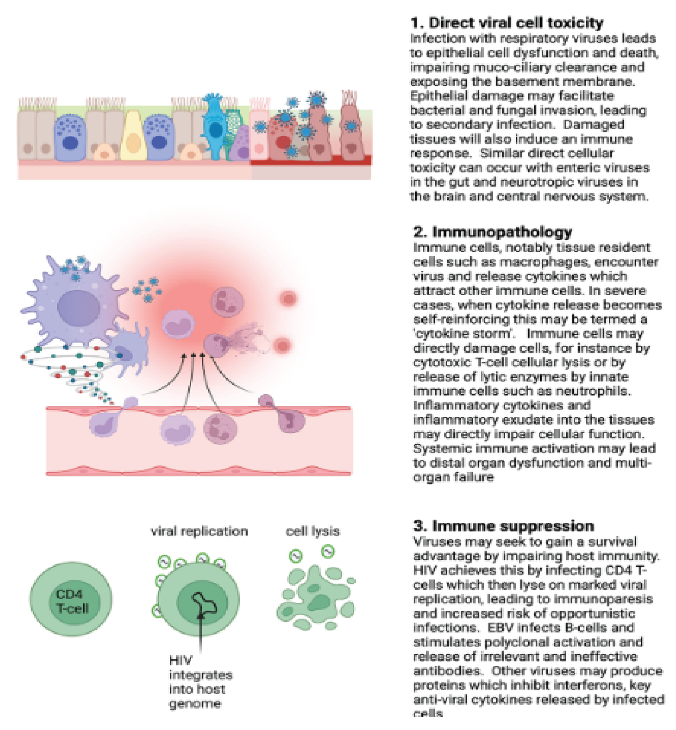
Figure 1.
General mechanisms of immune responses to viral infection, adapted from Conway Morris A and Smielewska A (2023) [24] https://doi.org/10.1111/anae.15946. Viruses may cause infection through direct cellular damage and lysis or induce host tissue injury via immune activation and immunopathology. Viral disease often arises from a combination of these mechanisms, potentially compounded by secondary infections or the consequences of secondary organ failure. An exhaustive overview of viral pathogenic mechanisms is beyond the scope of this review.
Figure 1.
General mechanisms of immune responses to viral infection, adapted from Conway Morris A and Smielewska A (2023) [24] https://doi.org/10.1111/anae.15946. Viruses may cause infection through direct cellular damage and lysis or induce host tissue injury via immune activation and immunopathology. Viral disease often arises from a combination of these mechanisms, potentially compounded by secondary infections or the consequences of secondary organ failure. An exhaustive overview of viral pathogenic mechanisms is beyond the scope of this review.
2. Selected Viruses Causing Urinary Tract Infection with Their Selective Potential Management
2.1. Severe Acute Respiratory Syndrome Coronavirus 2 (SARS_COV2)
Acute Severe Respiratory Syndrome Coronavirus 2 (SARS-CoV-2) is a spherical, enclosed, single-stranded RNA virus with a diameter of 120–160 nm that is a member of the genus Betacoronavirus, family Coronaviridae, and order Nidovirales [25,26,27,28]. Random changes in SARS-CoV-2 can result in either increased or decreased pathogenicity [29,30]. Inhaling respiratory droplets released by infected people when they cough or sneeze [31,32,33] and touching the mouth, nose, or possibly the eyes after coming into contact with virus-contaminated surfaces [30,31,32] are the main ways that the virus spreads. Furthermore, it has been determined that uninfected people may become infected by direct contact with the urine or feces of infected people [34].
While respiratory symptoms like dyspnea, coughing, and sputum production are the most common manifestation of SARS-CoV-2 infection, a number of studies have linked COVID-19 to urinary symptoms (US) [35,36], such as hematuria, frequency of urination, glucosuria, and proteinuria [37,38]. According to reports, 36.6% of COVID-19 patients had COVID-19-associated cystitis [39], with 14.7% to 22% of those patients developing symptoms of de novo overactive bladder (OAB) [39,40]. Urinary frequency and urgency were the most common symptoms, and patients with symptomatic COVID-19 had a roughly twice increased chance of developing OAB symptoms compared to asymptomatic patients [39].
One possible underlying cause of COVID-19-related urinary symptoms is cytokine cystitis. Elevated inflammatory cytokines may be released into the urine in reaction to SARS-CoV-2 infection, which can either worsen pre-existing US [41] or impact the bladder and cause de novo US [42,43]. Different studies revealed, COVID-19 infection caused de novo urine symptoms in patients who had no prior history of US [43].
Although the exact pathophysiology of SARS-CoV-2 infection is still unknown, both cell-mediated [44,45] and antibody-mediated [31] immune responses are triggered once the virus eludes the innate immune system [46]. CD4⁺ and CD8⁺ T cells are part of the cell-mediated immune response and are essential for the removal of viruses [44,45]. While interferon-gamma (IFN-γ) helps remove the virus from oligodendrocytes, perforin-mediated immune cells help remove the virus from microglia and astrocytes [47,48].
The main receptor for the SARS-CoV spike protein, angiotensin-converting enzyme 2 (ACE2), is found in around 2.4% of urothelial cells and is expressed in several organs, including the kidneys and lungs [49,50].This pattern of expression points to a possible pathway for urothelial infection through urine and capillaries. SARS-CoV-2 RNA was found in urine samples using RT-PCR in a case study involving a single COVID-19 patient, and it was demonstrated that the infectious virus isolated from the urine could successfully infect Vero cells [51]. Additionally, postmortem electron microscopy examinations of COVID-19 patients revealed viral infection of podocytes and renal tubular epithelial cells [52].
According to a Brazilian investigation, urine samples had a 5.71% (2/35) SARS-CoV-2 RNA positive rate, with virus loads ranging from 2.22 x 10² to 2.49 × 10¹ genome copies (G.C.)/mL and a median of 1.36 × 10¹ G.C./mL [53]. In a similar vein, an Italian investigation discovered that 14% of urine samples included SARS-CoV-2 RNA [54]. When combined, discoveries of direct renal infection, ultrastructural evidence from electron microscopy, and the isolation of infectious SARS-CoV-2 from urine demonstrate that SARS-CoV-2 is spread through infected urine [2].
Several antiviral medications have been tested against SARS-CoV-2 since the start of the COVID-19 pandemic. These include Paxlovid (nirmatrelvir co-administered with ritonavir), a protease inhibitor that disrupts viral polyprotein processing and replication [55], Favipiravir, which inhibits viral RNA-dependent RNA polymerase and suppresses viral replication [56]. and Remdesivir, which inhibits viral RNA-dependent RNA polymerase and thereby blocks viral replication [57]. These medications are among the most well-known antivirals that have been researched; they have been shown to be effective in shortening recovery times, especially for hospitalized patients with severe COVID-19.
2.2. Zika Virus
The Zika virus (ZIKV) is a positive-sense, single-stranded RNA virus that is a member of the genus Flavivirus and family Flaviviridae, which also includes viruses that cause dengue, West Nile, Japanese encephalitis, and yellow fever [58]. In 1947, while conducting surveys for yellow fever, the virus was initially isolated from sentinel monkeys in Uganda’s Zika Forest [59]. Humans contract ZIKV mostly via the bites of infected Aedes mosquitoes [60]. During a jaundice outbreak in eastern Nigeria in 1954, three patients were found to have the first human cases of ZIKV infection [61].
ZIKV infections can cause fever, rash, arthritis or arthralgia, conjunctivitis, myalgia, headache, retro-orbital pain, or Guillain-Barré syndrome [62] while some infections are asymptomatic [63]. Before the epidemic on the Yap Islands of Micronesia in 2007, ZIKV infections were rare. Between 2013 and 2014, they moved to the Pacific Islands [64] with relatively mild and self-limiting clinical outcomes described [64]. However, ZIKV was linked to severe congenital conditions in infants born to infected women in Brazil in 2015, including microcephaly, intrauterine growth restriction, congenital contractures, fetal death, and ocular abnormalities [64,65,66].
Numerous bodily fluids, including blood, saliva, urine, semen, vaginal secretions, rectal swabs, perspiration, cerebrospinal fluid, amniotic fluid, and breast milk, have been found to contain ZIKV RNA [67,68]. Furthermore, the majority of clinical research has shown that both adult and pediatric patients’ urine samples contain high levels of ZIKV virus loads [69]. After an infected mosquito bites a human kidney, the virus enters the bloodstream, travels to the kidney through the renal circulation, enters the glomerulus through the afferent arterioles and glomerular capillaries, and infects the renal corpuscle, including glomerular endothelial cells and other parts of the glomerular parenchyma [70].
Evidence suggests that human embryonic kidney (HEK) cells are susceptible to ZIKV infection, and it has been shown that ZIKV can reproduce in glomerular parenchymal cells of the adult human kidney. Notably, high viral loads and increased expression of the ZIKV nonstructural protein NS1 have been noted in the absence of overt cytopathic consequences, suggesting that the kidney may be a preferred site for persistent ZIKV replication [64,71]. The persistent high-level viruria seen in ZIKV-infected patients is probably caused by infection of podocytes, glomerular endothelial cells, and mesangial cells [70]. Podocyte damage, severe proteinuria, and eventually end-stage renal disease can be caused by a persistent infection of these cells [72].
The increased viral load seen in podocytes and the stimulation of the pro-inflammatory cytokine RANTES, whose expression is correlated with viral replication in glomerular cells, may be responsible for these adverse results [64,70]. After the acute phase of infection, the virus usually clears from bodily fluids in a few days to weeks; nevertheless, protracted persistence of ZIKV RNA has been documented in urine, saliva, and semen [63,67]. Reverse transcription polymerase chain reaction (RT-PCR) has been used to identify ZIKV genomic RNA, and real-time quantitative RT-PCR has been used to quantify it. Urine samples showed up to 2.53 x 10³ ZIKV RNA copies/mL in one documented instance [63].
The successful isolation of infectious ZIKV particles from patients’ urine using Vero cell cultures has demonstrated the existence of a replication-competent virus [63]. Interestingly, RT-PCR-based diagnostic tests reveal that ZIKV is more frequently detected in saliva and urine than in blood. It has been suggested that ZIKV concentrations are highest in saliva at the onset of the disease, even though viral RNA may be present in urine for a long time [73,74]. Although a number of substances with possible anti-ZIKV action have been found, there is currently no approved antiviral treatment for Zika virus (ZIKV) infection. These include: (i) emricasan, which lessens virus-induced cellular death by inhibiting caspase-3 activity [75], and (ii) bromocriptine, which inhibits the viral NS2B–NS3 protease [76], (iii) GSK126, which strongly inhibits ZIKV infection in pretreated cells [77], and (iv) sofosbuvir, a nucleoside inhibitor that binds to amino acid residues critical for ribonucleotide incorporation and interacts strongly with the ZIKV RNA-dependent RNA polymerase [78].
2.3. West Nile Virus
Humans are incidental dead-end hosts due to the low and fleeting levels of virus in their bloodstream. Serious symptoms develop in about 1% of infected individuals, with increased morbidity observed particularly in those over 50 years old, often manifesting as neurological complications. Additionally, rare modes of transmission include infection via infected donor blood, organs, breast milk, or through transplacental transmis-sio West Nile virus is an RNA virus belonging to the Flaviviridae family and Flavivirus genus [2]. It primarily infects humans through mosquito bites, with the Culex species being the predominant vector.
West Nile virus (WNV) is a positive-sense, single-stranded RNA virus that belongs to the Flaviviridae family [79,80]. WNV was first discovered in Uganda in 1937 [81], For over 60 years, the virus spread enzootically by mosquitoes throughout Africa, the Middle East, Russia, and Europe [82,83]. Lineage 2 was the most common strain during this time [84], and infections were usually subclinical or accompanied by a low fever [82]. Clinical symptoms typically start with an abrupt headache and can develop into muscle soreness, weakness, appetite loss, nausea, vomiting, and rash. Neurological symptoms such tremors resembling Parkinson’s disease have also been seen in severe cases [85,86].
Humans contract the disease mostly through mosquito bites, with Culex species acting as the main carriers. Because of their low and temporary viremia, humans are regarded as unintentional dead-end hosts. About 1% of infected people get severe illness, and those over 50 have higher morbidity, which often manifests as neurological symptoms. Blood transfusions, organ transplants, nursing, and transplacental transmission are examples of uncommon means of transmission [80,87]. About 20% of infected people experience mild, influenza-like symptoms that last two to seven days, while the majority of WNV infections are asymptomatic or subclinical. Meningitis, encephalitis, and/or acute flaccid paralysis are the hallmarks of neuroinvasive illness, which develops in less than 1% of cases [88,89].
When WNV-specific IgM antibodies are found in blood or cerebrospinal fluid, a likely diagnosis of West Nile virus (WNV) infection is made. These antibodies usually show up three days after the onset of symptoms and can last up to ninety days, possibly indicating a prior infection. IgG seroconversion is still the most accurate measure of prior exposure because immune responses differ from person to person. Viral isolation, the identification of viral antigens or nucleic acids, a fourfold or higher increase in antibody titers in paired serum samples, the presence of WNV-specific IgM along with neutralizing antibodies, or the detection of WNV-specific IgM in CSF in the absence of antibodies to other endemic arboviruses are all methods used in laboratories to confirm acute WNV infection [85,86].
Viral RNA can be found in urine even when it is no longer detectable in serum using nucleic acid amplification tests [90]. The West Nile virus has been effectively isolated and propagated in Vero E6 and BHK21 cell lines [90,91]. Urine may therefore be a more sensitive specimen for WNV detection than serum. There have been occasional reports of acute urine retention linked to infection, although urinary shedding of WNV has not been connected to structural damage of the urinary system [92].
In line with these conclusions, two out of five patients with suspected WNV infection who were admitted to Near East University Hospital in Nicosia, Northern Cyprus, in October 2023 had WNV RNA found in CSF, serum, and urine samples using an optimized multiplex RT-qPCR assay. The results were consistent with IgM and IgG serology. Interestingly, WNV RNA was found in urine more frequently than in CSF [86]. Recent research confirms that urine-based RT-qPCR offers higher sensitivity than plasma or CSF in symptomatic cases [86,90,93,94] and shows larger viral loads and extended viral shedding in urine during acute WNV infection [95].
These results confirm that the West Nile virus has not been known to induce acute damage to the urinary system [92], despite the fact that it is present in urine at greater viral levels [95] as shown by several modern diagnostic techniques. While supportive care is still the mainstay of treatment for symptomatic infection and there are presently no licensed antiviral treatments for West Nile virus (WNV) sickness, a number of medicines have shown in vitro action against WNV. These include: (i) remdesivir, which inhibits WNV replication [96], (ii) favipiravir, which appears to drive viral extinction and reduce virus-specific infectivity [97], (iii) rilpivirine, which inhibits WNV replication [98], and (iv) sofosbuvir, which also inhibits WNV replication [99].
2.4. BK Viruse/ Human Polyomavirus
BK virus (BKV) is a member of the Polyomaviridae family, which also includes other known human polyomaviruses like JC virus (JCV) and more recently identified members like KI virus (KIV), WU virus (WUV), polyomaviruses 6, 7, and 9, and Merkel cell polyomavirus (MCV). BKV and JCV share about 75% sequence homology [100]. Gardner et al. isolated the virus from a renal transplant recipient’s urine in 1971. It is a small, non-enveloped virus with a diameter of around 40 nm, an icosahedral capsid of 72 capsomers, and a single molecule of covalently closed circular double-stranded DNA [101,102]. BKV infection is more common in adults (up to 90%) than in children under five (around 50%) [103].
Fecal–oral transmission, pulmonary exposure, blood transfusion, organ transplantation, transplacental transmission, and seminal fluid are among the modes of infection [104,105]. BKV is not entirely eradicated from the host after the initial infection. Rather, it creates permanent latency, mostly in renal tubular epithelial cells, where the immune system inhibits viral multiplication [106]. Up to 20% of healthy people have asymptomatic and clinically inconsequential viruria; however, viral shedding is more common during pregnancy and in immunocompromised persons [105].
Renal tubular epithelial cells and bladder transitional epithelial cells are the main locations of BKV tenacity. BKV is dormant under normal circumstances, but in clinical contexts linked to compromised immunocompetence, viral replication may be triggered. These include the administration of immunosuppressive chemotherapy or biologic treatments, pregnancy, multiple sclerosis, solid organ transplantation, hematopoietic stem cell transplantation, and acquired immunodeficiency syndrome (AIDS) [104].
Immunohistological analysis of a renal biopsy specimen is the gold standard for diagnosing BK virus-associated nephropathy (BKVAN) [104,107]. In practical practice, however, noninvasive diagnostic techniques are more frequently employed. These include urine cytology and the microscopic identification of epithelial cells called “decoy cells,” which have a peripheral border of chromatin and distinctive intranuclear viral inclusions. The detection of viral load in plasma and urine using quantitative techniques, especially real-time quantitative polymerase chain reaction (qPCR), is the most dependable marker for patient follow-up.
This approach is a useful tool for preventing the clinical effects of BKV reactivation and for prospective surveillance of it. High levels of viruria and detectable BKV DNA in plasma (viraemia) are common in patients with active infection [108]. According to a number of studies, substantial viruria (with a suggested cut-off value of ≥10² copies/mL) may precede viraemia and the progression to BKVAN and is a risk factor for the development of nephropathy [109]. qPCR is an excellent method for tracking therapy response because of its high sensitivity and broad dynamic range. As a result, specialists in transplant programs advise at-risk patients to undergo routine BKV screening [102].
Midstream urine samples collected in sterile containers and subjected to real-time quantitative polymerase chain reaction (rt-qPCR) analysis were used in a study at the University of Alberta Hospital to assess the BK virus (BKV). Every receiver of a kidney transplant had BKV viruria. All urine samples and 60% of plasma samples proved positive for BKV DNA when BK virus-associated nephropathy (BKVAN) was diagnosed [109]. These results highlight the urinary tract as the main site of viral replication and a significant contributor to BKV-related urinary tract problems by showing that BKV is more commonly found in urine than in other bodily fluids.
Supportive treatment, immunosuppression reduction, and leflunomide have historically been used to treat BK virus (BKV) infection, with generally unsatisfactory results [110]. Cidofovir has shown efficacy with comparatively minimal toxicity when given intravenously or intravesically in recent years [111]. Since intravesical cidofovir lowers the risk of nephrotoxicity while maintaining action against BKV, CMV, and adenovirus, it may be recommended for hemorrhagic cystitis after supportive treatments [111]. Viral load reduction and hematuria resolution serve as indicators of treatment response. Even though BKV is the most common viral cause of cystitis, only a small percentage of viruria patients experience hemorrhagic cystitis, therefore routine prophylaxis is not recommended; nonetheless, early preventive treatment may lower morbidity [112]. Although the exact mechanism is yet unknown, ciprofloxacin prophylaxis has been linked to a decreased BKV viral load [112].
2.5. Adenoviruses
Adenoviruses (AdVs) are non-enveloped double-stranded DNA viruses that typically cause moderate or asymptomatic respiratory and gastrointestinal infections [113,114,115]. Their linear genomes range from 26 to 45 kb [113,114,115], with a diameter of 80–90 nm [116]. But AdVs can also cause serious illnesses such encephalitis, hepatitis, pancreatitis, hemorrhagic colitis, hemorrhagic cystitis, and nephritis [113]. Young children are more likely to experience these severe symptoms, mostly because their humoral immunity is still developing [113]. Rowe and associates first identified the virus in 1953 from tonsillar and adenoidal tissues taken from youngsters undergoing surgery. The same virus was then discovered in 1954 in individuals suffering from acute conjunctivitis and pharyngitis [116,117,118].
The same year, U.S. Army recruits with pharyngoconjunctival fever were found to have a virus known as I-67, which was later determined to be an adenovirus [116,119]. About 5–10% of all viral infections are caused by adenoviral illnesses, which are more common in the winter. There have been reports of both isolated incidents and epidemic breakouts. The most vulnerable groups are military personnel and children between the ages of six months and five years. During the first two to three months of close contact, a particularly high prevalence is seen in newly established groups of adults and children [113,116].
Nasopharyngeal secretions, sputum, conjunctival discharge, feces, and urine are the main sources of infection in immunocompromised people with acute illness symptoms, patients in the convalescent stage, or asymptomatic virus carriers. Adenoviral infections are mostly spread by airborne droplets, conjunctival inoculation, and possibly the feco-oral route, suggesting that they affect not only the respiratory system but also several other organs, including the urinary tract [113,116].
Human adenoviruses (HAdVs) have been linked to urethritis in a number of investigations [120,121,122], with HAdVs found in the urine of afflicted individuals. HAdV-B species type 11 and HAdV-D species types 8, 19, 37, 56, and 64 have been identified [120,123,124,125]. Since these viruses were first discovered, it has been known that HAdVs are involved in the urinary system, with special attention paid to HAdV-B type 11 (HAdV-B11) [126] and HAdV-D type 37 (HAdV-D37) [127]. Despite these findings, little is known about the clinical characteristics and underlying mechanisms of HAdV-associated urinary tract disease. HAdV-B11 has drawn a lot of interest as an opportunistic pathogen in immunocompromised people, especially patients of kidney and other solid-organ transplants, and is most frequently linked to hemorrhagic cystitis [125,128,129].
Additionally, utilizing real-time PCR as the diagnostic technique, a study carried out at the Melbourne Sexual Health Centre between January 2006 and April 2014 revealed that adenovirus was found in 93% of urethritis patients [120]. Adenovirus type 11 was isolated from the urine of 11 out of 25 patients in another study that assessed the viral etiologies of acute hemorrhagic cystitis using virological and serological analyses of unstored urine samples collected between 1967 and 1972; in total, viruses were isolated from 11 out of 12 subjects examined [126]. Adenovirus type 19-associated ocular infections were first reported in Perth, Western Australia, in 1977, despite the fact that eye swabs had been regularly examined in cell cultures for adenovirus type 19 isolations since 1968. Adenovirus type 19 was also recovered from urethral and cervical tissues from male and female patients visiting a Perth sexually transmitted illness clinic during and after this outbreak [130]. When taken as a whole, these results show that adenoviruses of various species can be important causes of UTIs and may be linked to a variety of clinical consequences.
Nevertheless, no antiviral medication is both extremely effective and toxic-free. Despite worries regarding nephrotoxicity[131], cidofovir is increasingly being utilized as a first-line treatment for adenoviral cystitis; however, lower-dose regimens used in recipients of renal transplants may be less effective against co-infections [132]. Compared to youngsters, recipients of HLA-matched bone marrow transplants have better results from ribavirin’s limited action against adenovirus [132]. Although it has been used to treat hemorrhagic cystitis, ganciclovir is mostly used for CMV prevention [133]. Although it is less successful in cases of disseminated adenoviral infection, vidarabine is an alternate treatment for hemorrhagic cystitis, probably as a result of high urinary drug concentrations [134].
2.6. Cytomegaloviruses
The archetypal member of the Betaherpesvirinae subfamily is human cytomegalovirus (HCMV), commonly referred to as human herpesvirus 5 [135]. Ribbert originally described it in 1881, and Goodpasture and Talbert suggested in 1921 that a viral agent was the cause of the disorder known as “cytomegalia.” Later, in 1950, Smith and Vellios proved that HCMV infection may happen in utero [136]. HCMV causes a lifetime of latency in the host after the initial infection [135]. Since efficient immune regulation restricts viral replication and avoids the large viral loads linked to end-organ illness, infection is typically asymptomatic in immunocompetent people. However, mounting evidence indicates that persistent immunological activation and chronic cell-mediated inflammation often referred to as indirect effects may contribute to long-term negative outcomes from chronic HCMV [137].
On the other hand, immunocompromised populations frequently have symptomatic CMV infection, which is linked to a wide range of clinical symptoms. While urinary tract involvement is uncommon and has mostly been described in recipients of hematopoietic stem cell transplants, intestinal, ophthalmic, and neurological problems are widely documented in AIDS patients [138]. It is extremely rare for HIV/AIDS individuals without a history of transplantation to develop CMV hemorrhagic cystitis. There have only been four identical occurrences reported globally, and a recent example from South Africa is the first known occurrence in this clinical environment. In every case that has been documented, a bladder biopsy was used to diagnose individuals who had bladder discomfort or haematuria [139].
In wealthy nations, the prevalence of CMV infection is believed to be 40–60% among adults, but in underdeveloped nations, it often surpasses 90% [140]. The detection and measurement of viral DNA in blood is the main method used in clinical practice to diagnose and track CMV infection. This method is frequently augmented by the identification of viral DNA or antigens in other biological fluids or tissue biopsies [140]. Urine has traditionally been one of the primary materials for viral isolation, and the viral shedding of CMV in urine is well known [141].
The primary cellular locations of viral replication are still unclear, despite the urogenital tract’s obvious involvement in CMV infection. The kidney is an important location of viral replication, as evidenced by in vitro studies showing that CMV may replicate in a variety of primary human renal cells of glomerular, tubular, and vascular origin [142]. Fibroblasts and macrophages are two further types of renal cells that are susceptible to CMV infection and may aid in the excretion of the virus in urine [140]. Local viral immunosuppressive effects and the compartmentalization of particular viral variants within different cells or tissues of the urinary tract may promote continued viral replication, even though the role of systemic immunosuppression in the development or persistence of CMV viruria cannot be ruled out [140].
Furthermore, renal illness linked to CMV has been reported in kidney transplant recipients. Renal biopsies may reveal CMV nephropathy, even when receivers who are CMV-negative receive kidneys from donors who are CMV-positive. On further biopsies, these cases have shown CMV glomerulopathy and CMV-associated interstitial nephritis; histological findings include CMV-positive endothelial cells in glomerular capillaries and CMV-infected monocytes in capillary lumens [143].
Most patients get ganciclovir prophylaxis following solid organ donation; however, foscarnet may be administered in cases of active disease [1,107]. When BK virus (BKV) co-infection is present, cidofovir can be utilized [1,144]. Therefore, cidofovir may be the recommended drug for patients who experience hemorrhagic cystitis following solid organ or hematopoietic stem cell transplantation [1].
3. Prevention and Control Strategies of Viral Infections
Effective prevention and control methods play a major role in shaping the transmission and impact of infectious illnesses in human populations, even if host genetics modulates host-pathogen interactions. Vaccination; environmental sanitation; vector control; population growth management and urban planning; sex education and promotion of safe sexual practices; testing, diagnosis, and treatment; promotion of hygienic practices; food security and adequate nutrition; reduction of contact with livestock and wildlife; reduction of social inequality; and surveillance of infectious diseases are some of the key tactics. Additionally, the prevention and management of infectious diseases brought on by viruses, bacteria, parasites, and other pathogens depend heavily on the preservation of biodiversity, the mitigation of climate change, and the creation of novel antimicrobial medications [145].
The review concludes that urinary tract infections (UTIs) continue to be a significant global public health issue, especially among immunocompromised people, despite the limited scope of this narrative review, which was restricted to identifying viral evidence for the occurrence of UTIs rather than their management. Even though bacteria are the primary cause of UTIs, new research suggests that viral infections are also playing a bigger role. Six viruses were the subject of this narrative review: adenoviruses, CMV, West Nile virus, BK virus (human polyomavirus), Zika virus, and Severe Acute Respiratory Syndrome Coronavirus 2 (SARS-CoV-2). With the exception of West Nile virus, which has been discovered in urine but does not seem to seriously harm the urinary tract, all of these viruses were found to be capable of causing serious damage to the urinary tract. Thus, infection rates and related problems can be greatly decreased by implementing treatment, prevention, and control techniques appropriately.
References
- Paduch DA. Viral lower urinary tract infections. Curr Urol Rep. 2007;8:324–35.
- Park S, Kim ET, Huh J-S. Virus in the Urine of Healthy People and Patients with Infectious Diseases. Urogenit Tract Infect. 2021;16:44–8.
- Agrati C, Bartolini B, Bordoni V, Locatelli F, Capobianchi MR, Di Caro A, et al. Emerging viral infections in immunocompromised patients: A great challenge to better define the role of immune response. Front Immunol. 2023;14 March:1–7.
- Singh D, Kiberd B, Gupta R, Alkhudair W, Lawen J. Polyoma virus-induced hemorrhagic cystitis in renal transplantation patient with polyoma virus nephropathy. Urology. 2006;67:423.e11-423.e12.
- Kogan MI, Naboka YL, Ibishev KS, Gudima IA, Naber KG. Human urine is not sterile - shift of paradigm. Urol Int. 2015;94:445–52.
- Morand A, Cornu F, Dufour J-C, Tsimaratos M, Lagier J-C, Raoult D. Human Bacterial Repertoire of the Urinary Tract: a Potential Paradigm Shift. J Clin Microbiol. 2019;57.
- Louten J. Detection and Diagnosis of Viral Infections. Essential Human Virology. 2016;:111–32.
- Brubaker L, Nager CW, Richter HE, Visco A, Nygaard I, Barber MD, et al. Urinary bacteria in adult women with urgency urinary incontinence. Int Urogynecol J. 2014;25:1179–84.
- Pichler M, Ortega AS, Conci N, Vargas S, Orsi WD. Running title : MiniSeq 16S rRNA gene sequencing. 2017;:1–17.
- Santiago-Rodriguez TM, Ly M, Bonilla N, Pride DT. The human urine virome in association with urinary tract infections. Front Microbiol. 2015;6:14.
- deVries CR, Freiha FS. Hemorrhagic cystitis: a review. J Urol. 1990;143:1–9.
- Haldar S, Dru C, Bhowmick NA. Mechanisms of hemorrhagic cystitis. Am J Clin Exp Urol. 2014;2:199–208.
- Gorczynska E, Turkiewicz D, Rybka K, Toporski J, Kalwak K, Dyla A, et al. Incidence, clinical outcome, and management of virus-induced hemorrhagic cystitis in children and adolescents after allogeneic hematopoietic cell transplantation. Biol blood marrow Transplant J Am Soc Blood Marrow Transplant. 2005;11:797–804.
- Luciani LG, Mattevi D, Hospital SC. Longitudinal Multi-omics Analyses Identify Responses of Megakaryocytes, Erythroid Cells, and Plasmablastsas Hallmarksof Severe COVID-19. 2020; January.
- Amico MJD, Foss H, Uhr A, Rudnick B, Kloniecke E, Gomella LG, et al. Literature and Treatment Options. 2022;:11276–83.
- Decker DB, Karam JA, Wilcox DT. Pediatric hemorrhagic cystitis. J Pediatr Urol. 2009;5:254–64.
- Peter J. The Global Challenge of Viral Infections: A Growing Threat to Public Health Corresponding Author*. Int J Collab Res Intern Med Public Heal. 2024;2024:1–002.
- Thomas J. Viral Infections : Medical Challenges and Advances in Treatment and Prevention Public health. 2024;16:1–2.
- Andrei G. Vaccines and Antivirals: Grand Challenges and Great Opportunities. Front Virol. 2021;1 May:1–8.
- Millan J, Sciences M. Antiviral Therapy : Advancements and challenges in the fight against viral infections . 2025;9:10–1.
- Gandhi RT, Landovitz RJ, Sax PE, Smith DM, Springer SA, Günthard HF, et al. Antiretroviral Drugs for Treatment and Prevention of HIV in Adults: 2024 Recommendations of the International Antiviral Society–USA Panel. JAMA. 2025;333:609–28.
- Rana D, Prajapati A, Karunakaran B, Vora L, Benival D, Jindal AB, et al. Recent Advances in Antiviral Drug Delivery Strategies. AAPS PharmSciTech. 2025;26:1–28.
- Ayouni I, Maatoug J, Dhouib W, Zammit N, Fredj S Ben, Ghammam R, et al. Effective public health measures to mitigate the spread of COVID-19: a systematic review. BMC Public Health. 2021;21:1015.
- Conway Morris A, Smielewska A. Viral infections in critical care: a narrative review. Anaesthesia. 2023;78:626–35.
- Frank MG, Fleshner M, Maier SF. Exploring the immunogenic properties of SARS-CoV-2 structural proteins: PAMP:TLR signaling in the mediation of the neuroinflammatory and neurologic sequelae of COVID-19. Brain Behav Immun. 2023;111:259–69.
- Cavanagh D. Nidovirales: a new order comprising Coronaviridae and Arteriviridae. Arch Virol. 1997;142:629—633.
- Vijgen L, Keyaerts E, Moës E, Thoelen I, Wollants E, Lemey P, et al. Complete genomic sequence of human coronavirus OC43: molecular clock analysis suggests a relatively recent zoonotic coronavirus transmission event. J Virol. 2005;79:1595–604.
- Ouassou H, Kharchoufa L, Bouhrim M, Daoudi NE, Imtara H, Bencheikh N, et al. The Pathogenesis of Coronavirus Disease 2019 (COVID-19): Evaluation and Prevention. J Immunol Res. 2020;2020:1357983.
- Theofilou P, Vlastos DD. Assessing the impact of the COVID-19 pandemic on the quality of life of cancer patients: A systematic review. Glob Heal Econ Sustain. 2024;2:2636.
- National Institutes of Health. Treatment Guidelines Panel. Coronavirus Disease 2019 (COVID-19). NIH. 2021;2019:1–243.
- Sarda SR, Tekale SU, Kótai L, Domb AJ, Pawar RP. COVID-19: A global pandemic. Eur Chem Bull. 2020;9:266–72.
- Zhejiang University School of Medicine. Handbook for COVID-19 treatment and Prevention. 2021.
- M. Kolawole O, I. Aun I, I. Ogah J, F. Folahan F, F. Kolawole C. Determinants of COVID-19 vaccine uptake in Kwara State, Nigeria: The role of public health management professionals in driving behavioral change. Glob Heal Econ Sustain. 2024;2:2462.
- Jones DL, Baluja MQ, Graham DW, Corbishley A, McDonald JE, Malham SK, et al. Shedding of SARS-CoV-2 in feces and urine and its potential role in person-to-person transmission and the environment-based spread of COVID-19. Sci Total Environ. 2020;749:141364.
- Khateeb J, Li Y, Zhang H. Emerging SARS-CoV-2 variants of concern and potential intervention approaches. Crit Care. 2021;25:1–8.
- Harvey WT, Carabelli AM, Jackson B, Gupta RK, Thomson EC, Harrison EM, et al. SARS-CoV-2 variants, spike mutations and immune escape. Nat Rev Microbiol. 2021;19:409–24.
- Liu L, Zuo K, Le W, Lu M, Liu Z, Xu W. Non-diabetic urine glucose in idiopathic membranous nephropathy. Ren Fail. 2022;44:1105–12.
- Mumm J-N, Osterman A, Ruzicka M, Stihl C, Vilsmaier T, Munker D, et al. Urinary Frequency as a Possibly Overlooked Symptom in COVID-19 Patients: Does SARS-CoV-2 Cause Viral Cystitis? Eur Urol. 2020;78:624–8.
- Hoang Roberts L, Zwaans BMM, Peters KM, Chancellor M, Padmanabhan P. Incidence of New or Worsening Overactive Bladder Among Patients with a Prior SARS-CoV-2 Infection: A Cohort Study. Eur Urol Open Sci. 2022;46:68–74.
- Moradi A, Rahimian N, Shekofteh Z, Shakiba B, Soleimani A, Maghsoudi R, et al. Lower Urinary Tract Symptoms in Hospitalized Patients with COVID-19. J Iran Med Counc. 2022;4:291–5.
- Lamb LE, Timar R, Wills M, Dhar S, Lucas SM, Komnenov D, et al. Long COVID and COVID-19-associated cystitis (CAC). Int Urol Nephrol. 2022;54:17–21.
- Lamb LE, Dhar N, Timar R, Wills M, Dhar S, Chancellor MB. COVID-19 inflammation results in urine cytokine elevation and causes COVID-19 associated cystitis (CAC). Med Hypotheses. 2020;145:110375.
- Tayebi S, Rahnama’i MS, Shafigh A, Salehi-Pourmehr H, Mostafaei H, Sepehran E, et al. Lower Urinary Tract Symptoms (LUTS) and COVID-19. Cont Reports. 2024;9:100044.
- Rijkers GT, Weterings N, Obregon-Henao A, Lepolder M, Dutt TS, van Overveld FJ, et al. Antigen Presentation of mRNA-Based and Virus-Vectored SARS-CoV-2 Vaccines. Vaccines. 2021;9.
- Burrell CJ, Howard CR, Murphy FA. Pathogenesis of Virus Infections. Fenner and White’s Medical Virology. 2017;:77–104.
- Iwasaki A, Medzhitov R. Control of adaptive immunity by the innate immune system. Nat Immunol. 2015;16:343–53.
- Savarin C, Bergmann CC. Fine Tuning the Cytokine Storm by IFN and IL-10 Following Neurotropic Coronavirus Encephalomyelitis. Front Immunol. 2018;9:3022.
- Gadotti AC, de Castro Deus M, Telles JP, Wind R, Goes M, Garcia Charello Ossoski R, et al. IFN-γ is an independent risk factor associated with mortality in patients with moderate and severe COVID-19 infection. Virus Res. 2020;289:198171.
- Zou X, Chen K, Zou J, Han P, Hao J, Han Z. Single-cell RNA-seq data analysis on the receptor ACE2 expression reveals the potential risk of different human organs vulnerable to 2019-nCoV infection. Front Med. 2020;14:185–92.
- Khazaal S, Harb J, Rima M, Annweiler C, Wu Y, Cao Z, et al. The Pathophysiology of Long COVID throughout the Renin-Angiotensin System. Molecules. 2022;27:1–21.
- Sun J, Zhu A, Li H, Zheng K, Zhuang Z, Chen Z, et al. Isolation of infectious SARS-CoV-2 from urine of a COVID-19 patient. Emerging microbes & infections. 2020;9:991–3.
- Farkash EA, Wilson AM, Jentzen JM. Ultrastructural Evidence for Direct Renal Infection with SARS-CoV-2. J Am Soc Nephrol. 2020;31:1683–7.
- Anjos D, Fiaccadori FS, do Prado Servian C, da Fonseca SG, Guilarde AO, Borges MASB, et al. SARS-CoV-2 loads in urine, sera and stool specimens in association with clinical features of COVID-19 patients. J Clin Virol Plus. 2022;2:100059.
- Perrella A, Brita M, Coletta F, Cotena S, De Marco G, Longobardi A, et al. Sars-cov-2 in urine may predict a severe evolution of covid-19. J Clin Med. 2021;10.
- Marzi M, Vakil MK, Bahmanyar M, Zarenezhad E. Paxlovid: Mechanism of Action, Synthesis, and In Silico Study. Biomed Res Int. 2022;2022:7341493.
- Pilkington V, Pepperrell T, Hill A. A review of the safety of favipiravir - a potential treatment in the COVID-19 pandemic? Journal of virus eradication. 2020;6:45–51.
- Jorgensen SCJ, Kebriaei R, Dresser LD. Remdesivir: Review of Pharmacology, Pre-clinical Data, and Emerging Clinical Experience for COVID-19. Pharmacotherapy. 2020;40:659–71.
- Hayes EB. Zika virus outside Africa. Emerg Infect Dis. 2009;15:1347–50.
- DICK GWA, KITCHEN SF, HADDOW AJ. Zika virus. I. Isolations and serological specificity. Trans R Soc Trop Med Hyg. 1952;46:509–20.
- Tetro JA. Zika and microcephaly: causation, correlation, or coincidence? Microbes and infection. 2016;18:167–8.
- MACNAMARA FN. Zika virus: a report on three cases of human infection during an epidemic of jaundice in Nigeria. Trans R Soc Trop Med Hyg. 1954;48:139–45.
- Rasmussen SA, Jamieson DJ, Honein MA, Petersen LR. Zika Virus and Birth Defects--Reviewing the Evidence for Causality. N Engl J Med. 2016;374:1981–7.
- Bonaldo MC, Ribeiro IP, Lima NS, dos Santos AAC, Menezes LSR, da Cruz SOD, et al. Isolation of Infective Zika Virus from Urine and Saliva of Patients in Brazil. PLoS Negl Trop Dis. 2016;10:1–17.
- Alcendor DJ. Zika virus infection and implications for kidney disease. J Mol Med (Berl). 2018;96:1145–51.
- Marrs C, Olson G, Saade G, Hankins G, Wen T, Patel J, et al. Zika Virus and Pregnancy: A Review of the Literature and Clinical Considerations. Am J Perinatol. 2016;33:625–39.
- McGrogan A, Madle GC, Seaman HE, de Vries CS. The epidemiology of Guillain-Barré syndrome worldwide. A systematic literature review. Neuroepidemiology. 2009;32:150–63.
- Calvet GA, Kara EO, Bôtto-Menezes CHA, da Costa Castilho M, de Oliveira Franca RF, Habib N, et al. Detection and persistence of Zika virus in body fluids and associated factors: a prospective cohort study. Sci Rep. 2023;13:1–18.
- Liu T, Tang L, Tang H, Pu J, Gong S, Fang D, et al. Zika Virus Infection Induces Acute Kidney Injury Through Activating NLRP3 Inflammasome Via Suppressing Bcl-2. Front Immunol. 2019;10:1925.
- Regadas VC, Silva M de CE, Abud LG, Labadessa LMPL, Oliveira RGG de, Miyake CH, et al. Microcephaly caused by congenital Zika virus infection and viral detection in maternal urine during pregnancy. Rev Assoc Med Bras. 2018;64:11–4.
- Alcendor DJ. Zika Virus Infection of the Human Glomerular Cells: Implications for Viral Reservoirs and Renal Pathogenesis. J Infect Dis. 2017;216:162–71.
- Chan JF-W, Yip CC-Y, Tsang JO-L, Tee K-M, Cai J-P, Chik KK-H, et al. Differential cell line susceptibility to the emerging Zika virus: implications for disease pathogenesis, non-vector-borne human transmission and animal reservoirs. Emerg Microbes Infect. 2016;5:e93.
- Nagata M. Podocyte injury and its consequences. Kidney Int. 2016;89:1221–30.
- Musso D, Roche C, Nhan T-X, Robin E, Teissier A, Cao-Lormeau V-M. Detection of Zika virus in saliva. J Clin Virol Off Publ Pan Am Soc Clin Virol. 2015;68:53–5.
- Gourinat A-C, O’Connor O, Calvez E, Goarant C, Dupont-Rouzeyrol M. Detection of Zika virus in urine. Emerg Infect Dis. 2015;21:84–6.
- Xu M, Lee EM, Wen Z, Cheng Y, Huang W-K, Qian X, et al. Identification of small-molecule inhibitors of Zika virus infection and induced neural cell death via a drug repurposing screen. Nat Med. 2016;22:1101–7.
- Chan JF-W, Chik KK-H, Yuan S, Yip CC-Y, Zhu Z, Tee K-M, et al. Novel antiviral activity and mechanism of bromocriptine as a Zika virus NS2B-NS3 protease inhibitor. Antiviral Res. 2017;141:29–37.
- Arbuckle JH, Gardina PJ, Gordon DN, Hickman HD, Yewdell JW, Pierson TC, et al. Inhibitors of the Histone Methyltransferases EZH2/1 Induce a Potent Antiviral State and Suppress Infection by Diverse Viral Pathogens. MBio. 2017;8.
- Sacramento CQ, Melo GR De, Freitas CS De, Marttorelli A, Ferreira AC, Barbosa-lima G, et al. The clinically approved antiviral drug sofosbuvir inhibits Zika virus replication. Nat Publ Gr. 2017; January:1–12.
- Mukhopadhyay S, Kuhn RJ, Rossmann MG. A structural perspective of the flavivirus life cycle. Nat Rev Microbiol. 2005;3:13–22.
- Pal M, Abdeta T. A Systematic Review on Epidemiology, Pathogenesis, Clinical Spectrum, Diagnostic Techniques and Control Strategies of West Nile Fever. Int J Med Front. 2024;7:43–9.
- Smithburn KC, Hughes TP, Burke AW, Paul JH. A Neurotropic Virus Isolated from the Blood of a Native of Uganda. 1940;20:471–2.
- HURLBUT HS, RIZK F, TAYLOR RM, WORK TH. A study of the ecology of West Nile virus in Egypt. Am J Trop Med Hyg. 1956;5:579–620.
- Ronca SE, Ruff JC, Murray KO. A 20-year historical review of West Nile virus since its initial emergence in North America: Has West Nile virus become a neglected tropical disease? PLoS Negl Trop Dis. 2021;15:e0009190.
- May FJ, Davis CT, Tesh RB, Barrett ADT. Phylogeography of West Nile virus: from the cradle of evolution in Africa to Eurasia, Australia, and the Americas. J Virol. 2011;85:2964–74.
- Colpitts TM, Conway MJ, Montgomery RR, Fikrig E. West Nile Virus: biology, transmission, and human infection. Clin Microbiol Rev. 2012;25:635–48.
- Tuncel G, Akan G, Kalaycı M, Baddal B, Bostancı A, Suer K, et al. Efficient Detection of West Nile Virus in Urine Specimens by a Novel In-House RT-qPCR Detection Kit. Can J Infect Dis Med Microbiol = J Can des Mal Infect la Microbiol medicale. 2025;2025:6513971.
- Petersen LR, Brault AC, Nasci RS. West Nile virus: review of the literature. JAMA. 2013;310:308–15.
- Gorchakov R, Gulas-Wroblewski BE, Ronca SE, Ruff JC, Nolan MS, Berry RM, et al. Optimizing PCR Detection of West Nile Virus from Body Fluid Specimens to Delineate Natural History in an Infected Human Cohort. Int J Mol Sci. 2019;20.
- Campbell GL, Marfin AA, Lanciotti RS, Gubler DJ. West Nile virus. Lancet Infect Dis. 2002;2:519–29.
- Barzon L, Pacenti M, Franchin E, Pagni S, Martello T, Cattai M, et al. Excretion of West Nile Virus in Urine During Acute Infection. J Infect Dis. 2013;208:1086–92.
- Barzon L, Pacenti M, Franchin E, Squarzon L, Sinigaglia A, Ulbert S, et al. Isolation of West Nile virus from urine samples of patients with acute infection. J Clin Microbiol. 2014;52:3411–3.
- Shpall AI, Varpetian A, Ginsberg DA. Urinary retention in a patient with West Nile virus. Urology. 2003;61:1259.
- Gdoura M, Fares W, Bougatef S, Inoubli A, Touzi H, Hogga N, et al. The value of West Nile virus RNA detection by real-time RT-PCR in urine samples from patients with neuroinvasive forms. Arch Microbiol. 2022;204:238.
- Cvjetković IH, Radovanov J, Kovačević G, Turkulov V, Patić A. Diagnostic value of urine qRT-PCR for the diagnosis of West Nile virus neuroinvasive disease. Diagn Microbiol Infect Dis. 2023;107:115920.
- Tonry JH, Brown CB, Cropp CB, Co JKG, Bennett SN, Nerurkar VR, et al. West Nile virus detection in urine. Emerg Infect Dis. 2005;11:1294–6.
- Konkolova E, Dejmek M, Hřebabecký H, Šála M, Böserle J, Nencka R, et al. Remdesivir triphosphate can efficiently inhibit the RNA-dependent RNA polymerase from various flaviviruses. Antiviral Res. 2020;182:104899.
- Escribano-Romero E, Jiménez de Oya N, Domingo E, Saiz JC. Extinction of West Nile Virus by Favipiravir through Lethal Mutagenesis. Antimicrob Agents Chemother. 2017;61.
- García-Zarandieta M, Quesada E, Martínez-Jiménez MI, Newnes C V, Fernández-Cabello V, Sáez-Álvarez Y, et al. Identification of West Nile virus RNA-dependent RNA polymerase non-nucleoside inhibitors by real-time high throughput fluorescence screening. Antiviral Res. 2023;212:105568.
- Dragoni F, Boccuto A, Picarazzi F, Giannini A, Giammarino F, Saladini F, et al. Evaluation of sofosbuvir activity and resistance profile against West Nile virus in vitro. Antiviral Res. 2020;175:104708.
- Frisque RJ, Bream GL, Cannella MT. Human polyomavirus JC virus genome. J Virol. 1984;51:458–69.
- Gardner SD, Field AM, Coleman D V, Hulme B. New human papovavirus (B.K.) isolated from urine after renal transplantation. Lancet (London, England). 1971;1:1253–7.
- Stec A, Polz D, Polz-Dacewicz M. BK-virus (BKV) – structure, epidemiology and pathogenesis. J Pre-Clinical Clin Res. 2014;7:90–2.
- Rogers DL, McClure GB, Ruiz JC, Abee CR, Vanchiere JA. Endemic Viruses of Squirrel Monkeys (Saimiri spp.). Comp Med. 2015;65:232–40.
- Krajewski W, Kamińska D, Poterek A, Małkiewicz B, Kłak J, Zdrojowy R, et al. Pathogenicity of BK virus on the urinary system. Cent Eur J Urol. 2020;73:94–103.
- Chong S, Antoni M, Macdonald A, Reeves M, Harber M, Magee CN. BK virus: Current understanding of pathogenicity and clinical disease in transplantation. Rev Med Virol. 2019;29:e2044.
- Shinohara T, Matsuda M, Cheng SH, Marshall J, Fujita M, Nagashima K. BK virus infection of the human urinary tract. J Med Virol. 1993;41:301–5.
- Drachenberg CB, Papadimitriou JC, Ramos E. Histologic versus molecular diagnosis of BK polyomavirus-associated nephropathy: a shifting paradigm? Clin J Am Soc Nephrol. 2006;1:374–9.
- William Tong CY, Hilton R, MacMahon EME, Brown L, Pantelidis P, Chrystie IL, et al. Monitoring the progress of BK virus associated nephropathy in renal transplant recipients. Nephrol Dial Transplant. 2004;19:2598–605.
- Pang XL, Doucette K, LeBlanc B, Cockfield SM, Preiksaitis JK. Monitoring of polyomavirus BK virus viruria and viremia in renal allograft recipients by use of a quantitative real-time PCR assay: one-year prospective study. J Clin Microbiol. 2007;45:3568–73.
- Chang CYM, Gangji A, Chorneyko K, Kapoor A. Urological manifestations of BK polyomavirus in renal transplant recipients. Can J Urol. 2005;12:2829–36.
- Bridges B, Donegan S, Badros A. Cidofovir bladder instillation for the treatment of BK hemorrhagic cystitis after allogeneic stem cell transplantation. Am J Hematol. 2006;81:535–7.
- Leung AYH, Chan MTL, Yuen K-Y, Cheng VCC, Chan K-H, Wong CLP, et al. Ciprofloxacin Decreased Polyoma BK Virus Load in Patients Who Underwent Allogeneic Hematopoietic Stem Cell Transplantation. Clin Infect Dis. 2005;40:528–37.
- Lynch JP 3rd, Kajon AE. Adenovirus: Epidemiology, Global Spread of Novel Serotypes, and Advances in Treatment and Prevention. Semin Respir Crit Care Med. 2016;37:586–602.
- Davison AJ, Benkő M, Harrach B. Genetic content and evolution of adenoviruses. J Gen Virol. 2003;84 Pt 11:2895–908.
- Robinson CM, Singh G, Lee JY, Dehghan S, Rajaiya J, Liu EB, et al. Molecular evolution of human adenoviruses. Sci Rep. 2013;3:1812.
- Desheva Y. Introductory Chapter: Human Adenoviruses. 2018.
- Huebner RJ, Rowe WP, Ward TG, Parrott RH, Bell JA. Adenoidal-Pharyngeal-Conjunctival Agents. N Engl J Med. 1954;251:1077–86.
- Effect C, Viruses P. Poliomyelitis viruses cytopathogenic effect. 1953.
- HILLEMAN MR, WERNER JH. Recovery of new agent from patients with acute respiratory illness. Proc Soc Exp Biol Med Soc Exp Biol Med (New York, NY). 1954;85:183–8.
- Samaraweera GR, Garcia K, Druce J, Williams H, Bradshaw CS, Fairley CK, et al. Characteristics of adenovirus urethritis among heterosexual men and men who have sex with men: a review of clinical cases. Sex Transm Infect. 2016;92:172–4.
- Frølund M, Lidbrink P, Wikström A, Cowan S, Ahrens P, Jensen JS. Urethritis-associated Pathogens in Urine from Men with Non-gonococcal Urethritis: A Case-control Study. Acta Derm Venereol. 2016;96:689–94.
- Ito S, Hanaoka N, Shimuta K, Seike K, Tsuchiya T, Yasuda M, et al. Male non-gonococcal urethritis: From microbiological etiologies to demographic and clinical features. Int J Urol Off J Japanese Urol Assoc. 2016;23:325–31.
- Numazaki Y, Kumasaka T, Yano N, Yamanaka M, Miyazawa T, Takai S, et al. Further study on acute hemorrhagic cystitis due to adenovirus type 11. N Engl J Med. 1973;289:344–7.
- Harnett GB, Newnham WA. Isolation of adenovirus type 19 from the male and female genital tracts. Br J Vener Dis. 1981;57:55–7.
- Hanaoka N, Ito S, Konagaya M, Nojiri N, Yasuda M, Fujimoto T, et al. Infectious human adenoviruses are shed in urine even after disappearance of urethral symptoms. PLoS One. 2019;14:1–18.
- Numazaki Y, Shigeta S, Kumasaka T, Miyazawa T, Yamanaka M, Yano N, et al. Acute hemorrhagic cystitis in children. Isolation of adenovirus type II. N Engl J Med. 1968;278:700–4.
- Swenson PD, Lowens MS, Celum CL, Hierholzer JC. Adenovirus types 2, 8, and 37 associated with genital infections in patients attending a sexually transmitted disease clinic. J Clin Microbiol. 1995;33:2728–31.
- Yagisawa T, Nakada T, Takahashi K, Toma H, Ota K, Yaguchi H. Acute hemorrhagic cystitis caused by adenovirus after kidney transplantation. Urol Int. 1995;54:142–6.
- Shindo K, Kitayama T, Ura T, Matsuya F, Kusaba Y, Kanetake H, et al. Acute hemorrhagic cystitis caused by adenovirus type 11 after renal transplantation. Urol Int. 1986;41:152–5.
- Harnett GB, Newnhamt WA. Isolation of adenovirus type 19 from the male and female genital tracts and the tPublic Health Venereal Disease. Br J Vener Dis. 1981;57 July 1980:55–7.
- Legrand F, Berrebi D, Houhou N, Freymuth F, Faye A, Duval M, et al. Early diagnosis of adenovirus infection and treatment with cidofovir after bone marrow transplantation in children. Bone Marrow Transplant. 2001;27:621–6.
- Gavin PJ, Katz BZ. Intravenous ribavirin treatment for severe adenovirus disease in immunocompromised children. Pediatrics. 2002;110 1 Pt 1:e9.
- Chen FE, Liang RH, Lo JY, Yuen KY, Chan TK, Peiris M. Treatment of adenovirus-associated haemorrhagic cystitis with ganciclovir. Bone Marrow Transplant. 1997;20:997–9.
- Kurosaki K, Miwa N, Yoshida Y, Kurokawa M, Endo S, Shiraki K. Therapeutic basis of vidarabine on adenovirus- induced haemorrhagic cystitis. 2004;:281–5.
- Zuhair M, Smit GSA, Wallis G, Jabbar F, Smith C, Devleesschauwer B, et al. Estimation of the worldwide seroprevalence of cytomegalovirus: A systematic review and meta-analysis. Rev Med Virol. 2019;29:e2034.
- Riley HDJ. History of the cytomegalovirus. South Med J. 1997;90:184–90.
- Griffiths P. The direct and indirect consequences of cytomegalovirus infection and potential benefits of vaccination. Antiviral Res. 2020;176:104732.
- Benson MC, Kaplan MS, O’Toole K, Romagnoli M. A report of cytomegalovirus cystitis and a review of other genitourinary manifestations of the acquired immune deficiency syndrome. J Urol. 1988;140:153–4.
- Padayachee WPR, Sadhwani S, Doherty SW, Mukendi AM, Van den Berg E, Botha AR. Haemorrhagic cystitis due to cytomegalovirus in a patient with AIDS. African J Urol. 2020;26.
- Scholes D. Risk factors for recurrent urinary tract infection in young women. J Infect Dis. 2000;182:1177–82.
- Ulrich W, Schlederer MP, Buxbaum P, Stummoll H, Rockenschaub S, Kovarik J, et al. The histopathologic identification of CMV infected cells in biopsies of human renal allografts: An evaluation of 100 transplant biopsies by in situ hybridization. Pathol - Res Pract. 1986;181:739–45.
- Platt JL, Sibley RK, Michael AF. Interstitial nephritis associated with cytomegalovirus infection. Kidney Int. 1985;28:550–2.
- Vichot AA, Formica RN, Moeckel GW. Cytomegalovirus Glomerulopathy and Cytomegalovirus Interstitial Nephritis on Sequential Transplant Kidney Biopsies. Am J Kidney Dis. 2014;63:536–9.
- Held TK, Biel SS, Nitsche A, Kurth A, Chen S, Gelderblom HR, et al. Treatment of BK virus-associated hemorrhagic cystitis and simultaneous CMV reactivation with cidofovir. Bone Marrow Transplant. 2000;26:347–50.
- Ellwanger JH, Veiga ABG da, Kaminski V de L, Valverde-Villegas JM, Freitas AWQ de, Chies JAB. Control and prevention of infectious diseases from a One Health perspective. Genet Mol Biol. 2021;44 1 Suppl 1:e20200256.
Disclaimer/Publisher’s Note: The statements, opinions and data contained in all publications are solely those of the individual author(s) and contributor(s) and not of MDPI and/or the editor(s). MDPI and/or the editor(s) disclaim responsibility for any injury to people or property resulting from any ideas, methods, instructions or products referred to in the content. |
© 2026 by the authors. Licensee MDPI, Basel, Switzerland. This article is an open access article distributed under the terms and conditions of the Creative Commons Attribution (CC BY) license (http://creativecommons.org/licenses/by/4.0/).
Copyright: This open access article is published under a Creative Commons CC BY 4.0 license, which permit the free download, distribution, and reuse, provided that the author and preprint are cited in any reuse.