

Submitted:
14 August 2026
Posted:
18 August 2026
You are already at the latest version
Abstract
Background. Modulation of the gut-liver axis with non-viable microbial components is a promising approach to correct metabolic disorders. The aim of the study was to evaluate the short-term efficacy and safety of the postbiotic lysate Lactobacillus rhamnosus DV-NRRL B-68023 on non-invasive measures of hepatic steatosis, trans-aminase activity, and anthropometric parameters in patients with MASLD. Methods. In a randomized, double-blind, placebo-controlled trial (NCT06352697), 52 patients were assigned 1:1 to receive the postbiotic Del-Immune V® Extra (100 mg twice daily; n=26) or placebo (n=26) for 3 months followed by a 3-month follow-up without treatment. The primary endpoints were FLI, HSI, and TyG. Additionally, lipid profile, ALT, AST and GGT activities, inflammatory markers and body composition indices were assessed. Results. In the ITT analysis, FLI decreased significantly more in the postbiotic group compared to placebo (p=0.049). HSI decreased in both groups without a significant difference between them (p=0.737), and a downward trend was observed for TyG. In the PP analysis (n=24 in each group), postbiotics were associated with a significant de-crease in waist circumference (p=0.003) and a trend towards a decrease in visceral fat (p=0.079). ALT activity decreased by 17.5% (p=0.011), AST by 10.4% (p=0.007), but gradually returned to baseline values after treatment discontinuation. Lipid profile and inflammation indices did not change significantly. All adverse events were mild and self-limiting. Conclusions. Three months of postbiotic L. rhamnosus lysate DV-NRRL B-68023 ad-ministration was well tolerated and improved noninvasive measures of steatosis and abdominal obesity in patients with MASLD. The effect on transaminases was transient. Postbiotics may be considered as an additional noninvasive approach to the treatment of MASLD in combination with lifestyle changes.
Keywords:
probiotic derivatives
; postbiotics
; metabiotics
; gut microbiota
; MASLD
; fatty liver index
; hepatic steatosis index
; triglyceride-to-glucose index
1. Introduction
Metabolic-associated steatohepatitis (MASLD) encompasses a wide spectrum of liver lesions, from simple steatosis to metabolic-associated steatohepatitis (MASH), fibrosis, cirrhosis, and MASLD-associated hepatocellular carcinoma (MASLD-HCC) [1,2]. MASLD is currently one of the most common liver diseases worldwide. The global burden of this disease has increased significantly between 1990 and 2023, posing significant economic and health-related social challenges [3]. Given the increasing prevalence of modifiable risk factors, the incidence and long-term burden of MASLD worldwide are expected to continue to increase through 2050 [3].
The pathogenesis of MASLD is heterogeneous and has traditionally been explained by the concept of “multiple hits,” in which genetic predisposition interacts with environmental factors to lead to hepatic lipid accumulation, insulin resistance, and obesity [4,5]. Genome-wide association studies have identified key single-nucleotide polymorphisms, including in the PNPLA3, TM6SF2, MBOAT7, and GCKR genes, that may significantly increase the risk of developing MASLD [6]. Although the disease is closely associated with components of the metabolic syndrome, such as obesity and type 2 diabetes [7,8], MASLD is also increasingly being diagnosed in individuals of normal or low body weight. This condition has been described as “underweight liver disease” [9].
At the cellular level, MASLD begins with the accumulation of fat in the liver, when it exceeds 5% of liver mass [10]. This contributes to the formation of reactive oxygen species (ROS), impaired mitochondrial function, and lipotoxicity [11,12]. The influx of free fatty acids increases oxidative stress and activates liver immune cells. As a result, the production of pro-inflammatory cytokines, including IL-6, IL-1β, and TNF-α, is increased, and triglyceride metabolism is dysregulated [13,14].
The gut microbiota plays an important role in maintaining metabolic balance through the so-called “gut–liver axis” [15,16]. A diet high in fat and refined carbohydrates can cause gut dysbiosis, reducing the number of beneficial bacteria and the overall diversity of the gut microbiota and viruses [17]. Disruption of the microbiota can weaken the intestinal epithelial barrier, which facilitates the penetration of bacterial endotoxins and other microbial components through the portal vein to the liver [18]. This maintains chronic inflammation in the liver and causes metabolic changes, forming a vicious cycle that contributes to the progression of MASLD [19].
Modification of the intestinal microbiota with pre-, pro-, para- and postbiotics is considered a promising therapeutic approach [20,21,22,23]. Traditional probiotics can improve intestinal barrier function and inhibit the growth of pathogenic microorganisms, but their use is associated with certain limitations in terms of viability, safety and stability, especially in patients at high risk of complications [24]. Therefore, in recent years, interest in postbiotics and metabiotics, which contain non-viable microbial components or their metabolic products, has increased [25]. Postbiotics containing cell wall fragments and nucleic acids have several advantages, including a well-defined chemical structure, longer shelf life, better pharmacokinetics, and a favorable safety profile [26,27].
Lactic acid bacteria (LAB), including Lactobacillus rhamnosus, and their lysates have demonstrated significant anti-inflammatory, antioxidant, and lipid-lowering properties. Their use may contribute to the reduction of triglyceride accumulation, dyslipidemia, and HOMA-IR levels [28,29,30]. However, clinical data on the efficacy of such products specifically in patients with MASLD are currently lacking.
Therefore, the aim of this study was to conduct a randomized, placebo-controlled clinical trial to evaluate the short-term efficacy and safety of the postbiotic lysate “Del-Immune V® Extra” (Lactobacillus rhamnosus DV-NRRL B-68023) in patients with MASLD. The effect of the drug on liver fat content was assessed using biochemical indices of steatosis, transaminase activity, lipid profile, and markers of chronic systemic inflammation.
2. Materials and Methods
2.1. Ethics Statement
This double-blind, placebo-controlled, parallel-group RCT was conducted in the Kyiv City Clinical Endocrinology Centre (Ukraine), with recruitment of patients beginning in May 2024. The research protocol received approval from the local Ethics Committee (protocol 2/2023) and was conducted in accordance with the Declaration of Helsinki (1975). The research was registered in the ClinicalTrials.gov database under entry number NCT06352697 (DELI_MASLD Study). Prior to the initiation of the RCT, the study’s objectives and methods were clearly explained to the participants, and all patients voluntarily provided informed consent.
2.2. Inclusion Criteria
The inclusion criteria were adult participants (ages 18–70), the presence of MASLD according to new EASL–EASD–EASO criteria [31], and a diagnosis of steatotic liver disease (SLD) on the basis of abdominal ultrasonography results. With respect to 4 known criteria (hepatorenal echo contrast, liver brightness, deep attenuation, and vascular blurring), the participants were required to have hepatorenal contrast and liver brightness to be given a diagnosis of SLD; a fatty liver index (FLI) greater than 60; a BMI of 25–39.9 kg/m2; and an aspartate transaminase (AST) and alanine transaminase (ALT) level ≤3x the upper limit of normal; and written informed consent was obtained.
2.3. Exclusion Criteria
The exclusion criteria were as follows: recent hepatitis or a positive screening test for hepatitis B (hepatitis B virus surface antigen) or hepatitis C (hepatitis C antibody); alcohol abuse (>20 g/day (2 standard drinks) in women or > 30 g/d (3 drinks) in men over a two-year period); drug-induced liver disease, Wilson's disease, hereditary deficiency of antitrypsin-1 and idiopathic hemochromatosis; a history of decompensated liver disease, including ascites or variceal bleeding; regular use of agents with gut microbiota modulation activity (antibiotic, pro, pre, post- or synbiotic supplements, etc.) within 3 months prior to enrollment; allergy to probiotics or their components; use of agents such as vitamin E, omega-3 fatty acids or medications with evidence for effects on MASLD (pioglitazone, GLP-1 analogues, dipeptidyl peptidase IV inhibitors, ursodeoxycholic acid); a history of bariatric surgery or significant weight loss (> 5% body weight) or rapid weight loss (> 1.6 kg/week) within 6 months prior to enrollment; uncontrolled cardiovascular or respiratory disease; and decompensated liver disease, including ascites, encephalopathy or variceal bleeding, active malignancy, or chronic infections; and participants who had a severe course of COVID-19.
2.4. Study Design
This RCT involved 52 individuals diagnosed with MASLD who satisfied all study criteria. The operational framework of the study was divided into three sequential phases, beginning with an initial 7-day screening window to verify participant eligibility, followed by a 12-week active intervention period, and concluding with a subsequent 3-month post-treatment monitoring phase (follow-up period). To eliminate potential bias from sudden dietary modifications on metabolic parameters, subjects underwent a 14-day pre-treatment stabilization run-in immediately after signing informed consent. During these two weeks, participants attended individual nutritional counseling sessions to adopt a lifestyle-change diet aligned with the National Cholesterol Education Program (NCEP) criteria, while concurrently maintaining their established stable doses of glucose-lowering medications and engaging in a structured, low-intensity physical exercise regimen lasting 60 minutes daily.
Following stabilization, participants were allocated to either the active treatment arm or the control cohort in an equal 1:1 ratio to receive "Del-Immune V® Extra" or an identical placebo for 12 weeks. To ensure balance between the groups regarding age, sex, and baseline disease severity, an independent statistician generated the allocation sequence electronically via www.randomization.com. The principal investigator recruited participants. Allocation to groups was carried out using sequentially numbered, opaque and sealed envelopes. The envelope was opened only after the patient’s baseline data were fully entered and their participation in the study was confirmed. The active capsules and placebo capsules were identical in appearance, smell and packaging. This allowed the blinding of participants, medical staff who dispensed the capsules and laboratory workers who performed blood tests to be maintained. Statistical analysis was also performed without information about the participants’ belonging to a particular group. The randomization code was revealed only after the analysis was completed and the database was finally closed. The condition of the participants and compliance with the protocol were monitored by monthly telephone contacts, participant diaries and objective examination during scheduled visits.
An adverse event (AE) was considered any unwanted symptom or condition that occurred during the study, regardless of whether it was related to the study drug. Adverse events were classified by severity as mild, moderate, and severe. Mild were considered to be short-term and well-tolerated symptoms that did not affect normal daily activities. Moderate adverse events caused noticeable discomfort and partially limited daily activities. Severe adverse events significantly impaired the participant's condition and completely prevented the participant from performing normal daily activities. If a minor AE occurred, patients retained the autonomy to either continue or cease product intake, though they were still encouraged to attend all subsequent follow-up evaluations. Individuals who experienced severe gastrointestinal disoder, such as severe diarrhea or vomiting, systemic infection, or required systemic antibiotic interventions during the study timeframe were discontinued from the protocol and excluded from the final assessments.
At the end of the 12th week, adherence to the treatment protocol was checked by comparing the number of returned capsules with the entries in the participants' diaries. Adherence to treatment was considered appropriate if the participant took at least 85% of the prescribed drug, that is, the number of capsules remaining was less than 15% of the total amount. Participants who took less than 85% of the prescribed doses were not included in the final per-protocol (PP) statistical analysis.
2.5. Supplements
During the study, participants received a twice-daily oral dose of "Del-Immune V® Extra" (containing cell lysate with DNA fragments of the probiotic strain Lactobacillus rhamnosus DV - NRRLB-68023) at a dose of 100 mg or a placebo in capsules. The dietary supplement "Del-Immune V® Extra" in capsules was produced by MirImmunoPharm LLC (Ukraine) in cooperation with Stellar Biotics, LLC (USA). The placebo capsules contained microcrystalline cellulose, which was identical in color, weight, and appearance to the active product capsules and were packaged the same way.
2.6. Outcomes Assessment and Measurement
After providing informed consent, patients submitted fasting serum samples that were immediately frozen at -20°C. The relevant clinical and demographic information was collected for each individual. Laboratory tests were performed in a certified medical laboratory, CSD, Kyiv, Ukraine.
The primary outcome measures included the fatty liver index (FLI), hepatic steatosis index (HSI) and triglyceride-to-glucose (TyG) index.The FLI was calculated via a formula that incorporates BMI, waist circumference (WC), triglyceride level, and GGT level [32]:
FLI = (e^(0.953*ln(triglycerides) + 0.139*BMI + 0.718*ln(GGT) + 0.053*waist circumference – 15.745))/(1 + e^(0.953*ln(triglycerides) + 0.139*BMI + 0.718*ln(GGT) + 0.053*waist circumference – 15.745)) * 100
To calculate the HSI, a formula that includes clinical and laboratory parameters such as body mass index (BMI) and aspartate aminotransferase (AST) and alanine aminotransferase (ALT) levels was used, with adjustments for diabetes and sex [33]:
HSI = 8 * ALT/AST + BMI (+ 2 if type 2 diabetes is yes, + 2 if female)
The TyG index is calculated on the basis of fasting blood triglyceride and glucose levels via the following formula [34]:
TyG = ln [Fasting triglyceride (mg/dl) × Fasting glucose (mg/dl)]/2
While the FLI, HSI, and TyG index are all validated non-invasive tools for managing metabolic liver disease, FLI was prioritized as the main primary outcome due to its superior sensitivity to short-term dynamic changes [35,36]. In a 12-week intervention trial design, HSI is constrained by static variables such as gender and diabetic status, and heavily relies on transaminase ratios, which can fluctuate due to extrahepatic factors. In contrast, FLI incorporates WC and TG, both of which serve as highly responsive proxies for visceral adiposity and are capable of rapid modification under effective postbiotic therapy. Furthermore, compared to the TyG index – which functions primarily as a high-quality surrogate marker for systemic insulin resistance rather than direct liver pathology – FLI provides greater organ-specific diagnostic weight by integrating GGT, a direct enzyme indicator of hepatic oxidative stress and micro-inflammation [37]. Therefore, utilizing FLI as the primary endpoint, supplemented by HSI and TyG as co-primary outcomes, establishes a comprehensive, multidimensional framework that captures concurrent shifts in visceral obesity, hepatocellular damage, and metabolic insulin sensitivity, thereby eliminating operator-dependent imaging bias while ensuring high clinical precision.
Secondary outcomes included changes in serum lipid content (total cholesterol (TC) and TG), transaminase activity (AST, ALT, GGT), markers of chronic inflammation (hs-CRP) and anthropomorphic variables (BMI) waist circumference WC, and visceral fat content).
All patients underwent anthropometry, in which body height (BH) was measured accurately to 0.001 m and body weight (BW) was measured accurately to 0.001 kg via medical scales. BMI was calculated via the Quetelet formula: BMI = BW/BH2.
The waist circumference (WC) was measured via flexible tape at the level of the belly button, with an accuracy of 0.001 m.
Visceral fat content was determined by bioimpedance measurement using the Tanita Scale BC-601 electronic body composition analyser.
2.7. Sample Size Calculation
The sample size calculation was performed using G*Power software (version 3.1.9.7) based on the primary efficacy outcome, the FLI, utilizing data from a reference trial by Kobyliak et al. (2018) [38]. In the reference study, the probiotic intervention induced a significant reduction in FLI as compared to placebo. Based on these parameters, the expected effect size (Cohen’s d=0.42) for the difference between groups was assumed. At a two-sided significance level (α=0.05) and a power of (1−β)=0.80 for a parallel-group study, it was determined that at least 24 participants in each group were required to detect a statistically significant difference, i.e., a total of 48 participants. To increase the reliability of the results and to account for possible dropout, the final enrollment plan was 52 participants, randomized in a 1:1 ratio, 26 in each group. This number of participants provided sufficient statistical power for the sequential hierarchical analysis of primary endpoints that was used.
2.8. Statistical Analysis
Statistical processing of the data was carried out using SPSS software, version 20.0 (SPSS Inc., Chicago, IL, USA), and GraphPad Prism, version 11.0 (GraphPad Software, La Jolla, CA, USA), with a two-sided p-value of less than 0.05 considered statistically significant. The baseline quantitative variables are expressed as mean values with standard deviations (M±SD), while categorical data are shown as percentages. The normality of data distribution was assessed using the Kolmogorov–Smirnov test, supplemented by visual inspection of Q-Q plots and histograms to ensure the reliability of the assumptions, especially given the sample size. To compare outcomes before and after the intervention within each group, paired t-tests were employed, while categorical attributes were compared using the Chi-square or Fisher's exact tests.
To optimize methodological rigor and address the specific longitudinal design involving three distinct timepoints (Baseline, Month 3, and Month 6), a hybrid analytical strategy based on two-way repeated measures analysis of variance (RM-ANOVA) was applied. Given that no participants were lost to follow-up during the trial, complete data sets are available for all 52 subjects across all timepoints. The intention-to-treat (ITT) approach was used exclusively for the primary outcomes based on their actual observed outcomes to ensure a conservative and unbiased assessment. To strictly control the family-wise error rate associated with multiple comparisons without sacrificing statistical power, a multi-stage fixed-sequence hierarchical testing procedure was implemented within this ITT primary framework. Hypothesis testing for the time*treatment effect between group interaction was follow a pre-specified clinical hierarchy: the main primary outcome, FLI, was evaluated first at α=0.05. If and only if the interaction for FLI achieves statistical significance (p<0.05), the additional primary metric for HSI and TyG index was futher proceed. For all RM-ANOVA models, Mauchly’s test was done to evaluate the assumption of sphericity, with the Greenhouse-Geisser correction applied where violations occur, followed by Bonferroni-adjusted post-hoc pairwise comparisons. The mean absolute values was calculated as post-treatment minus baseline and presented as mean differences and 95% confidence intervals (CI).
Conversely, all secondary endpoints – including changes in transaminase activities (ALT, AST), complete lipid profile markers, and systemic inflammatory biomarkers were evaluated using a per-protocol (PP) approach, encompassing only compliant individuals who completed the trial without protocol deviations or antibiotic treatment. To maintain statistical integrity, these secondary outcomes were interpreted as exploratory rather than confirmatory. Out of the 52 randomized participants, 4 were excluded due to protocol violations, such as nonadherence or antibiotic intake, resulting in a final PP cohort of 48 participants.
3. Results
3.1. Efficacy
The recruitment phase of this randomized controlled trial (RCT) lasted from May to September 2024 (Figure 1). At the initial screening stage, 128 individuals were examined, of whom 76 were excluded from further participation. In particular, 52 candidates did not meet the established inclusion and exclusion criteria, and 24 individuals did not provide informed consent to participate in the study.
As a result, 52 participants who met the criteria were included in the study, after which they were randomized in a 1:1 ratio to two parallel groups: the postbiotic therapy group (Del-Immune V® Extra, n=26) and the control group (placebo, n=26).
At the initial stage, demographic characteristics and main clinical indicators were similar in both groups. No statistically significant differences between the groups were found (p>0.05; Table 1).
The ITT dataset encompassed the total population of 26 randomized individuals per group for the assessment of primary efficacy endpoints. Every single participant across both study arms (n=26 in each) successfully received their assigned intervention, with no non-compliance recorded at the allocation phase. During the trial, no participants were lost to follow-up in either arm. However, 2 individuals in the Del-Immune V® Extra cohort (1 due to antibiotic therapy and 1 for personal reasons) and 2 individuals in the placebo cohort (both due to antibiotic use) were excluded from the final exploratory evaluations due to protocol deviations. Accordingly, the PP analysis framework was instituted using a complete-case dataset of 48 highly compliant participants (n=24 per arm), with no statistical imputation methods applied for missing metrics.
During the 6-month study period, all participants received standard medical care, MASLD management recommendations, and the same lifestyle modification recommendations. Participants in both groups maintained a stable diet and usual level of physical activity. High levels of dietary adherence, greater than 85%, were observed in the majority of study participants. In addition, during the double-blind phase, greater than 90% of participants adhered to the prescribed regimen of study product use in terms of the number of sachets consumed. The postbiotic and placebo were palatable and well tolerated by the participants. Additional baseline analyses confirmed the effectiveness of randomization. There were no statistically significant differences between the placebo and Del-Immune V® Extra groups in any of the study parameters at baseline (p>0.05). The primary finding of this RCT is a statistically significant superior trajectory for the main primary outcome, the FLI, in the intervention group compared to the placebo arm (time*treatment effect: p=0.049) (Table 2). The observed difference between the groups was associated with a significant decrease in FLI in the Del-Immune V® Extra group 3 months after the start of treatment (p=0.016; Fig. 2A). During this period, the mean FLI value decreased by 6.28 points compared to baseline (95% CI: -11.31 to -1.25). In the placebo group, no significant changes in FLI were observed during the treatment period. The mean difference compared to baseline was only 0.92 (95% CI: -2.07 to 3.92). At the follow-up at month 6, the Del-Immune V® Extra group showed a partial return of FLI towards baseline. At the same time, the value remained below baseline, and the mean difference was -1.80 (95% CI: -4.45 to 0.83) (Table 2).
Following the fixed-sequence hierarchical protocol, the HSI was evaluated next. The analysis revealed a highly significant main effect of time (p=0.001), indicating verified longitudinal improvements across the entire study population. Intra-group analyses confirmed that HSI significantly decreased by the end of the 3-month active phase in both the placebo group (p=0.002) and the Del-Immune V® Extra group, which demonstrated a mean difference from baseline of -1.79 (95% CI: -2.90 to -0.68; p=0.003) (Table 2, Figure 2B). However, the intergroup repeated-measures ANOVA analysis for HSI did not reveal a statistically significant difference between the two study arms (time*treatment interaction: p=0.737), indicating that the observed reductions were statistically parallel and the postbiotic did not induce a superior therapeutic benefit over the placebo. During the follow-up phase, a statistically significant return toward baseline values was verified in the placebo cohort (p=0.009; Figure 2B).
Finally, within the exploratory framework, the TyG index showed no statistically significant differences or interactions between the two study groups (time*treatment effect: p=0.261), remaining stable in the placebo cohort throughout the 6-month timeline (Table 2, Figure 2C).
The exploratory intergroup evaluation of secondary endpoints demonstrated a pronounced therapeutic impact on WC, alongside a notable clinical trend for visceral adiposity (Table 3, Figure 4 C, D). Although numerical declines in WC occurred across both cohorts at the 3-month time point, a statistically verified advantage was isolated exclusively in the Del-Immune V® Extra arm (mean reduction of -1.42 cm; 95% CI: -2.09 to -0.75; p=0.003 for the time*treatment effect). This anthropometric improvement was sustained through the follow-up phase (mean change of -1.13 cm; 95% CI: -1.66 to -0.60), whereas the placebo cohort maintained a static profile during the trial. In terms of visceral fat accumulation, statistical modeling highlighted a strong trend toward accelerated reduction in favor of the postbiotic group (p=0.079), which was confirmed by a substantial internal 3-month drop of 5.8% (mean change: -1.05%; 95% CI: -1.64 to -0.46; p=0.001) (Table 3, Figure 4 D).
The remaining exploratory parameters – encompassing body weight, BMI, and circulating lipid fractions and hs-CRP failed to exhibit statistically significant differences in the definitive between-group comparisons (p>0.05, for all interaction effects). It is worth noting that while the Del-Immune V® Extra group displayed marked intergroup improvements in liver enzyme activities – specifically ALT decreased by 17.5% (-5.88 U/L; 95% CI: -10.27 to -1.49; p=0.011; Figure 3A) and AST decreased by 10.4% (-2.78 U/L; 95% CI: –4.74 to –0.81; p=0.007; Figure 3B) – these shifts did not translate into statistically superior differences when contrasted against the placebo arm (p=0.340 and p=0.111, respectively).
Similarly, circulating lipid fractions (including TC and TG) and the systemic inflammatory marker hs-CRP remained statistically stable, showing no notable intergroup divergence or longitudinal modifications throughout the 6-month study period (p>0.05; Table 3, Figure 5). Given that no formal corrections for multiple testing were instituted for these secondary variables, all reported p-values must be treated as nominal, and these specific observations serve primarily as hypothesis-generating insights.
3.2. Safety
Regarding the safety and tolerability profiles, both the active postbiotic and the placebo formulations were exceptionally well accepted, with no severe adverse events (SAEs) documented throughout the entire RCT. All registered AEs were mild in intensity, temporary, and resolved spontaneously without requiring clinical intervention or leading to treatment discontinuation. In the Del-Immune V® Extra group, 4 mild adverse events (15.4%) were recorded. Two participants experienced abdominal pain and bloating, one had constipation, and one had transient diarrhea. In the placebo group, 3 adverse events (11.5%) were recorded: one participant had nausea, one had heartburn, and one had transient headache. There was no statistically significant difference in the frequency of adverse events between the groups (p=0.685). Thus, the postbiotic demonstrated good tolerability. During the next 3-month follow-up after the end of treatment, there were no new complaints or adverse events in participants in either group.
4. Discussion
Although MASLD remains strongly associated with obesity, recent epidemiological evidence suggests that hepatic steatosis is increasingly being detected in individuals of normal weight [39]. The mainstay of treatment for MASLD is lifestyle modification, including dietary modification, reduction of excessive caloric intake, and adequate physical activity [40]. At the same time, there is growing interest in additional pharmacological and metabolic approaches aimed at reducing hepatic steatosis and its associated metabolic disorders [41,42]. In this context, increasing attention is being paid to biotics as a possible additional therapeutic approach. The use of traditional probiotics may be beneficial in steatotic liver disease, as they contribute to the restoration of the normal composition of the intestinal microbiota, reduce systemic inflammation, and regulate lipid and glucose metabolism [43]. Postbiotics – comprising functional non-viable microbial cells, cell constituents, or metabolites – offer distinct clinical advantages in metabolic disorders [44]. Because they eliminate the requirement for microbial viability, postbiotics possess superior stability under diverse physiological conditions, exhibit a commendable safety profile even in immunocompromised states, and carry a negligible risk of systemic infection or bacteremia [45].
In this double-blind RCT, we evaluated the clinical efficacy of a 3-month intervention with a postbiotic complex derived from the lysate of L. rhamnosus DV-NRRLB-68023 (Del-Immune V® Extra). We demonstrated a statistically significant superior trajectory for the FLI in the postbiotic group compared to the placebo cohort (p=0.049). This meaningful attenuation of hepatic steatosis was achieved strictly as an add-on therapy to standard lifestyle modifications, without alterations to the patients' baseline regimens. The intention-to-treat (ITT) analysis of the primary endpoints is an important advantage of this study. This approach preserves the benefits of randomization and reduces the potential for bias due to dropout. HSI decreased in both groups, but there was no statistically significant difference between the groups (p=0.737). This may indicate that the lifestyle changes that all participants received had a positive effect on this indicator, while the additional effect of postbiotics was not statistically confirmed. TyG also showed a downward trend, but it did not reach statistical significance (p=0.261). The results obtained are consistent with recent meta-analyses showing that interventions targeting the gut microbiota can have a positive effect on glucose and lipid metabolism, particularly in patients with type 2 diabetes [46].
Our exploratory PP analysis (n=24 per arm) complemented the ITT findings by capturing the maximal biological potential of the postbiotic under conditions of high participant compliance. This additional analysis showed a statistically significant advantage of postbiotics on abdominal obesity. In the postbiotic group, waist circumference decreased to a greater extent than in the placebo group (p=0.003). There was also a trend towards a decrease in visceral fat (p=0.079). This result is of clinical importance, since excess visceral fat is associated with an increased risk of cardiovascular disease and progression of MASLD. At the same time, there was no statistically significant difference between the groups in terms of total body weight and body mass index (BMI). Lipid profile parameters and hs-CRP levels also did not differ significantly between the groups.
This response strongly reflects recent evidence published by Rodrigues e-Lacerda et al., which demonstrated that gut bacteria-derived postbiotic site-specific immunomodulators (SSIs) directly attenuate core hallmarks of MASLD – including liver steatosis, inflammation, and fibrosis – independently of any alterations in overall body weight or caloric intake [22]. The convergence of our clinical findings with these preclinical models suggests that the therapeutic impact of L. rhamnosus DV lysate is mediated via direct hepatoprotective and tissue-specific immunomodulatory axes rather than through generalized systemic weight reduction. Physiologically, this selective action can be explained by the unique metabolic properties of the visceral adipose tissue pool. By mitigating this underlying chronic metabolic inflammation, the postbiotic selectively halts pathological lipid deposition within the visceral areas [47]. This process manifests clinically as an isolated reduction in WC and visceral fat content before any substantial alterations can be captured in total body mass or BMI [48]. A 3-month treatment duration or the designated dosage appears sufficient to modify highly metabolically active visceral fat depots and downregulate hepatic lipid accumulation pathways, but insufficient to induce generalized systemic lipid or total body weight alterations.
Regarding hepatocellular injury, the postbiotic cohort experienced a pronounced intragroup reduction in transaminase activities, with ALT and AST levels declining by 17.5% (p=0.011) and 10.4% (p=0.007), respectively. Although these internal shifts did not achieve statistical superiority in direct intergroup comparative modeling (p>0.05) due to parallel fluctuations in the control group, they indicate a highly favorable hepatoprotective trend. Following the cessation of active therapy, a noticeable upward rebound toward baseline values was observed at Month 6, implying that the metabolic benefits exerted by non-viable microbial components are functionally dynamic and treatment-dependent rather than permanent. This clinical "rebound effect" strongly indicates that a single 3-month course may be insufficient for sustaining long-term hepatic remodeling in patients with MASLD [49]. Consequently, optimal clinical management may require either prolonged continuous administration or a structured cyclical therapeutic regimen (e.g., repeated 3-month treatment intervals alternating with brief observational windows).
The underlying mechanisms of these postbiotic-driven benefits are likely anchored in immunomodulatory and anti-inflammatory axes within the gut-liver axis [50]. The structural fragments within the L. rhamnosus DV lysate, such as peptidoglycans and teichoic acids, are hypothesized to act as functional ligands that interact directly with innate immune receptors – specifically toll-like receptors 2 and 4 (TLR-2 and TLR-4) – on intestinal epithelial and dendritic cells [51]. This interaction triggers an upregulation of intercellular tight junction proteins, reinforcing the intestinal mucosal barrier and effectively reversing the state of "leaky gut" frequently secondary to metabolic syndrome [52]. By restricting the paracellular translocation of pathogen-associated molecular patterns, the postbiotic ultimately limits the portal influx of bacterial lipopolysaccharides (LPS) and endotoxins into the liver parenchyma [53]. Reduced endotoxin delivery suppresses the pathological activation of hepatic Kupffer cells and stellate cells, downstream signaling cascades, and localized triglyceride accumulation [53]. This aligns perfectly with the paradigm established by Rodrigues e-Lacerda et al., wherein postbiotics prepared from inactivated gut-derived microbial strains selectively trigger protective host immunometabolic responses within the liver-gut axis to alleviate fatty liver pathology. Furthermore, in preclinical models of hepatic steatosis, postbiotics derived from Lactobacilli have been shown to significantly downregulate core lipogenic genes (such as PPAR-γ and SREBP-1c) while simultaneously augmenting PPAR-α expression, thereby suppressing pro-inflammatory cytokine release and preventing macrovesicular lipid accumulation [54,55].
Therefore, the results obtained support the possibility of using Del-Immune V® Extra as an additional non-invasive approach in patients with MASLD. The drug was well tolerated and demonstrated favorable safety profiles. The main limitations of this study were the small number of participants and the relatively short follow-up period. Larger multicenter studies with a longer follow-up period are needed in the future to confirm the stability of the metabolic changes detected and to evaluate long-term liver outcomes.
5. Conclusions
In this randomized, double-blind, placebo-controlled study, 3 months of Lactobacillus rhamnosus lysate DV-NRRL B-68023 (Del-Immune V® Extra) was associated with improvements in noninvasive measures of hepatic steatosis and reductions in abdominal obesity. The most pronounced reduction was observed for waist circumference, while visceral fat tended to decrease without a statistically significant difference between groups. These changes were not accompanied by significant changes in body weight or lipid profile. ALT and AST activity were also reduced during treatment, but this effect was not maintained after discontinuation of the postbiotic, and transaminase levels gradually returned to baseline values during follow-up. The postbiotic demonstrated a favorable safety profile, and adverse events reported were mild and self-limiting. The results obtained suggest that the postbiotic complex may be a promising and well-tolerated additional non-invasive approach to metabolic therapy of patients with MASLD in combination with standard lifestyle modification recommendations.
Author Contributions
Conceptualization, M.S., L.S. and N.K.; methodology, M.S.; software, Y.I.; validation, O.B., E.C., V.Y., D.K. and T.F.; formal analysis, Y.I.; investigation, M.S., O.B., E.C., V.Y., D.K. and T.F.; data curation, Y.I.; writing—original draft preparation, M.S., Y.I. and N.K.; writing—review and editing, O.B., E.C., V.Y., D.K., T.F., O.K., I,H., P.P. and L.S.; supervision, L.S. and N.K.; project administration, L.S. and N.K. All authors have read and agreed to the published version of the manuscript.
Funding
This research received no external funding.
Institutional Review Board Statement
The primary research protocols were approved by the local Ethics Committee (protocol 2/2023) and put into practice on the basis of the Declaration of Helsinki (1975).
Informed Consent Statement
Informed consent was obtained from all subjects involved in the study.
Data Availability Statement
The data presented in this study are available from the corresponding author upon reasonable request. The data are not publicly available because they contain information that could compromise the privacy of the research participants.
Conflicts of Interest
Author LS is employed by MirImmunoPharm LLC, Kyiv, Ukraine. The remaining authors declare that the research was conducted in the absence of any commercial or financial relationships that could be construed as a potential conflict of interest.
References
- Tilg, H.; Petta, S.; Stefan, N.; Targher, G. Metabolic Dysfunction-Associated Steatotic Liver Disease in Adults: A Review. JAMA 2026, 335, 163–174. [Google Scholar] [CrossRef] [PubMed]
- Qi, X.; Li, J.; Caussy, C.; Teng, G.-J.; Loomba, R. Epidemiology, screening, and co-management of type 2 diabetes mellitus and metabolic dysfunction-associated steatotic liver disease. Hepatology 2026, 83, 661–678. [Google Scholar] [CrossRef] [PubMed]
- Global burden of metabolic dysfunction-associated steatotic liver disease, 1990-2023, and projections to 2050: a systematic analysis for the Global Burden of Disease Study 2023. Lancet Gastroenterol. Hepatol. 2026, 11, 463–494. [CrossRef] [PubMed]
- Trépo, E.; Valenti, L. Update on NAFLD genetics: From new variants to the clinic. J. Hepatol. 2020, 72, 1196–1209. [Google Scholar] [CrossRef] [PubMed]
- Bai, W.; Zhu, Z. Multiorgan crosstalk in MASLD/MASH: from hepatic pathogenesis to systemic complications. Front Endocrinol. 2025, 16, 1720780. [Google Scholar] [CrossRef] [PubMed]
- Tian, Y.; Wang, B. Unraveling the pathogenesis of non-alcoholic fatty liver diseases through genome-wide association studies. J. Gastroenterol. Hepatol. 2023, 38, 1877–1885. [Google Scholar] [CrossRef] [PubMed]
- Kobyliak, N.; Falalyeyeva, T.; Virchenko, O.; Mykhalchyshyn, G.; Bodnar, P.; Spivak, M.; Yankovsky, D.; Beregova, T.; Ostapchenko, L. Comparative experimental investigation on the efficacy of mono- and multiprobiotic strains in non-alcoholic fatty liver disease prevention. BMC Gastroenterol. 2016, 16, 34. [Google Scholar] [CrossRef] [PubMed]
- Quek, J.; Chan, K.E.; Wong, Z.Y.; Tan, C.; Tan, B.; Lim, W.H.; Tan, D.J.H.; Tang, A.S.P.; Tay, P.; Xiao, J.; et al. Global prevalence of non-alcoholic fatty liver disease and non-alcoholic steatohepatitis in the overweight and obese population: a systematic review and meta-analysis. Lancet Gastroenterol. Hepatol. 2023, 8, 20–30. [Google Scholar] [CrossRef] [PubMed]
- Sookoian, S.; Pirola, C.J. Systematic review with meta-analysis: risk factors for non-alcoholic fatty liver disease suggest a shared altered metabolic and cardiovascular profile between lean and obese patients. Aliment Pharmacol. Ther. 2017, 46, 85–95. [Google Scholar] [CrossRef] [PubMed]
- Rinella, M.E.; Lazarus, J. V.; Ratziu, V.; Francque, S.M.; Sanyal, A.J.; Kanwal, F.; Romero, D.; Abdelmalek, M.F.; Anstee, Q.M.; Arab, J.P.; et al. A multisociety Delphi consensus statement on new fatty liver disease nomenclature. Hepatology 2023, 78, 1966–1986. [Google Scholar] [CrossRef] [PubMed]
- Kobyliak, N.; Abenavoli, L.; Falalyeyeva, T.; Virchenko, O.; Natalia, B.; Beregova, T.; Bodnar, P.; Spivak, M. Prevention of nafld development in rats with obesity via the improvement of pro/antioxidant state by cerium dioxide nanoparticles. Clujul Med. 2016, 89, 229–235. [Google Scholar] [CrossRef] [PubMed]
- H Z, J D, L Z, J M, J D, Y Z, Y Z, M G, G D. Non-alcoholic fatty liver disease: pathogenesis and models. Am. J. Transl. Res. 2024, 16, 387–399. [CrossRef] [PubMed]
- Mykhal’chyshyn, H.P.; Bodnar, P.M.; Kobyliak, N.M. Effect of probiotics on proinflammatory cytokines level in patients with type 2 diabetes and nonalcoholic fatty liver disease. Lik. Sprav. 2013, 56–62. [Google Scholar]
- Loomba, R.; Friedman, S.L.; Shulman, G.I. Mechanisms and disease consequences of nonalcoholic fatty liver disease. Cell 2021, 184, 2537–2564. [Google Scholar] [CrossRef] [PubMed]
- Fan, Y.; Pedersen, O. Gut microbiota in human metabolic health and disease. Nat. Rev. Microbiol. 2021, 19, 55–71. [Google Scholar] [CrossRef] [PubMed]
- Hsu, C.L.; Schnabl, B. The gut-liver axis and gut microbiota in health and liver disease. Nat. Rev. Microbiol. 2023, 21, 719–733. [Google Scholar] [CrossRef] [PubMed]
- Quesada-Vázquez, S.; Bone, C.; Saha, S.; Triguero, I.; Colom-Pellicer, M.; Aragonès, G.; Hildebrand, F.; del Bas, J.M.; Caimari, A.; Beraza, N.; et al. Microbiota Dysbiosis and Gut Barrier Dysfunction Associated with Non-Alcoholic Fatty Liver Disease Are Modulated by a Specific Metabolic Cofactors’ Combination. Int. J. Mol. Sci. 2022, 23, 13675. [Google Scholar] [CrossRef] [PubMed]
- Milosevic, I.; Vujovic, A.; Barac, A.; Djelic, M.; Korac, M.; Spurnic, A.R.; Gmizic, I.; Stevanovic, O.; Djordjevic, V.; Lekic, N.; et al. Gut-Liver Axis, Gut Microbiota, and Its Modulation in the Management of Liver Diseases: A Review of the Literature. Int. J. Mol. Sci. 2019, 20, 395. [Google Scholar] [CrossRef] [PubMed]
- Arab, J.P.; Martin-Mateos, R.M.; Shah, V.H. Gut-liver axis, cirrhosis and portal hypertension: the chicken and the egg. Hepatol. Int. 2018, 12, 24–33. [Google Scholar] [CrossRef] [PubMed]
- Xue, L.; He, J.; Gao, N.; Lu, X.; Li, M.; Wu, X.; Liu, Z.; Jin, Y.; Liu, J.; Xu, J.; et al. Probiotics may delay the progression of nonalcoholic fatty liver disease by restoring the gut microbiota structure and improving intestinal endotoxemia. Sci. Rep. 2017, 7, 45176. [Google Scholar] [CrossRef] [PubMed]
- Benedé-Ubieto, R.; Cubero, F.J.; Nevzorova, Y.A. Breaking the barriers: the role of gut homeostasis in Metabolic-Associated Steatotic Liver Disease (MASLD). Gut Microbes 2024, 16, 2331460. [Google Scholar] [CrossRef] [PubMed]
- Rodrigues e-Lacerda, R.; Barra, N.G.; Conn, M.O.; Kukje Zada, D.; Robin, N.; Fang, H.; Marko, D.M.; Punde, N.; Kalyan, S.; Gunn, H.D.; et al. Postbiotic gut bacteria site-specific immunomodulators improve liver steatosis, inflammation, and fibrosis in obese mice with MASLD. Am. J. Physiol. Endocrinol. Metab. 2026, 330. [Google Scholar] [CrossRef] [PubMed]
- Arellano-García, L.; Trepiana, J.; Martínez, J.A.; Portillo, M.P.; Milton-Laskibar, I. Beneficial Effects of Viable and Heat-Inactivated Lactobacillus rhamnosus GG Administration on Oxidative Stress and Inflammation in Diet-Induced NAFLD in Rats. Antioxidants 2023, 12, 717. [Google Scholar] [CrossRef] [PubMed]
- Ewaschuk, J.B.; Diaz, H.; Meddings, L.; Diederichs, B.; Dmytrash, A.; Backer, J.; Langen, M.L.; Van; Madsen, K.L. Secreted bioactive factors from Bifidobacterium infantis enhance epithelial cell barrier function. Am. J. Physiol. Gastrointest. Liver Physiol. 2008, 295, 1025–1034. [Google Scholar] [CrossRef] [PubMed]
- Żółkiewicz, J.; Marzec, A.; Ruszczyński, M.; Feleszko, W. Postbiotics-A Step Beyond Pre- and Probiotics. Nutrients 2020, 12, 1–17. [Google Scholar] [CrossRef] [PubMed]
- Biswas, I.; Das Mohapatra, P.K. Recent advancement in metabiotics: A consortium with bioactive molecules after fermentation by probiotic bacteria with multidisciplinary application potential and future solution in health sector. Bioresour. Technol. Rep. 2023, 23, 101583. [Google Scholar] [CrossRef]
- Savytska, M.; Baka, O.; Manzhalii, E.; Kyriienko, D.; Falalyeyeva, T.; Strubchevska, O.; Futornyi, S.; Pastukhova, V.; Lukyantseva, H.; Sichel, L.; et al. Effect of probiotics derivatives (postbiotics and metabiotics) on glycemic control in individuals with type 2 diabetes: a randomized clinical trial (DELI_Diab study). Front Endocrinol. 2026, 17, 1802045. [Google Scholar] [CrossRef] [PubMed]
- Dobreva, L.; Atanasova, N.; Donchev, P.; Krumova, E.; Abrashev, R.; Karakirova, Y.; Mladenova, R.; Tolchkov, V.; Ralchev, N.; Dishliyska, V.; et al. Candidate-Probiotic Lactobacilli and Their Postbiotics as Health-Benefit Promoters. Microorganisms 2024, 12, 1910. [Google Scholar] [CrossRef] [PubMed]
- Sanchez, M.; Darimont, C.; Drapeau, V.; Emady-Azar, S.; Lepage, M.; Rezzonico, E.; Ngom-Bru, C.; Berger, B.; Philippe, L.; Ammon-Zuffrey, C.; et al. Effect of Lactobacillus rhamnosus CGMCC1.3724 supplementation on weight loss and maintenance in obese men and women. Br. J. Nutr. 2014, 111, 1507–1519. [Google Scholar] [CrossRef] [PubMed]
- Choi, B.S.Y.; Brunelle, L.; Pilon, G.; Cautela, B.G.; Tompkins, T.A.; Drapeau, V.; Marette, A.; Tremblay, A. Lacticaseibacillus rhamnosus HA-114 improves eating behaviors and mood-related factors in adults with overweight during weight loss: a randomized controlled trial. Nutr. Neurosci. 2023, 26, 667–679. [Google Scholar] [CrossRef] [PubMed]
- Rinella, M.E.; Lazarus, J. V.; Ratziu, V.; Francque, S.M.; Sanyal, A.J.; Kanwal, F.; Romero, D.; Abdelmalek, M.F.; Anstee, Q.M.; Arab, J.P.; et al. A multisociety Delphi consensus statement on new fatty liver disease nomenclature. J. Hepatol. 2023, 79, 1542–1556. [Google Scholar] [CrossRef] [PubMed]
- Bedogni, G.; Bellentani, S.; Miglioli, L.; Masutti, F.; Passalacqua, M.; Castiglione, A.; Tiribelli, C. The fatty liver index: A simple and accurate predictor of hepatic steatosis in the general population. BMC Gastroenterol. 2006, 6, 33. [Google Scholar] [CrossRef] [PubMed]
- Lee, J.H.; Kim, D.; Kim, H.J.; Lee, C.H.; Yang, J.I.; Kim, W.; Kim, Y.J.; Yoon, J.H.; Cho, S.H.; Sung, M.W.; et al. Hepatic steatosis index: A simple screening tool reflecting nonalcoholic fatty liver disease. Dig. Liver Dis. 2010, 42, 503–508. [Google Scholar] [CrossRef] [PubMed]
- Zhang, S.; Du, T.; Zhang, J.; Lu, H.; Lin, X.; Xie, J.; Yang, Y.; Yu, X. The triglyceride and glucose index (TyG) is an effective biomarker to identify nonalcoholic fatty liver disease. Lipids Health Dis. 2017, 16, 15. [Google Scholar] [CrossRef] [PubMed]
- Fedchuk, L.; Nascimbeni, F.; Pais, R.; Charlotte, F.; Housset, C.; Ratziu, V. Performance and limitations of steatosis biomarkers in patients with nonalcoholic fatty liver disease. Aliment Pharmacol. Ther. 2014, 40, 1209–1222. [Google Scholar] [CrossRef] [PubMed]
- Han, A.L.; Lee, H.K. Comparison of the Diagnostic Performance of Steatosis Indices for Discrimination of CT-Diagnosed Metabolic Dysfunction-Associated Fatty Liver Disease. Metabolites 2022, 12, 664. [Google Scholar] [CrossRef] [PubMed]
- Demirci, S.; Sezer, S. Fatty Liver Index vs. Biochemical-Anthropometric Indices: Diagnosing Metabolic Dysfunction-Associated Steatotic Liver Disease with Non-Invasive Tools. Diagnostics 2025, 15, 565. [Google Scholar] [CrossRef] [PubMed]
- Kobyliak, N.; Abenavoli, L.; Mykhalchyshyn, G.; Kononenko, L.; Boccuto, L.; Kyriienko, D.; Dynnyk, O. A Multi-strain Probiotic Reduces the Fatty Liver Index, Cytokines and Aminotransferase levels in NAFLD Patients: Evidence from a Randomized Clinical Trial. J. Gastrointest. Liver Dis. 2018, 27, 41–49. [Google Scholar] [CrossRef] [PubMed]
- Ye, Q.; Zou, B.; Yeo, Y.H.; Li, J.; Huang, D.Q.; Wu, Y.; Yang, H.; Liu, C.; Kam, L.Y.; Tan, X.X.E.; et al. Global prevalence, incidence, and outcomes of non-obese or lean non-alcoholic fatty liver disease: a systematic review and meta-analysis. Lancet Gastroenterol. Hepatol. 2020, 5, 739–752. [Google Scholar] [CrossRef] [PubMed]
- Hamurcu Varol, P.; Kaya, E.; Alphan, E.; Yilmaz, Y. Role of intensive dietary and lifestyle interventions in the treatment of lean nonalcoholic fatty liver disease patients. Eur. J. Gastroenterol. Hepatol. 2020, 32, 1352–1357. [Google Scholar] [CrossRef] [PubMed]
- Alam, S. Effect of probiotics supplementation on liver stiffness and steatosis in patients with NAFLD. Hepatol. Forum 2023, 18–24. [Google Scholar] [CrossRef] [PubMed]
- Fogacci, F.; Giovannini, M.; Di Micoli, V.; Grandi, E.; Borghi, C.; Cicero, A.F.G. Effect of Supplementation of a Butyrate-Based Formula in Individuals with Liver Steatosis and Metabolic Syndrome: A Randomized Double-Blind Placebo-Controlled Clinical Trial. Nutrients 2024, 16, 2454. [Google Scholar] [CrossRef] [PubMed]
- Wang, Q.; Wang, Z.; Pang, B.; Zheng, H.; Cao, Z.; Feng, C.; Ma, W.; Wei, J. Probiotics for the improvement of metabolic profiles in patients with metabolic-associated fatty liver disease: A systematic review and meta-analysis of randomized controlled trials. Front Endocrinol. 2022, 13, 1014670. [Google Scholar] [CrossRef] [PubMed]
- Nataraj, B.H.; Ali, S.A.; Behare, P. V.; Yadav, H. Postbiotics-parabiotics: the new horizons in microbial biotherapy and functional foods. Microb. Cell Fact. 2020, 19, 168. [Google Scholar] [CrossRef] [PubMed]
- Liang, X.; Li, Y.; Zhao, Z.; Ding, R.; Sun, J.; Chi, C. Safety and efficacy of adding postbiotics in infant formula: a systematic review and meta-analysis. Pediatr. Res. 2024, 95, 43–51. [Google Scholar] [CrossRef] [PubMed]
- Xiao, R.; Wang, L.; Tian, P.; Jin, X.; Zhao, J.; Zhang, H.; Wang, G.; Zhu, M. The Effect of Probiotic Supplementation on Glucolipid Metabolism in Patients with Type 2 Diabetes: A Systematic Review and Meta-Analysis. Nutrients 2023, 15, 3240. [Google Scholar] [CrossRef] [PubMed]
- Monteiro, R.; Azevedo, I. Chronic inflammation in obesity and the metabolic syndrome. Mediat. Inflamm. 2010, 2010, 289645. [Google Scholar] [CrossRef] [PubMed]
- Chait, A.; den Hartigh, L.J. Adipose Tissue Distribution, Inflammation and Its Metabolic Consequences, Including Diabetes and Cardiovascular Disease. Front Cardiovasc Med. 2020, 7, 22. [Google Scholar] [CrossRef] [PubMed]
- Vinderola, G.; Sanders, M.E.; Salminen, S. The Concept of Postbiotics. Foods 2022, Vol 11 11, 1077. [Google Scholar] [CrossRef] [PubMed]
- Yilmaz, Y. Postbiotics as Antiinflammatory and Immune-Modulating Bioactive Compounds in Metabolic Dysfunction-Associated Steatotic Liver Disease. Mol. Nutr. Food Res. 2024, 68, e2400754. [Google Scholar] [CrossRef] [PubMed]
- Ma, L.; Tu, H.; Chen, T. Postbiotics in Human Health: A Narrative Review. Nutrients 2023, 15, 291. [Google Scholar] [CrossRef] [PubMed]
- Camilleri, M. Leaky gut: mechanisms, measurement and clinical implications in humans. Gut 2019, 68, 1516–1526. [Google Scholar] [CrossRef] [PubMed]
- Tilg, H.; Adolph, T.E.; Trauner, M. Gut-liver axis: Pathophysiological concepts and clinical implications. Cell Metab. 2022, 34, 1700–1718. [Google Scholar] [CrossRef] [PubMed]
- Pan, Z.; Mao, B.; Zhang, Q.; Tang, X.; Yang, B.; Zhao, J.; Cui, S.; Zhang, H. Postbiotics Prepared Using Lactobacillus paracasei CCFM1224 Prevent Nonalcoholic Fatty Liver Disease by Modulating the Gut Microbiota and Liver Metabolism. Int. J. Mol. Sci. 2022, 23, 13522. [Google Scholar] [CrossRef] [PubMed]
- Ul Hasan, A.; Rahman, A.; Kobori, H. Interactions between Host PPARs and Gut Microbiota in Health and Disease. Int. J. Mol. Sci. 2019, 20, 387. [Google Scholar] [CrossRef] [PubMed]
Figure 1.
Consolidated Standards of Reporting Trials (CONSORT) flow chart trial protocol.
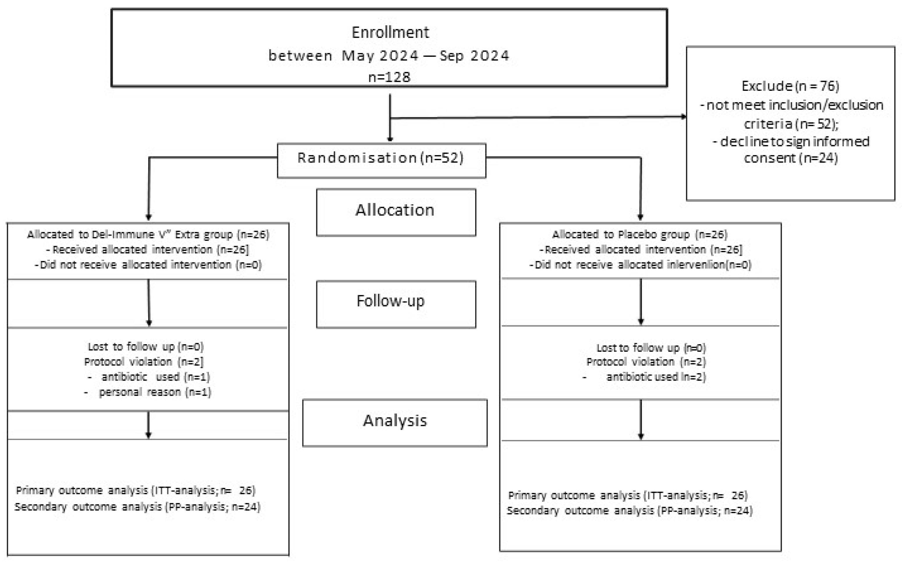
Figure 2.
Primary outcomes analysis with accent on hepatic steatosis indeces changes (A – FLI; B – HSI; C - TyG). Data presented as M±SD.
Figure 2.
Primary outcomes analysis with accent on hepatic steatosis indeces changes (A – FLI; B – HSI; C - TyG). Data presented as M±SD.
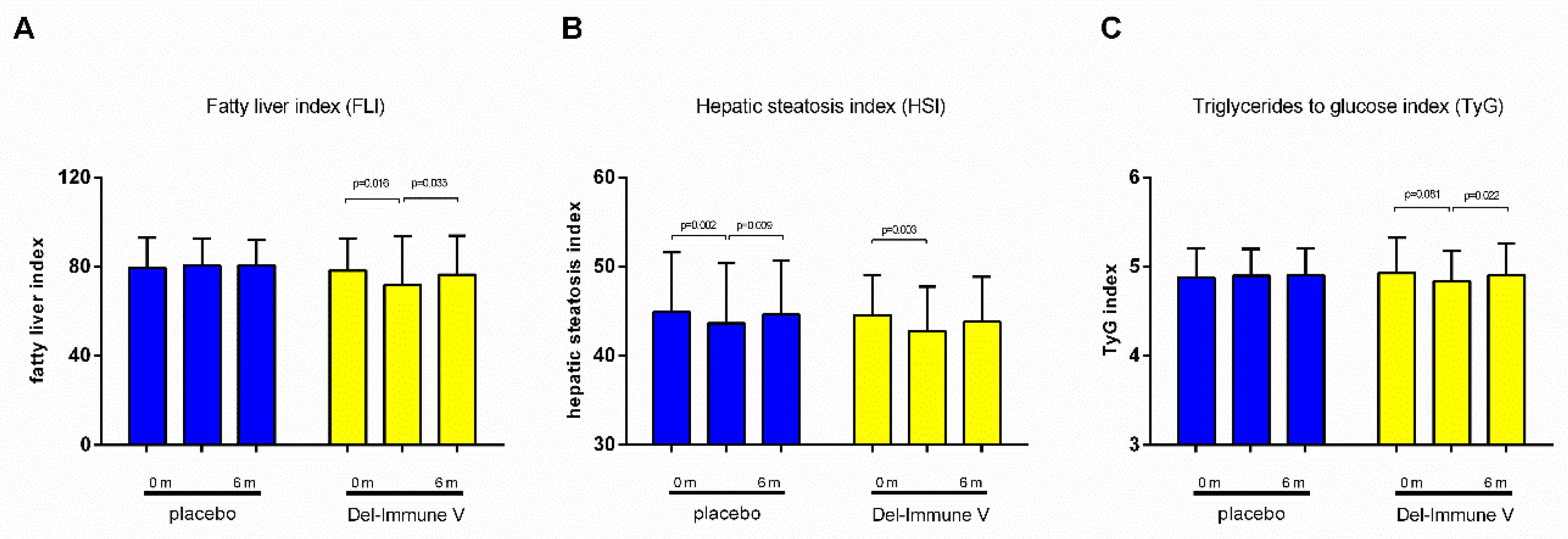
Figure 3.
Secondary outcome analysis with a focus on transaminase activity changes (A – ALT; B – AST; C – γ-GT). Data presented as M±SD.
Figure 3.
Secondary outcome analysis with a focus on transaminase activity changes (A – ALT; B – AST; C – γ-GT). Data presented as M±SD.
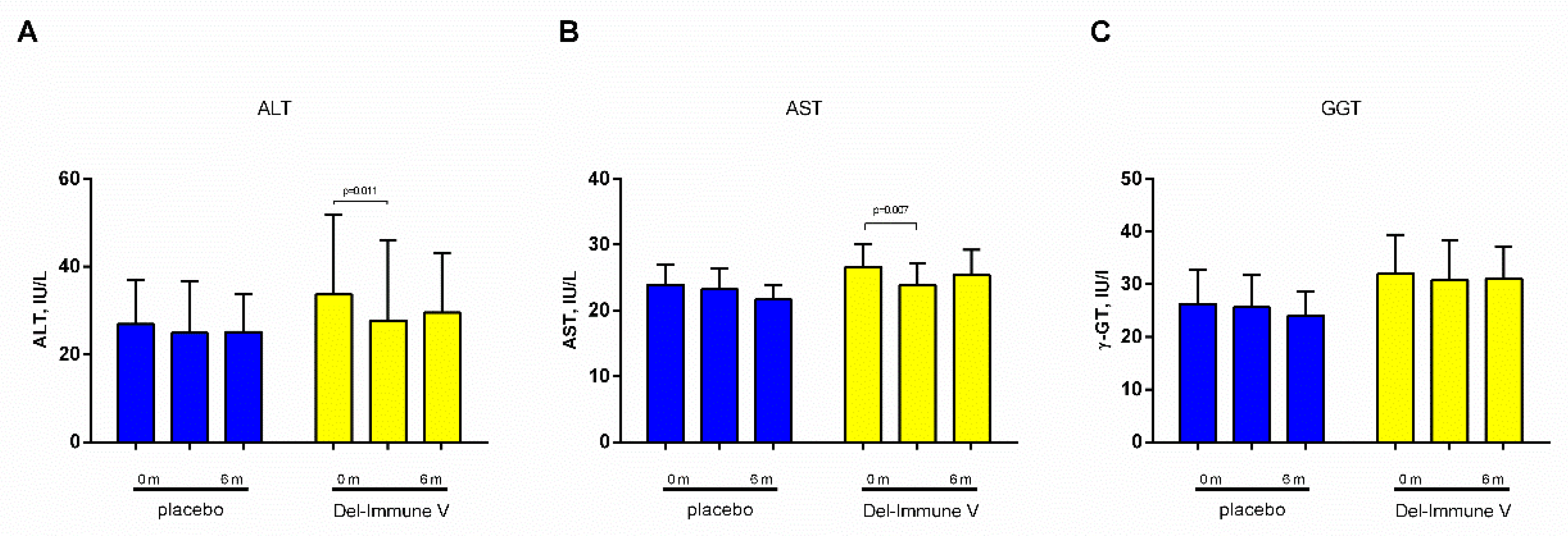
Figure 4.
Secondary outcome analysis with a focus on changes in anthropometric parameters (A – BMI; B – body weight; C – WC; D – visceral fat content). Data presented as M±SD.
Figure 4.
Secondary outcome analysis with a focus on changes in anthropometric parameters (A – BMI; B – body weight; C – WC; D – visceral fat content). Data presented as M±SD.
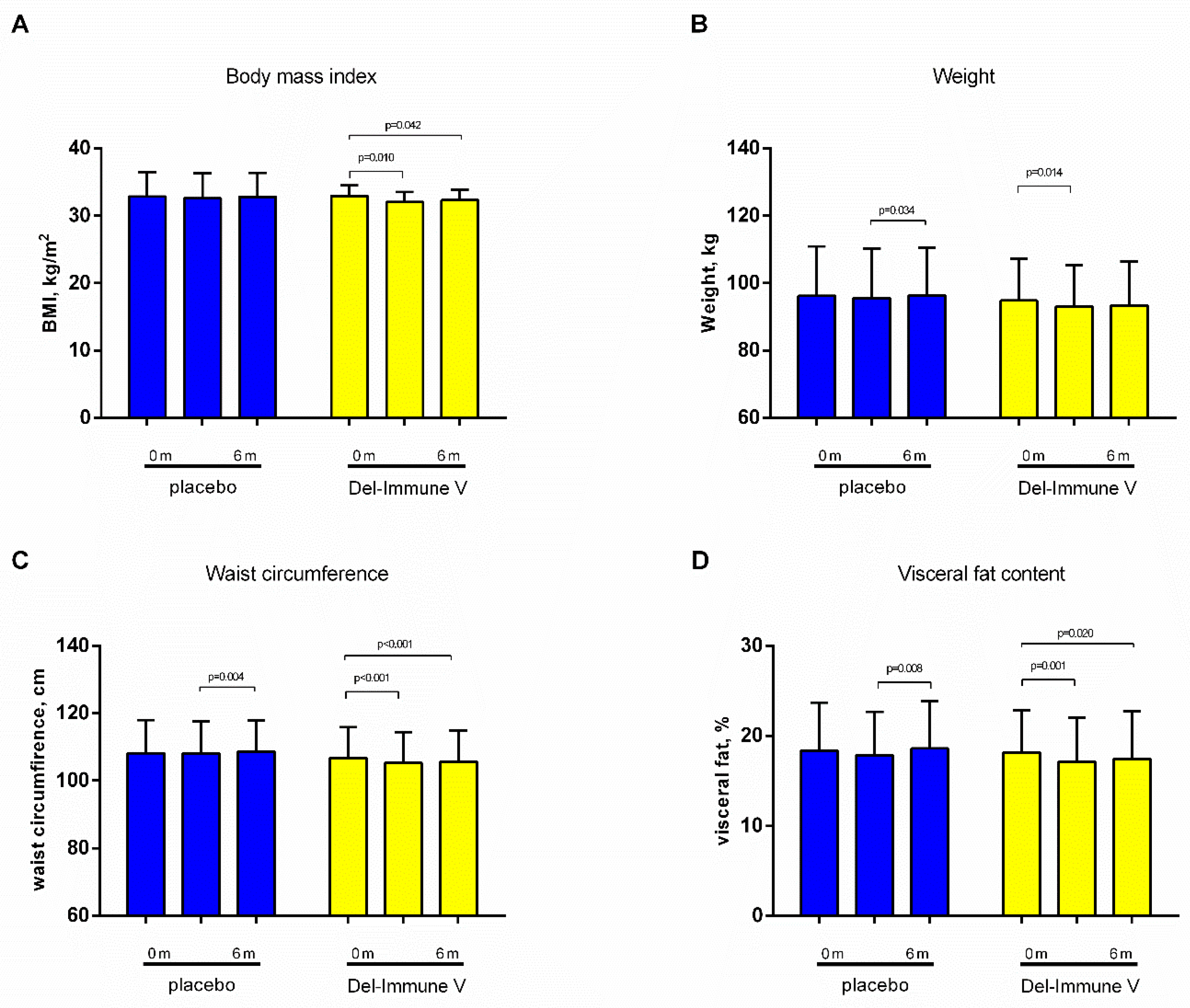
Figure 5.
Secondary outcome analysis with a focus on changes in lipids and chronic systemic inflammation markers (A – total cholesterol; B – triglycerides; C – hs-CRP). Data presented as M±SD.
Figure 5.
Secondary outcome analysis with a focus on changes in lipids and chronic systemic inflammation markers (A – total cholesterol; B – triglycerides; C – hs-CRP). Data presented as M±SD.
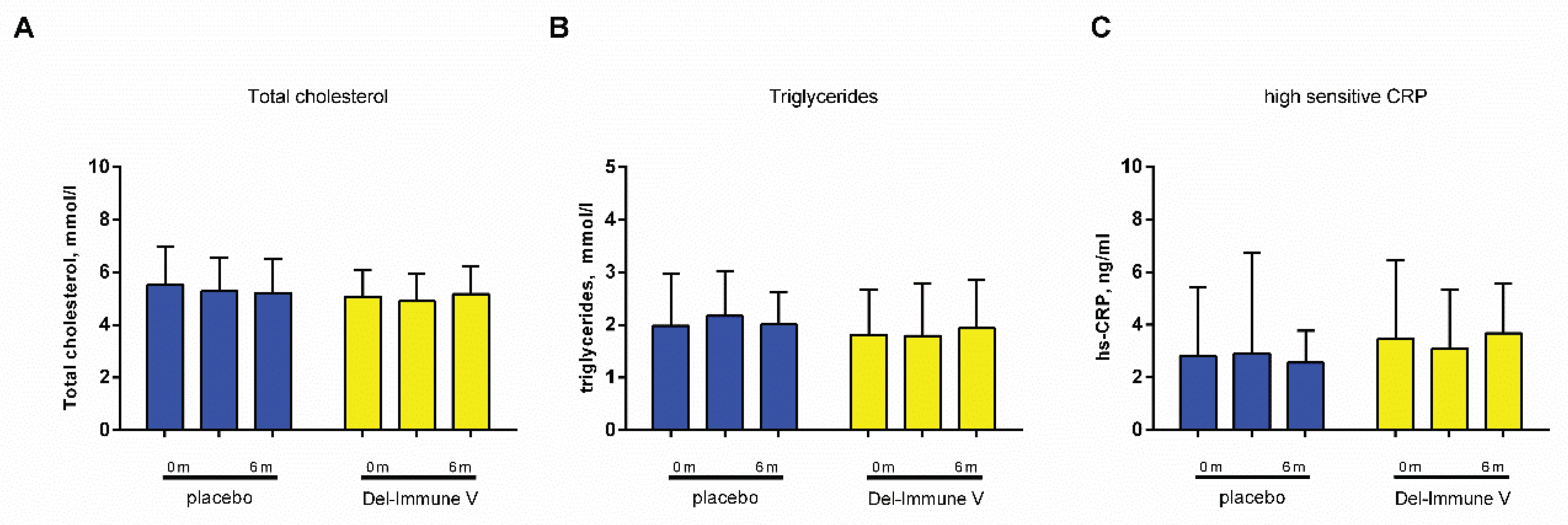

Table 1.
Baseline clinical parameters in examined patients (M±SD or %).
| Parameter |
Placebo group (n=26) |
Del-Immune (n=26) |
p |
| Age, years | 53.42±14.11 | 54.15±13.98 | 0.852 |
| T2D, % (n) | 46.2 (12) | 53.8 (14) | 0.579 |
| T2D duration, years | 15.58±8.08 | 15.14±10.20 | 0.905 |
| Women, % (n) | 57.7 (15) | 50.0 (13) | 0.578 |
| Metformin, % (n) | 34.6 (9) | 34.6 (9) | 0.999 |
| Insulin, % (n) | 15.4 (4) | 19.2 (5) | 0.714 |
Table 2.
Intergroup analysis of primary outcomes.
| Parameters | Baseline | p1 | Mean absolute changes from baseline | p2 | ||
| ∆ 3 month | ∆ 6 month | time effect | time*treatment effect | |||
| FLI | 0.103 | 0.049 | ||||
| Placebo |
79.5±13.4 | 0.92 (-2.07 to 3.92) |
1.19 (-1.50 to 3.88) |
|||
| Postbiotic | 78.23±14.47 | 0.744 | -6.28 (-11.31 to -1.25) |
-1.8 (-4.45 to 0.83) |
||
| HSI | 0.001 | 0.737 | ||||
| Placebo | 44.92±6.67 | -1.27 (-2.05 to -0.50) |
-0.30 (-1.27 to 0.67) |
|||
| Postbiotic | 44.55±4.44 | 0.813 | -1.79 (-2.90 to -0.68) |
-0.73 (-2.16 to 0.69) |
||
| TyG index | 0.342 | 0.261 | ||||
| Placebo | 4.88±0.32 | 0.01 (-0.07 to 0.11) |
0.02 (-0.07 to 0.12) |
|||
| Postbiotic | 4.92±0.39 | 0.629 | -0.08 (-0.19 to 0.01) |
-0.02 (-0.11 to 0.07) |
||
p1 – difference between baseline values; p2- difference between the two groups after the intervention, adjusted for baseline measurements and confounders (intergroup Baseline values presented as M±SD. The mean absolute values was calculated as post-treatment minus baseline and presented as mean differences and 95% confidence intervals (CI). p "time" - measures the effect of time across all three visits for both groups combined; p (time*treatment) evaluates if the intervention group showed a different pattern of change compared to the placebo group during both the active treatment (0–3 months) and the subsequent follow-up (3–6 months).
Table 3.
Intergroup analysis of secondary outcomes.
| Parameters | Baseline | p1 | Mean absolute changes from baseline | p2 | ||
| ∆ 3 month | ∆ 6 month | time effect | time*treatment effect | |||
| ALT, IU/L | 0.012 | 0.340 | ||||
| Placebo | 26.93±10.09 | -2.06 (-4.94 to 0.81) |
-1.80 (-4.76 to 1.15) |
|||
| Postbiotic | 33.64±18.15 | 0.106 | -5.88 (-10.27 to -1.49) |
-4.11 (-10.67 to 2.43) |
||
| AST, IU/L | 0.045 | 0.111 | ||||
| Placebo | 23.92±7.66 | -0.67 (-2.66 to 1.31) |
-2.22 (-4.68 to 0.23) |
|||
| Postbiotic | 26.66±8.60 | 0.230 | -2.78 (-4.74 to -0.81) |
-1.27 (-4.48 to 1.94) |
||
| γ-GT, IU/L | 0.639 | 0.753 | ||||
| Placebo | 26.24±16.22 | -0.58 (-3.07 to 1.89) |
-2.20 (-5.80 to 1.40) |
|||
| Postbiotic | 32.08±18.11 | 0.226 | -1.36 (-6.24 to 3.50) |
-1.00 (-6.98 to 4.97) |
||
| BMI, kg/m2 | 0.003 | 0.318 | ||||
| Placebo | 32.85±3.64 | -0.28 (-0.75 to 0.18) |
-0.05 (-0.67 to 0.57) |
|||
| Postbiotic | 32.95±3.87 | 0.927 | -0.88 (-1.53 to -0.23) |
-0.63 (-1.24 to -0.02) |
||
| Weight, kg | 0.010 | 0.364 | ||||
| Placebo | 96.26±14.65 | -0.69 (-1.95 to 0.55) |
0.01 (-1.51 to 1.53) |
|||
| Postbiotic | 94.86±12.40 | 0.712 | -1.83 (-3.27 to -0.39) |
-1.51 (-3.05 to 0.02) |
||
| WC, cm | 0.001 | 0.003 | ||||
| Placebo | 108.13±9.86 | -0.01 (-0.59 to 0.55) |
0.48 (-0.25 to 1.21) |
|||
| Postbiotic | 106.67±9.16 | 0.582 | -1.42 (-2.09 to -0.75) |
-1.13 (-1.66 to -0.60) |
||
| Visceral fat, % | 0.002 | 0.079 | ||||
| Placebo | 18.34±5.34 | -0.50 (-1.21 to 0.21) |
0.23 (-0.39 to 0.85) |
|||
| Postbiotic | 18.17±4.68 | 0.902 | -1.05 (-1.64 to -0.46) |
-0.75 (-1.37 to -0.12) |
||
| Total cholesterol, mmol/L | 0.419 | 0.349 | ||||
| Placebo | 5.51±0.45 | -0.22 (-0.67 to 0.23) |
-0.29 (-0.81 to 0.21) |
|||
| Postbiotic | 5.08±1.00 | 0.221 | -0.17 (-0.58 to 0.23) |
0.08 (-0.27 to 0.44) |
||
| Triglycerides, mmol/L | 0.724 | 0.243 | ||||
| Placebo | 1.98±0.99 | 0.18 (-0.16 to 0.54) |
0.02 (-0.36 to 0.41) |
|||
| Postbiotic | 1.81±0.85 | 0.511 | -0.02 (-0.29 to 0.25) |
0.12 (-0.10 to 0.35) |
||
| hs-CRP, ng/mL | 0.939 | 0.542 | ||||
| Placebo | 2.82±2.58 | 0.06 (-1.55 to 1.67) |
-0.25 (-1.14 to 0.62) |
|||
| Postbiotic | 3.46±3.00 | 0.417 | -0.36 (-0.99 to 0.27) |
0.21 (-0.63 to 1.05) |
||
p1 – difference between baseline values; p2- difference between the two groups after the intervention, adjusted for baseline measurements and confounders (intergroup Baseline values presented as M±SD. The mean absolute values was calculated as post-treatment minus baseline and presented as mean differences and 95% confidence intervals (CI). p "time" - measures the effect of time across all three visits for both groups combined; p (time*treatment ) evaluates if the intervention group showed a different pattern of change compared to the placebo group during both the active treatment (0–3 months) and the subsequent follow-up (3–6 months).
Disclaimer/Publisher’s Note: The statements, opinions and data contained in all publications are solely those of the individual author(s) and contributor(s) and not of MDPI and/or the editor(s). MDPI and/or the editor(s) disclaim responsibility for any injury to people or property resulting from any ideas, methods, instructions or products referred to in the content. |
© 2026 by the authors. Licensee MDPI, Basel, Switzerland. This article is an open access article distributed under the terms and conditions of the Creative Commons Attribution (CC BY) license.
Copyright: This open access article is published under a Creative Commons CC BY 4.0 license, which permit the free download, distribution, and reuse, provided that the author and preprint are cited in any reuse.