

Submitted:
13 August 2026
Posted:
17 August 2026
You are already at the latest version
Abstract
Fulgides are a class of organic compounds that have been extensively investigated because of their photochromic properties and the wide range of applications arising from them. Fulgimides, an important subclass of fulgides, contain a succinimide moiety in place of the succinic anhydride functionality found in fulgides. Isofulgimides are constitutional isomers of fulgimides in which the succinimide ring is replaced by a 5-iminodihydrofuran-2(3H)-one moiety. Although isofulgimides also exhibit photochromism, their synthesis and photochromic behavior have received considerably less attention than those of other fulgide derivatives. Reports on isofulgimides remain scarce, and crystallographic investigations are particularly limited. As part of our ongoing efforts to develop novel photochromic compounds, we synthesized a unique dimeric isofulgimide featuring an N–N single-bond linkage. Herein, we report its synthesis, crystal structure, and photochromic properties.

Keywords:
photochromic
; fulgide
; fulgimide
; isofulgimide
; crystal structure
; dimmer
1. Introduction
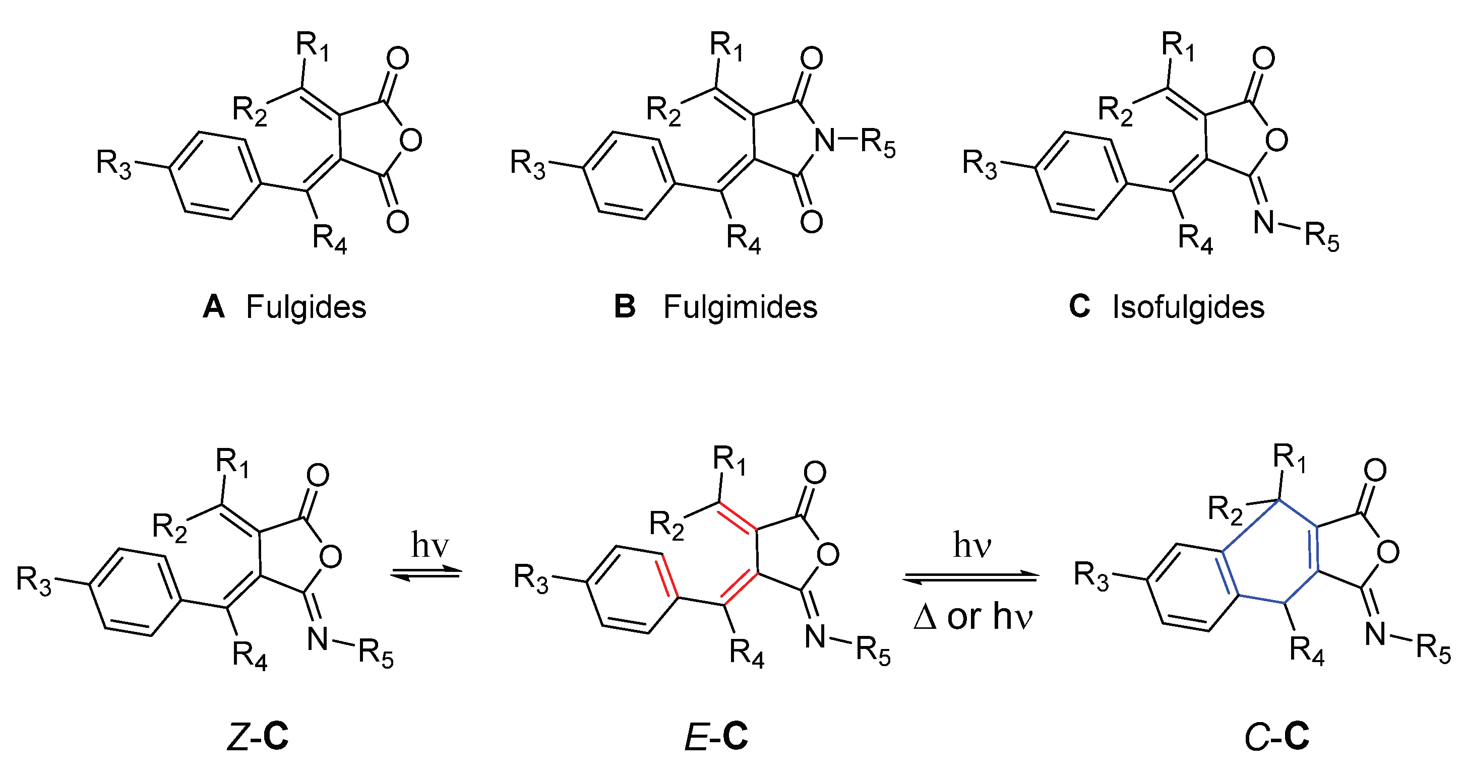
Photochromism is a reversible photochemical phenomenon in which a compound undergoes a color change upon irradiation with light, typically ultraviolet (UV) light, and reverts to its original state either upon exposure to light of a different wavelength or through a thermal process. At the molecular level, photochromism arises from a light-induced transformation of the parent molecule into an isomer with a different electronic structure and, consequently, a different absorption spectrum and color. The photogenerated isomer subsequently undergoes the reverse reaction, either thermally or photochemically, to regenerate the original compound [1,2,3]. Organic photochromic molecules are classified based on their organic features [1]. Fulgides (A) are one class of organic molecules among them. Fulgimides [4] (B) and isofulgimides [5] (C) are considered as fulgide derivatives with photochromic property based on the same chemical mechanism as illustrated in Figure 1. The Z-C and E-C are isomers of isofulgimide C of Z and E the configuration respectively according to the configuration of the double bond with the phenyl group directly attached. C-C is an isomer which contains a newly formed six-member ring in the photoreaction. Although the isomerization between Z-C and E-C is accompanied by color changes, the photoisomerization of the triene isomer E-C to its cyclized isomer C-C, which plays the dominant role in photochromism, has been the primary focus of studies on the photochromic properties of fulgides and their derivatives including isofulgimides [4,5].
Organic photochromic molecules and materials have attracted considerable attention because of their wide range of practical applications, leading to extensive studies on their synthesis and functional properties [1,4,6,7,8]. Conversion of fulgides A into fulgimides B or isofulgimides C introduces a nitrogen atom capable of bearing a variety of substituents, thereby greatly expanding the structural diversity of this family and providing a versatile site for further functionalization.
This transformation generally proceeds in two steps (Scheme 1). In the first step, a fulgide reacts with a primary amine, most commonly an aromatic amine, to afford an amide–acid intermediate (D) in high yield. In the second step, intermediate D undergoes cyclization to form either a fulgimide (B) or an isofulgimide (C), depending on the reaction conditions; in some cases, mixtures of both products are obtained.
Numerous methods have been developed to promote cyclization to fulgimides (pathway a). Most reported procedures employ dehydrating reagents such as acetyl chloride [9,10], acetic anhydride [11], CDI (1,1′-carbonyldiimidazole) [12], BOP (benzotriazol-1-yloxytris(dimethylamino)phosphonium hexafluorophosphate) [13], or HATU (1-[bis(dimethylamino)methylene]-1H-1,2,3-triazolo [4,5-b]pyridinium 3-oxide hexafluorophosphate) [14]. In contrast, N,N′-dicyclohexylcarbodiimide (DCC) was found to be ineffective for fulgimide synthesis. Subsequent studies demonstrated that DCC efficiently promotes the formation of isofulgimides in good yields (pathway b, Scheme 1), providing a practical and selective route to this class of photochromic compounds [5,8].
There are very few reports on the synthesis and applications of isofulgimides, and no crystal structures have been reported to date. Herein, we report the synthesis, crystal structure, and photochromic properties of a unique isofulgimide dimer DM, obtained during our exploration of new organic photochromic materials, (4E,4’E,5Z,5’Z)-5,5’-(hydrazine-1,2-diylidene)bis(4-(1-([1,1’-biphenyl]-4-yl)ethylidene)-3-(propan-2-ylidene)dihydrofuran-2(3H)-one).
2. Materials and Instrumentation
2.1. Materials
Reagent and solvents: the fulgide E is synthesized in our lab (see synthesis in 3. Results and Discussion) [15]. Reagents and solvents are purchased from Thermo Scientific Chemicals: hydrochloric acid aqueous solution (30% w/w), deuterated chloroform-d (99.8% atom); hexanes (98.5% Certified ACS); ethyl acetate 99%, toluene 99%, hydrazine monohydrate 98%, triethylamine 99%.
2.2. Instruments
1 H and 13 C NMR spectra are measured with Bruker, Ascend Evo 400 MHz. Chemical shift in part per million (ppm) is recorded with residual deuterated solvent (CDCl3: 7.260 ppm for 1H, and 77.16 for 13 C) signal as reference [16]. Electrospray ionization mass spectrometry (ESI-MS) experiments were performed using a Thermo Scientific Q Exactive Focus. Sample was injected into a 10 µL loop and was transferred to the mass spec using a mobile phase containing 70% methanol and 30% water with 0.1% formic acid at a flow rate of 600 µL/min. The Q Exactive Focus HESI source was operated in full MS in positive mode. Melting point is measured with thinkSRS: DigiMelt MPA 160. A Leica M80 microscope is used for crystal selection. A XtaLAB Synergy R, DW system, HyPix-Arc 100 diffractometer was used for crystal data collection. Data was measured using w scans with Cu Ka radiation. The structure was solved with the program CrysAlisPro system (CCD 44.138a 64-bit (release 19-01-2026)) and Olex2 1.5 as the graphical interface. UV-Vis spectrum was recorded with Agilent: Cary 7000 UV-Vis-Nir. Laser used for irradiation of DM solution: 402 nm, 150 milliwatt.
3. Methods, Results and Discussion
3.1. Synthesis and Crystallization of DM
The synthesis of DM was carried out following the pathway illustrated in Scheme 2. To a fulgide E (0.3185 g, 1.000 mmol) in toluene (5 mL) was added hydrazine monohydrate (0.0254 g, 0.5073 mmol) and triethylamine (0.1104 g, 1.091 mmol). The solution was stirred magnetically at room temperature overnight. The reaction mixture was washed with 1.0 mL of 1.0 M hydrochloric acid three times and dried with granule sodium sulfate. After removal of solvent with a rotatory evaporator, residual product F was mixed with DCC (0.1238g, 0.6000 mmol), and precipitate formed immediately. After the reaction mixture was stirred overnight, the solid was filtered out. A solid product yielded after the solvent was removed with a rotatory evaporator. The crude product was applied to silica gel chromatography with a solvent of mixed ethyl acetate and hexanes. DM formed single crystals in some test tubes of fractions containing DM after solvent partially evaporated at room temperature overnight. High-quality single crystals were chosen for single X-ray crystallography analysis. The rest of the fractions containing DM were combined. After removal of solvent, 0.1328 g of DM was obtained as a yellow solid with a yield of 21%. DM Formula: C42H36N2O4, Exact theoretical mass 632.27; Exact mass measured [M+H]+: 633.27, Melting point: 118 OC (decompose), 1H NMR in CDCl3 (chemical shift in ppm): 1.183, singlet, 3 H, 2.227, singlet, 3 H, 2.754, singlet, 3 H, 7.389, doublet, 3 H, 7.471, dd, 2 H, 7.629, triplet, 4 H. 13 C NMR in CDCl3 (chemical shift in ppm): 22.49, 22.61, 26.20, 119.89, 120.71, 126.97, 127.63, 128.15, 128.99, 139.70, 141.52, 141.81, 153.52, 155.10, 163.28, 163.90. (For spectrum, see Supporting Information).
3.2. Crystal Structure of DM
3.2.1. Data Collection
A suitable single, colorless, plate-shaped crystal of DM, with dimensions of 0.20 × 0.08 × 0.01 mm, was selected from a representative batch of crystals using a Leica M80 stereomicroscope. The crystal was mounted on a nylon loop and cooled under a stream of nitrogen gas maintained at 100.00(13) K using an Oxford Cryostream cooling device.
Crystal screening, unit-cell determination, and intensity data collection were performed on a XtaLAB Synergy-R DW diffractometer equipped with a HyPix-Arc 100 detector. Data collection and indexing were carried out using CrysAlisPro (version 1.171.44.138a, 64-bit, Rigaku Oxford Diffraction). The data collection strategy was optimized automatically by the software. Diffraction data were collected using ω scans with Cu Kα radiation (λ = 1.54184 Å) to a maximum resolution corresponding to θ = 75.717° (0.80 Å). The unit-cell parameters were refined using 4058 reflections, corresponding to approximately 85% of the observed reflections.
3.2.2. Data Reduction, Structure Solution, and Refinement
Integrated reflection intensities were obtained by processing the diffraction images using CrysAlisPro. The final data completeness was 99.9% to θ = 75.717°. An empirical absorption correction based on spherical harmonics was applied using the SCALE3 ABSPACK algorithm implemented in CrysAlisPro. The absorption coefficient for DM is μ = 0.661 mm⁻¹ (Cu Kα, λ = 1.54184 Å), with minimum and maximum transmission factors of 0.957.
Systematic absences and intensity statistics indicated the centrosymmetric space group P-1, which was subsequently confirmed during structure solution using SHELXL [17]. The structure was refined by full-matrix least-squares minimization on F² using SHELXL-2019/1[18]. All non-hydrogen atoms were refined anisotropically. Hydrogen atoms were placed in geometrically calculated positions and refined using the riding model.
Refinement Details
The crystals were extremely thin; therefore, diffraction data were collected using a Cu rotating-anode X-ray source to maximize diffraction intensity. Several crystals were examined, and all exhibited either twinning or cracking. The crystal with the best diffraction quality was selected for the final data collection.
The crystal was refined as a two-component non-merohedral twin. Diffraction data from both twin domains were integrated simultaneously using CrysAlisPro, which generated both HKLF4 and HKLF5 reflection files. The HKLF4 file, containing non-overlapping reflections, was used for the initial structure solution, whereas the HKLF5 file, containing reflections from both twin domains, was used for the final least-squares refinement. The second twin domain is related to the first by the twin-law matrix:
which corresponds to an approximately 180° rotation about the reciprocal [001] direction. Refinement against the HKLF5 reflection file converged to twin fractions of 0.8689(18) and 0.1311(18).
Elongated atomic displacement ellipsoids for atoms C12–C23, together with nearby residual electron density peaks, indicated positional disorder. This region was successfully modeled over two alternative positions with occupancies fixed at 0.50:0.50 using the PART −1 and PART −2 instructions in SHELXL. Appropriate geometric restraints and displacement-parameter restraints (including SAME, SIMU, and RIGU) were applied to maintain chemically reasonable bond distances, bond angles, and atomic displacement parameters.
Absorption Correction
An empirical absorption correction based on spherical harmonics was applied using the SCALE3 ABSPACK scaling algorithm implemented in CrysAlisPro (version 1.171.44.134a, Rigaku Oxford Diffraction).
Crystal Symmetry
The structure crystallizes in the triclinic space group P-1 with Z = 1 and Z′ = 0.5. Thus, only one-half of the molecular formula is present in the asymmetric unit, while the remaining half is generated by crystallographic inversion symmetry.
3.2.3. The Crstal Structure and Discussion
The Crystal and Molecular Structure of DM
The crystal structure of DM crystallizes in the centrosymmetric triclinic space group P-1. The crystallographic data and refinement parameters are summarized in Table 1.
The molecular structure of DM is shown in Figure 2, in which all atoms are well ordered except for all the carbon atoms in the phenyl rings (atoms C12–C23), which exhibits positional disorder and was successfully modeled over two alternative configurations. Apart from this disordered region, the molecular structure is well defined, with all non-hydrogen atoms refined anisotropically.
The two symmetry-related isofulgimide units are linked through an N–N single bond, forming a centrosymmetric dimer with an extended conjugated framework. The N3–N3’ bond length is 1.410(3) Å, which falls within the normal range reported for conjugated N–N single bonds and is comparable to values observed for hydrazine derivatives (typically 1.38–1.43 Å). This bond is slightly shorter than that in unsubstituted hydrazine (approximately 1.45 Å), suggesting partial π-electron delocalization across the N–N linkage [19,20]. The C2=N3 bond length of 1.281(2) Å is consistent with that of a typical imine C=N double bond (1.27–1.30 Å), confirming the presence of a well-defined imine functionality within the isofulgimide ring [21,22]. The remaining bond lengths in the ordered region of the molecule are all within the expected ranges for related isofulgimide derivatives. Selected bond lengths involving the non-disordered carbon, nitrogen, and oxygen atoms are listed in Table 2. The distances between atoms C8 and C17 (PART−1) and between C8 and C17A (PART−2), which represent the reactive carbon atoms involved in the photoisomerization process, are 3.156 Å and 3.531 Å, respectively. The average separation of 3.343 Å falls within the range commonly observed for photochromic fulgides [15]and is favorable for the photocyclization reaction.
Disoder of Phenyl Groups and Intermolecular Interactions
The phenyl ring comprising atoms C12–C23, located in the disordered region, was modeled over two alternative configurations, designated PART −1 (atoms C12–C23) and PART−2 (atoms C12A–C23A), as shown in Figure 2. The overall crystal packing of DM is illustrated in A. Because the closest intermolecular contacts are concentrated within the disordered region, a meaningful Hirshfeld surface analysis of the complete structure was not feasible. Therefore, the two disorder components were examined separately to illustrate the alternative local packing environments associated with each configuration.
The PART−1 configuration (B) exhibits a pronounced intermolecular π–π stacking interaction between adjacent phenyl rings, with a centroid-to-centroid distance of 3.594 Å. In contrast, the PART−2 configuration (C) does not exhibit significant π–π stacking but instead shows a close face-to-face aromatic contact with an intermolecular separation of 4.154 Å. These two disorder models represent alternative local packing environments arising from the positional disorder of the phenyl ring. Together, they indicate that aromatic intermolecular interactions, whether through π–π stacking or close aromatic contacts, contribute to the stabilization of the crystal packing of DM.
Figure 3.
Crystal packing of DM. (A) Overall crystal packing showing both disorder components (PART−1 and PART−2) of the disordered phenyl ring. (B) Local packing environment corresponding to PART−1, illustrating an intermolecular π–π stacking interaction between adjacent phenyl rings. (C) Local packing environment corresponding to PART −2, showing close face-to-face aromatic contact.
Figure 3.
Crystal packing of DM. (A) Overall crystal packing showing both disorder components (PART−1 and PART−2) of the disordered phenyl ring. (B) Local packing environment corresponding to PART−1, illustrating an intermolecular π–π stacking interaction between adjacent phenyl rings. (C) Local packing environment corresponding to PART −2, showing close face-to-face aromatic contact.

Comparison of an Isofulgimide and a Fulgimide
Isofulgimides are structural analogues of fulgides, differing only in that one carbonyl (C=O) group of the fulgide is replaced by an imine (C=N) group in the isofulgimide. To evaluate the structural effect of this substitution, the molecular structure of fulgide E was overlaid with one-half of the DM molecule. The two structures differ only at the corresponding heteroatom position, where an oxygen atom in the fulgide is replaced by a nitrogen atom in the isofulgimide moiety. The overlay was performed by matching the corresponding atoms of the five-membered ring together with the atoms directly attached to the ring, as shown in Figure 4. The least-squares superposition gave an RMSD of 0.037 Å, demonstrating that the two molecular frameworks are nearly identical and that replacement of the carbonyl oxygen by an imine nitrogen has a negligible effect on the overall molecular geometry.
In summary, the synthesized compound DM was characterized by mass spectrometry, NMR, and MS spectroscopy. Single-crystal X-ray diffraction unequivocally established its molecular structure as a dimeric isofulgimide with an N–N single-bond linkage. Although the terminal phenyl ring exhibits positional disorder, the disorder was successfully modeled, allowing accurate determination of the molecular geometry and crystal structure.
3.3. Photochromic Property of MD
3.3.1. Method and Results
The UV-Vis spectrum of a yellow solution of MD (5.0 mg) in 1 mL of CDCl3 in a quartz cuvette was measured with a UV spectrometer as shown in Figure 5. The solution was then taken out of the spectrometer and then irradiated with a 405 nm laser of 150 mW for about one minute. The solution turned red and was put back into the spectrometer, UV-Vis spectrum was measured again at different times after the irradiation until the original UV-Vis spectrum of MD is completely recovered. The results are shown in Figure 5. The process has repeated several times; no significant color difference of the irradiated sample was observed.
3.3.2. Discussion of UV Spectrum and Photochromism of MD
Compound DM, corresponding to the E–C form of the hexatriene moiety shown in Figure 1, exhibits strong absorption at wavelengths below 450 nm, whereas its absorption above 480 nm is relatively weak (Figure 5). Compared with the UV–Vis spectrum of the isofulgimide monomer, which exhibits strong absorption only at wavelengths below 400 nm [5], DM displays a pronounced bathochromic (red) shift. This significant bathochromic shift is attributed to the doubly extended π-conjugated system of the dimeric DM, which provides greater electron delocalization than the monomeric isofulgimide.
Upon irradiation with a 405 nm laser, a new absorption band centered at approximately 510 nm appeared, corresponding to the cyclized isomer of DM (the C–C form of the six-membered ring moiety). The appearance of this band indicates that photoisomerization from the E–C form to the C–C form is induced by 405 nm irradiation. After the irradiation was discontinued, the absorbance of the 510 nm band gradually decreased and disappeared completely within approximately 50 min, indicating a slow thermal back-isomerization from the C–C form to the E–C form. This photoisomerization cycle was repeated several times without any significant change in the intensity of the 510 nm absorption band, indicating that DM exhibits excellent fatigue resistance during the photochromic process. Further investigations of the photochromic properties of DM are currently underway.
5. Conclusions
The N–N single bond-linked dimeric isofulgimide DM was successfully synthesized using a straightforward synthetic method and characterized by MS, 1 H NMR, 13 C NMR, and UV–Vis spectroscopy. Its molecular structure was unequivocally confirmed by single-crystal X-ray diffraction using a high-quality diffraction dataset. Although the terminal phenyl rings exhibit crystallographic disorder, the 5-iminodihydrofuran-2(3H)-one core is well resolved.
The photochromic behavior of DM was investigated in a preliminary study. Photoisomerization of the hexatriene moiety to the cyclized six-membered ring isomer, followed by thermal back-isomerization, was clearly demonstrated by the appearance of an absorption band at approximately 510 nm upon irradiation with a 405 nm laser and its gradual disappearance after the irradiation was discontinued. These results confirm the reversible photochromic behavior of DM. The synthesis of structural analogues of DM and a comprehensive investigation of their photochromic properties is currently underway.
Supplementary Materials
The following supporting information can be downloaded at: Preprints.org, MD.cif, MDcheckcif.pdf, MD.pdb, MD.res, MD crystal structure report.doxc, and 1 H and 13 C NMR and MS spectra of DM.
Author Contributions
Conceptualization, synthesis, crystallization, photochromism study, writing original drat Y.L. X-ray crystallography, data collection, structural solving, refining, N.B, photochromic study, spectrum recording, T.T and Y.Z. Synthesis and NMR spectra measurement T.G and J.F. Draft reviewing and correction, all authors. All authors have read and agreed to the published version of the manuscript.
Funding
This research is supported by Faculty RISE Program of Prairie View A&M University.
Data Availability Statement
The crystal data has been provided in the supplementary materials and deposited to the Cambridge Crystallographic Data Centre (CCDC). CCDC 2574491. For any other information, please contact the corresponding author.
Acknowledgments
We would like to thank Dr. Marco Giles for help with Flash Column Chromatography, thank Dr. Xiqu Wang for rechecking the crystal structure, and thank Prairie View A&M University for providing Faculty RISE funding to support this project.
Conflicts of Interest
The authors declare no conflicts of interest.
Abbreviations
The following abbreviations are used in this manuscript:
| UV | Ultraviolet |
| UV-Vis | Ultraviolet-Visible |
| NMR | Nuclear Magnetic Resonance |
| MS | Mass Spectroscopy |
| CDI | 1,1′-carbonyldiimidazole |
| HATU | 1-[bis(dimethylamino)methylene]-1H-1,2,3-triazolo [4,5-b] pyridinium 3-oxide hexafluorophosphate |
| BOP | benzotriazol-1-yloxytris(dimethylamino)phosphonium hexafluorophosphate |
| RMSD | Root-Mean-Square Deviation |
| DCC | N, N′-dicyclohexylcarbodiimide |
References
- Irie, M. Photochromism: Memories and SwitchesIntroduction . Chem. Rev. 2000, 100(5), 1683–1684. [Google Scholar] [CrossRef] [PubMed]
- Marcon, M.; Haag, C.; König, B. Photoswitches beyond azobenzene: a beginner’s guide . Beilstein J. Org. Chem. 2025. 21, 1808–1853. [Google Scholar] [CrossRef] [PubMed]
- Volarić, J.; et al. Molecular photoswitches in aqueous environments . Chem. Soc. Rev. 2021, 50(22), 12377–12449. [Google Scholar] [CrossRef] [PubMed]
- Yokoyama, Y. Fulgides for Memories and Switches . Chem. Rev. 2000, 100(5), 1717–1740. [Google Scholar] [CrossRef] [PubMed]
- Heller, H.G.; et al. Fulgides and Fulgimides for Practical Applications . In Molecular Crystals and Liquid Crystals Science and Technology. Section A. Molecular Crystals and Liquid Crystals; 1994; Volume 246, 1, pp. 79–86. [Google Scholar]
- Chen, X.; et al. Photochromic properties of a water-soluble methyl carboxylic acid indolylfulgimide . Photochem. Photobiol. Sci. 2011, 10(6), 1023–1029. [Google Scholar] [CrossRef] [PubMed]
- Irie, M.; et al. Photochromism of Diarylethene Molecules and Crystals: Memories, Switches, and Actuators . Chem. Rev. 2014, 114(24), 12174–12277. [Google Scholar] [CrossRef] [PubMed]
- Rustler, K.; et al. Optical Control of GABAA Receptors with a Fulgimide-Based Potentiator . Chem. – A Eur. J. 2020, 26(56), 12722–12727. [Google Scholar] [CrossRef] [PubMed]
- Irie, M. Photo-Reactive Materials for Ultrahigh Density Optical Memory: MITI Research and Development Program on Basic Technologies for Future Industries 1994. [CrossRef]
- Matsushima, R.; Sakaguchi, H. Comparison of the photochromic properties of fulgides and fulgimides . J. Photochem. Photobiol. A Chem. 1997, 108(2), 239–245. [Google Scholar] [CrossRef]
- Darcy, P.J.; et al. Photochromic systems. Part 1. Photochemical studies on (E)-2-isopropylidene-3-[1-(3,4,5-trimethoxyphenyl)ethylidene]succinic anhydride and related compounds . J. Chem. Soc. Perkin Trans. 1986, 1 15, 315–319. [Google Scholar] [CrossRef]
- Ilyina, I.G.; et al. Synthesis and spectral-kinetic investigation of fulgimide-based photochromic autocomplexes of the dinitroquinoline series . Russ. Chem. Bull. 2008, 57(7), 1444–1450. [Google Scholar] [CrossRef]
- Krawczyk, K.K.; et al. Efficient BOP-mediated synthesis of fulgimides . Comptes Rendus Chim. 2012, 15(5), 384–388. [Google Scholar] [CrossRef]
- Copko, J.; Slanina, T. Multiplicity-driven photochromism controls three-state fulgimide photoswitches . Chem. Commun. 2024, 60(28), 3774–3777. [Google Scholar] [CrossRef] [PubMed]
- Li, Y.; et al. Crystal Structures of Novel Phenyl Fulgides . Crystals 2026, 16(1), 38. [Google Scholar] [CrossRef]
- Fulmer, G.R.; et al. NMR Chemical Shifts of Trace Impurities: Common Laboratory Solvents, Organics, and Gases in Deuterated Solvents Relevant to the Organometallic Chemist . Organometallics 2010, 29(9), 2176–2179. [Google Scholar] [CrossRef]
- Sheldrick, G.M. SHELXT - integrated space-group and crystal-structure determination . Acta Crystallogr. A Found. Adv. 2015, 71 Pt 1, 3–8. [Google Scholar] [CrossRef] [PubMed]
- Sheldrick, G. Crystal structure refinement with SHELXL . Acta Crystallogr. Sect. C 2015, 71(1), 3–8. [Google Scholar] [CrossRef] [PubMed]
- Poisson, J.; Butler, I.S.; Bohle, D.S. π-Delocalization in the vicinal lone pairs of hydrazines: Electronic effects in derivatives of 1-(2-nitrophenyl)-1-phenylhydrazine . J. Mol. Struct. 2016, 1116, 30–36. [Google Scholar] [CrossRef]
- Kriel, F.H.; M.A. Fernandes, J. Caddy, 1,2-Bis(diphenylphosphino)-1,2-diethylhydrazine. Acta Crystallogr. Sect. E 2010, 66(6), o1270. [Google Scholar] [CrossRef] [PubMed]
- Allen, F.H.; et al. Tables of bond lengths determined by X-ray and neutron diffraction. Part 1. Bond lengths in organic compounds . J. Chem. Soc. Perkin Trans. 2 1987, 16(12), S1–S19. [Google Scholar] [CrossRef]
- Allen, F. The Cambridge Structural Database: a quarter of a million crystal structures and rising . Acta Crystallogr. Sect. B 2002, 58(3 Part 1), 380–388. [Google Scholar] [CrossRef] [PubMed]
Figure 1.
Structure of Fulgides A, Fulgimides B and isofulgimides C, and Scheme of Photochromism of Isofuligimdes C.
Figure 1.
Structure of Fulgides A, Fulgimides B and isofulgimides C, and Scheme of Photochromism of Isofuligimdes C.
Scheme 1.
Synthetic pathways from Fulgides A to Fulgimides B and isofulgimides C via intermediate D.
Scheme 1.
Synthetic pathways from Fulgides A to Fulgimides B and isofulgimides C via intermediate D.

Scheme 2.
Synthesis of DM from fulgide E.

Figure 2.
The molecular structure of DM with the disordered phenyl groups (C12-C23) refined into two models PART-1 containing C23 and PART-2 containing C23A.
Figure 2.
The molecular structure of DM with the disordered phenyl groups (C12-C23) refined into two models PART-1 containing C23 and PART-2 containing C23A.

Figure 4.
Least-squares overlay the molecular structures of fulgide E with hydrogen atoms and one-half of the DM molecule without hydrogen atoms.
Figure 4.
Least-squares overlay the molecular structures of fulgide E with hydrogen atoms and one-half of the DM molecule without hydrogen atoms.

Figure 5.
UV spectrum of MD before (no light, blue) and after (orange) laser irradiation at different times.
Figure 5.
UV spectrum of MD before (no light, blue) and after (orange) laser irradiation at different times.


Table 1.
Crystallographic data and refinement parameters.
| Compound | DM | ||
| Formula | C42H36N2O4 | Z’ | 0.5 |
| Dcalc./ g cm-3 | 1.296 | Wavelength/Å | 1.54184 |
| m/mm-1 | 0.661 | Radiation type | Cu Ka |
| Formula Weight | 632.73 | Qmin/° | 3.363 |
| Color | colorless | Qmax/° | 75.717 |
| Shape | plate-shaped | Measured Refl’s. | 4799 |
| Size/mm3 | 0.20×0.08×0.01 | Indep’t Refl’s | 4799 |
| T/K | 100.00(13) | Refl’s I≥2s(I) | 4332 |
| Crystal System | triclinic | Rint | . |
| Space Group | P-1 | Parameters | 281 |
| a/Å | 7.6613(4) | Restraints | 480 |
| b/Å | 8.6920(4) | Largest Peak | 0.298 |
| c/Å | 13.4675(7) | Deepest Hole | -0.258 |
| a/° | 81.955(4) | GooF | 1.073 |
| b/° | 77.954(4) | wR2 (all data) | 0.1645 |
| g/° | 67.859(4) | wR2 | 0.1601 |
| V/Å3 | 810.54(7) | R1 (all data) | 0.0635 |
| Z | 1 | R1 | 0.0583 |
Table 2.
Bond distances between non-disordered C, N and O atoms.
| Atom | Atom | Length/Å | Atom | Atom | Length/Å | |||
| O1 | C2 | 1.385(2) | C4 | C11 | 1.363(3) | |||
| O1 | C6 | 1.398(2) | C5 | C6 | 1.477(2) | |||
| O7 | C6 | 1.195(2) | C5 | C8 | 1.358(3) | |||
| N3 | N31 | 1.410(3) | C8 | C9 | 1.492(3) | |||
| N3 | C2 | 1.281(2) | C8 | C10 | 1.511(3) | |||
| C2 | C4 | 1.471(2) | C11 | C24 | 1.503(3) | |||
| C4 | C5 | 1.478(2) | ||||||
Disclaimer/Publisher’s Note: The statements, opinions and data contained in all publications are solely those of the individual author(s) and contributor(s) and not of MDPI and/or the editor(s). MDPI and/or the editor(s) disclaim responsibility for any injury to people or property resulting from any ideas, methods, instructions or products referred to in the content. |
© 2026 by the authors. Licensee MDPI, Basel, Switzerland. This article is an open access article distributed under the terms and conditions of the Creative Commons Attribution (CC BY) license (http://creativecommons.org/licenses/by/4.0/).
Copyright: This open access article is published under a Creative Commons CC BY 4.0 license, which permit the free download, distribution, and reuse, provided that the author and preprint are cited in any reuse.