

Submitted:
13 August 2026
Posted:
14 August 2026
You are already at the latest version
Abstract
Colorectal, lung, breast, and melanoma malignancies remain major contributors to cancer-related morbidity and mortality worldwide, underscoring the need for therapeutic strategies that can be rapidly translated to the clinic. Drug repurposing offers an attractive route for accelerating oncology drug development, particularly for agents with established safety profiles. Background/Objectives: Niclosamide, an FDA-approved anthelmintic, has demonstrated broad anticancer activity through modulation of Wnt1/β-catenin signaling and other pathways associated with tumor growth, metastasis, and therapeutic resistance. However, poor pharmaceutical and pharmacokinetic properties have limited its advancement as a systemic cancer therapeutic. Methods: To investigate prodrug-based approaches for improving niclosamide delivery, we synthesized a panel of ester and carbonate derivatives bearing structurally diverse alkyl promoieties and evaluated their hydrolytic stability, anticancer activity, and drug-like properties through experimental and in silico analyses. Antiproliferative activity was assessed across a diverse panel of human and murine cancer models, including colorectal (HCT-116, HT-29), lung (A549), breast (MDA-MB-231), and melanoma (B16F10) cell lines. Results: All derivatives underwent rapid hydrolysis under physiologically relevant conditions, exhibiting half-lives of less than two hours and efficiently regenerating niclosamide. While alkyl substitution produced modest differences in hydrolytic stability, these variations did not substantially influence anticancer activity, with the prodrugs displaying comparable potency profiles across the evaluated cancer models. In silico pharmacokinetic and drug metabolism analyses further suggested that prodrug derivatization may offer opportunities to modulate developability while preserving pharmacological activity through rapid bioactivation. Conclusions: Collectively, these findings establish ester and carbonate derivatization as a viable therapeutic delivery strategy for niclosamide and provide a framework for the continued development of niclosamide-based cancer therapeutics with improved pharmaceutical properties and retained anticancer efficacy.
Keywords:
niclosamide
; prodrugs
; hydrolysis
; Wnt inhibitors
1. Introduction
Therapeutic repurposing of bioactive agents with known tolerability, safety, and efficacy profiles has offered an accelerated route towards preclinical advancement and clinical approval[1,2,3], and in many cases has led to the serendipitous discovery of novel modes of action for existing small molecules, including fingolimod[4,5], zidovudine[6], topiramate[7], and carmofur[8,9,10,11]. Similarly, niclosamide, which was originally approved by the Food and Drug Administration (FDA) as an anthelmintic agent[12], has been more recently investigated for other clinical indications, such as Parkinson’s disease[13], diabetes[14], bacterial infections[15], and cancer[16]. Putatively, niclosamide engages multiple intracellular therapeutic targets associated with cancers and other diseases[17], including the Signal Transducer and Activator of Transcription 3 (STAT3), Nuclear factor kappa-light-chain-enhancer of activated B cells (NF-κB), and Wnt1 signaling pathways (Table 1).
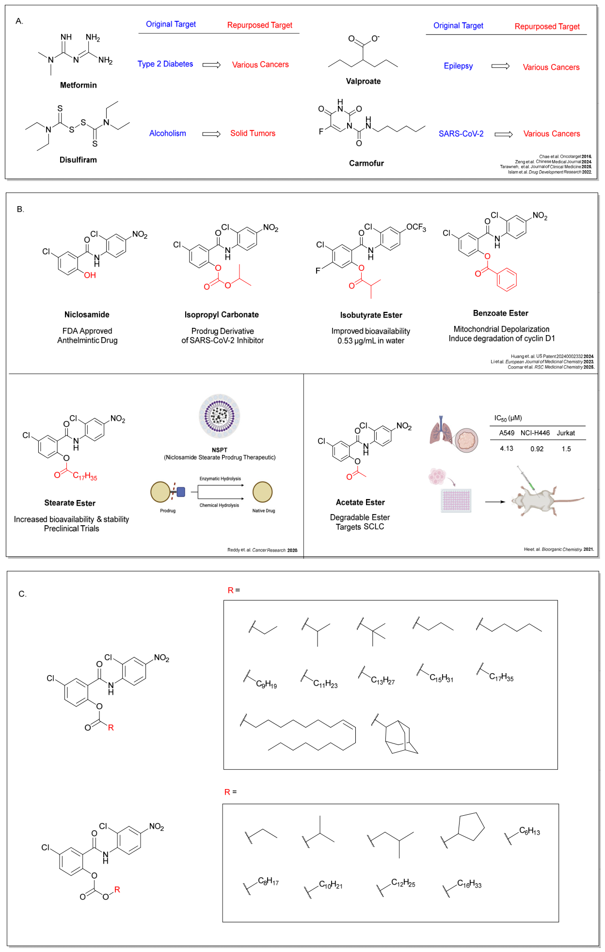
Notwithstanding its diverse therapeutic potential, niclosamide suffers from poor aqueous solubility (0.23 μg/mL)[30] and limited bioavailability (logP: 4.723, PSA: 95.15)[31], prompting newfound efforts to identify strategies to augment the bioavailability and therapeutic efficacy of niclosamide. Among these strategies, installation of lipophilic ester promoieties accounts for nearly fifty percent of FDA-approved prodrugs[32,33,34,35]. In context of the delivery challenges associated with niclosamide, its acetate ester prodrug has been investigated for improved therapeutic efficacy in a range of human diseases, including neuroblastoma[36], glioblastoma [37], and small cell lung cancer (SCLC) [38]. Coomar et al. likewise prepared a benzoate ester at the niclosamide phenol, which exhibited comparable activity as an anticancer agent through mitochondrial depolarization[39]. Yuan et al. prepared a niclosamide valproate ester[40] and Wang et al. prepared its butyrate ester [41], both of which exhibited enhanced antitumor activity. Analogously, a stearate ester prodrug of niclosamide, which was previously demonstrated to bear enhanced anthelmintic activity through improved lymphotropic uptake[42], was recently demonstrated to bear heightened therapeutic in vivo potency against osteosarcoma[43] through improvement of its ADME profile and bioavailability (50 mg/kg (1.9 mmol/L) intravenously (IV)).
Similarly, alkyl phosphate prodrugs of niclosamide were found to exhibit enhanced potency in SCLC by improving bioavailability profiles[38]. Others have acylated the phenol of niclosamide and tethered this conjugate to a genetically-encoded elastin-based chimeric polypeptide[44] or to polyethylene glycol (PEG) -ylated ester nanomaterials [45] as a similar functionalization strategy to improve delivery dynamics of the compound. In addition, Chen et al. reported a series of O-alkylamino-tethered derivatives of niclosamide, where improved aqueous solubility translated into improved biological performance[30]. Taken together, these observations indicate that acylation of the free phenol of niclosamide is an effective chemical strategy to augment its biological availability and improve its therapeutic potential. However, notwithstanding the diversity of alkyl, heteroalkyl, and alkylaryl esters and carbonates of niclosamide previously prepared and investigated as cancer therapeutics, a systematic study of niclosamide ester and carbonate promoiety stability and anticancer therapeutic efficacy is underreported in the literature.
In order to interrogate this, we prepared a systematic panel of twenty-one niclosamide analogs with varying ester and carbonate tail lengths. Of these, we investigated the potential role of steric demand of esters with different α-branched substituents[46] including isobutyrate ester 2b and pivaloate ester 2c. Among other esters, we prepared both previously reported stearate ester 2j and unsaturated oleate ester 2k in order to investigate the role of a single cis-alkene in this promoiety. Finally, adamanecarboxy ester 2l was prepared to study the effect of a rigid, aliphatic moiety. We further demonstrate that these ester and carbonate prodrugs exhibit comparable dose-dependent inhibitory activity in HCT-116, HT29, MDA-MB-231, B16F10, and A549 cell lines. Additionally, through hydrolysis assays, with the exception of hindered carbonate niclosamide prodrugs bearing isopropyl and cyclopentyl carbonate promoieties (Compounds 3b and 3d), we observed that all compounds exhibit similarly efficient hydrolysis into niclosamide within the first few hours of hydrolytic exposure under physiologically simulative pH conditions. To further assess the ability of niclosamide prodrugs to inhibit the Wnt1 pathway, we deployed a firefly luciferase (fLuc) T-Cell Factor/Lymphoid Enhancer-Binding Factor (TCF/LEF) -dependent reporter assay[47,48] and observed similar, niclosamide-like activity for this series of compounds, which is consistent with the relative hydrolysis kinetics observed. We further establish timepoint-based bioavailability profiles through washout experiments to identify compounds with the most rapid cellular internalization or membrane association. Finally, in silico models were utilized for predictions of ADME and DMPK properties of this series of niclosamide prodrugs with the open access SwissADME Suite. Collectively, these observations demonstrate that, with few exceptions, alkyl ester and carbonate prodrugs of niclosamide exhibit comparable hydrolysis release kinetics and bear similar activity in efficient aqueous release of niclosamide, retaining both niclosamide-like anticancer and Wnt1-modulating activity. More generally, these studies show that installation of alkyl ester and carbonate prodrug moieties on the free phenol of niclosamide are an effective therapeutic strategy for rapid-release niclosamide prodrugs with tunable bioavailability profiles that can be modulated to achieve desirable physicochemical properties relevant in future preclinical and clinical applications.
Figure 1.
Niclosamide Analogs (A) Representative examples of FDA-approved drugs repurposed for cancer therapeutics. (B) Previously synthesized niclosamide analogs demonstrating potency against various cancers, including stearate ester and acetate ester derivatives of niclosamide. (C) Our library of 21 niclosamide ester and carbonate derivatives.
Figure 1.
Niclosamide Analogs (A) Representative examples of FDA-approved drugs repurposed for cancer therapeutics. (B) Previously synthesized niclosamide analogs demonstrating potency against various cancers, including stearate ester and acetate ester derivatives of niclosamide. (C) Our library of 21 niclosamide ester and carbonate derivatives.
2. Materials and Methods
2.1. Colorimetric Hydrolysis Assay
The hydrolysis profiles of the niclosamide prodrugs were determined by monitoring the spectrophotometric appearance of the free parent drug. In a clear 96-well flat-bottom microplate (Nest, Costar), 5 µL of each prodrug stock solution (50 mM in DMSO) was added to 95 µL of isopropanol. The hydrolysis reaction was initiated by the addition of 10 µL of a 1.24 M TRIS buffer solution adjusted to pH 7.0 with 25% v/v phosphoric acid, and diluted to volume. The progress of the hydrolysis was tracked by measuring the increasing yellow coloration of the hydrolyzed niclosamide. Absorbance was recorded at 380 nm utilizing a Molecular Devices SpectraMax Plus 384 microplate reader at 5-minute intervals for 60 minutes. The percentage of hydrolysis over time was calculated in comparison to an equivalent concentration of hydrolyzed niclosamide, which was defined as 100% hydrolyzed.
2.2. Cell Culture
HCT-116, HT-29, MDA-MB-231, B16F10, and A549 cells were cultured in T25 and T75 flasks. The cultures were kept at 37 °C in a humidified incubator (5.0% CO2), and maintained by splitting cells at a ratio of 1:3 to 1:6 based on confluence and cultured up to passage 20. HCT-116 and HT-29 human colorectal cancer cell lines were cultured in McCoy’s 5A Medium (Tribioscience) supplemented with 10% v/v fetal bovine serum (FBS, Gibco) and 1% v/v 100x penicillin-streptomycin (Tribioscience). MDA-MB-231, a human breast cancer cell line, was cultured in RPMI-1640 Medium (Corning) supplemented with 10% v/v fetal bovine serum (FBS) and 1% v/v 100x penicillin-streptomycin (Tribioscience). A549, a human non-small cell lung carcinoma cell line, and B16F10, a mouse non-small melanoma cell line, was maintained in High Glucose Dulbecco’s Modified Eagle Medium (DMEM) (Hygia Reagents) supplemented with 10% fetal bovine serum (Gibco) and 1% penicillin-streptomycin (Tribioscience).
2.3. MTT Assay
Cell viability was determined by 3-(4,5-dimethylthiazol-2-yl)-2,5-diphenyltetrazolium bromide (MTT) assays through literature reported protocols. Cell cultures were prepared referencing the protocol above. Cells were seeded at 70% confluency in a 384-well flat bottom tissue culture treated plate (Nest, Costar) and incubated for 24 hours at 37 °C in a humidified incubator (5% CO2). Following the 24-hour incubation period, a drug medium solution was prepared by adding 100-fold DMSO solutions of compounds 1, 2a-l, 3a-3i to cell culture media. 20µL of the drugged media was then added to reach desired concentrations of 250 µM, 125 µM, 25 µM, 12.5 µM, 2.5 µM, 1.25 µM, 250 nM, and 125 nM. A negative control of 0.5% v/v DMSO was also included. The plates were then incubated for 72 hours at 37 °C in a humidified incubator (5% CO2). After the incubation period, 2 µL of a solution of 3-(4,5-dimethylthiazol-2-yl)-2,5-diphenyltetrazolium bromide (MTT) (AK Scientific) in 1x phosphate-buffered saline (PBS) (5 mg/mL) was added to each well, and the cells were allowed to incubate for 30-45 minutes. Then, the cell supernatant was aspirated before 40 µL of DMSO was added to each well and mixed until all formazan crystals were re-solubilized. Absorbance was measured with a Molecular Devices SpectraMax Plus 384 microplate reader at 570 nm. Cell viability was calculated and normalized against the negative control, and IC50 values were determined using GraphPad Prism 10.4.1 with an inhibition regression analysis.
2.4. Washout Assay
Cell cultures were prepared referencing the protocol above. To plate cells, a confluent culture was detached from a T75 flask (Corning), resuspended in media as per the previously described protocol, and plated in a 96-well flat bottom tissue culture treated plate (Corning) with 90 µL per well and incubated for 24 hours at 37 °C in a humidified incubator (5% CO2). Following the 24-hour incubation period, a drug medium solution was prepared by adding 200-fold DMSO solutions of compounds 2a-2l and 3a-3i to cell culture media to reach desired concentrations of 250 µM, 125 µM, 25 µM, 12.5 µM, 2.5 µM, 1.25 µM, 250 nM, and 125 nM. A negative control of 0.5% v/v DMSO was also included. Old cell media was aspirated off and replaced with 140 µL of drugged media in quadruplicate and incubated at 37 °C for 15, 30, 60 minutes, and 24 hours, respectively. The drugged media was subsequently removed and replaced with 150 µL of appropriate cell culture media. The plates were incubated for 72 additional hours at 37 °C in a humidified incubator (5% CO2). After the incubation period, 10 µL of a solution of 3-(4,5-dimethylthiazol-2-yl)-2,5-diphenyltetrazoliumbromide (MTT) (AK Scientific) in 1x phosphate-buffered saline (PBS) (5 mg/mL) was added to each well, and the cells were incubated for 2 hours. The cell supernatant was then aspirated off before 40 µL of DMSO was added to each well to resolubilize the formazan crystals. Absorbance was measured with a Molecular Devices SpectraMax 250 Microplate Spectrophotometer at 570 nm. Cell viability was calculated and normalized against the negative control, and IC50 values were determined using GraphPad Prism 10.4.1 by running an inhibition regression analysis.
2.5. Luciferase Assay
LEADING LIGHT® Wnt1 Reporter 3T3 mouse embryonic fibroblast cells were obtained from Enzo LifeSciences (Cat. #ENZ-61002–0001) and seeded at 80% confluency in 96-well flat bottom plates (Corning). After 24 h of incubation at 37 °C (5.0% CO2), the plates were treated for 24 h with compounds propanoate ester (2a), pivaloate ester (2c), decanoate ester (2f), ethyl carbonate (3a), isopropyl carbonate (3b), n-octyl carbonate (3f), and hexadecyl carbonate (3i) at final concentrations 250 µM and 25 µM. Administration of 10 µM CHIR-99021 (AK Scientific) was used to induce the overexpression of β-catenin/TCF/LEF signaling factors consistent with previous reports. A negative control of 0.5% v/v DMSO and a background control were used to establish background signal intensity in the absence of an inhibitor and background signal in the absence of luciferin chemiluminescence. After 24 h of incubation at 37 °C (5.0% CO2), 70 μL of the culture supernatant was transferred to a black wall, flat bottom opaque 96-well plate (Corning) and 70 μL of the 3 × Firefly Assay Buffer (15 mM dithiothreitol, 0.60 mM coenzyme A, 0.45 mM ATP, 4.2 mg/mL D-luciferin, Triton X-100 lysis buffer) was added on top. Luminescence was quantified using a Tecan FarCyte Ultra Plate Reader.
3. Results
3.1. Chemical Synthesis
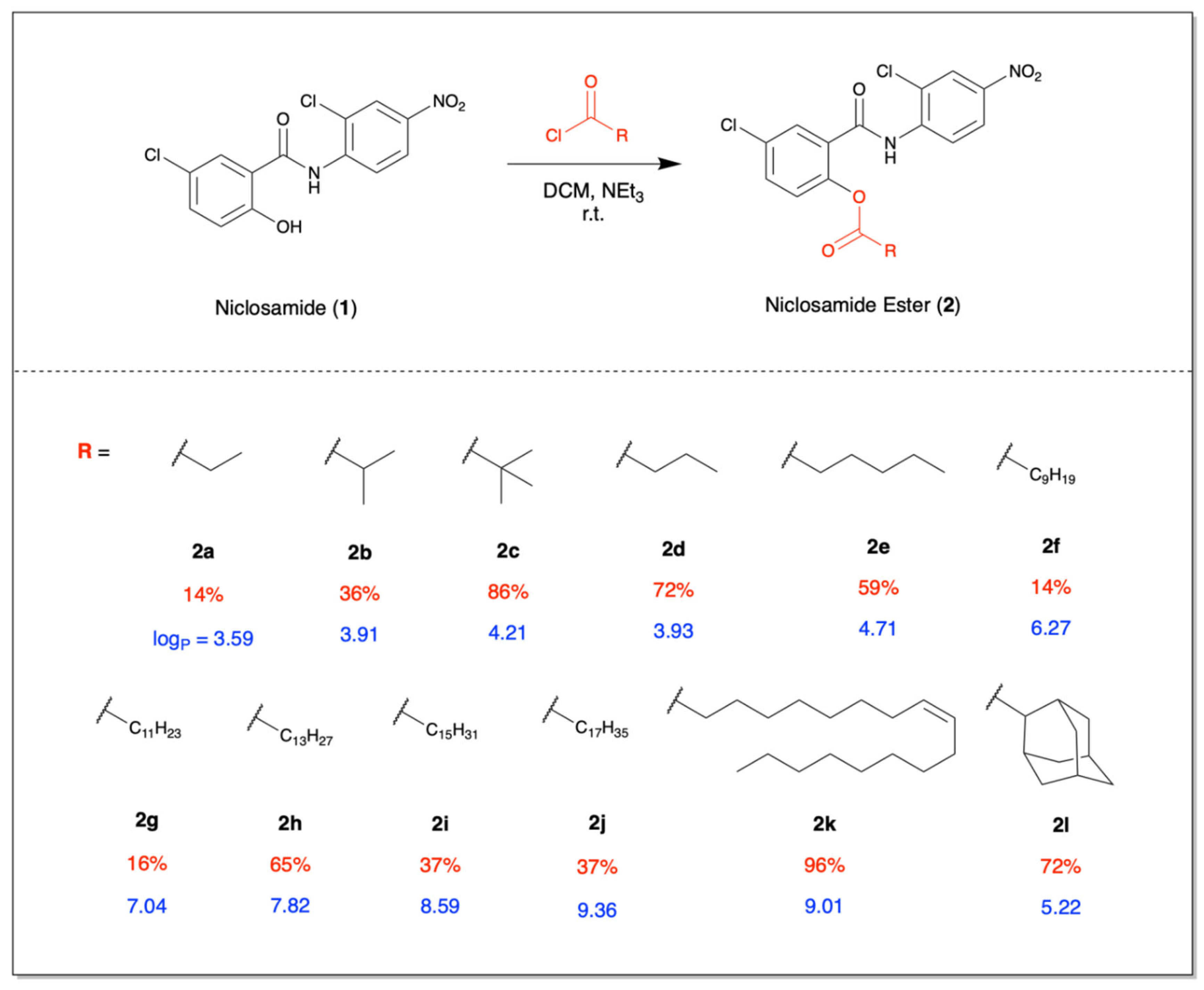
Esters 2a-2l were synthetically prepared from acylation of the phenol of niclosamide (1) with triethylamine and with an excess of the corresponding acid chloride, resulting in rapid conversion to the desired ester whose reaction progress can be monitored by thin layer chromatography (TLC). Upon complete consumption of niclosamide, the reaction was worked up in a brine/ethyl acetate solution, and the corresponding niclosamide ester was purified by silica gel flash chromatography. The propanoate (2a), isobutyrate (2b), pivaloate (2c), butanoate (2d), hexanoate (2e), decanoate (2f), myristate (2g), laurate (2h), palmitate (2i), stearate (2j), oleate (2k), and adamantate (2l) were synthesized in this manner, as shown in Scheme 1, in 14% to 96% isolated yield. Compound-specific experimentals and characterization data, including 1H and 13C NMR and FT-IR spectra, are found in the Supporting Information.
To synthesize niclosamide carbonates 3a-3i, we treated niclosamide (1) with pyridine followed by excess of the corresponding chloroformate, resulting in rapid conversion to the desired carbonate, with some reactions completing within 5 minutes. Reaction progress was monitored by thin layer chromatography, and upon complete consumption of niclosamide, the reaction was worked up in a brine/ethyl acetate solution and the corresponding niclosamide carbonate purified by silica gel flash chromatography. The ethyl (3a), isopropyl (3b), isobutyl (3c), cyclopentyl (3d), hexyl (3e), n-octyl (3f), decyl (3g), dodecyl (3h), and hexadecyl (3i) were synthesized, as shown in Scheme 2, in 26% to 76% yield. Compound-specific experimentals and characterization data, including 1H and 13C NMR and FT-IR spectra, are found in the Supporting Information.
3.2. SwissADME Results
In silico predicted ADMET and DMPK values for the ester and carbonate analogs was performed on the SwissADME suite[49] for each compound. Overall, the analogs maintained favorable drug-like properties while exhibiting systematic physiochemical changes associated with alkyl chain length. The predicted GI absorption values were found to be higher for the shorter and branched aliphatic esters, such as propanoate ester (2a), isobutyrate ester (2b), pivaloate ester (2c), and butanoate ester (2d). Additionally, for the esters, the bioavailability score decreased as the carbon chain length increased. However, the carbonates did not exhibit the same trend. When comparing tPSA (total polar surface area) and HBA (hydrogen bond acceptor) values, carbonate derivatives had higher values than the ester derivatives. RB (rotatable bonds), MR (Molar Refractivity), and log Kp values increased as the carbon chain length increased. Expectedly, as carbon chain length increases, the logP and tPSA values of both the esters and carbonates increase, indicating higher lipophilicity. Carbonate analogs also exhibited higher hydrogen bond acceptor counts than the corresponding esters, consistent with the additional oxygen atom within the carbonate functionality. Collectively, these in silico analyses suggest that promoiety selection can modulate the pharmaceutical properties of niclosamide prodrugs, providing useful guidance for the future optimization of niclosamide-based therapeutic candidates.

Table 2.
ADME valTable 2: Calculated metabolic and physiochemical values of ester and carbonate analogs, including select in silico swissADME values obtained for each compound. MW = Molecular weight, tPSA = Total Polarizable Surface Area, HBA = # of Hydrogen Bond Acceptors, HBD = # of Hydrogen Bond Donors, RB = # of Rotatable Bonds, MR = Molar Refractivity, GI = Gastrointestinal tract.
Table 2.
ADME valTable 2: Calculated metabolic and physiochemical values of ester and carbonate analogs, including select in silico swissADME values obtained for each compound. MW = Molecular weight, tPSA = Total Polarizable Surface Area, HBA = # of Hydrogen Bond Acceptors, HBD = # of Hydrogen Bond Donors, RB = # of Rotatable Bonds, MR = Molar Refractivity, GI = Gastrointestinal tract.
| Compound | MW | logP | tPSA | Fsp3 | HBA | HBD | RB | MR | Log Kp (cm/s) | Bioavailability Score | GI Absorption |
|---|---|---|---|---|---|---|---|---|---|---|---|
| Propanoate Ester (2a) | 383.18 | 3.96 | 107.21 | 0.12 | 5 | 1 | 7 | 95.8 | -5.75 | 0.55 | High |
| Isobutyrate Ester (2b) | 397.21 | 4.16 | 107.21 | 0.18 | 5 | 1 | 7 | 100.61 | -5.43 | 0.55 | High |
| Pivaloate Ester (2c) | 411.24 | 4.96 | 107.21 | 0.22 | 5 | 1 | 7 | 105.16 | -5.27 | 0.55 | High |
| Butanoate Ester (2d) | 397.21 | 4.42 | 107.21 | 0.18 | 5 | 1 | 8 | 100.61 | -5.58 | 0.55 | High |
| Hexanoate Ester (2e) | 425.26 | 5.33 | 107.21 | 0.26 | 5 | 1 | 10 | 110.23 | -4.99 | 0.55 | Low |
| Decanoate Ester (2f) | 481.37 | 7.15 | 107.21 | 0.39 | 5 | 1 | 14 | 129.45 | -3.79 | 0.55 | Low |
| Laurate Ester (2g) | 509.42 | 8.06 | 107.21 | 0.44 | 5 | 1 | 16 | 139.07 | -3.19 | 0.17 | Low |
| Myristate Ester (2h) | 537.48 | 8.98 | 107.21 | 0.48 | 5 | 1 | 18 | 148.68 | -2.59 | 0.17 | Low |
| Palmitate Ester (2i) | 565.53 | 9.89 | 107.21 | 0.52 | 5 | 1 | 20 | 158.3 | -2 | 0.17 | Low |
| Stearate Ester (2j) | 593.58 | 10.8 | 107.21 | 0.55 | 5 | 1 | 22 | 167.91 | -1.4 | 0.17 | Low |
| Oleate Ester (2k) | 591.57 | 10.09 | 107.21 | 0.48 | 5 | 1 | 21 | 167.44 | -2.05 | 0.17 | Low |
| Adamantate Ester (2l) | 489.35 | 5.89 | 107.21 | 0.42 | 5 | 1 | 7 | 127.92 | -4.58 | 0.55 | Low |
| Ethyl Carbonate (3a) | 399.18 | 4.16 | 116.44 | 0.12 | 6 | 1 | 8 | 97.28 | -5.64 | 0.56 | Low |
| Isopropyl Carbonate (3b) | 413.21 | 4.75 | 116.44 | 0.18 | 6 | 1 | 8 | 102.09 | -5.41 | 0.56 | Low |
| Isobutyl Carbonate (3c) | 427.24 | 4.82 | 116.44 | 0.22 | 6 | 1 | 9 | 106.89 | -5.12 | 0.56 | Low |
| Cyclopentyl Carbonate (3d) | 439.25 | 5.32 | 116.44 | 0.26 | 6 | 1 | 8 | 109.59 | -5.24 | 0.56 | Low |
| Hexyl Carbonate (3e) | 455.29 | 5.99 | 116.44 | 0.3 | 6 | 1 | 12 | 116.51 | -4.58 | 0.56 | Low |
| n-Octyl Carbonate (3f) | 483.34 | 6.9 | 116.44 | 0.53 | 6 | 1 | 14 | 126.12 | -3.98 | 0.56 | Low |
| Decyl Carbonate (3g) | 511.39 | 6.81 | 116.44 | 0.42 | 6 | 1 | 16 | 135.73 | -3.38 | 0.56 | Low |
| Dodecyl Carbonate (3h) | 539.45 | 8.72 | 116.44 | 0.46 | 6 | 1 | 18 | 145.35 | -2.79 | 0.56 | Low |
| Hexadecyl Carbonate (3i) | 595.55 | 10.55 | 116.44 | 0.53 | 6 | 1 | 22 | 164.58 | -1.59 | 0.56 | Low |
3.3. Hydrolysis Rates
To investigate the effects of these compounds as prodrugs, we performed a colorimetric hydrolysis assay, in which we tracked hydrolysis of each prodrug into niclosamide over a period of 60 minutes at physiological conditions, taking advantage of the unique yellow color and redshifted absorbance at 380 nm exhibited by niclosamide [Figure 2]. Within the first hour, almost all prodrugs demonstrated rapid conversion to niclosamide, as evidenced by a gradual increase in the absorbance at 380 nm, characteristic of niclosamide. Remarkably, the esters (compounds 2a - 2l) and carbonates (3a - 3i) exhibit hydrolysis rates within range of each other, with no distinct improvement in stability in the niclosamide carbonates in comparison to the amide series of prodrugs. Among carbonates, the dodecyl carbonate (3h) and cyclopentyl carbonate (3d) exhibited the lowest rate of release, while octyl carbonate (3f) and isobutyl carbonate (3c) appear to degrade most quickly in water. Similarly, the laurate ester (2g) and adamantate ester (2l) exhibited the lowest rate of ester autohydrolysis, while stearate ester (2j) exhibited much faster hydrolysis. In both of these cases, increasing alkyl demand or molecular weight did not result in a consistent trend.
Table 3.
Relative hydrolysis rates of ester and carbonate analogs at pH 7.0. Relative hydrolysis calculated from hydrolysis assay data.
Table 3.
Relative hydrolysis rates of ester and carbonate analogs at pH 7.0. Relative hydrolysis calculated from hydrolysis assay data.
| Compound | Rate Constant (min-1) | Time to 50% Hydrolysis |
|---|---|---|
| (min) | ||
| Propanoate Ester (2a) | 0.00817 | 84.8 |
| Isobutyrate Ester (2b) | 0.00539 | 129 |
| Pivaloate Ester (2c) | 0.00764 | 90.7 |
| Butanoate Ester (2d) | 0.00358 | 194 |
| Hexanoate Ester (2e) | 0.0126 | 54.9 |
| Decanoate Ester (2f) | 0.0134 | 51.7 |
| Laurate Ester (2g) | 0.00151 | 461 |
| Myristate Ester (2h) | 0.013 | 53.3 |
| Palmitate Ester (2i) | 0.0076 | 91.2 |
| Stearate Ester (2j) | 0.0434 | 16 |
| Oleate Ester (2k) | 0.00258 | 269 |
| Adamantate Ester (2l) | 0.0016 | 434 |
| Ethyl Carbonate (3a) | 0.0129 | 53.7 |
| Isopropyl Carbonate (3b) | 0.006073 | 114 |
| Isobutyl Carbonate (3c) | 0.0142 | 48.7 |
| Cyclopentyl Carbonate (3d) | 0.00155 | 447 |
| Hexyl Carbonate (3e) | 0.0115 | 60.3 |
| n-Octyl Carbonate (3f) | 0.0184 | 37.7 |
| Decyl Carbonate (3g) | 0.0138 | 50.4 |
| Dodecyl Carbonate (3h) | 0.00196 | 353 |
| Hexadecyl Carbonate (3i) | 0.00451 | 154 |
3.4. Cell Viability Assays
To evaluate the antiproliferative activity of our niclosamide ester and carbonate analogs in comparison to niclosamide, we measured their antiproliferative properties in a representative series of solid cancer models, including HCT-116, HT-29 human colon cancer cells, MDA-MB-231 human triple negative breast cancer cells, A549 human non-small cell lung cancer (NSCLC) cells, and B16F10 murine melanoma cancerous cell lines at a 72 hour timepoint. For both ester and carbonate analogs, similar antiproliferative activities were exhibited [Figure 3] with IC₅₀ (0.10 - 62.43 µM) of each compound comparable to that of the parent compound niclosamide (1) (0.25-11.7 µM)[50]. In most cases, the B16F10 cell line appears more sensitive to niclosamide and its prodrugs as compared to the human cell lines.
3.5. Washout Assay
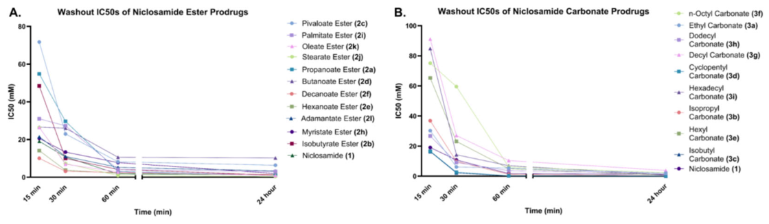
Next, to interrogate potential differences in the cellular uptake of our compounds, we examined the effects of drug washout at 15 minutes, 30 minutes, 60 minutes, and 24-hour timepoints. Expectedly, increasing timeframes for drug exposure led to a gradual decrease in observed 72 hour IC₅₀, with maximal potency observed at timeframes exceeding 60 minutes of drug exposure. Most compounds demonstrated similar IC₅₀ over these timepoints past 30 minutes. However, astute differences in 72 hr IC50 values observed from the 15 minutes washout timepoint revealed a precipitous loss in potency in short, branched aliphatic esters such as the pivaloate ester (2c, 15 min. washout IC50 = 71.72 mM), propanoate ester (2a, 15 min. washout IC50 = 54.82 mM), and isobutyrate ester (2b 15 min. washout IC50 = 48.43 mM) while long, aliphatic esters such as the decanoate ester (2f, 15 min. washout IC50 = 10.05 mM), hexanoate ester (2e, 15 min. washout IC50 = 14.14 mM) retained the most niclosamide-like anticancer activity (niclosamide 15 min. washout IC50 = 19.07 mM). Conversely, among carbonate prodrugs, we observed that long, linear aliphatic carbonates such as n-octyl carbonate (3f, 15 min. washout IC50 = 75.16), decyl carbonate (3g, 15 min. washout IC50 = 91.15), and hexadecyl carbonate (3i, 15 min. washout IC50 = 84.84) exhibited significantly attenuated activity upon 15 minute washout, while more compact carbonate moieties such as the cyclopentyl carbonate (3d, 15 min. washout IC50 = 16.67) retained the most potent niclosamide-like activity. These differences were largely diminished following 30 minutes of drug exposure, suggesting that the observed effects primarily reflect differences in the rate of early cellular accumulation rather than intrinsic antiproliferative potency. The opposing trends observed between the ester and carbonate series further indicate that the relationship between alkyl structure and short-term activity is influenced by the chemical nature of the promoiety, likely reflecting differences in membrane partitioning, intracellular uptake, and/or the rate of bioactivation during the initial period of drug exposure. Nevertheless, because all derivatives converged to comparable IC₅₀ values at longer exposure times, these early kinetic differences do not appear to substantially alter the overall therapeutic activity of the prodrugs under prolonged treatment conditions.
Figure 4.
Washout assay to determine sensitivity to prodrugs upon limited media drug exposure. IC₅₀ values of compounds evaluated in HT-29 cells across 15 minute, 30 minute, 60 minute, and 24 hour timepoints demonstrate gradually lower IC50 values upon longer drug incubation and exposure times.
Figure 4.
Washout assay to determine sensitivity to prodrugs upon limited media drug exposure. IC₅₀ values of compounds evaluated in HT-29 cells across 15 minute, 30 minute, 60 minute, and 24 hour timepoints demonstrate gradually lower IC50 values upon longer drug incubation and exposure times.
3.6. Luciferase Assay
To further investigate the biological mechanism of the niclosamide ester and carbonate derivatives, we selected a representative panel of carbonate and ester prodrugs for investigation a firefly luciferase reporter assay with a stably-transfected TCF/LEF-dependent fLuc reporter cell line, where results of select compounds highlighted a dose-dependent relationship in inhibition of Wnt1 signaling. All compounds exhibited potent reduction in luciferase activity, especially for pivaloate ester (2c), ethyl carbonate (3a), isopropyl carbonate (3b), n-octyl carbonate (3f), and hexadecyl carbonate (3i), highlighting their activity in inhibition of the Wnt1 signaling axis. Expectedly, these compounds retained or exceeded the original potency of niclosamide. Among esters, propanoate and decanoate esters exhibited lower activity in Wnt1 inhibition compared to niclosamide. By contrast, pivaloate ester (2c) exhibited improved potency in inhibition of Wnt1 signaling. On average, carbonates exhibited significantly amplified activity compared to the esters surveyed. It was determined via two-way ANOVA (n = 4)(p < 0.05) that, in comparison to niclosamide (1), several of these compounds are significantly better inhibitors of Wnt1 signaling, such as the pivaloate ester (2c), ethyl carbonate (3a), isopropyl carbonate (3b), n-octyl carbonate (3f), and hexadecyl carbonate (3i).
4. Discussion
Niclosamide, a small molecule therapeutic agent with demonstrated anticancer activity through multiple proposed pathways, has been previously converted into ester or carbonate prodrugs in efforts to improve its pharmacokinetic and pharmacodynamic properties. Here, we report a systematic structure-activity relationship (SAR) study of aliphatic carbonate and ester promoieties at the hydroxyl position of niclosamide in order to investigate the role of the aliphatic promoiety on in vitro anticancer activity. In a representative panel of human and murine cancer cell lines including HCT-116, HT-29, MDA-MB-231, A549, and B16F10, all compounds exhibited IC₅₀ values mostly in the single- to double-digit micromolar range, with similar potencies observed across several cell lines. All compounds were found to be slightly more potent in B16F10 murine melanoma cells. The similarity in potency of all carbonate and ester prodrugs of niclosamide with the parent drug itself is consistent with the observed hydrolysis kinetics exhibited by these prodrugs into the native drug within an hour. Additionally, in washout assays, differences in IC50 values could be observed at the 15 and 30 minute timepoints, wherein for esters, shorter carbon chain lengths demonstrated less potency, while among the carbonates, cyclopentyl (3d), dodecyl (3h), and ethyl carbonate (3a) were the most potent at the 15 minute time point. The origin of this selectivity at 15 minute washout may be due to a number of factors, including relative rates of membrane permeability. All compounds performed similarly to the native drug niclosamide by the 60 minute timepoint, which can be attributed to the rapid hydrolysis of the compounds back into niclosamide within an hour at a pH 7.0, which is the same pH as cell culture media.
This efficient hydrolysis to niclosamide by the 60 minute time point results in equal potency in cell viability assays when measured after 72 hours. This consistent trend of therapeutic efficacy across multiple time points is further corroborated by an fLuc-based Wnt1 reporter assay. A select panel of prodrugs [Figure 5] retained the dose-dependent Wnt1-inhibiting properties similar to niclosamide (4.2 - 42% at 250 µM, 18 - 72% at 25 µM), which is consistent with observed hydrolysis back into niclosamide well within this time frame. Most compounds exhibit better Wnt1-inhibiting activity than niclosamide as demonstrated by the two-way ANOVA test, suggesting that the promoiety improved the potency of niclosamide into the cell.
In a cell-free hydrolysis assay, most compounds in the library exhibited similar rates of hydrolysis. However, it was observed that isopropyl carbonate (3b) and cyclopentyl carbonate (3d) exhibited particularly sluggish hydrolysis relative to linear carbonates. Notably, the cyclopentyl carbonate exhibits the slowest hydrolysis (k = 0.00155 min-1) compared to isopropyl carbonate (3b) (k = 0.00607 min-1), which is slower still compared to linear carbonate prodrugs such as ethyl carbonate (3a) (k = 0.0129 min-1). While canonical mechanisms of carbonate hydrolysis involve nucleophilic attack of the solvent on the carbonate carbonyl [Figure 6B], the observed kinetics from this study suggest that an SN2 mechanism may be operative, in which a cyclic aliphatic carbonate results in the relatively inaccessibility of an electrophilic carbon directly flanking the carbonate functional group. [Figure 6C] This is consistent with our observation that sterically hindered carbonates, including the cyclopentyl carbonate (3d) and isopropyl carbonate (3b), bear significantly lower hydrolysis rates than other compounds, with respective rate constants of 0.00155 min-1 and 0.00607 min-1 and hydrolysis half lives of 447 and 114 minutes. In contrast, compounds with long but accessible alkyl chains and more steric bulk appear to undergo hydrolysis more rapidly. Among carbonate prodrugs, the hexyl carbonate (3e) showed the fastest hydrolysis rate with a rate constant of 0.0434 min-1 and half-time of 16.0 minutes. Importantly, the hydrolysis rates of these compounds do not correlate with differences in biological activity in antiproliferative assays. Additionally, through in silico predicted DMPK and PD/PK parameters obtained from SwissADME, we obtained expected trends in lipophilicity and other physiochemical properties, all of which were not observed to directly correlate with either in vitro anticancer activity or with cell-free hydrolysis kinetics.
Collectively, these results indicate that aliphatic carbonate and ester prodrugs of the therapeutic agent niclosamide have only a limited effect on the in vitro pharmacological activity of niclosamide, as rapid conversion to niclosamide under physiological conditions results in efficient regeneration of the parent drug. While alkyl structure influences hydrolysis kinetics, early cellular activity following brief drug exposure, and predicted physiochemical properties, these effects largely converge following prolonged incubation, yielding comparable antiproliferative activity across multiple cancer models. Accordingly, this work informs the future optimization of niclosamide prodrugs, which may prioritize optimization of formulation, target tissue distribution, and in vivo therapeutic efficacy; these findings provide a translational framework for the continued development of niclosamide-based anticancer therapeutics, particularly in the use of ester and carbonate prodrug design as a strategy to improve drug delivery while preserving the broad anticancer activity of the parent compound.
5. Conclusions
Since its initial discovery as an anthelmintic agent, niclosamide has since been repurposed for a number of human clinical indications, and has specifically demonstrated potency in several pathways connected to cancer, including inhibition of STAT3, Wnt1/β-catenin, Notch, and NF-kB signaling pathways. However, given its poor aqueous solubility, many have undertaken efforts to improve the compound’s bioavailability through modification of the phenol. Here, we report a systematic study of the biological activity and physiochemical properties of a library of alkyl derivatives of niclosamide.
Despite spanning a broad range of alkyl ester and carbonate promoieties, these derivatives displayed remarkably consistent antiproliferative activity across colorectal, breast, lung, and melanoma cancer models. Although measurable differences in hydrolysis kinetics were observed, these differences were insufficient to produce meaningful changes in cellular potency, washout behavior, or Wnt/β-catenin inhibition. Instead, the rapid hydrolysis of nearly all derivatives under physiologically relevant conditions resulted in efficient regeneration of niclosamide, indicating that the biological activity of this series is governed primarily by the parent drug rather than the identity of the promoiety. Collectively, these findings establish rapid bioactivation as a defining characteristic of niclosamide ester and carbonate prodrugs and suggest that modulation of alkyl chain length or branching are unlikely to substantially alter in vitro therapeutic efficacy.
The rapid and predictable conversion of these derivatives to the active drug demonstrates that ester and carbonate masking groups can function as transient delivery vehicles while preserving the established pharmacological profile of niclosamide. Together with favorable in silico pharmacokinetic and drug metabolism predictions, these findings provide a framework for the continued optimization of niclosamide prodrugs toward improved pharmaceutical performance, including enhanced formulation, systemic exposure, tissue distribution, and in vivo therapeutic efficacy. Future studies will focus on evaluating these properties in animal models to determine whether the favorable activation characteristics observed in vitro translate into improved anticancer activity in vivo.
Supplementary Materials
The following supporting information can be downloaded at website of this paper posted on Preprints.org. The full synthesis, 1H NMR, 13C NMR, and FT-IR characterization of all compounds, their inhibitory dose-response curves, as well as detailed procedures for cell culture, cell viability assays, reporter cell assays, and cell-free assays can be downloaded in the online Supporting Information Document.
Author Contributions
Conceptualization, E.N.; methodology, A.G., M.P., K.L., H.K., K.Z., E.N.; software, M.P., H.K.; formal analysis, A.G., M.P., K.L., H.K., K.Z., C.D., I.C., C.C., A.S., A.Y., E.H., J.S., M.T., E.N..; investigation, A.G., M.P., K.L., H.K., K.Z., C.D., I.C., C.C., A.S., A.Y., E.H., J.S., M.T., E.N.; resources, E.N.; data curation, A.G., M.P., K.L., H.K., K.Z., C.D., I.C., C.C., A.S., A.Y., E.H., J.S., M.T., E.N.; writing—original draft preparation, A.G., M.P., K.L, H.K., K.Z., C.D., E.N.; writing—review and editing, A.G., M.P., K.L., H.K., K.Z., C.D., I.C., A.S., E.N.; visualization, A.G., M.P., H.K.; supervision, E.N.; project administration, E.N. funding acquisition, E.N. All authors have read and agreed to the published version of the manuscript.
Funding
This research did not receive any external funding.
Informed Consent Statement
Not applicable.
Data Availability Statement
All experimental data, spectroscopic data, and additional raw data are available online in the Supporting Information document.
Acknowledgments
The authors gratefully acknowledge Dr. Feng Wang-Johanning and Dr. Gary Johanning from SunnyBay Biotech for their generous contribution of MDA-MB-231 and B16F10 cell lines. Additionally, the authors gratefully acknowledge Dr. Stephen Lynch from Stanford University Nuclear Magnetic Resonance (NMR) facility for access to high field NMR spectra.
Conflicts of Interest
The authors declare no conflicts of interest.
Abbreviations
The following abbreviations are used in this manuscript:
| FDA | Food and Drug Administration |
| STAT3 | Signal Transducer and Activator of Transcription 3 |
| NF-κB | Nuclear Factor kappa-light-chain-enhancer of activated B cells |
| Wnt | Wingless-related integration site |
| AKT | Protein Kinase B |
| ERK | Extracellular Signal-Regulated Kinase |
| Src | Sarcoma |
| c-Fos | cellular Fibroblast Osteosarcoma |
| c-Jun | cellular Junction |
| E2F1 | E2F Transcription Factor 1 |
| c-Myc | cellular Myelocytomatosis |
| AR-V7 | Androgen Receptor Splice Variant 7 |
| p-JNK | Phosphorylated c-Jun N-terminal kinase |
| JNK | c-Jun N-terminal kinase |
| mTORC1 | Mammalian Target of Rapamycin Complex 1 |
| CTNNB1 | Catenin Beta 1 |
| mTOR | mammalian target of rapamycin |
| miR-200 | microRNA-200 |
| EGFR | Epidermal Growth Factor Receptor |
| LRP6 | Low-Density Lipoprotein Receptor-Related Protein 6 |
| DMPK | Drug Metabolism and Pharmacokinetics |
| ADME | Absorption, Distribution, Metabolism, and Excretion |
| SCLC | Small Cell Lung Cancer |
| PEG | Polyethylene Glycol |
| fLuc | Firefly luciferase |
| TCF/LEF | T-Cell Factor/Lymphoid Enhancer-Binding Factor |
| DMSO | Dimethyl Sulfoxide |
| TRIS | Tris(hydroxymethyl)aminomethane |
| FBS | Fetal Bovine Serum |
| DMEM | Dulbecco’s Modified Eagle Medium |
| MTT | 3-(4,5-dimethylthiazol-2-yl)-2,5-diphenyltetrazolium bromide |
| PBS | Phosphate-Buffered Saline |
| TLC | Thin layer chromatography |
| FT-IR | Fourier Transform Infrared |
| tPSA | Total Polar Surface Area |
| HBA | Number of Hydrogen Bond Acceptors |
| HBD | Number of Hydrogen Bond Donors |
| RB | Number of Rotatable Bonds |
| MR | Molar Refractivity |
| GI | Gastrointestinal tract |
| NSCLC | Non-Small Cell Lung Cancer |
| IC50 | Half-Maximal Inhibitory Concentration |
| ANOVA | Analysis of Variance |
| SD | Standard Deviation |
| SN2 | Substitution Nucleophilic Bimolecular |
| SAR | Structure Activity Relationship |
| NMR | Nuclear Magnetic Resonance |
| UV-Vis | Ultraviolet-Visible |
References
- Gupta, S.C.; Sung, B.; Prasad, S.; Webb, L.J.; Aggarwal, B.B. Cancer Drug Discovery by Repurposing: Teaching New Tricks to Old Dogs. Trends Pharmacol. Sci. 2013, 34, 508–517. [Google Scholar] [CrossRef] [PubMed]
- Pushpakom, S.; Iorio, F.; Eyers, P.A.; Escott, K.J.; Hopper, S.; Wells, A.; Doig, A.; Guilliams, T.; Latimer, J.; McNamee, C.; et al. Drug Repurposing: Progress, Challenges and Recommendations. Nat. Rev. Drug Discov. 2018, 18, 41–58. [Google Scholar] [CrossRef] [PubMed]
- Oprea, T.I.; Bauman, J.E.; Bologa, C.G.; Buranda, T.; Chigaev, A.; Edwards, B.S.; Jarvik, J.W.; Gresham, H.D.; Haynes, M.K.; Hjelle, B.; et al. Drug Repurposing from an Academic Perspective. Drug Discov. Today Ther. Strateg. 2011, 8, 61–69. [Google Scholar] [CrossRef] [PubMed]
- Patmanathan, S.N.; Yap, L.F.; Murray, P.G.; Paterson, I.C. The Antineoplastic Properties of FTY720: Evidence for the Repurposing of Fingolimod. J. Cell. Mol. Med. 2015, 19, 2329–2340. [Google Scholar] [CrossRef] [PubMed]
- Jin, H.W.; Bae, J.H.; Youn, H.S.; Eom, Y. Bin Repurposing Fingolimod as an Antibacterial and Antibiofilm Agent against Carbapenem-Resistant Acinetobacter Baumannii. ACS Omega 2026, 11, 32158–32166. [Google Scholar] [CrossRef] [PubMed]
- Levenfors, J.J.; Bjerketorp, J.; Guss, B.; Nord, C.; Cao, S.; Hughes, D.; Broberg, A.; Öberg, B. Repurposing Zidovudine and 5-Fluoro-2′-Deoxyuridine as Antibiotic Drugs Made Possible by Synergy with Both Trimethoprim and the Mitochondrial Toxicity–Reducing Agent Uridine. J. Antimicrob. Chemother. 2025, 80, 509–517. [Google Scholar] [CrossRef] [PubMed]
- Tarawneh, N.; Hussein, S.A.; Abdalla, S. Repurposing Antiepileptic Drugs for Cancer: A Promising Therapeutic Strategy. J. Clin. Med. 2025, 14, 1–26. [Google Scholar] [CrossRef] [PubMed]
- Gu, T.; Lu, A.; Wang, X.; Brahan, N.; Xu, L.; Zhang, L.; Su, K.; Seow, K.; Vu, J.; Luk, C.; et al. Synthesis and Evaluation of Carmofur Analogs as Antiproliferative Agents, Inhibitors to the Main Protease (Mpro) of SARS-CoV-2, and Membrane Rupture-Inducing Agents. Discov. Chem. 2025, 2, 73. [Google Scholar] [CrossRef]
- Gao, J.; Zhang, L.; Liu, X.; Li, F.; Ma, R.; Zhu, Z.; Zhang, J.; Wu, J.; Shi, Y.; Pan, Y.; et al. Repurposing Low-Molecular-Weight Drugs against the Main Protease of Severe Acute Respiratory Syndrome Coronavirus 2. J. Phys. Chem. Lett. 2020, 11, 7267–7272. [Google Scholar] [CrossRef] [PubMed]
- Ma, C.; Hu, Y.; Townsend, J.A.; Lagarias, P.I.; Marty, M.T.; Kolocouris, A.; Wang, J. Ebselen, Disulfiram, Carmofur, PX-12, Tideglusib, and Shikonin Are Nonspecific Promiscuous SARS-CoV-2 Main Protease Inhibitors. ACS Pharmacol. Transl. Sci. 2020, 3, 1265–1277. [Google Scholar] [CrossRef] [PubMed]
- Jin, Z.; Zhao, Y.; Sun, Y.; Zhang, B.; Wang, H.; Wu, Y.; Zhu, Y.; Zhu, C.; Hu, T.; Du, X.; et al. Structural Basis for the Inhibition of SARS-CoV-2 Main Protease by Antineoplastic Drug Carmofur. Nat. Struct. Mol. Biol. 2020, 27, 529–532. [Google Scholar] [CrossRef] [PubMed]
- Imperi, F.; Massai, F.; Pillai, C.R.; Longo, F.; Zennaro, E.; Rampioni, G.; Visc, P.; Leoni, L. New Life for an Old Drug: The Anthelmintic Drug Niclosamide Inhibits Pseudomonas Aeruginosa Quorum Sensing. Antimicrob. Agents Chemother. 2013, 57, 996–1005. [Google Scholar] [CrossRef] [PubMed]
- Barini, E.; Miccoli, A.; Tinarelli, F.; Mulholland, K.; Kadri, H.; Khanim, F.; Stojanovski, L.; Read, K.D.; Burness, K.; Blow, J.J.; et al. The Anthelmintic Drug Niclosamide and Its Analogues Activate the Parkinson’s Disease Associated Protein Kinase PINK1. ChemBioChem 2018, 19, 425–429. [Google Scholar] [CrossRef] [PubMed]
- Tao, H.; Zhang, Y.; Zeng, X.; Shulman, G.I.; Jin, S. Niclosamide Ethanolamine–Induced Mild Mitochondrial Uncoupling Improves Diabetic Symptoms in Mice. Nat. Med. 2014, 20, 1263–1269. [Google Scholar] [CrossRef] [PubMed]
- Rajamuthiah, R.; Fuchs, B.B.; Conery, A.L.; Kim, W.; Jayamani, E.; Kwon, B.; Ausubel, F.M.; Mylonakis, E. Repurposing Salicylanilide Anthelmintic Drugs to Combat Drug Resistant Staphylococcus Aureus. PLoS ONE 2015, 10, e0124595. [Google Scholar] [CrossRef] [PubMed]
- Wang, A.; Ku, H.; Liang, Y.; Chen, Y.; Hwu, Y.; Yeh, T. The Autonomous Notch Signal Pathway Is Activated by Baicalin and Baicalein but Is Suppressed by Niclosamide in K562 Cells. J. Cell. Biochem. 2009, 106, 682–692. [Google Scholar] [CrossRef] [PubMed]
- Kadri, H.; Lambourne, O.A.; Mehellou, Y. Niclosamide, a Drug with Many (Re)Purposes. ChemMedChem 2018, 13, 1088–1091. [Google Scholar] [CrossRef] [PubMed]
- Liu, J.; Chen, X.; Ward, T.; Pegram, M.; Shen, K. Combined Niclosamide with Cisplatin Inhibits Epithelial-Mesenchymal Transition and Tumor Growth in Cisplatin-Resistant Triple-Negative Breast Cancer. Tumor Biol. 2016, 37, 9825–9835. [Google Scholar] [CrossRef] [PubMed]
- Liao, Z.; Nan, G.; Yan, Z.; Zeng, L.; Deng, Y.; Ye, J.; Zhang, Z.; Qiao, M.; Li, R.; Denduluri, S.; et al. The Anthelmintic Drug Niclosamide Inhibits the Proliferative Activity of Human Osteosarcoma Cells by Targeting Multiple Signal Pathways. Curr. Cancer Drug Targets 2015, 15, 726–738. [Google Scholar] [CrossRef] [PubMed]
- Ren, X.; Duan, L.; He, Q.; Zhang, Z.; Zhou, Y.; Wu, D.; Pan, J.; Pei, D.; Ding, K. Identification of Niclosamide as a New Small-Molecule Inhibitor of the STAT3 Signaling Pathway. ACS Med. Chem. Lett. 2010, 1, 454–459. [Google Scholar] [CrossRef] [PubMed]
- Li, R.; You, S.; Hu, Z.; Chen, Z.G.; Sica, G.L.; Khuri, F.R.; Curran, W.J.; Shin, D.M.; Deng, X. Inhibition of STAT3 by Niclosamide Synergizes with Erlotinib against Head and Neck Cancer. PLoS ONE 2013, 8, e74670. [Google Scholar] [CrossRef] [PubMed]
- Liu, C.; Lou, W.; Armstrong, C.; Zhu, Y.; Evans, C.P.; Gao, A.C. Niclosamide Suppresses Cell Migration and Invasion in Enzalutamide Resistant Prostate Cancer Cells via Stat3-AR Axis Inhibition. Prostate 2015, 75, 1341–1353. [Google Scholar] [CrossRef] [PubMed]
- Balgi, A.D.; Fonseca, B.D.; Donohue, E.; Tsang, T.C.F.; Lajoie, P.; Proud, C.G.; Nabi, I.R.; Roberge, M. Screen for Chemical Modulators of Autophagy Reveals Novel Therapeutic Inhibitors of MTORC1 Signaling. PLoS ONE 2009, 4, e7124. [Google Scholar] [CrossRef] [PubMed]
- Wieland, A.; Trageser, D.; Gogolok, S.; Reinartz, R.; Höfer, H.; Keller, M.; Leinhaas, A.; Schelle, R.; Normann, S.; Klaas, L.; et al. Anticancer Effects of Niclosamide in Human Glioblastoma. Clin. Cancer Res. 2013, 19, 4124–4136. [Google Scholar] [CrossRef] [PubMed]
- Suliman, M.A.; Zhang, Z.; Na, H.; Ribeiro, A.L.L.; Zhang, Y.; Niang, B.; Hamid, A.S.; Zhang, H.; Xu, L.; Zuo, Y. Niclosamide Inhibits Colon Cancer Progression through Downregulation of the Notch Pathway and Upregulation of the Tumor Suppressor MiR-200 Family. Int. J. Mol. Med. 2016, 38, 776–784. [Google Scholar] [CrossRef] [PubMed]
- Kumar, R.; Coronel, L.; Somalanka, B.; Raju, A.; Aning, O.A.; An, O.; Ho, Y.S.; Chen, S.; Mak, S.Y.; Hor, P.Y.; et al. Mitochondrial Uncoupling Reveals a Novel Therapeutic Opportunity for P53-Defective Cancers. Nat. Commun. 2018, 9, 3931. [Google Scholar] [CrossRef] [PubMed]
- Lu, W.; Lin, C.; Roberts, M.J.; Waud, W.R.; Piazza, G.A.; Li, Y. Niclosamide Suppresses Cancer Cell Growth By Inducing Wnt Co-Receptor LRP6 Degradation and Inhibiting the Wnt/β-Catenin Pathway. PLoS ONE 2011, 6, e29290. [Google Scholar] [CrossRef] [PubMed]
- Chen, M.; Wang, J.; Lu, J.; Bond, M.C.; Ren, X.-R.; Lyerly, H.K.; Barak, L.S.; Chen, W. The Anti-Helminthic Niclosamide Inhibits Wnt/Frizzled1 Signaling. Biochemistry 2009, 48, 10267–10274. [Google Scholar] [CrossRef] [PubMed]
- Sack, U.; Walther, W.; Scudiero, D.; Selby, M.; Kobelt, D.; Lemm, M.; Fichtner, I.; Schlag, P.M.; Shoemaker, R.H.; Stein, U. Novel Effect of Antihelminthic Niclosamide on S100A4-Mediated Metastatic Progression in Colon Cancer. JNCI J. Natl. Cancer Inst. 2011, 103, 1018–1036. [Google Scholar] [CrossRef] [PubMed]
- Chen, H.; Yang, Z.; Ding, C.; Chu, L.; Zhang, Y.; Terry, K.; Liu, H.; Shen, Q.; Zhou, J. Discovery of O -Alkylamino-Tethered Niclosamide Derivatives as Potent and Orally Bioavailable Anticancer Agents. ACS Med. Chem. Lett. 2013, 4, 180–185. [Google Scholar] [CrossRef] [PubMed]
- Spessard, G.O. ACD Labs/LogP DB 3.5 and ChemSketch 3.5. J. Chem. Inf. Comput. Sci. 1998, 38, 1250–1253. [Google Scholar] [CrossRef]
- Zaro, J.L. Lipid-Based Drug Carriers for Prodrugs to Enhance Drug Delivery. AAPS J. 2015, 17, 83–92. [Google Scholar] [CrossRef] [PubMed]
- Han, S.; Mei, L.; Quach, T.; Porter, C.; Trevaskis, N. Lipophilic Conjugates of Drugs: A Tool to Improve Drug Pharmacokinetic and Therapeutic Profiles. Pharm. Res. 2021, 38, 1497–1518. [Google Scholar] [CrossRef] [PubMed]
- Subbaiah, M.A.M.; Rautio, J.; Meanwell, N.A. Prodrugs as Empowering Tools in Drug Discovery and Development: Recent Strategic Applications of Drug Delivery Solutions to Mitigate Challenges Associated with Lead Compounds and Drug Candidates. Chem. Soc. Rev. 2024, 53, 2099–2210. [Google Scholar] [CrossRef] [PubMed]
- Rautio, J.; Kumpulainen, H.; Heimbach, T.; Oliyai, R.; Oh, D.; Järvinen, T.; Savolainen, J. Prodrugs: Design and Clinical Applications. Nat. Rev. Drug Discov. 2008, 7, 255–270. [Google Scholar] [CrossRef] [PubMed]
- Mito, S.; Cheng, B.; Garcia, B.A.; Yee Ooi, X.; Gonzalez, D.; Ruiz, T.C.; Elisarraras, F.X.; Tsin, A. SAR Study of Niclosamide Derivatives for Neuroprotective Function in SH-SY5Y Neuroblastoma. Bioorg. Med. Chem. Lett. 2023, 96, 129498. [Google Scholar] [CrossRef] [PubMed]
- Mito, S.; Cheng, B.; Garcia, B.A.; Gonzalez, D.; Ooi, X.Y.; Ruiz, T.C.; Elisarraras, F.X.; Tsin, A.; Chew, S.A.; Arriaga, M.A. SAR Study of Niclosamide Derivatives in the Human Glioblastoma U-87 MG Cells. Med. Chem. Res. 2022, 31, 1313–1322. [Google Scholar] [CrossRef] [PubMed]
- He, X.; Li, M.; Ye, W.; Zhou, W. Discovery of Degradable Niclosamide Derivatives Able to Specially Inhibit Small Cell Lung Cancer (SCLC). Bioorg. Chem. 2021, 107, 104574. [Google Scholar] [CrossRef] [PubMed]
- Coomar, S.; Gasser, J.A.; Słabicki, M.; Donovan, K.A.; Fischer, E.S.; Ebert, B.L.; Gillingham, D.; Thomä, N.H. Niclosamide: CRL4 AMBRA1 Mediated Degradation of Cyclin D1 Following Mitochondrial Membrane Depolarization. RSC Med. Chem. 2025, 16, 3049–3057. [Google Scholar] [CrossRef] [PubMed]
- Yuan, M.; Li, J.-M.; He, G.-W.; Zhong, G.-C.; Zhang, Y.-C. Design, Synthesis and Antitumor Activity of Valproic Acid Salicylanilide Esters. Yao Xue Xue Bao 2013, 48, 874–880. [Google Scholar] [PubMed]
- Wang, J.; Li, J.-M.; Yuan, M.; Zhou, P. Design, Synthesis and Antitumor Activity of n-Butyric Acid Salicylanilide Esters. Chin. J. New Drugs 2013, 22. [Google Scholar]
- Elkihel, L.; Loiseau, P.M.; Bourass, J.; Gayral, P.; Letourneux, Y. Synthesis and Orally Macrofilaricidal Evaluation of Niclosamide Lymphotropic Prodrugs. Arzneimittelforschung 1994, 44, 1259–1264. [Google Scholar] [PubMed]
- Reddy, G.B.; Kerr, D.L.; Spasojevic, I.; Tovmasyan, A.; Hsu, D.S.; Brigman, B.E.; Somarelli, J.A.; Needham, D.; Eward, W.C. Preclinical Testing of a Novel Niclosamide Stearate Prodrug Therapeutic (NSPT) Shows Efficacy Against Osteosarcoma. Mol. Cancer Ther. 2020, 19, 1448–1461. [Google Scholar] [CrossRef] [PubMed]
- Bhattacharyya, J.; Ren, X.-R.; Mook, R.A.; Wang, J.; Spasojevic, I.; Premont, R.T.; Li, X.; Chilkoti, A.; Chen, W. Niclosamide-Conjugated Polypeptide Nanoparticles Inhibit Wnt Signaling and Colon Cancer Growth. Nanoscale 2017, 9, 12709–12717. [Google Scholar] [CrossRef] [PubMed]
- Ma, R.; Ma, Z.; Gao, J.; Tai, Y.; Li, L.; Zhu, H.; Li, L.; Dong, D.; Sun, Z. Injectable Pegylated Niclosamide (Polyethylene Glycol-modified Niclosamide) for Cancer Therapy. J. Biomed. Mater. Res. Part A 2020, 108, 30–38. [Google Scholar] [CrossRef] [PubMed]
- Kocalar, S.; Njoo, E. Taft Linear Free-Energy Relationships in the Biocatalytic Hydrolysis of Sterically Hindered Nitrophenyl Ester Substrates. J. Emerg. Investig. 2022, 5, 2–7. [Google Scholar] [CrossRef] [PubMed]
- Gu, T.; Raval, R.; Bashkin, Z.; Zhou, C.; Ko, S.; Kong, N.; Hong, S.; Bhaskara, A.; Shah, S.; Joshi, A.; et al. Synthesis, Antiproliferative Activity, and Biological Profiling of C-19 Trityl and Silyl Ether Andrographolide Analogs in Colon Cancer and Breast Cancer Cells. Bioorganic Med. Chem. Lett. 2025, 121, 130163. [Google Scholar] [CrossRef] [PubMed]
- Gutti, M.; Luo, J.; Sharma, V.; Lee, E.; Desarkar, A.; Raval, R.; Soedarmono, A.; Zhu, J.; Njoo, E. Anticancer Synthetic Arylsulfonamides with Wnt1-Modulating Activity. Essent. Chem. 2026, 3, 2697084. [Google Scholar] [CrossRef]
- Daina, A.; Michielin, O.; Zoete, V. SwissADME: A Free Web Tool to Evaluate Pharmacokinetics, Drug-Likeness and Medicinal Chemistry Friendliness of Small Molecules. Sci. Rep. 2017, 7, 42717. [Google Scholar] [CrossRef] [PubMed]
- Pan, J.X.; Ding, K.; Wang, C.Y. Niclosamide, an Old Antihelminthic Agent, Demonstrates Antitumor Activity by Blocking Multiple Signaling Pathways of Cancer Stem Cells. Chin. J. Cancer 2012, 31, 178. [Google Scholar] [CrossRef] [PubMed]
Scheme 1.
Synthesis of niclosamide ester analogs Esters were synthesized in DCM by treating niclosamide (1) with triethylamine before the corresponding acid chloride was added to the reaction mixture. Isolated yields are shown in red and calculated logₚ shown in blue.t.
Scheme 1.
Synthesis of niclosamide ester analogs Esters were synthesized in DCM by treating niclosamide (1) with triethylamine before the corresponding acid chloride was added to the reaction mixture. Isolated yields are shown in red and calculated logₚ shown in blue.t.
Scheme 2.
Synthesis of niclosamide carbonate analogs Carbonates were synthesized in THF by treating niclosamide (1) with pyridine before the corresponding chloroformate was added dropwise to the reaction mixture at 0oC. Following that, the mixture was allowed to warm to room temperature. Isolated yields are shown in red and calculated logₚ shown in blue.
Scheme 2.
Synthesis of niclosamide carbonate analogs Carbonates were synthesized in THF by treating niclosamide (1) with pyridine before the corresponding chloroformate was added dropwise to the reaction mixture at 0oC. Following that, the mixture was allowed to warm to room temperature. Isolated yields are shown in red and calculated logₚ shown in blue.
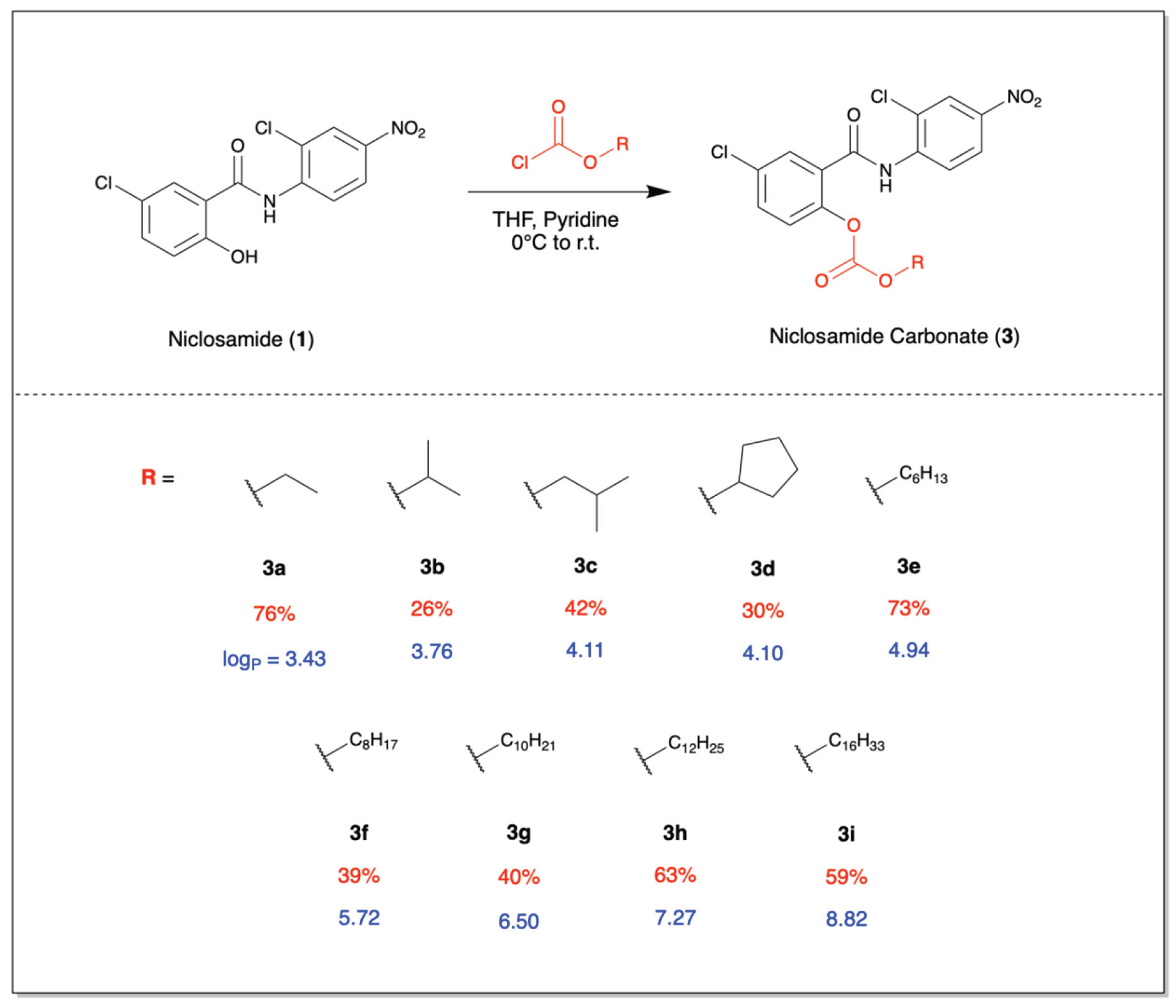
Figure 2.
Hydrolysis rates of ester and carbonate analogs Hydrolysis rates of niclosamide prodrugs over the course of 60 minutes in five-minute increments, tracked through a gradual increase in absorbance at 380 nm.
Figure 2.
Hydrolysis rates of ester and carbonate analogs Hydrolysis rates of niclosamide prodrugs over the course of 60 minutes in five-minute increments, tracked through a gradual increase in absorbance at 380 nm.
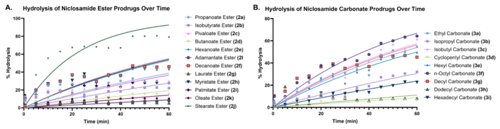
Figure 3.
Cell viabilities after 72 hour time point IC₅₀ values of compounds evaluated across HCT-116, HT-29, MDA-MB-231, A549, and B16F10 cell lines.
Figure 3.
Cell viabilities after 72 hour time point IC₅₀ values of compounds evaluated across HCT-116, HT-29, MDA-MB-231, A549, and B16F10 cell lines.
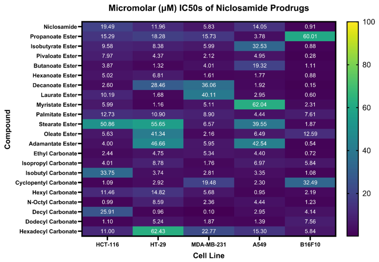
Figure 5.
Wnt1 Activity fLuc Reporter Assay: Percent luciferase activity of select compounds at 250 µM and 25 µM, with DMSO control. All compounds demonstrate decreased luciferase activity, correlating to their ability to inhibit Wnt1. Data is represented as means ± SD compared with the negative control using a two-way ANOVA (n = 4)(****p < 0.0001).
Figure 5.
Wnt1 Activity fLuc Reporter Assay: Percent luciferase activity of select compounds at 250 µM and 25 µM, with DMSO control. All compounds demonstrate decreased luciferase activity, correlating to their ability to inhibit Wnt1. Data is represented as means ± SD compared with the negative control using a two-way ANOVA (n = 4)(****p < 0.0001).
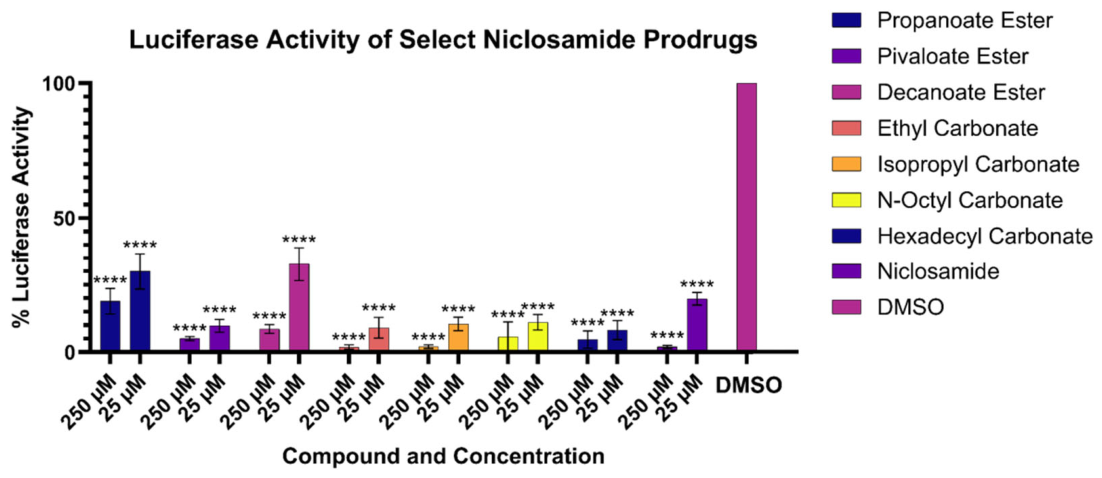
Figure 6.
Possible mechanisms of carbonate hydrolysis (A) Generalized schematic of hydrolysis of niclosamide carbonate prodrugs at physiological pH. (B) Possible mechanism with a carbonyl electrophile. (C) Possible alternate SN2 mechanism supported by the hydrolysis kinetics, wherein differences in hydrolysis kinetics arise from niclosamide analogs with long alkyl chains and more steric bulk. Hydrolysis rate constant (in min-1) is represented in red, while the time for 50% hydrolysis of compounds (in minutes) is represented in blue.
Figure 6.
Possible mechanisms of carbonate hydrolysis (A) Generalized schematic of hydrolysis of niclosamide carbonate prodrugs at physiological pH. (B) Possible mechanism with a carbonyl electrophile. (C) Possible alternate SN2 mechanism supported by the hydrolysis kinetics, wherein differences in hydrolysis kinetics arise from niclosamide analogs with long alkyl chains and more steric bulk. Hydrolysis rate constant (in min-1) is represented in red, while the time for 50% hydrolysis of compounds (in minutes) is represented in blue.
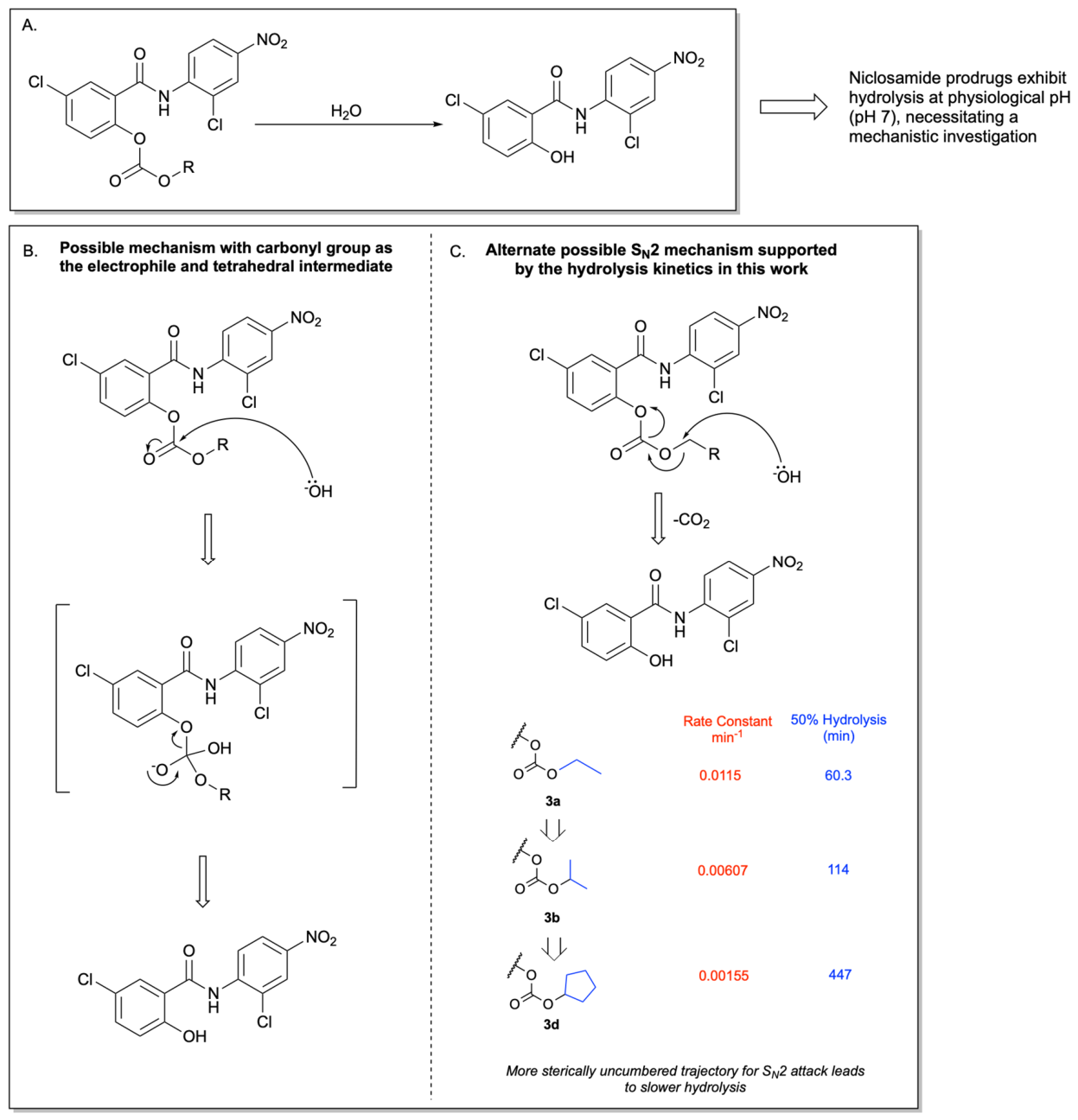
Table 1.
Putative biological targets of niclosamide in an anticancer context.
| Targets | Reference |
|---|---|
| AKT/ERK/Src | Liu et al. Tumour Bio. 2016 [18] |
| AMPK | Tao et al. Nature Med. 2014 [14] |
| C-Fos, C-Jun, E2F1, c-Myc | Liao et al. Curr. Cancer Drug Targets 2015 [19] |
| STAT3 | Ren et al. Med. Chem. Lett. 2010 [20,21] |
| AR-V7 | Liu et al. The Prostate 2015 [22] |
| p-JNK, JNK | Chen et al. Int. J. Med. Sci. 2025 |
| mTORC1 | Balgi et al. Plos One 2009 [23] |
| WNT/CTNNB1-, Notch-, mTOR-, NF-kB | Wieland et al. Clin. Cancer Res. 2013[24] |
| miR-200, Notch | Suliman et al. Int J of Mol. Med. 2016 [25] |
| EGFR | Li et al. PlOS One. 2013 [21] |
| Direct targeting of mutant p53 | Kumar et al. Nature Comm. 2018 [26] |
| LRP6 | Lu et al. PLoS One 2011 [27] |
| β-catenin | Chen et al. Biochem. 2009 [28] |
| S100A4 | Sack et al. JNCI. 2011 [29] |
Disclaimer/Publisher’s Note: The statements, opinions and data contained in all publications are solely those of the individual author(s) and contributor(s) and not of MDPI and/or the editor(s). MDPI and/or the editor(s) disclaim responsibility for any injury to people or property resulting from any ideas, methods, instructions or products referred to in the content. |
© 2026 by the authors. Licensee MDPI, Basel, Switzerland. This article is an open access article distributed under the terms and conditions of the Creative Commons Attribution (CC BY) license (http://creativecommons.org/licenses/by/4.0/).
Copyright: This open access article is published under a Creative Commons CC BY 4.0 license, which permit the free download, distribution, and reuse, provided that the author and preprint are cited in any reuse.