

Submitted:
14 August 2026
Posted:
14 August 2026
You are already at the latest version
Abstract
Marine invertebrates, particularly the sea cucumber (Isostichopus badionotus), represent a rich reservoir of bioactive collagen peptides with high biomedical potential. This study employed a combined proteomic and computational polypharmacology approach to identify and optimize low-molecular-weight (LMW) collagen peptides for regenerative and metabolic applications. LC-MS/MS analysis of a 1–3 kDa enzymatic hydrolysate derived exclusively from purified I. badionotus body wall collagen successfully identified Alpha-2 collagen as the primary constituent. Crucially, the analysis revealed the natural presence of both the angiogenic RGD (Arg-Gly-Asp) and osteogenic GPR (Gly-Pro-Arg) motifs. In silico blind molecular docking demonstrated that the native sequences possessed moderate polypharmacological affinities for Integrin α_V β_3, Dipeptidyl peptidase-4 (DPP4), and Cyclooxygenase-2 (COX-2). Rational structural modification yielded optimized variants (Loc1d and Loc2g) with vastly superior thermodynamic binding scores. Ultimately, a dual-motif chimeric peptide, Loc3a (YGPRGDPRG), was designed, achieving unparalleled binding against ACE (−225.12), DPP4 (−217.25), and Integrin αV β3 (−183.93). Physicochemical and ADMET profiling confirmed these peptides are exceptionally stable (Loc3a Instability Index = −36.17), non-allergenic, and possess a pharmacokinetic profile uniquely suited for safe, localized topical application. Supported by systems biology protein-protein interaction (PPI) networks, these findings position the rationally designed I. badionotus collagen derivatives as highly promising, multi-target therapeutic candidates for the simultaneous management of chronic wounds, oxidative stress, and metabolic dysregulation.
Keywords:
marine bioactive peptides
; Isostichopus badionotus
; LC-MS/MS
; molecular docking
; polypharmacology
; integrin αVβ3
; dipeptidyl peptidase-4 (DPP4)
; RGD motif
; wound healing
1. Introduction
The marine environment represents a vast, largely untapped reservoir of novel bioactive health-promoting compounds [1]. Among marine invertebrates, the sea cucumber has been extensively utilized in traditional medicine and is increasingly recognized in modern pharmacognosy [2,3,4,5]. Its body wall is a rich source of extracellular matrix proteins, particularly collagens. These may comprise over 70% of the total protein and are valued for their high nutritional value and therapeutic properties [2,6]. In recent years, the biomedical focus has shifted from intact collagen to low-molecular-weight collagen peptides (LMWCP), generated via enzymatic hydrolysis, which have superior bioavailability, enhanced water solubility, and diverse bioactivities compared to their parent proteins [7,8,9].
LMWCP usually comprise 3-10 amino acids and exhibit a broad range of physiological and health-promoting properties, including antihypertensive, lipid-lowering, anti-atherosclerotic, anti-fatigue, antimicrobial, antiviral, immune-supporting, anti-cancer, anti-aging, and antioxidant effects. [10,11,12,13,14,15,16]. However, collagens and their bioactive peptides can vary significantly between sea cucumber species, regional location, and growth environment [17,18].
Concurrently, the paradigm of drug discovery is evolving from the traditional “one-drug-one- target” model towards polypharmacology—the design or identification of therapeutic agents that simultaneously modulate multiple targets [19,20]. Complex, overlapping pathologies such as metabolic syndrome, chronic inflammation, and impaired diabetic wound healing are driven by highly interconnected biological networks. Consequently, a single molecular entity capable of simultaneously targeting cell adhesion receptors (e.g., Integrins), metabolic regulators (e.g., Dipeptidyl peptidase-4 [DPP4]), and cardiovascular enzymes (e.g., Angiotensin-converting enzyme [ACE]) would offer a profound clinical advantage.
Recently, a (1-3 kDa) peptide preparation isolated from pure collagen of the body wall of the sea cucumber Isostichopus badionotus, captured from off the coast of Yucatan (Mexico), was shown to have potent antioxidant, antibacterial, and wound-healing properties [21]. Therefore, the objective of this study was to isolate, characterize, and computationally optimize the bioactivity of the collagen peptides in this extract. By integrating LC-MS/MS proteomic identification with robust in silico molecular docking, ADMET profiling, and systems biology network analyses, we aimed to elucidate the multi-target regenerative and metabolic potential of these marine peptides and engineer optimized structural variants for future pharmacological development.
2. Results
Prior to proteomic identification, collagen was extracted exclusively from the purified body wall of I. badionotus. This purified collagen was subjected to enzymatic hydrolysis and ultrafiltration to isolate low-molecular-weight (1–3 kDa) peptides, a size range associated with high bioavailability and therapeutic efficacy. The most biologically active fraction from this process (designated Peak 2) was subsequently subjected to LC-MS/MS analysis to identify its constituent sequences.
Previous preliminary in vitro evaluations of the purified 1–3 kDa Peak 2 fraction demonstrated baseline bioactivity, including significant antioxidant capacity, local anti-inflammatory activity, and the promotion of keratinocyte and fibroblast proliferation (unpublished data), which aligns with recent findings on the potent nutraceutical and antioxidant applications of marine collagen hydrolysates [16].
2.1. Proteomic Identification of Alpha-2 Collagen Peptides and RGD Motifs
LC-MS/MS analysis of the active Peak 2 peptide fraction of I. badionotus collagen successfully identified the constituent peptides. Analysis returned a highly significant, exclusive hit for Alpha-2 collagen (I. badionotus; Accession A0A2g8LKB1), yielding 11 PSMs and 10 unique LMW peptide sequences (MW: ~1.8 kDa; calc. pI: 5.53). Crucially, sequence analysis revealed naturally occurring peptides harboring the highly conserved Integrin-binding motif (Arg-Gly-Asp; RGD), specifically within the sequences [G].RDGDQGPVG.[A] and [A].QGARGDAGARGANGPA.[G].
To assess sequence stability, an aliquot of Peak 2 was subjected to tryptic digestion. While common laboratory contaminants (e.g., Trypsin, Keratin K1C10/K2C1) were detected and filtered, Alpha-2 collagen remained the primary constituent of the sample. Notably, specific peptide sequences lacking the RGD motif—primarily [G].FDGPEGPR.[G] and [R].GFDGPEGPR.[G]—were identified in both the undigested and digested samples. The survival of these sequences post-trypsinization suggests they represent highly stable, protease-resistant structural motifs within the marine collagen hydrolysate.
2.2. Native Binding Profiling: The RGD and GPR Motifs
Based on the proteomic identification, three representative native peptide variants—Loc1 (RGFDGPEGPRG), Loc2 (GFDGPEGPR), and Loc3 (AQGARGDAGARGANGPAG)—were evaluated in silico against the therapeutic target panel. The docking results revealed that the native marine sequences possessed profound, inherent polypharmacological capabilities.
Notably, the native Loc3 sequence, which harbors a naturally occurring Arg-Gly-Asp (RGD) motif, demonstrated exceptional baseline affinities. Without any structural modification, Loc3 achieved a binding score of −200.25 against Integrin and −219.97 against DPP4. Furthermore, the native Loc1 and Loc2 sequences were identified to contain the highly conserved Gly-Pro-Arg (GPR) motif. The native Loc2 (GFDGPEGPR) peptide exhibited strong baseline affinities for DPP4 (−194.20) and COX-2 (−187.22), indicating that the natural marine hydrolysate possesses innate metabolic and anti-inflammatory regulatory capacities.
2.3. Rational Modification Yields High-Affinity Variants and Mechanistic Validation
To enhance their potential therapeutic efficacy, modified peptide variants were engineered to optimize receptor-ligand packing and electrostatic interactions. The structural modifications resulted in striking thermodynamic improvements (Figure 1).
The modified Loc1d variant (RGFDGPER) exhibited a dramatic optimization for cardio-metabolic targets, improving its binding to ACE to an exceptional score of −206.04, whilst its affinity for DPP4 peaked at −213.43. Similarly, the modification of the Loc2 variant into Loc2g (YGRDGPR)—which forces the exposure of the RGD pharmacophore—resulted in elevated scores of −211.83 against DPP4 and −220.93 against ACE.
Building upon the success of isolating the native motifs, a novel dual-motif chimeric variant, Loc3a (YGPRGDPRG), was rationally designed to combine the GPR and RGD sequences into a single continuous peptide. The in silico docking of Loc3a yielded unprecedented results, achieving the highest overall affinities across the regenerative and metabolic target panel: Integrin (−183.93), DPP4 (−217.25), and ACE (−225.12) (Table A1).
Crucially, residue-level interaction analysis confirmed that Loc3a acts as a precise structural mimic targeting established catalytic and receptor binding sites. Against Integrin , Loc3a anchored flawlessly into the canonical RGD-binding cleft at the heterodimeric interface, engaging key residues (Asp150, Tyr178, Gln180, Asp218) and crucial mechanotransduction residues (Ser121, Tyr122, Ser123, Arg214, Asn215). In the metabolic targeting of DPP4, Loc3a directly occluded the primary S1 catalytic pocket, forming robust polar contacts with the essential catalytic triad residue Ser630, alongside Glu205, Glu206, and His740. Furthermore, the exceptional binding affinity against ACE (−225.12) was mechanistically justified by Loc3a’s direct engagement with the enzyme’s critical zinc-coordinating residues, specifically His383, His387, and Glu411 (Table A2). These data confirm that concatenating distinct biological recognition motifs exponentially improves the multi-target energetic profile and active-site specificity compared to native counterparts.
2.4. Molecular Dynamics Validation of the Loc3a Complex
While static molecular docking provides an excellent predictive energetic model, all-atom MD simulations were conducted to confirm the dynamic stability of the Loc3a chimeric peptide within the target active sites in a simulated physiological water environment.
Analysis of the 50 ns trajectory for the Integrin – Loc3a complex revealed high structural stability (Figure 2). The Root Mean Square Deviation (RMSD) of the complex backbone exhibited an initial structural equilibration phase during the first 5 ns, before stabilizing to form a plateau at an average of 0.74 nm for the remainder of the simulation. For a massive, multi-domain heterodimeric receptor, this constrained deviation confirms that the overall quaternary structure remains intact and the peptide does not dissociate from the canonical RGD-binding cleft. This confirms that the dual GPR-RGD motif sequence possesses sufficient binding affinity to withstand the kinetic energy of an aqueous environment. Root Mean Square Fluctuation (RMSF) analysis further demonstrated that the specific receptor residues interacting with Loc3a (including Tyr122 and Arg214) exhibited strictly constrained flexibility (RMSF < 0.4 nm), physically validating the tight “lock-and-key” binding mechanism predicted during the docking phase.
2.5. Physicochemical Profiling and ADMET/Toxicity Prediction
To evaluate the therapeutic viability of the identified marine peptides, their physicochemical properties, pharmacokinetics, and toxicity profiles were assessed in silico. As detailed in Table 1, the computation of instability indices revealed a highly favorable structural profile across the dataset. All evaluated peptides scored well below the instability threshold of 40. The native sequences demonstrated robust inherent stability (ranging from −11.79 for Loc3 to 33.53 for Loc2). Remarkably, the rationally designed dual-motif variant, Loc3a, exhibited an exceptional instability index of −36.17, rendering it the most structurally stable molecule in the study and an ideal candidate for further pharmacological evaluations.
Furthermore, an analysis of the Boman index—which quantifies a peptide’s potential to bind to other proteins—mathematically corroborated the high binding affinities observed during the molecular docking phase. Values exceeding 2.48 indicate a high binding propensity; notably, the modified Loc1d and Loc2g variants achieved extraordinary scores of 5.06 and 5.26, respectively. The proposed Loc3a variant successfully balanced extreme stability with a high Boman index (3.99), confirming its strong intrinsic propensity for protein-protein interaction. Additionally, the modified variants (Loc2g, Loc3, and Loc3a) exhibited a net positive charge (~+1.00) at physiological pH (7.4), a characteristic known to facilitate electrostatic interactions with target cell membranes and the negatively charged residues within integrin active sites.
Safety profiling utilizing predictive machine learning models indicated a highly favorable toxicological profile for the lead compounds. The focused therapeutic sequences were definitively classified as non-toxins and non-allergens. Pharmacokinetic profiling confirmed the peptides are non-cell-penetrating (Non-CPP) and incapable of crossing the blood-brain barrier, ensuring a safe profile for localized topical applications without systemic neurotoxicity.
Pharmacokinetic profiling via Deep-PK and pkCSM revealed classic macromolecular characteristics. The peptides were classified as non-cell-penetrating (Non-CPP) and exhibited low predicted oral bioavailability and poor intestinal absorption. Crucially, the peptides were predicted to be incapable of crossing the blood-brain barrier (BBB permeability log BB = -2.987) and showed no significant inhibition of major cytochrome P450 (CYP) metabolic enzymes.
2.6. Systems Biology Analysis: Protein-Protein Interaction (PPI) Network
To determine if our computationally identified protein targets operate independently or as part of a connected biological system, a Protein-Protein Interaction (PPI) network was constructed using the STRING database. As illustrated in Figure 3, the targeted receptors do not act in isolation; rather, they form a tightly interconnected functional module. This network visually demonstrates that by hitting these specific targets (Integrin , DPP4, ACE, and COX-2), the marine peptides are theoretically capable of modulating a single, continuous biological pathway that links metabolic regulation (DPP4) and vascular blood pressure (ACE) directly to inflammation (COX-2) and tissue regeneration (Integrins).
The analysis revealed a highly connected functional module rather than isolated targets. Using high-confidence interaction scores (excluding text-mining to ensure experimental rigor), a clear linear linkage emerged connecting metabolic regulation to tissue regeneration. As expected, the strongest interaction was observed between ITGAV and ITGB3, confirming their obligate heterodimerization to form the functional RGD-binding integrin receptor. Crucially, this regenerative integrin complex displayed functional connectivity to the inflammatory mediator PTGS2 (COX-2). PTGS2, in turn, linked to the cardiovascular regulator ACE, which connected to the metabolic enzyme DPP4. This network topology suggests that the marine peptides act upon a concerted “cardio-metabolic-inflammatory axis,” rather than hitting unrelated pathways.
3. Discussion
Marine invertebrates, particularly sea cucumbers, are increasingly recognized as rich sources of bioactive collagen peptides. In this study, a combination of wet-lab proteomics and dry-lab molecular docking was employed to isolate, identify, and optimize the pharmacological potential of a 1–3 kDa collagen hydrolysate from I. badionotus.
The most biologically significant finding from the LC-MS/MS proteomic profiling was the identification of intact, naturally occurring peptides containing both the RGD (Arg-Gly-Asp) and GPR (Gly-Pro-Arg) motifs. The RGD sequence is a universally established ligand for Integrin , an essential transmembrane receptor that mediates angiogenesis, cell migration, and skin tissue regeneration [10]. Conversely, the GPR sequence is widely documented as an osteogenic and cell-differentiating motif that interacts with integrins, triggering FAK-ERK1/2 signaling pathways to facilitate deep tissue and bone repair [10]. The native presence of both motifs in the I. badionotus hydrolysate provides a definitive molecular rationale for the efficacy of this, and other, peptide preparations from other sea cucumber species, in modulating host metabolism, particularly accelerating complex wound healing [9,15,21].
The in silico findings in the present study support the biological premise that I. badionotus derived collagen peptides may act as multi-target regenerative and metabolic modulators. The native Loc3 peptide (AQGARGDAGARGANGPAG) showed high predicted affinity for Integrin (−200.25), while rational sequence trimming to expose key pharmacophores improved predicted binding performance. In particular, Loc1d (RGFDGPER) showed strong docking scores against ACE (−228.86) and DPP4 (−231.00), whereas Loc2g (YGRDGPR) combined enhanced integrin-binding potential with the highest DPP4 score (−244.00).
Rational modification of the peptides did not compromise their integrity and was generally associated with improved predicted receptor-binding capacity. The high Boman indices calculated for Loc1d and Loc2g are consistent with amphipathic protein-binding potential and align with their favorable docking profiles against Integrin αVβ3, DPP4, and ACE. The dual-motif Loc3a variant also showed a favorable predicted formulation profile, with an instability index of −36.17 and a net positive charge, indicating structural stability and potential interaction with biological membranes. Predicted systemic absorption and blood-brain barrier permeability of Loc3a and Loc2g suggest they are particularly suited to local delivery platforms, such as topical hydrogels or nanofilms, for use in the treatment of refractory diabetic and epithelial wounds. These are characterized by a combination of impaired angiogenesis, oxidative stress, and persistent inflammation [22,23,24], and peptides capable of simultaneously ameliorating major aspects of these deleterious processes offer major therapeutic potential [25].
Within this context, the dual-motif Loc3a peptide (YGPRGDPRG), which combines the osteogenic GPR motif with the angiogenic RGD motif, represents a particularly promising novel candidate for therapeutic use. Loc3a engaged the canonical RGD-binding MIDAS cleft of Integrin αVβ3 and the S1 catalytic pocket of DPP4 with favorable stability and was predicted to interact with the ACE active site, including occlusion of zinc-coordinating residues His383 and His387. These combined interactions indicate potent possible mechanisms for reducing local vasoconstrictive and oxidative stress [25].
The poor predicted oral bioavailability and limited cell-penetrating capacity of YGRDGPR suggests that it is best suited for topical wound-healing applications, because its activity would be concentrated on the local dermal interface, where it could directly modulate critical inflammatory, metabolic, vascular, and regenerative pathways. However, its predicted lack of blood-brain barrier penetration, limited cytochrome P450 (CYP) inhibition, and non-allergenic profile suggest that broader use to treat host local and systemic disorders is feasible. For example, protein-protein interaction networks place PTGS2 (COX-2), Integrin αVβ3, ACE, and DPP4 within a cardio-metabolic–inflammatory–regenerative axis, where Loc1d, Loc2g, and Loc3a may therefore have possible therapeutic uses.
4. Materials and Methods
4.1. Collagen 1-3 kDa Peak 2
For the present study, no de novo extraction or purification was performed; rather, we utilized the previously isolated peptide fraction from our prior work [21]. As previously described, this sample was generated by digesting collagen fibrils from the body wall of I. badionotus with papain, followed by differential ultrafiltration (1 and 3 kDa cut-offs). Chromatographic purification of the 1–3 kDa fraction yielded a distinct preparation (Peak 2) with potent antioxidant and wound-healing properties. Retained samples of this previously purified Peak 2 preparation were used exclusively for all further characterizations in the current study.
4.2. LC-MS/MS Proteomic Profiling
To identify the specific peptide sequences within the active fraction, liquid chromatography-tandem mass spectrometry (LC-MS/MS) was performed. A total of 910 µg of the lyophilized sample was divided into two distinct aliquots: an undigested sample (to identify naturally occurring functional LMW peptides) and a trypsin-digested sample.
The resulting spectra were analyzed using Proteome Discoverer (PD) version 2.4. Sequence identification was performed by searching the data against the Isostichopus badionotus protein database retrieved from UniProt (30,032 entries; downloaded May 2023). Given the targeted nature of the dataset, a FixedPSM validator was applied. To ensure high-confidence identifications, a strict threshold was set, and only Peptide-to-Spectrum Matches (PSMs) with a ∆Cn (Delta Correlation) of 0.05 were retained for downstream analysis [26].
4.3. In Silico Molecular Docking and Sequence Modification
To evaluate the therapeutic potential of the identified sequences, blind molecular docking was performed against a panel of six protein targets. High-resolution crystal structures were retrieved from the Protein Data Bank (PDB): Integrin (1L5G), DPP4 (6B1E), Cyclooxygenase-2 / COX-2 (5IKQ), ACE (1O86), DNA Gyrase B (6F86), and Myeloperoxidase / MPO (1DNU). Docking and thermodynamic scoring were executed utilizing the HPEPDOCK server [27], which employs a robust geometric energy function, and cross-validated with the HADDOCK protocol to calculate binding free energies [28]. Based on the baseline affinities of the native peptides (Pep1/Loc1 and Pep2/Loc2), rational sequence modifications were applied to generate the Loc1D and Loc2g variants, aiming to optimize receptor-ligand packing and electrostatic interactions.
4.4. Molecular Dynamics Simulations
To rigorously validate the thermodynamic stability of the top-ranked receptor-ligand complexes, all-atom Molecular Dynamics (MD) simulations were performed using GROMACS (version 2026.2). The topologies for the Integrin and ACE complexes with the lead Loc3a peptide were generated utilizing the AMBER99SB-ILDN force field. The complexes were solvated in a dodecahedron box with the TIP3P water model, maintaining a minimum distance of 1.0 nm from the box edge. The systems were neutralized and physiological conditions were simulated by the addition of 0.15 M NaCl. Energy minimization was conducted using the steepest descent algorithm until the maximum force was below 1000 kJ/mol/nm. The systems were subsequently equilibrated under NVT (constant volume and temperature) and NPT (constant pressure and temperature) ensembles for 100 ps each, utilizing a modified Berendsen thermostat (V-rescale) to maintain 310 K (37 °C) and a Parrinello-Rahman barostat to maintain 1.0 bar pressure. Finally, position restraints were removed, and 50 ns production MD simulations were executed. Trajectory analysis, including Root Mean Square Deviation (RMSD) and Root Mean Square Fluctuation (RMSF), was performed using intrinsic GROMACS utilities to evaluate complex stability and residue flexibility.
4.5. Physicochemical Profiling, ADMET, and Systems Biology
The physicochemical properties of the peptides, including molecular weight, theoretical isoelectric point (pI), instability index, and Boman index, were computed using the Peptides package [29] in the R programming environment. In silico toxicity and allergenicity profiles were predicted using the ToxinPred [30] and AllerTOP v.2 [31] web servers. Pharmacokinetic parameters (absorption, blood-brain barrier permeability, and CYP450 inhibition) were evaluated using the Deep-PK and pkCSM platforms [32]. Finally, functional interdependencies between the therapeutic protein targets were mapped by constructing a Protein-Protein Interaction (PPI) network using the STRING database (v11.5, Homo sapiens) [33], restricted to high-confidence experimental interactions excluding text-mining data.
5. Conclusions
In conclusion, this study identifies Isostichopus badionotus as a source of bioactive low-molecular-weight collagen peptides with predicted regenerative and metabolic activity. LC-MS/MS proteomics and computational polypharmacology identified stable RGD-containing motifs, including YGRDGPR, and supported the rational design of optimized variants such as Loc1d, Loc2g, and Loc3a. These peptides showed favourable predicted interactions with key therapeutic targets, including Integrin , ACE, DPP4, and COX-2, alongside ADMET properties consistent with local topical delivery. Network analysis further suggests that these peptides may act across an integrated cardio-metabolic–inflammatory–regenerative axis. Experimental validation is now required to confirm their efficacy, safety, and formulation potential in wound-healing models.
6. Patents
Patent applications resulting from the work reported in this manuscript have been filed for the Loc1d, Loc2g, and Loc3a peptides.
Author Contributions
L.O.: Conceptualization, Project administration, Investigation, Methodology, Supervision, Formal analysis, Writing—review and editing; B.H.: Investigation, Methodology, Writing—review and editing; C.P.: Methodology, Resources, Writing—review and editing; G.G.: Investigation, Supervision, Writing—review and editing; D.C.: Investigation, Writing—review and editing; M.O.: Writing—review and editing; R.R.: Investigation, Writing—review and editing; O.M.: Conceptualization, Project administration, Investigation, Supervision, Methodology, Formal analysis, Writing—original draft.
Funding
This research received no external funding.
Institutional Review Board Statement
The capture of wild I. badionotus was authorized and supervised by the National Fisheries Institute in Yucalpeten, Yucatan, Mexico (INAPESCA, CRIP-Yucalpeten), with their direct participation. All procedures involving animal handling and euthanasia were performed in accordance with the Mexican Official Standard for the Care and Use of Laboratory Animals (NOM-062-ZOO-1999) and the guidelines of the CINVESTAV Internal Committee for the Care and Use of Laboratory Animals. The overarching collection and research authorization (Permit PRMN/DGOPA-010/2017, initially approved on 01 June 2017) has been reviewed and renewed annually to date.
Data Availability Statement
Data is contained within the article or supplementary material.
Acknowledgments
The authors thank John Lindsay-Edwards, and Denisse Castro-Eguiluz for editing and proofreading an early draft of this manuscript, and to Cesar Puerto-Castillo for technical support.
Conflicts of Interest
The authors declare no conflicts of interest.
Abbreviations
The following abbreviations are used in this manuscript:
| ACE | Angiotensin-converting enzyme |
| ADMET | Absorption, Distribution, Metabolism, Excretion, and Toxicity |
| BBB | Blood-brain barrier |
| COX-2 | Cyclooxygenase-2 |
| CPP | Cell-penetrating (used in the text as “Non-CPP” for non-cell-penetrating) |
| CYP | Cytochrome P450 |
| Da | Daltons |
| DH | Degree of hydrolysis |
| DPP4 | Dipeptidyl peptidase-4 |
| EDTA | Ethylenediaminetetraacetic acid |
| GPR | Gly-Pro-Arg (amino acid motif) |
| HPLC | High-performance liquid chromatography |
| ITGAV | Integrin subunit alpha V |
| ITGB3 | Integrin subunit beta 3 |
| LC-MS/MS | Liquid chromatography-tandem mass spectrometry |
| LMW | Low-molecular-weight |
| MPO | Myeloperoxidase |
| MW | Molecular weight |
| NAC | n-Acetyl-L-cysteine |
| PD | Proteome Discoverer |
| PDB | Protein Data Bank |
| pI | Theoretical isoelectric point |
| PPI | Protein-protein interaction |
| PSMs | Peptide-to-Spectrum Matches |
| PTGS2 | Cyclooxygenase-2 (gene/protein designation) |
| RGD | Arg-Gly-Asp (amino acid motif) |
| ΔCn | Delta Correlation |
Appendix A
Appendix A.1

Table A1.
HPEPDOCK Docking.
| Complex | Docking Score |
|---|---|
| ITG-1d | -160.891 |
| ITG-2g | -174.625 |
| ITG-3a | -183.936 |
| DPP-1d | -213.434 |
| DPP-2g | -211.830 |
| DPP-3a | -217.249 |
| ACE-1d | -206.045 |
| ACE-2g | -220.932 |
| ACE-3a | -225.127 |
Table A2.
Interacting residues.
| Complex | Residues |
|---|---|
| Integrin_Loc1d | ALA2218, ARG2214, ARG2216, ASN2215, ASP150, ASP218, ASP2217, GLN180, GLU2220, SER2121, SER2123, TYR178, TYR2122 |
| Integrin_Loc2g | ALA2218, ARG2214, ARG2216, ARG3, ARG7, ASN2215, ASP150, ASP218, ASP2217, ASP4, GLN180, GLU2220, GLY2, GLY5, PRO6, SER2121, SER2123, TYR178, TYR2122 |
| Integrin_Loc3a | ALA2218, ARG2214, ARG2216, ASN2215, ASP150, ASP218, ASP2217, GLN180, GLU2220, SER2121, SER2123, TYR178, TYR2122 |
| DPP_Loc1d | ALA743, ARG125, ARG669, ASN562, GLU205, GLU206, GLY741, HIS126, HIS748, ILE742, PHE357, SER209, SER630, TRP563, TRP627, TRP629, TYR48, TYR547, TYR662, TYR666, TYR752 |
| DPP_Loc2g | ALA743, ARG125, ARG358, ASN710, ASP709, GLU205, GLU206, GLY741, HIS740, PHE357, SER209, SER630, TYR547 |
| DPP_Loc3a | ARG125, ASN51, ASN710, GLU205, GLU206, GLY741, HIS126, HIS740, HIS748, LEU561, SER209, SER630, TRP659, TYR43, TYR48, TYR53, TYR547, TYR631, TYR662, TYR666, VAL656, VAL711 |
| ACE_Loc1d | ARG124, ARG402, ARG522, ASN66, ASP121, ASP358, GLU123, GLU403, GLY404, HIS410, ILE204, LEU139, LYS118, PHE391, SER219, SER222, SER516, SER517, TRP220, TRP357, TYR135, TYR213, TYR360, TYR394 |
| ACE_Loc2g | ALA354, ALA356, ARG402, ARG522, ASP121, ASP358, GLU123, GLU403, HIS353, HIS383, HIS513, HIS91, LYS118, MET223, NXA702, PHE512, PRO407, SER355, SER55, THR92, TRP59, TYR360, TYR394, TYR51, TYR523, VAL119, VAL399, VAL518 |
| ACE_Loc3a | ALA354, ALA63, ARG124, ARG522, ASN66, ASN70, ASP358, GLU123, GLU143, GLU384, GLU403, GLU411, HIS353, HIS383, HIS387, HIS513, LEU139, LEU140, LEU81, NXA702, PHE512, SER355, SER516, TRP357, TYR360, TYR523, TYR62, TYR69, VAL518 |
References
- Santhiravel, S.; Dave, D.; Shahidi, F. Bioactives from Marine Resources as Natural Health Products: A Review. Pharmacol. Rev. 2025, 77, 100006. [Google Scholar] [CrossRef] [PubMed]
- Maskur, M.; Sayuti, M.; Widyasari, F.; Setiarto, R.H.B. Bioactive Compound and Functional Properties of Sea Cucumbers as Nutraceutical Products. Rev. Agric. Sci. 2024, 12, 45–64. [Google Scholar] [CrossRef] [PubMed]
- Bordbar, S.; Anwar, F.; Saari, N. High-Value Components and Bioactives from Sea Cucumbers for Functional Foods—A Review. Mar. Drugs 2011, 9, 1761–1805. [Google Scholar] [CrossRef] [PubMed]
- Lin, S.; Zhang, X.; Dong, S. others Sea Cucumber Peptides: Preparation, Biological Activity, and Potential Applications. J. Funct. Foods 2020, 69, 103938. [Google Scholar]
- Man, J.; El-Aty, A.M.A.; Wang, Z.; Tan, M. Recent Advances in Sea Cucumber Peptide: Production, Bioactive Properties, and Prospects. Food Front. 2023, 4, 131–163. [Google Scholar] [CrossRef]
- Barzkar, N.; Sukhikh, S.; Babich, O.; Maran, B.A.V.; Jahromi, S.T. Marine Collagen: Purification, Properties and Application. Front. Mar. Sci. 2023, 10, 1245077. [Google Scholar] [CrossRef]
- León-Ĺopez, A.; Morales-Peñaloza, A.; Martínez-Juárez, V.M.; Vargas-Torres, A.; Zeugolis, D.I.; Aguirre-Álvarez, G. Hydrolyzed Collagen—Sources and Applications. Molecules 2019, 24, 4031. [Google Scholar] [CrossRef] [PubMed]
- Ashaolu, T.J. Collagen Peptides from Marine By-Products: From Waste to Functional Ingredients. Food Res. Int. 2026, 233, 118982. [Google Scholar] [CrossRef] [PubMed]
- Shahidi, F.; Saeid, A. Bioactivity of Marine-Derived Peptides and Proteins: A Review. Mar. Drugs 2025, 23, 157. [Google Scholar] [CrossRef] [PubMed]
- Mi, R.; Chen, B.; Leng, J.; Zhao, W.; Gao, S.; Jiang, J.; Lan, J.; Zhou, Z. Sea Cucumber Collagen Peptides Exert an Anti-Skin Aging Effect by Inhibiting Endoplasmic Reticulum Stress in Fibroblasts. Foods 2026, 15, 1147. [Google Scholar] [CrossRef] [PubMed]
- Xu, Z.; Chang, J.; Qin, Z.; Liu, H.; Yan, X.; Guan, S.; Dong, X.; Wu, J.; Li, T. Preparation and Characterization of a Sea Cucumber Collagen-Derived Peptide and the Inhibition of Pancreatic Lipase. Food Biosci. 2025, 65, 106013. [Google Scholar] [CrossRef]
- Xu, Z.; Yan, X.; Qin, Z.; Zhang, R.; Dong, X.; Li, T.; Zhang, Z.; Chang, J.; Li, T. Preparation of Sea Cucumber Collagen Hydrolysate and Its Inhibitory Effect on α-Glucosidases. Food Chem. X 2026, 33, 103472. [Google Scholar] [CrossRef] [PubMed]
- Yang, X.; Zhao, H.; Liu, Y.; Pan, J.; Yang, G.; Tang, Q. Components and Bioactivities of Sea Cucumber: An Update. J. Food Bioact. 2025, 25–32. [Google Scholar] [CrossRef]
- Zhang, B.; Ren, L.; Lu, Y.; Wang, Z.; Zhang, G.; Liu, Z.; Wang, Y.; Zhu, F.; Li, X.; Jiang, Y.; et al. Novel Antioxidative and Hypoglycemic Collagen Peptides from Apostichopus Japonicus: In Silico Preparation, Functional Mechanism, and Structure–Activity Relationship Study. Food Chem. 2025, 493, 145695. [Google Scholar] [CrossRef] [PubMed]
- Shou, Y.; Feng, C.; Lu, Q.; Mao, X.; Huang, H.; Su, Z.; Guo, H.; Huang, Z. Research Progress on the Chemical Components and Biological Activities of Sea Cucumber Polypeptides. Front. Pharmacol. 2023, 14, 1290175. [Google Scholar] [CrossRef] [PubMed]
- Cadar, E.; Pesterau, A.-M.; Prasacu, I.; Ionescu, A.-M.; Pascale, C.; Dragan, A.-M.L.; Sirbu, R.; Tomescu, C.L. Marine Antioxidants from Marine Collagen and Collagen Peptides with Nutraceuticals Applications: A Review. Antioxidants 2024, 13, 919. [Google Scholar] [CrossRef] [PubMed]
- Nabilla, N.; Shofiyah, I.; Sugiharto; Alvitasari, D.; Sumarsih, S.; Khaleyla, F.; Wirawati, I.; Winarni, D. Organization, Density, and Content of Collagen in the Body Wall of Sea Cucumbers Acaudina Rosettis and Phyllophorus Sp. Aquac. Fish. 2025, 10, 816–824. [Google Scholar] [CrossRef]
- Senadheera, T.R.L.; Dave, D.; Shahidi, F. Sea Cucumber Derived Type I Collagen: A Comprehensive Review. Mar. Drugs 2020, 18, 471. [Google Scholar] [CrossRef] [PubMed]
- Hopkins, A.L. Network Pharmacology: The next Paradigm in Drug Discovery. Nat. Chem. Biol. 2008, 4, 682–690. [Google Scholar] [CrossRef] [PubMed]
- Proschak, E.; Stark, H.; Merk, D. Polypharmacology in Drug Discovery: A Review. Drug Discov. Today 2019, 24, 257–264. [Google Scholar]
- Olivera-Castillo, L.; Grant, G.; Medina-Contreras, O.; Cruz-López, H.; Carrillo-Cocom, L.; Cruz-Córdova, A.; Segura-Cadiz, F.; Fernández-Velasco, D.A.; Rodríguez-Morales, S.; Cauich-Rodríguez, J.V.; et al. Sea Cucumber (Isostichopus Badionotus): Bioactivity and Wound Healing Capacity In Vitro of Small Peptide Isolates from Digests of Whole-Body Wall or Purified Collagen. Mar. Drugs 2025, 23, 411. [Google Scholar] [CrossRef] [PubMed]
- Jreije, S.; Fadel, M.; Karam, C.; Ghadieh, H.E.; Ghanem, A. Diabetes and Delayed Wound Healing: Molecular Mechanisms and Dermatological Interventions. Int. Wound J. 2026, 23, e70972. [Google Scholar] [CrossRef] [PubMed]
- Dawi, J.; Tumanyan, K.; Tomas, K.; Misakyan, Y.; Gargaloyan, A.; Gonzalez, E.; Hammi, M.; Tomas, S.; Venketaraman, V. Diabetic Foot Ulcers: Pathophysiology, Immune Dysregulation, and Emerging Therapeutic Strategies. Biomedicines 2025, 13, 1076. [Google Scholar] [CrossRef] [PubMed]
- Mamun, A.A.; Shao, C.; Geng, P.; Wang, S.; Xiao, J. Recent Advances in Molecular Mechanisms of Skin Wound Healing and Its Treatments. Front. Immunol. 2024, 15, 1395479. [Google Scholar] [CrossRef] [PubMed]
- Ng, I.V.; Lee, S.H. Therapeutic Peptides as Targeting Vectors and Smart Therapeutics: Emerging Trends and Future Perspectives. Curr. Protein Pept. Sci. 2026, 27. [Google Scholar] [CrossRef] [PubMed]
- Thermo Scientific. Proteome Discoverer Software, Version 2.4.
- Zhou, P.; Jin, B.; Li, H.; Huang, S.-Y. HPEPDOCK: A Web Server for Blind Peptide--Protein Docking Based on a Hierarchical Algorithm. Nucleic Acids Res. 2018, 46, W443–W450. [Google Scholar] [CrossRef] [PubMed]
- van Zundert, G.C.P.; Rodrigues, J.P.G.L.M.; Trellet, M.; Schmitz, C.; Kastritis, P.L.; Karaca, E.; Melquiond, A.S.J.; van Dijk, M.; de Vries, S.J.; Bonvin, A.M.J.J. The HADDOCK2.2 Web Server: User-Friendly Integrative Modeling of Biomolecular Complexes. J. Mol. Biol. 2016, 428, 720–725. [Google Scholar] [CrossRef] [PubMed]
- Osorio, D.; Rondón-Villarreal, P.; Torres, R. Peptides: A Package for Data Mining of Antimicrobial Peptides. R J. 2015, 7, 4–14. [Google Scholar] [CrossRef]
- Gupta, S.; Kapoor, P.; Chaudhary, K.; Gautam, A.; Kumar, R.; Raghava, G.P.S. others In Silico Approach for Predicting Toxicity of Peptides and Proteins. PLoS ONE 2013, 8, e73957. [Google Scholar] [CrossRef] [PubMed]
- Dimitrov, I.; Bangov, I.; Flower, D.R.; Doytchinova, I. AllerTOP v.2—a Server for in Silico Prediction of Allergens. J. Mol. Model. 2014, 20, 2278. [Google Scholar] [CrossRef] [PubMed]
- Pires, D.E.V.; Blundell, T.L.; Ascher, D.B. pkCSM: Predicting Small-Molecule Pharmacokinetic and Toxicity Properties Using Graph-Based Signatures. J. Med. Chem. 2015, 58, 4066–4072. [Google Scholar] [CrossRef] [PubMed]
- Szklarczyk, D.; Gable, A.L.; Nastou, K.C.; Lyon, D.; Kirsch, R.; Pyysalo, S.; Doncheva, N.T.; Legeay, M.; Fang, T.; Bork, P.; et al. The STRING Database in 2021: Customizable Protein--Protein Networks, and Functional Characterization of User-Uploaded Gene/Measurement Sets. Nucleic Acids Res. 2021, 49, D605–D612. [Google Scholar] [CrossRef] [PubMed]
Figure 1.
3D Docking analysis. Interaction analysis shows the modified Loc1d (a,d,g), Loc2g (b,e,h), and Loc3a (c,f,i) sitting inside the Integrin (a,b,c), DPP4 (d,e,f), or ACE (g,h,i) receptor pocket, with dashed yellow lines pointing to the specific amino acids being bound.
Figure 1.
3D Docking analysis. Interaction analysis shows the modified Loc1d (a,d,g), Loc2g (b,e,h), and Loc3a (c,f,i) sitting inside the Integrin (a,b,c), DPP4 (d,e,f), or ACE (g,h,i) receptor pocket, with dashed yellow lines pointing to the specific amino acids being bound.
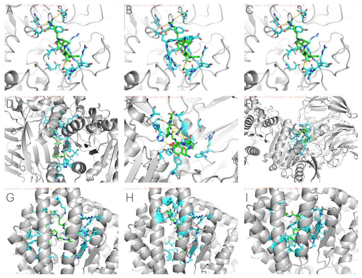
Figure 2.
Molecular dynamics trajectory analysis of the Integrin– Loc3a complex over 50 ns. (A) Root Mean Square Deviation (RMSD) of the complex backbone throughout the 50 ns simulated aqueous environment. Following an initial structural relaxation phase of ~5 ns, the trajectory achieves a highly stable plateau. The red dashed line represents the post-equilibration average RMSD (0.74 nm), confirming that the massive heterodimeric complex remains intact and the dual-motif Loc3a peptide does not dissociate from the binding cleft. (B) Root Mean Square Fluctuation (RMSF) demonstrating local residue flexibility. The analysis reveals strict conformational constraint (RMSF < 0.4 nm) across the majority of the receptor, particularly within the subunit (residues 2100–2250), validating a tight, highly stable “lock-and-key” physical interaction at the canonical RGD-binding interface. (Note: The gap in the x-axis corresponds to the transition between the and subunit chain numbering).
Figure 2.
Molecular dynamics trajectory analysis of the Integrin– Loc3a complex over 50 ns. (A) Root Mean Square Deviation (RMSD) of the complex backbone throughout the 50 ns simulated aqueous environment. Following an initial structural relaxation phase of ~5 ns, the trajectory achieves a highly stable plateau. The red dashed line represents the post-equilibration average RMSD (0.74 nm), confirming that the massive heterodimeric complex remains intact and the dual-motif Loc3a peptide does not dissociate from the binding cleft. (B) Root Mean Square Fluctuation (RMSF) demonstrating local residue flexibility. The analysis reveals strict conformational constraint (RMSF < 0.4 nm) across the majority of the receptor, particularly within the subunit (residues 2100–2250), validating a tight, highly stable “lock-and-key” physical interaction at the canonical RGD-binding interface. (Note: The gap in the x-axis corresponds to the transition between the and subunit chain numbering).
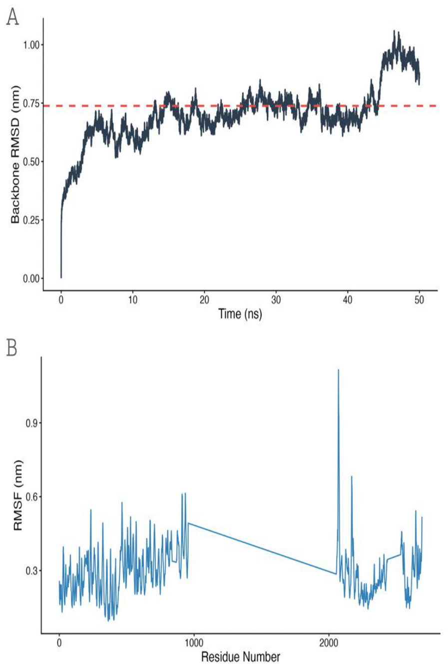
Figure 3.
Systems biology analysis of the target protein interaction network. The Protein-Protein Interaction (PPI) network generated via STRING database (v11.5, Homo sapiens) shows functional connectivity between the high-affinity targets identified in this study: DPP4, ACE, PTGS2 (COX-2), and the Integrin subunits ITGB3 and ITGAV. Edges (lines) represent specific, high-confidence functional protein associations based on experimental data and curated databases (excluding text-mining). The network reveals a connected functional module linking metabolic, cardiovascular, inflammatory, and cell-adhesion pathways.
Figure 3.
Systems biology analysis of the target protein interaction network. The Protein-Protein Interaction (PPI) network generated via STRING database (v11.5, Homo sapiens) shows functional connectivity between the high-affinity targets identified in this study: DPP4, ACE, PTGS2 (COX-2), and the Integrin subunits ITGB3 and ITGAV. Edges (lines) represent specific, high-confidence functional protein associations based on experimental data and curated databases (excluding text-mining). The network reveals a connected functional module linking metabolic, cardiovascular, inflammatory, and cell-adhesion pathways.
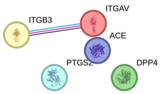
Table 1.
Physicochemical properties and stability indices of the native and modified I. badionotus collagen peptides.
Table 1.
Physicochemical properties and stability indices of the native and modified I. badionotus collagen peptides.
| Peptide ID | Sequence | MW (Da) | Charge (pH 7.4) | Instability Index | Boman Index |
|---|---|---|---|---|---|
| Loc1 (Native) | RGFDGPEGPRG | 1144.21 | −0.004 | 13.82 | 3.51 |
| Loc1d (Modified) | RGFDGPER | 932.99 | −0.004 | 35.29 | 5.06 |
| Loc2 (Native) | GFDGPEGPR | 930.97 | −1.004 | 33.53 | 2.74 |
| Loc2g (Modified) | YGRDGPR | 819.88 | +0.993 | −14.33 | 5.26 |
| Loc3 (Native) | AQGARGDAGARGANGPAG | 1553.61 | +0.995 | −11.79 | 1.90 |
| Loc3a (Modified) | YGPRGDPRG | 974.04 | +0.993 | −36.17 | 3.99 |
Disclaimer/Publisher’s Note: The statements, opinions and data contained in all publications are solely those of the individual author(s) and contributor(s) and not of MDPI and/or the editor(s). MDPI and/or the editor(s) disclaim responsibility for any injury to people or property resulting from any ideas, methods, instructions or products referred to in the content. |
© 2026 by the authors. Licensee MDPI, Basel, Switzerland. This article is an open access article distributed under the terms and conditions of the Creative Commons Attribution (CC BY) license (http://creativecommons.org/licenses/by/4.0/).
Copyright: This open access article is published under a Creative Commons CC BY 4.0 license, which permit the free download, distribution, and reuse, provided that the author and preprint are cited in any reuse.