

Submitted:
13 August 2026
Posted:
14 August 2026
You are already at the latest version
Abstract
Serratia marcescens complex bacteria are recognized causes of outbreaks in neonatal intensive care units. We investigated a cluster of VIM-producing Serratia spp. in the Neonatal Unit of Azienda Ospedaliera di Perugia, Italy, using epidemiological surveillance, antimicrobial susceptibility testing, environmental sampling, and whole-genome sequencing (WGS). In November 2024, ten neonates were colonized and/or infected with Serratia. Eight carried VIM-producing isolates, including two patients with bloodstream infections. Nine VIM-producing Serratia isolates were recovered because one patient yielded both a rectal and a blood culture isolate; one non-VIM-producing bloodstream isolate was also available and included for comparison. WGS showed that the ten isolates, initially identified as S. marcescens by routine diagnostic methods, belonged to two species within the S. marcescens complex: seven Serratia bockelmannii ST388 isolates and three ST382-like Serratia sarumanii isolates. All VIM-producing Serratia spp. isolates carried blaVIM-1 and blaSHV-12 and showed multidrug-resistant phenotypes. blaVIM-1 was also detected in Escherichia coli, Citrobacter freundii, and Pseudomonas putida isolates from the same ward. A shared IncA-ST12 replicon suggested possible horizontal dissemination of a related mobile resistance element. Environmental and healthcare-worker screening did not detect Serratia. These findings highlight the value of WGS for distinguishing clonal transmission from dissemination of mobile resistance determinants during complex healthcare-associated outbreaks.
Keywords:
Serratia marcescens complex
; Serratia bockelmannii
; Serratia sarumanii
; nosocomial out-break
; neonatal intensive care unit
; carbapenemase-producing bacteria
; blaVIM-1
; whole-genome sequencing
; antimicrobial resistance
Abstract
Serratia marcescens complex bacteria are recognized causes of outbreaks in neonatal intensive care units. We investigated a cluster of VIM-producing Serratia spp. in the Neonatal Unit of Azienda Ospedaliera di Perugia, Italy, using epidemiological surveillance, antimicrobial susceptibility testing, environmental sampling, and whole-genome sequencing (WGS). In November 2024, ten neonates were colonized and/or infected with Serratia. Eight carried VIM-producing isolates, including two patients with bloodstream infections. Nine VIM-producing Serratia isolates were recovered because one patient yielded both a rectal and a blood culture isolate; one non-VIM-producing bloodstream isolate was also available and included for comparison. WGS showed that the ten isolates, initially identified as S. marcescens by routine diagnostic methods, belonged to two species within the S. marcescens complex: seven Serratia bockelmannii ST388 isolates and three ST382-like Serratia sarumanii isolates. All VIM-producing Serratia spp. isolates carried blaVIM-1 and blaSHV-12 and showed multidrug-resistant phenotypes. blaVIM-1 was also detected in Escherichia coli, Citrobacter freundii, and Pseudomonas putida isolates from the same ward. A shared IncA-ST12 replicon suggested possible horizontal dissemination of a related mobile resistance element. Environmental and healthcare-worker screening did not detect Serratia. These findings highlight the value of WGS for distinguishing clonal transmission from dissemination of mobile resistance determinants during complex healthcare-associated outbreaks.
Introduction
Healthcare-associated infections remain a major concern in neonatal intensive care units (NICUs), where premature and critically ill neonates are particularly susceptible because of immune immaturity, prolonged hospitalization, invasive procedures, and frequent exposure to broad-spectrum antimicrobial agents[1,2,3]. Among Gram-negative bacteria, Serratia spp. are well-recognized causes of NICU outbreaks and may be associated with asymptomatic gastrointestinal colonization and infections like conjunctivitis, pneumonia, sepsis, meningitis, and other invasive infections. Colonized and/or infected neonates may act as reservoirs for cross-transmission, while contaminated sinks, drains, medical devices, disinfectants, expressed breast milk, and the hands of healthcare workers have all been implicated as potential sources or vehicles of transmission[4]. Nevertheless, a definite environmental source remains unresolved in many of outbreak investigations[2,5]. The management of Serratia spp. outbreaks is further complicated by the ability of these bacteria to persist in the hospital environment, including in water-associated reservoirs and biofilms, and by their documented tolerance to some antiseptics and disinfectants, which may allow survival despite routine cleaning and disinfection procedures, as well as by their capacity to acquire diverse antimicrobial resistance determinants[6]. Members of the genus possess intrinsic resistance mechanisms, including chromosomally encoded AmpC-type β-lactamases, and may additionally acquire extended-spectrum β-lactamases, including carbapenemases[7,8]. Antimicrobial susceptibility testing is essential for therapeutic decision-making, but resistance profiles alone provide limited epidemiological resolution. Unrelated isolates may share similar phenotypes, whereas closely related isolates may differ in susceptibility to individual agents[9].
Verona integron-encoded metallo-β-lactamases (VIM) are Ambler class B carbapenemases that hydrolyse a broad range of β-lactam, including carbapenems. Their activity is not inhibited by available inhibitors such as avibactam or vaborbactam[10], with aztreonam-avibactam being the combination with potential residual activity only. The genes encoding these enzymes, particularly blaVIM-1 and blaVIM-2, are commonly inserted within class 1 integrons and may be associated with transposons and conjugative plasmids[11]. These mobile genetic contexts facilitate dissemination both within bacterial lineages and between species occupying the same healthcare environment. The identification of blaVIM-positive isolates from several bacterial species may therefore reflect both clonal transmission and horizontal mobilization of resistance determinants. Interspecies clusters of blaVIM-1-positive Enterobacterales have been reported in Italian hospitals, including cases in which individual patients were simultaneously colonized by multiple VIM-producing species. In one such cluster, blaVIM-1, together with blaSHV-12 and other resistance determinants, was carried on a highly conjugative, broad-host-range IncA plasmid[12,13].
Accurate tracking of Serratia spp. outbreaks has also been complicated by recent taxonomic revisions within the S. marcescens complex. Isolates identified as S. marcescens by biochemical methods, 16S rRNA gene analysis, or mass spectrometry may belong to several closely related species, such as Serratia sarumanii and Serratia bockelmannii [14,15]. Recent genomic studies have confirmed that both species are represented among clinical isolates previously reported as S. marcescens, including isolates recovered during neonatal unit outbreaks[14,15]. Routine identification may therefore obscure the simultaneous circulation of distinct lineages.
Whole-genome sequencing (WGS) has consequently become an important component of healthcare-associated outbreak investigation. WGS can refine taxonomic assignment, distinguish co-circulating lineages, define resistance genes and plasmid replicons profiles, and examine relationships between clinical and environmental isolates. Its application has improved the resolution of Serratia spp. outbreak investigations in neonatal settings and has enabled the identification of transmission patterns that may remain undetected by routine microbiological methods[16,17].
In November 2024, the emergence of VIM-type carbapenemase-producing S. marcescens was detected in the NICU and Neonatal Unit of Azienda Ospedaliera di Perugia, involving both colonization and infection. We investigated the epidemiological and genomic features of this cluster by integrating clinical and environmental surveillance, antimicrobial susceptibility testing, and WGS. The analysis focused on species identification, antimicrobial resistance determinants, plasmid replicons, and the potential contribution of the hospital environment to the circulation of blaVIM genes.
Materials and Methods
Study Design
In the neonatal intensive care unit of the Santa Maria della Misericordia hospital, rectal swab surveillance is routinely performed at admission and every 7 days thereafter to screen for carbapenemase-producing bacteria and vancomycin-resistant enterococci. A retrospective genomic epidemiology study was conducted following the emergence of VIM-producing S. marcescens among neonates admitted to the Neonatal Intensive Care Unit (NICU) and Neonatal Unit of Azienda Ospedaliera di Perugia, Italy, in November 2024. Clinical Serratia isolates recovered during the November 2024 cluster were considered for genomic analysis. Nine VIM-producing isolates from eight neonates were available, including two isolates from one patient, together with one non-VIM-producing isolate used as a comparator. These ten clinical Serratia isolates were subjected to whole-genome sequencing. The genomic analysis also included four additional VIM-producing Gram-negative isolates recovered from neonatal surveillance specimens during the outbreak investigation: two Escherichia coli isolates, one Citrobacter freundii isolate and one Pseudomonas putida isolate. Finally, two environmental Pseudomonas isolates recovered during environmental surveillance were selected for comparative microbiological and genomic characterization. In total, 16 isolates were analysed by WGS: 10 clinical Serratia isolates, 4 additional clinical VIM-producing Gram-negative isolates, and 2 environmental Pseudomonas isolates.
Clinical Specimen Processing and Bacterial Identification
Clinical specimens were cultured on MacConkey agar for the selective isolation of Gram-negative bacteria, including S. marcescens. Plates were incubated aerobically at 37 °C for 24-48 h. Species identification was performed by matrix-assisted laser desorption/ionization time-of-flight mass spectrometry (MALDI-TOF MS; Bruker Daltonics GmbH & Co. KG, Bremen, Germany). All isolates selected for subsequent phenotypic and genomic analyses were stored at -80 °C until further processing.
Antimicrobial Susceptibility Testing and Carbapenemase Detection
Antimicrobial susceptibility testing was performed using the VITEK® automated system (BioMérieux, Marcy-l’Étoile, France) and broth microdilution with the Micronaut system (Merlin Diagnostika GmbH, Bornheim, Germany). Minimum inhibitory concentrations (MICs) were interpreted according to the clinical breakpoints established by the European Committee on Antimicrobial Susceptibility Testing (EUCAST). Carbapenemase production was initially assessed using the NG-Test® CARBA 5 immunochromatographic assay. This assay differentiates among KPC-, NDM-, OXA-48-like-, VIM-, and IMP-type carbapenemases. The presence of carbapenemase genes, including the Verona integron-encoded metallo-β-lactamase gene (blaVIM), was subsequently confirmed using the Xpert® Carba-R molecular assay (Cepheid, Sunnyvale, CA, USA) and further characterized by whole-genome sequencing analysis.
Environmental Investigation: High-Touch Surface Sampling and Water Sampling
High-touch surfaces were sampled in several areas of the NICU, including the intensive care room, intermediate care room, isolation room, and corridor. Sterile swabs were used to sample an area of approximately 100 cm² at each site. The complete list of sampled surfaces is provided in Supplementary Table S1. Swabs were inoculated onto blood agar and CHROMID® Carba chromogenic agar for the for the recovery of total cultivable bacteria and carbapenemase-producing Enterobacterales, respectively. Plates were incubated aerobically at 37 °C for 48 h. Following incubation, colony-forming units were counted and recorded. Representative colonies growing on CHROMID® Carba agar were identified by MALDI-TOF MS and screened for carbapenemase production using the phenotypic and molecular methods described above. Water samples were collected from sinks at different locations within the NICU. At each sampling point, 1 L of water was collected in a sterile polypropylene bottle containing sodium thiosulfate (Na₂S₂O₃) at a final concentration of 18 mg/mL to neutralize residual disinfectants and reduce the risk of false-negative culture results. Each 1-L water sample was filtered through a mixed cellulose ester membrane with a pore size of 0.45 µm and a diameter of 47 mm, using a sterile vacuum filtration apparatus. Following filtration, the membrane was aseptically transferred onto blood agar and CHROMID® Carba chromogenic agar. Plates were incubated at 37 °C for 24-48 h. After incubation colony were counted and representative isolates were identified by MALDI-TOF MS.
Genomic DNA Extraction and Whole-Genome Sequencing
For each isolate, a pure colony was inoculated into broth medium and cultured to obtain sufficient bacterial biomass. Genomic DNA was extracted using the QIAsymphony DSP Virus/Pathogen Kit (Qiagen, Hilden, Germany) in accordance with the manufacturer’s instructions. Sequencing libraries were prepared using the Illumina DNA Prep Kit with Nextera DNA CD Indexes (Illumina, San Diego, CA, USA). Library concentrations were measured using a Qubit™ Flex Fluorometer (Thermo Fisher Scientific, Waltham, MA, USA), while library fragment size distribution and overall quality were assessed using an Agilent 4150 TapeStation System with High Sensitivity DNA ScreenTape (Agilent Technologies, Santa Clara, CA, USA). Whole-genome sequencing was performed on an Illumina MiSeq platform using MiSeq Reagent Kit v3 and MiSeq Reagent Kit v2. Sequencing was conducted using a paired-end protocol generating 2 x 150-bp reads.
Bioinformatic and Comparative Genomic Analyses
Raw paired-end sequencing reads in FASTQ format were first processed using the DRAGEN pipeline for quality control and read preprocessing. The resulting quality-filtered reads were subsequently analysed using tools available through the Galaxy Europe platform and services provided by the Center for Genomic Epidemiology (CGE). Species-level identification was performed using SpeciesFinder v4.0.4[18], which compares sequencing reads against a curated database of complete genomes retrieved from the National Center for Biotechnology Information (NCBI) by K-mer alignment using KMA. De novo genome assembly was performed using SPAdes v3.15.5[19] through Galaxy, and assembled genomes were annotated using Prokka v1.14.6[20]. Pan-genome analysis was performed using Roary v3.13.0[21]. The analysis included the study isolates and the following reference genomes: S. marcescens ELP1.10 (GenBank accession no. GCA_030291735.1), S. marcescens subsp. marcescens Db11 (GCA_000513215.1), Serratia sarumanii strain K-M0228 (GCA_035749905.1), and Serratia bockelmannii strain K-L0361 (GCA_051804595.1). Genes present in at least 95% of the analysed genomes were defined as core-genome genes. Whole-genome average nucleotide identity (ANI) was calculated using FastANI v1.3[22]. An ANI threshold of >95% was used to assign isolates to the same bacterial species. Phylogenetic relationships based on the core genome alignment were inferred using IQ-TREE v2.4.0[23]. Genomic alignments and associated isolate metadata were explored using Phandango (https://jameshadfield.github.io/phandango/#/), and the final phylogenetic trees were visualized and annotated using the Interactive Tree of Life (iTOL)[24]. Plasmid replicons were identified using PlasmidFinder v2.1[25], whereas plasmid mobility and predicted mobilization characteristics were assessed using MOB-Typer v3.1.9[26]. Plasmid sequence types were assigned using the plasmid multilocus sequence typing approach implemented in the CGE pMLST platform[25,27]. Acquired antimicrobial resistance genes and resistance-associated mutations were investigated using ResFinder v4.6.0[28], ABRicate v1.0.1[29], and the Comprehensive Antibiotic Resistance Database (CARD)[30]. Virulence-associated genes were identified by ABRicate v1.0.1[29] based on virulence factor database (VFDB) with 80% of coverage and 80% of identity. Multilocus sequence typing was performed using MLST v2.22.0 and the resulting sequence-type assignments were confirmed using the PubMLST database[31].
Statistical Analysis
The statistical analysis was performed using GraphPad Prism software version 8.4.3 (GraphPad Software, San Diego, CA, USA). Differences in the number of accessory genes between the S. bockelmannii and S. sarumanii groups were assessed using the two-tailed Mann–Whitney U test. Statistical significance was defined as P < 0.05.
Ethics Statement
The study was conducted in accordance with the principles of the Declaration of Helsinki and applicable national regulations. The investigation was undertaken as part of routine infection prevention and control activities in response to a suspected healthcare-associated outbreak. Clinical and microbiological data were collected retrospectively and anonymized before analysis. No additional samples were collected, and no diagnostic or therapeutic interventions were performed specifically for research purposes. The study was approved by the Ethics Committee of Azienda Ospedaliera di Perugia (approval no. 4457/23).
Availability of Data and Materials
The bacterial genome sequencing data generated and analysed during the present study have been deposited in the National Center for Biotechnology Information Sequence Read Archive (SRA) under BioProject accession n° PRJNA1498554.
Results
Epidemiological Characteristics of the Outbreak
Between September and December 2024, 17 neonates admitted to the NICU tested positive for S. marcescens in clinical and/or surveillance specimens. The temporal distribution of cases is shown in Figure 1. Before November 2024, none of the S. marcescens cases was associated with carbapenem resistance or VIM-type carbapenemase production. In November 2024, ten neonates tested positive for S. marcescens. Eight carried VIM-producing, carbapenem-resistant isolates, whereas two carried non-VIM-producing isolates. The first VIM-positive case was identified on 6 November 2024 in a neonate with an ocular infection (patient A; Table 1). During the following two weeks, seven additional neonates tested positive for VIM-producing S. marcescens (patients B-H), defining a cluster of eight affected patients. Two of these neonates developed bloodstream infections caused by VIM-producing S. marcescens.
Nine VIM-producing S. marcescens isolates (Sm1-Sm9) were available from the eight VIM-positive neonates because patient D yielded both a rectal isolate (Sm4) and a subsequent blood culture isolate (Sm9). These isolates were recovered from an eye swab (n = 1), rectal swabs (n = 6), and blood cultures (n = 2), collected between 2 and 56 days after admission. Of the two neonates carrying non-VIM-producing S. marcescens in November, one bloodstream isolate (Sm10), recovered from patient I, was available and included as a comparator for genomic analysis. Thus, ten clinical Serratia isolates were selected for WGS: nine VIM-producing isolates from eight neonates and one non-VIM-producing isolate from a ninth neonate. Apart from the eight patients colonized and/or infected by VIM-producing isolates associated with the November NICU cluster, no additional carbapenem-resistant or VIM-producing S. marcescens isolates were identified.
Four additional VIM-producing Gram-negative isolates recovered from the same NICU during the outbreak investigation were also selected for genomic analysis: two Escherichia coli isolates (Ec12 and Ec14), one Citrobacter freundii isolate (Cf13), and one Pseudomonas putida isolate (Pp15). Following implementation of outbreak-control measures, no further Serratia cases were detected in the NICU during 2025. The demographic, clinical, and microbiological characteristics of the clinical and environmental isolates selected for further analysis are summarized in Table 1.
Clinical Characteristics of Affected Neonates
Patient A was a female neonate born at 29 weeks of gestation. Seventeen days after admission, she developed an ocular infection caused by VIM-producing S. marcescens (Sm1). The microorganism was also detected in a rectal swab during the same period. Patient B was a female neonate born at 30 weeks of gestation who developed neonatal sepsis 18 days after admission. VIM-producing S. marcescens was recovered from a blood culture (Sm2). Patient C was a male neonate born at 27 weeks of gestation, colonized by VIM-producing S. marcescens in a rectal swab 25 days after admission (Sm3) and in a pharyngeal aspirate. Patient D was a female neonate born at 29 weeks of gestation. Rectal colonization with VIM-producing S. marcescens was detected 11 days after admission (Sm4). Four days later, VIM-producing E. coli was recovered from a rectal swab (Ec14). On day 19, the patient developed a bloodstream infection caused by VIM-producing S. marcescens (Sm9), following the earlier detection of rectal colonization. Patient E was a female neonate born at 24 weeks of gestation. Rectal colonization with VIM-producing S. marcescens was detected 56 days after admission (Sm5), together with VIM-producing E. coli (Ec12). The patient subsequently developed neonatal sepsis on day 63 after admission. Patient F was a male neonate in whom rectal colonization with VIM-producing S. marcescens was detected 21 days after admission (Sm6). Patient G was a female neonate born at 34 weeks of gestation. Rectal colonization with VIM-producing S. marcescens was detected seven days after admission (Sm7). Patient H was a male neonate in whom rectal colonization with VIM-producing S. marcescens was identified two days after admission (Sm8). A subsequent rectal swab, collected on day 7, yielded VIM-producing C. freundii (Cf13). Patient I was a female neonate born at 24 weeks of gestation who developed neonatal sepsis 85 days after admission. A non-VIM-producing isolate identified as S. marcescens was recovered from blood culture (Sm10). Patient L was a female neonate who developed an ocular infection caused by non-VIM-producing S. marcescens 52 days after birth. A rectal swab collected 69 days after admission was positive for VIM-producing P. putida (Pp15).
Environmental Investigation
Environmental sampling was conducted to investigate whether the NICU and neonatal environment represented a potential reservoir of VIM-producing S. marcescens. High-touch surfaces, water-associated sites, and the hands of healthcare personnel were sampled in different areas of the unit, as detailed in Supplementary Table S1. No S. marcescens was recovered from any environmental or hand sample. Several other bacterial species were identified from high-touch surfaces, including Pseudomonas mosselii, Pseudomonas putida, Klebsiella pneumoniae, Enterobacter asburiae, Pseudomonas aeruginosa, Pseudomonas oleovorans, Stenotrophomonas maltophilia, Enterococcus faecium, and Escherichia coli. The environmental E. faecium isolate showed a vancomycin-resistant phenotype. Bacteria species recovered from water-associated samples included Acinetobacter bereziniae, Acinetobacter junii, Pseudomonas mendocina, Pseudomonas alcaliphila, Pseudomonas aeruginosa, and Stenotrophomonas maltophilia. In addition, a VIM-producing P. oleovorans isolate was also recovered from a sink trap.
Two environmental Pseudomonas isolates were selected for whole-genome sequencing: non-VIM-producing P. putida (Pp16) and VIM-producing P. oleovorans (Po17). Pp16 was included to assess its possible relatedness to the VIM-producing clinical P. putida isolate Pp15 recovered from patient L. Po17 was sequenced to compare the genetic context of its blaVIM determinant with that of the clinical isolates. These analyses were undertaken to evaluate the potential contribution of the neonatal care environment to the dissemination of blaVIM. All environmental isolates were identified to the species level by MALDI-TOF mass spectrometry.
Phenotypic Antimicrobial Susceptibility Testing of Clinical and Environmental Isolates
The nine VIM-producing Serratia isolates showed similar multidrug-resistant phenotype with resistance to most of antimicrobial agents tested (Figure 2). Resistance was observed to all tested cephalosporins and β-lactam/β-lactamase inhibitor combinations, including cefotaxime, ceftazidime, ceftazidime-avibactam, and ceftolozane-tazobactam. Resistance was also observed to all carbapenems tested, including imipenem, meropenem and meropenem-vaborbactam (Supplementary Table S2). Trimethoprim-sulfamethoxazole resistance was also observed in all VIM-producing Serratia isolates. By contrast, fluoroquinolones (ciprofloxacin and levofloxacin) and aminoglycoside amikacin retained in vitro activity against all nine VIM-producing Serratia isolates (Figure 2).
The non-VIM-producing Serratia isolate Sm10 showed a different susceptibility profile. The isolate was resistant to cefotaxime and ceftolozane-tazobactam but remained susceptible to all the other antibiotics tested. Consistent with the absence of blaVIM, carbapenem MICs for Sm10 remained below the EUCAST resistance breakpoints (Figure 2).
Antimicrobial susceptibility testing was also performed on four clinically relevant VIM-producing Gram-negative bacteria isolates recovered from rectal specimens. The two E. coli, Ec12 and Ec14, C. freundii Cf13, and P. putida Pp15 isolates exhibited multidrug-resistant phenotypes. All four isolates were resistant to the cephalosporins and β-lactam/β-lactamase inhibitor combinations tested. Carbapenem resistance was observed in Cf13, Ec14, and Pp15, whereas Ec12 was categorized as susceptible at increased exposure to both imipenem and meropenem. All four isolates were resistant to trimethoprim-sulfamethoxazole. In addition, P. putida Pp15 was resistant to ciprofloxacin and categorized as susceptible at increased exposure to levofloxacin (Figure 2).
The two environmental Pseudomonas spp. isolates had clearly different susceptibility profiles. The non-VIM-producing P. putida isolate Pp16 was susceptible to all β-lactam agents tested but resistant to trimethoprim-sulfamethoxazole; both fluoroquinolones were categorized as susceptible at increased exposure. In contrast, the VIM-producing P. oleovorans isolate Po17 was resistant to ceftolozane-tazobactam and imipenem, categorized as susceptible at increased exposure to the third-generation cephalosporins and fluoroquinolones tested. It remained susceptible to ceftazidime-avibactam, meropenem, trimethoprim-sulfamethoxazole, and amikacin (Figure 2).
Whole-Genome Sequencing and Genomic Characterization
Whole-genome sequencing was performed on ten clinical isolates identified as Serratia marcescens and on six additional non-Serratia isolates recovered from clinical and environmental samples. Detailed sequencing output and assembly quality metrics, including raw and quality-filtered read counts, are reported in Supplementary Table S3. Taxonomic classification assigned the majority of reads from the ten Serratia isolates to the genus Serratia. De novo assembly produced genomes with a median size of 5.3 Mb, within the expected range for members of the S. marcescens complex.
Species-level analysis resolved the ten isolates into two groups within the S. marcescens complex. Isolates Sm1, Sm2, Sm3, Sm5, Sm6, Sm7, and Sm8 were assigned to Serratia bockelmannii, whereas Sm4, Sm9, and Sm10 were assigned to Serratia sarumanii. The isolates reported as S. marcescens by routine identification therefore comprised two distinct genomic lineages. This classification was supported independently by multilocus sequence typing and core-genome phylogenetic analysis. For the six non-Serratia isolates, SpeciesFinder confirmed the species assignments obtained by MALDI-TOF mass spectrometry. These comprised the clinical isolates E. coli Ec12 and Ec14, C. freundii Cf13 and P. putida Pp15, together with the environmental isolates P. putida Pp16 and P. oleovorans Po17.
Distribution of Antimicrobial Resistance Determinants
To further investigate the genetic relatedness among the isolates, whole-genome sequence data were analysed to characterize the antimicrobial resistance gene profiles of the clinical and environmental isolates (Figure 3A). All nine VIM-producing Serratia isolates (Sm1-Sm9) carried the metallo-β-lactamase gene blaVIM-1 and the extended-spectrum β-lactamase gene blaSHV-12. Neither blaVIM-1 nor blaSHV-12 was identified in the non-VIM-producing isolate Sm10. Moreover, the distribution of intrinsic β-lactamase genes was consistent with species assignment: blaSST-1 was detected in the S. bockelmannii isolates Sm1, Sm2, Sm3, Sm5, Sm6, Sm7, and Sm8, while the S. sarumanii isolates Sm4, Sm9, and Sm10 carried blaSRT-1, encoding an inducible AmpC-type β-lactamase. Genes associated with resistance to aminoglycosides were also identified. Sm1-Sm9 carried aac(6′)-Ic, aph(3′)-XV, and ant(3″)-Ia, whereas Sm10 carried only aac(6′)-Ic. In addition, the sulfonamide- and trimethoprim-resistance genes sul1 and dfrA14 were detected in all VIM-producing Serratia isolates but were absent from Sm10. This difference in acquired resistance-gene content was consistent with the more susceptible phenotype of Sm10 and clearly separated it from the VIM-producing group.
The two VIM-positive E. coli isolated from the rectal swab of patients E and D (Ec12 and Ec14 respectively), were assigned to sequence type 12 and showed a broad range of acquired resistance genes. Both isolates carried blaVIM-1, blaSHV-12, and blaTEM-1B, together with the aminoglycoside-resistance determinants aph(3′)-XV, aph(3″)-Ib, and ant(3″)-Ia. The genes sul1, sul2, and dfrA14 were also detected, supporting resistance to sulfonamides and trimethoprim. Both isolates additionally carried the plasmid-mediated quinolone-resistance gene qnrS1, which may contribute to reduced fluoroquinolone susceptibility. The C. freundii isolate Cf13 carried blaVIM-1, blaSHV-12, and blaCYM-110, consistent with its broad β-lactam resistance phenotype. The isolate also harbored the aminoglycoside-resistance genes aph(3′)-XV and ant(3″)-Ia, together with sul1 and dfrA14. The clinical P. putida isolate Pp15 carried blaVIM-1, ant(3″)-Ia, and sul1. In addition, Pp15 was the only isolate in which qnrVC6 was detected, in agreement with its reduced susceptibility to fluoroquinolone (Figure 3A).
The two environmental Pseudomonas isolates exhibited distinct resistance gene profiles. P. putida Pp16 carried only aph(3″)-Ib and sul2 and did not harbor a VIM-type metallo-β-lactamase gene. By contrast, P. oleovorans Po17 carried blaVIM-2. This allele differed from blaVIM-1 determinant found in the clinical Serratia, E. coli, C. freundii, and P. putida isolates, distinguishing Po17 from the clinical VIM-positive group.
Overall, the resistance profiles detected by WGS were broadly consistent with the antimicrobial susceptibility results. They differentiated the non-VIM-producing Sm10 isolate from the VIM-producing Serratia isolates and separated the blaVIM-2-positive environmental Po17 isolate from the blaVIM-1 positive clinical isolates.
Plasmid Replicon Content and Mobility Typing
Plasmid replicons were detected in 13 of the 16 sequenced isolates. The detected replicon families included IncA, IncM1, IncFIA, IncFIB, IncQ2, Col156, and a pKPC-associated replicon (Figure 3B). An IncA replicon was detected in all VIM-producing Serratia isolates, as well as in E. coli Ec12 and Ec14 and C. freundii Cf13. In the Serratia and E. coli isolates, the IncA replicon showed 100% sequence coverage and 98.3% nucleotide identity to the corresponding reference sequence. Plasmid multilocus sequence typing assigned these replicons to plasmid sequence type 12 (pST12), based on the allelic profile A053-3, parA-7, parB-8, and repA-6. Mobility analysis classified them within the MOBH relaxase group. The IncA replicon detected in Cf13 showed 100% sequence coverage and 100% nucleotide identity to the reference sequence. An IncM1 replicon was identified in all the S. bockelmannii isolates but was absent from the S. sarumani isolates. The IncM1 replicons showed 100% sequence coverage and 99.5% nucleotide identity to the reference sequence and were assigned to the MOBP relaxase group. The IncFIA, IncFIB, and Col156 replicons were detected in both E. coli isolates, with 100% sequence coverage and nucleotide identities of 99.7%, 99.4%, and 98.7%, respectively. Col156 was also assigned to the MOBP relaxase group. IncA was also presente in C. freundii with 100% sequence coverage and nucleotide identities of 100%. A pKPC-associated replicon was identified exclusively in C. freundii Cf13, with 100% sequence coverage and 100% nucleotide identity to the reference sequence. An IncQ2 replicon was detected only in the environmental P. putida isolate Pp16.
No plasmid replicons were detected in the clinical P. putida isolate Pp15 or in the environmental Pseudomonas isolates Po17 by the applied in silico methods. Nevertheless, in Pp15, blaVIM-1 was identified on an assembled contig, in agreement with the positive Xpert® Carba-R result. Short-read sequencing did not allow its chromosomal or plasmid-associated location to be resolved. By contrast, blaVIM-2 was chromosomally located in P. oleovorans Po17, indicating that this resistance determinant was not plasmid mediated in the environmental isolate.
Virulence Genes
The distribution of virulence-associated genes was examined in the ten clinical Serratia isolates (Figure 3C). Both S. bockelmannii and S. sarumanii harboured a largely conserved repertoire of genes associated with flagellar assembly, motility, and chemotaxis, with no evidence of a distinctive high-virulence gene profile. The chemotaxis response regulator gene cheY and the flagellar biosynthesis gene flhA were detected in the S. sarumanii isolates but were absent from the S. bockelmannii isolates. Conversely, flgH and fliF, which encode components of the flagellar basal body, were detected in S. bockelmannii but were absent from S. sarumanii. The remaining flagella-associated genes shown in Figure 3C, including flgG, fliG, fliU, and flgP, were conserved across both lineages. Additional virulence-associated determinants, grouped under the category “Others” in Figure 3C, included acrB, gndA, rcsB, rpoS, and tssC1; all were detected in both lineages. Overall, the virulence-associated gene profiles were broadly conserved between the two species, while the differential distribution of cheY, flhA, flgH, and fliF provided an additional genomic feature distinguishing S. bockelmannii from S. sarumanii.
Comparative Genomic Analysis and Population Structure of the Serratia Isolates
Pan-genome analysis of the ten clinical Serratia isolates shown two distinct gene-content profiles corresponding to the species assignments obtained by whole-genome sequencing. The S. sarumanii isolates Sm4, Sm9, and Sm10 clustered separately from the S. bockelmannii isolates Sm1, Sm2, Sm3, Sm5, Sm6, Sm7, and Sm8 in the gene presence-absence matrix (Figure 4A). The S. bockelmannii isolates carried a significantly higher number of accessory genes than the S. sarumanii isolates (P < 0.05; Figure 4B). Comparison of the two lineage gene sets identified a total of 5,936 genes of which 3,687 were shared by both groups. A further, 1,213 genes were associated only with the S. bockelmannii group, including 43 cloud genes, whereas 1,036 genes were restricted to the S. sarumanii group, including 15 cloud genes (Figure 4C). Overall, the pan-genome structure therefore separated the clinical isolates into two genomic lineages and showed a broader accessory-gene repertoire in the S. bockelmannii group. Given the limited number of isolates, genes found in only one group are more appropriately regarded as group-associated rather than species-specific.
Multi Locus Sequence Typing
Finally, multilocus sequence typing was performed using the allelic profiles of adk, fumC, gyrB, icd, mdh, and recA. The S. bockelmannii isolates Sm1, Sm2, Sm3, Sm5, Sm6, Sm7, and Sm8 were assigned to sequence type 388 and shared the allelic profile adk-3, fumC-253, gyrB-191, icd-3, mdh-187, and recA-5 (Figure 5). The S. sarumanii isolates Sm4, Sm9, and Sm10 showed the closest match to ST382, with the allelic profile adk-135, fumC-189, icd-161, mdh-131, and recA-147 (Figure 5). However, the gyrB locus was not recovered from their genome assemblies, preventing definitive sequence-type assignment. These isolates were therefore classified as nearest ST382. The MLST findings were consistent with the pan-genome analyses and further separated the clinical isolates into two distinct Serratia lineages.
Discussion and Conclusion
This study describes a cluster of VIM-producing members of the Serratia marcescens complex among neonates admitted to the Neonatal Intensive Care Unit and Neonatal Unit of Azienda Ospedaliera di Perugia. The investigation combined epidemiological surveillance, antimicrobial susceptibility testing, environmental sampling, and whole-genome sequencing. Eight neonates were colonized or infected with VIM-producing Serratia, including two patients who developed bloodstream infections. The event therefore extended beyond the spread of a single resistant species and involved the circulation of blaVIM-1 positive Gram-negative bacteria within the same neonatal care setting. The predominance of rectal isolates is consistent with gastrointestinal colonization playing an important role in the epidemiology of the outbreak, while the two bloodstream infections demonstrate the risk of progression to invasive disease in this highly vulnerable population. Similar observations have been reported in neonatal outbreaks, including one in which approximately one-quarter of colonized neonates subsequently developed infection, underscoring the value of active surveillance and early identification of colonized patients[32,33].
The clustering of cases in November 2024, together with similar multidrug-resistant phenotypes, initially suggested dissemination of a single VIM-producing S. marcescens strain. However, WGS showed that the epidemiological picture was more complex. The ten isolates identified as S. marcescens by routine diagnostic methods belonged to two species within the S. marcescens complex: seven isolates were assigned to the Serratia bockelmannii ST388 lineage, whereas three belonged to Serratia sarumanii ST382-like lineage, including the non-VIM-producing isolate Sm10. Thus, at least two Serratia populations were circulating in the neonatal setting and that the acquisition or dissemination of blaVIM-1 occurred in distinct genomic backgrounds. The pan-genome analysis supported this separation: although the two groups shared a substantial proportion of their gene content, S. bockelmannii isolates carried a larger accessory-gene repertoire than S. sarumanii isolates. Outbreaks involving multiple Serratia lineages have also been described in other NICUs, where WGS separated epidemiologically coexisting isolates into genetically unrelated or partially related groups that could not be reliably distinguished by antimicrobial susceptibility profiles alone[34,35,36].
The resistance profile of the VIM-producing Serratia isolates has relevant therapeutic implications, particularly in neonates, for whom antimicrobial options are already limited. Resistance to carbapenems and to β-lactam/β-lactamase inhibitor combinations such as ceftazidime-avibactam and meropenem-vaborbactam was expected because VIM metallo-β-lactamases are not inhibited by avibactam or vaborbactam[37]. Amikacin and fluoroquinolones retained in vitro activity, although their use in neonates requires careful assessment.
The comparison between phenotypic susceptibility and WGS data also clarified the basis of the multidrug-resistant phenotype. The VIM-producing Serratia isolates carried not onlyblaVIM-1 but a broader set of acquired resistance determinants, including blaSHV-12, sul1, dfrA14, and aminoglycoside-resistance genes. This indicates that carbapenem resistance occurred within a more complex multidrug-resistance background. Sm10 provides a useful contrast. Although it belonged to the same S. sarumanii ST382-like lineage as Sm4, it lacked these acquired determinants and showed a substantially more susceptible phenotype. This difference suggests that the resistance pattern was driven primarily by gain or loss of mobile resistance elements rather than by species assignment alone. At the same time, the distribution of the intrinsic AmpC genes, with blaSST-1 in S. bockelmannii and blaSRT-1 in S. sarumanii, reflected the chromosomal background of the two lineages.
The detection of blaVIM-1 in several bacterial species was another notable feature of the outbreak. In addition to the two Serratia lineages, blaVIM-1 was detected in E. coli, C. freundii, and the clinical P. putida isolate Pp15. Although the resistomes of these organisms differed, the presence of the same carbapenemase allele across taxonomically distinct hosts is compatible with horizontal dissemination of a mobile resistance element[38,39]. The plasmid analysis provides partial support to this interpretation. An IncA replicon assigned to ST12 was detected in the VIM-producing Serratia isolates, both E. coli isolates and C. freundii Cf13. Because IncA plasmids are conjugative and have a broad host range, the shared replicon profile may reflect circulation of a related plasmid backbone among different Enterobacterales[12,40]. However, this remains a working hypothesis rather than direct evidence of transfer because short-read sequencing can identify replicons and resistance genes but cannot determine whether blaVIM-1 is physically located on the same plasmid in each isolate or whether the plasmids are structurally identical. Long-read or hybrid sequencing would be needed to reconstruct these elements and establish their relationship. The IncM1 replicon was restricted to the S. bockelmannii isolates and was absent from the S. sarumanii group, providing an additional genomic marker separating the two lineages. Other plasmid families were detected in the E. coli and C. freundii isolates, further illustrating the diversity of mobile elements present during the outbreak. In Pp15, no plasmid replicon was detected, despite the presence of blaVIM-1 on an assembled contig. Its genomic location therefore remains unresolved and may reflect chromosomal integration or an incompletely assembled mobile element.
Environmental sampling and screening of healthcare personnel’s hands did not detect Serratia, and no direct environmental or personnel-associated reservoir of the clinical lineages was identified. Nevertheless, the recovery of VIM-producing P. oleovorans (Po17) from a sink trap showed that carbapenemase-producing Gram-negative bacteria were present in the water associated environment. Po17 carried chromosomally located blaVIM-2, whereas the clinical VIM-positive Serratia carried blaVIM-1. The different VIM allele, its chromosomal location and the absence of a shared detectable plasmid replicon argue against Po17 being the direct source of the clinical cluster. Its presence is better interpreted as evidence of an independent environmental reservoir of carbapenem resistance.
Several factors limit the reconstruction of transmission. The number of sequenced isolates was small, and the outbreak was concentrated within a short period. Short-read sequencing did not permit complete reconstruction of the plasmids, integrons, or other mobile elements associated with blaVIM-1, preventing direct comparison of their structure across species. Environmental sampling was restricted to selected sites and time points and may have missed intermittent or low-level contamination.
Despite these limitations, WGS data substantially refined the interpretation of the outbreak. What initially appeared to be an increase in VIM-producing S. marcescens was resolved into the simultaneous circulation of two species within the S. marcescens complex, together with blaVIM-1 positive isolates from several other Gram-negative species. Overall, these findings support the use of WGS not only to define isolate relatedness and identify co-circulating lineages, but also to investigate the distribution of resistance genes and their potential mobile genetic vehicles during complex healthcare-associated outbreaks.
Supplementary Materials
The following supporting information can be downloaded at the website of this paper posted on Preprints.org.
Author Contributions
Conceptualization, AM, DP, RS, ST; Methodology, RS, GB, F.AL; Validation, RS, AM; Formal analysis, GB, AYWW; Investigation, GB, DP; Resources, AM, RS; Data curation, GB, DP, AYWW; Writing, GB, RS, AM, DP, F.AR; Visualization, GB, AYWW; Supervision, RS, AM; Project Administration, AM, RS; Funding acquisition, AM, RS.
Funding
This work was supported by the Umbria Region; resolution of the regional council (No. 524, 05/06/2024).
Institutional Review Board Statement
The study was approved by the Ethics Committee of Azienda Ospedaliera di Perugia (approval no. 4457/23).
Informed Consent Statement
The investigation was undertaken as part of routine infection prevention and control activities in response to a suspected healthcare-associated outbreak. Clinical and microbiological data were collected retrospectively and anonymized before analysis. No additional samples were collected, and no diagnostic or therapeutic interventions were performed specifically for research purposes.
Conflicts of Interest
The author(s) declared that this work was conducted in the absence of any commercial or financial relationships that could be construed as a potential conflict of interest.
References
- European Centre for Disease Prevention and Control. European Surveillance of Healthcare-Associated Infections in Intensive Care Units: HAI Net ICU Protocol, Version 1.02 . Publications Office. 2015. Available online: https://data.europa.eu/doi/10.2900/371526 (accessed on 2025).
- Donkor, E.S.; Asantewaa, A.A.; Odoom, A.; Dame, J.A. A systematic review and meta-analysis of outbreaks in neonatal intensive care units. J. Hosp. Infect. 2026, 172, 188–209. [Google Scholar] [CrossRef] [PubMed]
- Tzialla, C.; Berardi, A.; Mondì, V. Outbreaks in the Neonatal Intensive Care Unit: Description and Management. Trop. Med. Infect. Dis. 2024, 9(9), 212. [Google Scholar] [CrossRef] [PubMed]
- Guo, Q.; Zhao, X.; Ma, J.; et al. Serratia marcescens outbreak in a neonatal intensive care unit associated with contaminated handwashing sinks. Indian J. Med. Microbiol. 2024, 52, 100741. [Google Scholar] [CrossRef] [PubMed]
- Cristina, M.L.; Sartini, M.; Spagnolo, A.M. Serratia marcescens Infections in Neonatal Intensive Care Units (NICUs). Int. J. Env. Res. Public Health 2019, 16(4), 610. [Google Scholar] [CrossRef] [PubMed]
- Hanczvikkel, A.; Tóth, Á.; Kopcsóné Németh, I.A.; et al. Nosocomial outbreak caused by disinfectant-resistant Serratia marcescens in an adult intensive care unit, Hungary, February to March 2022. Euro Surveill. Bull. Eur. Sur Mal. Transm. Eur. Commun. Dis. Bull. 2024, 29(26), 2300492. [Google Scholar] [CrossRef] [PubMed]
- Rezzoug, I.; Bernabeu, S.; Ouacel, K.; et al. Spread of carbapenemase-producing Serratia spp. in France from 2016 to 2024: a comparative genomic study. Emerg. Microbes Infect. 15(1), 2671515. [CrossRef] [PubMed]
- Peirano, G.; Matsumura, Y.; Pitout, J.D.D. Molecular epidemiology of global carbapenemase-producing Serratia spp. (2015-2017). Eur. J. Clin. Microbiol. Infect. Dis. Off. Publ. Eur. Soc. Clin. Microbiol. 2025, 44(12), 3037–3046. [Google Scholar] [CrossRef] [PubMed]
- Toorop, M.M.A.; Hoogendijk, I.V.; Dogterom-Ballering, H.C.M.; et al. Implications of ad-hoc molecular typing for infection control measures in a multi-cluster, multi-phenotypic Serratia marcescens outbreak in a neonatal intensive care unit. J. Hosp. Infect. 2024, 150, 26–33. [Google Scholar] [CrossRef] [PubMed]
- Boyd, S.E.; Livermore, D.M.; Hooper, D.C.; Hope, W.W. Metallo-β-Lactamases: Structure, Function, Epidemiology, Treatment Options, and the Development Pipeline. Antimicrob. Agents Chemother. 2020, 64(10), e00397-20. [Google Scholar] [CrossRef] [PubMed]
- Lagatolla, C.; Edalucci, E.; Dolzani, L.; et al. Molecular Evolution of Metallo-β-Lactamase-Producing Pseudomonas aeruginosa in a Nosocomial Setting of High-Level Endemicity. J. Clin. Microbiol. 2006, 44(7), 2348–2353. [Google Scholar] [CrossRef] [PubMed]
- Arcari, G.; Di Lella, F.M.; Bibbolino, G.; et al. A Multispecies Cluster of VIM-1 Carbapenemase-Producing Enterobacterales Linked by a Novel, Highly Conjugative, and Broad-Host-Range IncA Plasmid Forebodes the Reemergence of VIM-1. Antimicrob. Agents Chemother. 2020, 64(4), e02435-19. [Google Scholar] [CrossRef] [PubMed]
- Olivieri, R.; Riccobono, E.; Gonnelli, S.; et al. Large, protracted, multi-species and multi-clonal spread of VIM-type metallo-β-lactamase-producing Enterobacterales in an Italian hospital. J. Hosp. Infect. 2025, 157, 10–18. [Google Scholar] [CrossRef] [PubMed]
- Harch, S.A.J.; Jenkins, F.; Farhat, R.; van Hal, S.J. Complexities in species identification for Serratia marcescens complex for the modern microbiology laboratory. Microbiol. Spectr. 2025, 13(1), e0136124. [Google Scholar] [CrossRef] [PubMed]
- Klages, L.J.; Hassa, J.; Busche, T.; et al. The Fires of Isengard Have Spread: Serratia sarumanii Is the Dominant Species in Clinical Isolates of the “Serratia marcescens Complex.”. Pathogens 2026, 15(2), 140. [Google Scholar] [CrossRef] [PubMed]
- Muyldermans, A.; Crombé, F.; Bosmans, P.; Cools, F.; Piérard, D.; Wybo, I. Serratia marcescens outbreak in a neonatal intensive care unit and the potential of whole-genome sequencing. J. Hosp. Infect. 2021, 111, 148–154. [Google Scholar] [CrossRef] [PubMed]
- Merla, C.; Ramus, M.; Kuka, A.; et al. Impact of Whole Genome Sequencing to investigate transmission of Serratia marcescens in Neonatal Intensive Care Unit. J. Infect. Public Health 2024, 17(5), 918–921. [Google Scholar] [CrossRef] [PubMed]
- Center for Genomic Epidemiology. Available online: https://genepi.dk/speciesfinder/ (accessed on 2 July 2026).
- Bankevich, A.; Nurk, S.; Antipov, D.; et al. SPAdes: A New Genome Assembly Algorithm and Its Applications to Single-Cell Sequencing. J. Comput Biol. 2012, 19(5), 455–477. [Google Scholar] [CrossRef] [PubMed]
- Seemann, T. Prokka: rapid prokaryotic genome annotation. Bioinformatics 2014, 30(14), 2068–2069. [Google Scholar] [CrossRef] [PubMed]
- Page, A.J.; Cummins, C.A.; Hunt, M.; et al. Roary: rapid large-scale prokaryote pan genome analysis. Bioinformatics 2015, 31(22), 3691–3693. [Google Scholar] [CrossRef] [PubMed]
- Jain, C.; Rodriguez-R, L.M.; Phillippy, A.M.; Konstantinidis, K.T.; Aluru, S. High throughput ANI analysis of 90K prokaryotic genomes reveals clear species boundaries. Nat. Commun. 2018, 9(1), 5114. [Google Scholar] [CrossRef] [PubMed]
- Nguyen, L.T.; Schmidt, H.A.; von Haeseler, A.; Minh, B.Q. IQ-TREE: A Fast and Effective Stochastic Algorithm for Estimating Maximum-Likelihood Phylogenies. Mol. Biol. Evol. 2015, 32(1), 268–274. [Google Scholar] [CrossRef] [PubMed]
- Letunic, I.; Bork, P. Interactive Tree of Life (iTOL) v6: recent updates to the phylogenetic tree display and annotation tool. Nucleic Acids Res. 2024, 52(W1), W78–W82. [Google Scholar] [CrossRef] [PubMed]
- Carattoli, A.; Hasman, H. PlasmidFinder and In Silico pMLST: Identification and Typing of Plasmid Replicons in Whole-Genome Sequencing (WGS). Methods Mol. Biol. Clifton NJ 2020, 2075, 285–294. [Google Scholar] [CrossRef] [PubMed]
- phac-nml/mob-suite. 2 July 2026. Available online: https://github.com/phac-nml/mob-suite (accessed on 28 July 2026).
- Center for Genomic Epidemiology. Available online: https://www.genomicepidemiology.org/services/ (accessed on 11 August 2024).
- Florensa, A.F.; Kaas, R.S.; Clausen, P.T.L.C.; Aytan-Aktug, D.; Aarestrup, F.M. ResFinder – an open online resource for identification of antimicrobial resistance genes in next-generation sequencing data and prediction of phenotypes from genotypes. Microb. Genom. 2022, 8(1), 000748. [Google Scholar] [CrossRef] [PubMed]
- Seemann, T. tseemann/abricate. 20 July 2026. Available online: https://github.com/tseemann/abricate (accessed on 28 July 2026).
- Alcock, B.P.; Huynh, W.; Chalil, R.; et al. CARD 2023: expanded curation, support for machine learning, and resistome prediction at the Comprehensive Antibiotic Resistance Database. Nucleic Acids Res. 2023, 51(D1), D690–D699. [Google Scholar] [CrossRef] [PubMed]
- Multi-Locus Sequence Typing | PubMLST. Available online: https://pubmlst.org/multilocus-sequence-typing (accessed on 3 March 2025).
- Villari, P.; Crispino, M.; Salvadori, A.; Scarcella, A. Molecular epidemiology of an outbreak of Serratia marcescens in a neonatal intensive care unit. Infect. Control Hosp. Epidemiol. 2001, 22(10), 630–634. [Google Scholar] [CrossRef] [PubMed]
- Vogiantzi, G.; Metallinou, D.; Tigka, M.; Deltsidou, A.; Nanou, C.I. Bloodstream Infections in the Neonatal Intensive Care Unit: A Systematic Review of the Literature. Cureus 2024, 16(8), e68057. [Google Scholar] [CrossRef] [PubMed]
- Sansom, S.E.; Logan, L.K.; Green, S.J.; Moore, N.M.; Hayden, M.K. Whole-genome sequencing for neonatal intensive care unit outbreak investigations: Insights and lessons learned. Antimicrob. Steward Healthc. Epidemiol. 2021, 1(1), e2. [Google Scholar] [CrossRef] [PubMed]
- Henares, D.; Cubero, M.; Martinez-de-Albeniz, I.; et al. Rapid identification of a Serratia marcescens outbreak in a neonatal intensive care unit by third-generation long-read nanopore sequencing. Antimicrob. Resist Infect. Control 2025, 14(1), 63. [Google Scholar] [CrossRef] [PubMed]
- Rath, A.; Klages, L.J.; Kieninger, B.; et al. Genome-oriented outbreak management reveals the first detection of concurrent transmissions of Serratia sarumanii and Serratia bockelmannii in a neonatal department. J. Hosp. Infect. 2026, 170, 60–68. [Google Scholar] [CrossRef] [PubMed]
- Tavares-Carreon, F.; De Anda-Mora, K.; Rojas-Barrera, I.C.; Andrade, A. Serratia marcescens antibiotic resistance mechanisms of an opportunistic pathogen: a literature review. PeerJ. 2023, 11, e14399. [Google Scholar] [CrossRef] [PubMed]
- Mobile Genetic Elements Associated with Antimicrobial Resistance | Clinical Microbiology Reviews. Available online: https://journals.asm.org/doi/10.1128/cmr.00088-17 (accessed on 28 July 2026).
- Wang, X.; Zhang, H.; Yu, S.; et al. Inter-plasmid transfer of antibiotic resistance genes accelerates antibiotic resistance in bacterial pathogens. ISME J. 2024, 18(1), wrad032. [Google Scholar] [CrossRef] [PubMed]
- Esposito, E.P.; Gaiarsa, S.; Del Franco, M.; et al. A Novel IncA/C1 Group Conjugative Plasmid, Encoding VIM-1 Metallo-Beta-Lactamase, Mediates the Acquisition of Carbapenem Resistance in ST104 Klebsiella pneumoniae Isolates from Neonates in the Intensive Care Unit of V. Monaldi Hospital in Naples. Front Microbiol. 2017, 8, 2135. [Google Scholar] [CrossRef] [PubMed]
Figure 1.
Temporal distribution of Serratia colonization among neonates admitted to the Neonatal Intensive Care Unit and Neonatal Unit of Azienda Ospedaliera di Perugia, Italy, from September to December 2024. Each circle represents one affected neonate. Orange circles indicate neonates colonized with non-VIM-producing S. marcescens, whereas green circles indicate neonates colonized or infected with VIM-producing S. marcescens. The y-axis indicates the number of affected neonates recorded during each month.
Figure 1.
Temporal distribution of Serratia colonization among neonates admitted to the Neonatal Intensive Care Unit and Neonatal Unit of Azienda Ospedaliera di Perugia, Italy, from September to December 2024. Each circle represents one affected neonate. Orange circles indicate neonates colonized with non-VIM-producing S. marcescens, whereas green circles indicate neonates colonized or infected with VIM-producing S. marcescens. The y-axis indicates the number of affected neonates recorded during each month.
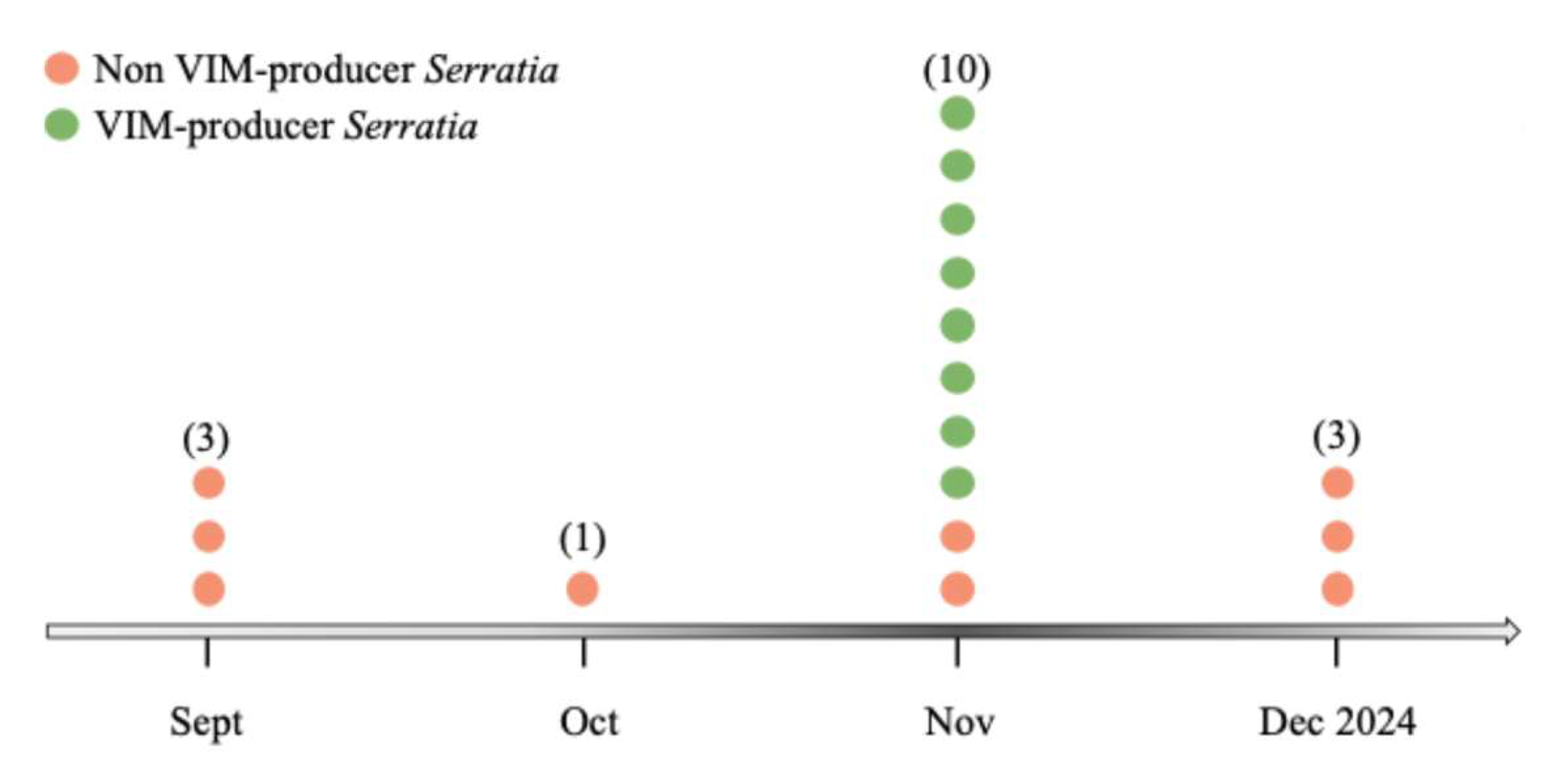
Figure 2.
Phenotypic antimicrobial susceptibility profiles of clinical and environmental isolates. Heatmap of antimicrobial susceptibility profile of the VIM-producing Serratia isolates Sm1-Sm9, the non-VIM-producing Serratia isolate Sm10, four additional clinical Gram-negative bacteria, and two environmental Pseudomonas isolates. Red indicates resistant (R), green indicates susceptible (S), yellow indicates susceptible at increased exposure (I), and grey indicates not tested (NT).
Figure 2.
Phenotypic antimicrobial susceptibility profiles of clinical and environmental isolates. Heatmap of antimicrobial susceptibility profile of the VIM-producing Serratia isolates Sm1-Sm9, the non-VIM-producing Serratia isolate Sm10, four additional clinical Gram-negative bacteria, and two environmental Pseudomonas isolates. Red indicates resistant (R), green indicates susceptible (S), yellow indicates susceptible at increased exposure (I), and grey indicates not tested (NT).
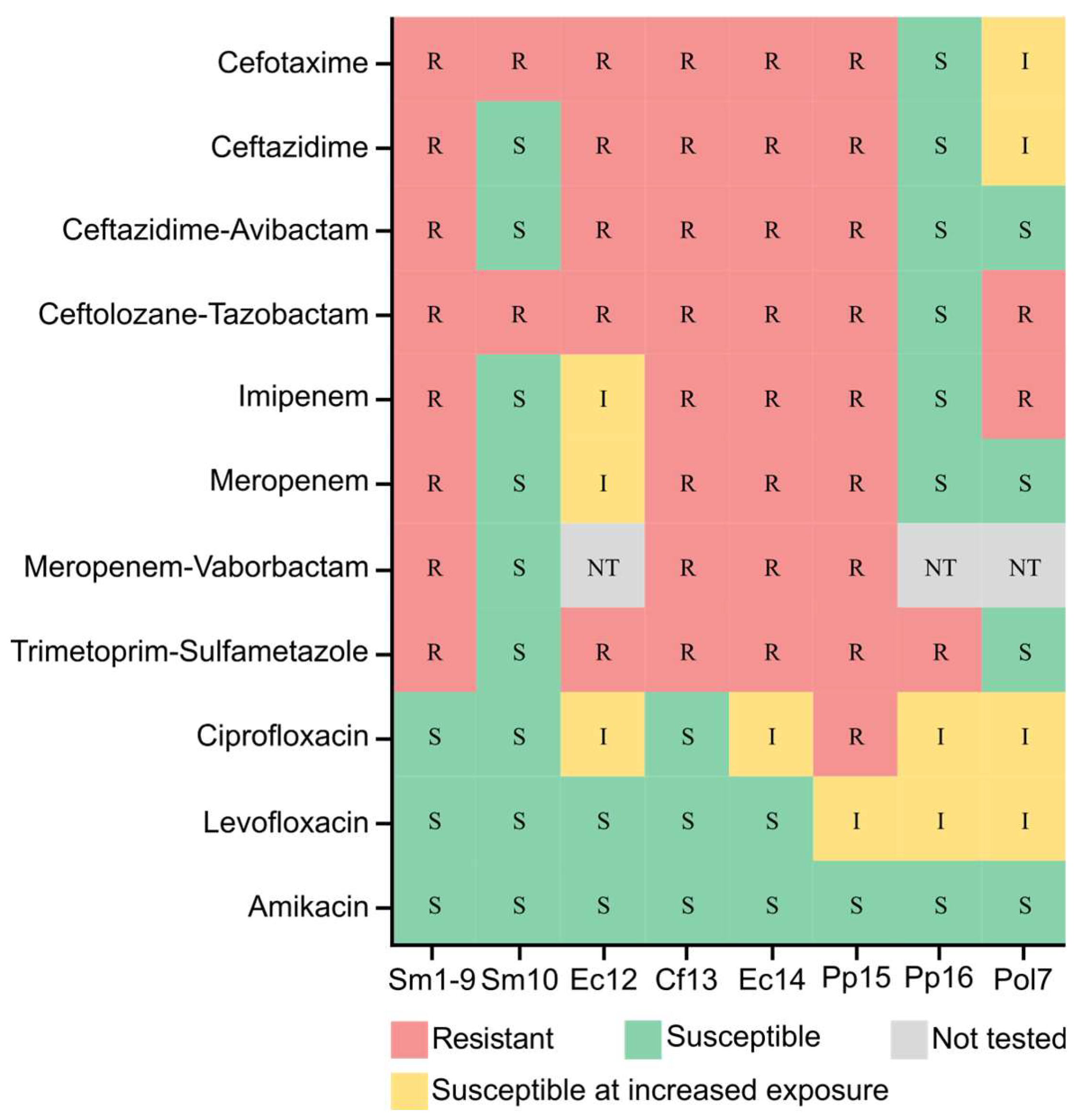
Figure 3.
Distribution of antimicrobial resistance genes, plasmid replicons and virulence-associated genes among clinical and environmental isolates. (A) Heatmap showing the presence or absence of acquired and intrinsic antimicrobial resistance determinants identified by whole-genome sequencing. Isolates are arranged according to bacterial species and clinical or environmental origin. (B) Heatmap showing the presence or absence of plasmid replicon families identified using PlasmidFinder. (C) Heatmap showing the presence or absence of selected virulence-associated genes in the ten clinical Serratia isolates.
Figure 3.
Distribution of antimicrobial resistance genes, plasmid replicons and virulence-associated genes among clinical and environmental isolates. (A) Heatmap showing the presence or absence of acquired and intrinsic antimicrobial resistance determinants identified by whole-genome sequencing. Isolates are arranged according to bacterial species and clinical or environmental origin. (B) Heatmap showing the presence or absence of plasmid replicon families identified using PlasmidFinder. (C) Heatmap showing the presence or absence of selected virulence-associated genes in the ten clinical Serratia isolates.
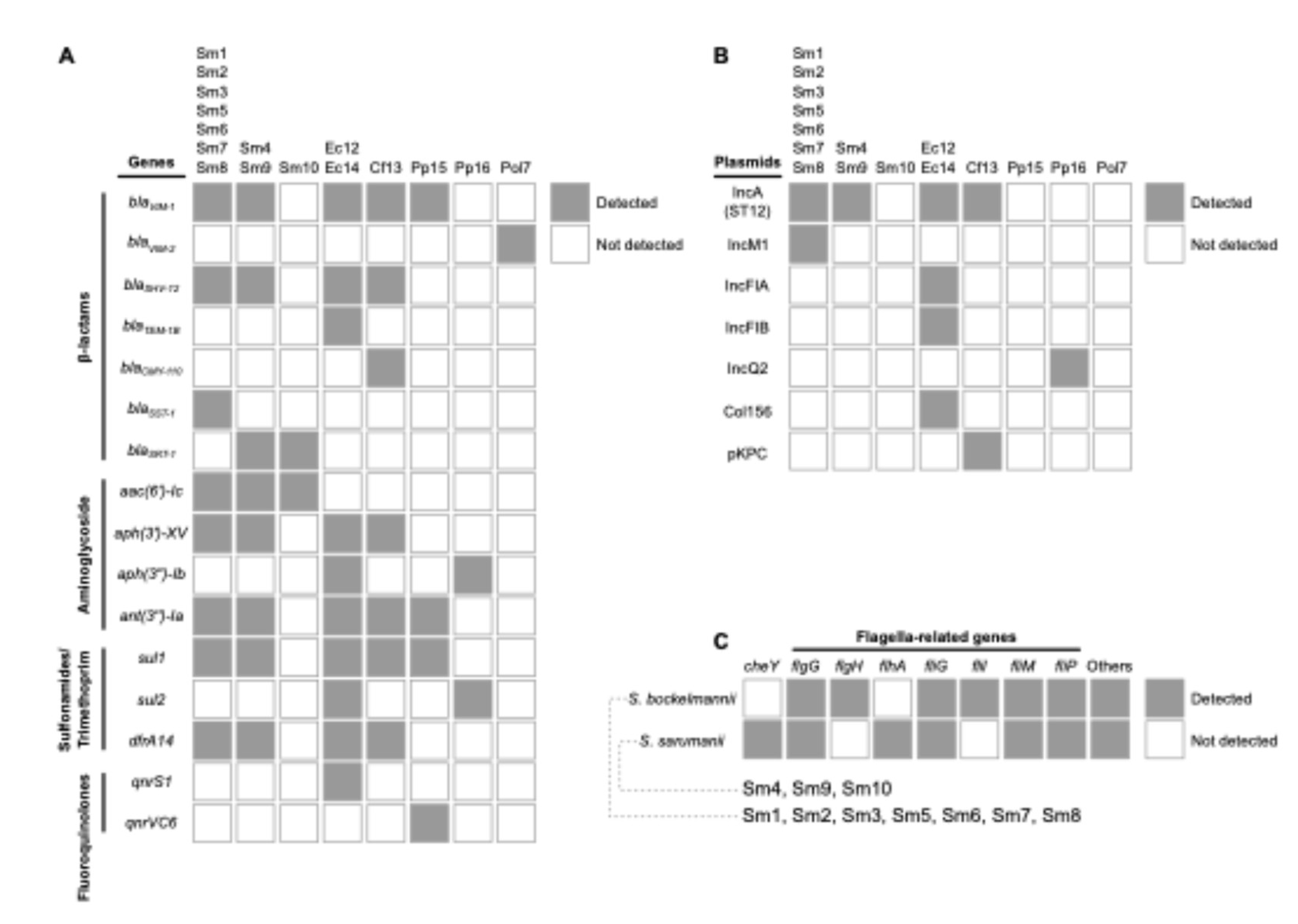
Figure 4.
Pan-genome analysis of the ten clinical Serratia isolates. (A) Gene presence-absence matrix showing the distribution of core and accessory genes among the isolates. Blue indicates gene presence and white indicates gene absence. (B) Distribution of accessory gene counts in S. sarumanii and S. bockelmannii. P < 0.05. (C) Venn diagram showing the genes shared between the two lineages and those associated exclusively with either the S. sarumanii or S. bockelmannii group.
Figure 4.
Pan-genome analysis of the ten clinical Serratia isolates. (A) Gene presence-absence matrix showing the distribution of core and accessory genes among the isolates. Blue indicates gene presence and white indicates gene absence. (B) Distribution of accessory gene counts in S. sarumanii and S. bockelmannii. P < 0.05. (C) Venn diagram showing the genes shared between the two lineages and those associated exclusively with either the S. sarumanii or S. bockelmannii group.
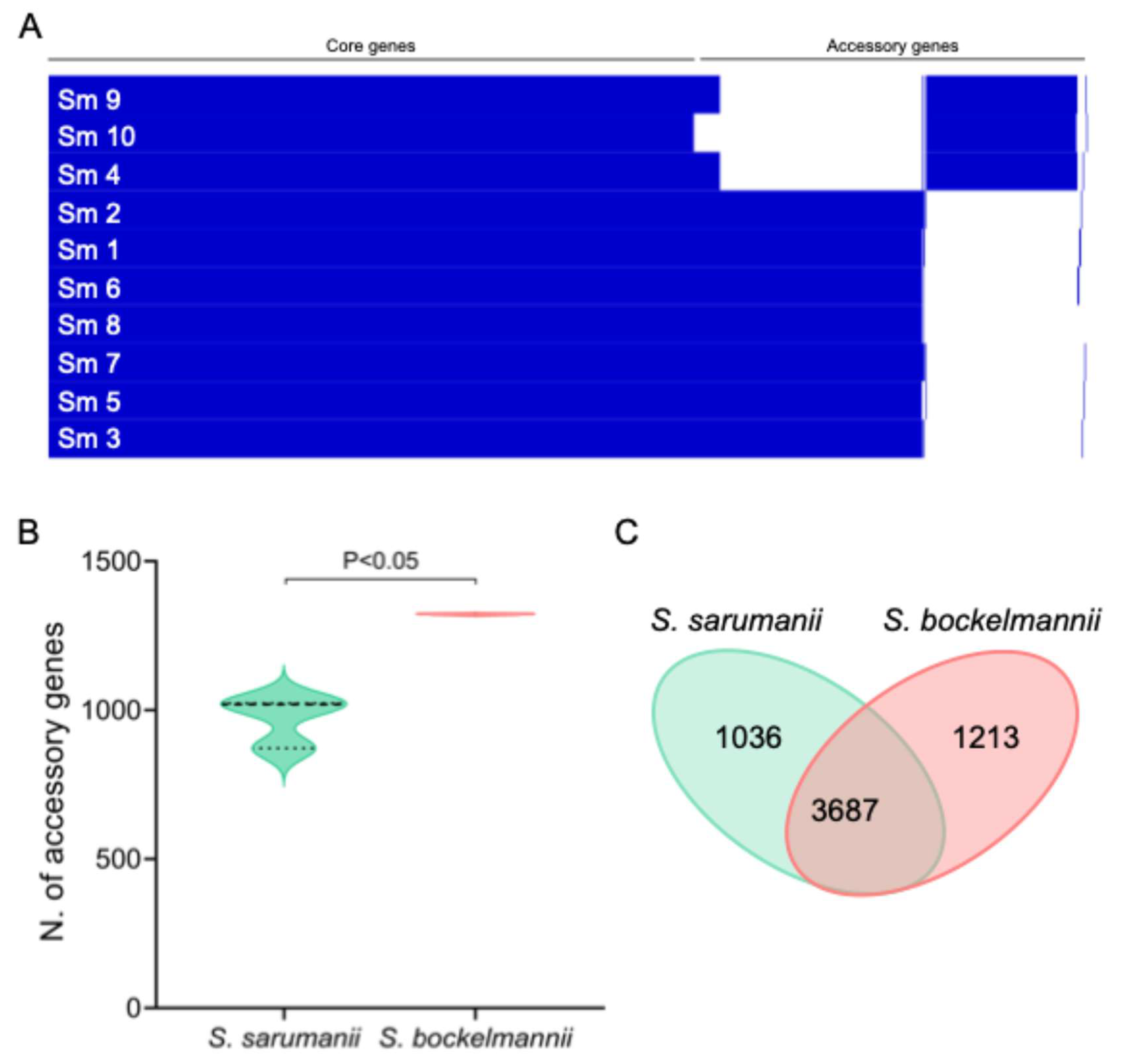
Figure 5.
Core-genome phylogeny of the clinical Serratia isolates. Phylogenetic tree of the 10 samples of Serratia and the reference genomes S. marcescens ELP1.10, S. marcescens subsp. marcescens Db11, S. sarumanii K-M0228, and S. bockelmannii K-L0361. Branches are coloured according to species assignment, with S. sarumanii shown in green and S. bockelmannii in orange.
Figure 5.
Core-genome phylogeny of the clinical Serratia isolates. Phylogenetic tree of the 10 samples of Serratia and the reference genomes S. marcescens ELP1.10, S. marcescens subsp. marcescens Db11, S. sarumanii K-M0228, and S. bockelmannii K-L0361. Branches are coloured according to species assignment, with S. sarumanii shown in green and S. bockelmannii in orange.
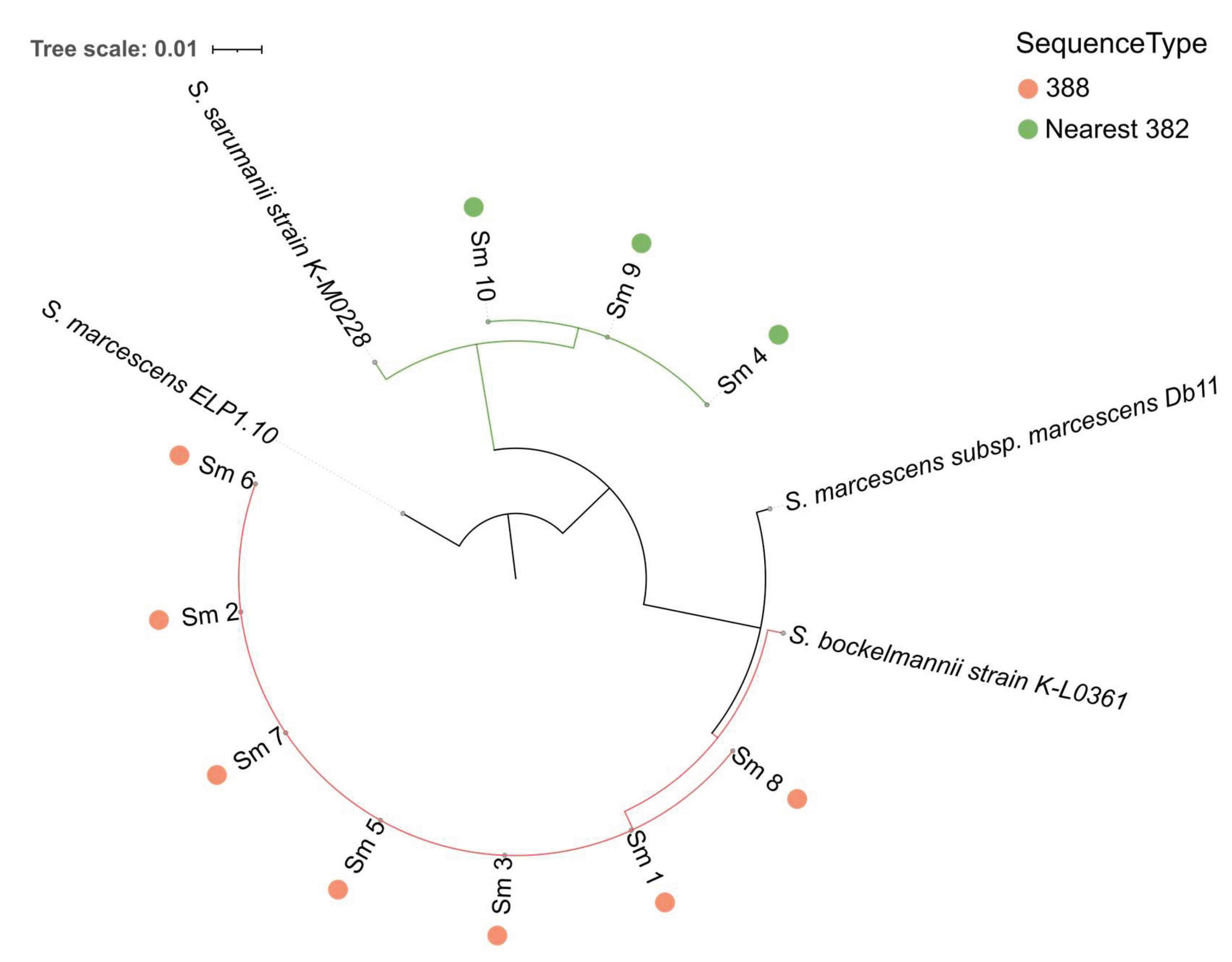

Table 1.
Demographic, clinical and microbiological characteristics of the clinical and environmental isolates selected for genomic analysis during the investigation of a Serratia marcescens outbreak in the Neonatal Intensive Care Unit and Neonatal Unit.
Table 1.
Demographic, clinical and microbiological characteristics of the clinical and environmental isolates selected for genomic analysis during the investigation of a Serratia marcescens outbreak in the Neonatal Intensive Care Unit and Neonatal Unit.
|
Isolate ID |
Species | Patient | Sampling date | Sex |
Days after admission to sampling |
Specimen type |
blaVIM status |
Hospital unit |
| Sm1 | Serratia marcescens | A | 06/11/2024 | F | 17 | Eye swab | Positive | NICU |
| Sm2 | Serratia marcescens | B | 10/11/2024 | F | 18 | Blood culture | Positive | NICU |
| Sm3 | Serratia marcescens | C | 11/11/2024 | M | 25 | Rectal swab | Positive | NICU |
| Sm4 | Serratia marcescens | D | 11/11/2024 | F | 11 | Rectal swab | Positive | NICU |
| Sm5 | Serratia marcescens | E | 11/11/2024 | F | 56 | Rectal swab | Positive | NICU |
| Sm6 | Serratia marcescens | F | 11/11/2024 | M | 21 | Rectal swab | Positive | Neonatal |
| Sm7 | Serratia marcescens | G | 12/11/2024 | F | 7 | Rectal swab | Positive | Neonatal |
| Sm8 | Serratia marcescens | H | 12/11/2024 | M | 2 | Rectal swab | Positive | Neonatal |
| Sm9 | Serratia marcescens | D | 19/11/2024 | F | 19 | Blood culture | Positive | NICU |
| Sm10 | Serratia marcescens | I | 17/11/2024 | F | 85 | Blood culture | Negative | NICU |
| Ec12 | Escherichia coli | E | 14/11/2024 | F | 59 | Rectal swab | Positive | NICU |
| Cf13 | Citrobacter freundii | H | 17/11/2024 | M | 7 | Rectal swab | Positive | Neonatal |
| Ec14 | Escherichia coli | D | 15/11/2024 | F | 15 | Rectal swab | Positive | Neonatal |
| Pp15 | Pseudomonas putida | L | 21/11/2024 | F | 69 | Rectal swab | Positive | Neonatal |
| Pp16 | Pseudomonas putida | - | 13/11/2024 | - | - | Sink swab | Negative | Neonatal |
| Po17 | Pseudomonas oleovorans | - | 13/11/2024 | - | - | Sink swab | Positive | Neonatal |
blaVIM: Verona integron-encoded metallo-β-lactamase gene; NICU: Neonatal Intensive Care Unit; F, female; M, male.
Disclaimer/Publisher’s Note: The statements, opinions and data contained in all publications are solely those of the individual author(s) and contributor(s) and not of MDPI and/or the editor(s). MDPI and/or the editor(s) disclaim responsibility for any injury to people or property resulting from any ideas, methods, instructions or products referred to in the content. |
© 2026 by the authors. Licensee MDPI, Basel, Switzerland. This article is an open access article distributed under the terms and conditions of the Creative Commons Attribution (CC BY) license.
Copyright: This open access article is published under a Creative Commons CC BY 4.0 license, which permit the free download, distribution, and reuse, provided that the author and preprint are cited in any reuse.