

Submitted:
12 August 2026
Posted:
13 August 2026
You are already at the latest version
Abstract
Counter-arguing against the proposition that homology between the coleoptile and the leaf sheath is impossible because a sheath cannot be located above the lamina of the same leaf (Mavrodiev 2025), while defending the bipartite interpretation of the grass cotyledon (BIGC), Scanlon et al. (2026) advance five principal objections: (1) during embryo ontogeny, the coleoptile initiates proximal to the initiation point of the distal scutellum; (2) the coleoptile must be part of the cotyledon because of embryo distichy; (3) tissue continuity between the scutellum and coleoptile supports the interpretation that these two structures together constitute a single composite organ, the cotyledon; (4) BOP1a marker expression in coleoptile supports coleoptile-sheath homology; and (5) ligule and leaf-sheath homologies of the coleoptile are compatible because the ligule represents an extension of the sheath margin. In this reply, I show that all five objections are incorrect. (1) Along the embryo's morphological axis, rather than the proximodistal axis of the leaf, the coleoptile occupies a position distal relative to the scutellum, thus lying above the latter. Therefore, homology between the coleoptile and the leaf sheath is impossible. (2) The disruption of embryonic distichous phyllotaxy by the placement of the coleoptile does not necessarily constitute a morphological argument, and the interpretation of the epiblast as a reduced leaf preserves embryo distichy. (3) Tissue continuity does not imply organ identity. (4) BOP1a expression in coleoptile provides evidence against, rather than in support of the BIGC. (5) Coleoptile-leaf sheath homology does not constitute evidence for coleoptile-ligule homology (or vice versa). Moreover, the claim that the ligule originates from the sheath margin remains open to question because it is formulated without consideration of the basipetal pattern of maize leaf maturation. The reply to Objection 1 affects the very foundation of the argument of Scanlon et al. (2026). Therefore, further discussion on the topic is optional (sublato fundamento, cadit tota propositio).
Keywords:
grass embryo
; proximodistal polarity
; coleoptile
; ligule
; mesocotyl
; ontogeny
; plant morphology
; scutellum
Despite the argument that homology between the coleoptile and the leaf sheath is impossible because a leaf sheath cannot be located above the lamina of the same leaf (Mavrodiev 2025), Scanlon et al. (2026), while defending the bipartite interpretation of the grass cotyledon (BIGC hereinafter), continue to maintain that in maize (Zea mays L.) the coleoptile is homologous to the leaf sheath and therefore represents the proximal sheathing part of the cotyledon, whereas the scutellum, homologous to the leaf blade, represents its distal part.
The objections of Scanlon et al. (2026) are as follows:
Objection 1
During maize embryo ontogeny, the coleoptile initiates proximal to the initiation point of the distal scutellum. But in the maize leaf, the sheath constitutes the proximal region and the lamina (blade) the distal region; therefore, the maize embryo can be viewed as comprising a distal scutellum (homologous to the leaf blade) and a proximal coleoptile (homologous to the leaf sheath). The expression of the YABBY14 marker in the scutellum supports the leaf-blade homology of the latter and, thus, provides an argument for its distal position relative to the coleoptile.
Objection 2
The coleoptile can only be interpreted as part of the cotyledon, not as a separate leaf, because in ontogeny it is initiated on the same side of the embryonic axis as the scutellum, keeping the same position, when mature, while a leaf would be expected on the opposite side under the distichous phyllotaxy of the embryo. Therefore, the maize cotyledon is considered to consist of two parts: the sheathing coleoptile and the laminar scutellum.
Objection 3
Contrary to the leaves developed from the plumule, the coleoptile and scutellum share continuous tissue and are therefore physically connected, supporting the interpretation of both structures as parts of a single composite organ, the cotyledon.
Objection 4
Transcriptomic data support the homology of the coleoptile and the leaf sheath because the sheath marker BOP1a is expressed in the coleoptile and in the proximal regions of the embryo's first leaf after the coleoptile, but not in the scutellum.
Objection 5
Homology between the coleoptile and the ligule is compatible with homology between the coleoptile and the leaf sheath because the ligule is interpreted as a distal extension of the sheath margin.
On the contrary. Ontogenetic evidence cannot support morphologically impossible interpretations. A leaf sheath is necessarily proximal to its blade. But the coleoptile is distal to the scutellum of the maize embryo. Therefore, the coleoptile cannot be the sheath of a cotyledon whose blade is represented by the scutellum.
I answer that the statement in Objection 1 is correct when considered along the cotyledon proximodistal axis. In the mature maize leaf, the sheath is proximal and the lamina is distal; the ligule is still proximal to the lamina but distal to the leaf sheath. In the embryo, this axis is directed from the first embryonic node, the base of the plumule (which includes the shoot apical meristem), toward the tip of the scutellum (Figure 1). Indeed, “the coleoptile initiates ... proximal to the initiation point of the scutellum” (Scanlon et al. 2026, their Figure 1c).
A proximodistal axis, however, may also be defined for the embryo, not only for the leaf or cotyledon. Assuming the plumule to be apical, this axis matches the morphological axis of the embryo and extends from the embryo base toward the plumule (Figure 1). The inversion of proximodistal polarity relative to this axis is purely terminological, since the positional relationship between the plumule, coleoptile, and scutellum remains unchanged: along the embryo morphological axis, the coleoptile (when initiated or developed) always lies between the scutellum and the plumule, regardless of the axis’ direction (Figure 1).
In ontogeny, the coleoptile is initiated closer to the plumule than the scutellum and subsequently encloses it (e.g., Kiesselbach 1949; Wu et al. 2024, 2025; Scanlon et al. 2026). Consequently, it occupies a proximal position relative to the scutellum along the cotyledon proximodistal axis and a distal position along the morphological embryo axis (Figure 1). Thus, in agreement with morphological observations on seedlings, direct ontogenetic evidence indicates that the coleoptile occupies a position closer to the plumule than the scutellum and therefore lies above it on the morphological axis of the embryo (Mavrodiev 2025).
Discussing the spatial relations between the scutellum and the coleoptile, Scanlon et al. (2026), who provided transcriptomic evidence supporting the lateral position of the scutellum and, therefore, the apical placement of the plumule based on the expression of the YABBY14 marker in the scutellum, in fact reduce the morphological axis of the embryo to the point of cotyledon attachment (the morphological node), that is, the origin of the cotyledon proximodistal axis (Figure 1). The morphological axis of the embryo appears again only when embryonic distichy is considered by them. Thus, when the proximodistal relationship between the scutellum and the coleoptile is under discussion, the plumule functions in Scanlon et al. (2026) only as a reference point for establishing the cotyledon proximodistal axis, not as a reference point for the morphological axis of the embryo, as it supposed to be. In other words, for Scanlon et al. (2026), the observation that the coleoptile initiates closer to the plumule simply indicates its proximal position relative to the scutellum along the proximodistal axis of the cotyledon. The point that the coleoptile simultaneously occupies a position above the scutellum on the morphological axis of the embryo is missing in Scanlon et al. (2026), since that axis is considered by these authors only when they discuss the embryonic phyllotaxis. As a result, a clear observation, which is also evident from ontogeny, namely the position of the coleoptile between the scutellum and the plumule (see above), is overlooked by these authors. However, the latter observation directly rejects the homology between the coleoptile and the scutellum sheath, since in that case the sequence would have to be: coleoptile, scutellum, plumule.
The confusion of the coleoptile, a cotyledonary sheath, with the leaf sheath itself, which is the central morphological error in the studies of Wu et al. (2024, 2025) (Mavrodiev 2025), is only possible when the embryo's morphological axis, along which the coleoptile is positioned above the scutellum, is lost from view. But the homology of the coleoptile can be discussed only with respect to the latter axis. As the ligule is also proximal to the lamina along the proximodistal axis of the leaf (see above), ontogenetic evidence alone cannot resolve the homology of the coleoptile: if it is a ligule rather than a leaf sheath, it must still be initiated closer to the plumule than the scutellum. Thus, consideration of the morphological axis of the embryo immediately leads either to the (a) the ligular interpretation of the coleoptile within the BIGC or (b) to the alternative interpretation of the coleoptile as the first (separate) leaf (e.g., Kiesselbach 1949), while simultaneously excluding its homology with the leaf sheath as morphologically meaningless. Neither of these two morphologically correct interpretations (a and b) is mentioned in the papers by Wu et al. (2024, 2025), the main source of the arguments advanced by Scanlon et al. (2026).
The reply to Objection 1 affects the very foundation of the argument of Scanlon et al. (2026). Therefore, further discussion of the topic is logically optional (sublato fundamento, cadit tota propositio). However, if morphologically impossible interpretations, such as the leaf-sheath homology of the coleoptile restated in Scanlon et al. (2026) (Objection 1), can nevertheless become established within an evo-devo framework, then the limitations of the paradigm itself merit serious debate.
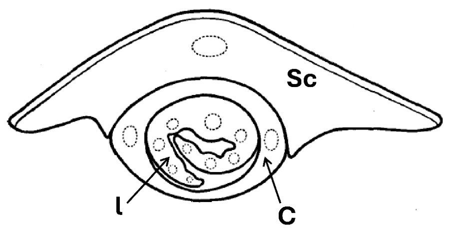
Leaving aside the question of where the morphological axis of the embryo suddenly comes from, given that it did not serve as the embryo's proximodistal axis in the description of the coleoptile’s placement relative to the scutellum, it should be noted that the explanatory power of Objection 2 is limited. Roth (1955, Fig. 8, pp. 580-581) described the first leaf of a wheat (Triticum aestivum L.) seedling positioned on the same side of the embryo morphological axis as the coleoptile, rather than on the opposite side as expected (Figure 2). Thus, according to her interpretation, there is nothing problematic in having not two but three leaves arranged one above another on the same side of the morphological axis of the embryo (Roth 1955). She tentatively attributed this disruption of seedling phyllotaxis to physiological processes that warrant separate study, especially in embryos (Roth 1955). From this perspective, some morphologists focusing specifically on grass embryos would probably not regard the position of the coleoptile as a morphological problem per se if such placement can be explained by physiological processes (cf. Weberling 1999). The epiblast interpretation as a reduced leaf, however, is regarded by many as a straightforward morphological way of restoring distichous phyllotaxy within the embryo (e.g., Bruns 1892; Roth 1955).
Objection 3 is not morphological: the presence of connecting tissue does not demonstrate that the connected structures belong to the same organ (e.g., the collet region). At the same time, the issue of mesocotyl, a structure that indeed connects the scutellum and the coleoptile ontogenetically, is once again completely missing from the discussion by Scanlon et al. (2026). Their equating the mesocotyl with the hypocotyl (Wu et al. 2024; see the legend of their Figure 1d, p. 1611) is a clear morphological error and therefore provides no basis for resolving the mesocotyl problem in future research. The hypocotyl is the sub-cotyledonary axis and therefore cannot be located above the cotyledon or part of it, for example, above the scutellum. In contrast, the mesocotyl is always positioned above the latter (reviewed in Mavrodiev 2025). Resolving the morphological nature of the mesocotyl is crucial for the defense of the BIGC because an axis cannot be situated between the distal and proximal parts of the same leaf (see Mavrodiev 2025 for references).
Any evidence supporting the homology between the coleoptile and the leaf sheath, such as the expression of the BOP1a marker in the coleoptile, as emphasized in Objection 4, constitutes evidence not in favor of the BIGC, but rather against it (Mavrodiev 2025).
This follows because the coleoptile is positioned above the scutellum and therefore cannot represent the sheath of the scutellum (lamina). Therefore, if the plumule is apical, then BOP1a expression supports the interpretation that the coleoptile is the sheath of the leaf immediately following the scutellum on the morphological axis of the embryo (Mavrodiev 2025). The example of BOP1a expression in the coleoptile illustrates that transcriptomic data alone are insufficient to establish organ identity and can be properly interpreted only in the context of the positional relationships among organs within the whole organism, in the discussed case the embryo.
Even if the ligule is interpreted as a distal extension of the leaf sheath margin, this interpretation does not support the conclusion that homology between the coleoptile and the ligule is compatible with homology between the coleoptile and the leaf sheath, as argued in Objection 5. In other words, the fact that the ligule is interpreted as an extension of the leaf sheath margin does not justify the conclusion that the coleoptile, assumed to be homologous to the ligule, is homologous to the leaf sheath. The ligule is a distinct morphological structure with its own unique position within the leaf, gene expression patterns, functions, and ontogeny (e.g., Strable & Aragón-Raygoza 2024; Wang et al. 2024). To demonstrate the ligular nature of the coleoptile from an evo-devo perspective, it would be necessary, at minimum, to present evidence showing similar patterns of gene expression, at least for selected developmental markers, in the coleoptile and the collar region of the maize leaf. However, Wu et al. (2024, 2025) and Scanlon et al. (2026) provide no such data.
In maize leaf ontogeny, the ligule differentiates before the sheath becomes recognizable (e.g., Kiesselbach 1949; Orkwiszewski & Poethig 2000). However, the implications of this well-known basipetal pattern of maize leaf maturation were surprisingly not considered by the authors cited by Scanlon et al. (2026), who either interpreted the ligule as a distal outgrowth of the sheath margin (Satterlee et al. 2023) or provided evidence in favor of such interpretation. Given this pattern, treating the ligule as a distal extension of that margin appears inconsistent with evidence that the ligule is initiated and differentiated before the sheath itself, even if both ligule and sheath margin share a WOX3-dependent developmental program (Satterlee et al. 2023).
Reply to Objection 1
The statement in Objection 1 that the coleoptile initiates proximal to the initiation point of the distal scutellum is correct when considered along the cotyledon’s proximodistal axis. However, along the morphological axis of the embryo, the coleoptile occupies a distal position relative to the scutellum because it is initiated and placed closer to the plumule than the scutellum. Therefore, contrary to Scanlon et al. (2026), the coleoptile cannot be considered a scutellum sheath because it lies above the scutellum on the morphological axis of the embryo (Figure 1).
Reply to Objection 2
The observed disruption of embryo distichy by the placement of the coleoptile is not necessarily a morphological argument. A similar disruption has been reported in grasses (e.g., wheat) and may be caused by physiological factors (Figure 2). By contrast, interpreting the epiblast as a reduced leaf provides a morphological argument against Objection 2 because such interpretation preserves the embryo's distichous phyllotaxis.
Reply to Objection 3
The existence of connecting tissue does not demonstrate that two structures (e.g., scutellum and coleoptile) belong to the same organ (e.g., collet region).
Reply to Objection 4
Because along the embryo morphological axis the coleoptile is located above the scutellum, it cannot be the sheath of the scutellum. Therefore, expression of the BOP1a marker indicates relationship between the coleoptile and the leaf sheath but does not support BIGC. Instead, this evidence is consistent with the view that the coleoptile is a sheath-like structure separate from the scutellum, representing the sheath of the leaf immediately following the scutellum, assuming the plumule is apical.
Reply to Objection 5
The proposition that ligular homology of the coleoptile is consistent with its leaf-sheath homology because the ligule is a distal outgrowth of the sheath margin is logically flawed, as it improperly extends the identity of a part to the whole (a composition fallacy). Thus, evidence for coleoptile-ligule homology does not constitute evidence for coleoptile-leaf sheath homology, and vice versa. It is therefore not surprising that the ligular homology of the coleoptile is compatible with its position in the embryo (Mavrodiev 2025), whereas the coleoptile-leaf sheath homology defended by Scanlon et al. (2026) is not (Mavrodiev 2025). Moreover, in view of the basipetal pattern of maize leaf development, the origin of the ligule from the leaf sheath margin is in question.
Whether plant morphology is still alive within contemporary academia, I leave to the judgment of the reader.
References
- Abbe, E.C.; Stein, O.L. The growth of the shoot apex in maize: embryogeny. Am. J. Bot. 1954, 41, 285–293. [Google Scholar] [CrossRef]
- Bruns, E. Der Grasembryo; Val. Höfling: Munich, Germany, 1892. [Google Scholar]
- Kiesselbach, T.A. The structure and reproduction of corn. Univ. Neb. Agric. Stat. Res. Bull. 1949, 161, 1–96. [Google Scholar]
- Mavrodiev, E. Is plant morphology still alive? Fundamental flaws in genetic and transcriptomic support for the bipartite interpretation of the grass cotyledon. MDPI Prepr.> 2025. [Google Scholar] [CrossRef]
- Orkwiszewski, J.A.; Poethig, R.S. Phase identity of the maize leaf is determined after leaf initiation. Proc. Natl. Acad. Sci. USA 2000, 97, 10631–10636. [Google Scholar] [CrossRef] [PubMed]
- Roth, I. Zur morphologischen Deutung des Grasembryos und verwandter Embryotypen. Flora 1955, 142, 564–600. [Google Scholar] [CrossRef]
- Satterlee, J.W.; Evans, L.J.; Conlon, B.R.; Conklin, P.; Martinez-Gomez, J.; Yen, J.R.; Wu, H.; Sylvester, A.W.; Specht, C.D.; Cheng, J.; et al. A Wox3-patterning module organizes planar growth in grass leaves and ligules. Nat. Plants 2023, 9, 720–732. [Google Scholar] [CrossRef] [PubMed]
- Scanlon, M.J.; Niklas, K.J.; Zhang, R.; Wu, H. Comment on “Is plant morphology still alive? Fundamental flaws in genetic and transcriptomic support for the bipartite interpretation of the grass cotyledon”. MDPI Prepr.> 2026. [Google Scholar] [CrossRef]
- Strable, J.; Aragón-Raygoza, A. Development and maintenance of the ligular region of maize leaves. Mol. Plant 2024, 17, 1175–117. [Google Scholar] [CrossRef] [PubMed]
- Wang, Q.; Guo, Q.; Shi, Q.; Yang, H.; Liu, M.; Niu, Y.; Quan, S.; Xu, D.; Chen, X.; Li, L.; et al. Histological and single-nucleus transcriptome analyses reveal the specialized functions of ligular sclerenchyma cells and key regulators of leaf angle in maize. Mol. Plant 2024, 17, 920–934. [Google Scholar] [CrossRef] [PubMed]
- Weberling, F. Wilhelm Troll, his work and influence. Syst. Geogr. Plants 1999, 68, 9–24. [Google Scholar] [CrossRef]
- Wu, H.; Zhang, R.; Scanlon, M.J. Genetic analyses of embryo homology and ontogeny in the model grass Zea mays subsp. mays. New Phytol. 2024, 243, 1610–1619. [Google Scholar] [CrossRef] [PubMed]
- Wu, H.; Zhang, R.; Niklas, K.J.; Scanlon, M.J. Multiplexed transcriptomic analyses of the plant embryonic hourglass. Nat. Commun. 2025, 16, 802. [Google Scholar] [CrossRef] [PubMed]
Figure 1.
Figure 1. The proximodistal axes of the maize embryo and its cotyledon. Proximodistal axis of the maize embryo (A, solid line or cross) and its cotyledon (B, dotted line). Axis A is identical to the morphological embryo axis. Images 1–3 are redrawn and modified from Wu et al. (2024, Figure 1b–d). For each panel (1–3), the left and right images show diagrams of the median longitudinal (sagittal) and paradermal sections of the maize embryo, respectively. For simplicity, the first node is indicated by the star at every developmental stage, and the radicle is referred as the primary root. All abbreviations are from Wu et al. (2024): c, coleoptile; h, hypocotyl; l, leaf; lc, lower coleoptile; r, radicle (root); SAM, shoot apical meristem; sc, scutellum; su, suspensor; uc, upper coleoptile; v, vasculature. The coleorhiza is not shown, as in Wu et al. (2024). Panel 1 shows the embryo at the transitional stage (Abbe & Stein, 1954; Wu et al., 2024). Panel 2 shows the embryo at the coleoptilar stage (Abbe & Stein, 1954; Wu et al., 2024), and Panel 3 shows 'Stage 1' as defined by Abbe & Stein (1954) and adopted by Wu et al. (2024). The coleoptile (c) occupies a proximal position relative to the scutellum (sc) along the cotyledon proximodistal axis (B) and a distal position along the morphological embryo axis (A).
Figure 1.
Figure 1. The proximodistal axes of the maize embryo and its cotyledon. Proximodistal axis of the maize embryo (A, solid line or cross) and its cotyledon (B, dotted line). Axis A is identical to the morphological embryo axis. Images 1–3 are redrawn and modified from Wu et al. (2024, Figure 1b–d). For each panel (1–3), the left and right images show diagrams of the median longitudinal (sagittal) and paradermal sections of the maize embryo, respectively. For simplicity, the first node is indicated by the star at every developmental stage, and the radicle is referred as the primary root. All abbreviations are from Wu et al. (2024): c, coleoptile; h, hypocotyl; l, leaf; lc, lower coleoptile; r, radicle (root); SAM, shoot apical meristem; sc, scutellum; su, suspensor; uc, upper coleoptile; v, vasculature. The coleorhiza is not shown, as in Wu et al. (2024). Panel 1 shows the embryo at the transitional stage (Abbe & Stein, 1954; Wu et al., 2024). Panel 2 shows the embryo at the coleoptilar stage (Abbe & Stein, 1954; Wu et al., 2024), and Panel 3 shows 'Stage 1' as defined by Abbe & Stein (1954) and adopted by Wu et al. (2024). The coleoptile (c) occupies a proximal position relative to the scutellum (sc) along the cotyledon proximodistal axis (B) and a distal position along the morphological embryo axis (A).
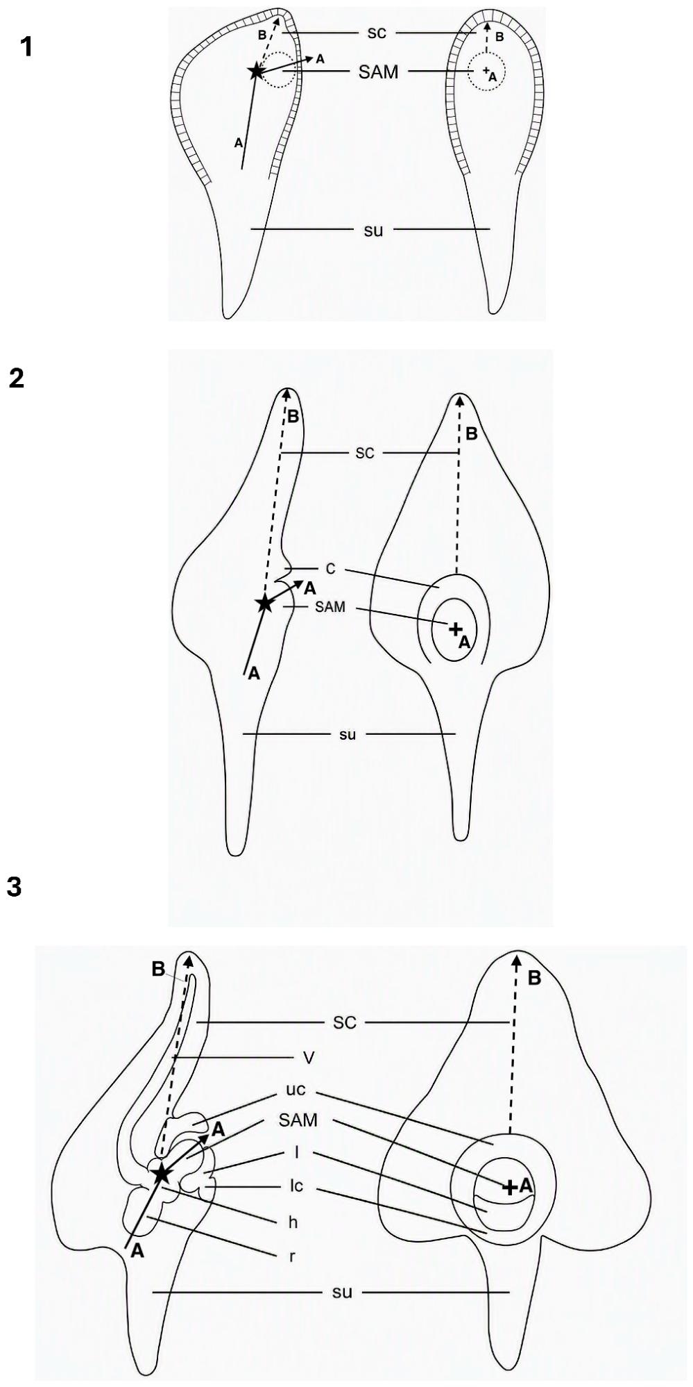
Figure 2.
The first leaf of a wheat seedling positioned on the same side of the morphological axis as the coleoptile. Transverse section through an embryo of the wheat cultivar Walthari, showing the first foliage leaf positioned above the coleoptile and on the same side of the embryonic axis. Vascular bundles are indicated by dotted lines. Modified from Roth (1955, her Fig. 8). Abbreviations: c, coleoptile; l, leaf; sc, scutellum.
Figure 2.
The first leaf of a wheat seedling positioned on the same side of the morphological axis as the coleoptile. Transverse section through an embryo of the wheat cultivar Walthari, showing the first foliage leaf positioned above the coleoptile and on the same side of the embryonic axis. Vascular bundles are indicated by dotted lines. Modified from Roth (1955, her Fig. 8). Abbreviations: c, coleoptile; l, leaf; sc, scutellum.
Disclaimer/Publisher’s Note: The statements, opinions and data contained in all publications are solely those of the individual author(s) and contributor(s) and not of MDPI and/or the editor(s). MDPI and/or the editor(s) disclaim responsibility for any injury to people or property resulting from any ideas, methods, instructions or products referred to in the content. |
© 2026 by the author. Licensee MDPI, Basel, Switzerland. This article is an open access article distributed under the terms and conditions of the Creative Commons Attribution (CC BY) license.
Copyright: This open access article is published under a Creative Commons CC BY 4.0 license, which permit the free download, distribution, and reuse, provided that the author and preprint are cited in any reuse.


