

Submitted:
06 August 2026
Posted:
13 August 2026
You are already at the latest version
Abstract
Colorectal cancer (CRC) is one of the most common malignancies worldwide, and while metastasis commonly occurs in the liver and lungs, brain metastases from CRC are rare. Solitary brain metastasis in young adults is particularly uncommon. This case report a rare presentation of brain metastasis in a patient with a history of colorectal cancer. Emergency radiotherapy is often considered for symptomatic patients with intracranial hypertension or rapid neurological deterioration, but its benefits may be transient in aggressive tumor biology. We report the case of a 35-year-old male with a history of T3N2bM1a colorectal adenocarcinoma who presented with new-onset neurological symptoms, including headaches and right-sided hemiparesis. CT imaging revealed multiple brain lesions, including solid and hemorrhagic components with erosion of the left frontalis bone and brain edema, suggestive of metastatic disease. Given the rapid neurological decline, the patient was managed with a multidisciplinary approach, including corticosteroids to reduce cerebral edema, followed by whole-brain radiotherapy (WBRT) to address the metastatic lesions. Neurological symptoms improved within days, with partial resolution of hemiparesis. Surgical resection was deferred due to high perioperative risk and patient preference. The clinical improvement was short-lived. Follow-up imaging at 3 months demonstrated lesion progression with increased mass effect, accompanied by worsening neurological function after 4 months. This case highlights the rare occurrence of solitary brain metastasis from CRC in a young patient. It underscores the potential but transient benefit of emergency radiotherapy in rapidly deteriorating neurological states. Early recognition, individualized treatment planning remain essential in optimizing quality of life.
Keywords:
colorectal cancer
; brain metastasis
; case report
; management
; radiotherapy
; chemotherapy
Budhi Ida Bagus 1,*, Nafa Unnisa 2 and Febriagi Bayu Aji 2
1 Department of Surgery, Sebelas Maret University, Indonesia
2 Medical Faculty, Sebelas Maret University, Indonesia
Introduction
Colorectal cancer (CRC) is the third most commonly diagnosed malignancy and the second leading cause of cancer-related mortality worldwide, with an estimated 1.93 million new cases and 935,000 deaths reported in 2020 [1]. The global number of new CRC cases is expected to increase to 3.2 million by 2040, driven by population growth, aging, and lifestyle-related risk factors [2]. Traditionally regarded as a disease of older adults, CRC incidence peaks between the ages of 60 and 75. However, there is a concerning rise in early-onset CRC (diagnosed before age 50), which accounts for approximately 10% of all new diagnoses, with a nearly 1.4% annual increase in CRC incidence among adults under 50 [3,4]. Early-onset colorectal cancer (EOCRC) refers to colorectal cancer diagnosed in individuals younger than 50 years and is generally detected at more advanced stages. Its histological characteristics are frequently associated with a poor prognosis [5]. Patients with early-onset colorectal cancer are more frequently diagnosed at advanced stages, with 27% presenting with distant metastases compared to 20% of older patients [6].
CRC metastasizes most commonly to the liver and lungs, reflecting the venous and lymphatic drainage from the colorectal region [7]. Brain metastases from CRC are relatively rare, with the overall average being 2.10%, ranging from 0.1% to 11.5%. BM from CRC typically occur late in the disease course, often develops after extracranial metastasis, including liver and lung involvement [8,9]. Risk factors for brain involvement include younger age, rectal primary tumors, KRAS mutations, and the presence of liver and pulmonary metastases [10]. Solitary brain metastasis from CRC is particularly uncommon, as most cases involve intracranial lesions in the setting of lung or liver metastasis before [9].
We present a case of a 35-year-old patient with early-onset CRC who developed a solitary brain metastasis with acute neurological deterioration and transient improvement following emergency radiotherapy. This case highlights the importance of early recognition, multidisciplinary decision-making, and realistic prognostic discussions in managing rare presentations of CRC brain metastases.
Case Report
Patient Information
A 35-year-old male with no significant family history of colorectal cancer or hereditary syndromes presented to our institution in June 2021 with complaints of abdominal pain, hematochezia, and worsening constipation. He had no prior history of abdominal surgery or inflammatory bowel disease.
Clinical Findings
On physical examination, the patient was hemodynamically stable. Abdominal palpation revealed tenderness in the lower abdomen, particularly in the left lower quadrant. Digital rectal examination confirmed the presence of a firm, irregular mass within the rectum.
Timeline
- June 2021: The patient underwent a contrast-enhanced CT scan, which revealed a large mass located in the rectum. Colonoscopy with biopsy confirmed adenocarcinoma of the rectum.
- June–Sept 2021: Initial chemotherapy with 5-fluorouracil and leucovorin was started, consisting of twelve cycles. Despite good tolerance, the patient showed no significant clinical or radiological improvement following chemotherapy.
- Oct 2021–Mar 2022: The chemotherapy regimen was escalated to FOLFOX (5-fluorouracil, leucovorin, and oxaliplatin) in response to the lack of improvement. However, repeat imaging showed persistent disease progression.
- Aug 2022: The patient received external beam radiotherapy (30 Gy in 10 fractions) in an effort to control the local tumor burden. Following radiotherapy, the patient was transitioned to palliative chemotherapy.
- 2024: Surveillance imaging revealed recurrent tumor infiltration involving the lamina propria, submucosa, muscularis propria, and muscularis mucosa. Multiple enlarged lymph nodes were seen in the paraaortic, mesenteric, parailiac, inguinal, and pericolic regions. No hepatic or pulmonary metastases were detected on further imaging studies. The tumor staging was re-evaluated according to the AJCC 2017 criteria as T3N2bM1a (Stage IVa).
- Mar 2025: Four years after the initial diagnosis, the patient developed neurological symptoms consisting of right hemiparesis and persistent headache. A CT scan revealed multiple intra-axial brain metastases with solid and hemorrhagic components located in the left parietal lobe, left centrum semiovale, left corona radiata extending into the basal ganglia, and left cerebellar hemisphere. The lesions were associated with significant vasogenic edema. The left parietal lesion caused compression and narrowing of the anterior and posterior horns of the left lateral ventricle, with evidence of a 0.5 cm subfalcine herniation (Figure 1). The patient was started on high-dose corticosteroids and underwent three cycles of whole-brain radiotherapy. Partial clinical improvement was observed, with some recovery of motor function and reduction in headache.
- June 2025: Despite the patient's stable condition, a follow-up CT scan showed no improvement in the metastatic lesions (Figure 2).
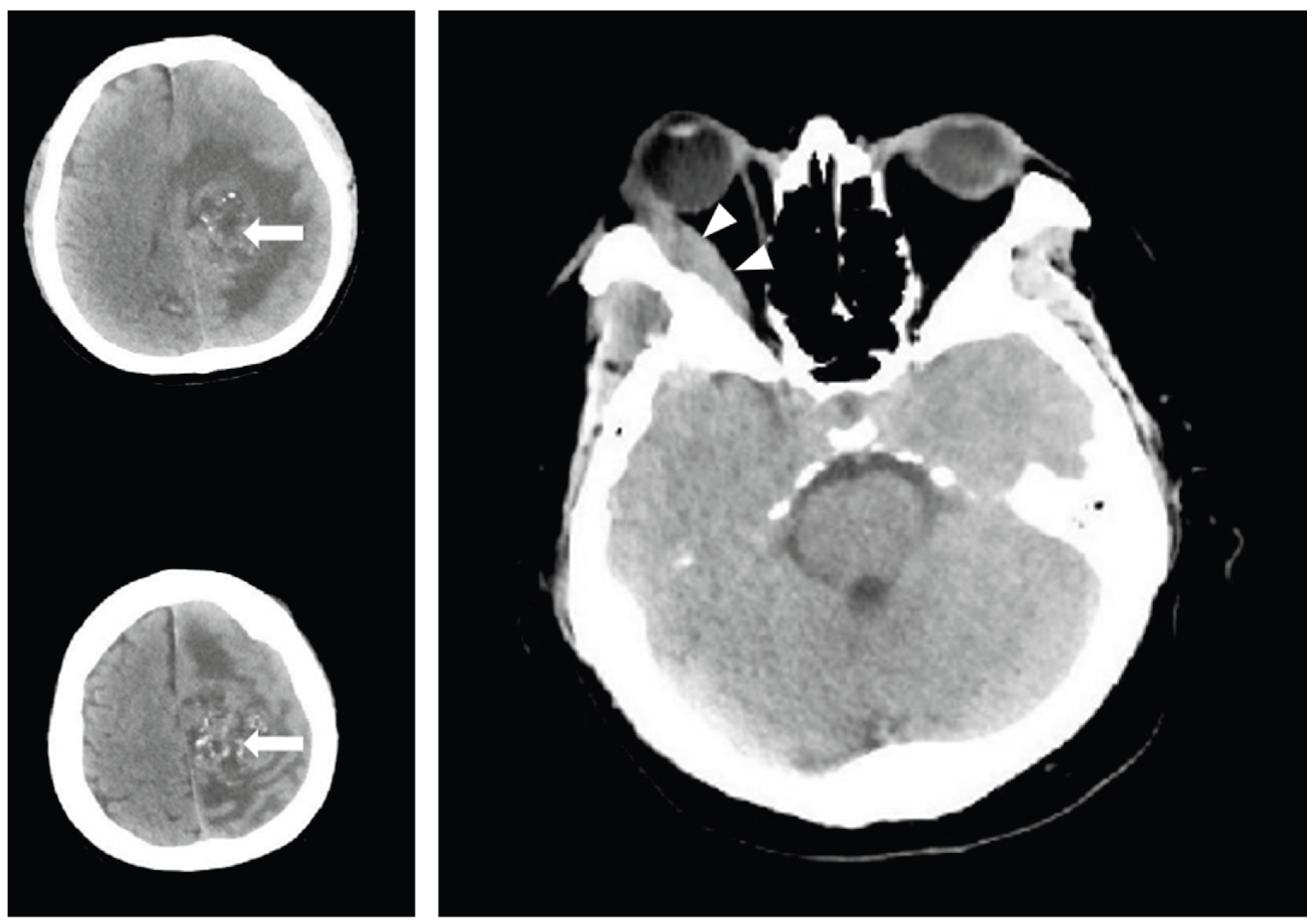
- July 2025: The patient was readmitted with worsened neurological status, including seizures and a consciousness impairment. Repeat brain CT revealed disease progression (Figure 3), and the patient was transferred to the High Care Unit (HCU) for intensive management with high-dose corticosteroids. The patient's condition showed improvement, and he was subsequently discharged for continued care at home.
- August 2025: The patient was readmitted on August 9th with complaints of seizures, nausea, vomiting, and generalized weakness. Palliative care was initiated, focusing on symptomatic management with appropriate medications.
Diagnostic Assessment
The diagnosis of colorectal cancer was initially confirmed through colonoscopy and biopsy. Imaging studies, including CT scans of the abdomen and pelvis, indicated a large rectal mass without distant metastasis at the time of diagnosis. Subsequent MRI confirmed local recurrence and peritoneal spread. Brain metastases were suspected after the patient developed new neurological symptoms 4 years after the first visit. A CT scan of the brain confirmed the presence of multiple intra-axial brain metastases, involving both supratentorial and infratentorial compartments, with hemorrhagic and solid components. The frontal lesion raised concern for meningeal metastasis, with calcifications and bone erosion on imaging.
Therapeutic Intervention
The patient was initially treated with adjuvant chemotherapy (5-fluorouracil and leucovorin) followed by FOLFOX due to the lack of response. Upon disease progression, he underwent external beam radiotherapy to control local tumor growth. Given the worsening prognosis and metastatic progression, the patient was transitioned to palliative chemotherapy. In March 2025, after the development of neurological symptoms, the patient received high-dose corticosteroids and emergency whole-brain radiotherapy. Although there was initial partial clinical improvement, further disease progression led to the need for intensive management in the HCU.
Follow-Up and Outcomes
During follow-up, the patient’s clinical status fluctuated. Initially, after receiving emergency treatment with steroids and radiotherapy, partial improvement was noted in his hemiparesis. However, repeated imaging demonstrated no significant change in the metastatic lesions. By July 2025, the patient experienced worsening neurological deficits, including frequent seizures and a decreased level of consciousness, necessitating HCU care. Given the extent of progression and resistance to multiple treatments, the patient was discharged and continued with outpatient treatment.
Discussion
This case illustrates the rare manifestation of solitary and multiple brain metastases in early-onset colorectal cancer (EORC), a finding that is uncommon and generally associated with advanced disease and unfavorable prognosis [8,11]. Early-onset colorectal cancer (EOCRC) typically exhibits more aggressive biological behavior, is diagnosed at more advanced stages, and demonstrates a higher propensity for metastasis [12].
Most patients —up to 96%— with BM in CRC remain asymptomatic, often resulting in delayed diagnosis. When symptoms of brain metastases do occur, the most commonly reported manifestations include seizures, signs of increased intracranial pressure, and other neurological deficits[9]. In this case, the patient presented with neurological symptoms of right-sided hemiparesis and headache approximately four years after his initial diagnosis of CRC.
Several risk factors have been identified for the development of brain metastases in patients with colon cancer. At the time of diagnosis, up to 90% of patients with brain metastases also have metastases at other sites, most frequently affecting the liver and lungs [13]. Specifically, the presence of lung and bone metastases has been recognized as an independent risk factor for brain involvement [9,14]; however, none were observed in this patient. Additional risk factors include KRAS mutations, elevated CEA levels, and tumors originating in the rectum [9].
The molecular mechanisms underlying the ability of tumor cells to cross the blood–brain barrier (BBB) remain incompletely understood. Most current evidence derives from studies on brain metastases in breast cancer, which demonstrate that CXCR4, the receptor for chemokine CXCL12, can destabilize blood vessels and increase the permeability of brain endothelial cells. Inhibition of the CXCR4/CXCL12 pathway reduces both cancer cell migration and vascular permeability. Similarly, colorectal carcinoma cells exhibit elevated CXCR4 expression compared to normal intestinal epithelium, and immunohistochemical analyses have confirmed strong CXCR4 expression in brain metastases, as well as in liver and lymph node metastases, suggesting that the CXCR4/CXCL12 pathway may play a comparable role in facilitating metastasis in colorectal cancer [15].
Brain metastases in CRC typically occur in patients with widespread systemic involvement, often involving the lungs and liver. However, three potential routes for hematogenous dissemination of colorectal cancer leading to brain metastases have been proposed. The first involves tumor cells spreading via the rectal venous plexus to the inferior vena cava, then to the lungs, and ultimately to the brain, bypassing the liver. The second route is through Batson’s vertebral plexus, allowing direct access to the brain without transit through the liver or lungs. The third pathway follows the portal venous system, with tumor cells first reaching the liver, then the lungs, and finally the brain [16]. In this patient, the most probable route of dissemination appears to be the second mechanism, as there was no evidence of metastasis to the liver or lungs. Rectal tumors may seed Batson’s venous plexus, a network of valveless veins extending up the vertebral column, and reach the cranial venous sinuses followed by the ophthalmic vein [17].
The standard management of brain metastases includes surgical resection, stereotactic radiosurgery, and whole-brain radiotherapy (WBRT), either alone or in combination. Surgical intervention is generally reserved for patients with a single, large metastasis causing significant edema or located in eloquent brain regions. Stereotactic radiosurgery is typically indicated for patients with a limited number of metastases, whereas WBRT is preferred in cases of multiple metastases, large oligometastases with uncontrolled extracranial disease, or in patients with poor performance status. According to the American Society of Clinical Oncology–Society for Neuro-Oncology–American Society for Radiation Oncology guidelines, symptomatic brain metastases should be treated with local therapies, including surgery or radiation-based approaches, independent of systemic therapy [18,19]. Considering these recommendations and the patient’s clinical presentation, WBRT represents an appropriate therapeutic choice in this case . The patient's response to emergency radiotherapy and corticosteroids provided transient symptomatic relief but did not result in long-term stabilization of the disease.
In conclusion, this case underscores the importance of early identification of neurological symptoms in patients with advanced colorectal cancer, as well as the need for a multidisciplinary approach in the management of brain metastases. While emergency radiotherapy and corticosteroids can provide transient symptomatic relief, these treatments are not curative and should be considered as part of a palliative care strategy when brain metastases are present.
Declaration of generative AI in scientific writing
During the preparation of this manuscript, ChatGPT was used to improve the clarity of this manuscript. The authors reviewed and edited the text and take full responsibility for the final content.
Financial Support
The authors received no financial support for the research, authorship, and/or publication of this article.
Informed consent
The patient has already been informed about the purpose of this case study and get patient permission on using of the clinical material such as clinical finding during operation, radiological exam for the publication. The patient already signed the written informed consent for the usage of the information and clinical images for publication.
Ethical Approval
This case study has already been approved by Health Research Ethic Committee of Moewardi General Hospital, Surakarta, Indonesia.
Ethical clearance number
315 / IV / HREC / 2025.
Author Contributions
Conceptualization and design: Budhi Ida Bagus; Administrative support: Budhi Ida Bagus; Provision of study materials or patients: Budhi Ida Bagus, Febriagi Bayu Aji, Nafa Unnisa; Collection and assembly of data: Budhi Ida Bagus, Febriagi Bayu Aji, Nafa Unnisa; Manuscript writing: All authors; Final approval of manuscript: All authors; Accountable for all aspects of the work: All authors.
Conflict of Interest
The author declared there is no conflict of interest to be reported.
Acknowledgement
No potential conflict of interest relevant to this article was reported.
References
- Sung, H.; Ferlay, J.; Siegel, R.L.; Laversanne, M.; Soerjomataram, I.; Jemal, A.; et al. Global Cancer Statistics 2020: GLOBOCAN Estimates of Incidence and Mortality Worldwide for 36 Cancers in 185 Countries. CA Cancer J. Clin. 2021, 71(3), 209–49. [Google Scholar] [CrossRef] [PubMed]
- Xi, Y.; Xu, P. Global colorectal cancer burden in 2020 and projections to 2040. Transl. Oncol. 2021, 14(10), 101174. [Google Scholar] [CrossRef] [PubMed]
- Ahmed, S.Z.; Cirocchi, N.; Saxton, E.; Brown, M.K. Incidence of age migration of colorectal cancer in younger population: Retrospective single centred-population based cohort study. Ann. Med. Surg. 2022, 74. [Google Scholar] [CrossRef] [PubMed]
- Ullah, F.; Pillai, A.B.; Omar, N.; Dima, D.; Harichand, S. Early-Onset Colorectal Cancer: Current Insights. Cancers 2023, 15(12), 3202. [Google Scholar] [CrossRef] [PubMed]
- Saraiva, M.R.; Rosa, I.; Claro, I. Early-onset colorectal cancer: A review of current knowledge. World J. Gastroenterol. 2023, 29(8), 1289–303. [Google Scholar] [CrossRef] [PubMed]
- Siegel, R.L.; Wagle, N.S.; Cercek, A.; Smith, R.A.; Jemal, A. Colorectal cancer statistics, 2023. CA Cancer J. Clin. 2023, 73(3), 233–54. [Google Scholar] [CrossRef] [PubMed]
- Riihimäki, M.; Hemminki, A.; Sundquist, J.; Hemminki, K. Patterns of metastasis in colon and rectal cancer. Sci. Rep. 2016, 6(1), 29765. [Google Scholar] [CrossRef] [PubMed]
- Li, W.; Wang, T.; Zhu, Y.; Yu, H.; Ma, L.; Ding, Y.; et al. Brain metastasis from colorectal cancer: Treatment, survival, and prognosis. Medicine 2022, 101(40), e30273. [Google Scholar] [CrossRef] [PubMed]
- Müller, S.; Köhler, F.; Hendricks, A.; Kastner, C.; Börner, K.; Diers, J.; et al. Brain Metastases from Colorectal Cancer: A Systematic Review of the Literature and Meta-Analysis to Establish a Guideline for Daily Treatment. Cancers 2021, 13(4), 900. [Google Scholar] [CrossRef] [PubMed]
- Shaikh, A.S.; Pavurala, R.; Gou, E. Brain Metastasis in a Young Patient: Consider the Rectum. Cureus 2021. [Google Scholar] [CrossRef] [PubMed]
- Majd, N.; Simon, M.; Wagman, R.; Gesner, L. A rare case of colon cancer metastasis to the brain and a brief review of its treatment and prognosis. Radiol. Case Rep. 2022, 17(3), 696–9. [Google Scholar] [CrossRef] [PubMed]
- Medici, B.; Riccò, B.; Caffari, E.; Zaniboni, S.; Salati, M.; Spallanzani, A.; et al. Early Onset Metastatic Colorectal Cancer: Current Insights and Clinical Management of a Rising Condition. Cancers 2023, 15(13). [Google Scholar] [CrossRef] [PubMed]
- Damiens, K.; Ayoub, J.P.M.; Lemieux, B.; Aubin, F.; Saliba, W.; Campeau, M.P.; et al. Clinical Features and Course of Brain Metastases in Colorectal Cancer: An Experience from a Single Institution. Curr. Oncol. 2012, 19(5), 254–8. [Google Scholar] [CrossRef] [PubMed]
- Nozawa, H.; Ishihara, S.; Kawai, K.; Sasaki, K.; Murono, K.; Otani, K.; et al. Brain Metastasis from Colorectal Cancer: Predictors and Treatment Outcomes. Oncology 2017, 93(5), 309–14. [Google Scholar] [CrossRef] [PubMed]
- Zang, Y.W.; Gu, X.D.; Xiang, J.B.; Chen, Z.Y. Brain Metastases from Colorectal Cancer: Microenvironment and Molecular Mechanisms. Int. J. Mol. Sci. 2012, 13(12), 15784–800. [Google Scholar] [CrossRef] [PubMed]
- Vargas-Urbina, J.; Martinez-Silva, R.; Rojas-Panta, G.; Ponce-Manrique, G.; Flores-Castillo, J.; Anicama-Lima, W. Unusual brain metastasis from colon cancer. Surg. Neurol. Int. 2025, 16, 5. [Google Scholar] [CrossRef] [PubMed]
- Pearlman, M.; Kwong, W.T. A long and distant journey: a case of rectal cancer with metastasis to the orbit. Ann. Gastroenterol. 2015, 28(1), 151–2. [Google Scholar] [PubMed]
- Vogelbaum, M.A.; Brown, P.D.; Messersmith, H.; Brastianos, P.K.; Burri, S.; Cahill, D.; et al. Treatment for Brain Metastases: ASCO-SNO-ASTRO Guideline. J. Clin. Oncol. 2022, 40(5), 492–516. [Google Scholar] [CrossRef] [PubMed]
- Nguyen, H.; Phung, D.; Nguyen, T.; Tran, B.; Thi Mai, K.; Trinh, H. Management of brain metastasis from rectal cancer using whole-brain radiation therapy followed by bevacizumab and chemotherapy: A case report. Oncol. Lett. 2023, 26(4), 446. [Google Scholar] [CrossRef] [PubMed]
Figure 1.
Head CT Scan March 2025. Brain metastases with solid and hemorrhagic components located in the left cerebral hemisphere (white arrow) and left cerebellar (black arrow). The lesions were associated with significant vasogenic edema (black arrowhead).
Figure 1.
Head CT Scan March 2025. Brain metastases with solid and hemorrhagic components located in the left cerebral hemisphere (white arrow) and left cerebellar (black arrow). The lesions were associated with significant vasogenic edema (black arrowhead).
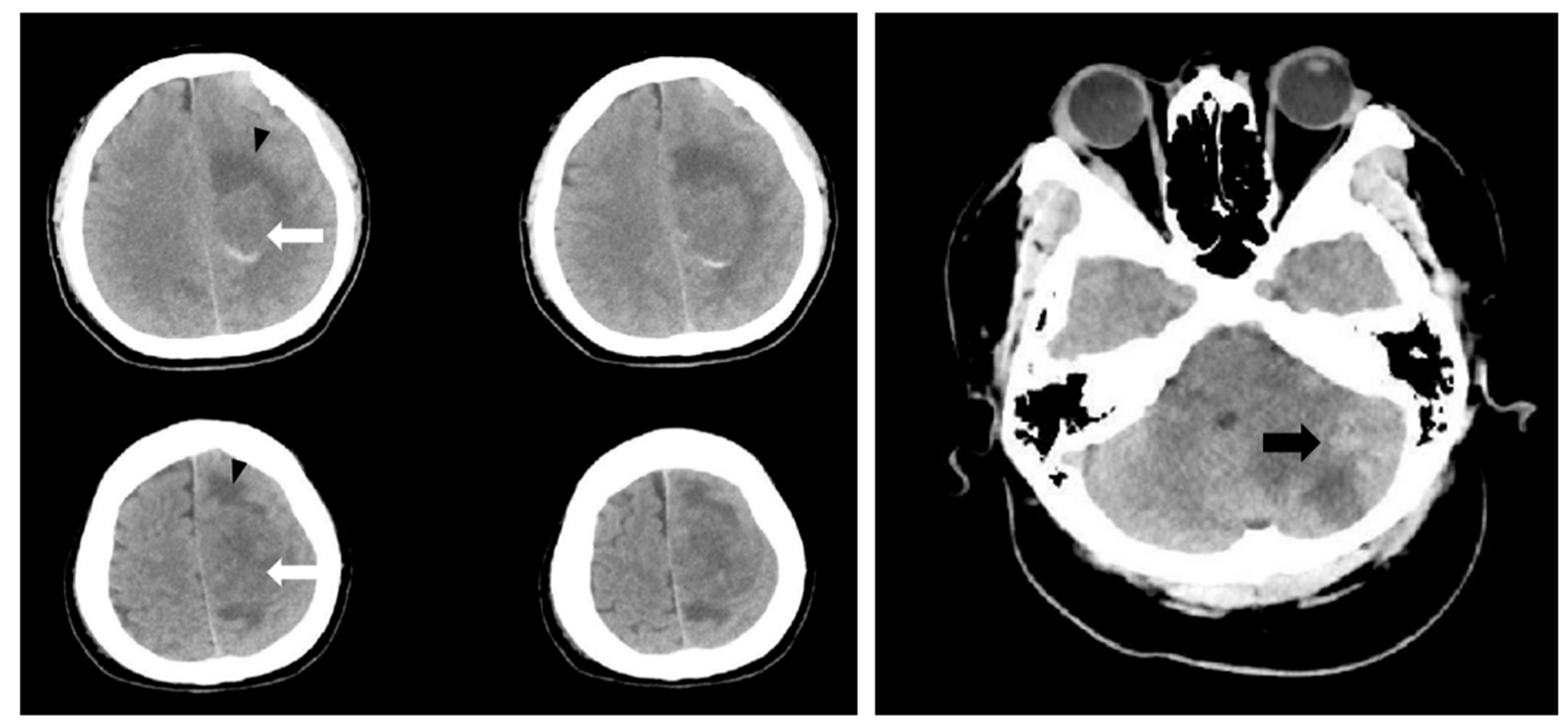
Figure 2.
Head CT-Scan June 2025. Intracranial metastatic lesions in the left cerebral hemisphere (white arrow) and left cerebellar (black arrow). A solid lesion was identified in the left frontal region, extending into the dural space and causing destruction of the frontal bone (black arrowhead). Additionally, a solid lesion in the right orbital region extending into the dural space of the right temporal region (white arrowhead).
Figure 2.
Head CT-Scan June 2025. Intracranial metastatic lesions in the left cerebral hemisphere (white arrow) and left cerebellar (black arrow). A solid lesion was identified in the left frontal region, extending into the dural space and causing destruction of the frontal bone (black arrowhead). Additionally, a solid lesion in the right orbital region extending into the dural space of the right temporal region (white arrowhead).
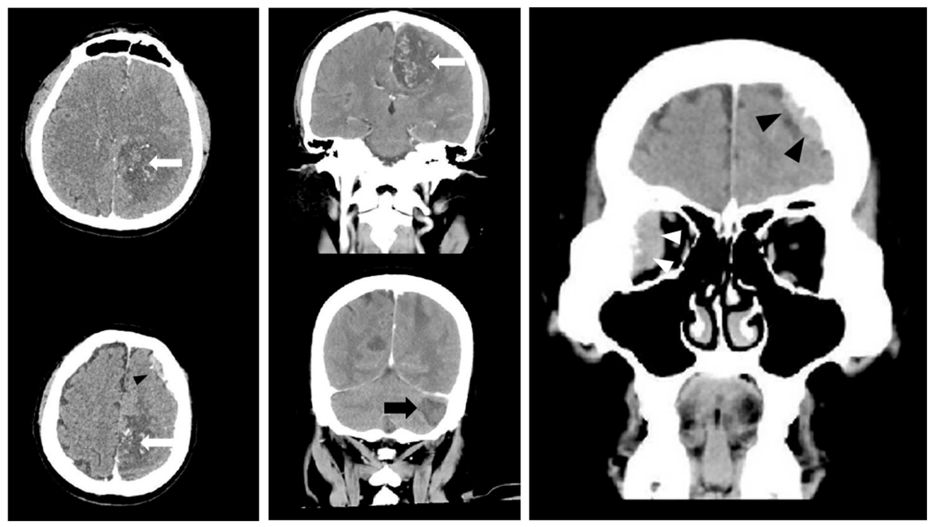
Figure 3.
Head CT-Scan July 2025. Intracranial metastatic lesions in the left cerebral hemisphere (white arrow). A solid lesion in the right orbital region extending into the dural space of the right temporal region (white arrowhead).
Figure 3.
Head CT-Scan July 2025. Intracranial metastatic lesions in the left cerebral hemisphere (white arrow). A solid lesion in the right orbital region extending into the dural space of the right temporal region (white arrowhead).
Disclaimer/Publisher’s Note: The statements, opinions and data contained in all publications are solely those of the individual author(s) and contributor(s) and not of MDPI and/or the editor(s). MDPI and/or the editor(s) disclaim responsibility for any injury to people or property resulting from any ideas, methods, instructions or products referred to in the content. |
© 2026 by the authors. Licensee MDPI, Basel, Switzerland. This article is an open access article distributed under the terms and conditions of the Creative Commons Attribution (CC BY) license.
Copyright: This open access article is published under a Creative Commons CC BY 4.0 license, which permit the free download, distribution, and reuse, provided that the author and preprint are cited in any reuse.