

Submitted:
11 August 2026
Posted:
13 August 2026
You are already at the latest version
Abstract
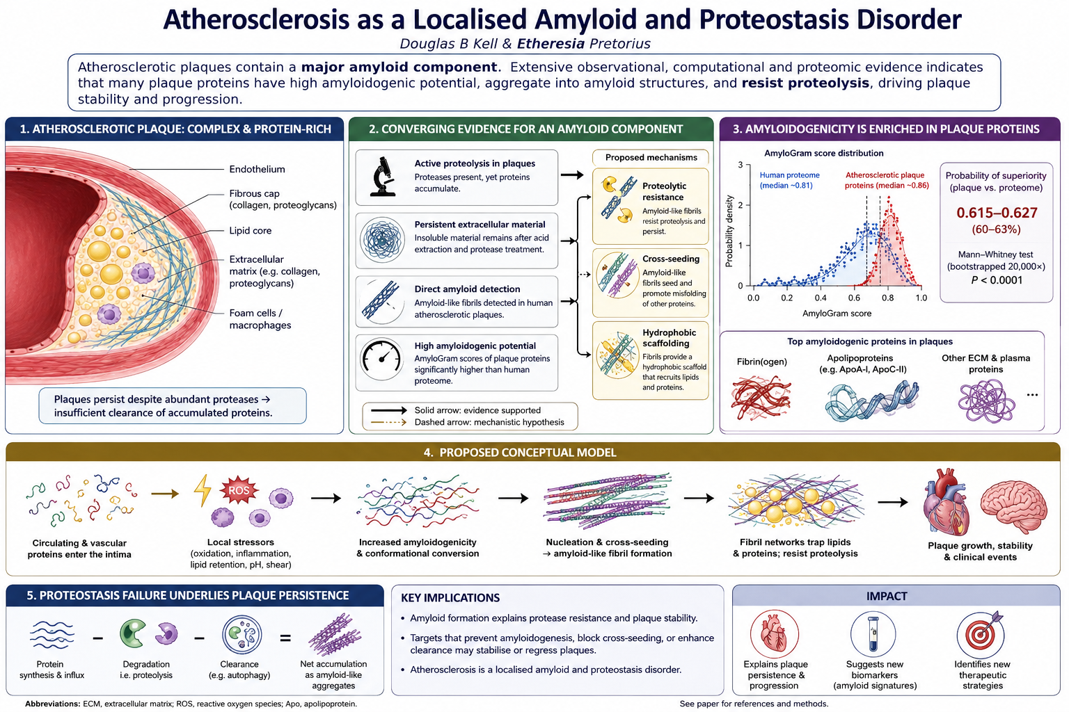
Atherosclerosis and atherosclerotic plaque formation underpin a variety of acute cardiovascular events including myocardial infarction and ischaemic stroke, and vascular diseases of this type are effectively the leading causes of death worldwide. Despite the presence of many proteases that are normally capable of degrading the proteins that these plaques contain, the existence of atherosclerotic plaques necessarily implies that their rate of growth or accretion exceeds the rate of their degradation. As is well known, amyloid forms of proteins can be much more resistant to proteolysis than are their non-amyloid forms. Thus, one possibility is that many of these atherosclerotic proteins have morphed into amyloid forms. First, we summarise the extensive evidence for this, that is both observational (using various amyloid-structure-determining methods) and computational (using the program AmyloGram), showing that the amyloid(ogenic) potential of proteins found in plaques is significantly greater than that of most typical proteins. Thus, while 79% of human proteins have an AmyloGram score exceeding 0.7, with a median value of 0.81, typically more than 90% of proteins in plaques have AmyloGram scores exceeding 0.7, with a median score around 0.86. The probability-of-superiority values are around 0.615–0.627 against the human proteome. In addition, a Mann-Whitney test bootstrapped 20,000 times gave a 95% CI 0.0411–0.0497 for the difference in median AmyloGram score in Lewy bodies vs the human proteome. Secondly, while there is no significant relation between the overall hydropathy and amyloidogenic tendency of individual proteins, we recognise that amyloid forms of proteins expose many hydrophobic surface patches. These can act as scaffolds for other hydrophobic constituents of plaque. Overall the extensive new evidence provided here of an enrichment of amyloid forms of proteins in atherosclerotic and similar plaques can account, at least in part, for their resistance to proteolytic degradation. This has therapeutic implications.
Keywords:
atherosclerosis
; plaque
; Lewy bodies
; amyloid
; amyloidogenesis
; AmyloGram
; resistance to proteolysis
Introduction
Atherosclerosis is a chronic, progressive disease of medium and large arteries characterised by the focal accumulation within the arterial intima of ApoB-containing lipoproteins, inflammatory cells, vascular smooth-muscle cells, extracellular matrix, cellular debris and, in advanced lesions, calcification (e.g. (Jebari-Benslaiman et al. 2022; Libby 2021; Libby et al. 2019; Stary et al. 1995)). The resulting plaques may cause disease through progressive arterial narrowing, impaired vasomotor function, plaque rupture or erosion with superimposed thrombosis, along with weakening of the arterial wall. It is increasingly prevalent, and with atherosclerotic cardiovascular problems being aetiological for acute cardiovascular events such as myocardial infarction, ischaemic stroke and unstable angina, such vascular diseases are effectively the leading causes of death worldwide (e.g. (Chen et al. 2023; Libby et al. 2019; Nedkoff et al. 2023; Song et al. 2020; Watanabe and Fan 2025)).
We recognise that according to the modified American Heart Association classification, there are eight types of atherosclerotic lesions: intimal thickening, fibroatheroma, late fibroatheroma, healed plaque rupture, fibrocalcific plaques, erosions, thin-capped atheroma, and ruptured plaques (Cai et al. 2002). The size and thickness of such plaques are necessarily determined by the difference between their rates of formation and removal. Indeed, human plaque degradomics shows extensive proteolytic processing of plaque proteins by matrix metalloproteinases, cathepsins, elastase, kallikreins and related enzymes, while plasma-derived fibrinolytic proteases such as plasmin are capable of degrading fibrin and several extracellular proteins present in plaques (Dollery and Libby 2006; Dollery et al. 1995; Garcia-Touchard et al. 2005; Lorentzen et al. 2024; Lorentzen et al. 2025; Lu et al. 2011; Luttun et al. 2004; Newby 2005; Smith 1994; Stirk et al. 1993). This creates a “proteolysis paradox”: advanced plaques contain many proteins that should, in principle, be vulnerable to extracellular proteases, yet they persist for years and often accumulate further. Consequently, our focus is on why the rate of plaque formation seemingly exceeds that of its degradation in atherosclerosis. Amyloid conversion offers a parsimonious explanation because amyloid formation changes the physical state of the substrate, additional to other oxidative changes, not merely the abundance or activity of the proteases. Specifically, since amyloid forms of proteins are known to be far more resistant to proteolysis than are their native forms, our main focus will be on the question of the extent to which any of these proteins can or do display amyloid properties.
The term ‘amyloid’ (Almeida and Brito 2020; Chiti and Dobson 2009, 2006; Eisenberg and Jucker 2012; Greenwald and Riek 2010; Ke et al. 2020; Kelly 1998, 1996; Sawaya et al. 2021; Taylor and Staniforth 2022; Toyama and Weissman 2011) is used to describe conformations of proteins that have no necessary change in primary structure but that adopt an entirely different, stabler conformation that may elongate (including heterologously (Kalitnik et al. 2025)) via recruitment and conformational templating of further monomers and oligomers. A defining structural element of amyloids is the cross-β feature (e.g. (Gallardo et al. 2020; Greenwald and Riek 2010; Ke et al. 2020; Nelson et al. 2005; Riek 2017; Serpell et al. 2007; Siemer 2022; Taylor and Staniforth 2022)) (that can be detected with various stains such as thioflavin T (Biancalana and Koide 2010; Gade Malmos et al. 2017) or conjugated thiophene oligomers (Nilsson et al. 2012; Nilsson 2009; Nyström et al. 2017) (‘Amytrackers’TM)).
Histology is an important means of detecting amyloidoses (Riefolo et al. 2022). Thus, following the self-consistent discoveries that blood can clot into an anomalous, amyloid form (e.g. (Ahn et al. 2017; Kell and Pretorius 2017; Pretorius et al. 2016; Pretorius et al. 2017b; Pretorius et al. 2013a; Zamolodchikov et al. 2016) that is resistant to proteolysis (fibrinolysis), that such structures can also be observed in the thrombi from ischaemic stroke (Grixti et al. 2025), and that the aggregates involved are highly enriched in amyloidogenic proteins (Kell et al. 2025a; Kell and Pretorius 2025b; Kell and Pretorius 2024; Kell et al. 2026a), it was of interest to enquire as to the extent to which atherosclerotic plaques are also amyloid in nature, as this seems not to be widely recognised and would contribute an important mechanistic basis for plaques’ stability and resistance to proteolysis. The purpose of the present article is therefore to highlight the significance of amyloidogenesis in atherosclerotic plaque formation and stability. Figure 1 gives an overview.
Proteostasis (protein homeostasis) refers to the biological processes that maintain a proper balance between the synthesis, folding, trafficking, and degradation of proteins (Shukla and Narayan 2025). Pathological amyloidogenesis may be viewed as a manifestation of failed proteostasis, arising when protein-folding, chaperone, degradation and clearance systems can no longer prevent the accumulation of aggregation-prone conformers; the resulting aggregates may in turn further impair the proteostasis network. As such, pathological amyloidogenesis can therefore be viewed as both a consequence and a cause of impaired proteostasis (Chiti and Dobson 2017; Goto et al. 2024; Hipp and Hartl 2024; Hipp et al. 2014; Klaips et al. 2018; Powers et al. 2009; Stroo et al. 2017). A shorter version of a very small subset of some of the analyses rehearsed here has been preprinted (Kell et al. 2025b).
Results
Classical Composition of Atheromatous Plaques
As widely reviewed (e.g. (Bentzon et al. 2014; Di Nubila et al. 2024; Libby 2021; Libby et al. 2019; Otsuka et al. 2016; Zhang et al. 2024)), the precise constituents of plaques, and their ratios, will vary with a plaque’s age. However, an advanced atheromatous plaque typically comprises an intimal accumulation of extracellular and intracellular lipid, macrophage- and smooth-muscle-derived foam cells, inflammatory leukocytes, extracellular matrix and a collagen-rich fibrous cap overlying a lipid-rich, acellular or hypocellular necrotic core. Variable additional features include cholesterol crystals, calcification, neovascularisation, intraplaque haemorrhage and superimposed thrombosis. However, although advanced plaques are recognised to contain abundant extracellular protein, retained apolipoproteins, proteolytic fragments, and long-lived necrotic material, aggregated proteins (including fibrin) and amyloid forms thereof are comparatively rarely listed among their canonical constituents.
Many diseases are comorbid with classical cardiovascular diseases, and each involve inflammation, oxidative stress, iron dysregulation and the production of microthrombi (Kell and Pretorius 2018), as may be observed using assays such as capillaroscopy (Kell and Pretorius 2025a), thermal imaging (Kell and Pretorius 2026) and laser scattering (Kell et al. 2026b) methods. Given the role of oxidative stress, it is very noteworthy and interesting that LDL oxidation (Chan et al. 2015; Stewart et al. 2005), cholesterol oxidation (Stewart et al. 2007b), and Apo-A1 oxidation (Wong et al. 2010) can lead to amyloid formation. Specifically, oxidised lipoproteins provide a particularly important mechanistic bridge between conventional atherogenesis and amyloidogenesis. Oxidation of LDL can induce amyloid-like structures displaying cross-β characteristics, and these modified particles are recognised by macrophages through pathways already central to foam-cell formation (Stewart et al. 2005). Thus, amyloidogenesis need not be viewed as an alternative to the lipid-retention/oxidation model of atherosclerosis, but as a conformational extension of it.
Evidence for an Amyloid Component in Atherosclerotic Plaques
As summarised in a number of articles (e.g. (Bucerius et al. 2017; Das and Gursky 2015; De Meyer et al. 2002; Hellberg et al. 2019; Howlett and Moore 2006; Howlett et al. 2019; Kell et al. 2025b; Medeiros et al. 2004; Mucchiano et al. 2001a; Mucchiano et al. 2001b; Röcken et al. 2006; Stewart et al. 2007a; Teoh et al. 2011b; Wang et al. 2017; Westermark et al. 1995), amyloid has in fact been widely observed in atherosclerotic plaques. We consider here both structural/ observational and computational evidence (the former summarised in Figure 2).
As indicated in Figure 2, the structural or observational evidence for the presence of amyloid in plaques comes from a variety of techniques, and these are listed with references in Table 1.
In particular, amyloid-targeting PET ligands provide a further translational line of evidence, albeit not yet a definitive clinical diagnostic test for plaque amyloid. 18F-flutemetamol has shown plaque accumulation ex vivo/in experimental systems with histological support (Hellberg et al. 2019), while 18F-florbetaben PET/MR has been explored for human carotid vascular amyloid-β imaging (Bucerius et al. 2017). These studies support the plausibility of imaging plaque amyloid in vivo, but larger studies with matched histology are still needed.
The Main Structural Feature of Amyloid Proteins
Non-amyloid forms of amyloidogenic proteins tend to have multiple α-helices. By contrast, the amyloid variants are much enriched in β−sheets. In particular, as mentioned, the defining feature of amyloid forms of proteins is a so-called cross-β motif in which β-sheets stack in a parallel or antiparallel form. Figure 3 gives an example, using the normal PrPC and ‘rogue’ PrPSc forms of the human prion protein.
Scoring Amyloidogenicity
A number of methods have been developed for calculating the aggregating or amyloid-forming potential of proteins from their sequences or (more occasionally) experimental structures. Some are listed in (Ahmed and Kajava 2013; Gonay et al. 2025; Hassan et al. 2025; Housmans et al. 2023; Kell and Pretorius 2024; Pintado-Grima et al. 2023; Prabakaran et al. 2021a; Prabakaran et al. 2021b; Roland et al. 2013; Santos et al. 2020). Most use sliding windows to detect amyloidogenic hotspots, but comparing these hotspots between proteins is rather cumbersome. Our experience is that a program which provides a convenient amyloidogenicity score between 0 and 1 for a whole protein is far more useful for large-scale surveys, and that the program AmyloGram (Burdukiewicz et al. 2017; Szulc et al. 2021) does this to great effect (Kell et al. 2025a; Kell and Pretorius 2025b; Kell and Pretorius 2024; Kell et al. 2025b; Roberts et al. 2026). We first ‘calibrated’ the system with known amyloids as annotated at UniProt, finding that any protein with an AmyloGram score over 0.7 was potentially amyloidogenic (Kell et al. 2025a), and on this basis the majority of proteins, in a variety of taxa from viruses upwards, were in fact potentially amyloidogenic (Kell et al. 2025a; Kell and Pretorius 2025b; Kell and Pretorius 2024; Roberts et al. 2026), with median AmyloGram scores per organism’s proteome in the range 0.81-0.85. The point is that while the forms (conformations) of these kinds of proteins as normally made by the ribosome are kinetically stable, they are much less thermodynamically stable than are the amyloid forms (Kell and Pretorius 2017), but they are separated therefrom by a high kinetic energy barrier. Prion proteins (Aguzzi and Calella 2009; Aguzzi and Lakkaraju 2016; Prusiner 1991, 1998), in which a minute quantity of PrPSc can seed an autocatalytic process that ultimately converts billions of normal PrPC molecules into the disease-associated PrPSc conformation (Meisl et al. 2021; Saá et al. 2006; Saborio et al. 2001), have particularly high amyloidogenic scores, with the human prion protein (Uniprot P04156) having an AmyloGram score of 0.916 (Kell et al. 2025a). Note that proteins with a high AmyloGram score also tended to have a high aggregation score, as judged (Roberts et al. 2026) by comparison with the outputs of the program PASTA2 (Walsh et al. 2014).
Flipping into an Amyloid Form, and Cross-Seeding
Because amyloid forms of proteins, with their multiple tight (but non-covalent) cross-β linkages, are thermodynamically significantly more stable than are the ‘normal’ forms of these proteins, in principle all that is needed to effect the amyloidogenic ‘flip’ is a catalytic trigger. Fibrinogen itself is a highly amyloidogenic protein (Kell and Pretorius 2017; Kranenburg et al. 2002; Kumar et al. 2022; Litvinov et al. 2012; Mai et al. 2024; Pretorius et al. 2016; Serpell et al. 2007; Zhmurov et al. 2012), with AmyloGram scores for fibrinogens A, B, and G of 0.828, 0.863, and 0.922, respectively (Kell et al. 2025a). Molecules that have been demonstrated experimentally to help trigger it to polymerise (under the influence of thrombin) into an amyloid form include ferric ions (Pretorius et al. 2013b), bacterial lipopolysaccharide (LPS, at a ratio of 1 LPS: 108 fibrinogens (Pretorius et al. 2016)), lipoteichoic acid (Pretorius et al. 2018a), and the SARS-CoV-2 spike protein (Grobbelaar et al. 2022; Grobbelaar et al. 2021) (itself again highly amyloidogenic (Nyström and Hammarström 2022; Westman et al. 2025)).
‘Homologous’ polymerisation in which a preformed amyloid version of an amyloidogenic protein molecule such as Aβ or α-synuclein causes non-amyloid conformations of the same protein to flip into an amyloid form and thus be incorporated into the growing polymer fibril, is probably seen as the most common occurrence. However, ‘cross-seeding’, in which a given amyloid molecule causes the incorporation of a molecule of a different protein, also in an amyloid form, is also commonplace (e.g. (Babu et al. 2025; Basha et al. 2026; Chaudhuri et al. 2019; Fan et al. 2024; Ge et al. 2023; Hu et al. 2017; Ivanova et al. 2021; Kell and Pretorius 2024; Larsson et al. 2011; Padilla-Godínez et al. 2025; Ren et al. 2018; Ren et al. 2019; Vaneyck et al. 2021; Westermark et al. 2018; Yuzu et al. 2021; Zhou et al. 2012)). Proteomic analyses of amyloid complexes are the means to establish the nature and extent of this cross-seeding.
Figure 4, adapted from an Open Access publication (Bondarev et al. 2018), illustrates the key features of two different classes of fibril complexes, each with two subsets.
In the case of blood clotting, the proteome of normal thrombi more or less reflects that (e.g. (Kalaidopoulou Nteak et al. 2024)) of the soluble plasma proteome (Kell and Pretorius 2024; Ząbczyk et al. 2019). By contrast, the proteome of amyloid-containing microclots is quite different (Kell and Pretorius 2025b; Kell and Pretorius 2024; Kruger et al. 2022; Pretorius et al. 2021; Schofield et al. 2024). Some normally soluble proteins that are abundant in plasma are virtually absent from the microclots, while other proteins that are rather low in concentration in plasma are highly enriched. As per Figure 4, we consider that the normal clot proteome reflects the behaviour in panel A, while the amyloid thrombi more closely reflect the architecture of Panel B. The explanation for this large difference turns out to be straightforward (Kell et al. 2025a; Kell and Pretorius 2025b; Kell and Pretorius 2024): the proteins enriched in the fibrinaloid microclots are found to be highly amyloidogenic as judged by AmyloGram. Figure 5 summarises this in pictorial form.
Hydrophobicity and Amyloidogenicity
In a classic paper (Kyte and Doolittle 1982), Kyte and Doolittle established an overall hydrophobicity index for a protein by summing the hydropathy values of each of its amino acid residues and dividing by the total number of residues, so as to obtain a so-called ‘GRAVY’ (GRand AVerage of HydropathY) score. We first ran this algorithm against the human proteome and compared the data with the AmyloGram scores (as are recorded in Supplementary Table 2 of (Kell et al. 2025a)), with the outputs in Figure 6. A line of best fit is shown but its r2 value is just 0.089. In other words there is no significant relationship between hydropathy as based on the full sequence of a protein and its amyloidogenicity. This said, a GRAVY-vs-Amylogram plot provides a convenient means of separating out the latter for the purposes of visualisation and is adopted here multiple times.
Lewy bodies represent a class of abnormal protein aggregates, occurring as microscopic neuronal inclusions, and the primary neuropathological hallmark of neurodegenerative diseases known as α-synucleinopathies (Goedert et al. 2017). We thus analysed the GRAVY scores and AmyloGram scores of 6,919 proteins found in two papers (Canal-Garcia et al. 2025; Killinger et al. 2022) that measured the proteome of Lewy bodies. In practice AmyloGram returned only 6,910 scores. When these proteins are in their native format, hydrophobic residues tend to lie in the centre of the protein, and are not (or are much less) surface exposed. Consequently, Figure 7 shows that there is only a very marginal relationship (r2 = 0.10) between overall hydropathy and amyloidogenicity.
Figure 7 also indicates that there is a high tendency of the proteins in Lewy bodies to be amyloidogenic. The median AmyloGram score of these proteins is 0.86, which is of course notably high. Such proteins clearly tend to be substantially more amyloidogenic as compared to those of the overall human proteome as were illustrated in Figure 6. Because it is hard to assess such distributions in dot plots of this type, Figure 8 shows the smoothed cumulative and probability density distributions for the two sets of proteomes (human (Figure 6) and amalgamated Lewy body work (Canal-Garcia et al. 2025; Killinger et al. 2022) (Figure 7). Such a plot makes very clear the differential amyloidogenicity of the two proteomes, and a Mann-Whitney test bootstrapped 20,000 times indicated that Lewy-body AmyloGram scores have a 0.0435 higher median (95% CI 0.0411–0.0497).
Unlike normally folded globular proteins, in which hydrophobic side chains are largely buried, amyloidogenic misfolding and assembly exposes hydrophobic patches and creates non-polar fibril surfaces/ interfaces (e.g. (Bode et al. 2019; Campioni et al. 2010; Halipi et al. 2024; Krishnan et al. 2012; Mannini et al. 2014; Ramella et al. 2011; Sanderson 2022; Stefani 2010; Stewart et al. 2007a; Stewart et al. 2007b; Thacker et al. 2020; Thacker et al. 2022)); these surfaces promote further self-association, secondary nucleation, membrane interaction and resistance to disaggregation (Bolognesi et al. 2010; Dhami et al. 2022; Krishnan et al. 2012; Mishra et al. 2011; Muchowski 2002; Ow and Dunstan 2014; Thacker et al. 2020; van Gils et al. 2020).
Amyloid-Forming Proteins in Plaques
Having established the widespread nature of amyloid in plaque, and the ability of fibrinogen to polymerise into an amyloid form of fibrin, it is next of interest to look at the proteins commonly seen in atherosclerotic plaques, starting with a proteomic overview (Figure 9 and Table 2).
As an example from Table 2, Langley and colleagues (Langley et al. 2017) sought biomarkers of high-risk atherosclerotic plaques, and identified a 4-biomarker signature (matrix metalloproteinase 9, S100A8/S100A9 (calprotectin), cathepsin D, and galectin-3-binding protein). Their AmyloGram scores are respectively 0.9148, 0.7768 (for S100A8), 0.7941, and 0.8661. Clearly, each is highly amyloidogenic, and we previously highlighted galectin-3-binding protein (LG3BP, Uniprot Q08380) (Kell and Pretorius 2025b) as being a major player in essentially every kind of persistent thrombus.
Rocchiccioli and colleagues (Rocchiccioli et al. 2013) found 31 proteins that were differentially secreted from atherosclerotic plaques. ELISA assays of plasma samples confirmed a significantly higher concentration of thrombospondin-1 and vitamin D binding protein in atherosclerotic subjects; as with LG3BP above, we had previously highlighted thrombospondin-1 as being a major player in essentially every kind of persistent thrombus (Kell and Pretorius 2025b), not least as it has long been known (Bale 1987; Bale and Mosher 1986; Bale et al. 1985) that it is actually incorporated into fibrin fibrils during thrombus formation.
Similarly, Alonso-Orgaz and colleagues (Alonso-Orgaz et al. 2014) assessed the proteome of the human coronary thrombus in patients with ST-segment elevation acute myocardial infarction, using means of three different analytical approaches involving a separation step followed by mass spectrometry. 46 proteins could be identified using all three methods (Alonso-Orgaz et al. 2014), and these are illustrated in Figure 10. All 46 have an AmyloGram score exceeding 0.7, 35 have scores that exceed 0.8, while 29 (labelled in the Figure) exceed more than 0.85, and three exceed 0.9. The median value (lying between proteins scoring 0.856 and 0.863) is 0.86. Interestingly, talin-1, with a score of 0.851 is among them and is a known amyloidogen (Ellis et al. 2024).
Wang and colleagues (Wang et al. 2025b) identified 11 proteins associated with coronary atherosclerosis (their Table 1), and their AmyloGram scores are given in Figure 11. Every single entry has an AmyloGram score exceeding 0.75, the median being 0.87.
Hansmeier et al. (Hansmeier et al. 2018) identified 20 proteins that were raised in atherosclerotic plaques (their Supplementary Table 3), and these are tabulated, along with their AmyloGram scores and lengths, in Figure 12. All proteins again have AmyloGram scores exceeding 0.7, many being considerably greater. The median AmyloGram score is 0.83.
Figure 13 shows the GRAVY and AmyloGram scores for 1,459 core plaque proteins as recorded in Supplementary Table 1 of the paper of Theofilatos and colleagues (Theofilatos et al. 2023). 1,343 of the 1,459 proteins (92%) have an AmyloGram score exceeding 0.7, while the median AmyloGram score for all 1,459 proteins is 0.84, again showing a strong tendency towards amyloidogenicity.
Figure 14 shows the same data as a set of probability and cumulative distribution functions, showing a strong peak above an AmyloGram score of 0.8.
In a similar vein, Figure 15 shows GRAVY and AmyloGram scores for 283 proteins taken from a calcified plaque (data from Supplementary Table 2 of (Theofilatos et al. 2023)). In this case the median AmyloGram score is 0.85, and 269 of the 283 have an Amylogram score of 0.7 or above.
As noted in Table 2, Lorentzen and colleagues (Lorentzen et al. 2025) studied 128 peptides observed in a variety of plaque types (referred to therein as clusters 1-3). Figure 16 shows that 123 of the 128 (96%) had AmyloGram scores exceeding 0.7 (the median was 0.853)
Aragonès and colleagues (Aragonès et al. 2016) compared the proteome of carotid atherosclerotic plaque and non-diseased mammary artery; 25 proteins showed statistically significant differences. Their median AmyloGram score is 0.845, and they are listed, along with their AmyloGram scores, in Figure 17.
Finally, here, we analysed the data in Supplementary Table 2 of the paper of Sinha and colleagues (Sinha et al. 2026). They studied a total of 12,248 protein-cluster rows across three subregions (Fibrous Cap (Fcap), Media, and, necrotic core (Ncore), amounting to 4,893 unique proteins. For each subregion the median AmyloGram score exceeded 0.85, with more than 94% of proteins having an AmyloGram Score of 0.7 or above. Data are diplayed in Figure 18.
This said, if plaques are partly amyloid/proteostasis lesions, amyloid burden should not be spatially uniform: it should be enriched where proteins are long-lived, oxidatively modified, proteolytically clipped, lipid-associated and poorly cleared: necrotic core, intraplaque haemorrhage regions, thrombus–plaque interfaces and possibly mechanically stressed cap shoulders. This provides testable predictions for spatial proteomics, LCO staining, Raman/FTIR mapping and laser-capture proteomics.
For convenience, Figure 19 shows the same data as probability density and cumulative distribution plots for each of the subregions and the total, illustrating the close similarity of their distributions of amyloidogenicity.
Quantitative Proteomics of Amyloid Plaques
Most analyses as per the above are of relative abundances (and some high-throughput proteomics methods can exhibit significant differences (Singh et al. 2026)), but it is of more interest to assess absolute levels where available. Hansmeier and colleagues provided such a dataset (Table ST3 in their Supplementary information) of 72 proteins enriched in atherosclerotic plaques. As indicated in Figure 20 and Figure 68 of these (94%) have AmyloGram scores exceeding 0.7. It is especially noteworthy that the most abundant protein also has the highest AmyloGram score, and is a well-known amyloid in the form of serum amyloid P (SAP, see also (Stewart et al. 2007a), who showed that SAP colocalises with apoA-I, apoB, apoC-II and apoE in human atheroma and can modulate apoC-II amyloid fibril formation and macrophage recognition).
‘Probability of Superiority’ Analyses
Although we often use the median or the fraction of proteins with an AmyloGram score exceeding 0.7 to discriminate distributions of AmyloGram scores, another useful statistic for such comparisons is the probability of superiority (PS) (see (McGraw and Wong 1992; Vargha and Delaney 2000)) as the primary, threshold-free distributional effect size. This is related to Cliff’s δ (Cliff 1993) by δ = 2.PS − 1. Specifically, the probability of superiority (sometimes known as the common language effect size) is the probability that, when sampling a pair of observations from two groups, the observation from the second group will be larger than the sample from the first group. As effectively an ordinal method, it has a clear benefit that it is robust to non-normality. The values for the Theofilatos data (Theofilatos et al. 2023) (PS 0.6274, Cliff’s δ +0.2547) and the Sinha plaque subregions (Sinha et al. 2026) are similarly and consistently shifted upward (PS ~0.615–0.619), providing further support for the view that the proteins in plaque are significantly more amyloidogenic than are those of the overall human proteome.
We next analyse the occurrence of fibrin(ogen) and apolipoproteins in atherosclerotic plaques.
Fibrin(ogen)
Table 3 summarises a number of quantitative studies in which fibrin(ogen) has been found in atherosclerotic plaques.
The data in Table 3 leave little room for doubt that fibrin(ogen) can provide a major scaffold for the formation of atherosclerotic plaques. The significance of this for the present article is the extensive evidence (e.g. (Cortes-Canteli et al. 2010; Dalton et al. 2024; Grixti et al. 2025; Grixti et al. 2026; Irimia et al. 2025; Kell et al. 2025a; Kell et al. 2022; Kell and Pretorius 2017; Kell and Pretorius 2025b; Kell and Pretorius 2024; Kruger et al. 2022; Okuducu et al. 2024; Page et al. 2019; Pretorius et al. 2016; Pretorius et al. 2017a; Pretorius et al. 2017b; Pretorius et al. 2018a; Pretorius et al. 2018b; Pretorius et al. 2022; Pretorius et al. 2020; Pretorius et al. 2025; Roberts et al. 2026; Schofield et al. 2024; Steifman et al. 2026; Thierry et al. 2025; Turner et al. 2023; Zamolodchikov et al. 2016; Zamolodchikov and Strickland 2012)) that it can polymerise into an amyloid form that stains with fluorogenic amyloid stains such as thioflavin T or the AmytrackerTM luminescent conjugated oligothiophene dyes.
Importantly, the amyloid-like nature of fibrin is not dependent solely on observations in disease-associated microclots. Independent biophysical work has shown that fibrin contains cross-β-forming motifs (Kranenburg et al. 2002), that ThT-positive regions in fibrin colocalise with tPA-binding sites (Longstaff et al. 2011), and that mechanical deformation of intact fibrin clots can drive an α-helix-to-β-sheet transition (Kumar et al. 2022; Litvinov et al. 2012). These findings are especially relevant to atherosclerotic plaques, where fibrin is abundant and exposed to chronic mechanical, oxidative and proteolytic stress.
Apolipoproteins
Apolipoproteins are a major constituent of amyloid plaques (e.g. (Hoff et al. 1975; O'Brien et al. 1998)). Table 4 provides a brief summary of some that have been widely detected, while Table 5 lists their AmyloGram scores.
Each of the lipoproteins in Table 5 has an AmyloGram score exceeding 0.7, with a median score of 0.835.
A particularly compelling example is apoA-I, for which plaque amyloid is not merely inferred from sequence or staining: fibril protein purified from human atherosclerotic plaque amyloid has been identified as an N-terminal apoA-I fragment (Mucchiano et al. 2001a; Mucchiano et al. 2001b; Westermark et al. 1995).
Methods
For analysing Lewy body proteomes, data were obtained from the supplementary infrormation of (Killinger et al. 2022) and (Canal-Garcia et al. 2025). AmyloGram scores for these (Canal-Garcia et al. 2025; Killinger et al. 2022) and other data were calculated as described previously (Kell et al. 2025a; Kell and Pretorius 2024; Kell et al. 2025b; Roberts et al. 2026). Hydropathy (the GRAVY score (Kyte and Doolittle 1982)) was obtained using Biopython (Cock et al. 2009) (ProteinAnalysis.gravy()). Statistics were calculated using NumPy. Other data were obtained from the sources stated.
Discussion, Conclusions and Forward Look
Overall, we sought to assess the question of whether – as with thrombi removed mechanically following an ischaemic stroke (Grixti et al. 2024a; Grixti et al. 2025; Kell et al. 2025a) – atherosclerotic proteomes might be strongly amyloid in character, something that would provide a ready explanation for both their existence and their seeming resistance to the normal processes of proteolysis (Figure 21).
The conclusion was strongly in the affirmative, and was based on a variety of lines of evidence. First, a simple literature review that found considerable evidence in support of this idea. Secondly, a variety of structural methods confirmed the presence of amyloid in atherosclerotic and other plaques. Thirdly, a computational analysis based on AmyloGram showed the strongly amyloidogenic potential of proteins found in various atherosclerotic plaques.
Note that the proposal is not that every plaque protein is amyloid, nor that amyloidogenesis replaces lipid retention, inflammation, calcification, extracellular-matrix remodelling or thrombosis. Rather, amyloidogenesis may explain why a subset of plaque proteins and protein–lipid complexes become unusually persistent, protease-resistant, self-templating and capable of acting as scaffolds for further plaque accretion.
An interesting question arises as to how these plaques then grow. Cross seeding, in which one amyloid fibril catalyses the incorporation of amyloid(ogenic) proteins or fibrils into growing amyloid polymers is well established (e.g. (Babu et al. 2025; Chaudhuri et al. 2019; Daskalov et al. 2021; Ge et al. 2023; Ivanova et al. 2021; Kell and Pretorius 2024; Koloteva-Levine et al. 2021; Larsson et al. 2011; Padilla-Godínez et al. 2025; Ren et al. 2018; Ren et al. 2019; Vaneyck et al. 2021; Zhang et al. 2021; Zhou et al. 2012)). So do the amyloid(ogenic) surfaces of nascent plaques cross-seed individual molecules, or is the accretion more of amyloid microclots, as are widely present in a variety of cardiovascular diseases (e.g. (Dalton et al. 2024; Kell et al. 2022; Kell and Pretorius 2018; Kell and Pretorius 2017; Kruger et al. 2022; Okuducu et al. 2024; Page et al. 2019; Pretorius et al. 2017a; Pretorius et al. 2017b; Pretorius et al. 2022; Pretorius et al. 2020; Pretorius et al. 2025; Pretorius et al. 2021; Schofield et al. 2024; Steifman et al. 2026))? Perhaps more likely, do both processes occur? This cannot presently be determined, but the proteomic case for accretion of pre-existing microclots seems strong.
A necessary limitation of the present analysis is that sequence-based amyloidogenicity estimates indicate potential rather than actual conformation in situ. Thus, a high AmyloGram score is not, by itself, evidence that a given plaque protein is present in a cross-β state. The argument made here is therefore cumulative rather than inferential from any single method: in particular, it rests on the principle of coherence (Thagard 2007, 1989, 2008, 1999), whereby multiple lines of orthogonal evidence that all point the same way can strongly strengthen a scientific conclusion. The concordance here is between histochemical amyloid detection, amyloid-binding molecular probes, direct fibrillar ultrastructure, the presence of known amyloid-forming plaque proteins, quantitative proteomics, and the striking enrichment of amyloidogenic proteins in plaque datasets. Conversely, the hypothesis could be weakened by spatially resolved studies (using methods such as (Klykov et al. 2020)) if they showed that high-scoring plaque proteins remain predominantly native-folded, soluble, protease-sensitive and unassociated with Congo-red-, thioflavin-, luminescent-oligothiophene-, FTIR/Raman- or diffraction-defined amyloid regions.
To this end, Table 6 lists a few prediction and means by which they might be ested.
Finally, the recognition that such plaques might be amyloid in character leads one to seek suitable therapeutics, where proteolytic enzymes such as nattokinase (that can degrade amyloid structures (Grixti et al. 2024b; Hsu et al. 2009)) have shown some promise (Chen et al. 2022; Eckey et al. 2025; Kell et al. 2022). Alternatively, there may be small molecules that inhibit proteins from flipping into the amyloid form (some are already known in specific cases (e.g. (Ehrnhoefer et al. 2008; Hirohata et al. 2007; Soldi et al. 2006; Yang et al. 2005; Zhu et al. 2004)) and these would seem well worth exploring.
Author Contributions
Conceptualization, DBK; Formal Analysis, DBK & EP; Resources, DBK & EP; Writing – Original Draft Preparation, DBK; Writing – Review & Editing, DBK & EP; Funding Acquisition, DBK & EP.
Funding
DBK thanks Balvi (grant 18) for funding. EP received funding from the National Research Foundation of South Africa (grant 142142), the South African Medical Research Council (Self-Initiated Research grant), and the Balvi Foundation. The content and findings reported and illustrated are the sole deduction, view and responsibility of the researchers and do not reflect the official position and sentiments of the funders. The funders had no role in study design, data collection and analysis, decision to publish, or preparation of the manuscript.
Declaration of Competing Interests
E.P. is a named inventor on a patent application related to the use of fluorescence-based methods for microclot detection in Long COVID, and a Founding Director of Biocode Technologies.
Declaration of generative AI and AI-assisted technologies in the manuscript preparation process
During the preparation of this work the author(s) used ChatGPT in order to improve literature searching, and Cursor to plot and visualise spreadsheet data. After using this tool/service, the author(s) reviewed and edited the content as needed (i.e. read the papers highlighted, and checked the data plots) and take full responsibility for the content of the published article.
References
- Aguzzi A, Calella AM: Prions: protein aggregation and infectious diseases. Physiol Rev 2009; 89:1105-1152. [CrossRef]
- Aguzzi A, Lakkaraju AKK: Cell Biology of Prions and Prionoids: A Status Report. Trends Cell Biol 2016; 26:40-51. [CrossRef]
- Ahmed AB, Kajava AV: Breaking the amyloidogenicity code: methods to predict amyloids from amino acid sequence. FEBS Lett 2013; 587:1089-1095. [CrossRef]
- Ahn HJ, Chen ZL, Zamolodchikov D, Norris EH, Strickland S: Interactions of beta-amyloid peptide with fibrinogen and coagulation factor XII may contribute to Alzheimer's disease. Curr Opin Hematol 2017; 24:427-431. [CrossRef]
- Almeida ZL, Brito RMM: Structure and Aggregation Mechanisms in Amyloids. Molecules 2020; 25:1195. [CrossRef]
- Alonso-Orgaz S, Moreno-Luna R, López JA, Gil-Dones F, Padial LR, Moreu J, de la Cuesta F, Barderas MG: Proteomic characterization of human coronary thrombus in patients with ST-segment elevation acute myocardial infarction. J Proteomics 2014; 109:368-381. [CrossRef]
- Aragonès G, Auguet T, Guiu-Jurado E, Berlanga A, Curriu M, Martinez S, Alibalic A, Aguilar C, Hernandez E, Camara ML, Canela N, Herrero P, Ruyra X, Martin-Paredero V, Richart C: Proteomic profile of unstable atheroma plaque: increased neutrophil defensin 1, clusterin, and apolipoprotein E levels in carotid secretome. J Proteome Res 2016; 15:933-944. [CrossRef]
- Babu PR, Turner C, Ryznar R: Cross-seeding of IAPP and Abeta42: A review of the molecular link between type 2 diabetes and Alzheimer's disease. Metab Brain Dis 2025; 41:8. [CrossRef]
- Bagnato C, Thumar J, Mayya V, Hwang SI, Zebroski H, Claffey KP, Haudenschild C, Eng JK, Lundgren DH, Han DK: Proteomics analysis of human coronary atherosclerotic plaque: a feasibility study of direct tissue proteomics by liquid chromatography and tandem mass spectrometry. Mol Cell Proteomics 2007; 6:1088-1102. [CrossRef]
- Bale MD: Noncovalent and covalent interactions of thrombospondin with polymerizing fibrin. Semin Thromb Hemost 1987; 13:326-334. [CrossRef]
- Bale MD, Mosher DF: Effects of thrombospondin on fibrin polymerization and structure. J Biol Chem 1986; 261:862-868. [CrossRef]
- Bale MD, Westrick LG, Mosher DF: Incorporation of thrombospondin into fibrin clots. J Biol Chem 1985; 260:7502-7508. [CrossRef]
- Basha S, Nadkarni PP, Pai AR, Mahato KK: Co-aggregation of amyloidogenic proteins in age-related neurodegenerative diseases. Ageing Res Rev 2026; 118:103148. [CrossRef]
- Bentzon JF, Otsuka F, Virmani R, Falk E: Mechanisms of plaque formation and rupture. Circ Res 2014; 114:1852-1866. [CrossRef]
- Biancalana M, Koide S: Molecular mechanism of Thioflavin-T binding to amyloid fibrils. Biochim Biophys Acta 2010; 1804:1405-1412. [CrossRef]
- Bini A, Fenoglio J, Jr., Sobel J, Owen J, Fejgl M, Kaplan KL: Immunochemical characterization of fibrinogen, fibrin I, and fibrin II in human thrombi and atherosclerotic lesions. Blood 1987; 69:1038-1045.
- Bode DC, Freeley M, Nield J, Palma M, Viles JH: Amyloid-beta oligomers have a profound detergent-like effect on lipid membrane bilayers, imaged by atomic force and electron microscopy. J Biol Chem 2019; 294:7566-7572. [CrossRef]
- Bolognesi B, Kumita JR, Barros TP, Esbjorner EK, Luheshi LM, Crowther DC, Wilson MR, Dobson CM, Favrin G, Yerbury JJ: ANS binding reveals common features of cytotoxic amyloid species. ACS Chem Biol 2010; 5:735-740. [CrossRef]
- Bondarev SA, Antonets KS, Kajava AV, Nizhnikov AA, Zhouravleva GA: Protein co-aggregation related to amyloids: methods of investigation, diversity, and classification. Int J Mol Sci 2018; 19:2292. [CrossRef]
- Bucerius J, Barthel H, Tiepolt S, Werner P, Sluimer JC, Wildberger JE, Patt M, Hesse S, Gertz HJ, Biessen EAL, Mottaghy FM, Sabri O: Feasibility of in vivo (18)F-florbetaben PET/MR imaging of human carotid amyloid-beta. Eur J Nucl Med Mol Imaging 2017; 44:1119-1128. [CrossRef]
- Burdukiewicz M, Sobczyk P, Rödiger S, Duda-Madej A, Mackiewicz P, Kotulska M: Amyloidogenic motifs revealed by n-gram analysis. Sci Rep 2017; 7:12961. [CrossRef]
- Cai JM, Hatsukami TS, Ferguson MS, Small R, Polissar NL, Yuan C: Classification of human carotid atherosclerotic lesions with in vivo multicontrast magnetic resonance imaging. Circulation 2002; 106:1368-1373. [CrossRef]
- Campioni S, Mannini B, Zampagni M, Pensalfini A, Parrini C, Evangelisti E, Relini A, Stefani M, Dobson CM, Cecchi C, Chiti F: A causative link between the structure of aberrant protein oligomers and their toxicity. Nat Chem Biol 2010; 6:140-147. [CrossRef]
- Canal-Garcia A, Branca RM, Francis PT, Ballard C, Winblad B, Lehtiö J, Nilsson P, Aarsland D, Pereira JB, Bereczki E: Proteomic signatures of Alzheimer's disease and Lewy body dementias: A comparative analysis. Alzheimers Dement 2025; 21:e14375. [CrossRef]
- Chan GK, Witkowski A, Gantz DL, Zhang TO, Zanni MT, Jayaraman S, Cavigiolio G: Myeloperoxidase-mediated Methionine Oxidation Promotes an Amyloidogenic Outcome for Apolipoprotein A-I. J Biol Chem 2015; 290:10958-10971. [CrossRef]
- Chaudhuri P, Prajapati KP, Anand BG, Dubey K, Kar K: Amyloid cross-seeding raises new dimensions to understanding of amyloidogenesis mechanism. Ageing Res Rev 2019; 56:100937. [CrossRef]
- Chen H, Chen J, Zhang F, Li Y, Wang R, Zheng Q, Zhang X, Zeng J, Xu F, Lin Y: Effective management of atherosclerosis progress and hyperlipidemia with nattokinase: A clinical study with 1,062 participants. Front Cardiovasc Med 2022; 9:964977. [CrossRef]
- Chen W, Li Z, Zhao Y, Chen Y, Huang R: Global and national burden of atherosclerosis from 1990 to 2019: trend analysis based on the Global Burden of Disease Study 2019. Chin Med J (Engl) 2023; 136:2442-2450. [CrossRef]
- Chiti F, Dobson CM: Amyloid formation by globular proteins under native conditions. Nat Chem Biol 2009; 5:15-22. [CrossRef]
- Chiti F, Dobson CM: Protein Misfolding, Amyloid Formation, and Human Disease: A Summary of Progress Over the Last Decade. Annu Rev Biochem 2017; 86:27-68. [CrossRef]
- Chiti F, Dobson CM: Protein misfolding, functional amyloid, and human disease. Annu Rev Biochem 2006; 75:333-366.
- Cliff N: Dominance Statistics: Ordinal Analyses to Answer Ordinal Questions. Psychol Bull 1993; 114:494-509. [CrossRef]
- Cock PJA, Antao T, Chang JT, Chapman BA, Cox CJ, Dalke A, Friedberg I, Hamelryck T, Kauff F, Wilczynski B, de Hoon MJL: Biopython: freely available Python tools for computational molecular biology and bioinformatics. Bioinformatics 2009; 25:1422-1423. [CrossRef]
- Cortes-Canteli M, Paul J, Norris EH, Bronstein R, Ahn HJ, Zamolodchikov D, Bhuvanendran S, Fenz KM, Strickland S: Fibrinogen and beta-amyloid association alters thrombosis and fibrinolysis: a possible contributing factor to Alzheimer's disease. Neuron 2010; 66:695-709. [CrossRef]
- Dalton CF, de Oliveira MIR, Stafford P, Peake N, Kane B, Higham A, Singh D, Jackson N, Davies H, Price D, Duncan R, Tattersall N, Barnes A, Smith DP: Increased fibrinaloid microclot counts in platelet-poor plasma are associated with Long COVID. medRxiv 2024:2024.2004.2004.24305318. [CrossRef]
- Das M, Gursky O: Amyloid-Forming Properties of Human Apolipoproteins: Sequence Analyses and Structural Insights. Adv Exp Med Biol 2015; 855:175-211. [CrossRef]
- Daskalov A, Martinez D, Coustou V, El Mammeri N, Berbon M, Andreas LB, Bardiaux B, Stanek J, Noubhani A, Kauffmann B, Wall JS, Pintacuda G, Saupe SJ, Habenstein B, Loquet A: Structural and molecular basis of cross-seeding barriers in amyloids. Proc Natl Acad Sci U S A 2021; 118:e2014085118 . [CrossRef]
- De Meyer GRY, De Cleen DMM, Cooper S, Knaapen MWM, Jans DM, Martinet W, Herman AG, Bult H, Kockx MM: Platelet phagocytosis and processing of beta-amyloid precursor protein as a mechanism of macrophage activation in atherosclerosis. Circ Res 2002; 90:1197-1204. [CrossRef]
- Dhami KB, Karki S, Parks A, Nichols CG, Nichols MR: Development of beta-sheet structure in Abeta aggregation intermediates diminishes exposed hydrophobic surface area and enhances proinflammatory activity. Biochim Biophys Acta Proteins Proteom 2022; 1870:140817. [CrossRef]
- Di Nubila A, Dilella G, Simone R, Barbieri SS: Vascular Extracellular Matrix in Atherosclerosis. Int J Mol Sci 2024; 25. [CrossRef]
- Dollery CM, Libby P: Atherosclerosis and proteinase activation. Cardiovasc Res 2006; 69:625-635. [CrossRef]
- Dollery CM, McEwan JR, Henney AM: Matrix metalloproteinases and cardiovascular disease. Circ Res 1995; 77:863-868. [CrossRef]
- Eckey M, Li P, Morrison B, Bergquist J, Davis RW, Xiao W: Patient-reported treatment outcomes in ME/CFS and long COVID. Proc Natl Acad Sci U S A 2025; 122:e2426874122. [CrossRef]
- Ehrnhoefer DE, Bieschke J, Boeddrich A, Herbst M, Masino L, Lurz R, Engemann S, Pastore A, Wanker EE: EGCG redirects amyloidogenic polypeptides into unstructured, off-pathway oligomers. Nat Struct Mol Biol 2008; 15:558-566. [CrossRef]
- Eisenberg D, Jucker M: The amyloid state of proteins in human diseases. Cell 2012; 148:1188-1203. [CrossRef]
- Ellis C, Ward NL, Rice M, Ball NJ, Walle P, Najdek C, Kilinc D, Lambert JC, Chapuis J, Goult BT: The structure of an amyloid precursor protein/talin complex indicates a mechanical basis of Alzheimer's disease. Open Biol 2024; 14:240185. [CrossRef]
- Fan X, Zhang X, Yan J, Xu H, Zhao W, Ding F, Huang F, Sun Y: Computational Investigation of Coaggregation and Cross-Seeding between Abeta and hIAPP Underpinning the Cross-Talk in Alzheimer's Disease and Type 2 Diabetes. J Chem Inf Model 2024; 64:5303-5316. [CrossRef]
- Gade Malmos K, Blancas-Mejia LM, Weber B, Buchner J, Ramirez-Alvarado M, Naiki H, Otzen D: ThT 101: a primer on the use of thioflavin T to investigate amyloid formation. Amyloid 2017; 24:1-16. [CrossRef]
- Gallardo R, Ranson NA, Radford SE: Amyloid structures: much more than just a cross-beta fold. Curr Opin Struct Biol 2020; 60:7-16. [CrossRef]
- Garcia-Touchard A, Henry TD, Sangiorgi G, Spagnoli LG, Mauriello A, Conover C, Schwartz RS: Extracellular proteases in atherosclerosis and restenosis. Arterioscler Thromb Vasc Biol 2005; 25:1119-1127. [CrossRef]
- Ge WY, Deng X, Shi WP, Lin WJ, Chen LL, Liang H, Wang XT, Zhang TD, Zhao FZ, Guo WH, Yin DC: Amyloid protein cross-seeding provides a new perspective on multiple diseases in vivo. Biomacromolecules 2023; 24:1-18. [CrossRef]
- Goedert M, Jakes R, Spillantini MG: The Synucleinopathies: Twenty Years On. J Parkinsons Dis 2017; 7:S51-S69. [CrossRef]
- Gonay V, Dunne MP, Caceres-Delpiano J, Kajava AV: Developing machine-learning-based amyloidogenicity predictors with Cross-Beta DB. Alzheimers Dement 2025; 21:e14510. [CrossRef]
- Goto Y, Nakajima K, Yamamoto S, Yamaguchi K: Supersaturation, a Critical Factor Underlying Proteostasis of Amyloid Fibril Formation. J Mol Biol 2024; 436:168475. [CrossRef]
- Greenwald J, Riek R: Biology of amyloid: structure, function, and regulation. Structure 2010; 18:1244-1260. [CrossRef]
- Grixti JM, Chandran A, Pretorius J-H, Walker M, Sekhar A, Pretorius E, Kell DB: The clots removed from ischaemic stroke patients by mechanical thrombectomy are amyloid in nature. medRxiv 2024a:2024.2011.2001.24316555. [CrossRef]
- Grixti JM, Chandran A, Pretorius JH, Walker M, Sekhar A, Pretorius E, Kell DB: Amyloid Presence in Acute Ischemic Stroke Thrombi: Observational Evidence for Fibrinolytic Resistance. Stroke 2025; 56:e165-e167. [CrossRef]
- Grixti JM, Pretorius E, Kell DB: Uncovering the hidden biology of fibrinaloid microclot complexes in complex, inflammatory diseases. QRB Discovery 2026; 7:e9. [CrossRef]
- Grixti JM, Theron CW, Salcedo-Sora JE, Pretorius E, Kell DB: Automated microscopic measurement of fibrinaloid microclots and their degradation by nattokinase, the main natto protease J Exp Clin Appl Chin Med 2024b; 5:30-55. [CrossRef]
- Grobbelaar LM, Kruger A, Venter C, Burger EM, Laubscher GJ, Maponga TG, Kotze MJ, Kwaan HC, Miller JB, Fulkerson D, Huff W, Chang E, Wiarda G, Bunch CM, Walsh MM, Raza S, Zamlut M, Moore HB, Moore EE, Neal MD, Kell DB, Pretorius E: Relative hypercoagulopathy of the SARS-CoV-2 Beta and Delta variants when compared to the less severe Omicron variants is related to TEG parameters, the extent of fibrin amyloid microclots, and the severity of clinical illness. Semin Thromb Haemost 2022; 48:858-868. [CrossRef]
- Grobbelaar LM, Venter C, Vlok M, Ngoepe M, Laubscher GJ, Lourens PJ, Steenkamp J, Kell DB, Pretorius E: SARS-CoV-2 spike protein S1 induces fibrin(ogen) resistant to fibrinolysis: implications for microclot formation in COVID-19. Biosci Rep 2021; 41:BSR20210611. [CrossRef]
- Halipi V, Sasanian N, Feng J, Hu J, Lubart Q, Bernson D, van Leeuwen D, Ahmadpour D, Sparr E, Esbjorner EK: Extracellular Vesicles Slow Down Abeta(1-42) Aggregation by Interfering with the Amyloid Fibril Elongation Step. ACS Chem Neurosci 2024; 15:944-954. [CrossRef]
- Hansmeier N, Buttigieg J, Kumar P, Pelle S, Choi KY, Kopriva D, Chao TC: Identification of mature atherosclerotic plaque proteome signatures using aata-independent acquisition mass spectrometry. J Proteome Res 2018; 17:164-176. [CrossRef]
- Hao P, Ren Y, Pasterkamp G, Moll FL, de Kleijn DPV, Sze SK: Deep proteomic profiling of human carotid atherosclerotic plaques using multidimensional LC-MS/MS. Proteomics Clin Appl 2014; 8:631-635. [CrossRef]
- Hassan M, Shahzadi S, Li MS, Kloczkowski A: Prediction and Evaluation of Protein Aggregation with Computational Methods. Methods Mol Biol 2025; 2867:299-314. [CrossRef]
- Hellberg S, Silvola JMU, Liljenback H, Kiugel M, Eskola O, Hakovirta H, Horkko S, Morisson-Iveson V, Hirani E, Saukko P, Yla-Herttuala S, Knuuti J, Saraste A, Roivainen A: Amyloid-Targeting PET Tracer [(18)F]Flutemetamol Accumulates in Atherosclerotic Plaques. Molecules 2019; 24. [CrossRef]
- Hipp MS, Hartl FU: Interplay of Proteostasis Capacity and Protein Aggregation: Implications for Cellular Function and Disease. J Mol Biol 2024; 436:168615. [CrossRef]
- Hipp MS, Park SH, Hartl FU: Proteostasis impairment in protein-misfolding and -aggregation diseases. Trends Cell Biol 2014; 24:506-514. [CrossRef]
- Hirohata M, Hasegawa K, Tsutsumi-Yasuhara S, Ohhashi Y, Ookoshi T, Ono K, Yamada M, Naiki H: The anti-amyloidogenic effect is exerted against Alzheimer's beta-amyloid fibrils in vitro by preferential and reversible binding of flavonoids to the amyloid fibril structure. Biochemistry 2007; 46:1888-1899. [CrossRef]
- Hoff HF, Heideman CL, Noon GP, Meyer JS: Localization of apo-lipoproteins in human carotid artery plaques. Stroke 1975; 6:531-534. [CrossRef]
- Housmans JAJ, Wu G, Schymkowitz J, Rousseau F: A guide to studying protein aggregation. FEBS J 2023; 290:554-583. [CrossRef]
- Howlett GJ, Moore KJ: Untangling the role of amyloid in atherosclerosis. Curr Opin Lipidol 2006; 17:541-547. [CrossRef]
- Howlett GJ, Ryan TM, Griffin MDW: Lipid-apolipoprotein interactions in amyloid fibril formation and relevance to atherosclerosis. Biochim Biophys Acta Proteins Proteom 2019; 1867:502-507. [CrossRef]
- Hsu RL, Lee KT, Wang JH, Lee LY, Chen RP: Amyloid-degrading ability of nattokinase from Bacillus subtilis natto. J Agric Food Chem 2009; 57:503-508. [CrossRef]
- Hu R, Ren B, Zhang M, Chen H, Liu Y, Liu L, Gong X, Jiang B, Ma J, Zheng J: Seed-Induced Heterogeneous Cross-Seeding Self-Assembly of Human and Rat Islet Polypeptides. ACS Omega 2017; 2:784-792. [CrossRef]
- Irimia D, Gill K, Alvarez-Carcamo B, Steifman C, Swank Z, Walt D, VanElzakker M, Yonker L: Quantification of fibrinaloid clots in plasma from pediatric Long COVID patients using a microfluidic assay. Research Square 2025. [CrossRef]
- Ivanova MI, Lin Y, Lee YH, Zheng J, Ramamoorthy A: Biophysical processes underlying cross-seeding in amyloid aggregation and implications in amyloid pathology. Biophys Chem 2021; 269:106507. [CrossRef]
- Jebari-Benslaiman S, Galicia-García U, Larrea-Sebal A, Olaetxea JR, Alloza I, Vandenbroeck K, Benito-Vicente A, Martín C: Pathophysiology of Atherosclerosis. Int J Mol Sci 2022; 23. [CrossRef]
- Kalaidopoulou Nteak S, Völlmy F, Lukassen MV, van den Toorn H, den Boer MA, Bondt A, van der Lans SPA, Haas PJ, van Zuilen AD, Rooijakkers SHM, Heck AJR: Longitudinal fluctuations in protein concentrations and higher-order structures in the plasma proteome of kidney failure patients subjected to a kidney transplant. J Proteome Res 2024; 23:2124-2136. [CrossRef]
- Kalitnik A, Lassota A, Polańska O, Gąsior-Głogowska M, Szefczyk M, Barbach A, Chilimoniuk J, Jęśkowiak-Kossakowska I, Wojciechowska AW, Wojciechowski JW, Szulc N, Kotulska M, Burdukiewicz M: Experimental methods for studying amyloid cross-interactions. Protein Sci 2025; 34:e70151. [CrossRef]
- Ke PC, Zhou R, Serpell LC, Riek R, Knowles TPJ, Lashuel HA, Gazit E, Hamley IW, Davis TP, Fandrich M, Otzen DE, Chapman MR, Dobson CM, Eisenberg DS, Mezzenga R: Half a century of amyloids: past, present and future. Chem Soc Rev 2020; 49:5473-5509. [CrossRef]
- Kell DB, Doyle KM, Salcedo-Sora JE, Sekhar A, Walker M, Pretorius E: AmyloGram reveals amyloidogenic potential in stroke thrombus proteomes. Biochem J 2025a; 482:1689-1706. [CrossRef]
- Kell DB, Laubscher GJ, Pretorius E: A central role for amyloid fibrin microclots in long COVID/PASC: origins and therapeutic implications. Biochem J 2022; 479:537-559. [CrossRef]
- Kell DB, Pretorius E: Assessing the Health and Functionality of the Microcirculation Using Thermal Imaging. J Biophotonics 2026; 19:e70270. [CrossRef]
- Kell DB, Pretorius E: No effects without causes. The Iron Dysregulation and Dormant Microbes hypothesis for chronic, inflammatory diseases. Biol Rev 2018; 93:1518-1557. [CrossRef]
- Kell DB, Pretorius E: On the Utility of Nailfold Capillaroscopy in Detecting the Effects of Fibrinaloid Microclots in Diseases Involving Blood Stasis. Immune Discov 2025a; 1:10011. [CrossRef]
- Kell DB, Pretorius E: Proteins behaving badly. Substoichiometric molecular control and amplification of the initiation and nature of amyloid fibril formation: lessons from and for blood clotting. Progr Biophys Mol Biol 2017; 123:16-41. [CrossRef]
- Kell DB, Pretorius E: The proteome content of blood clots observed under different conditions: successful role in predicting clot amyloid(ogenicity). Molecules 2025b; 30:668. [CrossRef]
- Kell DB, Pretorius E: Proteomic Evidence for Amyloidogenic Cross-Seeding in Fibrinaloid Microclots. Int J Mol Sci 2024; 25:10809. [CrossRef]
- Kell DB, Roberts I, Tan X, Moore JB, Pretorius E: Bioinformatic assessment of the potential amyloidogenicity of the human and evolutionarily more ancient proteomes. Biochem J 2026a:BCJ20260336. [CrossRef]
- Kell DB, Salcedo-Sora JE, Pretorius E: Amyloidogenic Potential of Plaque and Thrombus Proteomes and of Fold-Switching Metamorphic Proteins. Preprints 2025b:2025081049. [CrossRef]
- Kell DB, Zhao H, Pretorius E: Fibrinaloid Microclots-Induced Microcirculation Dysfunction: Mechanism and Laser-Based Haemodynamic Validation. Cardiovasc Sci 2026b; 3:10005. [CrossRef]
- Kelly JW: The alternative conformations of amyloidogenic proteins and their multi-step assembly pathways. Curr Opin Struct Biol 1998; 8:101-106. [CrossRef]
- Kelly JW: Alternative conformations of amyloidogenic proteins govern their behavior. Curr Opin Struct Biol 1996; 6:11-17. [CrossRef]
- Kholová I, Niessen HWM: Amyloid in the cardiovascular system: a review. J Clin Pathol 2005; 58:125-133. [CrossRef]
- Killinger BA, Marshall LL, Chatterjee D, Chu Y, Bras J, Guerreiro R, Kordower JH: In situ proximity labeling identifies Lewy pathology molecular interactions in the human brain. Proc Natl Acad Sci U S A 2022; 119. [CrossRef]
- Klaips CL, Jayaraj GG, Hartl FU: Pathways of cellular proteostasis in aging and disease. J Cell Biol 2018; 217:51-63. [CrossRef]
- Klykov O, van der Zwaan C, Heck AJR, Meijer AB, Scheltema RA: Missing regions within the molecular architecture of human fibrin clots structurally resolved by XL-MS and integrative structural modeling. Proc Natl Acad Sci U S A 2020; 117:1976-1987. [CrossRef]
- Koloteva-Levine N, Aubrey LD, Marchante R, Purton TJ, Hiscock JR, Tuite MF, Xue WF: Amyloid particles facilitate surface-catalyzed cross-seeding by acting as promiscuous nanoparticles. Proc Natl Acad Sci U S A 2021; 118:e2104148118. [CrossRef]
- Kranenburg O, Bouma B, Kroon-Batenburg LMJ, Reijerkerk A, Wu YP, Voest EE, Gebbink MFBG: Tissue-type plasminogen activator is a multiligand cross-beta structure receptor. Curr Biol 2002; 12:1833-1839.
- Krishnan R, Goodman JL, Mukhopadhyay S, Pacheco CD, Lemke EA, Deniz AA, Lindquist S: Conserved features of intermediates in amyloid assembly determine their benign or toxic states. Proc Natl Acad Sci U S A 2012; 109:11172-11177. [CrossRef]
- Kruger A, Vlok M, Turner S, Venter C, Laubscher GJ, Kell DB, Pretorius E: Proteomics of fibrin amyloid microclots in Long COVID/ Post-Acute Sequelae of COVID-19 (PASC) shows many entrapped pro-inflammatory molecules that may also contribute to a failed fibrinolytic system. Cardiovasc Diabetol 2022; 21:190. [CrossRef]
- Kumar S, Wang Y, Hedayati M, Fleissner F, Rausch MK, Parekh SH: Structural control of fibrin bioactivity by mechanical deformation. P Natl Acad Sci USA 2022; 119:e2117675119. [CrossRef]
- Kyte J, Doolittle RF: A simple method for displaying the hydropathic character of a protein. J Mol Biol 1982; 157:105-132. [CrossRef]
- Lai Z, Wang C, Liu X, Sun H, Guo Z, Shao J, Li K, Chen J, Wang J, Lei X, Shu K, Feng Y, Kong D, Sun W, Liu B: Characterization of the proteome of stable and unstable carotid atherosclerotic plaques using data-independent acquisition mass spectrometry. J Transl Med 2024; 22:247. [CrossRef]
- Langley SR, Willeit K, Didangelos A, Matic LP, Skroblin P, Barallobre-Barreiro J, Lengquist M, Rungger G, Kapustin A, Kedenko L, Molenaar C, Lu R, Barwari T, Suna G, Yin X, Iglseder B, Paulweber B, Willeit P, Shalhoub J, Pasterkamp G, Davies AH, Monaco C, Hedin U, Shanahan CM, Willeit J, Kiechl S, Mayr M: Extracellular matrix proteomics identifies molecular signature of symptomatic carotid plaques. J Clin Invest 2017; 127:1546-1560. [CrossRef]
- Larsson A, Malmström S, Westermark P: Signs of cross-seeding: aortic medin amyloid as a trigger for protein AA deposition. Amyloid 2011; 18:229-234. [CrossRef]
- Libby P: The changing landscape of atherosclerosis. Nature 2021; 592:524-533. [CrossRef]
- Libby P, Buring JE, Badimon L, Hansson GK, Deanfield J, Sommer Bittencourt M, Tokgözoğlu L, Lewis EF: Atherosclerosis. Nat Rev Dis Primers 2019; 5:56. [CrossRef]
- Litvinov RI, Faizullin DA, Zuev YF, Weisel JW: The alpha-helix to beta-sheet transition in stretched and compressed hydrated fibrin clots. Biophys J 2012; 103:1020-1027. [CrossRef]
- Longstaff C, Thelwell C, Williams SC, Silva MMCG, Szabó L, Kolev K: The interplay between tissue plasminogen activator domains and fibrin structures in the regulation of fibrinolysis: kinetic and microscopic studies. Blood 2011; 117:661-668. [CrossRef]
- Lorentzen LG, Yeung K, Eldrup N, Eiberg JP, Sillesen HH, Davies MJ: Proteomic analysis of the extracellular matrix of human atherosclerotic plaques shows marked changes between plaque types. Matrix Biol Plus 2024; 21:100141. [CrossRef]
- Lorentzen LG, Yeung K, Zitkeviciute A, Yang-Jensen KC, Eldrup N, Eiberg JP, Davies MJ: N-Terminal Proteomics Reveals Distinct Protein Degradation Patterns in Different Types of Human Atherosclerotic Plaques. J Proteome Res 2025; 24:144-157. [CrossRef]
- Lu P, Takai K, Weaver VM, Werb Z: Extracellular matrix degradation and remodeling in development and disease. Cold Spring Harb Perspect Biol 2011; 3. [CrossRef]
- Luttun A, Lutgens E, Manderveld A, Maris K, Collen D, Carmeliet P, Moons L: Loss of matrix metalloproteinase-9 or matrix metalloproteinase-12 protects apolipoprotein E-deficient mice against atherosclerotic media destruction but differentially affects plaque growth. Circulation 2004; 109:1408-1414. [CrossRef]
- Mai X, Hu Y, Wu Z, Guo X, Dong M, Jia L, Ren J: Self-assembly of human fibrinogen into microclot-mimicking antifibrinolytic amyloid fibrinogen particles. ACS Appl Bio Mater 2024; 8:825-834. [CrossRef]
- Mannini B, Mulvihill E, Sgromo C, Cascella R, Khodarahmi R, Ramazzotti M, Dobson CM, Cecchi C, Chiti F: Toxicity of protein oligomers is rationalized by a function combining size and surface hydrophobicity. ACS Chem Biol 2014; 9:2309-2317. [CrossRef]
- McGraw KO, Wong SP: A common language effect size statistic Psychol Bull 1992; 111:361-365. [CrossRef]
- Medeiros LA, Khan T, El Khoury JB, Pham CL, Hatters DM, Howlett GJ, Lopez R, O'Brien KD, Moore KJ: Fibrillar amyloid protein present in atheroma activates CD36 signal transduction. J Biol Chem 2004; 279:10643-10648. [CrossRef]
- Meisl G, Kurt T, Condado-Morales I, Bett C, Sorce S, Nuvolone M, Michaels TCT, Heinzer D, Avar M, Cohen SIA, Hornemann S, Aguzzi A, Dobson CM, Sigurdson CJ, Knowles TPJ: Scaling analysis reveals the mechanism and rates of prion replication in vivo. Nat Struct Mol Biol 2021; 28:365-372. [CrossRef]
- Mishra R, Sjolander D, Hammarström P: Spectroscopic characterization of diverse amyloid fibrils in vitro by the fluorescent dye Nile red. Mol Biosyst 2011; 7:1232-1240. [CrossRef]
- Mucchiano GI, Haggqvist B, Sletten K, Westermark P: Apolipoprotein A-1-derived amyloid in atherosclerotic plaques of the human aorta. J Pathol 2001a; 193:270-275. [CrossRef]
- Mucchiano GI, Jonasson L, Häggqvist B, Einarsson E, Westermark P: Apolipoprotein A-I-derived amyloid in atherosclerosis. Its association with plasma levels of apolipoprotein A-I and cholesterol. Am J Clin Pathol 2001b; 115:298-303. [CrossRef]
- Muchowski PJ: Protein misfolding, amyloid formation, and neurodegeneration: a critical role for molecular chaperones? Neuron 2002; 35:9-12. [CrossRef]
- Murase T, Oka T, Yamada N, Mori N, Ishibashi S, Takaku F, Mori W: Immunohistochemical localization of apolipoprotein E in atherosclerotic lesions of the aorta and coronary arteries. Atherosclerosis 1986; 60:1-6. [CrossRef]
- Nedkoff L, Briffa T, Zemedikun D, Herrington S, Wright FL: Global Trends in Atherosclerotic Cardiovascular Disease. Clin Ther 2023; 45:1087-1091. [CrossRef]
- Nelson R, Sawaya MR, Balbirnie M, Madsen AO, Riekel C, Grothe R, Eisenberg D: Structure of the cross-beta spine of amyloid-like fibrils. Nature 2005; 435:773-778. [CrossRef]
- Newby AC: Dual role of matrix metalloproteinases (matrixins) in intimal thickening and atherosclerotic plaque rupture. Physiol Rev 2005; 85:1-31. [CrossRef]
- Nguyen BA, del Carmen Fernandez-Ramirez M, Bassett P, Singh V, Singh P, Pękała M, Villalon L, Ahmed Y, Lemoff A, Evers B, Lopez C, Kluve-Beckerman B, Saelices L: Cryo-EM reveals structural variability of apolipoprotein A-I amyloid fibrils across organs, mutations, and clinical presentations. Nat Commun 2026; 17. [CrossRef]
- Nilsson KP, Lindgren M, Hammarström P: A pentameric luminescent-conjugated oligothiophene for optical imaging of in vitro-formed amyloid fibrils and protein aggregates in tissue sections. Methods Mol Biol 2012; 849:425-434. [CrossRef]
- Nilsson KPR: Small organic probes as amyloid specific ligands--past and recent molecular scaffolds. FEBS Lett 2009; 583:2593-2599. [CrossRef]
- Nyström S, Bäck M, Nilsson KPR, Hammarström P: Imaging amyloid tissues stained with luminescent conjugated oligothiophenes by hyperspectral confocal microscopy and fluorescence lifetime imaging. J Vis Exp 2017:56279. [CrossRef]
- Nyström S, Hammarström P: Amyloidogenesis of SARS-CoV-2 Spike Protein. J Amer Chem Soc 2022; 144:8945-8950. [CrossRef]
- O'Brien KD, Deeb SS, Ferguson M, McDonald TO, Allen MD, Alpers CE, Chait A: Apolipoprotein E localization in human coronary atherosclerotic plaques by in situ hybridization and immunohistochemistry and comparison with lipoprotein lipase. Am J Pathol 1994; 144:538-548.
- O'Brien KD, Olin KL, Alpers CE, Chiu W, Ferguson M, Hudkins K, Wight TN, Chait A: Comparison of apolipoprotein and proteoglycan deposits in human coronary atherosclerotic plaques: colocalization of biglycan with apolipoproteins. Circulation 1998; 98:519-527. [CrossRef]
- Okuducu YK, Boribong B, Ellett F, Hajizadeh S, VanElzakker M, Haas W, Pillai S, Fasano A, Irimia D, Yonker L: Evidence Circulating Microclots and Activated Platelets Contribute to Hyperinflammation Within Pediatric Post Acute Sequala of COVID. Am J Respir Crit Care Med 2024; 209:A2247.
- Otsuka F, Yasuda S, Noguchi T, Ishibashi-Ueda H: Pathology of coronary atherosclerosis and thrombosis. Cardiovasc Diagn Ther 2016; 6:396-408. [CrossRef]
- Ow SY, Dunstan DE: A brief overview of amyloids and Alzheimer's disease. Protein Sci 2014; 23:1315-1331. [CrossRef]
- Padilla-Godínez FJ, Vázquez-García ER, Trujillo-Villagran MI, Soto-Rojas LO, Palomero-Rivero M, Hernández-González O, Pérez-Eugenio F, Collazo-Navarrete O, Arias-Carrión O, Guerra-Crespo M: alpha-synuclein and tau: interactions, cross-seeding, and the redefinition of synucleinopathies as complex proteinopathies. Front Neurosci 2025; 19:1570553. [CrossRef]
- Page MJ, Thomson GJA, Nunes JM, Engelbrecht AM, Nell TA, de Villiers WJS, de Beer MC, Engelbrecht L, Kell DB, Pretorius E: Serum amyloid A binds to fibrin(ogen), promoting fibrin amyloid formation. Sci Rep 2019; 9:3102. [CrossRef]
- Pintado-Grima C, Bárcenas O, Bartolomé-Nafría A, Fornt-Suñé M, Iglesias V, Garcia-Pardo J, Ventura S: A Review of Fifteen Years Developing Computational Tools to Study Protein Aggregation. Biophysica 2023; 3:1-20. [CrossRef]
- Powers ET, Morimoto RI, Dillin A, Kelly JW, Balch WE: Biological and chemical approaches to diseases of proteostasis deficiency. Annu Rev Biochem 2009; 78:959-991. [CrossRef]
- Prabakaran R, Rawat P, Kumar S, Gromiha MM: Evaluation of in silico tools for the prediction of protein and peptide aggregation on diverse datasets. Brief Bioinform 2021a; 22. [CrossRef]
- Prabakaran R, Rawat P, Thangakani AM, Kumar S, Gromiha MM: Protein aggregation: in silico algorithms and applications. Biophys Rev 2021b; 13:71-89. [CrossRef]
- Pretorius E, Mbotwe S, Bester J, Robinson CJ, Kell DB: Acute induction of anomalous and amyloidogenic blood clotting by molecular amplification of highly substoichiometric levels of bacterial lipopolysaccharide. J R Soc Interface 2016; 123:20160539. [CrossRef]
- Pretorius E, Mbotwe S, Kell DB: Lipopolysaccharide-binding protein (LBP) reverses the amyloid state of fibrin seen in plasma of type 2 diabetics with cardiovascular comorbidities. Sci Rep 2017a; 7:9680. [CrossRef]
- Pretorius E, Page MJ, Engelbrecht L, Ellis GC, Kell DB: Substantial fibrin amyloidogenesis in type 2 diabetes assessed using amyloid-selective fluorescent stains. Cardiovasc Diabetol 2017b; 16:141. [CrossRef]
- Pretorius E, Page MJ, Hendricks L, Nkosi NB, Benson SR, Kell DB: Both lipopolysaccharide and lipoteichoic acids potently induce anomalous fibrin amyloid formation: assessment with novel Amytracker™ stains. J R Soc Interface 2018a; 15:20170941. [CrossRef]
- Pretorius E, Page MJ, Mbotwe S, Kell DB: Lipopolysaccharide-binding protein (LBP) can reverse the amyloid state of fibrin seen or induced in Parkinson's disease. PlosOne 2018b; 13:e0192121. [CrossRef]
- Pretorius E, Venter C, Laubscher GJ, Kotze MJ, Oladejo S, Watson LR, Rajaratnam K, Watson BW, Kell DB: Prevalence of symptoms, comorbidities, fibrin amyloid microclots and platelet pathology in individuals with Long COVID/ Post-Acute Sequelae of COVID-19 (PASC) Cardiovasc Diabetol 2022; 21:148. [CrossRef]
- Pretorius E, Venter C, Laubscher GJ, Lourens PJ, Steenkamp J, Kell DB: Prevalence of readily detected amyloid blood clots in 'unclotted' Type 2 Diabetes Mellitus and COVID-19 plasma: A preliminary report. Cardiovasc Diabetol 2020; 19:193. [CrossRef]
- Pretorius E, Venter C, Nunes JM, Thierry AR, Kell DB: Surface cues shape procoagulant properties of amyloidogenic microclots. Cell Death Dis 2025:online.
- Pretorius E, Vermeulen N, Bester J, Lipinski B: Novel use of scanning electron microscopy for detection of iron-induced morphological changes in human blood. Microsc Res Tech 2013a; 76:268-271. [CrossRef]
- Pretorius E, Vermeulen N, Bester J, Lipinski B, Kell DB: A novel method for assessing the role of iron and its functional chelation in fibrin fibril formation: the use of scanning electron microscopy. Toxicol Mech Methods 2013b; 23:352-359. [CrossRef]
- Pretorius E, Vlok M, Venter C, Bezuidenhout JA, Laubscher GJ, Steenkamp J, Kell DB: Persistent clotting protein pathology in Long COVID/ Post-Acute Sequelae of COVID-19 (PASC) is accompanied by increased levels of antiplasmin. Cardiovasc Diabetol 2021; 20:172. [CrossRef]
- Prusiner SB: Molecular biology of prion diseases. Science 1991; 252:1515-1522. [CrossRef]
- Prusiner SB: Prions. Proc Natl Acad Sci 1998; 95:13363-13383.
- Ramella NA, Rimoldi OJ, Prieto ED, Schinella GR, Sanchez SA, Jaureguiberry MS, Vela ME, Ferreira ST, Tricerri MA: Human apolipoprotein A-I-derived amyloid: its association with atherosclerosis. PLoS One 2011; 6:e22532. [CrossRef]
- Ren B, Hu R, Zhang M, Liu Y, Xu L, Jiang B, Ma J, Ma B, Nussinov R, Zheng J: Experimental and Computational Protocols for Studies of Cross-Seeding Amyloid Assemblies. Methods Mol Biol 2018; 1777:429-447. [CrossRef]
- Ren B, Zhang Y, Zhang M, Liu Y, Zhang D, Gong X, Feng Z, Tang J, Chang Y, Zheng J: Fundamentals of cross-seeding of amyloid proteins: an introduction. J Mater Chem B 2019; 7:7267-7282. [CrossRef]
- Riefolo M, Conti M, Longhi S, Fabbrizio B, Leone O: Amyloidosis: What does pathology offer? The evolving field of tissue biopsy. Front Cardiovasc Med 2022; 9:1081098. [CrossRef]
- Riek R: The Three-Dimensional Structures of Amyloids. Cold Spring Harb Perspect Biol 2017; 9:a023572. [CrossRef]
- Roberts I, Tan X, Moore JB, Pretorius E, Kell DB: Bioinformatic assessment of the potential amyloidogenicity of the human and evolutionarily more ancient proteomes. Biochem J 2026; 483:1337-1355. [CrossRef]
- Rocchiccioli S, Pelosi G, Rosini S, Marconi M, Viglione F, Citti L, Ferrari M, Trivella MG, Cecchettini A: Secreted proteins from carotid endarterectomy: an untargeted approach to disclose molecular clues of plaque progression. J Transl Med 2013; 11:260. [CrossRef]
- Röcken C, Tautenhahn J, Bühling F, Sachwitz D, Vöckler S, Goette A, Bürger T: Prevalence and pathology of amyloid in atherosclerotic arteries. Arterioscler Thromb Vasc Biol 2006; 26:676-677. [CrossRef]
- Roland BP, Kodali R, Mishra R, Wetzel R: A serendipitous survey of prediction algorithms for amyloidogenicity. Biopolymers 2013; 100:780-789. [CrossRef]
- Saá P, Castilla J, Soto C: Ultra-efficient replication of infectious prions by automated protein misfolding cyclic amplification. J Biol Chem 2006; 281:35245-35252. [CrossRef]
- Saborio GP, Permanne B, Soto C: Sensitive detection of pathological prion protein by cyclic amplification of protein misfolding. Nature 2001; 411:810-813. [CrossRef]
- Sanderson JM: The association of lipids with amyloid fibrils. J Biol Chem 2022; 298:102108. [CrossRef]
- Santos J, Pujols J, Pallarès I, Iglesias V, Ventura S: Computational prediction of protein aggregation: Advances in proteomics, conformation-specific algorithms and biotechnological applications. Comput Struct Biotechnol J 2020; 18:1403-1413. [CrossRef]
- Sawaya MR, Hughes MP, Rodriguez JA, Riek R, Eisenberg DS: The expanding amyloid family: Structure, stability, function, and pathogenesis. Cell 2021; 184:4857-4873. [CrossRef]
- Schofield J, Abrams ST, Jenkins R, Lane S, Wang G, Toh CH: Microclots, as defined by amyloid-fibrinogen aggregates, predict risks of disseminated intravascular coagulation and mortality. Blood Adv 2024; 8:2499-2508. [CrossRef]
- Serpell LC, Benson M, Liepnieks JJ, Fraser PE: Structural analyses of fibrinogen amyloid fibrils. Amyloid 2007; 14:199-203. [CrossRef]
- Shekhonin BV, Tararak EM, Samokhin GP, Mitkevich OV, Mazurov AV, Vinogradov DV, Vlasik TN, Kalantarov GF, Koteliansky VE: Visualization of apo B, fibrinogen/fibrin, and fibronectin in the intima of normal human aorta and large arteries and during atherosclerosis. Atherosclerosis 1990; 82:213-226. [CrossRef]
- Shukla M, Narayan M: Proteostasis and Its Role in Disease Development. Cell Biochem Biophys 2025; 83:1725-1741. [CrossRef]
- Siemer AB: What makes functional amyloids work? Crit Rev Biochem Mol Biol 2022; 57:399-411. [CrossRef]
- Singh B, Karpov OA, Mayr M: Clinical proteomics in cardiovascular medicine: Current capabilities, limitations, and future directions. Atherosclerosis 2026; 413:120637. [CrossRef]
- Sinha A, Sachs N, Kratz E, Pauli J, Steigerwald S, Albrecht V, Nordmann TM, Ugur E, Rodriguez EH, Engl ML, Skowronek P, Oliinyk D, Metousis A, von Scheidt M, Wierer M, Winter H, Schunkert H, Branzan D, Maegdefessel L, Mann M: Proteomics reveals spatial and molecular heterogeneities in advanced atherosclerotic carotid artery plaques. Nat Cardiovasc Res 2026. [CrossRef]
- Smith EB: Fibrin deposition and fibrin degradation products in atherosclerotic plaques. Thromb Res 1994; 75:329-335. [CrossRef]
- Smith EB, Alexander KM, Massie IB: Insoluble "fibrin" in human aortic intima. Quantitative studies on the relationship between insoluble "fibrin", soluble fibrinogen and low density lipoprotein. Atherosclerosis 1976; 23:19-39. [CrossRef]
- Soldi G, Plakoutsi G, Taddei N, Chiti F: Stabilization of a native protein mediated by ligand binding inhibits amyloid formation independently of the aggregation pathway. J Med Chem 2006; 49:6057-6064. [CrossRef]
- Song P, Fang Z, Wang H, Cai Y, Rahimi K, Zhu Y, Fowkes FGR, Fowkes FJI, Rudan I: Global and regional prevalence, burden, and risk factors for carotid atherosclerosis: a systematic review, meta-analysis, and modelling study. Lancet Glob Health 2020; 8:e721-e729. [CrossRef]
- Stakhneva EM, Meshcheryakova IA, Demidov EA, Starostin KV, Sadovski EV, Peltek SE, Voevoda MI, Chernyavskii AM, Volkov AM, Ragino YI: A Proteomic Study of Atherosclerotic Plaques in Men with Coronary Atherosclerosis. Diagnostics (Basel) 2019; 9:177. [CrossRef]
- Stary HC, Chandler AB, Dinsmore RE, Fuster V, Glagov S, Insull W, Jr., Rosenfeld ME, Schwartz CJ, Wagner WD, Wissler RW: A definition of advanced types of atherosclerotic lesions and a histological classification of atherosclerosis. A report from the Committee on Vascular Lesions of the Council on Arteriosclerosis, American Heart Association. Arterioscler Thromb Vasc Biol 1995; 15:1512-1531. [CrossRef]
- Stefani M: Biochemical and biophysical features of both oligomer/fibril and cell membrane in amyloid cytotoxicity. FEBS J 2010; 277:4602-4613. [CrossRef]
- Steifman CB, Alvarez-Carcamo B, Verma S, McCarthy R, Guthrie LB, Gill KK, Swank Z, Walt DR, Grabowski EF, Fasano A, VanElzakker MB, Irimia D, Yonker LM: Endovascular profiles linked to neutrophil activation in children and young adults with long COVID. Pediatr Res 2026. [CrossRef]
- Stewart CR, Haw A, 3rd, Lopez R, McDonald TO, Callaghan JM, McConville MJ, Moore KJ, Howlett GJ, O'Brien KD: Serum amyloid P colocalizes with apolipoproteins in human atheroma: functional implications. J Lipid Res 2007a; 48:2162-2171. [CrossRef]
- Stewart CR, Tseng AA, Mok YF, Staples MK, Schiesser CH, Lawrence LJ, Varghese JN, Moore KJ, Howlett GJ: Oxidation of low-density lipoproteins induces amyloid-like structures that are recognized by macrophages. Biochemistry 2005; 44:9108-9116. [CrossRef]
- Stewart CR, Wilson LM, Zhang Q, Pham CL, Waddington LJ, Staples MK, Stapleton D, Kelly JW, Howlett GJ: Oxidized cholesterol metabolites found in human atherosclerotic lesions promote apolipoprotein C-II amyloid fibril formation. Biochemistry 2007b; 46:5552-5561. [CrossRef]
- Stirk CM, Kochhar A, Smith EB, Thompson WD: Presence of growth-stimulating fibrin degradation products containing fragment E in human atherosclerotic plaques. Atherosclerosis 1993; 103:159-169. [CrossRef]
- Stroo E, Koopman M, Nollen EAA, Mata-Cabana A: Cellular Regulation of Amyloid Formation in Aging and Disease. Front Neurosci 2017; 11:64. [CrossRef]
- Swuec P, Lavatelli F, Tasaki M, Paissoni C, Rognoni P, Maritan M, Brambilla F, Milani P, Mauri P, Camilloni C, Palladini G, Merlini G, Ricagno S, Bolognesi M: Cryo-EM structure of cardiac amyloid fibrils from an immunoglobulin light chain AL amyloidosis patient. Nat Commun 2019; 10:1269. [CrossRef]
- Szulc N, Burdukiewicz M, Gąsior-Głogowska M, Wojciechowski JW, Chilimoniuk J, Mackiewicz P, Šneideris T, Smirnovas V, Kotulska M: Bioinformatics methods for identification of amyloidogenic peptides show robustness to misannotated training data. Sci Rep 2021; 11:8934. [CrossRef]
- Tavora F, Cresswell N, Li L, Ripple M, Burke A: Immunolocalisation of fibrin in coronary atherosclerosis: implications for necrotic core development. Pathology 2010; 42:15-22. [CrossRef]
- Taylor AIP, Staniforth RA: General Principles Underpinning Amyloid Structure. Front Neurosci 2022; 16:878869. [CrossRef]
- Teoh CL, Bekard IB, Asimakis P, Griffin MD, Ryan TM, Dunstan DE, Howlett GJ: Shear flow induced changes in apolipoprotein C-II conformation and amyloid fibril formation. Biochemistry 2011a; 50:4046-4057. [CrossRef]
- Teoh CL, Griffin MD, Howlett GJ: Apolipoproteins and amyloid fibril formation in atherosclerosis. Protein Cell 2011b; 2:116-127. [CrossRef]
- Thacker D, Sanagavarapu K, Frohm B, Meisl G, Knowles TPJ, Linse S: The role of fibril structure and surface hydrophobicity in secondary nucleation of amyloid fibrils. P Natl Acad Sci USA 2020; 117:25272-25283. [CrossRef]
- Thacker D, Willas A, Dear AJ, Linse S: Role of Hydrophobicity at the N-Terminal Region of Abeta42 in Secondary Nucleation. ACS Chem Neurosci 2022; 13:3477-3487. [CrossRef]
- Thagard P: Coherence, truth, and the development of scientific knowledge. Philosophy of Science 2007; 74:28-47.
- Thagard P: Explanatory Coherence. Behav Brain Sci 1989; 12:435-502.
- Thagard P: Explanatory Coherence. Reasoning: Studies of Human Inference and Its Foundations 2008:471-513.
- Thagard P: How scientists explain disease. Princeton, NJ: Princeton University Press, 1999.
- Theofilatos K, Stojkovic S, Hasman M, van der Laan SW, Baig F, Barallobre-Barreiro J, Schmidt LE, Yin S, Yin X, Burnap S, Singh B, Popham J, Harkot O, Kampf S, Nackenhorst MC, Strassl A, Loewe C, Demyanets S, Neumayer C, Bilban M, Hengstenberg C, Huber K, Pasterkamp G, Wojta J, Mayr M: Proteomic Atlas of Atherosclerosis: The Contribution of Proteoglycans to Sex Differences, Plaque Phenotypes, and Outcomes. Circ Res 2023; 133:542-558. [CrossRef]
- Thierry AR, Usher T, Sanchez C, Turner S, Venter C, Pastor B, Waters M, Thompson A, Mirandola A, Pisareva E, Prevostel C, Laubscher GJ, Kell DB, Pretorius E: Circulating Microclots Are Structurally Associated With Neutrophil Extracellular Traps and Their Amounts Are Elevated in Long COVID Patients. J Med Virol 2025; 97:e70613. [CrossRef]
- Toyama BH, Weissman JS: Amyloid structure: conformational diversity and consequences. Annu Rev Biochem 2011; 80:557-585. [CrossRef]
- Turner S, Laubscher GJ, Khan MA, Kell DB, Pretorius E: Accelerating discovery: A novel flow cytometric method for detecting fibrin(ogen) amyloid microclots using long COVID as a model Heliyon 2023; 9:e19605. [CrossRef]
- van Gils JHM, van Dijk E, Peduzzo A, Hofmann A, Vettore N, Schutzmann MP, Groth G, Mouhib H, Otzen DE, Buell AK, Abeln S: The hydrophobic effect characterises the thermodynamic signature of amyloid fibril growth. PLoS Comput Biol 2020; 16:e1007767. [CrossRef]
- Vaneyck J, Segers-Nolten I, Broersen K, Claessens MMAE: Cross-seeding of alpha-synuclein aggregation by amyloid fibrils of food proteins. J Biol Chem 2021; 296:100358. [CrossRef]
- Vargha A, Delaney HD: A critique and improvement of the "CL" common language effect size statistics of McGraw and Wong J Educ Behav Stat 2000; 25:101-132. [CrossRef]
- Walsh I, Seno F, Tosatto SC, Trovato A: PASTA 2.0: an improved server for protein aggregation prediction. Nucleic Acids Res 2014; 42:W301-307. [CrossRef]
- Wang C, Feng Y, Liu X, Sun H, Guo Z, Shao J, Li K, Chen J, Shu K, Kong D, Wang J, Li Y, Lei X, Li C, Liu B, Sun W, Lai Z: Molecular landscape of atherosclerotic plaque progression: insights from proteomics, single-cell transcriptomics and genomics. BMC Med 2025a; 23:254. [CrossRef]
- Wang H, Xie F, Wang M, Ji J, Song Y, Dai Y, Wang L, Kang Z, Cao L: Identification of potential therapeutic targets for coronary atherosclerosis from an inflammatory perspective through integrated proteomics and single-cell omics. Int J Mol Sci 2025b; 26:6201. [CrossRef]
- Wang Y, Feng X, Shen B, Ma J, Zhao W: Is Vascular Amyloidosis Intertwined with Arterial Aging, Hypertension and Atherosclerosis? Front Genet 2017; 8:126. [CrossRef]
- Watanabe T, Fan J: Atherosclerosis Is a Global No. 1 Killer. Atherosclerosis: Pathology, Pathogenesis, and Clinical Significance. Singapore: Springer Nature Singapore, 2025:3-14.
- Westermark GT, Fandrich M, Lundmark K, Westermark P: Noncerebral Amyloidoses: Aspects on Seeding, Cross-Seeding, and Transmission. Cold Spring Harb Perspect Med 2018; 8:a024323. [CrossRef]
- Westermark P, Mucchiano G, Marthin T, Johnson KH, Sletten K: Apolipoprotein A1-derived amyloid in human aortic atherosclerotic plaques. Am J Pathol 1995; 147:1186-1192.
- Westman H, Hammarström P, Nyström S: SARS-CoV-2 Spike Protein Amyloid Fibrils Impair Fibrin Formation and Fibrinolysis. Biochemistry 2025; 64:4818-4829. [CrossRef]
- Wong YQ, Binger KJ, Howlett GJ, Griffin MD: Methionine oxidation induces amyloid fibril formation by full-length apolipoprotein A-I. Proc Natl Acad Sci U S A 2010; 107:1977-1982. [CrossRef]
- Yang F, Lim GP, Begum AN, Ubeda OJ, Simmons MR, Ambegaokar SS, Chen PP, Kayed R, Glabe CG, Frautschy SA, Cole GM: Curcumin inhibits formation of amyloid beta oligomers and fibrils, binds plaques, and reduces amyloid in vivo. J Biol Chem 2005; 280:5892-5901. [CrossRef]
- Yuzu K, Yamamoto N, Noji M, So M, Goto Y, Iwasaki T, Tsubaki M, Chatani E: Multistep Changes in Amyloid Structure Induced by Cross-Seeding on a Rugged Energy Landscape. Biophys J 2021; 120:284-295. [CrossRef]
- Ząbczyk M, Stachowicz A, Natorska J, Olszanecki R, Wiśniewski JR, Undas A: Plasma fibrin clot proteomics in healthy subjects: relation to clot permeability and lysis time. J Proteomics 2019; 208:103487. [CrossRef]
- Zamolodchikov D, Berk-Rauch HE, Oren DA, Stor DS, Singh PK, Kawasaki M, Aso K, Strickland S, Ahn HJ: Biochemical and structural analysis of the interaction between beta-amyloid and fibrinogen. Blood 2016; 128:1144-1151. [CrossRef]
- Zamolodchikov D, Strickland S: Abeta delays fibrin clot lysis by altering fibrin structure and attenuating plasminogen binding to fibrin. Blood 2012; 119:3342-3351. [CrossRef]
- Zhang L, Feng Q, Kong W: ECM Microenvironment in Vascular Homeostasis: New Targets for Atherosclerosis. Physiology (Bethesda) 2024; 39:0. [CrossRef]
- Zhang Y, Zhang M, Liu Y, Zhang D, Tang Y, Ren B, Zheng J: Dual amyloid cross-seeding reveals steric zipper-facilitated fibrillization and pathological links between protein misfolding diseases. J Mater Chem B 2021; 9:3300-3316. [CrossRef]
- Zhmurov A, Kononova O, Litvinov RI, Dima RI, Barsegov V, Weisel JW: Mechanical transition from alpha-helical coiled coils to beta-sheets in fibrin(ogen). J Amer Chem Soc 2012; 134:20396-20402. [CrossRef]
- Zhou Y, Smith D, Leong BJ, Brännström K, Almqvist F, Chapman MR: Promiscuous cross-seeding between bacterial amyloids promotes interspecies biofilms. J Biol Chem 2012; 287:35092-35103. [CrossRef]
- Zhu M, Rajamani S, Kaylor J, Han S, Zhou F, Fink AL: The flavonoid baicalein inhibits fibrillation of alpha-synuclein and disaggregates existing fibrils. J Biol Chem 2004; 279:26846-26857. [CrossRef]
Figure 1.
An overview of the ideas and evidence presented here. The figure was created using Cursor (https://cursor.com/) following a prompt written by the authors.
Figure 1.
An overview of the ideas and evidence presented here. The figure was created using Cursor (https://cursor.com/) following a prompt written by the authors.
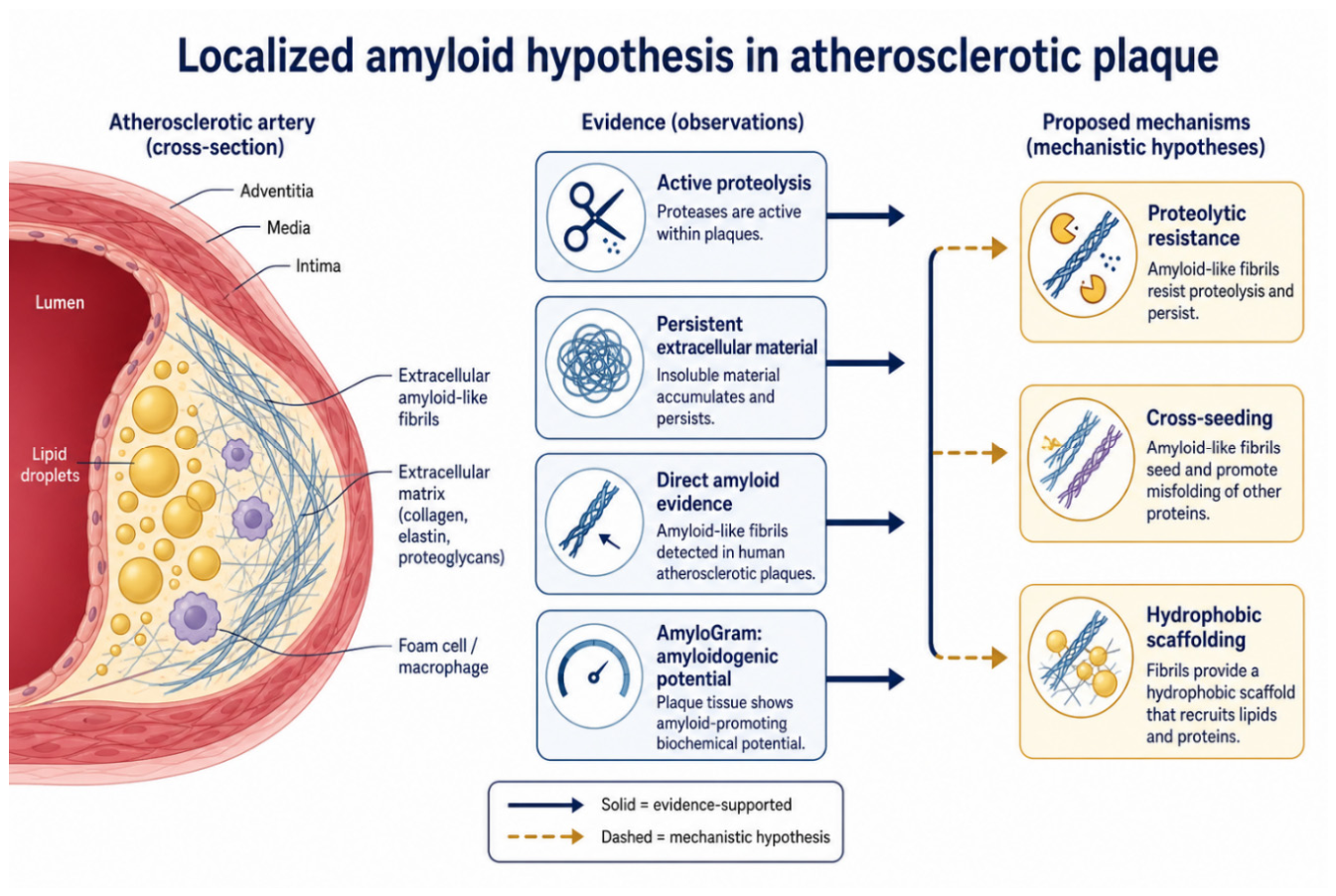
Figure 2.
An illustration of the kinds of observational evidence leading to the recogniion that plaques contain a major amyloid component. The figure was created using Cursor (https://cursor.com/) following a prompt written by the authors.
Figure 2.
An illustration of the kinds of observational evidence leading to the recogniion that plaques contain a major amyloid component. The figure was created using Cursor (https://cursor.com/) following a prompt written by the authors.
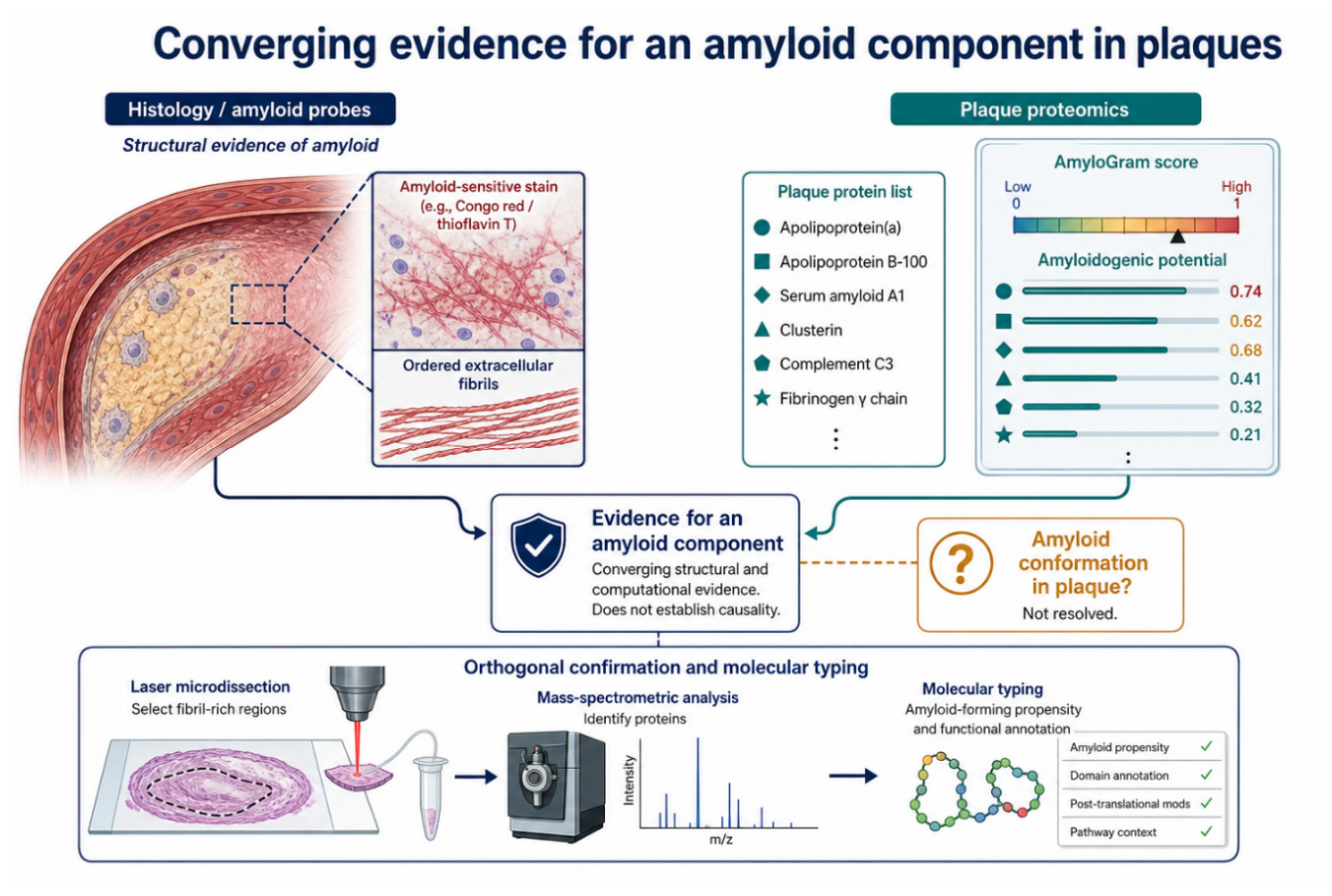
Figure 3.
Structures of the normal (PrPC) and ‘rogue’ (PrPSc) forms of the human prion protein. These are taken from the PDB entries indicated. Note the massive enrichment of β-sheets in the latter, and their arrangement in a cross-β motif.
Figure 3.
Structures of the normal (PrPC) and ‘rogue’ (PrPSc) forms of the human prion protein. These are taken from the PDB entries indicated. Note the massive enrichment of β-sheets in the latter, and their arrangement in a cross-β motif.
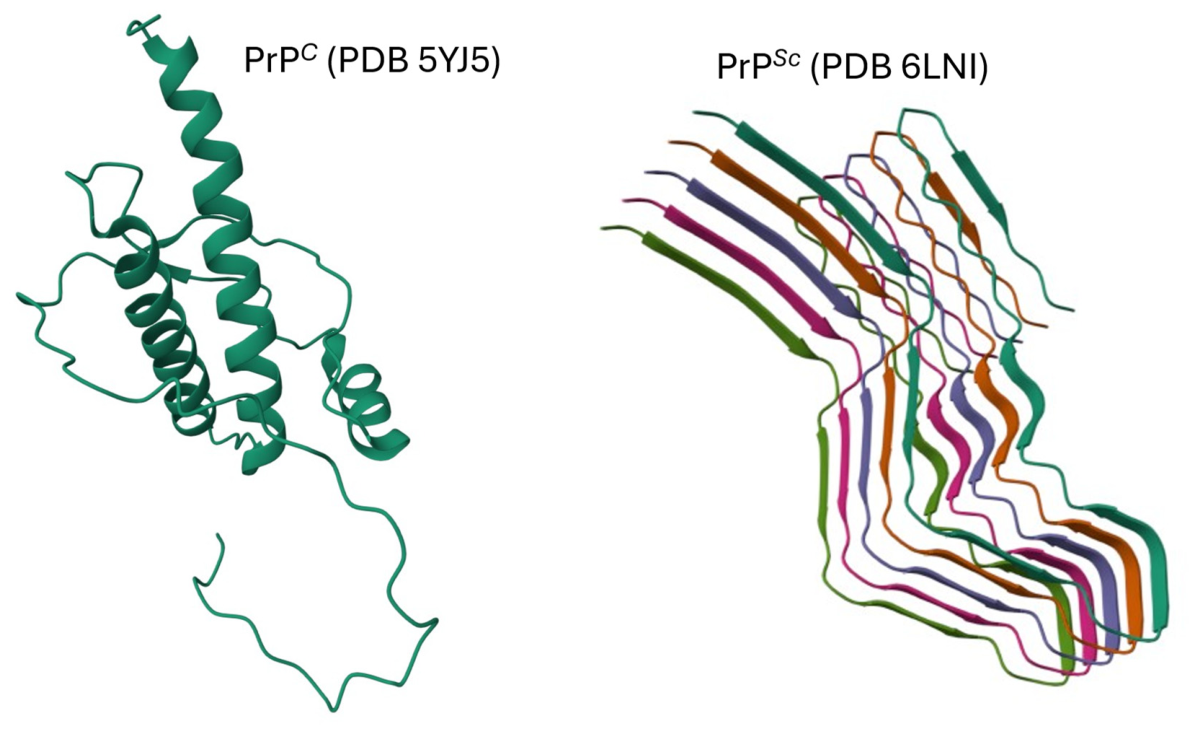
Figure 4.
Two broad classes of complex fibre structure. In A, a variety of proteins that are not part of the main fibril structure are bound with varying degrees of specificity to the fibril chains. Such binding will reflect, and potentially be in equilibrium with, the proteins in the solution in which the fibrils reside. In B, different proteins are incorporated irreversibly into the fibril chains themselves; under these circumstances their composition will not necessarily reflect that of the solution in which the fibrils are suspended. This figure is redrawn from the CC-BY 4.0 Open Access publication by Bondarev et al. (Bondarev et al. 2018).
Figure 4.
Two broad classes of complex fibre structure. In A, a variety of proteins that are not part of the main fibril structure are bound with varying degrees of specificity to the fibril chains. Such binding will reflect, and potentially be in equilibrium with, the proteins in the solution in which the fibrils reside. In B, different proteins are incorporated irreversibly into the fibril chains themselves; under these circumstances their composition will not necessarily reflect that of the solution in which the fibrils are suspended. This figure is redrawn from the CC-BY 4.0 Open Access publication by Bondarev et al. (Bondarev et al. 2018).
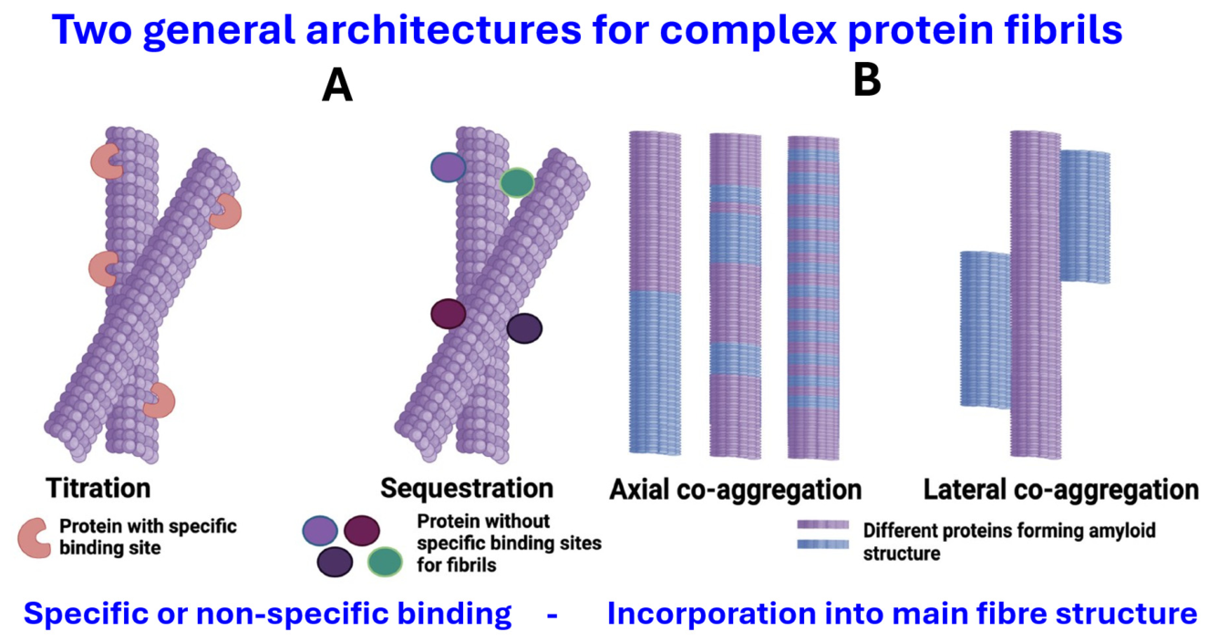
Figure 5.
A pictorial representation of the key differences between the proteomes of normal blood clots (thrombi) and those observed in amyloid-containing fibrinaloid microclot complexes. Image generated by ChatGPT following a prompt given by the authors.
Figure 5.
A pictorial representation of the key differences between the proteomes of normal blood clots (thrombi) and those observed in amyloid-containing fibrinaloid microclot complexes. Image generated by ChatGPT following a prompt given by the authors.
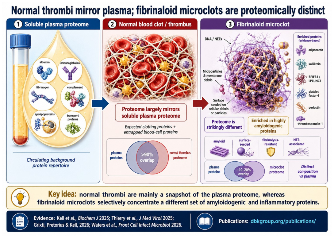
Figure 6.
GRAVY and AmyloGram scores for the human proteome. The line of best fit is shown, but the r2 value is just 0.089. Amylogram scores taken from the Supplementary information of (Kell et al. 2025a).
Figure 6.
GRAVY and AmyloGram scores for the human proteome. The line of best fit is shown, but the r2 value is just 0.089. Amylogram scores taken from the Supplementary information of (Kell et al. 2025a).
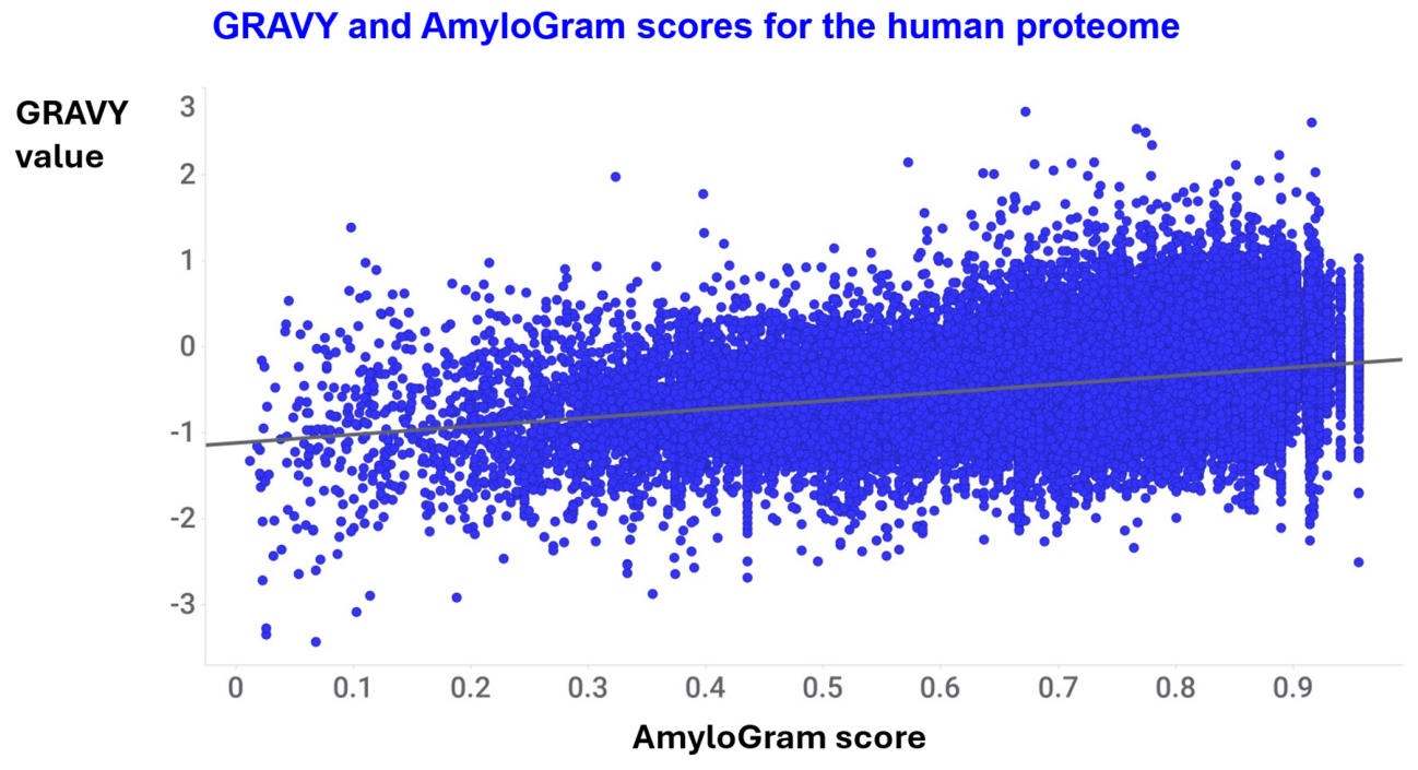
Figure 7.
GRAVY and AmyloGram scores for 6,910 proteins of 6919 observed (Canal-Garcia et al. 2025; Killinger et al. 2022) in Lewy bodies.The line of best fit is shown, but the r2 value is just 0.10.
Figure 7.
GRAVY and AmyloGram scores for 6,910 proteins of 6919 observed (Canal-Garcia et al. 2025; Killinger et al. 2022) in Lewy bodies.The line of best fit is shown, but the r2 value is just 0.10.
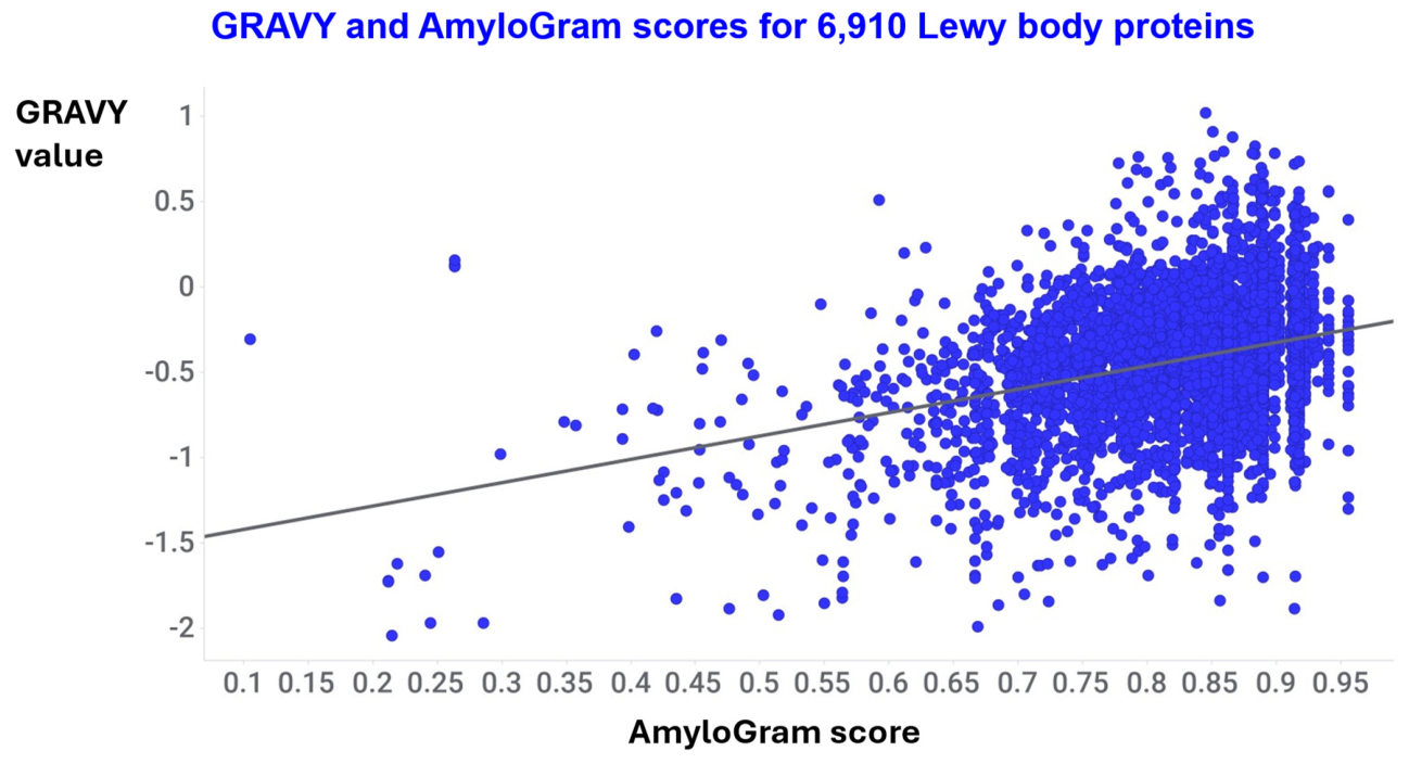
Figure 8.
Smoothed cumulative and probability density distributions for the AmyloGram scores of proteins in the human proteome (data taken from the Supplementary information of (Kell et al. 2025a)) and as calculated here for two sets of Lewy body proteomes (Canal-Garcia et al. 2025; Killinger et al. 2022)).
Figure 8.
Smoothed cumulative and probability density distributions for the AmyloGram scores of proteins in the human proteome (data taken from the Supplementary information of (Kell et al. 2025a)) and as calculated here for two sets of Lewy body proteomes (Canal-Garcia et al. 2025; Killinger et al. 2022)).
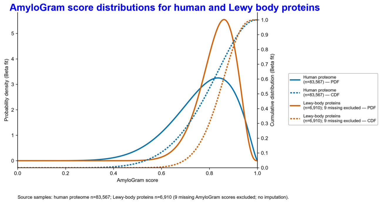
Figure 9.
A graphical summary of the proteomic studies and AmyloGram scores in plaques and Lewy bodies vs the overall human proteome, recognising that an AmyloGram score is an indication of potential rather than experimental amyloid formation. The image was created using Cursor following a prompt provided by the authors.
Figure 9.
A graphical summary of the proteomic studies and AmyloGram scores in plaques and Lewy bodies vs the overall human proteome, recognising that an AmyloGram score is an indication of potential rather than experimental amyloid formation. The image was created using Cursor following a prompt provided by the authors.
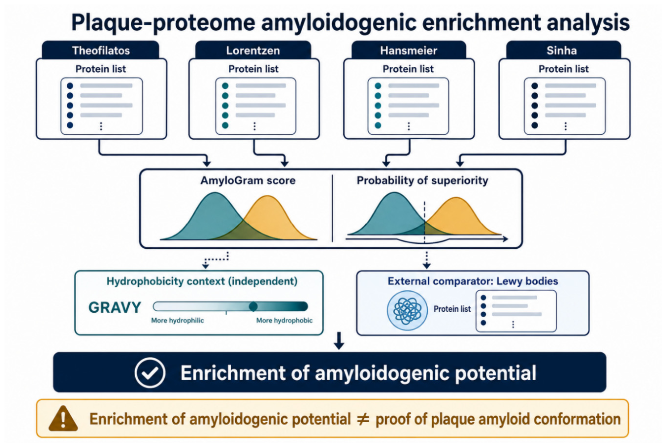
Figure 10.
AmyloGram scores of proteins in the proteome of the human coronary thrombus in patients with ST-segment elevation acute myocardial infarction. 46 proteins were found present in each case when assessed with three different separation/mass spectrometric methods. Proteomic data are taken from the Supplementary information provided with reference (Alonso-Orgaz et al. 2014).
Figure 10.
AmyloGram scores of proteins in the proteome of the human coronary thrombus in patients with ST-segment elevation acute myocardial infarction. 46 proteins were found present in each case when assessed with three different separation/mass spectrometric methods. Proteomic data are taken from the Supplementary information provided with reference (Alonso-Orgaz et al. 2014).
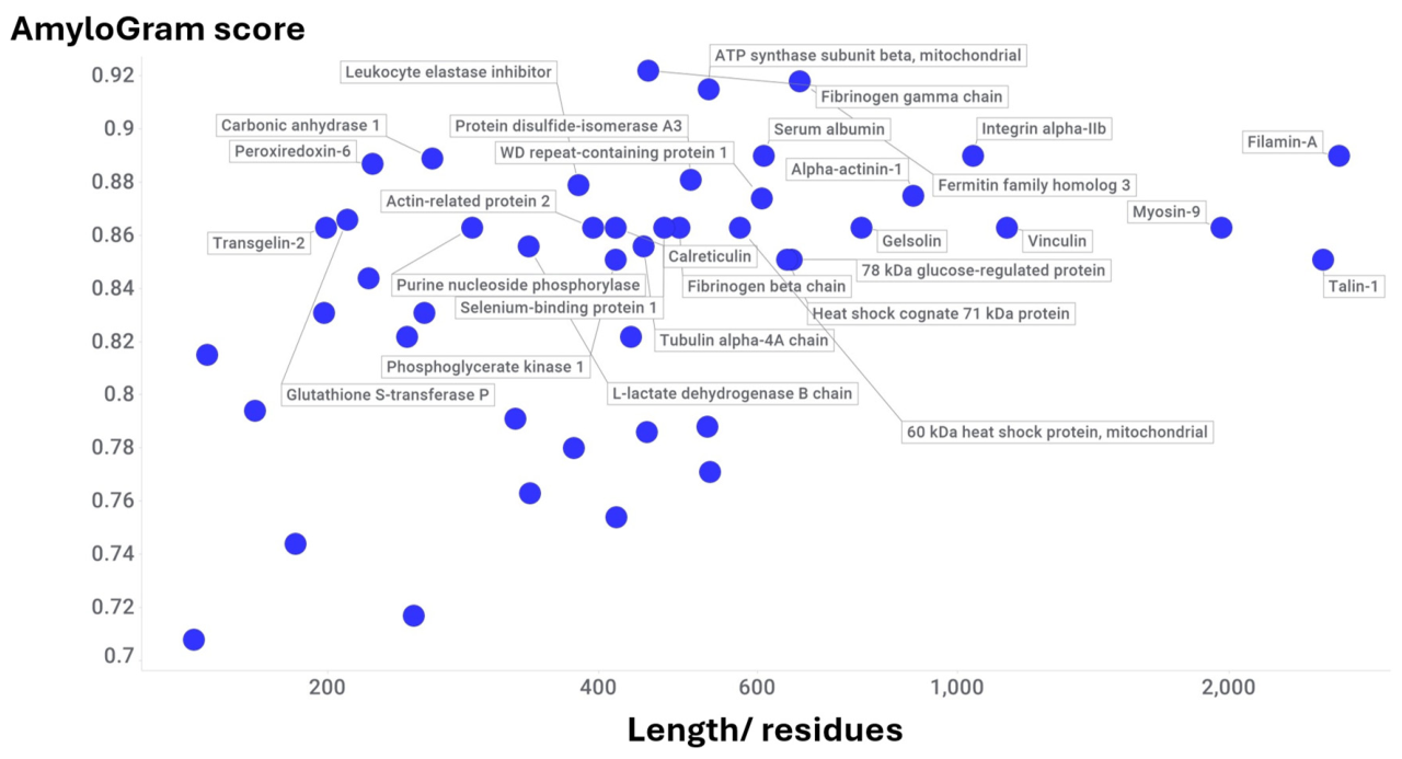
Figure 11.
11 proteins identified by Wang and colleagues (Table 1 of (Wang et al. 2025b)) as being associated with coronary atherosclerosis. The colouring reflects the AmyloGram score.
Figure 11.
11 proteins identified by Wang and colleagues (Table 1 of (Wang et al. 2025b)) as being associated with coronary atherosclerosis. The colouring reflects the AmyloGram score.
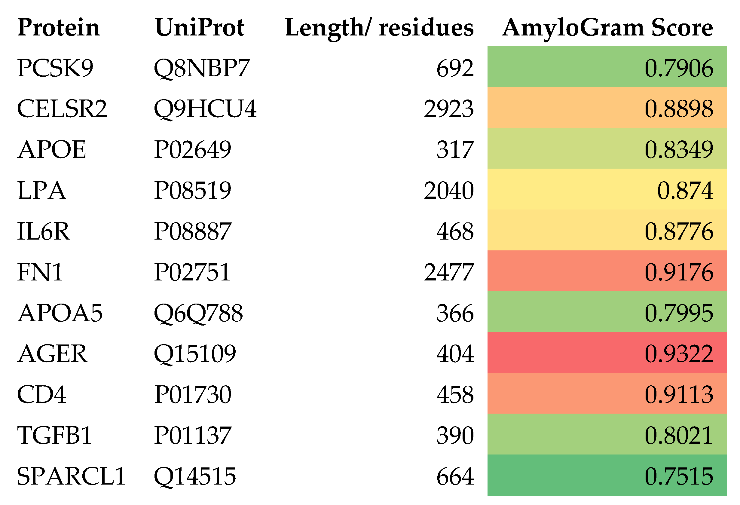
Figure 12.
20 proteins that were raised in atherosclerotic plaques (Supplementary Table 3 of (Hansmeier et al. 2018)) and their AmyloGram scores.
Figure 12.
20 proteins that were raised in atherosclerotic plaques (Supplementary Table 3 of (Hansmeier et al. 2018)) and their AmyloGram scores.
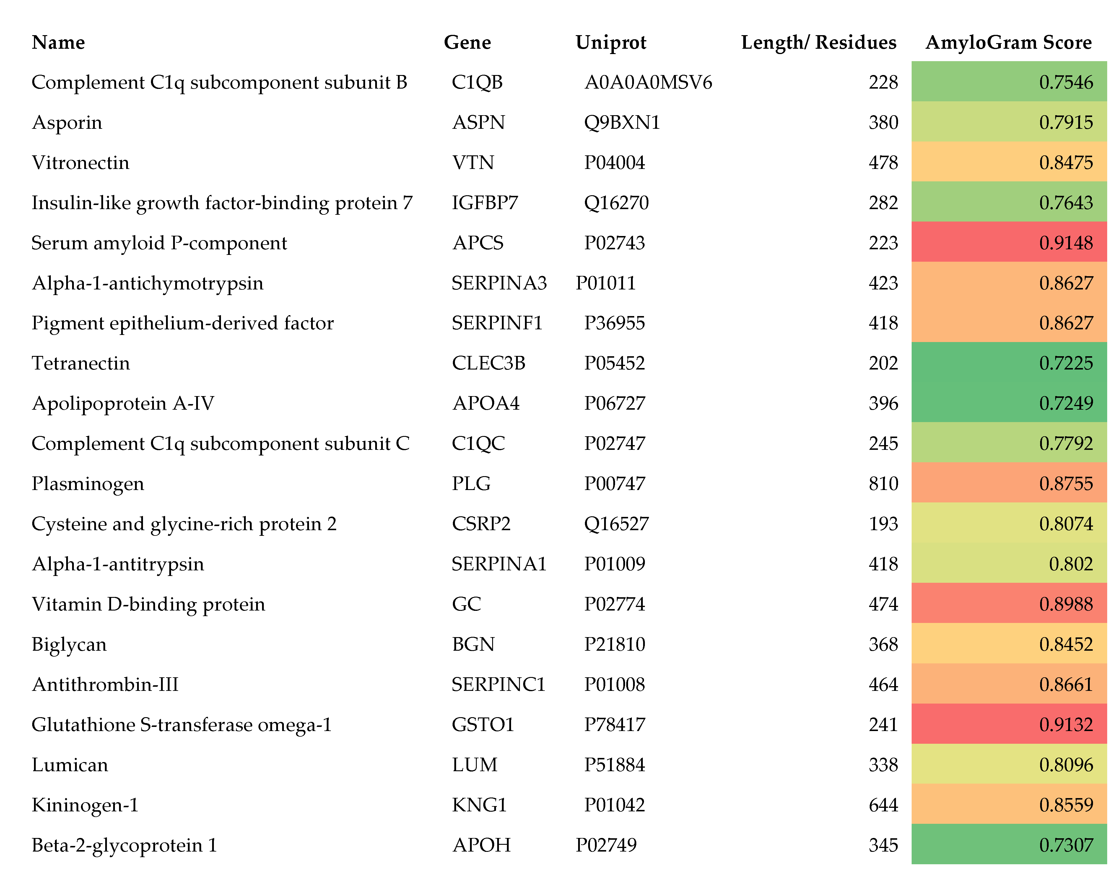
Figure 13.
GRAVY and AmyloGram scores for 1459 core plaque proteins as recorded in Supplementary Table 1 of the paper of Theofilatos and colleagues (Theofilatos et al. 2023). The fibrinogen molecules are labelled and have red symbols.
Figure 13.
GRAVY and AmyloGram scores for 1459 core plaque proteins as recorded in Supplementary Table 1 of the paper of Theofilatos and colleagues (Theofilatos et al. 2023). The fibrinogen molecules are labelled and have red symbols.
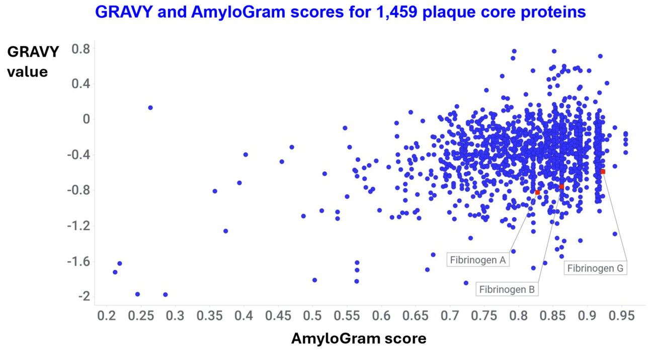
Figure 14.
Smoothed probability density (red) and cumulative distribution functions (blue) of the data of Figure 13.
Figure 14.
Smoothed probability density (red) and cumulative distribution functions (blue) of the data of Figure 13.
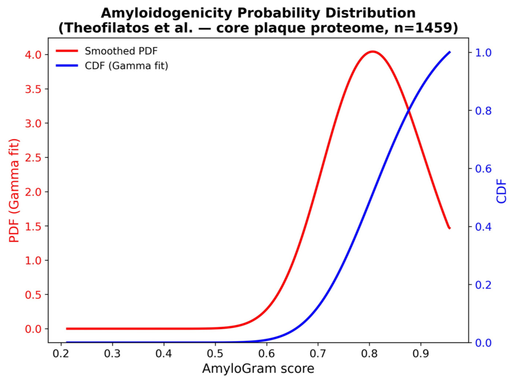
Figure 15.
GRAVY and AmyloGram scores for 283 proteins from a calcified plaque as recorded in Supplementary Table 2 of the paper of Theofilatos and colleagues (Theofilatos et al. 2023). The fibrinogen molecules are labelled and have red symbols.
Figure 15.
GRAVY and AmyloGram scores for 283 proteins from a calcified plaque as recorded in Supplementary Table 2 of the paper of Theofilatos and colleagues (Theofilatos et al. 2023). The fibrinogen molecules are labelled and have red symbols.
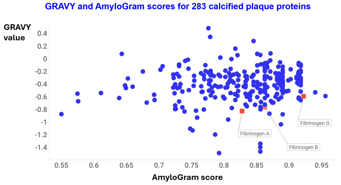
Figure 16.
GRAVY and AmyloGram scores for 128 peptides (from 83 proteins) proteins from a plaque as recorded in Supplementary Table 2 of the paper of (Lorentzen et al. 2025).
Figure 16.
GRAVY and AmyloGram scores for 128 peptides (from 83 proteins) proteins from a plaque as recorded in Supplementary Table 2 of the paper of (Lorentzen et al. 2025).
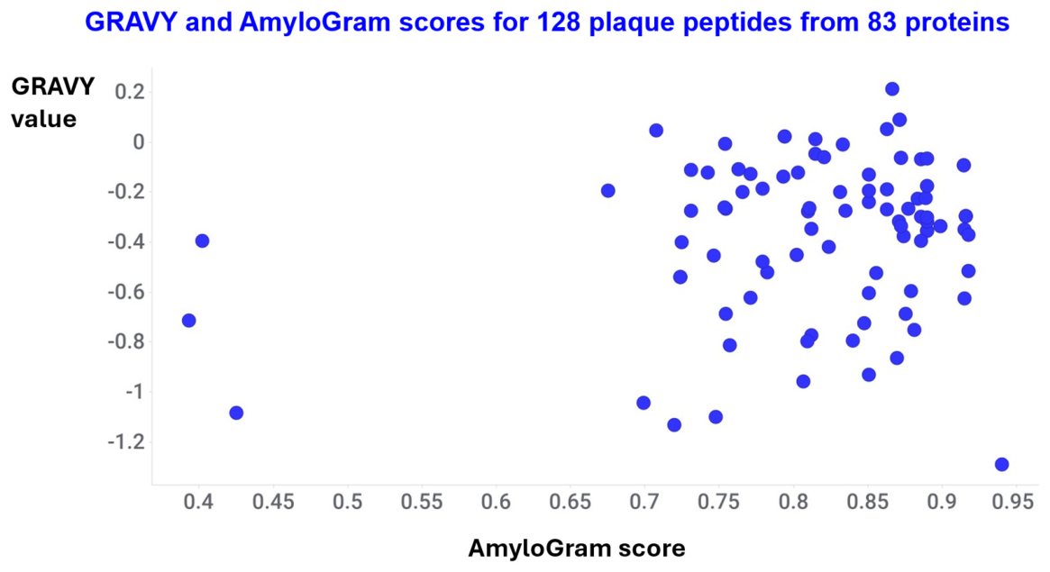
Figure 17.
25 proteins that were raised in the proteome of carotid atherosclerotic plaque relative to non-diseased mammary artery (Aragonès et al. 2016), along with their Amyogram scores (that are also encoded in colour).
Figure 17.
25 proteins that were raised in the proteome of carotid atherosclerotic plaque relative to non-diseased mammary artery (Aragonès et al. 2016), along with their Amyogram scores (that are also encoded in colour).
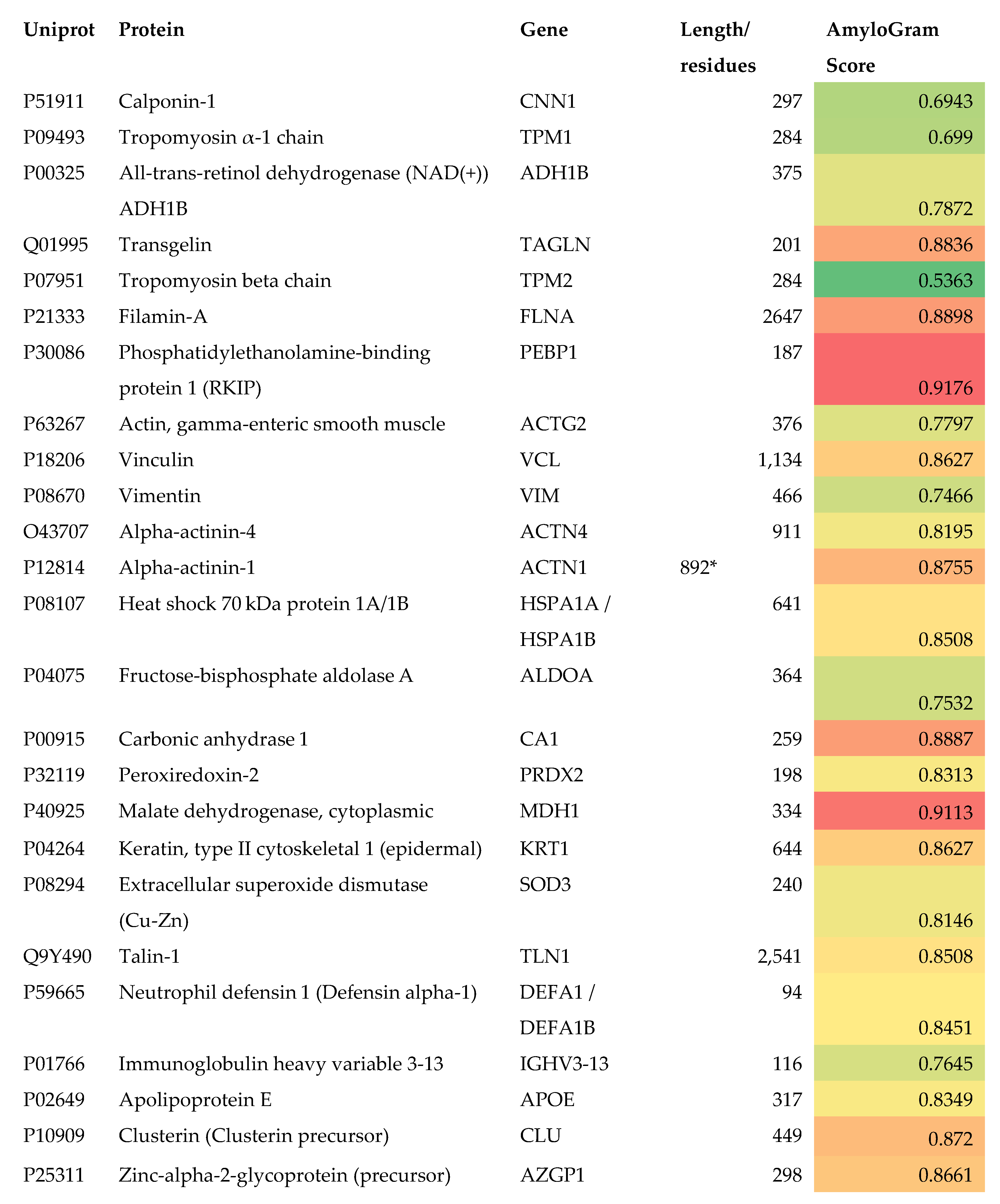
Figure 18.
GRAVY and AmyloGram scores for 4,893 unique proteins observed in advanced atherosclerotic carotid artery plaques. Data are taken from Supplementary Table 2 of (Sinha et al. 2026). Subregions are discriminated by colour: Fibrous cap blue, Media red, Necrotic core yellow.
Figure 18.
GRAVY and AmyloGram scores for 4,893 unique proteins observed in advanced atherosclerotic carotid artery plaques. Data are taken from Supplementary Table 2 of (Sinha et al. 2026). Subregions are discriminated by colour: Fibrous cap blue, Media red, Necrotic core yellow.
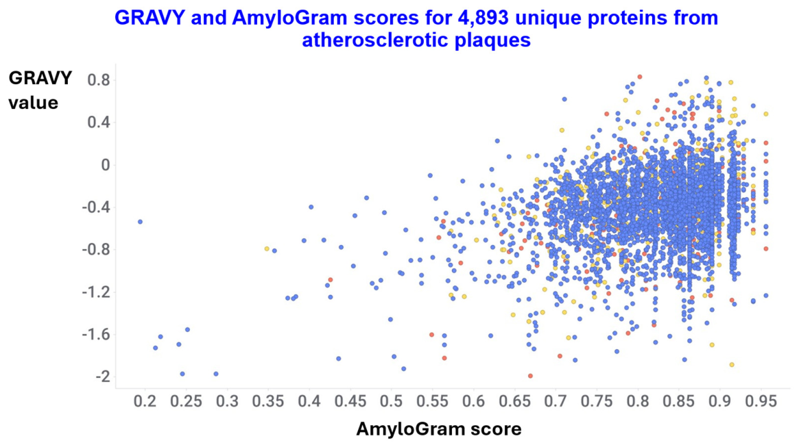
Figure 19.
Probability density and cumulative distributions of AmyloGram scores for 12,248 proteins (4,893 unique) as observed in advanced atherosclerotic carotid artery plaques. Data are taken from Supplementary Table 2 of (Sinha et al. 2026).
Figure 19.
Probability density and cumulative distributions of AmyloGram scores for 12,248 proteins (4,893 unique) as observed in advanced atherosclerotic carotid artery plaques. Data are taken from Supplementary Table 2 of (Sinha et al. 2026).
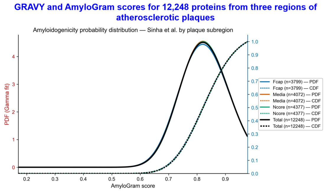
Figure 20.
Plaque abundance and AmyloGram scores for 72 proteins identified as being enriched in atherosclerotic plaques. Data from Table ST3 in the supplementary information of (Hansmeier et al. 2018). The six proteins with the highest AmyloGram scores are labelled.
Figure 20.
Plaque abundance and AmyloGram scores for 72 proteins identified as being enriched in atherosclerotic plaques. Data from Table ST3 in the supplementary information of (Hansmeier et al. 2018). The six proteins with the highest AmyloGram scores are labelled.
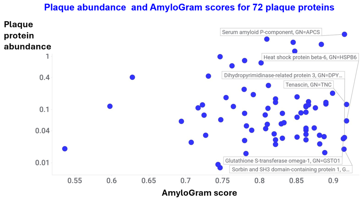
Figure 21.
A graphical summary of the proposed mechanisms by which amyloidogenesis contributes to proteolytic resistance and hence the accretion and persistence of plaques. The image was created using Cursor following a prompt provided by the authors.
Figure 21.
A graphical summary of the proposed mechanisms by which amyloidogenesis contributes to proteolytic resistance and hence the accretion and persistence of plaques. The image was created using Cursor following a prompt provided by the authors.
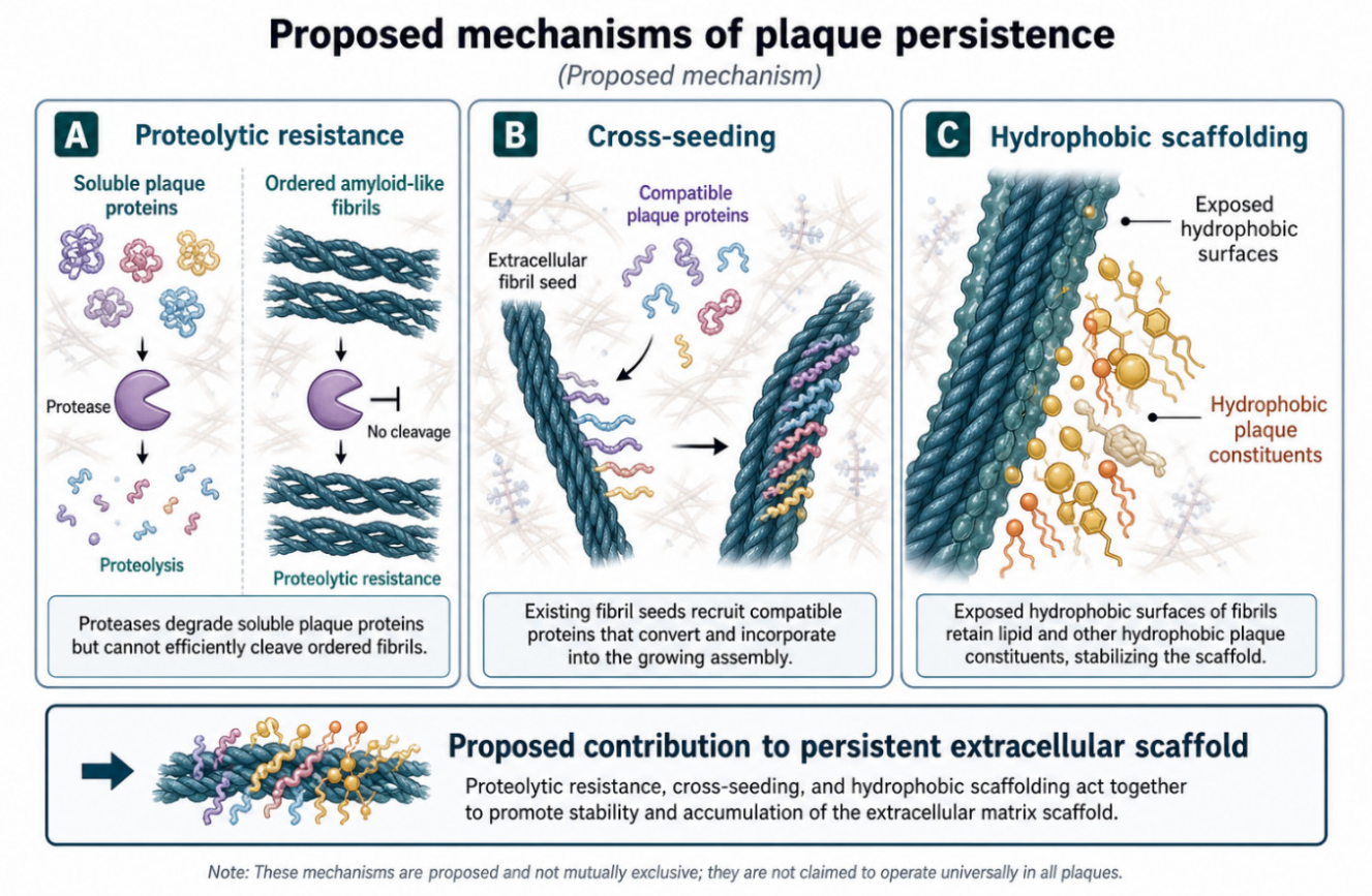

Table 1.
Some of the techniques used to detect the presence of amyloid in atheromatous plaques and related structures.
Table 1.
Some of the techniques used to detect the presence of amyloid in atheromatous plaques and related structures.
| Technique | Comments | Selected references relating to atherosclerotic plaques |
| Congo-red birefringence | Classical amyloid stain | (Kholová and Niessen 2005; Mucchiano et al. 2001a; Mucchiano et al. 2001b; Röcken et al. 2006; Stewart et al. 2007a; Westermark et al. 1995) |
| Binding of multiple amyloid conformation-sensitive fluorophores and other probes | Thioflavin S, another classical fluorogenic amyloid stain; other amyloid-binding PET ligands. | (Bucerius et al. 2017; Hellberg et al. 2019; Medeiros et al. 2004) |
| Electron microscopy | Direct observation | (Mucchiano et al. 2001a; Westermark et al. 1995) |
| X-ray diffraction or cryo-EM demonstration of cross-β structure | As related to light-chain amyloidosis or apolipoprotein-A1 or Apo-C-II in vitro. | (Nguyen et al. 2026; Swuec et al. 2019; Teoh et al. 2011a; Teoh et al. 2011b; Wong et al. 2010) |
| Extraction followed by proteomics to identify the constituent proteins | Also relates to amyloidogenesis calculations described previously (Kell and Pretorius 2025b; Kell et al. 2025b) and below | (Alonso-Orgaz et al. 2014; Langley et al. 2017; Rocchiccioli et al. 2013; Theofilatos et al. 2023) |
Table 2.
A summary of some proteomic studies of atherosclerotic plaques.
| Material / method | Comments | Paper reference |
| 219 carotid endarterectomy samples from 120 patients; core and periphery; discovery plus targeted proteomics. | Large cohort, plaque phenotypes, outcomes, sex differences, proteoglycans, inflammation/ calcification signatures. | (Theofilatos et al. 2023) |
| Human carotid plaques; DIA LC-MS; extracellular-matrix-focused but broad coverage. | Reports 4,498 proteins, including 354 ECM proteins, and 714 differentially abundant proteins between plaque groups. | (Lorentzen et al. 2024) |
| 39 stable and 49 unstable carotid plaques; DIA quantitative proteomics. | Identified 6,143 proteins, used 3,999 common proteins for PCA, and reports 397 differentially expressed proteins. | (Lai et al. 2024) |
| Mature human atherosclerotic plaque; data-independent acquisition mass spectrometry. | ~4,181 proteins identified using a quantitative DIA approach; Useful quantitative assessment of mature human plaque tissue. 20 raised in atherosclerotic plaques. | (Hansmeier et al. 2018) |
| Human carotid plaques; multidimensional LC-MS/MS. | “Deep-identification” paper: 4,702 proteins identified, 3,846 with at least two unique peptides. Substantial plaque proteome list. | (Hao et al. 2014) |
| Symptomatic versus asymptomatic carotid plaques; tissue/cell proteomics; ECM emphasis. | MMP9-S100A8/S100A9-cathepsin D-LG3BP signature. | (Langley et al. 2017) |
| Secretome/ proteome comparison of carotid plaque and non-diseased mammary artery. | Quantified 162 proteins, with 25 statistically significant differences. | (Aragonès et al. 2016) |
| 35 human coronary atherosclerotic plaque samples; direct tissue LC-MS/MS. | Coronary plaque proteome. Identified 806 proteins. | (Bagnato et al. 2007) |
| Coronary plaques at different developmental stages. | Coronary plaque progression dataset; compares plaque proteomes across stages of coronary atherosclerosis. | (Stakhneva et al. 2019) |
| Human plaque N-terminomics/ protein degradation analysis. | Focus on proteolysis, plaque stability, insolubility, fibrinolytic/ proteolytic resistance. Supplementary Table 2 contains 128 peptides and 83 unique proteins. | (Lorentzen et al. 2025) |
| DIA proteomics plus single-cell genomics and transcriptomics. | Uses DIA to analyse plaque progression and integrate proteomics with single-cell transcriptomics and genomics. | (Wang et al. 2025a) |
| Histomorphology-guided spatial proteomics in 112 carotid endarterectomy specimens. | Focus on cap thickness. Fibrous cap, necrotic core, media. Supplementary Table 2 provides a quantitative proteomics data matrix. | (Sinha et al. 2026) |
| Proteins differentially secreted from atherosclerotic plaques | ELISA assays showed raised thrombospondin-1 | (Rocchiccioli et al. 2013) |
| Poteomics of the human coronary thrombus in patients with ST-segment elevation acute myocardial infarction. | 46 proteins identified using three mass spectrometric methods | (Alonso-Orgaz et al. 2014) |
Table 3.
Some quantitative studies of the fibrin(ogen) content of atherosclerotic plaques.
| Material / methods | Findings/ comments | Reference |
| Human aortic intima / plaques; biochemical extraction and quantification of insoluble “fibrin”, soluble fibrinogen and LDL | Insoluble fibrin increased strongly with plaque severity. At the edges of larger plaques it reached about 10% of the tissue dry weight. | (Smith et al. 1976) |
| Human thrombi and atherosclerotic aortas; CNBr cleavage plus radioimmunoassays for NDSK and fibrinopeptides | Fibrinogen-derived protein was <10% of total protein in normal and atherosclerotic aortas; fibrinogen decreased and fibrin II increased with lesion severity, consistent with progressive in situ fibrin formation. | (Bini et al. 1987) |
| Normal and atherosclerotic human aorta and large arteries; immunofluorescence / immunohistochemistry | Visualised apoB, fibrinogen/fibrin and fibronectin in normal intima and during atherosclerosis. | (Shekhonin et al. 1990) |
| Human atherosclerotic plaques; biochemical detection of fibrin degradation products containing fragment E | Demonstrated growth-stimulating fibrin degradation products in plaques, supporting active fibrin turnover rather than passive deposition alone. | (Stirk et al. 1993) |
| Human atherosclerotic plaques; assays of soluble fibrin/ fibrinogen-related antigens and insoluble fibrin | Summarised and extended the evidence for both fibrin deposition and fibrin degradation products in plaques. | (Smith 1994) |
| Human coronary atherosclerosis; immunolocalisation of fibrin | More recent histological evidence for fibrin in coronary atherosclerotic lesions, with implications for necrotic core development. | (Tavora et al. 2010) |
Table 4.
Examples of the presence of various apolipoproteins in human atherosclerotic plaques.
| Example | Apolipoproteins observed | Reference |
| Human atherosclerotic lesions | apoB; apoA-I; apoC-III; | (Hoff et al. 1975) |
| Human atherosclerotic lesions | apoE | (Murase et al. 1986) |
| Plaque macrophage surfaces | apoE | (O'Brien et al. 1994) |
| Human aortic atherosclerotic plaques | apoA1 (in amyloid form) | (Mucchiano et al. 2001b; Westermark et al. 1995) |
| Human coronary atherosclerotic plaques | apoE, apoA-I and apoB | (O'Brien et al. 1998) |
| Carotid secretome | apoE, apoJ (clusterin) | (Aragonès et al. 2016) |
| Human atherosclerotic plaques | apoA-1, A-II, A-IV, B, C-1, C-II, C-III, D, E, L1, M | (Hansmeier et al. 2018) |
| Human atherosclerotic plaques | apoA-1, A-2, B, C-1, C-3, D, E, H, L1, M | (Lorentzen et al. 2025) |
Table 5.
AmyloGram scores for typical lipoproteins found in atherosclerotic plaques as listed in Table 4.
Table 5.
AmyloGram scores for typical lipoproteins found in atherosclerotic plaques as listed in Table 4.
| Gene symbol | Uniprot ID | AmyloGram score |
| APOA1 | P02647 | 0.833 |
| APOA2 | P02652 | 0.856 |
| APOA4 | P06727 | 0.725 |
| APOB | P04114 | 0.916 |
| APOC1 | P02654 | 0.890 |
| APOC2 | P02655 | 0.702 |
| APOC3 | P02656 | 0.731 |
| APOD | P05090 | 0.886 |
| APOE | P02649 | 0.835 |
| APOH | P02749 | 0.731 |
| CLU | P10909 | 0.872 |
| APOL1 | O14791 | 0.851 |
| APOM | O95445 | 0.822 |
Table 6.
Some proposed predictions and tests for the ‘amyloid hypothesis of atherosclerotic plaque formation and persistence.
Table 6.
Some proposed predictions and tests for the ‘amyloid hypothesis of atherosclerotic plaque formation and persistence.
| Prediction | Test |
| Amyloid signal colocalises with specific plaque proteins | multiplex LCO/Congo red + immunofluorescence for fibrin, apoA-I, apoB, apoE, SAP, clusterin |
| Amyloid-rich plaque regions are protease-resistant | ex vivo digestion with plasmin, cathepsins, MMPs followed by residual proteomics |
| Amyloid regions are β-sheet-rich | FTIR/Raman mapping of serial sections |
| Amyloid regions have distinct proteomes | laser-capture microdissection + DIA proteomics |
| Intraplaque thrombus/microclot accretion contributes | compare plaque thrombus-interface proteome with circulating microclot proteome |
| Hydrophobic plaque constituents bind amyloid surfaces | lipidomics of amyloid-positive vs amyloid-negative microdissected regions |
Disclaimer/Publisher’s Note: The statements, opinions and data contained in all publications are solely those of the individual author(s) and contributor(s) and not of MDPI and/or the editor(s). MDPI and/or the editor(s) disclaim responsibility for any injury to people or property resulting from any ideas, methods, instructions or products referred to in the content. |
© 2026 by the authors. Licensee MDPI, Basel, Switzerland. This article is an open access article distributed under the terms and conditions of the Creative Commons Attribution (CC BY) license (http://creativecommons.org/licenses/by/4.0/).
Copyright: This open access article is published under a Creative Commons CC BY 4.0 license, which permit the free download, distribution, and reuse, provided that the author and preprint are cited in any reuse.