Preprint
Article

This version is not peer-reviewed.

Sustainable Antimicrobial Textiles Functionalized with Gold and Silver Nanoparticles Biosynthesized Using Undaria pinnatifida Extracts

A peer-reviewed version of this preprint was published in:
Nanomaterials 2026, 16(17), 1112. https://doi.org/10.3390/nano16171112

Submitted:

11 August 2026

Posted:

12 August 2026

You are already at the latest version

Abstract

Functionalizing textiles with nanoparticles is a promising strategy for developing sustainable materials with antimicrobial activity and reduced potential for resistance. This study aimed to develop antimicrobial cotton and polyester (PE) textiles functionalized with gold and silver nanoparticles (Au@UP and Ag@UP) biosynthesized using Undaria pinnatifida (UP) aqueous extracts. Nanoparticle-functionalized textiles were characterized by UV–vis spectroscopy, SEM, and FTIR, and nanoparticle attachment–detachment and stability were evaluated on both substrates. Antioxidant activity was assessed by the DPPH assay, while antimicrobial activity against Staphylococcus aureus and Pseudomonas aeruginosa and cytocompatibility using the L-929 cell line were determined. UV–vis spectroscopy revealed a slow-release profile, while SEM, UV–vis, and FTIR confirmed nanoparticle binding. Cotton exhibited higher nanoparticle affinity and stability (KS=0.5007 for Au@UP and 0.4817 for Ag@UP), attributed to its abundance of reactive hydroxyl groups. Although antioxidant activity decreased after nanoparticle binding (<5%), functionalized cotton gauzes retained antimicrobial activity. Au@UP and Ag@UP textiles were non-cytotoxic (>80% cell viability) and inhibited the growth of S. aureus (~15% and 90%, respectively) and P. aeruginosa (~90% for both). These findings support the potential of UP-mediated nanoparticle-functionalized textiles, particularly Ag@UP-containing materials, as sustainable antimicrobial surfaces.
Keywords: 
;  ;  ;  ;  ;  

1. Introduction

Healthcare-associated infections remain a major global challenge, particularly due to the increasing prevalence of multidrug-resistant bacteria and the limited efficacy of conventional antimicrobial treatments [1]. In this context, antimicrobial textiles have emerged as promising materials for reducing microbial transmission and preventing infections in medical and healthcare settings [2]. Among the various strategies explored, textile functionalization with bioactive agents has attracted considerable attention because it enables the development of materials with enhanced antimicrobial performance while maintaining desirable properties such as biocompatibility, durability and biodegradability [3,4]. In particular, the use of naturally derived functionalizing agents represents an attractive approach for developing safer and more sustainable antimicrobial textile systems [5].
Cotton is a cellulosic material widely used in the biomedical industry due to its favourable mechanical properties, including absorbency, permeability and biodegradability [6]. Polyester-based textiles also exhibit excellent characteristics for medical applications, particularly due to their inert nature and generally high biocompatibility [3,7]. As both of these types of textiles are biocompatible and can be functionalized with non-toxic agents to impart additional bioactivities, such as antimicrobial properties, they have been widely used [3,4,6].
Among the various textile functionalization strategies, metal-based nanoparticles (NPs) have emerged as promising antimicrobial agents due to their broad-spectrum activity and low likelihood of inducing microbial resistance [3]. Their antimicrobial potential arises from direct interactions with the bacterial cell wall and membranes and includes mechanisms such as oxidative stress induction, metal ion release and non-oxidative mechanisms [8]. Since these structural barriers are highly conserved, only extensive genetic mutation would alter bacterial susceptibility to NPs, making the development of resistance unlikely [9]. In particular, silver and gold NPs have attracted considerable interest because they can interact directly with bacterial cell structures and disrupt essential cellular processes without requiring specific molecular targets [10,11,12,13]. Silver nanoparticles are widely recognized for their potent antibacterial activity and chemical stability, whereas gold nanoparticles offer additional advantages such as high biocompatibility and unique physicochemical properties [13,14]. These characteristics make both nanomaterials attractive candidates for the development of functionalized antimicrobial textiles for biomedical applications.
Green synthesis has emerged as a sustainable, low-cost and non-toxic approach for NPs production [15]. This strategy combines the intrinsic properties of NPs with bioactive compounds from organisms, such as marine seaweeds, that often possess potent defence mechanisms against ecological pressures [16,17]. Marine macroalgae such as Undaria pinnatifida (UP) contain polysaccharides, phenolic compounds and other metabolites capable of acting as both reducing and stabilizing agents during NP synthesis. These biomolecules may enhance nanoparticle biocompatibility and antimicrobial performance while avoiding toxic chemical reagents commonly used in conventional synthesis methods. Notably, NPs synthesized using UP extracts have demonstrated very strong antibacterial activity against Staphylococcus aureus and Pseudomonas aeruginosa [18]. However, the incorporation of UP-mediated nanoparticles into functionalized textile substrates, particularly regarding the combined assessment of antimicrobial activity and biocompatibility, remains unexplored.
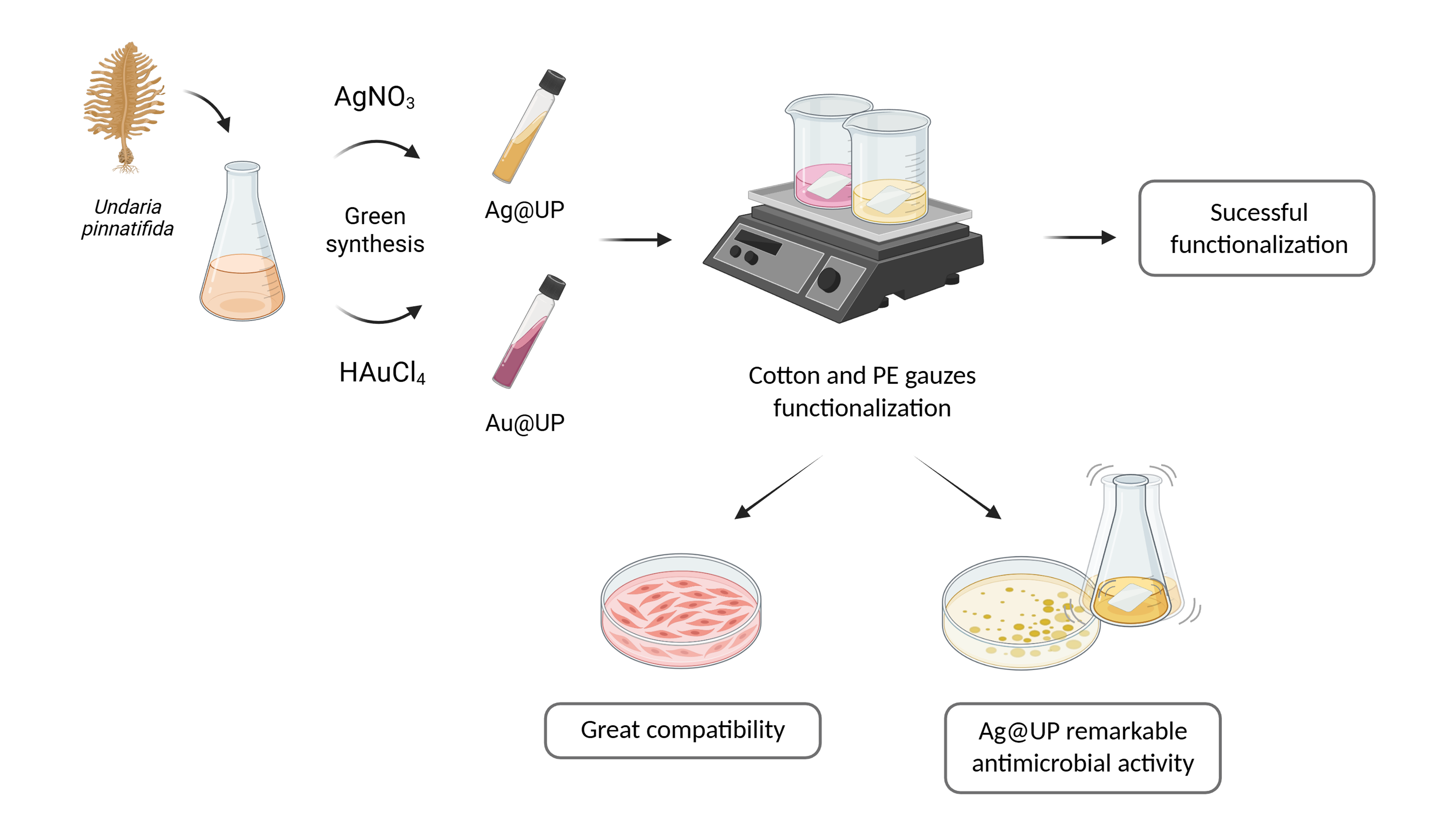
In this study, green-synthesized gold (Au@UP) and silver (Ag@UP) nanoparticles produced using UP extracts were immobilized onto cotton and non-woven polyester (PE) gauzes to investigate the influence of nanoparticle composition and textile substrate on antimicrobial performance and biocompatibility. By combining marine-derived nanoparticle synthesis with textile functionalization, this work aims to develop sustainable textile substrates that leverage the potent antimicrobial properties of these nanoparticles, providing a novel, efficient and environmentally friendly approach for the prevention and management of bacterial infections.

2. Materials and Methods

2.1. Materials

UP seaweed samples were collected at the north-west coast of Spain (42°12’2.9’’ N; 8°47’6.2’’ W) and stored at -20 °C until use. Polyester-based (PE) (MaiaLab, Portugal) and cotton gauzes (Disfasan, Barcelona) were obtained from a local pharmacy. Sodium chloride (NaCl), sodium phosphate dibasic (Na₂HPO₄), L-histidine monohydrochloride, lactic acid, sodium hydroxide (NaOH), ethanol, 1,1-difenil-2-picril-hidrazil (DPPH), 6-hydroxy-2,5,7,8-tetramethylchroman-2-carboxylic acid (Trolox), Dulbecco’s Modified Eagle’s Medium (DMEM), dimethyl sulfoxide (DMSO), 3-(4,5-dimethyl-2-thiazolyl)-2,5-diphenyl-2H-tetrazolium bromide (MTT), chloroauric acid (HAuCl4) and silver nitrate (AgNO3) were purchased from Sigma-Aldrich (USA). Phosphate-buffered saline (PBS) was prepared using NaCl and potassium chloride (KCl) from Fisher Bioreagents (USA) and potassium phosphate monobasic (KH₂PO4) from Panreac AppliChem (Germany). Mus musculus fibroblast cell line L-929 (CCL-1), Staphylococcus aureus (ATCC 6538) and Pseudomonas aeruginosa (ATCC 10145) were obtained from ATCC® (USA). Luria broth was obtained from Grisp (Porto, Portugal). Fetal bovine serum (FBS) was purchased from Biochrom GmbH (Berlin, Germany).

2.2. Methods

2.2.1. UP Extracts Preparation and Nanoparticle Synthesis

The extraction procedure was performed as previously described [18]. Briefly, UP thalli were defrosted, washed, dried, and finely chopped. An aqueous extract (UP-extract) was prepared with 1 g/mL in endotoxin-free water at 100 °C for 15 min. The biomass was removed by filtration through a 0.22 µm membrane, followed by centrifugation at 4500 rpm for 20 min. The resulting supernatant was subjected to a final filtration step.
Green synthesis of gold (Au@UP) and silver (Ag@UP) nanoparticles was performed as previously reported [18]. Briefly, Au@UP were synthesized at room temperature (RT) under stirring, with chloroauric acid (HAuCl4) being slowly added over 24 h. The optimal conditions were achieved using an initial UP-extract concentration of 1 g/mL and a final HAuCl4 concentration of 0.4 mM. Regarding Ag@UP, the synthesis was conducted at 100 °C, with silver nitrate (AgNO3) being added under stirring over 30 min. The optimal conditions were obtained using an initial UP-extract concentration of 0.5 g/mL and with a final solution of 0.25 mM of AgNO3. Both Au@UP and Ag@UP were frozen at -80 °C and lyophilized in a Christ Beta 2-8 LSC Plus lyophilizer (Martin Christ, Germany) at −64 °C under a vacuum of 0.0060 mbar. A secondary drying step was then applied at −76 °C under a vacuum of 0.0010 mbar to preserve their properties, until completely dry, which in total took about one day. Before use, the samples were reconstituted with ultrapure water when required, yielding final concentrations of HAuCl4 and AgNO3 of 0.17 and 0.34 mM.

2.2.2. Characterization of Au@UP and Ag@UP

UV-vis analysis was performed using a BioTek Synergy H1 plate reader (USA) at RT in the range of 300 – 700 nm. Dynamic light scattering (DLS) and electrophoretic light scattering (ELS) analysis was conducted to assess mean diameter size, polydispersity index and surface charge, as ζ-potential, of the NPs using a Zetasizer nano ZS (Malvern, Worcestershire, UK).

2.2.3. Functionalization of Gauzes with Au@UP and Ag@UP - Exhaustion Method

Lyophilized green synthesized NPs were reconstituted in ultrapure water to obtain final concentrations of 0.17 or 0.34 mM and sonicated using a 20 kHz probe for 10 s, with a 3-second pulse duration and 35 % amplitude, to ensure homogeneity. PE and cotton gauzes of 3 x 3 cm were used for functionalization. The gauzes were immersed in 3 mL of Au@UP or Ag@UP solutions with non-altered pH and incubated at RT, under constant agitation of 100 rpm, for 45 min. After incubation, the gauzes were transferred to a Petri Box and cured in a memmert INB 200 oven (Germany) at 180 °C for 7 min. The immersion and curing cycle were repeated five times for the 0.17 mM nanoparticle solutions and three times for the 0.34 mM solutions. Afterwards, the gauzes were washed three times with ultrapure water and air dried in the same oven at 70 °C until completely dry. Control samples were prepared following the same procedure using UP-extract at 0.85 g/mL (control for Au@UP) and 1 g/mL (control for Ag@UP). Additional control gauzes (cotton and PE) were incubated only with ultrapure water as the functionalizing solution.
The optimal conditions were established by minimizing tissue damage at elevated temperatures and by assessing colour intensity after each functionalization cycle. The best results were obtained following the method represented on Figure 1.

2.2.4. Functionalization Efficiency

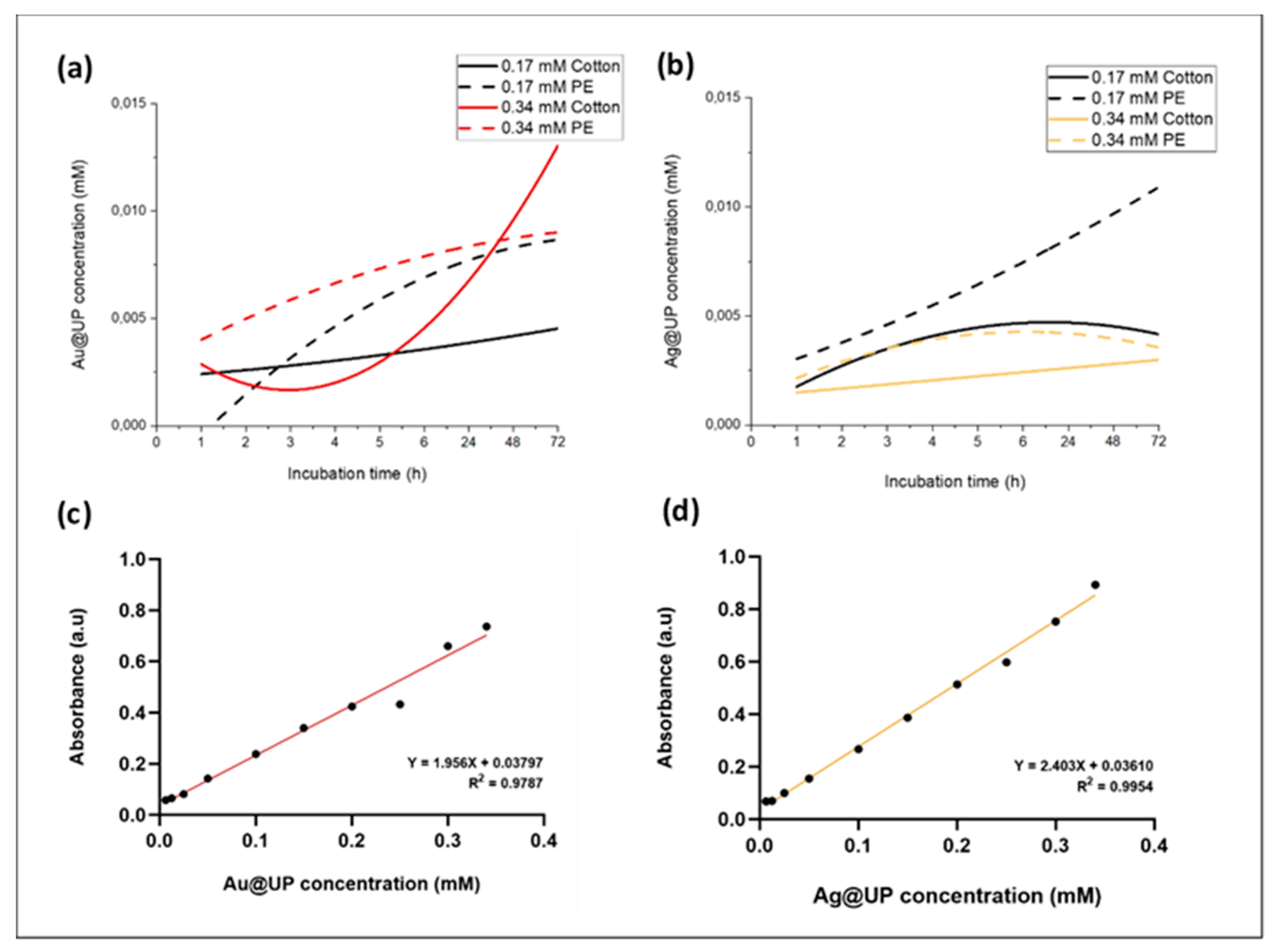
The efficiency of Au@UP and Ag@UP deposition on the textiles surface was determined by UV-vis analysis, using a BioTek Synergy H1 plate reader (USA). The concentration of nanoparticles on the supernatant at the end of the functionalizing cycles was determined by measuring the absorbance at the previously determined surface plasmon resonance (SPR) wavelengths (530 nm for Au@UP and 430 nm for Ag@UP; Figure S1 (a), (b)). The concentration values were then calculated using calibration curves (absorbance versus nanoparticle concentration) for each formulation.

2.3. Physicochemical and Structural Characterization of Functionalized Gauzes

2.3.1. Stereo Microscope Imaging

Micrographs of the gauzes functionalized with Au@UP, Ag@UP and UP-extracts were obtained using a SZ61/SZ51 Stereo Microscope coupled to a EP50 Wireless LAN Camera. Images were recorded from both sides of each sample against a white background and from the top view against a black background, using magnifications of 1x and 3x. Control gauzes functionalized only with ultrapure water were used as control.

2.3.2. Evaluation of Functionalization Degree Evaluation through Colour Depth

The efficiency of the gauze’s functionalization with Au@UP and Ag@UP was assessed by quantification of colour staining levels (K/S values) at the maximum absorbance wavelength. Measurements were performed using a Colour Reflectance Spectrophotometer (Spectraflash 600 Plus CT, Datacolor International) connected to a computer. These measurements were conducted at the maximum wavelength for each sample and were used to evaluate the functionalization efficiency.

2.3.3. Surface Morphology of Functionalized Gauzes - Scanning Electron Microscopy (SEM)

The surface morphology of the functionalized gauzes was examined using a desktop scanning electron microscope (FlexSEM 1000, Hitachi, Japan) to confirm the attachment of Au@UP and Ag@UP onto the gauzes. The samples were mounted on aluminium pin stubs with electrically conductive carbon adhesive tape and sputter-coated with a 2.5 nm gold layer to reduce charging. Imaging was performed under high vacuum at an accelerating voltage of 7, 10 or 15 kV with magnifications of 0.150, 1.5, 2, 4, 4.5, 5 and 7 K x. Measurements of the fibers diameter were conducted using ImageJ 1.54d software.

2.3.4. Fourier Transform Infrared Spectroscopy (FTIR-ATR) Analysis

Fourier Transform Infrared Spectroscopy with Attenuated Total Reflectance (FTIR-ATR) measurements were performed using an ALPHA II Compact FT-IR Spectrometer. Samples were placed directly onto the crystal and spectra were acquired in the range of 4000 - 500 cm-1 with a resolution of 2 cm-1. The resulting spectral bands were then analysed using OriginPro software (version 10.10) (OriginLab Corporation, USA).

2.3.5. Release Profile of NPs from the Functionalized Gauzes

The release profile of Au@UP and Ag@UP from the functionalized gauzes was evaluated using an artificial sweat solution formulated to mimic the human sweat composition [19]. The solution contained 10 g/L NaCl, 1 g/L of 85 % lactic acid, 0.25 g/L of L-histidine monohydrochloride and 1 g/L of sodium phosphate dibasic anhydrous. The pH was adjusted to 4.3 with an alkaline solution of 1 M NaOH.
For this assay, gauzes were cut into 1 x 1 cm pieces and immersed in 5 mL of artificial sweat at 37 °C under constant agitation of 30 rpm. At determined timepoints, 1, 2, 3, 4, 5, 6, 24, 48 and 72 h, 600 µL of the solution was collected for UV-Vis analysis in triplicates. At each timepoint, the collected volume was reinserted in order to maintain constant volume. Final NPs release concentrations were determined using calibration curves generated from known concentration of Au@UP and Ag@UP.

2.4. Bioactive Assessment of Functionalized Gauzes: Antioxidant and Antimicrobial Properties

2.4.1. Radical Scavenging Activity - DPPH Assay

The DPPH assay was performed as previously described, with slight alterations [20]. Briefly, gauzes were cut into 0.5 x 0.5 cm pieces and placed into tubes. In each tube, 450 µL of DPPH 200 µM were added, followed by an incubation in the dark for 2 and 6 h under constant agitation of 200 rpm. At these timepoints, 70 µL of the supernatant was collected and the absorbance was measured at 515 nm using a BioTek Synergy H1 plate reader (USA). All measurements were performed as triplicates. Positive control was performed with Trolox 0.5 mg/mL. Both DPPH and Trolox solutions were prepared in 99 % ethanol. The percentage of radical scavenging activity was calculated using the equation:
% D P P H :   A b s b l a n k A b s s a m p l e A b s b l a n k   × 100
where Abssample corresponds to the absorbance of the samples and Absblank corresponds to the absorbance of wells containing only DPPH 200 µM.

2.4.2. Cytocompatibility of the Au@UP and Ag@UP Functionalized Gauzes

Mus musculus fibroblast cell line L-929 (ATCC®) was selected as a reference cell line due to their widespread use in biocompatibility and cytotoxicity studies of biomedical materials, particularly in accordance with ISO 10993 guidelines for the biological evaluation of medical devices [21]. The cells were cultured in high-glucose Dulbecco’s Modified Eagle Medium (DMEM) supplemented with 10 % fetal bovine serum (FBS) and 1 % antimycotic/antibiotic mix and maintained in a humidified incubator at 37 °C with 5 % CO₂.
The textiles were cut into 1.5 x 1.5 cm pieces and sterilized with UV radiation for 15 min. To obtain conditioned solutions, the functionalized gauzes were immersed on DMEM without fetal bovine serum (FBS) for 24 h at 37 °C. The proportion of medium employed on the gauzes were calculated according to the ISO guidelines for medical devices (ISO 10993-12), 3 cm2/mL [22]. At the same moment, the cells were seeded in 96-well plates at a density of 5 × 10⁴ cells/mL and allowed to adhere overnight (100 µL per well). After this period, 10 % FBS was added to the conditioned media, which was then transferred to the 96-well plates containing L-929 fibroblasts. After 24 h of incubation at 37 °C and 5 % CO₂, cell metabolic activity was assessed using the MTT assay (3-(4,5-dimethylthiazol-2-yl)-2,5-diphenyltetrazolium bromide) (Sigma, USA).
Formazan crystals were dissolved in 100 µL of an ethanol:DMSO (1:1) solution (DMSO, Sigma, USA), and the absorbance was measured at 570 nm on a microplate reader (Tecan, Infinite MPlex). The assay included a viability control (fresh culture medium) and a death control (medium containing 30 % DMSO).

2.4.3. Antibacterial Activity of the Au@UP and Ag@UP Functionalized Gauzes

A direct contact assay was conducted to evaluate the antimicrobial activity of the materials. Samples were tested against Staphylococcus aureus (ATCC 6538) (Gram-positive) and Pseudomonas aeruginosa (ATCC 10145) (Gram-negative). Bacterial inocula were prepared according to the 0.5 McFarland standard (OD600 = 0.08 – 0.1), corresponding to approximately 1.5 × 10⁸ CFU/mL, following EUCAST/CLSI guidelines. All the assays were conducted in a Bio II Advance Plus laminar flow cabinet (Teslar, Spain), tested according to performance criteria for microbiological safety cabinets - EN 12469:2000 [23].
For the assay, the materials were cut into 1.5 × 1.5 cm squares and sterilised under UV radiation for 30 min. Each sample was placed at the bottom of a well in non-treated, sterile 24-well microplates, and 30 µL of bacterial suspension was added directly onto the material. The microplates were incubated at 37 °C for 30 and 120 min. After incubation, each sample was washed with 970 µL of sterile 1x PBS (NaCl 8 g, Na2HPO4 1.44 g, KH2PO4 0.24 g, KCl 0.2 g, per litre, pH 7.4) and transferred to a 1.5 mL microcentrifuge tube. To determine bacterial survival, 100 µL of the solution was serially diluted 1:1000 and plated on Luria Broth (LB) agar. The plates were incubated overnight at 37 °C, and colony-forming units (CFU) were quantified using ImageJ software (National Institutes of Health, USA) [24].
Samples of cotton and PE gauzes without nanoparticles were used as negative controls.

2.5. Statistical Analysis

Data are presented as mean ± standard deviation (SD) from three independent experiments. The significance of the data was determined using GraphPad Prism® software (version 8.0) by One-way ANOVA with comparisons between each sample and its respective control. Results are expressed as ns p > 0.05, * p ≤ 0.05, ** p ≤ 0.01, *** p ≤ 0.001, **** p ≤ 0.0001.

3. Results

3.1. Characterization of Au@UP and Ag@UP

As reported by us, the physicochemical characterization of Au@UP and Ag@UP nanoparticles was performed using UV–Vis spectroscopy, zeta potential analysis, TEM/HRTEM, EDX/EELS, FTIR and size exclusion chromatography, allowing the evaluation of their optical properties, morphology, crystallinity, surface charge and chemical composition [18]. The nanoparticles were shown to be spherical, crystalline and colloidally stable, with average sizes of 6.8 ± 1.0 nm for Au@UP and 14.1 ± 2.8 nm for Ag@UP, and negative zeta potential values indicating stable dispersions. FTIR and carbohydrate analyses further suggested that polysaccharides and proteins from the UP extract acted as reducing and stabilizing agents during nanoparticle synthesis. To confirm reproducibility and to achieve similar properties regarding Au@UP and Ag@UP, the reconstituted green synthesized nanoparticles produced with UP-extracts were characterized by UV-vis spectroscopy, DLS and ELS analysis. The UV-vis spectroscopy confirmed the characteristic absorbance SPR bands for gold and silver nanoparticles, showing distinct absorbance peaks around 530 nm, for Au@UP, and 430 nm, for Ag@UP. The ζ-potential obtained through ELS revealed highly negative surface charges of - 27.7 mV for Au@UP and – 34.3 mV for Ag@UP, indicating good colloidal stability. The diameter size obtained by DLS showed mean sizes of 168.2 nm for Au@UP and 180.2 for Ag@UP (Supplementary Data, Figure S1).

3.2. Functionalization of Gauzes with Au@UP and Ag@UP - Exhaustion Method

Functionalization of the gauzes was performed using the optimized conditions of 45 min of immersion in either Au@UP or Ag@UP at 0.34 mM followed by 7 min of curing at 180 °C. The optimization was conducted varying the incubation (30 to 60 min) and curing times (7 to 10 min), as well as the curing temperature (150 to 180 °C). Different nanoparticle concentrations were also tested (0.17 and 0.34 mM).

3.2.1. Functionalization Efficiency

The UV-vis analysis of the supernatant at the end of the functionalization, coupled with the construction of calibration curves for Au@UP and Ag@UP, allowed the obtention of incorporated nanoparticle concentrations. Regarding the cotton gauzes, the functionalization with Au@UP and Ag@UP at 0.34 mM achieved ~ 62 % (~ 0.21 mM) and ~ 57 % (~ 0.20 mM) of incorporation, respectively. Regarding PE fabrics, Au@UP and Ag@UP 0.34 mM functionalization achieved ~ 60 % (~ 0.20 mM) and ~ 56 % (~ 0.19 mM) of incorporation, respectively.

3.3. Physicochemical and Structural Characterization of Functionalized Gauzes

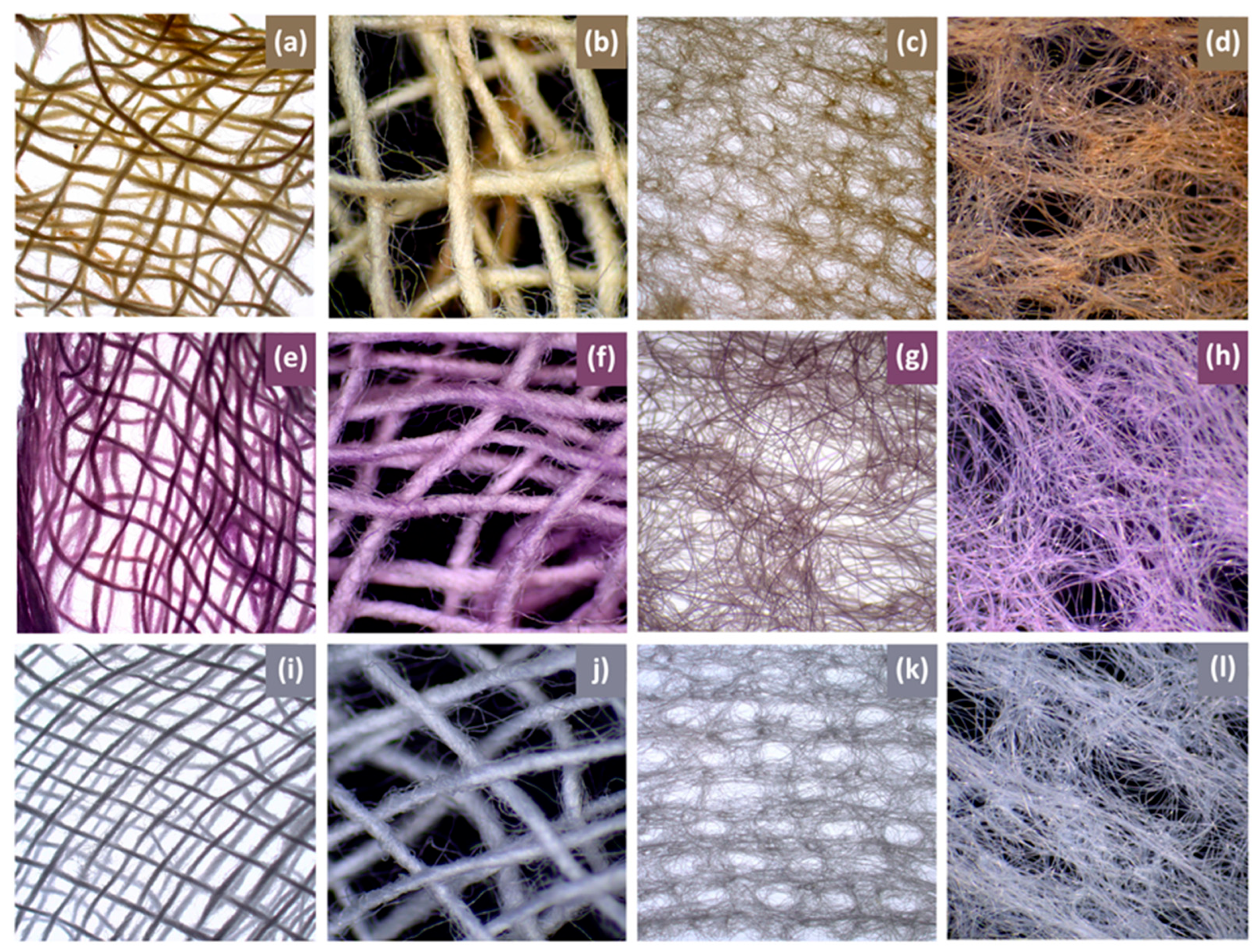
The colouration of the functionalized gauzes was examined resorting to a stereo microscope, allowing a more detailed observation of the differences between each condition. The resulting images clearly show the distinct colours instilled by each nanoparticle type, as the gauzes acquired the characteristic colour of the corresponding functionalizing solution – lilac for Au@UP and yellow for Ag@UP (Figure 2).
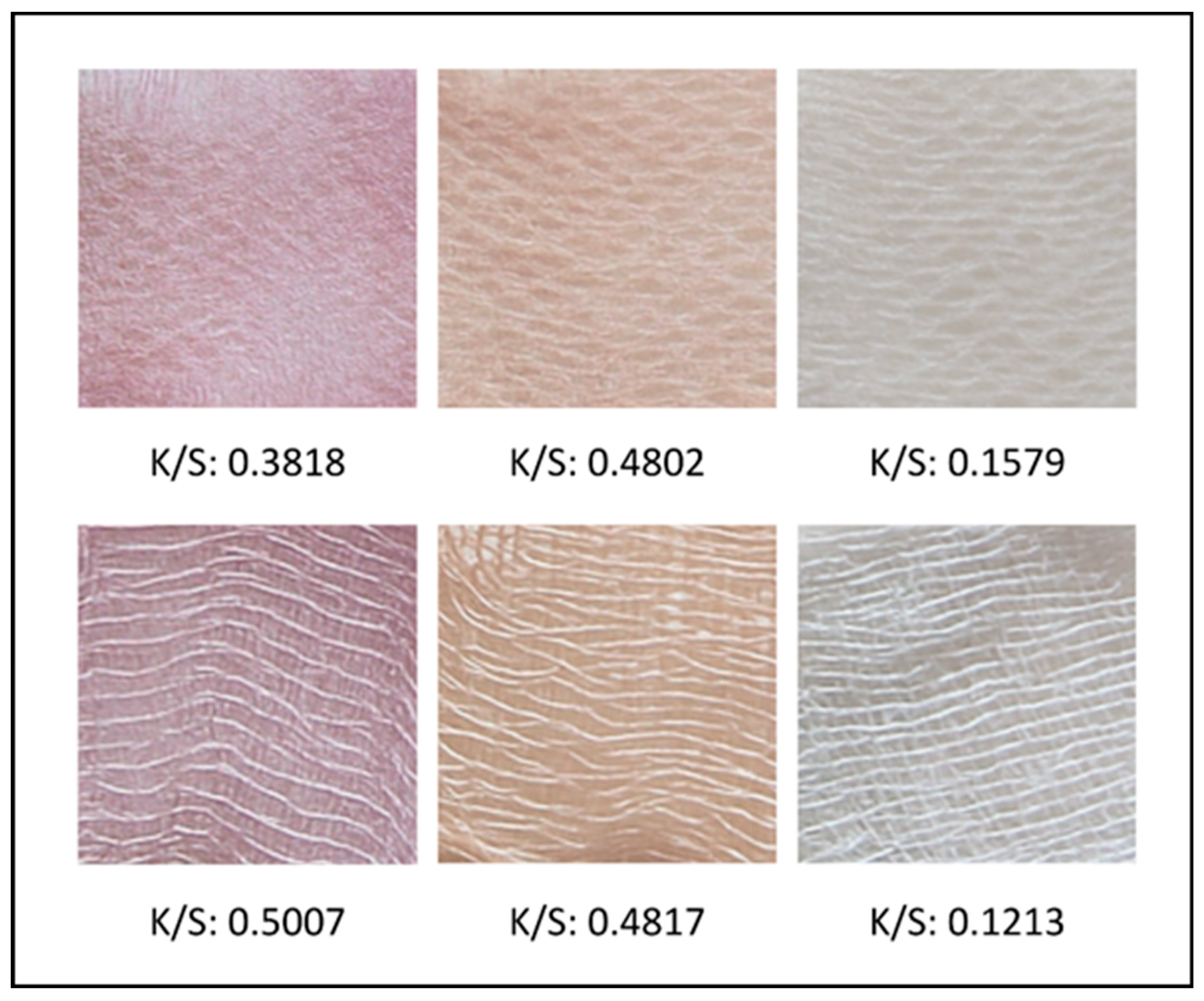
To further evaluate the colouration of the fabrics upon NPs incorporation, the colour strength was analysed before and after functionalization, using the K/S parameter. The clear differences observed between the functionalized samples and the control gauzes are supported by the higher K/S values, which reflects the intensity and depth of the coloured materials by correlating light absorption with light scattering coefficients. The functionalization efficiency appeared comparable for both fabrics treated with Ag@UP (~ 0.48). However, a substantially higher value was obtained for Au@UP functionalized cotton (0.5007) compared to Au@UP functionalized PE (0.3818). Images of the functionalized gauzes and their corresponding K/S values are shown in Figure 3.
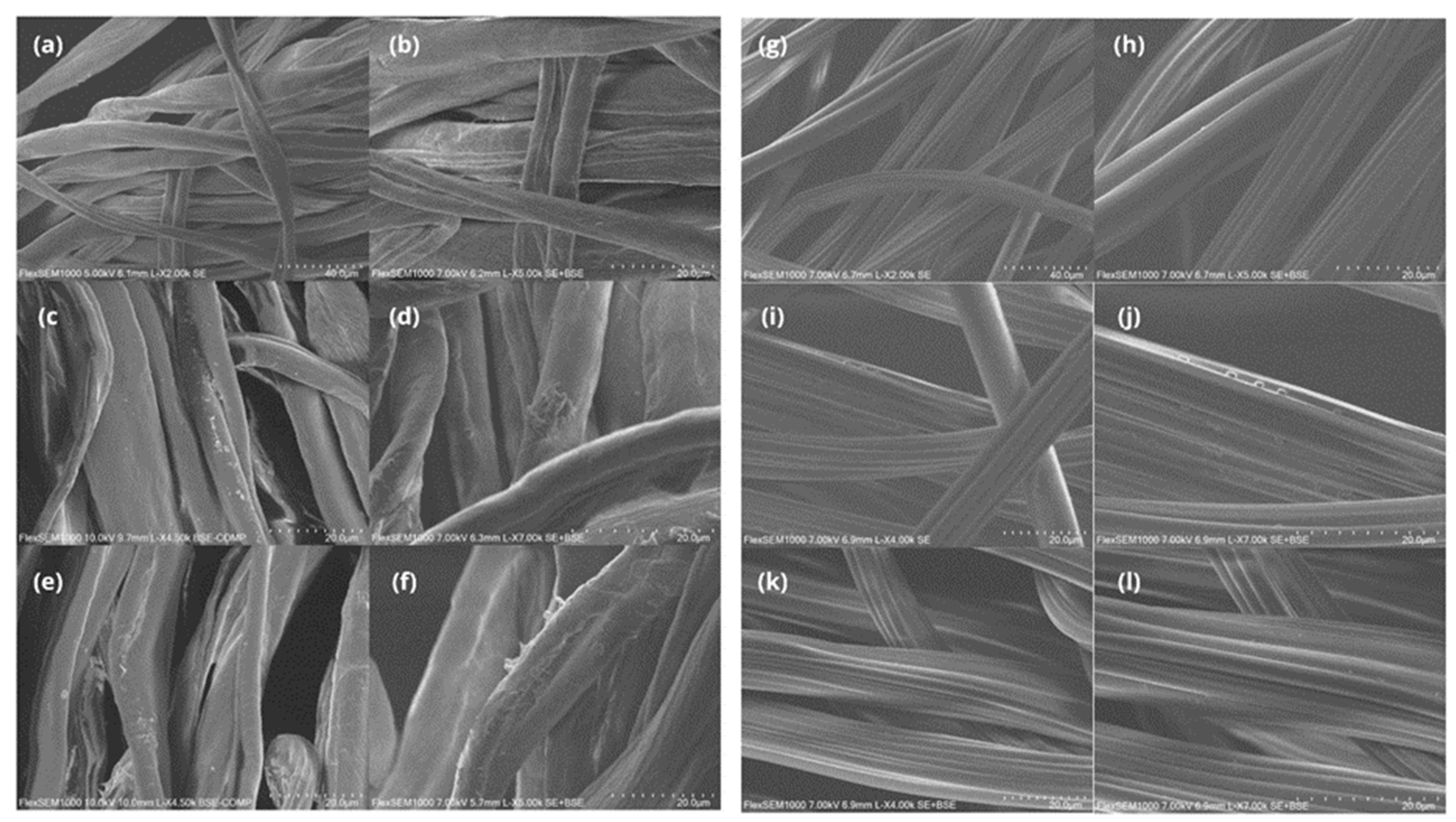
The surface morphology of the samples was examined by Scanning Electron Microscopy (SEM) (Figure 4). The resulting images demonstrated that both Au@UP and Ag@UP were successfully coated onto the cotton and PE gauzes, as evidenced by increased surface roughness due to the deposition of NPs and UP-extract compounds. This phenomenon was more pronounced in the cotton gauzes. Some aggregates were also observed. However, since particle size strongly influences adhesion, larger agglomerates are expected to be more easily leached from the textile surface [25]. Measurements of cotton fibers demonstrated an increase of 1.06 µm (Ag@UP, *) and 2.84 µm (Au@UP, ****) when comparing to the untreated cotton control. For PE gauzes, more slight changes were observed, with increases of 1.05 µm (Ag@UP, ns) and 1.48 µm (Au@UP, *). These differences reinforce successful deposition of Au@UP, Ag@UP and the extract compounds on the surface of the fabrics.
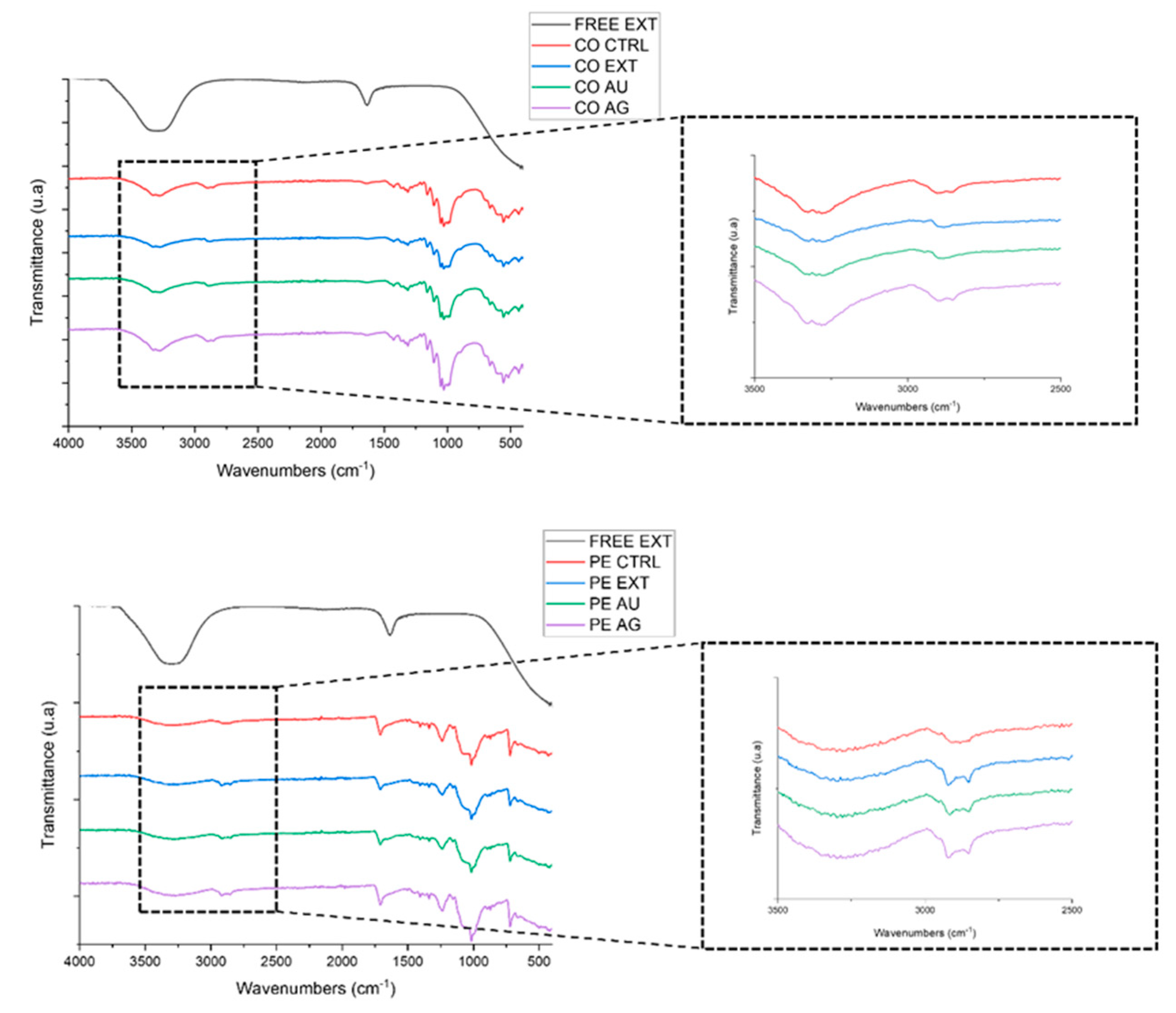
FTIR-ATR analysis was performed to characterize the functionalized textile samples and assess changes in chemical interactions. The analysis detected the presence of chemical groups typical of UP extracts, responsible for stabilizing and capping the metal nanoparticles core, as well as functional groups present in the textiles [26]. The FTIR spectra of the free UP extract, functionalized gauzes and their respective textile controls, are presented on Figure 5.
FTIR analysis revealed that the functionalized gauzes exhibited similar spectral profiles when compared to their respective fabric controls, indicating that the textiles matrices were preserved upon treatment. In all samples the characteristic bands of the textile remained dominant, particularly the broad O–H stretching region around 3300 cm⁻¹ and the C–H stretching vibrations at 2900 cm⁻¹ [27]. Although slight variations were observed on the fingerprint region (1200 – 900 cm⁻¹), no distinct absorption bands or peaks can be attributed to the UP extract, according to its spectrum. This behavior was observed for both textiles, as the spectral characteristics of the fabrics masked any potential signals from the deposited extract, Au@UP or Ag@UP. However, some slight differences can be noticed between functionalized textiles, suggesting changes on the chemical composition of the textile substrates due to possible interactions with the functionalizing agents.

3.4. Release Profile of NPs from the Functionalized Gauzes

A cumulative release assay was carried out to obtain an insight into the leaching behaviour of the NPs from the gauzes under conditions simulating human skin contact. For this purpose, an artificial sweat solution with an adjusted pH was used. The absorbance of the supernatant was measured at predefined timepoints, and a calibration curve was established to determine the concentration of NPs in the supernatant at each interval. The results are presented in Figure 6. For Ag@UP, UV–vis endpoint measurements revealed that samples functionalized with the lower NP concentration (0.17 mM) exhibited a more pronounced and faster release, particularly at later time points. This behaviour suggests that the number of functionalization cycles—five for 0.17 mM and three for 0.34 mM—may influence the strength of the NPs attachment to the textile surface. In contrast, this trend was not observed for the Au@UP functionalized gauzes, where the higher concentration (0.34 mM) displayed a greater release for both textile types. The release profiles of both Au@UP and Ag@UP displayed fluctuations throughout the assay. The seemingly inconsistent variations in NPs concentration on the supernatant and absorbance over time may indicate that the NPs undergo cyclic attachment and detachment processes. However, similar fluctuations were also observed in the control gauzes, suggesting that this behaviour may also be related to the leaching of intrinsic textile compounds rather than to nanoparticle release alone.

3.5. Bioactive Assessment of Functionalized Gauzes: Antioxidant and Antimicrobial Properties

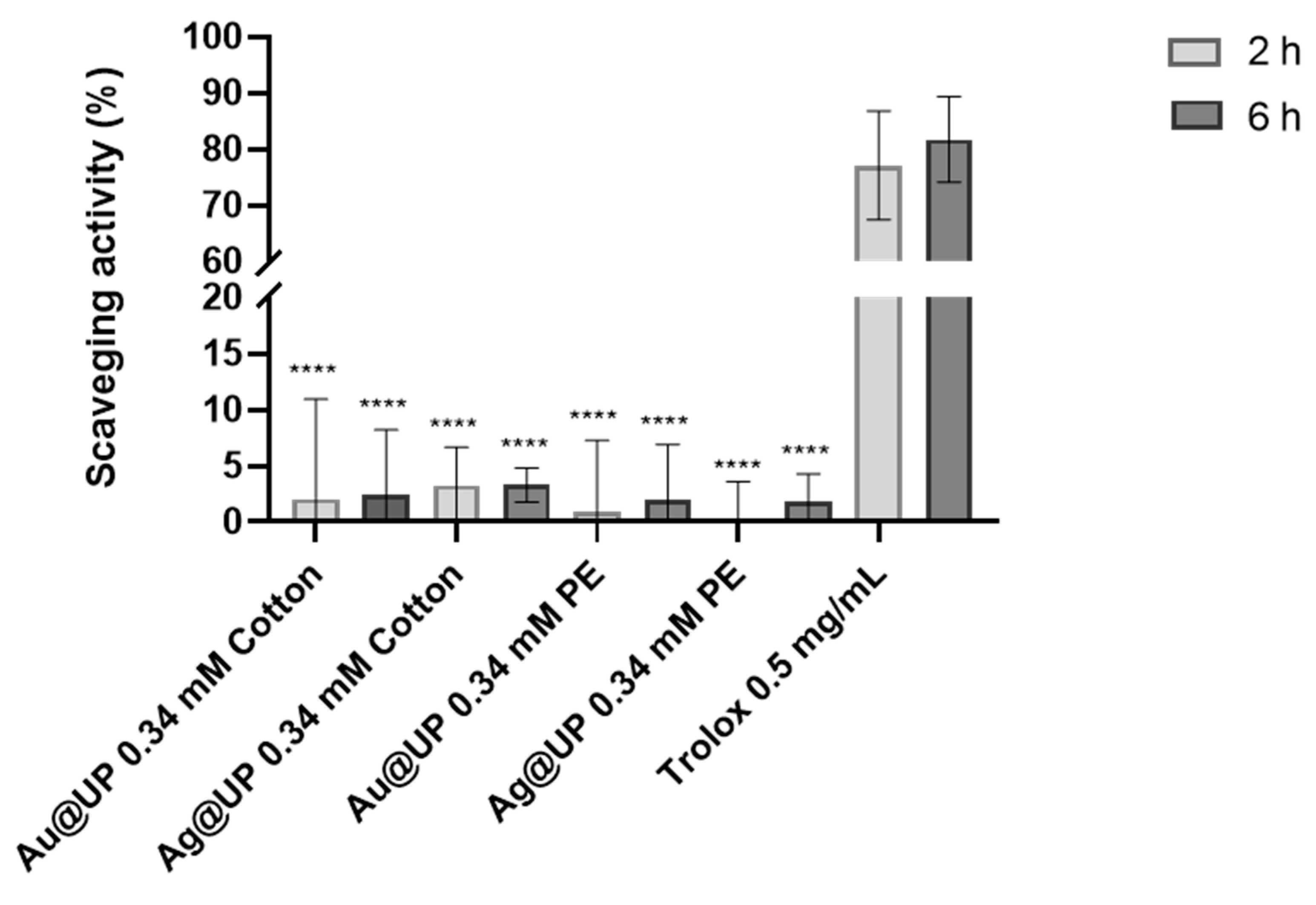
To assess the antioxidant potential of the functionalized gauzes, a DPPH radical scavenging assay was conducted. Since the antioxidant activity of both the UP extracts and the UP-mediated nanoparticles had already been reported [18], the objective here was to determine whether the attachment to the textiles would alter this activity. Given the minimal release observed during the initial hours of the release study, the DPPH assay was performed under agitation and supernatants were collected after 2 and 6 h of incubation. The scavenging activity of the control was subtracted from all conditions to obtain clearer results (Figure 7). After 2 h of exposure to the functionalized gauzes, all the tested conditions exhibited higher scavenging activity than the untreated textiles, with exception of the Ag@UP 0.34 mM PE sample. Nonetheless, all samples showed increased activity at the endpoint measurement (6 h), with cotton gauzes tended to exhibit slightly higher activity than the PE ones, although no statistical differences were identified between conditions. Additionally, all samples demonstrated a significantly lower antioxidant activity when compared to the positive control performed with Trolox 0.5 mg/mL (p < 0.0001).
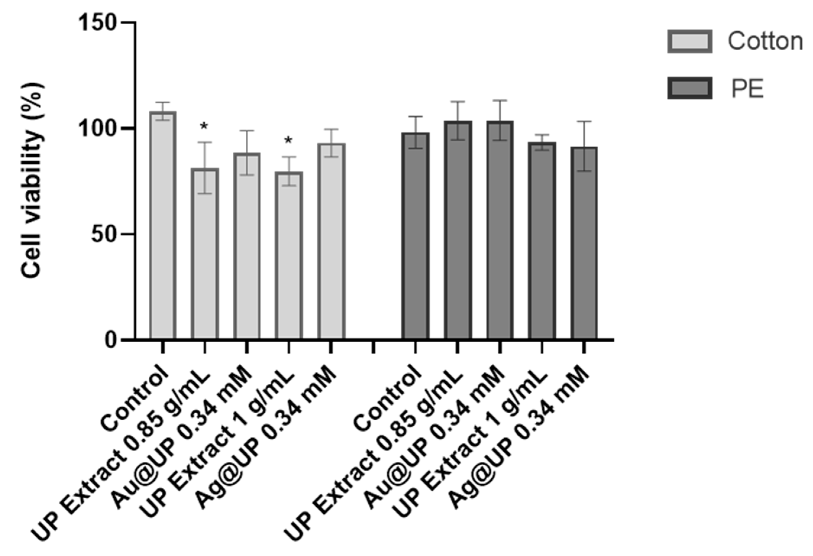
As part of evaluating the potential of these functionalized textiles for biomedical applications, an indirect contact cytotoxicity assay was performed using the widely used reference, murine fibroblast cell line, L-929. For this purpose, culture medium was preconditioned by incubation with the samples for 24 h prior to cell exposure. After incubation, metabolic cell activity was assessed using the MTT assay and results were normalized to the life control (100 % viability on fresh culture medium). Additionally, gauzes functionalized with crude UP-extract (0.85 g/mL for Au@UP and 1 g/mL for Ag@UP) were included as controls to evaluate whether the extracts themselves exhibited cytotoxicity. The results are presented on Figure 8. IC50 values were not emphasized because it was not possible to achieve 50% inhibition within the tested concentration range of NPs, as a constraining factor of the nanoparticle synthesis process.
For the functionalized cotton gauzes, a significant decrease in cell viability was observed for both crude UP-extract conditions (0.85 and 1 g/mL) (p < 0.05), compared to the untreated gauzes. Meanwhile, none of the functionalized gauzes with Au@UP or Ag@UP displayed cytotoxicity. This suggests that the UP extracts display greater cytotoxicity than the nanoparticles synthesized with them, although no significant differences were detected between the NP-treated samples and their respective UP-extract controls (0.85 g/mL for Au@UP; 1 g/mL for Ag@UP). For the PE functionalized gauzes, no significant differences were observed among any of the tested conditions or controls. Likewise, no statistically significant differences were identified between the two textiles.
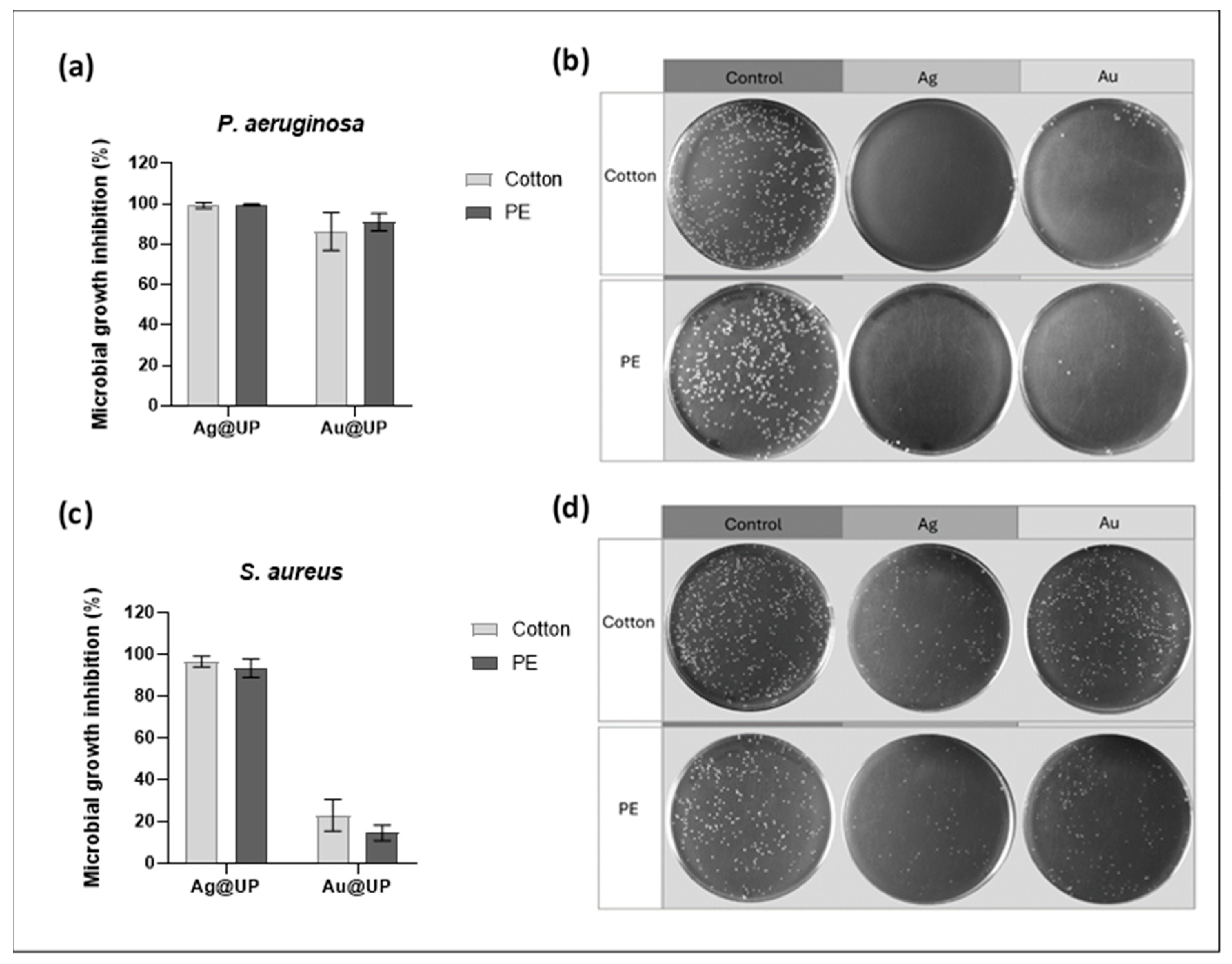
To evaluate the antimicrobial activity of the functionalized gauzes, a direct contact assay was performed by applying Staphylococcus aureus and Pseudomonas aeruginosa suspensions directly onto the samples. These bacteria were used as models of Gram-positive and Gram-negative bacteria, respectively. An initial optimization assay was conducted by varying incubation time (30 or 120 min) and NP concentration (0.17 or 0.34 mM). The highest antimicrobial activity was achieved with Ag@UP functionalized gazes, for both textile substrates and against both bacterial strains (Supplementary Data, Tables S1 and S2). Optimal conditions corresponded to the higher NP concentration (0.34 mM) and the prolonged incubation time (120 min) (Supplementary Data, Table S3).
Antimicrobial testing (Figure 9) demonstrated that Ag@UP functionalized gauzes exhibited high antimicrobial efficacy, achieving inhibition values exceeding 90 % against both S. aureus and P. aeruginosa. Au@UP also showed strong antimicrobial activity against P. aeruginosa (~ 90 %), whereas substantially lower inhibition was observed against S. aureus (~ 15 % for PE and ~ 20 % for cotton). These results suggest that the differences in bacterial cell wall composition may influence the efficacy of the functionalized materials.

4. Discussion

Nanotechnology-based antibacterial strategies have received considerable attention in recent years due to their unique modes of action and lower likelihood of developing resistance [9]. As textiles typically provide an ideal environment for microorganism growth, with adequate moisture and oxygen availability, the need to develop multi-functional textiles is emerging in response to increasing awareness of health and hygiene [3,32]. Therefore, in this study, silver and gold green synthesized NPs were used to functionalize widely used medical textiles [6,7]. The synthesis of Ag@UP and Au@UP was carried out employing UP aqueous extracts in order to obtain nanoparticles with antioxidant and antibacterial activity previously reported [18]. Further characterization was performed, confirming reproducibility of the synthesis. The functionalization was performed on cotton and non-woven PE gauzes through the exhaustion method.
The exhaustion method is affected by several parameters, such as incubation time and temperature, the type and concentration of the functionalization solution, and posttreatment conditions, such as heat setting/cure temperature [33]. Incubation times usually range from 5 to 90 min. Temperature, considered the most decisive factor, is reported to enhance the process efficacy [34]. To maintain the functionalization process as environmentally friendly and sustainable as possible, incubation was performed at room temperature. Given the high thermal stability of metal nanoparticles, the heat setting/cure of the gauzes was carried out at a higher temperature than commonly reported for this method [35,36,37]. The curing process is crucial due to the rearrangement of dipole interactions and hydrogen bonds, which may help to stabilize and attach the NPs onto the textile surface [38,39]. Accordingly, the gauzes were successfully functionalized through exhaustion, using cycles of 45 min of incubation in the respective NPs solution, followed by curing for 7 min at 180 °C. These were the conditions that exhibited stronger final coloration of the gauzes. Morphological alterations and shrinkage of the gazes were monitored throughout the process to guarantee avoiding textile damage.
The efficacy of the functionalization was initially confirmed by determining the incorporation of Au@UP and Ag@UP through UV-Vis analysis of the supernatant and through visual and stereo microscopy inspection. Textile colour changes, from pale yellow to lilac for Au@UP or yellow/orange for Ag@UP, were readily observed during gauze functionalization process. These colours are associated with the SPR, as for metallic nanoparticles, the superficial electrons generate an intense absorption band in the UV-Vis spectrum when exposed to light [40,41]. As such, the UV-vis analysis was conducted mainly on these parts of the NPs formulation’s spectra. The lilac and yellow also correspond to the coloration of the original Au@UP and Ag@UP formulations. As such, their appearance indicates that the particles were successfully attached to the textiles [42,43].
To further confirm the NP-derived adsorption onto the gauzes, the colour strength values (K/S) were obtained through the absorption coefficient (K) and the scattering coefficient (S), demonstrating the depth of colour on functionalized fabrics. The results demonstrate nanoparticle deposition through significant increase on K/S values for Au@UP and Ag@UP-functionalized textiles, while much lower colour depth values were obtained regarding the cotton and PE controls. This has been previously reported for silver nanoparticle-functionalized cotton fabrics [44]. Since Au@UP and Ag@UP absorb light at the previously described wavelengths due to the SPR phenomenon, the reflectometer can be used as an indirect measure of nanoparticle adhesion through absorbance increase [45]. The difference of values between textiles on Au@UP samples might be attributed to polyester based textiles usually presenting fewer free active groups as well as higher hydrophobicity, when compared to cotton, promoting less nanoparticle attachment [46]. However, this behaviour was not noticeable on Ag@UP functionalized textiles, probably because these nanoparticles present lower surface charge, when compared to Au@UP, exhibiting higher colloidal stability and therefore lower reactivity [18,47]. Additionally, the presence of UP-extract compounds bound to the different textile substrates might also be a key factor for the differences found in nanoparticle adhesion efficiency [32]. Nevertheless, the results indicated higher colour adsorption on cotton gauzes, particularly those functionalized with Au@UP.
SEM analysis revealed significant changes in the textiles’ morphology, with the original smooth surface of the untreated textiles becoming rough upon functionalization. The clear deposition of UP-extract compounds, the deposition of nearly spherical NPs and an increase on fiber diameter upon functionalization was confirmed by SEM, especially on functionalized cotton gauzes. These alterations on surface morphology have been described previously regarding nanoparticle-functionalized textiles such as cotton and polyester [42,48]. These results further support the successful deposition of Au@UP and Ag@UP, as well as UP-extract compounds on the textile surfaces.
Since the metallic NPs cannot be detected by FTIR due to the lack of molecular vibrations under infrared radiation, the main spectral differences observed in the functionalized gauzes are attributed to the UP-extract components [49]. Although slight alterations seem to appear at the fingerprint region of the spectra, FTIR analysis of the functionalized gauzes did not reveal direct chemical changes attributed to the functionalization with UP extracts, Au@UP and Ag@UP. This masking phenomenon has been previously reported on natural extract-functionalized textiles, aligning with our results on the fabrics matrix groups dominating FTIR spectra [50,51]. Nevertheless, in both tested fabrics, the changes observed in the O-H absorption band region suggests that these groups are responsible for effectively distributing and stabilizing the interactions between the NPs or the UP-extract compounds and the textile substrates [32,52]. The successful deposition of the UP-extract compounds, present on the nanoparticle’s formulations, is further supported by the increased presence of organic groups such as -CH2 and -CH3, associated with the presence of carboxylic acids, lipids and hydrocarbon chains in the functionalized samples [29,53]. This effect was more pronounced in the Ag@UP functionalized gauzes, possibly because silver nanoparticle solutions contained a higher extract concentration (1 g/mL) than the gold counterpart (0.85 g/mL), owing to the dilution adjustments required to achieve equivalent concentration of 0.34 mM of nanoparticles content. These results align with previous reports, as these groups are widely present on proteins, that play a crucial role in stabilizing and capping agents during green synthesis [29,32].
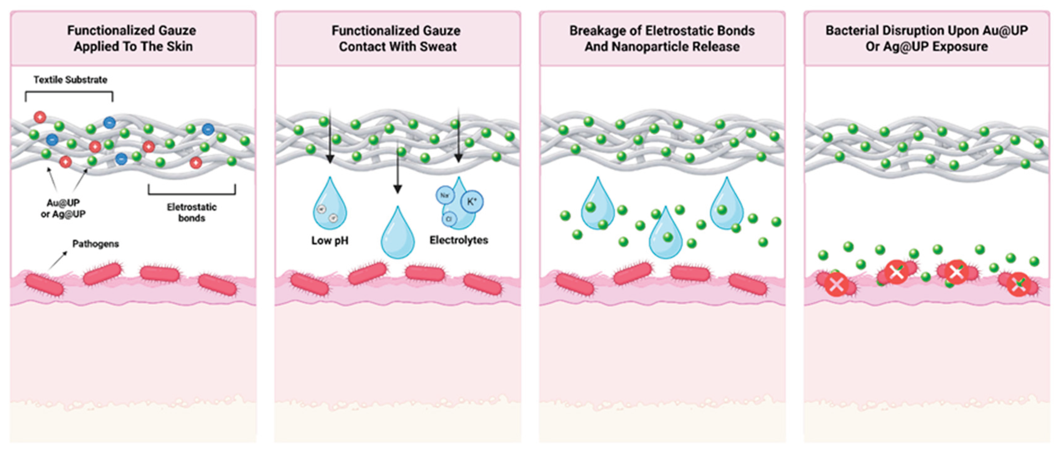
The physical bonding between textiles and the NPs is believed to be caused by electrostatic interactions, which are weak interactions that might allow NPs detachment under certain environmental triggers [32,54]. This property is very interesting in this study, as the antimicrobial potential of the Au@UP and Ag@UP might require their detachment from the textiles. The cumulative release assay was evaluated by UV-vis spectroscopic analysis, measuring the absorbance of the supernatant at different timepoints over a 72 h incubation period. An artificial sweat solution was used to simulate skin-contact conditions and to assess leaching behaviour of the nanoparticles from the functionalized gauzes during direct contact with human skin [55]. The functionalized gauzes with different Au@UP and Ag@UP concentrations demonstrated a fluctuating release profile, suggesting that both the number of cycles involved in the functionalization - five for 0.17 mM and three for 0.34 mM – and type of NPs influence their attachment to the textiles. This observation aligns with the understanding that surface functionalization represents a critical parameter governing particle migration [56]. Moreover, higher nanoparticle concentrations are associated with increased UP extract content, resulting in a greater amount of compounds rich in hydroxyl groups that promote stronger and more extensive interactions with the textile surface [32,54,57]. Despite the fact that our results did not fit the usual controlled release models, probably due to the absorbance fluctuations, a fitting line was used to evaluate release. These fluctuations may be explained by the leaching of impurities from the textiles [58], as similar profiles were detected on the control gauzes and similar behaviours were previously reported, even when employing comparable artificial sweat solutions [19,59]. Nevertheless, it is possible to notice a slow-release profile tendency of the textiles functionalized with either Au@UP or Ag@UP that may prolong the desired bioactivities, such as antibacterial, while reducing the risk of toxicity associated with gold and silver NPs exposure, especially at higher concentrations [60]. The release assay reinforces the hypothesis that the NPs might behave through dynamic processes, not following a typical release profile.
Altogether, our results suggest that the nanoparticles interact with the textile substrates through electrostatic forces, allowing their dynamic attachment and detachment during both the functionalization cycles and the release assay. This behaviour has been previously reported by many authors, that hypothesize that the binding of metallic nanoparticles is through van der Waals interactions [32,54]. These interactions, when in contact with sweat – including exposure to parameters such as low pH and the presence of electrolytes - may be weakened, allowing the release of the nanoparticles and their action as antimicrobial agents against pathogens. This hypothesis is further supported by the lack of nanoparticle loss during the washing performed at the end of the functionalization cycles, which supernatant was analyzed by UV-vis spectroscopy (data not shown). This pattern has also been previously described for silver nanoparticles functionalized textiles, as acidic environments promoted nanoparticles leaching mostly in form of silver ions [56,61]. The presence of free ions may also be a key factor in weakening electrostatic interactions by increasing ionic strength. In these conditions, electrolytes may compete with bound species for interaction sites, reducing the strength and stability of electrostatic associations [62]. These authors further suggest that the nanoparticle release profile is directly proportional to their initial concentration during functionalization. Our mechanism suggestion is therefore presented in the following Figure 10.
Regarding the antioxidant testings of the functionalized gauzes, neither the UP extracts nor the NPs exhibited significant antioxidant potential. This finding is consistent with previous reports showing that the antioxidant capacity of aqueous UP-extracts is relatively low compared to other brown seaweeds and is further reduced by 2.5- and 1.5-fold for Au@UP and Ag@UP, respectively, as antioxidant compounds in the extract play a crucial role during NP synthesis [18,63]. This decrease on antioxidant activity is mainly due to the loss of hydroxyl groups during the synthesis of metallic nanoparticles [64]. Nevertheless, it is important to note that the masking of the bioactives antioxidant activity might be due to a combination of factors such as partial degradation during the cure thermal treatment (180 °C) and low mobility upon immobilization on the textile’s matrices [65,66]. As the scavenging capacity is related to the arrangement and availability of hydroxyl groups, the attachment of NPs to the textile substrates could be further hindering their antioxidant potential [67]. This might also be associated with potential textile–oxygen ligations, that could mask the bioactives action and enhance nanoparticle retention on the textile fibers [64]. This is consistent with the DPPH assay results obtained for the functionalized gauzes, which showed a peak around 3 % for cotton gauzes functionalized with Au@UP at 0.34 mM.
Since the aim of this work was to assess the suitability of the functionalized gauzes for biomedical applications without posing toxicity risks, murine fibroblast cell line L-929 were exposed to culture media preconditioned with the samples. The results showed that the cotton textiles functionalized with crude UP extracts induced some degree of cytotoxicity after overnight exposure. In contrast, this effect was not observed for cotton textiles functionalized with either Au@UP or Ag@UP. This reduced toxicity of NPs compared to their synthesis extracts in fibroblast-like cells has been previously reported for both gold and silver particles [68,69]. Moreover, cotton textiles functionalized with different NPs, including green-synthesized silver NPs obtained using natural extracts, have been shown to be safe for biological applications [70,71]. A marked reduction in cell viability was observed for cotton gauzes compared to PE gauzes. This behaviour may be explained by the attachment of metabolites originating from the UP-extract to the cotton substrate, resulting in higher concentrations than on the PE substrate.
The strong antimicrobial activity of Ag@UP functionalized textiles against S. aureus (Gram-positive) and the even greater activity observed against P. aeruginosa (Gram-negative) are in agreement with previous reports on silver and other green synthesized metallic particles, which typically show higher resistance in Gram-positive bacteria [72,73,74]. This difference in susceptibility is usually attributed to variations in the bacterial cell wall structure, as the thinner peptidoglycan layer on Gram-negative bacteria facilitates penetration of metallic ions into the cell [75,76]. A similar behaviour was observed for Au@UP functionalized gauzes, which exhibited a strong decrease of antimicrobial activity against S. aureus, compared to P. aeruginosa. These results are consistent with previous reports with these NPs, as the minimum inhibitory concentration against these bacteria was 7.88 µg/mL for Ag@UP and 11.81 µg/mL for Au@UP [18].
The differences in antimicrobial activity observed for Au@UP and Ag@UP nanoparticles are likely due to distinct modes of action. Silver nanoparticles exhibit intrinsic antimicrobial effects through direct interactions with metabolic systems involved in the respiratory chain or through interference with DNA synthesis [77]. Additionally, silver ions are reported to induce oxidative stress [12]. These effects are largely mediated by the release of positively charged silver ions, which interact with the bacterial cell membrane, inducing the formation of silver pits [78]. Gold particles, conversely, and generally, display weaker antimicrobial effects, as their activity is mainly with the presence of antimicrobial conjugates, such as bioactive natural compounds, rather than to the metallic core itself [79,80,81,82].
The incorporation of green synthesized NPs into textiles to impart antimicrobial properties has been extensively explored. Silver NPs synthesized using plant extracts, such as Acorus calamus and microalgae extracts, such as Spirulina, as well as gold NPs produced from aqueous plant extracts of Coleus aromaticus, Croton sparsiflorus and Ginkgo biloba Linn, have demonstrated effective microbial growth inhibition when incorporated into cotton and polyester-based fabrics [32,43,79,83,84]. However, information regarding the use of brown seaweed extracts for the green synthesis of NPs and textile functionalization remain limited. Cotton fabrics functionalized with zinc oxide nanoparticles from the brown seaweed Padina sp. aqueous extracts exhibited only moderate antimicrobial activity [85]. Moreover, these materials required approximately 5 h of incubation to achieve significant inhibition of P. aeruginosa, in contrast to the UP-mediated nanoparticles used in this study, which achieved approximately 90 % inhibition within just 2 h. Furthermore, preliminary assays with Ag@UP nanoparticles demonstrated that considerable antimicrobial activity was attained after only 30 min of incubation. Our previous work on UP-mediated silver and gold NPs demonstrated strong antimicrobial properties, which are retained upon incorporation into both cotton and PE gauzes [18]. Accordingly, several studies have shown that cotton and polyester-based fabrics functionalized with biosynthesized copper, zinc oxide and silver NPs retain antimicrobial activity, whereas untreated textiles exhibited no bioactivity either before or after washing [29,86,87].

5. Conclusions

Green synthesized silver and gold NPs were previously obtained using aqueous extracts of the invasive seaweed UP and shown to exhibit strong antimicrobial activity. The present study extends this work by demonstrating the functionalization of widely used medical textiles, namely cotton and polyester-based gauzes, with these biogenic NPs. The textiles acquired the characteristic colouration of the functionalizing solutions, indicating NP attachment by electrostatic interactions and stabilized with hydroxyl groups present in UP-extract compounds. This attachment was confirmed through UV-Vis spectroscopy, K/S measurements, FTIR spectroscopy and SEM analysis, which revealed the presence of extract-derived functional groups and efficient nanoparticle deposition on the textile substrates. The functionalized gauzes exhibited a slow-release profile, with a dynamic attachment–detachment mechanism that may contribute to the prolonged durability of their bioactive properties, particularly antimicrobial activity. Moreover, no cytotoxic effects were observed in fibroblasts following exposure to the functionalized gauzes. Regarding the functionalized gauzes, Ag@UP demonstrated remarkable inhibitory effects against both S. aureus and P. aeruginosa, while Au@UP showed strong activity exclusively against P. aeruginosa within just 2 h of contact. Overall, these results highlight UP-mediated NP-functionalized textiles as a sustainable and promising approach for inhibiting microbial growth and preventing bacterial infections.

Supplementary Materials

The following supporting information can be downloaded at the website of this paper posted on Preprints.org, Figure S1: UV-vis spectroscopy of Au@UP (a) and Ag@UP (b) nanoparticles. ζ-potential (c), diameter size mean and polydispersity index (d) determined by ELS and DLS for both nanoparticles; Table S1: Optimization antimicrobial assay conducted with different incubation times (30 or 120 min) and NPs concentration (0.17 or 0.34 mM) using Pseudomonas aeruginosa as a gram-negative bacterial model; Table S2: Optimization of the antimicrobial assay conducted with different incubation times (30 or 120 min) and NPs concentration (0.17 or 0.34 mM) using Staphylococcus aureus as a gram-positive bacterial model; Table S3: CFU count from the optimization antimicrobial assays performed with the functionalized gauzes varying incubation times (30 or 120 min) and NPs concentration (0.17 or 0.34 mM) and using Staphylococcus aureus (gram-positive) and Pseudomonas aeruginosa (gram-negative) as bacterial models.

Author Contributions

Conceptualization, M.F., R.M., A.G. and C.S.; methodology, J.A., M.F., A.R.B., B.S., D.R., R.M. and A.R.; validation, J.A. and A.R.; formal analysis, J.A., A.R.B. and R.M.; investigation, J.A., M.F., A.R.B. and B.S.; resources, A.R. and M.C.R.-A.; data curation, J.A., A.R.B. and B.S.; writing—original draft preparation, J.A., A.R.B. and B.S.; writing—review and editing, J.A., R.M., M.C.R.-A., A.G. and C.S.; supervision, R.M., M.C.R.-A., A.G. and C.S.; project administration, A.G.; funding acquisition, M.C.R.-A., A.G. and C.S. All authors have read and agreed to the published version of the manuscript.

Funding

This work is a result of the projects ATLANTIDA (ref. NORTE-01-0145-FEDER-000040) and ATLANTIDA II (ref. NORTE2030-FEDER-01799200), supported by the Norte Portugal Regional Operational Program (NORTE2020 and NORTE2030, respectively), under the PORTUGAL 2020 Partnership Agreement and through the European Regional Development Fund (ERDF). It was further supported by the Portuguese Foundation for Science and Technology (FCT) under the scope of the strategic funding of the project LA/P/0069/2020 granted to the Associate Laboratory ARNET (https://doi.org/10.54499/LA/P/0069/2020), by the “Contrato-Programa” UID/04050/2025 funded by FCT I.P. (https://doi.org/10.54499/UID/04050/2025) and by LABBELS—Associate Laboratory in Biotechnology, Bioengineering and Microelectromechanical Systems (LA/P/0029/2020).

Data Availability Statement

All data can be made available upon request to the corresponding author(s) through the institutional resource https://datarepositorium.uminho.pt/. Raw data regarding DLS and ELS analysis are available at https://doi.org/10.34622/datarepositorium/26GZLS.

Acknowledgments

n.a.

Conflicts of Interest

The authors declare no conflicts of interest.

Abbreviations

The following abbreviations are used in this manuscript:
ANOVA Analysis of variance
ATCC American Type Culture Collection
CFU Colony-forming units
CLSI Clinical and Laboratory Standards Institute
DLS Dynamic light scattering
DMEM Dulbecco’s Modified Eagle Medium
DMSO Dimethyl sulfoxide
DPPH 2,2-Diphenyl-1-picrylhydrazyl
EDX Energy-dispersive X-ray spectroscopy
EELS Electron energy-loss spectroscopy
ELS Electrophoretic light scattering
EUCAST European Committee on Antimicrobial Susceptibility Testing
FBS Fetal bovine serum
FTIR Fourier-transform infrared spectroscopy
FTIR-ATR Fourier-transform infrared spectroscopy–attenuated total reflectance
HRTEM High-resolution transmission electron microscopy
IC₅₀ Half-maximal inhibitory concentration
K/S Colour strength parameter
LB Luria broth
MTT 3-(4,5-Dimethyl-2-thiazolyl)-2,5-diphenyl-2H-tetrazolium bromide
NPs Nanoparticles
OD₆₀₀ Optical density at 600 nm
PBS Phosphate-buffered saline
PE Polyester
RT Room temperature
SD Standard deviation
SEM Scanning electron microscopy
SPR Surface plasmon resonance
TEM Transmission electron microscopy
UP Undaria pinnatifida
UV–Vis Ultraviolet–visible spectroscopy

References

  1. Frieri, M.; Kumar, K.; Boutin, A. Antibiotic resistance. J. Infect. Public Health 2017, 10, 369–378. [Google Scholar] [CrossRef] [PubMed]
  2. Owen, L.; Laird, K. The role of textiles as fomites in the healthcare environment: A review of the infection control risk. PeerJ 2020, 8, e9790. [Google Scholar] [CrossRef] [PubMed]
  3. Allehyani, E.S. Surface functionalization of polyester textiles for antibacterial and antioxidant properties. Polymers 2022, 14, 5512. [Google Scholar] [CrossRef] [PubMed]
  4. Gulati, R.; Sharma, S.; Sharma, R.K. Antimicrobial textile: Recent developments and functional perspective. Polym. Bull. 2022, 79, 5747–5771. [Google Scholar] [CrossRef] [PubMed]
  5. Yusuf, M.; Kiran, S. Biomedical Textiles: Introduction and Applications; Routledge: New York, NY, USA, 2025. [Google Scholar] [CrossRef]
  6. Shahriari Khalaji, M.; Lugoloobi, I. Biomedical application of cotton and its derivatives. In Cotton Science and Processing Technology: Gene, Ginning, Garment and Green Recycling; Wang, H., Memon, H., Eds.; Springer: Singapore, 2020; pp. 393–416. [Google Scholar] [CrossRef]
  7. Darie-Niță, R.N.; Râpă, M.; Frąckowiak, S. Special features of polyester-based materials for medical applications. Polymers 2022, 14, 951. [Google Scholar] [CrossRef] [PubMed]
  8. Mondal, S.K.; Chakraborty, S.; Manna, S.; Mandal, S.M. Antimicrobial nanoparticles: Current landscape and future challenges. RSC Pharm. 2024, 1, 388–402. [Google Scholar] [CrossRef]
  9. Wang, L.; Hu, C.; Shao, L. The antimicrobial activity of nanoparticles: Present situation and prospects for the future. Int. J. Nanomed. 2017, 12, 1227–1249. [Google Scholar] [CrossRef] [PubMed]
  10. Yuan, P.; Ding, X.; Yang, Y.Y.; Xu, Q.H. Metal nanoparticles for diagnosis and therapy of bacterial infection. Adv. Healthc. Mater. 2018, 7, 1701392. [Google Scholar] [CrossRef] [PubMed]
  11. Slavin, Y.N.; Asnis, J.; Häfeli, U.O.; Bach, H. Metal nanoparticles: Understanding the mechanisms behind antibacterial activity. J. Nanobiotechnol. 2017, 15, 65. [Google Scholar] [CrossRef] [PubMed]
  12. Stensberg, M.C.; Wei, Q.; McLamore, E.S.; Porterfield, D.M.; Wei, A.; Sepúlveda, M.S. Toxicological studies on silver nanoparticles: Challenges and opportunities in assessment, monitoring and imaging. Nanomedicine 2011, 6, 879–898. [Google Scholar] [CrossRef] [PubMed]
  13. Sánchez-López, E.; et al. Metal-based nanoparticles as antimicrobial agents: An overview. Nanomaterials 2020, 10, 292. [Google Scholar] [CrossRef] [PubMed]
  14. Giljohann, D.A.; Seferos, D.S.; Daniel, W.L.; Massich, M.D.; Patel, P.C.; Mirkin, C.A. Gold nanoparticles for biology and medicine. Angew. Chem. Int. Ed. 2010, 49, 3280–3294. [Google Scholar] [CrossRef] [PubMed]
  15. Jadoun, S.; Arif, R.; Jangid, N.K.; Meena, R.K. Green synthesis of nanoparticles using plant extracts: A review. Environ. Chem. Lett. 2021, 19, 355–374. [Google Scholar] [CrossRef]
  16. Ciko, A.M.; Jokić, S.; Šubarić, D.; Jerković, I. Overview on the application of modern methods for the extraction of bioactive compounds from marine macroalgae. Mar. Drugs 2018, 16, 348. [Google Scholar] [CrossRef] [PubMed]
  17. Giordano, D. Bioactive molecules from extreme environments. Mar. Drugs 2020, 18, 640. [Google Scholar] [CrossRef] [PubMed]
  18. González-Ballesteros, N.; et al. Valorisation of the invasive macroalgae Undaria pinnatifida (Harvey) Suringar for the green synthesis of gold and silver nanoparticles with antimicrobial and antioxidant potential. Mar. Drugs 2023, 21, 397. [Google Scholar] [CrossRef] [PubMed]
  19. Costa, A.F.; Luís, S.; Noro, J.; Silva, S.; Silva, C.; Ribeiro, A. Therapeutic textiles functionalized with keratin-based particles encapsulating terbinafine for the treatment of onychomycosis. Int. J. Mol. Sci. 2022, 23, 13999. [Google Scholar] [CrossRef] [PubMed]
  20. Rocha, D.; et al. NADES-based cork extractives as green ingredients for cosmetics and textiles. Processes 2023, 11, 309. [Google Scholar] [CrossRef]
  21. International Organization for Standardization. ISO 10993-5: Biological Evaluation of Medical Devices—Part 5: Tests for In Vitro Cytotoxicity; ISO: Geneva, Switzerland, 2009.
  22. International Organization for Standardization. ISO 10993-12: Biological Evaluation of Medical Devices—Part 12: Sample Preparation and Reference Materials; ISO: Geneva, Switzerland, 2021.
  23. British Standards Institution. BS EN 12469:2000 Biotechnology: Performance Criteria for Microbiological Safety Cabinets; BSI Standards: London, UK, 2000. [CrossRef]
  24. Schneider, C.A.; Rasband, W.S.; Eliceiri, K.W. NIH Image to ImageJ: 25 years of image analysis. Nat. Methods 2012, 9, 671–675. [Google Scholar] [CrossRef] [PubMed]
  25. Becheri, A.; Dürr, M.; Lo Nostro, P.; Baglioni, P. Synthesis and characterization of zinc oxide nanoparticles: Application to textiles as UV-absorbers. J. Nanopart. Res. 2008, 10, 679–689. [Google Scholar] [CrossRef]
  26. El-Sheekh, M.M.; El-Kassas, H.Y. Algal production of nano-silver and gold: Their antimicrobial and cytotoxic activities: A review. J. Genet. Eng. Biotechnol. 2016, 14, 299–310. [Google Scholar] [CrossRef] [PubMed]
  27. Delicana, J.D.P.; et al. Tailoring color and antibacterial properties of cotton fabric materials using gold nanoparticles synthesized from Mangifera indica peel extract. Sci. Rep. 2025, 15. [Google Scholar] [CrossRef] [PubMed]
  28. Adekoya, M.A.; Liu, S.; Oluyamo, S.S.; Oyeleye, O.T.; Ogundare, R.T. Influence of size classifications on the crystallinity index of Albizia gummifera cellulose. Heliyon 2022, 8, e12019. [Google Scholar] [CrossRef] [PubMed]
  29. Eid, A.M.; et al. Endophytic Streptomyces laurentii mediated green synthesis of Ag-NPs with antibacterial and anticancer properties for developing functional textile fabric properties. Antibiotics 2020, 9, 641. [Google Scholar] [CrossRef] [PubMed]
  30. Volkov, D.S.; Rogova, O.B.; Proskurnin, M.A. Organic matter and mineral composition of silicate soils: FTIR comparison study by photoacoustic, diffuse reflectance, and attenuated total reflection modalities. Agronomy 2021, 11, 1879. [Google Scholar] [CrossRef]
  31. Cao, T.; Zhou, Y.; Zhang, J.; Zhao, B. Clean preparation of Ag@Au nanoparticle-modified cotton fabric with durable antimicrobial properties by adsorption. Cellulose 2025, 32, 613–628. [Google Scholar] [CrossRef]
  32. Ganesan, R.M.; Gurumallesh Prabu, H. Synthesis of gold nanoparticles using herbal Acorus calamus rhizome extract and coating on cotton fabric for antibacterial and UV blocking applications. Arab. J. Chem. 2019, 12, 2166–2174. [Google Scholar] [CrossRef]
  33. Ibrahim, N.A.; Youssef, M.A.; Helal, M.H.; Shaaban, M.F. Exhaust dyeing of polyester-based textiles using high-temperature-alkaline conditions. J. Appl. Polym. Sci. 2003, 89, 3659–3667. [Google Scholar] [CrossRef]
  34. Cay, A.; Tarakçioǧlu, I.; Hepbasli, A. Assessment of finishing processes by exhaustion principle for textile fabrics: An exergetic approach. Appl. Therm. Eng. 2009, 29, 2554–2561. [Google Scholar] [CrossRef]
  35. Saranya, K.S.; Padil, V.V.T.; Senan, C.; Pilankatta, R.; Saranya, K.; George, B.; Wacławek, S.; Černík, M. Green synthesis of high temperature stable anatase titanium dioxide nanoparticles using gum kondagogu: Characterization and solar driven photocatalytic degradation of organic dye. Nanomaterials 2018, 8, 1002. [Google Scholar] [CrossRef] [PubMed]
  36. Sun, J.; et al. Toward monodispersed silver nanoparticles with unusual thermal stability. J. Am. Chem. Soc. 2006, 128, 15756–15764. [Google Scholar] [CrossRef] [PubMed]
  37. Radetić, M. Functionalization of textile materials with silver nanoparticles. J. Mater. Sci. 2013, 48, 95–107. [Google Scholar] [CrossRef]
  38. Chowdhury, M.; et al. A feasibility study to analyze the behavior of heat settings on the cleaner production of knitted fabrics. Clean. Eng. Technol. 2022, 7, 100429. [Google Scholar] [CrossRef]
  39. Besler, N.; Gloy, Y.S.; Gries, T. Analysis of the heat setting process. In IOP Conference Series: Materials Science and Engineering; Institute of Physics Publishing: Bristol, UK, 2016; Volume 141, p. 012018. [Google Scholar] [CrossRef]
  40. Ismail, E.H.; Saqer, A.M.A.; Assirey, E.; Naqvi, A.; Okasha, R.M. Successful green synthesis of gold nanoparticles using a Corchorus olitorius extract and their antiproliferative effect in cancer cells. Int. J. Mol. Sci. 2018, 19, 2612. [Google Scholar] [CrossRef] [PubMed]
  41. Darroudi, M.; Khorsand Zak, A.; Muhamad, M.R.; Huang, N.M.; Hakimi, M. Green synthesis of colloidal silver nanoparticles by sonochemical method. Mater. Lett. 2012, 66, 117–120. [Google Scholar] [CrossRef]
  42. Hasan, K.M.F.; et al. A novel coloration of polyester fabric through green silver nanoparticles (G-AgNPs@PET). Nanomaterials 2019, 9, 569. [Google Scholar] [CrossRef] [PubMed]
  43. Velmurugan, P.; Shim, J.; Bang, K.S.; Oh, B.T. Gold nanoparticles mediated coloring of fabrics and leather for antibacterial activity. J. Photochem. Photobiol. B 2016, 160, 102–109. [Google Scholar] [CrossRef] [PubMed]
  44. Ramaiah, G.B.; Ari, A.P. Evaluation of color strength (K/S) values of cotton fabrics dyed with reactive dye and treated with silver nanoparticles. In AIP Conference Proceedings; American Institute of Physics: Melville, NY, USA, 2019; Volume 2162, p. 020111. [Google Scholar] [CrossRef]
  45. Dubas, S.T.; Kumlangdudsana, P.; Potiyaraj, P. Layer-by-layer deposition of antimicrobial silver nanoparticles on textile fibers. Colloids Surf. A Physicochem. Eng. Asp. 2006, 289, 105–109. [Google Scholar] [CrossRef]
  46. Tarbuk, A.; Grancarić, A.M.; Begović, S.; Dekanić, T. Interfacial phenomena of cotton/polyester blended fabric modified with enzyme and chitosan. Polymers 2026, 18, 867. [Google Scholar] [CrossRef] [PubMed]
  47. d’Errico, A.; Schröpfer, M.; Mondschein, A.; Wösten, H.A.B. Characterization of the surface charge and reactivity of Schizophyllum commune mycelium material. Colloids Surf. B Biointerfaces 2025, 254, 114852. [Google Scholar] [CrossRef] [PubMed]
  48. Shaheen, T.I.; El-Naggar, M.E.; Abdelgawad, A.M.; Hebeish, A. Durable antibacterial and UV protections of in situ synthesized zinc oxide nanoparticles onto cotton fabrics. Int. J. Biol. Macromol. 2016, 83, 426–432. [Google Scholar] [CrossRef] [PubMed]
  49. Pasieczna-Patkowska, S.; Cichy, M.; Flieger, J. Application of Fourier transform infrared (FTIR) spectroscopy in characterization of green synthesized nanoparticles. Molecules 2025, 30, 684. [Google Scholar] [CrossRef] [PubMed]
  50. Nortjie, E.; Basitere, M.; Moyo, D.; Nyamukamba, P. Assessing the efficiency of antimicrobial plant extracts from Artemisia afra and Eucalyptus globulus as coatings for textiles. Plants 2024, 13, 514. [Google Scholar] [CrossRef] [PubMed]
  51. Kramar, A.; et al. Selected aromatic plants extracts as an antimicrobial and antioxidant finish for cellulose fabric—Direct impregnation method. Fibers Polym. 2021, 22, 3317–3325. [Google Scholar] [CrossRef]
  52. Abd El-Aziz, E.; Zayed, M.; Mohamed, A.L.; Hassabo, A.G. Enhancement of the functional performance of cotton and polyester fabrics upon treatment with polymeric materials having different functional groups in the presence of different metal nanoparticles. Polymers 2023, 15, 3047. [Google Scholar] [CrossRef] [PubMed]
  53. Zhang, L.; Li, X.; Zhang, S.; Gao, Q.; Lu, Q.; Peng, R.; Xu, P.; Shang, H.; Yuan, Y.; Zou, H. Micro-FTIR combined with curve fitting method to study cellulose crystallinity of developing cotton fibers. Anal. Bioanal. Chem. 2021, 413, 1313–1320. [Google Scholar] [CrossRef] [PubMed]
  54. Ratnasari, A.; Endarko, E.; Syafiuddin, A. A green method for the enhancement of antifungal properties of various textiles functionalized with silver nanoparticles. Biointerface Res. Appl. Chem. 2020, 10, 7284–7294. [Google Scholar] [CrossRef]
  55. Midander, K.; Julander, A.; Kettelarij, J.; Lidén, C. Testing in artificial sweat—Is less more? Comparison of metal release in two different artificial sweat solutions. Regul. Toxicol. Pharmacol. 2016, 81, 381–386. [Google Scholar] [CrossRef] [PubMed]
  56. Wagener, S.; et al. Textile functionalization and its effects on the release of silver nanoparticles into artificial sweat. Environ. Sci. Technol. 2016, 50, 5927–5934. [Google Scholar] [CrossRef] [PubMed]
  57. Princy, K.F.; Gopinath, A. Green synthesis of silver nanoparticles using polar seaweed Fucus gardneri and its catalytic efficacy in the reduction of nitrophenol. Polar Sci. 2021, 30, 100692. [Google Scholar] [CrossRef]
  58. Cortese, B.; Caschera, D.; Padeletti, G.; Ingo, G.M.; Gigli, G. A brief review of surface-functionalized cotton fabrics. Surf. Innov. 2013, 1, 140. [Google Scholar] [CrossRef]
  59. Khadeja, L.; Grigoriants, I.; Halperin-Sternfeld, M.; Yona, A.; Adler-Abramovich, L. Sonochemical functionalization of cotton and non-woven fabrics with bio-inspired self-assembled nanostructures. Isr. J. Chem. 2020, 60, 1190–1196. [Google Scholar] [CrossRef]
  60. Üreyen, M.E.; Doğan, A.; Koparal, A.S. Antibacterial functionalization of cotton and polyester fabrics with a finishing agent based on silver-doped calcium phosphate powders. Text. Res. J. 2012, 82, 1731–1742. [Google Scholar] [CrossRef]
  61. Kulthong, K.; Srisung, S.; Boonpavanitchakul, K.; Kangwansupamonkon, W.; Maniratanachote, R. Determination of silver nanoparticle release from antibacterial fabrics into artificial sweat. Part. Fibre Toxicol. 2010, 7, 8. [Google Scholar] [CrossRef] [PubMed]
  62. Sakhawoth, Y.; et al. Real time observation of the interaction between aluminium salts and sweat under microfluidic conditions. Sci. Rep. 2021, 11, 6484. [Google Scholar] [CrossRef] [PubMed]
  63. Ma, X.T.; Sun, X.Y.; Yu, K.; Gui, B.S.; Gui, Q.; Ouyang, J.M. Effect of content of sulfate groups in seaweed polysaccharides on antioxidant activity and repair effect of subcellular organelles in injured HK-2 cells. Oxid. Med. Cell. Longev. 2017, 2017, 2542950. [Google Scholar] [CrossRef] [PubMed]
  64. Rehan, M.; Mashaly, H.M.; Abdel-Aziz, M.S.; Abdelhameed, R.M.; Montaser, A.S. Viscose fibers decorated with silver nanoparticles via an in-situ green route: UV protection, antioxidant activities, antimicrobial properties, and sensing response. Cellulose 2024, 31, 5899–5930. [Google Scholar] [CrossRef]
  65. Antony, A.; Farid, M. Effect of temperatures on polyphenols during extraction. Appl. Sci. 2022, 12, 2107. [Google Scholar] [CrossRef]
  66. Volf, I.; Ignat, I.; Neamtu, M.; Popa, V.I. Thermal stability, antioxidant activity, and photo-oxidation of natural polyphenols. Chem. Pap. 2014, 68, 121–129. [Google Scholar] [CrossRef]
  67. Hong, K.H. Functionalization of cotton textiles via screen printing of Aronia melanocarpa berry extract–chitosan mixtures. J. Nat. Fibers 2024, 21. [Google Scholar] [CrossRef]
  68. Jalilian, F.; Chahardoli, A.; Sadrjavadi, K.; Fattahi, A.; Shokoohinia, Y. Green synthesized silver nanoparticle from Allium ampeloprasum aqueous extract: Characterization, antioxidant activities, antibacterial and cytotoxicity effects. Adv. Powder Technol. 2020, 31, 1323–1332. [Google Scholar] [CrossRef]
  69. Machado, S.; et al. Toxicity in vitro and in zebrafish embryonic development of gold nanoparticles biosynthesized using Cystoseira macroalgae extracts. Int. J. Nanomed. 2021, 16, 5017–5036. [Google Scholar] [CrossRef] [PubMed]
  70. dos Santos, O.A.L.; de Araujo, I.; Dias da Silva, F.; Sales, M.N.; Christoffolete, M.A.; Backx, B.P. Surface modification of textiles by green nanotechnology against pathogenic microorganisms. Curr. Res. Green Sustain. Chem. 2021, 4, 100206. [Google Scholar] [CrossRef]
  71. Ballottin, D.; et al. Antimicrobial textiles: Biogenic silver nanoparticles against Candida and Xanthomonas. Mater. Sci. Eng. C 2017, 75, 582–589. [Google Scholar] [CrossRef] [PubMed]
  72. Geethalakshmi, R.; Sarada, D.V.L. Characterization and antimicrobial activity of gold and silver nanoparticles synthesized using saponin isolated from Trianthema decandra L. Ind. Crops Prod. 2013, 51, 107–115. [Google Scholar] [CrossRef]
  73. Ahmad, T.; Wani, I.A.; Manzoor, N.; Ahmed, J.; Asiri, A.M. Biosynthesis, structural characterization and antimicrobial activity of gold and silver nanoparticles. Colloids Surf. B Biointerfaces 2013, 107, 227–234. [Google Scholar] [CrossRef] [PubMed]
  74. Gurunathan, S.; Han, J.W.; Kwon, D.N.; Kim, J.H. Enhanced antibacterial and anti-biofilm activities of silver nanoparticles against Gram-negative and Gram-positive bacteria. Nanoscale Res. Lett. 2014, 9, 373. [Google Scholar] [CrossRef] [PubMed]
  75. Khan, S.A.; Shahid, S.; Lee, C.S. Green synthesis of gold and silver nanoparticles using leaf extract of Clerodendrum inerme: Characterization, antimicrobial, and antioxidant activities. Biomolecules 2020, 10, 835. [Google Scholar] [CrossRef] [PubMed]
  76. Emami-Karvani, Z. Antibacterial activity of ZnO nanoparticle on Gram-positive and Gram-negative bacteria. Afr. J. Microbiol. Res. 2012, 5, 4521–4526. [Google Scholar] [CrossRef]
  77. Castillo-Henríquez, L.; Alfaro-Aguilar, K.; Ugalde-Álvarez, J.; Vega-Fernández, L.; de Oca-Vásquez, G.M.; Vega-Baudrit, J.R. Green synthesis of gold and silver nanoparticles from plant extracts and their possible applications as antimicrobial agents in the agricultural area. Nanomaterials 2020, 10, 1763. [Google Scholar] [CrossRef] [PubMed]
  78. Rahisuddin; Akrema. Extracellular synthesis of silver dimer nanoparticles using Callistemon viminalis (bottlebrush) extract and evaluation of their antibacterial activity. Spectrosc. Lett. 2016, 49, 268–275. [Google Scholar] [CrossRef]
  79. Mishra, A.; Tripathy, S.K.; Yun, S.I. Bio-synthesis of gold and silver nanoparticles from Candida guilliermondii and their antimicrobial effect against pathogenic bacteria. J. Nanosci. Nanotechnol. 2011, 11, 243–248. [Google Scholar] [CrossRef] [PubMed]
  80. Yadi, M.; Azizi, M.; Dianat-Moghadam, H.; Akbarzadeh, A.; Abyadeh, M.; Milani, M. Antibacterial activity of green gold and silver nanoparticles using ginger root extract. Bioprocess Biosyst. Eng. 2022, 45, 1905–1917. [Google Scholar] [CrossRef] [PubMed]
  81. El Domany, E.B.; Essam, T.M.; Ahmed, A.E.; Farghali, A.A. Biosynthesis physico-chemical optimization of gold nanoparticles as anti-cancer and synergetic antimicrobial activity using Pleurotus ostreatus fungus. J. Appl. Pharm. Sci. 2018, 8, 119–128. [Google Scholar] [CrossRef]
  82. Shankar, S.; Jaiswal, L.; Aparna, R.S.L.; Prasad, R.G.S.V. Synthesis, characterization, in vitro biocompatibility, and antimicrobial activity of gold, silver and gold silver alloy nanoparticles prepared from Lansium domesticum fruit peel extract. Mater. Lett. 2014, 137, 75–78. [Google Scholar] [CrossRef]
  83. Gouda, M.; Khalaf, M.M.; Abou Taleb, M.F.; Abdelaziz, M.A.; Abd El-Lateef, H.M. Functionalization of cotton fabric using the biogenic synthesized silver nanoparticles for enhanced dye reduction and antimicrobial efficiency: Response surface methodology. Int. J. Biol. Macromol. 2025, 307, 141853. [Google Scholar] [CrossRef] [PubMed]
  84. Boomi, P.; Ganesan, R.M.; Poorani, G.; Gurumallesh Prabu, H.; Ravikumar, S.; Jeyakanthan, J. Biological synergy of greener gold nanoparticles by using Coleus aromaticus leaf extract. Mater. Sci. Eng. C 2019, 99, 202–210. [Google Scholar] [CrossRef] [PubMed]
  85. Rohaeti, E.; Helmiyati; Joronavalona, R.; Taba, P.; Sondari, D.; Kamari, A. The role of brown algae as a capping agent in the synthesis of ZnO nanoparticles to enhance the antibacterial activities of cotton fabrics. Mar. Drugs 2025, 23, 71. [Google Scholar] [CrossRef] [PubMed]
  86. Asmat-Campos, D.; et al. Cu2O nanoparticles synthesized by green and chemical routes, and evaluation of their antibacterial and antifungal effect on functionalized textiles. Biotechnol. Rep. 2023, 37, e00785. [Google Scholar] [CrossRef] [PubMed]
  87. Giedraitienė, A.; Ružauskas, M.; Šiugždinienė, R.; Tučkutė, S.; Grigonis, K.; Milčius, D. ZnO nanoparticles enhance the antimicrobial properties of two-sided-coated cotton textile. Nanomaterials 2024, 14, 1264. [Google Scholar] [CrossRef] [PubMed]
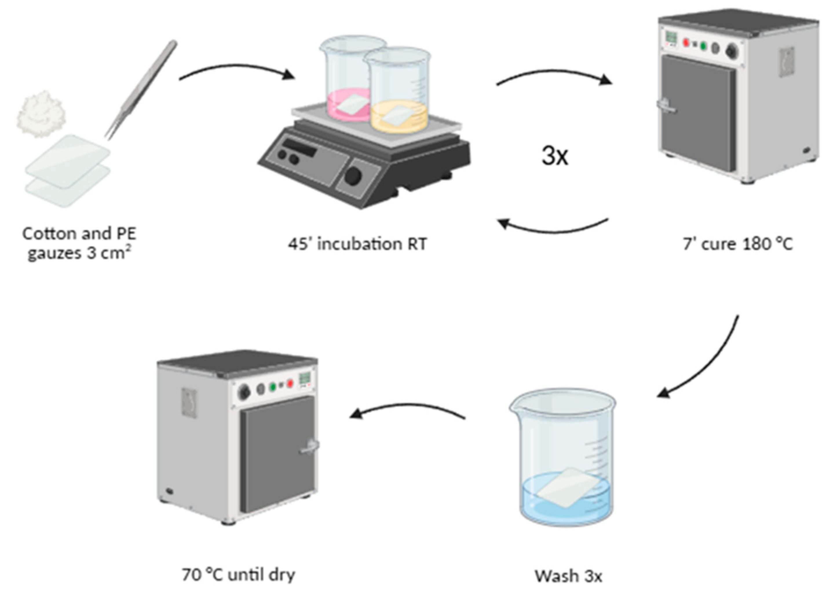
Figure 1. This is a figure. Schemes follow the same formatting.
Preprints 227912 g001
Figure 2. Stereo microscope images of cotton gauzes (a, b) and PE gauzes (c, d) functionalized with Ag@UP 0.34 mM and cotton gauzes (e, f) and PE gauzes (g, h) functionalized with Au@UP 0.34 mM. Cotton (i, j) and PE (k, l) gauzes treated with ultrapure water, as control. Magnifications of 1x (first and third columns) and 3x with a black background (second and fourth columns) used for imaging.
Preprints 227912 g002
Figure 3. K/S values of functionalized cotton gauzes with Au@UP 0.34 mM (a), Ag@UP 0.34 mM (b) and ultrapure water as control (c) and functionalized PE gauzes with Au@UP 0.34 mM (d), Ag@UP 0.34 mM (e) and ultrapure water as control (f).
Preprints 227912 g003
Figure 4. Scanning electron microscopy (SEM) images at different magnifications ranging from 0.15 to 7 K of the gauzes before and after functionalization. Cotton gauzes functionalized with ultrapure water as control (a, b), Au@UP 0.34 mM (c, d) and Ag@UP 0.34 mM (e, f). PE gauzes functionalized with ultrapure water as control (g, h), Au@UP 0.34 mM (i, j) and Ag@UP 0.34 mM (k, l).
Preprints 227912 g004
Figure 5. Fourier Transform Infrared Spectroscopy - Attenuated Total Reflectance (FTIR-ATR) analysis of cotton (a) and PE (b) gauzes functionalized with UP extract at 1 g/mL and Au@UP and Ag@UP 0.34 mM. Non-treated gauzes and free UP extract used as control.
Preprints 227912 g005
Figure 6. Cumulative release concentration measured using artificial sweat on functionalized cotton and PE gauzes with Au@UP (a) and Ag@UP (b). Values were obtained from calibration curves prepared with Au@UP (c) and Ag@UP (d) solutions and are represented as polynomial trend lines.
Preprints 227912 g006
Figure 7. DPPH radical scavenging activity (%) of the functionalized gauzes and their respective controls. The assay was conducted under agitation (200 rpm) for 2 and 6 h of incubation. Values are presented after subtraction of the corresponding textile control and are represented as the mean from three independent experiments (n = 3). Trolox 0.5 mg/mL was used as the positive control. Statistical differences compared to the respective timepoint positive control at every condition (**** for p < 0.0001). No statistical differences between functionalized gauzes.
Preprints 227912 g007
Figure 8. Cell viability (%) after 24 h of incubation in culture medium pre-conditioned with functionalized gauzes. Viability was assessed using the MTT assay and normalized to the live control values cultured in fresh medium (100 %). Extract control gauzes were functionalized with UP-extracts at 0.85 and 1 g/mL for Au@UP and Ag@UP, respectively. * p < 0.05, compared to the respective control. No further statistically significant differences observed between any conditions tested.
Preprints 227912 g008
Figure 9. Antimicrobial activity of cotton and PE gauzes functionalized with Au@UP and Ag@UP (0.34 mM) after 120 minutes of contact. Microbial growth inhibition (%) against Pseudomonas aeruginosa (a) and Staphylococcus aureus (c) was determined by CFU counting and calculated relative to non-treated cotton and PE gauzes. Representative agar plate images after exposure to functionalized gauzes are shown for P. aeruginosa (b) and S. aureus (d).
Preprints 227912 g009
Figure 10. Suggested mechanism of release profile and antimicrobial action of Au@UP and Ag@UP functionalized textiles when in contact with the skin. Figure created with BioRender.
Preprints 227912 g010
Disclaimer/Publisher’s Note: The statements, opinions and data contained in all publications are solely those of the individual author(s) and contributor(s) and not of MDPI and/or the editor(s). MDPI and/or the editor(s) disclaim responsibility for any injury to people or property resulting from any ideas, methods, instructions or products referred to in the content.
Copyright: This open access article is published under a Creative Commons CC BY 4.0 license, which permit the free download, distribution, and reuse, provided that the author and preprint are cited in any reuse.