

Submitted:
11 August 2026
Posted:
12 August 2026
You are already at the latest version
Abstract
Background: Because falls can lead to loss of autonomy in individuals with post-tuberculosis lung disease (iwPTLD), assessing fall risk is an important health issue. This study aimed to evaluate balance control (BC) in iwPTLD and correlate it with functional capacity during exertion (FCE), pulmonary function, muscle strength, and fatigue. Methods: This cross-sectional study assessed BC in iwPTLD using the Berg Balance Scale (BBS), the Tinetti Balance Scale (TBS), and the Timed "Up and Go" Test (TUGT). Participants also underwent assessments of pulmonary function, respiratory muscle strength, handgrip strength (HGS), quadriceps strength (QMS), general fatigue, and functional capacity during exertion (FCE) using the six-minute step test (6MST). Results: Of the 50 individuals with iwPTLD, 66%, 38%, and 46% had a BBS, TBS, and TUGT, respectively, indicating a moderate/high risk of falls. The degree of agreement between BC classifications was weak to poor (Kappa between 0.15 and 0.35). The BBS correlated significantly with the QMS (rs=0.373, p=0.007) and the 6MST (rs=0.290, p=0.041). The TBS correlated significantly with fatigue (rs= 0.376, p=0.016), HGS (rs= 0.325, p=0.021), BBS (rs= 0.291, p=0.040), and 6MST (rs= 0.310, p=0.028). The TUGT correlated significantly with fatigue (rs= -0.340, p=0.015), residual volume/total lung capacity (rs=-0.500, p=0.0004), QMS (rs=-0.641, p<0.0001), and 6MST (rs=0.696, p<0.0001). Conclusions: In iwPTLD, BC damage appears to be common, with alterations observed in up to two-thirds of cases. In these individuals, the worse the BC, the greater the static pulmonary hyperinflation, the worse the peripheral muscle strength, and the more deteriorated the FCE.
Keywords:
tuberculosis
; balance control
; exercise
; muscle strength
; pulmonary function
1. Introduction
Post-tuberculosis lung disease (PTLD) is a widely unrecognized global health challenge affecting millions of people worldwide who have undergone treatment for pulmonary tuberculosis (PTB). While most individuals with PTB are cured during the acute phase of the disease, nearly half of the annual health impact of tuberculosis (TB)—58 million disability-adjusted life years—is attributable to post-TB complications [1]. PTLD is associated with chronic symptoms and persistent imaging abnormalities, such as residual pulmonary fibrosis, cavitation, and structural distortion, which ultimately lead to lung remodeling that affects respiratory function [2]. Consequently, PTLD can cause airflow obstruction, restrictive ventilatory disorders, and impaired gas exchange [3].
In addition to pulmonary symptoms, the systemic effects of PTLD include reduced exercise tolerance, fatigue, weight loss, and psychological distress [4]. Following TB treatment, residual lung damage triggers physical dysfunction and reduced exercise capacity. This is often further impaired by musculoskeletal dysfunction in these patients [5]. In PTLD, exercise intolerance is associated with sedentary behavior and peripheral muscle dysfunction. This can lead to functional inactivity, creating a vicious cycle [6]. In fact, a recent study showed that handgrip strength (HGS) and quadriceps muscle strength (QMS) were reduced in 34% and 25.5% of adults with PTLD, respectively [7]. Furthermore, the authors showed that alongside reduced diffusion capacity of the lungs for carbon monoxide (DLco), QMS and HGS explain approximately 40% of the variability in functional capacity during exertion (FCE) in this patient population. Thus, the interrelationship between peripheral muscle strength and low FCE suggests that extrapulmonary manifestations should be routinely assessed in individuals with PTLD (iwPTLD), as is the case in other chronic lung diseases.
Several factors directly influence balance control (BC), including muscle strength, reaction time, joint flexibility, sensory integration, and various adaptive neuromotor strategies. BC depends on neuromuscular synergy and the integration of information from multiple subsystems, such as the vestibular and somatosensory systems [8]. In iwPTLD, limited thoracic expansion and altered respiratory mechanics can interfere with trunk mobility and center of mass control. Thus, anterior trunk tilt and semi-flexed posture can reduce the base of support and increase instability, leading to alterations in BC [9]. In addition to the respiratory system, central nervous system (CNS) involvement by TB (CNSTB) can lead to gait disturbances and impact BC. However, CNSTB is frequently overlooked and underdiagnosed in clinical contexts such as active PTB and PTLD. The diagnosis of CNSTB is difficult due to the disease's rarity, nonspecific presentation, and low-sensitivity diagnostic tests. Subtle neuromuscular sequelae may persist in iwPTLD [10]. Proposed causes of CNS injury by TB include vascular injury, inflammatory lesions, immunological mechanisms, and drug-related effects [11].
Individuals with pulmonary dysfunction, such as PTLD, often have muscular and ventilatory impairments that affect motor coordination and posture. Furthermore, alterations in gait pattern, compensatory body stiffness, and cognitive deficits related to chronic oxygen deprivation are common in individuals with chronic lung diseases. These factors compromise anticipatory postural control and reactive mechanisms, increasing the risk of falls [12]. Therefore, assessing balance control (BC) is essential to prevent accidents and guide physiotherapy interventions focused on improving overall function. It is important to note that alterations in BC may persist or worsen after PTLD treatment because many of these alterations are not diagnosed during the acute TB phase [11]. Moreover, musculoskeletal dysfunction can deteriorate physical function and impair BC in iwPTLD, similar to what occurs in chronic obstructive pulmonary disease (COPD). Thus, this study aimed to evaluate BC in iwPTLD and correlate it with FCE, pulmonary function, muscle strength, and fatigue.
2. Materials and Methods
2.1. Study Design and Participants
From April 2024 to May 2025, a cross-sectional study of iwPTLD [13] was conducted at the Hospital Universitário Pedro Ernesto of the Universidade do Estado do Rio de Janeiro, Brazil. The inclusion criteria were iwPTLD patients [13] aged ≥18 years who had completed anti-TB treatment up to three years prior. The following exclusion criteria were applied: prior diagnosis of neurological or musculoskeletal diseases, prior history of CNSTB, and cognitive limitations that prevented understanding of the recommendations. The study was conducted in accordance with the Declaration of Helsinki, and approved by the Research Ethics Committee of the Hospital Universitário Pedro Ernesto of the Universidade do Estado do Rio de Janeiro (CAAE-70493823.5.0000.5259; 25 August 2023). All participants provided informed consent.
2.2. Instruments and Measurements
BC was assessed through three evaluations. First, participants underwent the Berg Balance Scale (BBS), which uses 14 items common to daily life. Each item has an ordinal scale with five alternatives, ranging from 0 to 4 points, for a maximum score of 56. In the BBS, a lower score indicates a greater risk of falling. Scores between 53 and 46 indicate moderate risk, and scores <46 indicate high risk [14]. Next, participants underwent the Tinetti Balance Scale (TBS). In the TBS, scores range from 0 to 1 or 0 to 2 for each exercise. A lower score indicates poorer physical ability. The total score is the sum of the BC and gait scores. A maximum score of 28 is possible, and a score between 19 and 24 indicates moderate risk, while a score below 19 indicates high risk [15]. Finally, the participant took the Timed "Up and Go" test (TUGT). After sitting in an armchair, the participant was instructed to stand up, walk to a mark on the floor, turn around, and sit back down. The TUGT can classify the risk of falls as low (<10 seconds), moderate (10–20 seconds), or high (>20 seconds) [16]. The value of 8.42 seconds in the TUGT has been previously validated as a cutoff point for normal physical capacity in patients with COPD [17].
We assessed pulmonary function using spirometry, body plethysmography, DLco measurement, and respiratory muscle strength measurement with the HDpft 3000 (nSpire Health, Inc., Longmont, CO, USA). We used nationally predicted values to interpret the subjects' pulmonary function [18,19,20,21].
Handgrip strength (HGS) was measured using an isometric dynamometer (SH5001, Saehan Corporation, Korea) on the dominant hand. The highest value was recorded after three maximal voluntary contractions, with one minute between each contraction. Cutoff points of 16 and 27 kgf were used for women and men, respectively [22]. QMS was assessed using a traction dynamometer (model E-lastic 5.0, E-sporte SE, Brazil) on the dominant leg. The highest value among three 5-second sustained contractions with one-minute intervals between them was selected for analysis. The cutoff points used were 14.8 and 25.3 kgf for women and men, respectively [23].
We used the Functional Assessment of Chronic Illness Therapy-Fatigue (FACIT-F) scale to measure fatigue. The scale has 13 questions, and scores range from 0 to 52. A higher score indicates lower fatigue. A cut-off point of ≤34 indicates fatigue [24].
The FCE was assessed using the six-minute step test (6MST) with a 20-cm-high, 40-cm-wide, 60-cm-long wooden step without armrests. After receiving standardized instructions at the beginning of the test, participants were instructed to step up and down on the step as many times as possible for six minutes. The 6MST was interrupted if participants experienced intolerable dyspnea, chest pain, dizziness, leg cramps, or oxygen desaturation <85%. National predicted values were used for comparison with the participants' absolute values [25].
2.3. Statistical Analysis
The sample size was calculated using MedCalc® version 8.2 software (MedCalc Software, Mariakerke, Belgium). Since the primary analysis was an investigation of the correlation between functional variables such as functional capacity and fatigue, an expected Spearman correlation coefficient (rs) of 0.5 was adopted with a 5% significance level and 80% statistical power (1−β=0.80). Based on these parameters, the minimum estimated sample size was 29 participants.
We verified the normality of the data distribution using the Shapiro–Wilk test. Considering that the scale scores did not present a normal distribution, the correlation between BC assessments and numerical variables was assessed using Spearman's correlation coefficient. We performed a Mann–Whitney test to compare BC assessments according to categorical variables. The degree of agreement between BC assessment classifications was analyzed using the linear weighted Kappa coefficient according to Cohen. The following cut-off points were used for the interpretation of Kappa: ≤0.20 = poor agreement, 0.21–0.40 = weak agreement, 0.41–0.60 = moderate agreement, 0.61–0.80 = good agreement, and >0.80 = excellent agreement [26]. The significance level adopted was 5%. Statistical analyses were performed using IBM SPSS Statistics version 26.0 (IBM Corp., Armonk, NY, USA).
3. Results
3.1. Patient Characteristics
Of the 53 patients evaluated for inclusion in the study, three were excluded due to neurological and/or musculoskeletal diseases unrelated to TB. None of the participants had drug-resistant PTB. Thirty-four participants (68%) were women with a mean age of 50.8 ± 15.4 years. The median body mass index and time since the end of PTB treatment were 25.2 ± 6.7 kg/m² and 24 (18–30) months, respectively. Thirteen (26%) of the iwPTLD had a history of smoking, with an average smoking load of 14 (6–23) pack-years.
3.2. Assessment of Balance Control
Upon evaluating the BC, we found that the median BBS score was 52 (46–56). Thirty-three (66%) iwPTLD had a BBS score of ≤53, indicating a moderate/high risk of falls. In the TBS, the median score was 26 (22–27), and 19 (38%) participants had a score of ≤24, indicating a moderate/high risk of falls. In the TUGT, the median was 9.3 (8.2–11), and 23 (46%) participants took ≥10 seconds to complete the test, indicating a moderate/high risk of falls. Considering the cut-off point of 8.42 seconds used for normal physical capacity in COPD patients, only 11 (22%) participants had good performance. Table 1 shows the anthropometric data, BC, and functionality of the participants.
3.3. Agreement Between Balance Control Assessments
We observed weak, yet statistically significant, agreement between the BBS and TBS classifications when measuring the degrees of agreement between the BC classifications (Kappa=0.35; p=0.001). Weak but statistically significant agreement was also found between TBS and TUGT (Kappa=0.25, p=0.014). However, the comparison between the BBS and the TUGT showed poor agreement (Kappa=0.15, p=0.085). Table 2 shows the degree of agreement between the BC assessments using the percentage of agreement, the linear weighted Kappa coefficient, and the 95% confidence interval of Kappa.
3.4. Pulmonary Function, Muscle Function, and Functional Capacity During Exertion
Of the participants who underwent pulmonary function testing, only 14 (28%) had normal results. However, 14 (28%), 13 (26%), and nine (18%) participants exhibited obstructive, restrictive, and mixed patterns, respectively. Thirty-one (62%) participants had elevated airway resistance, and 22 (44%) had reduced DLco. In the muscle function assessment, 38 (78%) and 30 (60%) iwPTLD exhibited reduced maximum expiratory pressure and maximum inspiratory pressure (MIP), respectively. HGS and QMS were reduced in 8% and 20% of participants, respectively. In the sample, 21 (42%) iwPTLD had a FACIT-F scale of ≤34, indicating fatigue. The median number of steps climbed in the 6MST was 83 (49–101), with 43 (86%) participants obtaining values <80% of predicted for the Brazilian population. Data on pulmonary function, muscle function, and FCE are shown in Table 3.
3.5. Correlations Between Balance Control Measures and Clinical Characteristics, Pulmonary Function, Muscle Function, and Functional Capacity During Exertion
Finally, we examined the relationship between BC measures (BBS, TBS, and TUGT) and clinical characteristics, pulmonary function, muscle function, and FCE (Table 4 and Figure 1). BBS correlated significantly with age (rs=-0.362, p=0.009), QMS (rs=0.373, p=0.007), and 6MST (rs=0.290, p=0.041). The TBS score was significantly correlated with the FACIT-F scale (rs=0.376, p=0.016), HGS (rs=0.325, p=0.021), MIP (rs=0.293, p=0.039), QMS (rs=0.291, p=0.040), and 6MST (rs=0.310, p=0.028). TUGT significantly correlated with smoking load (rs=0.620, p=0.023), the FACIT-F scale (rs=-0.340, p=0.015), residual volume (RV)/total lung capacity (TLC, rs=-0.500, p=0.0004), MIP (rs=-0.285, p=0.044), HGS (rs=-0.335, p=0.017), QMS (rs=-0.641, p<0.0001), and 6MST (rs=0.696, p<0.0001).
4. Discussion
A better understanding of common PTLD phenotypes will help guide optimal treatment and allow for the identification of patient subgroups with distinct clinical characteristics and "treatable traits." Therefore, assessing BC using validated functional scales enables the early detection of postural deficits, the prevention of falls, and the development of physiotherapy treatment plans. The main finding of this study is that up to two-thirds of iwPTLD have body imbalance. In these individuals, there is generally poor agreement between BC tests, suggesting that more than one type of test may be necessary for adequate assessment. Nearly all of these individuals have a low FCE, as assessed by the 6MST, which is correlated with body imbalance. In iwPTLD, lower QMS indicates worse BC. Furthermore, fatigue, static pulmonary hyperinflation (SPH), and reduced respiratory muscle strength correlate with body imbalance, particularly with the TUGT time. To our knowledge, this is the first study to evaluate BC in iwPTLD.
In clinical practice, the risk of falls should be investigated, as falls can limit activities of daily living and induce post-fall syndromes, such as immobilization, dependence, and loss of autonomy [27]. In our sample, up to two-thirds of iwPTLD exhibited body imbalance, depending on the balance test used. We observed the best agreement between the BBS and TBS classifications, possibly because they involve overlapping measures of static balance. In contrast, the TUGT uses a more objective measure to assess functional mobility over a timed period [17]. This suggests that these tests are complementary, meaning the use of a single tool to study BC may be insufficient for iwPTLD. Interestingly, gait disturbances related to TB present with manifestations that vary depending on the location of the lesion. Basal ganglia lesions are associated with chorea/dystonia, while cerebellar lesions are associated with ataxia [28]. Although we excluded individuals with a history of CNSTB, it is possible that CNSTB may have been subclinical during the active phase of TB and contributed to the high prevalence of body imbalance in our sample.
In our sample, nearly half (46%) of iwPTLD exhibited some degree of airway obstruction, either in isolation or as part of a mixed ventilatory disorder. The pathophysiology of functional sequelae of PTB in PTLD is likely heterogeneous. It involves structural lung changes resulting from aberrant tissue repair and poorly understood mechanisms leading to airflow obstruction. These mechanisms include smoking and the development of COPD [3]. Interestingly, we observed relationships between the TUGT time and both smoking load and SPH (increased RV/TLC). This points to the importance of airflow obstruction and chest wall derangement in the genesis of poor chest wall function in this population. In PTLD, chest wall rigidity can limit the range of motion necessary for rapid postural adjustments. Dyspnea can also lead to inadequate postural strategies, such as forward trunk tilt and semi-flexed posture. These strategies reduce the base of support and increase instability [9]. Thus, rehabilitative strategies that prevent the deterioration of pulmonary function are essential in iwPTLD. These strategies can potentially minimize postural instability and BC in this population.
Peripheral muscle strength, particularly QMS, has been linked to limitations in heart rate, dyspnea, and the risk of hospitalization [29]. Similar to de Souza and colleagues [7], we found that approximately 20% of our sample had reduced QMS. Significant correlations were observed between QMS and all BC assessments, particularly with the TUGT (rs=-0.641, P<0.0001). These results highlight the importance of incorporating motor physiotherapy into a comprehensive rehabilitation program for iwPTLD. In iwPTLD, impaired muscle function negatively affects quality of life and has socioeconomic implications when it compromises the ability to perform work activities [30]. In agreement with the study by de Souza and colleagues [7], we also observed reduced HGS in a portion of our sample. In clinical practice, this finding is important because HGS assessment has proven to be an efficient, easily applicable method for estimating overall muscle strength and for early detection of sarcopenia in populations with chronic respiratory diseases [29]. Similar to QMS, the correlation between reduced HGS and poor performance on the TUGT reinforces the importance of routinely assessing peripheral muscle strength in iwPTLD to improve BC in this population. It is also worth noting that nearly half of our iwPTLD experienced general fatigue, which was associated with poor BC. A better understanding of the factors associated with PTLD-related fatigue may elucidate new approaches to managing it, thus offering relief to a greater number of patients.
Of the field tests used to assess functional capacity, the 6MST is notable for its low cost and strong correlation with other functional tests, such as the 6-minute walk test (6MWT) [25]. In our sample, performance during the 6MST was low, with only 14% of iwPTLD reaching values >80% of those predicted for the Brazilian population. While the healthy Brazilian population climbed an average of 175 ± 45 steps in the 6MST, the median in our sample was only 83 (49–101) steps. This value is very close to that observed in COPD patients, who climbed an average of 74 ± 29 steps [31]. Importantly, we found that performance in the 6MST significantly correlated with the three balance assessment tools used in our study, particularly the TUGT. These results suggest that 6MST performance may predict postural instability and poor BC in iwPTLD. It is worth noting that climbing stairs involves working against gravity and using muscle groups that are not frequently used in daily activities. This leads to distinct physiological responses from those observed in the 6MWT and makes the metabolic and ventilatory demands more intense [32].
This study has several limitations. First, it is a cross-sectional study, which prevents the determination of a cause-and-effect relationship. Second, we did not use neuroimaging methods that could reveal CNS lesions, such as tuberculomas and vasculitis [28]. Third, we lacked comprehensive measures of respiratory health, lung damage, and lung function at the beginning of PTB treatment. Finally, because there are no studies specific to iwPTLD, we used cut-off points from other clinical conditions instead of PTLD to assess BC. Therefore, we could not determine whether PTLD features developed during treatment or were present before diagnosis.
5. Conclusions
In iwPTLD, the BC appears to be damaged, with alterations observed in up to two-thirds of cases using the BC tests employed in routine practice. In these individuals, the worse the BC, the greater the SPH, the poorer the peripheral muscle strength, and the more impaired the FCE. Thus, these results may serve as a starting point for randomized controlled trials evaluating BC in this population, particularly following physical reconditioning programs.
Author Contributions
Conceptualization, P.H.P.d.L.S. and A.J.L.; methodology, I.d.N.F., A.d.S.B., N.M.d.S., E.R.d.S.P., A.S.C., W.C., B.C.P. and A.P.S.; formal analysis, P.H.P.d.L.S. and A.J.L.; investigation, P.H.P.d.L.S., I.d.N.F., A.d.S.B., N.M.d.S., E.R.d.S.P., A.S.C., W.C., B.C.P. and A.P.S.; data curation, A.J.L.; writing—original draft preparation, A.J.L.; writing—review and editing, P.H.P.d.L.S., I.d.N.F. and A.d.S.B.; supervision, A.J.L.; project administration, P.H.P.d.L.S. and A.J.L.; funding acquisition, A.J.L. All authors have read and agreed to the published version of the manuscript.
Funding
This work was supported by the Conselho Nacional de Desenvolvimento Científico e Tecnólogico (CNPq) under Grant number #401633/2023-3, Brazil, the Fundação Carlos Chagas Filho de Amparo à Pesquisa do Estado do Rio de Janeiro (FAPERJ) under Grant number #E-26/200.037/2026, Brazil, and the Coordenação de Aperfeiçoamento de Pessoal de Nível Superior (CAPES) under Grant number Finance Code 001, Brazil.
Institutional Review Board Statement
The study was conducted in accordance with the Declaration of Helsinki, and approved by the Research Ethics Committee of the Hospital Universitário Pedro Ernesto of the Universidade do Estado do Rio de Janeiro (CAAE-70493823.5.0000.5259; 25 August 2023).
Informed Consent Statement
Informed consent was obtained from all subjects involved in the study.
Data Availability Statement
Data is contained within the article.
Conflicts of Interest
The authors declare no conflicts of interest.
Abbreviations
The following abbreviations are used in this manuscript:
| PTLD | Post-tuberculosis lung disease |
| PTB | Pulmonary tuberculosis |
| TB | Tuberculosis |
| HGS | Handgrip strength |
| QMS | Quadriceps muscle strength |
| DLco | Diffusion capacity of the lungs for carbon monoxide |
| FEC | Functional capacity during exertion |
| iwPTLD | Individuals with post-tuberculosis lung disease |
| BC | Balance control |
| CNS | Central nervous system |
| CNSTB | Central nervous system involvement by tuberculosis |
| COPD | Chronic obstructive pulmonary disease |
| BBS | Berg Balance Scale |
| TBS | Tinetti Balance Scale |
| TUGT | Timed "Up and Go" test |
| FACIT-F | Functional Assessment of Chronic Illness Therapy-Fatigue |
| 6MST | Six-minute step test |
| MIP | Maximum inspiratory pressure |
| RV | Residual volume |
| TLC | Total lung capacity |
| SPH | Static pulmonary hyperinflation |
| 6MWT | 6-minute walk test |
References
- Menzies, N.A.; Quaife, M.; Allwood, B.W.; Byrne, A.L.; Coussens, A.K.; Harries, A.D.; Marx, F.M.; Meghji, J.; Pedrazzoli, D.; Salomon, J.A.; et al. Lifetime burden of disease due to incident tuberculosis: a global reappraisal including post-tuberculosis sequelae. Lancet Glob. Health 2021, 9, e1679–e1687. [Google Scholar] [CrossRef] [PubMed]
- Bisson, G.P.; Allwood, B.; Byrne, A.; Günther, G.; Khosa, C.; Navuluri, N.; Nightingale, R.; Schoeman, I.; van der Zalm, M.M.; Meghji, J.; et al. Post-tuberculosis lung disease: a case definition for use in research studies. Lancet Infect. Dis. 2026, 26, e315–e325. [Google Scholar] [CrossRef] [PubMed]
- Ratnakumar, S.; Hayward, S.E.; Denneny, E.K.; Goldsmith, L.P.; Evans, R.; Checkley, W.; Goletti, D.; Ong, C.W.M.; Gotowiec, M.; Zhu, J.; et al. Post-pulmonary tuberculosis lung function: a systematic review and meta-analysis. Lancet Glob. Health 2025, 13, e1020–e1029. [Google Scholar] [CrossRef] [PubMed]
- Thu, W.P.P.; Hu, T.H.; D'Ambrosio, L.; Centis, R.; Ong, C.W.M.; Migliori, G.B. Charting the course in post-tuberculosis lung disease: from inflammation to intervention. Arch. Bronconeumol. 2025, 61, 757–765. [Google Scholar] [CrossRef] [PubMed]
- Daniels, K.J.; Irusen, E.; Pharaoh, H.; Hanekom, S. Post-tuberculosis health-related quality of life, lung function and exercise capacity in a cured pulmonary tuberculosis population in the Breede Valley District, South Africa. S. Afr. J. Physiother. 2019, 75, 1319. [Google Scholar] [CrossRef] [PubMed]
- Silva, S.N.L.C.; Sarges, E.S.N.F.; Normando, V.M.F.; Rocha, R.S.B.; Santos, M.C.S.; Reis, T.C.; Corrêa, V.A.C.; Falcão, L.F.M. Evaluation of functional capacity, quality of life and quality of sleep in people with chronic obstructive pulmonary disease. Rev. Bras. Ciênc. Saúde 2019, 23, 503–512. [Google Scholar]
- de Souza, A.M.L.R.; da Silva, D.L.O.; da Silva, M.M.; da Silva, L.L.; da Silva, U.D.P.V.; Dos Santos Beserra, A.; de Freitas, Y.O.; Ferreira, I.N.; Lopes, A.J. Peripheral muscle strength and pulmonary function negatively impact functional capacity in patients with post-tuberculosis lung disease: a cross-sectional study. Physiother. Res. Int. 2025, 30, e70115. [Google Scholar] [CrossRef] [PubMed]
- Feldman, A.G. The relationship between postural and movement stability. Adv. Exp. Med. Biol. 2016, 957, 105–120. [Google Scholar] [CrossRef] [PubMed]
- Núñez-Cortés, R.; Padilla-Acevedo, P.; Vergara-Peña, F.; Mollà-Casanova, S.; Espinoza-Bravo, C.; Torres-Castro, R.; Cruz-Montecinos, C. Clinical assessment of balance and functional impairments in people with stable chronic obstructive pulmonary disease: a systematic review and meta-analysis. ERJ Open Res. 2022, 8, 00164–2022. [Google Scholar] [CrossRef] [PubMed]
- Parchinski, K.; Pascopella, L.; Barry, P. Central nervous system tuberculosis: characteristics, risks, and outcomes in California adults, 2010-2022. J. Clin. Tuberc. Other Mycobact. Dis. 2025, 42, 100574. [Google Scholar] [CrossRef] [PubMed]
- Garg, R.K.; Pandey, S.; Mahdi, R.A.; Singhal, S. Movement disorders in central nervous system tuberculosis: a systematic review. Can. J. Neurol. Sci. 2025. [Google Scholar] [CrossRef] [PubMed]
- Ahmad, I.; Mathiyakom, W.; Tantisuwat, A. Balance, gait, and postural control impairment and fall risk in patients with chronic obstructive pulmonary disease and breathing pattern disorder: A case-control study. Medicine 2025, 104, e43609. [Google Scholar] [CrossRef] [PubMed]
- Allwood, B.W.; van der Zalm, M.M.; Amaral, A.F.S.; Byrne, A.; Datta, S.; Egere, U.; Evans, C.A.; Evans, D.; Gray, D.M.; Hoddinott, G.; et al. Post-tuberculosis lung health: perspectives from the First International Symposium. Int. J. Tuberc. Lung Dis. 2020, 24, 820–828. [Google Scholar] [CrossRef] [PubMed]
- de Sousa, K.C.A.; Gardel, D.G.; Lopes, A.J. Postural balance and its association with functionality and quality of life in non-hospitalized patients with post-acute COVID-19 syndrome. Physiother. Res. Int. 2022, 27, e1967. [Google Scholar] [CrossRef] [PubMed]
- Tinetti, M.E. Performance-oriented assessment of mobility problems in elderly patients. J. Am. Geriatr. Soc. 1986, 34, 119–126. [Google Scholar] [CrossRef] [PubMed]
- Wall, J.C.; Bell, C.; Campbell, S.; Davis, J. The Timed Get-up-and-Go test revisited: measurement of the component tasks. J. Rehabil. Res. Dev. 2000, 37, 109–113. [Google Scholar] [PubMed]
- Albarrati, A.M.; Gale, N.S.; Enright, S.; Munnery, M.M.; Cockcroft, J.R.; Shale, D.J. A simple and rapid test of physical performance in chronic obstructive pulmonary disease. Int. J. Chron. Obstruct. Pulmon. Dis. 2016, 11, 1785–1791. [Google Scholar] [CrossRef] [PubMed]
- Pereira, C.A.C.; Sato, T.; Rodrigues, S.C. New reference values for forced spirometry in white adults in Brazil. J. Bras. Pneumol. 2007, 33, 397–406. [Google Scholar] [PubMed]
- Neder, J.A.; Andreoni, S.; Castelo-Filho, A.; Nery, L.E. Reference values for lung function tests. I. Static volumes. Braz. J. Med. Biol. Res. 1999, 32, 703–717. [Google Scholar] [CrossRef] [PubMed]
- Neder, J.A.; Andreoni, S.; Peres, C.; Nery, L.E. Reference values for lung function tests. III. Carbon monoxide diffusing capacity (transfer factor). Braz. J. Med. Biol. Res. 1999, 32, 729–737. [Google Scholar] [CrossRef] [PubMed]
- Neder, J.A.; Andreoni, S.; Lerario, M.C.; Nery, L.E. Reference values for lung function tests. II. Maximal respiratory pressures and voluntary ventilation. Braz. J. Med. Biol. Res. 1999, 32, 719–727. [Google Scholar] [CrossRef] [PubMed]
- Cruz-Jentoft, A.J.; Bahat, G.; Bauer, J.; Boirie, Y.; Bruyère, O.; Cederholm, T.; Cooper, C.; Landi, F.; Rolland, Y.; Sayer, A.A.; et al. Sarcopenia: revised European consensus on definition and diagnosis. Age Ageing 2019, 48, 16–31. [Google Scholar] [CrossRef] [PubMed]
- Canavan, J.L.; Maddocks, M.; Nolan, C.M.; Jones, S.E.; Kon, S.S.C.; Clark, A.L.; Polkey, M.I.; Man, W.D.-C. Functionally relevant cut point for isometric quadriceps muscle strength in chronic respiratory disease. Am. J. Respir. Crit. Care Med. 2015, 192, 395–397. [Google Scholar] [CrossRef] [PubMed]
- Van Belle, S.; Paridaens, R.; Evers, G.; Kerger, J.; Bron, D.; Foubert, J.; Ponnet, G.; Steichel, D.V.; Heremans, C.; Rosillon, D. Comparison of proposed diagnostic criteria with FACT-F and VAS for cancer-related fatigue: proposal for use as a screening tool. Support. Care Cancer 2005, 13, 246–254. [Google Scholar] [CrossRef] [PubMed]
- Albuquerque, V.S.; Dal Corso, S.; do Amaral, D.P.; de Oliveira, T.M.D.; Souza, G.F.; de Souza, R.N.S.; Nogueira, A.K.M.; Dal Lago, P.; Dadalt, M.L.R.; Correa, I.F.; et al. Normative values and reference equation for the six-minute step test to evaluate functional exercise capacity: a multicenter study. J. Bras. Pneumol. 2022, 48, e20210511. [Google Scholar] [CrossRef]
- Landis, J.R.; Koch, G.G. The measurement of observer agreement for categorical data. Biometrics 1977, 33, 159–174. [Google Scholar] [CrossRef]
- Park, S.H. Tools for assessing fall risk in the elderly: a systematic review and meta-analysis. Aging Clin. Exp. Res. 2018, 30, 1–16. [Google Scholar] [CrossRef] [PubMed]
- Desai, S.; Yadav, R.; Garg, D.; Schneider, S.A.; Pal, P.K. Tuberculosis related movement disorders: a systematic scoping review highlighting geographic disparities, phenotypic patterns, treatment responses, and knowledge gaps in global reporting. Mov. Disord. Clin. Pract. 2025, 12, 1043–1052. [Google Scholar] [CrossRef] [PubMed]
- Lima, T.R.L.; Almeida, V.P.; Ferreira, A.S.; Guimarães, F.S.; Lopes, A.J. Handgrip strength and pulmonary disease in the elderly: what is the link? Aging Dis. 2019, 10, 1109–1129. [Google Scholar] [CrossRef] [PubMed]
- Muniyandi, M.; Rajeswari, R.; Balasubramanian, R.; Nirupa, C.; Gopi, P.G.; Jaggarajamma, K.; Sheela, F.; Narayanan, P.R. Evaluation of post-treatment health-related quality of life (HRQoL) among tuberculosis patients. Int. J. Tuberc. Lung Dis. 2007, 11, 887–892. [Google Scholar] [PubMed]
- Santos-de-Araújo, A.D.; da Luz Goulart, C.; Marinho, R.S.; Dourado, I.M.; Mendes, R.G.; Roscani, M.G.; Bassi-Dibai, D.; Phillips, S.A.; Arena, R.; Borghi-Silva, A. The six-minute step test can predict COPD exacerbations: a 36-month follow-up study. Sci. Rep. 2024, 14, 3649. [Google Scholar] [CrossRef] [PubMed]
- Marrara, K.T.; Marino, D.M.; Jamami, M.; de Oliveira Junior, A.D.; Di Lorenzo, V.A.P. Responsiveness of the six-minute step test to a physical training program in patients with COPD. J. Bras. Pneumol. 2012, 38, 579–587. [Google Scholar] [PubMed]
Figure 1.
Relationship between Timed "Up and Go" test (TUGT) and residual volume / total lung capacity (RV/TLC, rs=0.500, p=0.0004) (A), quadriceps muscle strength (QMS, rs=-0.641, p<0.0001) (B), and six-minute step test (6MST, rs=-0.696, p<0.0001) (C).
Figure 1.
Relationship between Timed "Up and Go" test (TUGT) and residual volume / total lung capacity (RV/TLC, rs=0.500, p=0.0004) (A), quadriceps muscle strength (QMS, rs=-0.641, p<0.0001) (B), and six-minute step test (6MST, rs=-0.696, p<0.0001) (C).
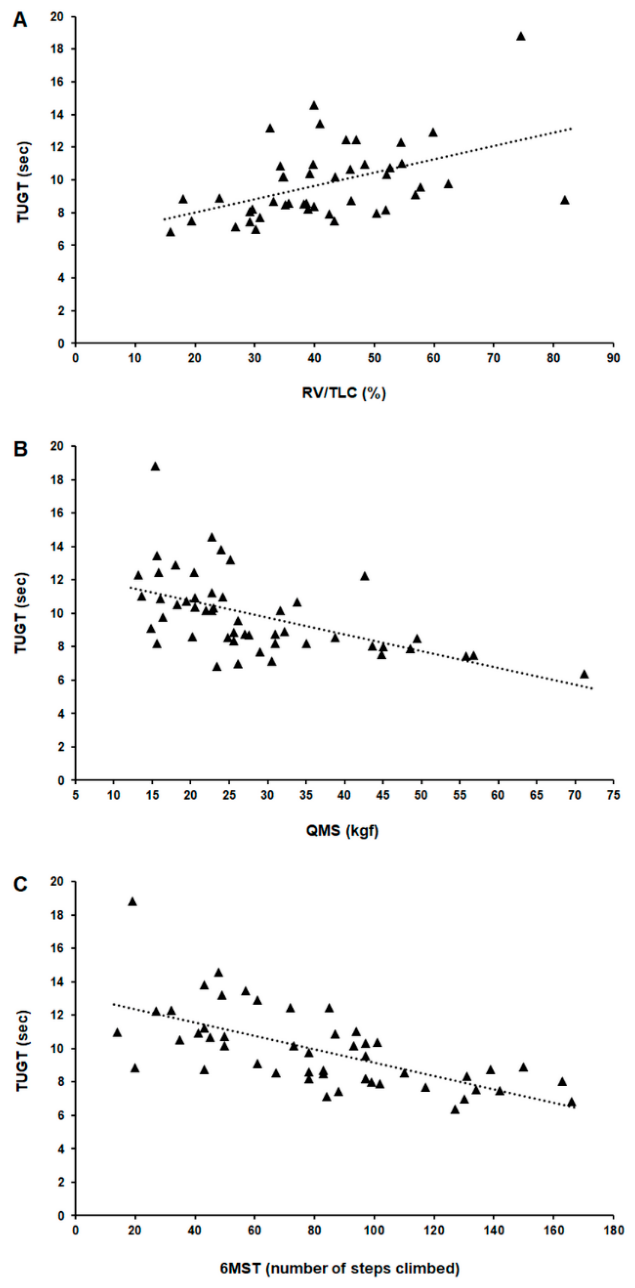

Table 1.
Anthropometry data, comorbidities, and balance control in the sample.
| Variables | Values | |
| Sex (male/female) | 34/16 | |
| Age (years) | 50.8 ± 15.4 | |
| BMI (kg/m2) | 25.2 ± 6.7 | |
| Comorbidities | Hypertension | 18 (36%) |
| Diabetes | 6 (12%) | |
| Asthma | 3 (6%) | |
| BBS | Low risk of falling | 17 (34%) |
| Moderate risk of falling | 22 (44%) | |
| High risk of falling | 11 (22%) | |
| TBS | Low risk of falling | 31 (62%) |
| Moderate risk of falling | 15 (30%) | |
| High risk of falling | 4 (8%) | |
| TUGT | Low risk of falling | 27 (54%) |
| Moderate risk of falling | 23 (46%) | |
| High risk of falling | 0 (0%) |
The values shown are the mean ± SD, median (interquartile range) or number (frequency). BMI, body mass index; BBS, Berg Balance Scale; TBS, Tinetti Balance Scale; TUGT, Timed "Up and Go" test.
Table 2.
Degree of agreement between the classifications used to assess balance control in the sample.
Table 2.
Degree of agreement between the classifications used to assess balance control in the sample.
| Classification | % Agreement | Weighted Kappa | 95% CI Kappa | p-value |
| BBS and TBS BBS and TUGT TBS and TUGT |
60 | 0.35 | 0.16–0.52 | 0.001 |
| 42 | 0.15 | -0.02–0.32 | 0.085 | |
| 62 | 0.25 | 0.05–0.43 | 0.014 | |
| BBS and TBS | 60 | 0.35 | 0.16–0.52 | 0.001 |
CI, confidence interval; BMI, body mass index; BBS, Berg Balance Scale; TBS, Tinetti Balance Scale; TUGT, Timed "Up and Go" test.
Table 3.
Pulmonary function, muscle function, and functional capacity during exertion in the sample.
Table 3.
Pulmonary function, muscle function, and functional capacity during exertion in the sample.
| Variables | Values | |
| Pulmonary function | FVC (% predicted) | 76 ± 24.1 |
| FEV1 (% predicted) | 69.3 ± 26.6 | |
| FEV1/FVC (%) | 74 ± 15.1 | |
| FEF25-75% (% predicted) | 62.2 ± 39.7 | |
| TLC (% predicted) | 86.5 ± 18.9 | |
| RV (% predicted) | 103.4 ± 44.7 | |
| RV/TLC (%) | 41.8 ± 13.7 | |
| Raw (cm H2O/L/s) | 4 (2–11) | |
| DLco (% predicted) | 94.5 ± 34.9 | |
| Muscle function | MIP (% predicted) | 47 (37–73) |
| MEP (% predicted) | 38 (26–56) | |
| HGS (kgf) | 26 (20–31) | |
| QMS (kgf) | 25 (20–33) | |
| FACIT-F scale (points) | 34.3 ± 11.5 | |
| Functional capacity during exertion | 6MST (number of steps climbed) | 83 (49–101) |
| 6MST (% predicted) | 50 (34–74) |
The values shown are the mean ± SD, median (interquartile range) or number (frequency). FVC, forced vital capacity; FEV1, forced expiratory volume in 1 second; FEF25-75%, forced expiratory flow during the middle half of the FVC manoeuvre; TLC, total lung capacity; RV, residual volume; Raw, airway resistance; DLco, diffusion capacity of the lungs for carbon monoxide; MIP, maximum inspiratory pressure; MEP, maximum expiratory pressure; HGS, quadriceps muscle strength; QMS, quadriceps muscle strength; FACIT-F, Functional Assessment of Chronic Illness Therapy-Fatigue; 6MST, six-minute step test.
Table 4.
Spearman's correlation coefficients between postural balance measures, clinical characteristics, pulmonary function, muscle function, and functional capacity during exertion in people with post-tuberculosis lung disease.
Table 4.
Spearman's correlation coefficients between postural balance measures, clinical characteristics, pulmonary function, muscle function, and functional capacity during exertion in people with post-tuberculosis lung disease.
| Balance control | ||||||
| BBS (points) | TBS (points) | TUGT (sec) | ||||
| rs | p-value | rs | p-value | rs | p-value | |
| Age (years) | -0.362 | 0.009 | -0.220 | 0.12 | 0.272 | 0.056 |
| BMI (kg/m2) | -0.118 | 0.41 | 0.064 | 0.66 | -0.034 | 0.81 |
| Time since the end of treatment (months) | -0.271 | 0.057 | -0.053 | 0.72 | 0.147 | 0.31 |
| Smoking load (pack-years) | -0.403 | 0.17 | -0.080 | 0.79 | 0.620 | 0.023 |
| FVC (% predicted) | 0.169 | 0.24 | 0.097 | 0.50 | -0.131 | 0.36 |
| FEV1 (% predicted) | 0.136 | 0.35 | 0.028 | 0.85 | -0.166 | 0.25 |
| FEV1/FVC (%) | -0.091 | 0.53 | -0.180 | 0.21 | -0.180 | 0.21 |
| FEF25-75% (% predicted) | 0.027 | 0.85 | -0.128 | 0.37 | -0.226 | 0.11 |
| TLC (% predicted) | 0.087 | 0.58 | 0.065 | 0.68 | 0.029 | 0.86 |
| RV (% predicted) | 0.145 | 0.36 | 0.197 | 0.21 | 0.299 | 0.052 |
| RV/TLC (%) | -0.128 | 0.40 | -0.068 | 0.66 | 0.500 | 0.0004 |
| Raw (cm H2O/L/s) | -0.118 | 0.40 | -0.074 | 0.63 | 0.284 | 0.058 |
| DLco (% predicted) | 0.059 | 0.70 | 0.258 | 0.091 | -0.267 | 0.079 |
| MIP (% predicted) | 0.198 | 0.17 | 0.293 | 0.039 | -0.285 | 0.044 |
| MEP (% predicted) | 0.145 | 0.31 | 0.275 | 0.053 | -0.262 | 0.066 |
| HGS (kgf) | 0.251 | 0.079 | 0.136 | 0.35 | -0.335 | 0.017 |
| QMS (kgf) | 0.373 | 0.007 | 0.291 | 0.040 | -0.641 | <0.0001 |
| FACIT-F (points) | 0.255 | 0.74 | 0.325 | 0.021 | -0.340 | 0.015 |
| 6MST (number of steps climbed) | 0.290 | 0.041 | 0.310 | 0.028 | -0.696 | <0.0001 |
| 6MST (% predicted) | 0.183 | 0.20 | 0.263 | 0.065 | -0.615 | <0.0001 |
Bold type indicates significant correlations. BBS, Berg Balance Scale; TBS, Tinetti Balance Scale; TUGT, Timed "Up and Go" test; BMI, body mass index; FVC, forced vital capacity; FEV1, forced expiratory volume in 1 second; FEF25-75%, forced expiratory flow during the middle half of the FVC manoeuvre; TLC, total lung capacity; RV, residual volume; Raw, airway resistance; DLco, diffusion capacity of the lungs for carbon monoxide; MIP, maximum inspiratory pressure; MEP, maximum expiratory pressure; HGS, quadriceps muscle strength; QMS, quadriceps muscle strength; FACIT-F, Functional Assessment of Chronic Illness Therapy-Fatigue; 6MST, six-minute step test.
Disclaimer/Publisher’s Note: The statements, opinions and data contained in all publications are solely those of the individual author(s) and contributor(s) and not of MDPI and/or the editor(s). MDPI and/or the editor(s) disclaim responsibility for any injury to people or property resulting from any ideas, methods, instructions or products referred to in the content. |
© 2026 by the authors. Licensee MDPI, Basel, Switzerland. This article is an open access article distributed under the terms and conditions of the Creative Commons Attribution (CC BY) license (http://creativecommons.org/licenses/by/4.0/).
Copyright: This open access article is published under a Creative Commons CC BY 4.0 license, which permit the free download, distribution, and reuse, provided that the author and preprint are cited in any reuse.