Preprint
Article

This version is not peer-reviewed.

Immune Activation of P. gingivalis—Infected Gingiva Fibroblasts Is Determined by Heterogeneity of Peptidylarginine Deiminase Gene and Biochemical Properties of Produce Variant(s)

Submitted:

11 August 2026

Posted:

12 August 2026

You are already at the latest version

Abstract

This study compared immune activation of gingival fibroblasts (GFs) by Porphyromonas gingivalis strains carrying different variants of peptidylarginine deiminase (PPAD). To this aim, GFs from donors with periodontitis (PD) (n=4) and healthy controls (CTRL) (n=2) were infected with P. gingivalis strains expressing either ppad with missense mutations (A390T + T421I) (n=2, moderate PD) or clusters of specific polymorphic variants (S203P + G231N, E232T, N235D) and (S203P + G231N, E232T, N235D + N291D + A515V + S528G) (n=2, advanced PD). Enzymatic activity of PPAD from these strains was determined using a colorimetric assay. Immune activation of infected GFs was analysed using qRT-PCR. P. gingivalis strains carrying clusters of specific polymorphic variants of ppad induced ~two-fold increase of PPAD activity and upregulated TNF-α, IL-6, and COX-2 expression by infected GFs compared to the reference ATCC 33277 strain. Contrary, P. gingivalis strains containing missense mutations showed decreased PPAD activity and elicited inflammatory response comparable to that presented by the ATCC 33277 strain. This study demonstrated that the inflammatory outcome of P. gingivalis infection results from the interplay between bacterial virulence driven by PPAD sequence diversity and enzymatic activity, and the intrinsic hyperreactivity of host GFs associated with PD.

Keywords: 
;  ;  ;  ;  ;  ;  

1. Introduction

PD is a prevalent chronic disease characterized by persistent connective tissue inflammation and progressive destruction of tooth-supporting structures and the alveolar bone [1,2]. It is a pathogen-driven dysbiotic disease, which entails multiple cycles of progression and remission mediated by the modulation of pro-inflammatory signaling networks [3,4,5]. As one of major causative agents of PD is considered an anaerobic, Gram-negative bacterium, P. gingivalis. The pathogenicity of this species is attributed to a number of virulence factors and effective strategies of immune evasion [6,7,8,9,10,11,12]. GFs represent an abundant cell population of oral connective tissue. Their primary function is production and remodeling of extracellular matrix (ECM) to maintain tissue integrity [13,14,15]. Besides the main structural function, numerous studies have underscored their important role in the progression of PD. It has been shown that P. gingivalis enters gingival epithelial cells, spreads through the upper layers of the gingival epithelial barrier, penetrates the basement membrane and invades deeper into the connective tissue, where fibroblasts are the most prevalent cell type constantly exposed to oral pathogens and their products [16,17]. In response to pathogens, their virulence factors and the local inflammatory microenvironment, fibroblasts produce a range of cytokines, chemokines and other inflammatory mediators [7,18,19,20,21]. Activation of GFs modulate immunity primarily through their interactions with immune cells infiltrating gingival tissue. GFs can promote neutrophil recruitment to gingival tissue through the synergistic induction of prostaglandin E2 (PGE2) production in response to simultaneous exposure to oral pathogens and inflammatory cytokines [12]. A key role of GFs in immunity was also demonstrated by Williams et al. (2021), who performed single cell transcriptomic analysis of the oral mucosa and identified the interplay between fibroblasts and neutrophils, also indicating it as a key aspect of pathogenesis of PD [22]. Uncontrolled activation of GFs in PD leads to chronic inflammation and tissue damage caused by the excessive recruitment of leukocytes, the secretion of proteolytic enzymes, e.g., matrix metalloproteinases (MMPs) and cathepsins, and the induction of osteoclastogenesis [23]. PPAD, a non-proteolytic enzyme which catalyzes the citrullination of arginine in proteins is considered as an unique enzyme in the Porphyromonas species. The citrullination of free arginine provides energy for anaerobic growth, while the ammonia generated by this system enables P. gingivalis survival during acid-cleansing cycles of the oral cavity [24,25]. Citrullination of an arginine residue with the release of ammonium ion decreases the charge of modified proteins and can therefore affect their stability, sensitivity to proteolysis, and biological activity affecting the virulence of P. gingivalis, host-pathogen interaction and immune defense in the periodontum [8,11,26,27]. For example, it was reported that PPAD citrullinates the C-terminal arginine of epidermal growth factor (EGF), which subsequently impair its biological activity. Decreased activity of EGF in gingival pockets may at least partially contribute to the tissue damage and delayed healing of the periodontium observed during P. gingivalis infection [26]. In another study, Bielecka et al. analyzed the impact of bacterial citrullination on the pro-inflammatory properties of anaphylatoxin C5a and demonstrated reduced chemotactic properties of neutrophils as a result of the reaction catalyzed by PPAD [27]. Moreover, recent studies showed variability of PPAD activity driven by heterogeneity of ppad gene sequence among clinical strains of P. gingivalis from PD associated with pathogenicity of P. gingivalis strains and the severity of PD [20,28,29].
Considering previously reported heterogeneity of ppad gene sequence and the importance of PPAD activity in P. gingivalis virulence, this study investigated immune activation of GFs by P. gingivalis strains carrying clusters of specific polymorphic variants and compared it to the effects exerted by P. gingivalis strains with missense mutations in ppad gene.

2. Materials and Methods

2.1. Clinical Examination of Study Participants and Sample Collection

The study was carried out in accordance with the Declaration of Helsinki and was approved by the Bioethics Committee of the Jagiellonian University, Medical College in Krakow, Poland (KBET/310/B/2012 and 1072.6120.156.2019). Gingival biopsies were collected from 4 PD and 2 CTRL donors, and gingival crevicular fluid (GCF) samples from 4 PD donors examined and qualified to the study at the Department of Periodontology, Preventive Dentistry and Oral Pathology, Medical College, Jagiellonian University, Krakow, Poland. All donors received information on the inclusion and exclusion criteria, as described elsewhere [20,30], read and signed a written informed consent form prior to inclusion in the study. PD diagnosis was confirmed by oral radiographs and periodontal examination of approximal plaque index (API), bleeding on probing (BOP), probing pocket depth (PPD) and clinical attachment loss (CAL). Healthy donors with no signs of oral inflammation or systemic diseases were qualified for orthodontic treatment. Gingival biopsies (1 × 1 mm per each donor) were collected in cold Dulbecco’s Modified Eagle Medium (DMEM, PAA, GmbH, Poland) with antibiotics and nystatin (Sigma-Aldrich, Poland) for cells isolation [8,31]. GCF was collected from five inflamed periodontal pockets per each PD donor and from five random gingival crevices from each control. GCF samples collected per each study donor were pooled and used to grow of one representative P. gingivalis strain, as described elsewhere [20,28].

2.2. Cells Isolation and Culture

The isolation procedure and the homogeneity of GFs cultures have been standardized and described previously [8,14,15]. Cells were cultured in DMEM (PAA, GmbH, Poland) with high glucose, supplemented with 10% heat-inactivated fetal bovine serum (FBS, PAA, GmbH, Poland), 50 U/mL penicillin, 50 μg/mL streptomycin and 50 μg/mL gentamycin (Thermo Fisher Scientific, Waltham, MA, USA) at 37 °C in a humidified atmosphere with 5% CO2 until 95% confluency. Cells were washed with sterile phosphate-buffered saline (PBS) and detached from the culture vessel using 3 mL of 0.05% trypsin/EDTA for 5 min at 37 °C, followed by trypsin neutralization with 3 mL DMEM and 10% FBS and centrifugation at 300 rcf for 10 min at 4 °C. Cell pellet was then resuspended in 3 mL of culture medium, diluted in trypan blue (1:10), and cell counts were determined using a TC20 automated cell counter (Bio-Rad, San Francisco, CA, USA). For infection experiments, cells were seeded in 24-well plates at a density of 250,000 cells per well and cultured for ~20 h. Next day, prior the infection, culture medium was removed, cells were washed with sterile PBS, and replaced with antibiotic-free DMEM containing 2% FBS. The experiments were carried out between 5 and 8 passage.

2.3. Bacterial Growth from GCF

GCF samples from PD donors were diluted with sterile PBS (1:2) and cultured for 10-14 days on agar plates with brain-heart infusion (BHI) broth (Becton Dickinson), supplemented with 5% defibrinated sheep blood, 5 g/L yeast extract, 0.5 mg/mL L-cysteine, 10 μg/mL hemin, and 0.5 μg/mL of menadione in anaerobic conditions (85% N2, 10% CO2, 5% H2). Black-pigmented colonies were then subcultured into fresh blood agar plates, with passages repeated every 7-10 days until homogeneous cultures were obtained [8,20,28], and then preserved in BHI with 20% glycerol and stored at -80 °C for analyses.

2.4. Isolation of Bacterial Genomic DNA

Genomic DNA was extracted from black pigment producing bacterial colonies, presumptively P. gingivalis strains, using the GeneJET Genomic DNA Purification Kit (Thermo Fisher Scientific, Waltham, MA, USA) according to the manufacturer’s instructions. Briefly, after anaerobic culture for 10-14 days at 37 °C, bacteria were collected and suspended in PBS to obtain 2 × 10⁹ cells, as estimated by optical density at 600 nm (OD600). Samples were centrifuged at 5,000 rcf, 4 °C for 10 min, supernatant was discarded, and pellet was resuspended in 180 μL of digestion solution with 20 μL proteinase K. The suspension was mixed until homogeneous and incubated at 56 °C for 30 min in a shaking thermoblock to ensure complete lysis. Next, 20 μL of RNase A solution was added, mixed, and incubated for 10 min at RT. 200 μL of lysis solution was added to each sample, mixed for 15 s, then 400 μL of 50% ethanol was added. The lysates were transferred to a purification column (GeneJET kit) and centrifuged at 6,000 rcf for 1 min. The flow-through was discarded, and the column was transferred to a new collection tube. A total of 500 μL of wash buffer (with ethanol) was added and centrifuged at 8,000 rcf for 1 min. After removing the filtrate, a second wash step with 500 μL of wash buffer was performed, followed by centrifugation at ≥12,000 rcf for 3 min. Next, 200 μL of elution buffer was added directly to the membrane, incubated for 2 min at RT, and centrifuged at 8,000 rcf for 1 min. Purified DNA was stored at -20 °C until analysis.

2.5. Confirmation of P. gingivalis Species

Genomic DNA from black pigment producing bacterial strains, indicating P. gingivalis species was used for PCR with specific primers targeting the 16S rRNA and ppad genes (primer sequences listed in Supplementary Materials, Table S1). PCR was performed using 4 μL of genomic DNA, 1 μM of each primer and DreamTaq Green PCR Master Mix (Thermo Fisher Scientific, Waltham, MA, USA) in a total of 12 μL reaction volume. Reaction profiles included: i) denaturation at 95 °C for 3 min, ii) 40 cycles of 30 s at 95 °C, 30 s at 53 °C, 60 s/2 min (16S rRNA/ppad) at 72 °C, and iii) final extension at 72 °C for 5 min. P. gingivalis species was identified by electrophoretic separation of PCR products in 1.2% agarose gel with ethidium bromide in TAE buffer (45 min, 90 V). Amplicons corresponding to 16S rRNA and ppad genes were visualized under UV light and archivized with gel documentation system (SYNGENE, Cambridge, UK) equipped with photo camera (Canon, Tokyo, Japan).

2.6. The ppad Gene Sequences Analysis

The ppad gene sequences from P. gingivalis strains were generated based on the consensus sequence from 3 sequencing reads and analyzed vs. reference wild-type P. gingivalis ATCC 33277 strain using NCBI, BLAST, UNIPROT PROTEIN DATABASE and Expasy, as previously described [20,28,29].

2.7. PPAD Activity Assay

The enzymatic activity of PPAD was determined as described elsewhere [28,32]. Briefly, P. gingivalis strains were cultured in supplemented BHI broth at 37 °C for 20-24 h in anaerobic conditions. Overnight cultures were then diluted in fresh medium to OD600 = 0.1 and incubated under the same conditions. Subsequently, bacterial cells were harvested, washed and resuspended in PBS to final OD600 = 1.0. For enzymatic activity assay, 10 μL of each bacterial strain suspension was mixed with 40 μL of reaction buffer containing 100 mM Tris-HCl (pH 7.5), 5 mM dithiothreitol (DTT), and 10 mM N-acetyl-L-arginine as the substrate. The reaction mixtures were incubated at 37 °C for 1 h and subsequently terminated by the addition of 10 μL of 5 M perchloric acid. The amount of citrulline generated during the reaction was quantified using a colorimetric assay and compared with a standard curve for L-citrulline. Immediately before use, the citrulline detection reagent was prepared by mixing one part of solution A (0.5% 2,3-butanedione monoxime and 0.01% thiosemicarbazide) with two parts of solution B (0.25 mg/mL FeCl3, 24.5% sulfuric acid, and 17% orthophosphoric acid) and added to each experimental and standard curve sample, followed by incubation at 110 °C for 20 min and absorbance measurement at 535 nm. The amount of produced citrulline was calculated using the standard curve and expressed relative to that of the reference P. gingivalis ATCC 33277 strain.

2.8. Bacterial Culture and Cell Infection

After anaerobic culture for 10-14 days at 37 °C, bacterial colonies were inoculated into 10 mL of liquid BHI supplemented with 0.5 mg/mL L-cysteine, 10 μg/mL hemin, and 0.5 μg/mL of menadione, and cultured o/n in anaerobic conditions (85% N2, 10% CO2, and 5% H2). Bacteria were then washed in PBS, centrifuged at 4,500 rcf for 10 min at 4 °C and resuspended in fresh BHI broth at OD600 = 0.1 and cultured for ~20 h. Bacterial suspension at OD600 = 1.0, corresponding to 109 colony-forming units (CFU)/ml in PBS was prepared. The experiments were carried out at a multiplicity of infection (MOI) of 1:100 for 24 h in DMEM with 2% FBS w/o antibiotics. Following infection, cells were washed with PBS, lysed in TRIzol reagent (Invitrogen, Waltham, MA, USA), and stored at -80 °C for analyses.

2.9. RNA Isolation

RNA isolation was performed using TRIzol reagent (Invitrogen, Waltham, MA, USA) according to the manufacturer’s instructions. To 0.5 mL of cell lysate, 100 μL of chloroform was added, vortexed for 10 s, incubated for 10 min at RT, and centrifuged at 12,000 rcf for 15 min at 4 °C. The aqueous phase was mixed with 250 μL of isopropanol, incubated for 10 min at RT and frozen at -20 °C for ~2 h. After centrifugation at 12,000 rcf for 15 min at 4 °C, supernatant was removed, and the RNA pellet was washed twice with 200 μL of cold 70% ethanol, followed by centrifugation at 12,000 rcf for 10 min at 4 °C. Ethanol was discarded, and the RNA pellet was air-dried and resuspended in 20 μL of nuclease-free water. Next, samples were treated with 1 μL of Turbo DNAse (Invitrogen, Waltham, MA, USA), 5 μL of 10× Turbo DNase buffer, and 25 μL of nuclease-free water. Samples were incubated at 37 °C for 30 min, and subsequently, 0.5 mL of TRIzol (Invitrogen, Waltham, MA, USA) was added to each sample, and the RNA isolation procedure was repeated, as described above. The RNA concentration and purity were assessed using a NanoDrop spectrophotometer (Thermo Fisher Scientific, Waltham, MA, USA), and stored at -80 °C for analyses.

2.10. Reverse Transcription Reaction

Reverse transcription was performed using the High-Capacity cDNA Reverse Transcription Kit (Invitrogen, Waltham, MA, USA). 400 ng of total RNA was resuspended in nuclease-free water to a final volume of 10 μL, followed by the addition of 2 μL of 10× random hexamers. Samples were incubated at 70 °C for 10 min, cooled to 25 °C, and placed on ice. Next, to each sample was added 1 μL of reverse transcriptase, 0.8 μL of 10 mM dNTP mix, 2 μL of 10× reverse transcriptase buffer and 4.2 μL of nuclease-free water. The reverse transcription was carried out at 37 °C for 2 h, followed by enzyme inactivation at 85 °C for 5 min. The resulting cDNA was stored at -20 °C until use.

2.11. Quantitative Real-Time PCR

The qRT-PCR was performed using 2 μL of 5× diluted cDNA template, 6 μL of reaction mixture containing 2× SYBR Green Master Mix (Merck, Darmstadt, Hesja, Germany), 1 μL of standardized primers sets (1 μM each) (Genomed S.A., Warsaw, Poland) (Supplementary Materials, Table S1) and 1 μL of nuclease-free water. The reaction profile used was as follows: i) 3 min at 95 °C, ii) 40 cycles of 30 s at 95 °C, 30 s at 57 °C, 45 s at 72 °C, iii) 30 s at 72 °C. For relative quantification of analyzed genes expression the ΔΔCt method was used. As an internal control, β-actin was used. Amplification was performed with a LightCycler 480 II system (Roche, Basel, Switzerland).

2.12. Statistical Analysis

The normality of the distribution of quantitative data was assessed using the Shapiro-Wilk test. Quantitative data are expressed as means ± SEM. For multiple comparisons of normally distributed variables, one-way analysis of variance (ANOVA) was applied, followed by Tukey’s post-hoc test, if significant differences were observed. Cases with missing data were excluded from the relevant analyses. The differences among compared results were considered statistically significant at p < 0.05. Data were analyzed using GraphPad Prism v. 10 (GraphPad Software, San Diego, CA, USA).

3. Results

3.1. Demographic and Clinical Data of Cells and Bacterial Strains Donors

The study was performed using GFs from 4 PD (2 females and 2 males; mean age 53 years, range 46 - 62 years) and 2 CTRL donors with healthy periodontium (2 females; range 23 - 27 years). Oral radiographs and periodontal examination of clinical parameters of periodontium (API, BOP, PPD, and CAL) revealed moderate PD in two donors and advanced PD in the other two donors (Table 1).
P. gingivalis strains were cultured from 4 donors with PD (2 females, 2 males; mean age 57 years, range 47 - 65 years). The X-rays and the API, BOP, PPD and CAL examination showed moderate PD in two donors and advanced PD in two other (Table 2).

3.2. P. gingivalis Strains

Clinical strains of P. gingivalis (Pg1-Pg4) were tested for expression of the 16S rRNA and ppad genes. Amplicons of both markers were detected for each strain, as depicted in Figure 1.

3.3. Variants of ppad Gene

Previous studies revealed significant diversity of ppad gene sequences in P. gingivalis strains from PD donors and identified clusters of specific polymorphic variants of ppad as critical determinants of enzymatic function, host immune modulation and periodontal tissue destruction in PD [20,28,29]. To further verify this thesis, we used for infections P. gingivalis strains from two donors with advanced PD carrying clusters of specific polymorphic variants of ppad (S203P + G231N, E232T, N235D) and (S203P + G231N, E232T, N235D + N291D + A515V + S528G), respectively and P. gingivalis strains carrying ppad with missense mutations (A390T + T421I), cultured from two patients with moderate PD (Table 2).

3.4. PPAD Activity

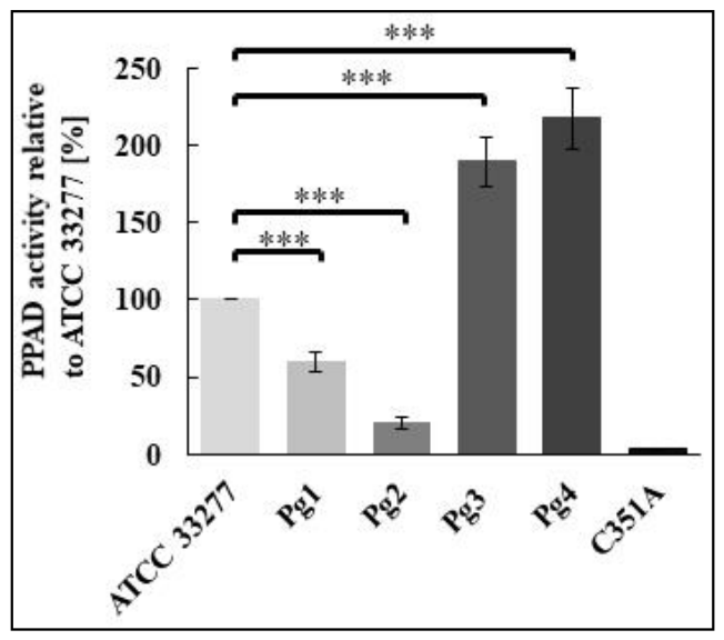
PPAD enzymatic activity was assessed in four P. gingivalis strains isolated from PD donors and compared with the reference P. gingivalis ATCC 33277 strain (control) (Figure 2). P. gingivalis strains (Pg3, Pg4) harboring clusters of specific polymorphic variants of ppad (S203P + G231N, E232T, N235D) and (S203P + G231N, E232T, N235D + N291D + A515V + S528G), respectively showed an approximately two-fold increase in PPAD enzymatic activity relative to the reference ATCC 33277 strain (p < 0.001). In opposite, bacterial strains (Pg1, Pg2) carrying missense mutations of ppad (A390T + T421I) revealed significantly decreased PPAD activity in comparison to the reference P. gingivalis strain (p < 0.001). Consistently with previous data [28,29], these results indicated that PPAD activity varied dependently on the ppad sequence variation underpinning an association of its elevated values with co-occurrence of specific polymorphic variants.

3.5. GFs Activation by Infection with P. gingivalis Strains Expressing ppad with Clusters of Specific Polymorphic Variants and Missense Mutations

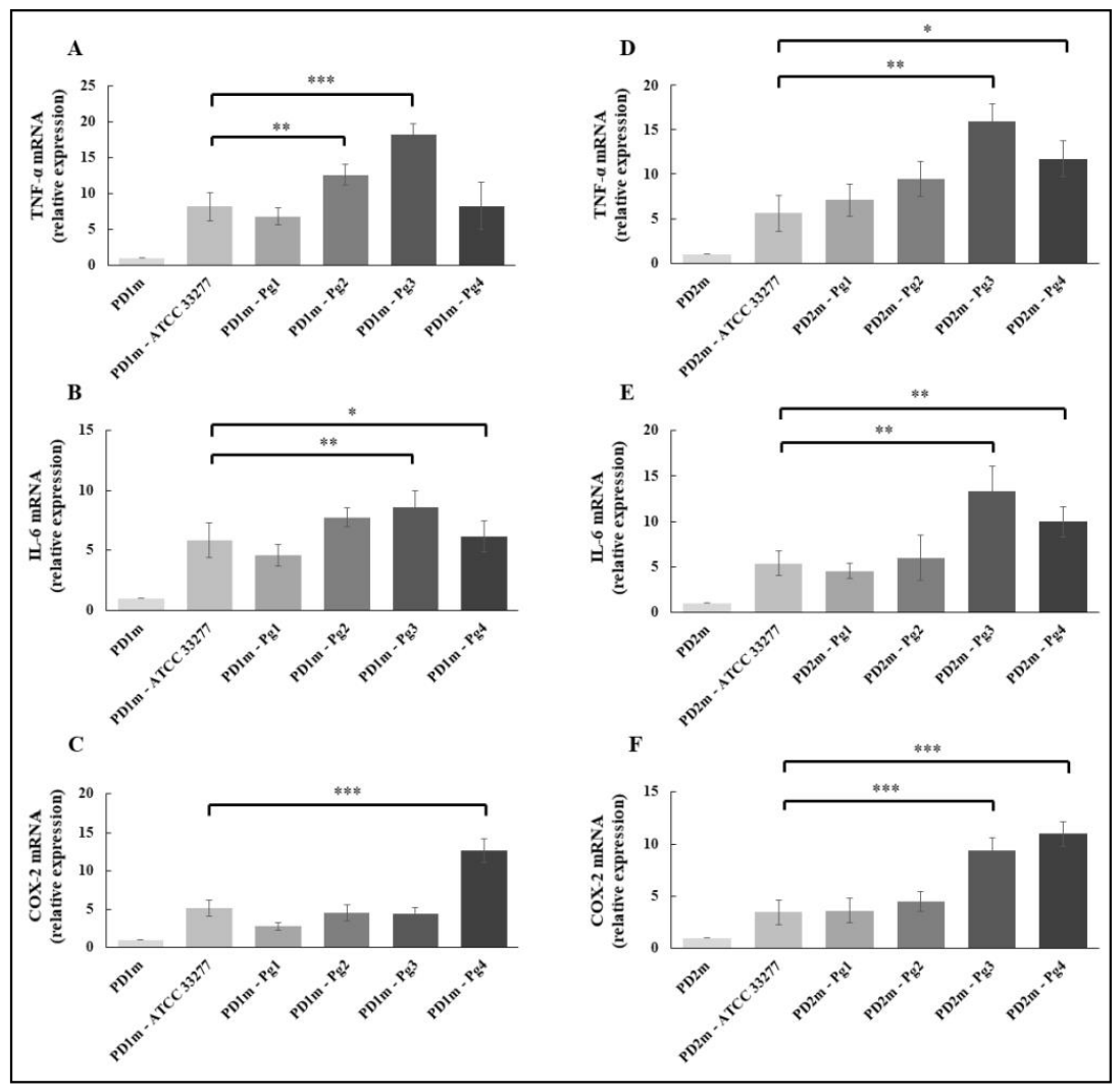
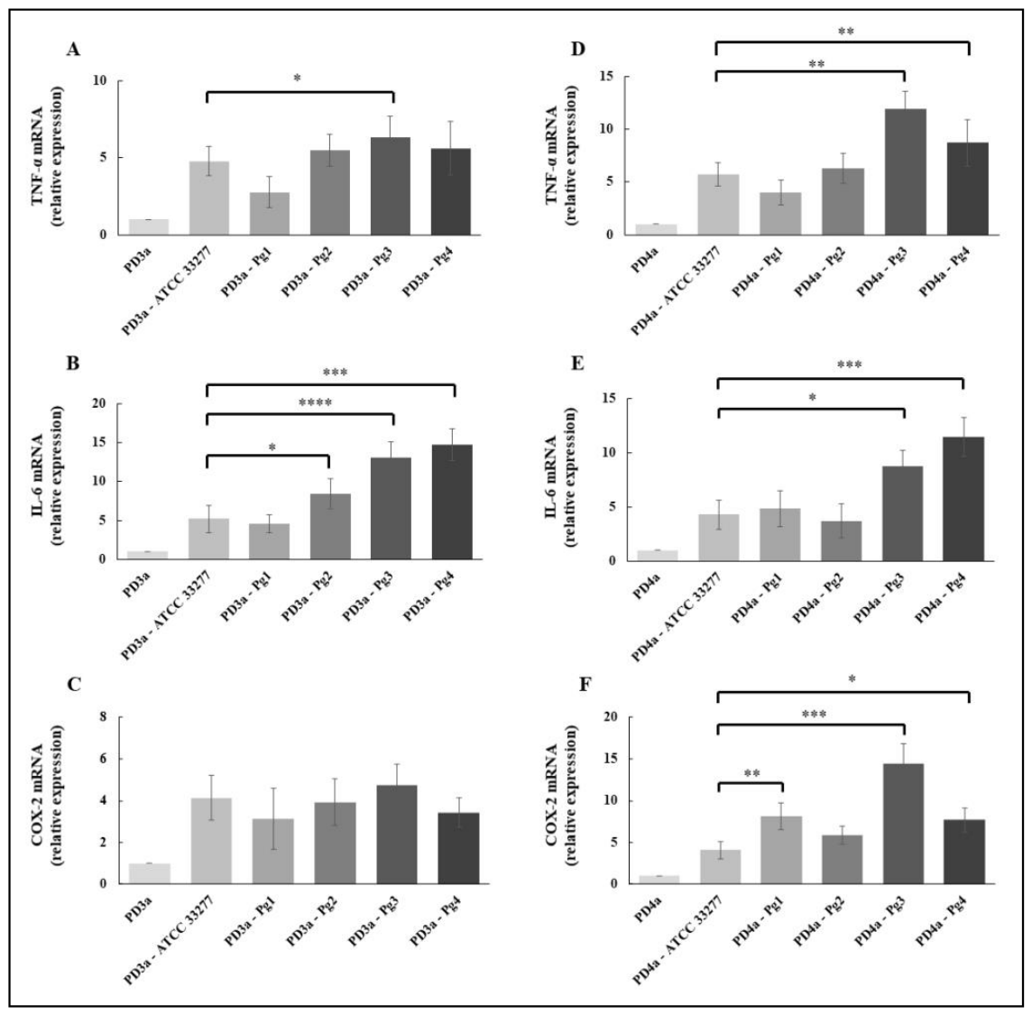
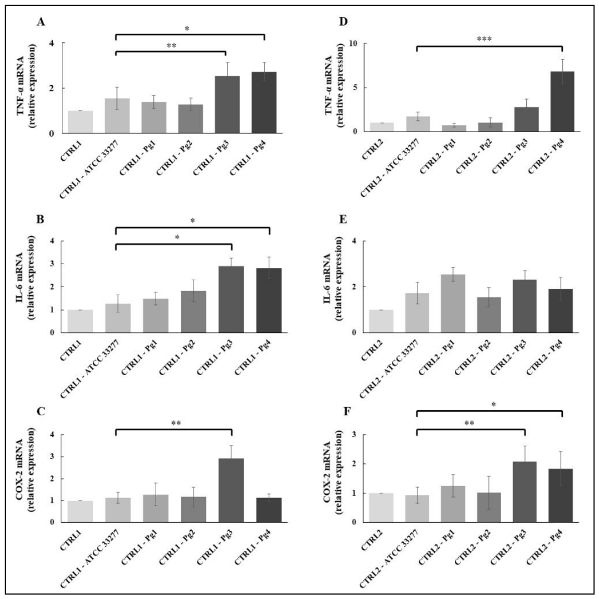
Beyond the canonical structural function, GFs represent the first line of defense against oral pathogens and are considered an important component of the innate immune system. However, their chronic activation due to persistent interaction with oral bacteria and/or their components, which involves the secretion of large quantities of cytokines, chemokines, matrix-degrading enzymes, and prostaglandins, significantly contributes to pathogenesis of PD [7,8,19]. Hence, the impact of P. gingivalis strains carrying polymorphic variants and missense mutations in ppad gene on host response was analyzed in GFs. As shown in Figure 3, upregulation of tumor necrosis factor α (TNF-α) and interleukin-6 (IL-6) expression was observed after GFs infection with Pg3 (p < 0.001, p < 0.01) and to the lesser extent with Pg4 (p < 0.05, p < 0.01) producing clusters of co-occurring polymorphic variants. In case of cyclooxygenase-2 (COX-2), the effect was observed in cells infected with Pg4 strain (p < 0.001) or Pg3 and Pg4 (p < 0.001), dependently on the individual donor. Expression of all tested genes in cells infected with strains expressing ppad with missense mutations (Pg1 and Pg2) was comparable with the effect observed after infection with reference ATCC 33277 strain. Only in the case of TNF-α, significant differences also occurred after GFs infection with Pg2 (p < 0.01) (Figure 3). Similar trends of expression were observed due to infections of GFs from advanced PD and healthy periodontium (CTRL). Cells infection with Pg3 and Pg4 resulted in significant upregulation of TNF-α (advanced PD, * p < 0.05, ** p < 0.01; CTRL, * p < 0.05, ** p < 0.01, *** p < 0.001), IL-6 (advanced PD, * p < 0.05, *** p < 0.001, **** p < 0.0001; CTRL, * p < 0.05) and COX-2 (advanced PD, * p < 0.05, *** p < 0.001; CTRL, * p < 0.05, ** p < 0.01) while in case of Pg1 and Pg2 infection, insignificant effects were observed (Figure 4 and Figure 5). In some cases, donor-dependent variations in GFs response were noted (Figure 4C,F). These data shown that immune activation of GFs is determined by specific PPAD variants produced by P. gingivalis, characterized by elevated enzymatic activity.

4. Discussion

PD is currently classified as one of the most common chronic inflammatory destructive diseases of bacterial etiology worldwide [33]. It is characterized by the progressive degradation of the tissues supporting the teeth, including the periodontal ligament and alveolar bone, a direct result of the multifaceted conflict between dysbiotic bacterial biofilm and dysregulated host immune response [6,34,35]. Among oral pathogens that drive pathogenic changes in the gingival tissue, a Gram-negative anaerobic bacterium P. gingivalis plays a central role [36]. Similar to immune cells, GFs actively interact with and respond to oral pathogens penetrating the epithelial barrier, and produce a range of cytokines, chemokines, MMPs and other inflammatory mediators [16,17,37]. Moreover, previous studies in vitro demonstrated that P. gingvalis efficiently invades and activates GFs [8,10,38,39,40]. It has been demonstrated that invasion of GFs and activation of the PGE2 signaling pathway by P. gingivalis is dependent on PPAD activity. These findings were confirmed by the observation of upregulated expression of crucial pro-inflammatory mediators, COX-2 and microsomal prostaglandin E synthase 1 (mPGES-1) and elevated levels of PGE2 after cells treatment with wild-type P. gingivalis strain (ATCC 33277), but not by P. gingivalis expressing PPAD mutants (ΔPPAD or inactive C351A). Because the addition of active PPAD alone to sham-infected control fibroblast cultures did not induce the cells to produce PGE2, we showed that citrullinated P. gingivalis cell surface protein(s) act as ligand(s) for activating the prostaglandin synthesis pathway, thus contributing to alveolar bone loss at infected periodontal sites [8]. These observations were further confirmed by the study of Wielento et al., which shown PPAD activity and expression of major fimbriae as indispensable factors for interaction with toll-like receptor 2 (TLR2). There has been also showed that fimbriae isolated from the PPAD-deficient P. gingivalis mutant strain failed to activate TLR2, indicating that citrullination of fimbriae components or other proteins involved in fimbriae assembly is required for host cells activation through TLR2 [11]. Recently, we identified and described a hyperactive PPAD variant containing 7-nucleotides substitution, inducing three amino acids substitution (G231N, E232T, N235D) in protein sequence, which exhibited approximately double the enzymatic activity of the reference strain ATCC 33277 and was detected in about 25% of P. gingivalis strains from advanced PD [28]. The analyses of ppad sequence revealed the presence of polymorphic variants, missense mutations and synonymous variants in the ppad gene from P. gingivalis strains at various stages of PD, indicating its significant heterogeneity [20]. Furthermore, we detected clusters of specific polymorphic variants of ppad as critical determinants of its enzymatic function, host immune modulation and periodontal tissue destruction in PD [29].
Considering previously reported heterogeneity of ppad gene sequence and the importance of PPAD activity in P. gingivalis virulence, this study investigated immune activation of GFs by P. gingivalis strains carrying clusters of specific polymorphic variants and compared it to the effects exerted by P. gingivalis strains with missense mutations in ppad gene.
Infection of PHGFs from PD donors with P. gingivalis strains (Pg3, Pg4) producing clusters of co-occurring polymorphic variants of ppad gene resulted in meaningful upregulation of crucial pro-inflammatory mediators - TNF-α, IL-6 and COX-2 expression, which was in opposite to the effects observed after infection with P. gingivalis strains carrying ppad with missense mutations (Pg1, Pg2). In the latter case, relative expression of all analyzed genes was slightly upregulated, but comparable with their expression after infection with reference P. gingivalis strain (ATCC 33277). These findings are consistent with previous studies, which demonstrated that expression of TNF-α, IL-6 and major components of the PGE2-signaling pathway, i.e., cyclooxygenase-1 (COX-1) and COX-2 in GFs from PD was activated by a laboratory mutant of the G231N, E232T, N235D variant introduced to the ATCC 33277 P. gingivalis strain (ATCC T2) and clinical P. gingivalis strains carrying one of most frequently occurring clusters of polymorphic variants of ppad (S203P + G231N, E232T, N235D + N291D + A515V + S528G), i.e., corresponding to those expressed by P. gingivalis strain tested in the current study (Pg4) [20,29].
Furthermore, P. gingivalis strains expressing clusters of co-occurring polymorphic variants revealed ~2-fold increased activity of PPAD compared to the reference ATCC 33277 strain. The G231N, E232T, N235D and N291D polymorphic variants present in clusters of PPAD produced by P. gingivalis strains (Pg3 and Pg4) were located in the vicinity of the catalytic triad of PPAD, i.e., the G231N, E232T, N235D close to the His236, and N291D close to the Asn297. Additionally, crystallographic models of the PPAD protein structure indicated the differences between PPAD expressing the G231N, E232T, N235D variant and PPAD w/o this variant (ATCC 33277). The E232T mutation involves the change of glutamic acid to neutral threonine and is located in the area essential to the deimination reaction. It forms the channel through which the water molecule passes and ammonia during citrullination. The substitution of glycine to asparagine (G231N) may also influence the conformation of this channel. Asparagine 235 is located in the channel through which the hydroxyl ion passes to restore the active center of the enzyme to a state ready to accept the substrate. Thus, the N235D induces the change of asparagine to negatively charged aspartic acid. The N291D substitution, which induces the change of asparagine to negatively charged aspartic acid can alter the electrostatic environment, potentially affecting substrate binding or catalytic efficiency. Moreover, the introduction of a negative charge may disrupt existing hydrogen bonds or introduce new electrostatic interactions that could either stabilize or destabilize the active site [41,42]. Therefore, the changes of amino acids of individual polymorphic variants expressed within clusters discussed herein can clearly explain the change of catalytic properties of the enzyme [28,43,44,45]. The missense mutations (A390T and T421I) of ppad were identified in 8.62% of P. gingivalis isolates obtained from patients with PD, whereas neither substitution was detected among P. gingivalis isolates from periodontally healthy individuals [20]. Although their relatively low prevalence suggests that they are not common substitutions within the ppad gene, their exclusive occurrence in disease-associated isolates indicates that they may contribute to the genetic diversity of P. gingivalis strains associated with PD. Nevertheless, unlike the clusters of polymorphic variants expressed by Pg3 and Pg4 strains, neither the A390T nor T421I formed clusters and have not been directly associated with increased PPAD activity and/or enhanced virulence of P. gingivalis [20,28,29,42]. These findings suggest that individual amino acid substitutions may have limited biological consequences when present alone and that PPAD activity is more likely influenced by the cumulative effects of multiple interacting mutations. This thesis also aligns with the observation of insignificant immune activation of PHGFs by P. gingivalis strains expressing missense mutations in ppad gene, which exhibited PPAD activity comparable (Pg1) or much below (Pg2) the activity of the reference ATCC 33277 strain.
Infection experiments performed in this study also demonstrated enhanced activation of gingival fibroblasts from PD donors comparing to immune response of fibroblasts derived from healthy periodontium. It was particularly observed for fibroblasts from PD infected with P. gingivalis strains expressing clusters of co-occurring polymorphic variants. This observation is consistent with previous studies which showed upregulated expression of inflammatory mediators (IL-6, IL-1β, IL-8, TNF-α) by fibroblasts from PD patients compared to control group upon infection with virulent P. gingivalis strain or stimulation by P. gingivalis LPS [10,39,46,47]. These data may suggest that cells isolated from the inflammatory microenvironment exhibit persistent and epigenetically marked inflammatory phenotype that become fully apparent upon exposure to infectious stimulus.

5. Conclusions

The findings of the present study emphasize the importance of two complementary factors that determine the magnitude of host response to P. gingivalis infection. First, the genetic heterogeneity of the ppad gene, particularly the presence of clusters of co-occurring polymorphic variants, directly influences PPAD activity, which in turn modulates the pro-inflammatory potential of P. gingivalis strains. Bacterial strains expressing hyper-active PPAD variants induced a markedly stronger immune response in GFs than those carrying missense mutations, highlighting the cumulative effect of multiple polymorphic variants rather than individual substitutions as a key determinant of bacterial virulence. Moreover, our results demonstrated that fibroblasts derived from PD patients exhibit an enhanced inflammatory responsiveness compared with cells isolated from healthy gingival tissues, supporting the concept that PD-derived GFs retain an inflammation-associated phenotype that amplifies their response to pathogenic stimuli. Together, these findings indicate that the inflammatory outcome of P. gingivalis infection results from the interplay between bacterial virulence driven by PPAD sequence diversity and enzymatic activity, as well as the intrinsic hyperreactivity of host GFs associated with PD.

Supplementary Materials

The following supporting information can be downloaded at the website of this paper posted on Preprints.org, Supplementary Materials, Table S1: Primer sequences used in the study (synthesized by Genomed S.A., Warsaw, Poland).

Author Contributions

Conceptualization, K.G.; Methodology, A.D.-K., K.S., K.F., M.A., and K.G.; Software, A.D.-K., B.M.; Validation, A.D.-K., K.S., and K.G.; Formal Analysis, A.D.-K., K.S., M.R., M.C.-G., T.J.W., and K.G.; Investigation, A.D.-K., K.S., K.F., M.A., B.M., and K.G.; Resources, T.J.W., and K.G.; Data Curation, M.R., M.C.-G., and K.G.; Writing – Original Draft Preparation, K.G.; Writing – Review & Editing, A.D.-K., K.S., K.F., M.R., M.A., M.C.-G., B.M., T.J.W., and K.G.; Visualization, K.S., K.F., and K.G.; Supervision, K.G.; Project Administration, K.G.; Funding Acquisition, K.G.All authors approved final version to be published and agree to be accountable for all aspects of the work by ensuring that questions relating to the accuracy or integrity of any part of the work are appropriately investigated and resolved.

Funding

This research was funded by grants to K.G. from the National Science Centre, Krakow, Poland (UMO-2012/07/B/NZ6/03524, UMO-2018/29/B/NZ2/01930) and from the Medical University of Silesia in Katowice, Poland (BNW-1-130/N/4/O).

Institutional Review Board Statement

The study was carried out in accordance with the Declaration of Helsinki and was approved by the Bioethics Committee of the Jagiellonian University, Medical College in Krakow, Poland (KBET/310/B/2012 and 1072.6120.156.2019).

Data Availability Statement

The original contributions presented in the study are included in the article, further inquiries can be directed to the corresponding author.

Conflicts of Interest

The authors declare no conflicts of interest. The funders had no role in the design of the study; in the collection, analyses, or interpretation of data; in the writing of the manuscript; or in the decision to publish the results.

References

  1. Eke PI, Dye BA, Wei L, Thornton-Evans GO, Genco RJ, Beck James, Douglass Gordon, Page Roy, Slade Gray, Taylor George W, Borgnakke Wenche. CDC Periodontal Disease Surveillance workgroup and representatives of the American Academy of Periodontology. Prevalence of periodontitis in adults in the United States: 2009 and 2010. J Dent Res. 2012;91(10):914–920. [CrossRef]
  2. Eke PI, Thornton-Evans GO, Wei L, Borgnakke WS, Dye BA, Genco RJ. Periodontitis in US Adults: National Health and Nutrition Examination Survey 2009-2014. J Am Dent Assoc. 2018;149(7):576–588.e6. [CrossRef]
  3. Hajishengallis G, Lamont RJ. Beyond the red complex and into more complexity: the polymicrobial synergy and dysbiosis (PSD) model of periodontal disease etiology. Mol Oral Microbiol. 2012;27(6):409–419. [CrossRef]
  4. Wright CJ, Burns LH, Jack AA, Back CR, Dutton LC, Nobbs AH, Lamont RJ, Jenkinson HF. Microbial interactions in building of communities. Mol Oral Microbiol. 2013;28(2):83–101. [CrossRef]
  5. Demmer RT, Papapanou PN. Epidemiologic patterns of chronic and aggressive periodontitis. Periodontol 2000. 2010;53:28–44. [CrossRef]
  6. Hajishengallis G. Immunomicrobial pathogenesis of periodontitis: keystones, pathobionts, and host response. Trends Immunol. 2014;35(1):3–11. [CrossRef]
  7. Ferenc K, Bożek A, Gawron K. Immunological pathways triggered by Porphyromonas gingivalis in periodontitis: New insights into molecular mechanisms. Mol Immunol. 2026;189: 119–132. [CrossRef]
  8. Gawron K, Bereta G, Nowakowska Z, Lazarz-Bartyzel K, Lazarz M, Szmigielski B, Mizgalska D, Buda A, Koziel J, Oruba Z, Chomyszyn-Gajewska M, Potempa J. Peptidylarginine deiminase from Porphyromonas gingivalis contributes to infection of gingival fibroblasts and induction of prostaglandin E2-signaling pathway. Mol Oral Microbiol. 2014;29(6):321–32. [CrossRef]
  9. Benedyk M, Mydel PM, Delaleu N, Płaza K, Gawron K, Milewska A, Maresz K, Koziel J, Pyrc K, Potempa J. Gingipains: Critical Factors in the Development of Aspiration Pneumonia Caused by Porphyromonas gingivalis. J Innate Immun. 2016;8(2):185–198. [CrossRef]
  10. Lagosz KB, Bysiek A, Macina JM, Bereta GP, Kantorowicz M, Lipska W, Sochalska M, Gawron K, Kaczmarzyk T, Chomyszyn-Gajewska M, Fossati G, Potempa J, Grabiec AM. HDAC3 Regulates Gingival Fibroblast Inflammatory Responses in Periodontitis. J Dent Res. 2020;99(1):98–106. [CrossRef]
  11. Wielento A, Bereta GP, Łagosz-Ćwik KB, Eick S, Lamont RJ, Grabiec AM, Potempa J. TLR2 Activation by Porphyromonas gingivalis Requires Both PPAD Activity and Fimbriae. Front Immunol. 2022;13:823685. [CrossRef]
  12. Nieboga E, Schuster A, Drapala DM, Melnykova M, Gut A, Lipska W, Kwitniewski M, Migaczewski M, Czesnikiewicz-Guzik M, Kaczmarzyk T, Potempa J, Grabiec AM. Synergistic induction of PGE2 by oral pathogens and TNF promotes gingival fibroblast-driven stromal-immune cross-talk in periodontitis. mBio. 2025;16(5):e0004625. [CrossRef]
  13. Gawron K, Łazarz-Bartyzel K, Fertala A, Plakwicz P, Potempa J, Chomyszyn-Gajewska M. Gingival Fibromatosis with Significant De Novo Formation of Fibrotic Tissue and a High Rate of Recurrence. Am J Case Rep. 2016;17:655-9. [CrossRef]
  14. Gawron K, Bereta G, Nowakowska Z, Łazarz-Bartyzel K, Potempa J, Chomyszyn-Gajewska M, Górska R, Plakwicz P. Analysis of mutations in the SOS-1 gene in two Polish families with hereditary gingival fibromatosis. Oral Dis. 2017;23(7):983-89. [CrossRef]
  15. Gawron K, Łazarz-Bartyzel K, Kowalska A, Bereta G, Nowakowska Z, Plakwicz P, Potempa J, Fertala A, Chomyszyn-Gajewska M. Fibroblasts from recurrent fibrotic overgrowths reveal high rate of proliferation in vitro - findings from the study of hereditary and idiopathic gingival fibromatosis. Connect Tissue Res. 2019;60(1):29-39. [CrossRef]
  16. Yilmaz O, Verbeke P, Lamont RJ, Ojcius DM. Intercellular spreading of Porphyromonas gingivalis infection in primary gingival epithelial cells. Infect Immun. 2006;74(1):703–10. [CrossRef]
  17. Amano A. Disruption of epithelial barrier and impairment of cellular function by Porphyromonas gingivalis. Front Biosci. 2007;12:3965–74. [CrossRef]
  18. Krutyhołowa A, Strzelec K, Dziedzic A, Bereta GP, Łazarz-Bartyzel K, Potempa J, Gawron K. Host and bacterial factors linking periodontitis and rheumatoid arthritis. Front Immunol. 2022;13:980805. [CrossRef]
  19. Wielento A, Lagosz-Cwik KB, Potempa J, Grabiec AM. The Role of Gingival Fibroblasts in the Pathogenesis of Periodontitis. J Dent Res. 2023;102(5):489-496. [CrossRef]
  20. Strzelec K, Dziedzic-Kowalska A, Sieron Ł, Bereta GP, Stepien KL, Ferenc K, Łazarz-Bartyzel K, Olszewska-Czyż I, Rąpalska I, Aptekorz M, Kaczmarzyk T, Cześnikiewicz-Guzik M, Gawron K. Polymorphic Variants of Peptidylarginine Deiminase Gene from P. gingivalis-Searching for Targets for Supportive Therapy of Periodontitis. Int J Mol Sci. 2025;26(4):1662. [CrossRef]
  21. Benakanakere M, Kinane DF. Innate cellular responses to the periodontal biofilm. Front Oral Biol. 2012;15:41–55. [CrossRef]
  22. Williams DW, Greenwell-Wild T, Brenchley L, Dutzan N, Overmiller A, Sawaya AP, Webb S, Martin D; NIDCD/NIDCR Genomics and Computational Biology Core; Hajishengallis G, Divaris K, Morasso M, Haniffa M, Moutsopoulos NM. Human oral mucosa cell atlas reveals a stromal-neutrophil axis regulating tissue immunity. Cell. 2021;184(15):4090–4104.e15. [CrossRef]
  23. Davidson S, Coles M, Thomas T, Kollias G, Ludewig B, Turley S, Brenner M, Buckley CD. Fibroblasts as immune regulators in infection, inflammation and cancer. Nat Rev Immunol. 2021;21(11):704–717. [CrossRef]
  24. Lamont RJ, Jenkinson HF. Life below the gum line: pathogenic mechanisms of Porphyromonas gingivalis. Microbiol Mol Biol Rev. 1998;62(4):1244–63. [CrossRef]
  25. Gerits E, Verstraeten N, Michiels J. New approaches to combat Pophyromonas gingivalis biofilms. J Oral Microbiol. 2017;9(1):1300366. [CrossRef]
  26. Pyrc K, Milewska A, Kantyka T, Sroka A, Maresz K, Kozieł J, Nguyen K-A, Enghild JJ, Knudsen AD, Potempa J. Inactivation of epidermal growth factor by Porphyromonas gingivalis as a potential mechanism for periodontal tissue damage. Infect Immun. 2013;81(1):55–64. [CrossRef]
  27. Bielecka E, Scavenius C, Kantyka T, Jusko M, Mizgalska D, Szmigielski B, Potempa B, Enghild JJ, Prossnitz ER, Blom AM, Potempa J. Peptidyl arginine deiminase from Porphyromonas gingivalis abolishes anaphylatoxin C5a activity. J Biol Chem. 2014;289(47):32481–87. [CrossRef]
  28. Bereta GP, Strzelec K, Łazarz-Bartyzel K, Dziedzic-Kowalska A, Nowakowska Z, Krutyhołowa A, Bielecka E, Kantyka T, Grabiec AM, Kaczmarzyk T, Chomyszyn-Gajewska M, Potempa J, Gawron K. Identification of a new genetic variant (G231N, E232T, N235D) of peptidylarginine deiminase from P. gingivalis in advanced periodontitis. Front Immunol. 2024;15:1355357. [CrossRef]
  29. Strzelec K, Dziedzic-Kowalska A, Ferenc K, Łazarz-Bartyzel K, Olszewska-Czyż I, Rąpalska I, Sieroń Ł, Aptekorz M, Romanik M, Mendrek B, Oleszko-Torbus N, Kowalczuk A, Kusz D, Kaczmarzyk T, Cześnikiewicz-Guzik M, Niemiec P, Wasik TJ, Gawron K. Pathogenicity of P. gingivalis strains carrying clusters of specific polymorphic variants of peptidylarginine deiminase gene. Front Cell Infect Microbiol. 2026;16:1820133. [CrossRef]
  30. Gawron K, Wojtowicz W, Łazarz-Bartyzel K, Łamasz A, Qasem B, Mydel P, Chomyszyn-Gajewska M, Potempa J, Mlynarz P. Metabolomic Status of The Oral Cavity in Chronic Periodontitis. In Vivo 2019;33(4):1165–1174. [CrossRef]
  31. Gawron K, Ochała-Kłos A, Nowakowska Z, Bereta G, Łazarz-Bartyzel K, Grabiec AM, Plakwicz P, Górska R, Fertala A, Chomyszyn-Gajewska M, Potempa J. TIMP-1 association with collagen type I overproduction in hereditary gingival fibromatosis. Oral Dis. 2018;24(8):1581–1590. [CrossRef]
  32. Boyde TR, Rahmatullah M. Optimization of conditions for the colorimetric determination of citrulline, using diacetyl monoxime. Anal Biochem. 1980;107(2):424-31. [CrossRef]
  33. Kinane DF, Stathopoulou PG, Papapanou PN. Periodontal diseases. Nat Rev Dis Primers. 2017;3:17038. [CrossRef]
  34. Armitage GC. Periodontal diagnoses and classification of periodontal diseases. Periodontol 2000. 2004:34:9–21. [CrossRef]
  35. Hajishengallis G. Periodontitis: from microbial immune subversion to systemic inflammation. Nat Rev Immunol. 2015;15(1):30–44. [CrossRef]
  36. Hajishengallis G, Diaz PI. Porphyromonas gingivalis: Immune subversion activities and role in periodontal dysbiosis. Curr Oral Health Rep. 2020;7(1):12–21. [CrossRef]
  37. Wong ZY, Nee E, Coles M, Buckley CD. Why does understanding the biology of fibroblasts in immunity really matter? PLoS Biol. 2023;21(2):e3001954. [CrossRef]
  38. Yucel-Lindberg T, Båge T. Inflammatory mediators in the pathogenesis of periodontitis. Expert Rev Mol Med. 2013;15:e7. [CrossRef]
  39. Maksylewicz A, Bysiek A, Lagosz KB, Macina JM, Kantorowicz M, Bereta G, Sochalska M, Gawron K, Chomyszyn-Gajewska M, Potempa J, Grabiec AM. BET Bromodomain Inhibitors Suppress Inflammatory Activation of Gingival Fibroblasts and Epithelial Cells From Periodontitis Patients. Front Immunol. 2019;10:933. [CrossRef]
  40. Schuster A, Nieboga E, Kantorowicz M, Lipska W, Kaczmarzyk T, Potempa J, Grabiec AM. Gingival fibroblast activation by Porphyromonas gingivalis is driven by TLR2 and is independent of the LPS-TLR4 axis. Eur J Immunol. 2024;54(3):e2350776. [CrossRef]
  41. DePristo MA, Weinreich DM, Hartl DL. Missense meanderings in sequence space: a biophysical view of protein evolution. Nat Rev Genet. 2005;6(9):678–87. [CrossRef]
  42. Betts MJ, Russell RB. Amino acid properties and consequences of substitutions. In: Bioinformatics for Geneticists; John Wiley & Sons: Hoboken, NJ, USA, 2003; pp. 289–316. [CrossRef]
  43. Montgomery AB, Kopec J, Shrestha L, Thezenas M-L, Burgess-Brown NA, Fischer R, Yue WW, Venables PJ. Crystal structure of Porphyromonas gingivalis peptidylarginine deiminase: implications for autoimmunity in rheumatoid arthritis. Ann Rheum Dis. 2016;75(6):1255–61. [CrossRef]
  44. Lasica AM,Ksiazek M, Madej M, Potempa J. The Type IX Secretion System (T9SS): Highlights and Recent Insights into Its Structure and Function. Front Cell Infect Microbiol. 2017;7:215. [CrossRef]
  45. Goulas T, Mizgalska D, Garcia-Ferrer I, Kantyka T, Guevara T, Szmigielski B, Sroka A, Millán C, Usón I, Veillard F, Potempa B, Mydel P, Solà M, Potempa J, Gomis-Rüth FX. Structure and mechanism of a bacterial host-protein citrullinating virulence factor, Porphyromonas gingivalis peptidylarginine deiminase. Sci Rep. 2015;5:11969. [CrossRef]
  46. Baek KJ, Choi Y, Ji S. Gingival fibroblasts from periodontitis patients exhibit inflammatory characteristics in vitro. Arch Oral Biol. 2013;58(10):1282–92. [CrossRef]
  47. Kang W, Hu Z, Ge S. Healthy and Inflamed Gingival Fibroblasts Differ in Their Inflammatory Response to Porphyromonas gingivalis Lipopolysaccharide. Inflammation. 2016;39(5):1842–52. [CrossRef]
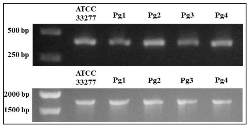
Figure 1. Expression of 16S rRNA and ppad in P. gingivalis strains. PCR products of 16S rRNA (upper panel) and ppad (lower panel) genes visualized on agarose gels. ATCC 33277, P. gingivalis reference strain; Pg1-Pg4, clinical P. gingivalis strains.
Preprints 227897 g001
Figure 2. Enzymatic activity of PPAD in clinical P. gingivalis strains. PPAD enzymatic activity was measured using N-acetyl-L-arginine as a substrate in colorimetric assay with citrulline production quantified relative to the standard curve. PPAD, peptidylarginie deiminase; ATCC 33277, P. gingivalis reference strain with enzymatic activity set at 100% (control); Pg1-Pg4, P. gingivalis strains from patients with PD; C351A, a control ATCC 33277 strain, which produces a catalytically inactive form of PPAD; Results are expressed as a percentage of activity relative to the control ATCC 33277 strain. Data represent mean values ± SEM from three independent experiments. Statistical analysis was performed using one-way ANOVA followed by Tukey’s post hoc test in comparison to the ATCC 33277 strain. ***p < 0.001.
Preprints 227897 g002
Figure 3. Relative mRNA expression of pro-inflammatory genes in GFs from donors with moderate PD infected with P. gingivalis strains expressing ppad with missense mutations or clusters of specific polymorphic variants. Relative mRNA expression level of TNF-α (A,D), IL-6 (B,E), COX-2 (C,F) in GFs from moderate PD in uninfected conditions and infected with P. gingivalis clinical strains expressing ppad with missense mutations (Pg1, Pg2) and clusters of specific polymorphic variants (Pg3, Pg4); β-actin gene, reference gene; Results were obtained from three independent experiments. Graphs present mean values ± SEM. Statistical analysis was performed using one-way analysis of variance (ANOVA) and Tukey’s post-hoc test. * p < 0.05, ** p < 0.01, *** p < 0.001.
Preprints 227897 g003
Figure 4. Relative mRNA expression of pro-inflammatory genes in GFs from donors with advanced PD infected with P. gingivalis strains expressing ppad with missense mutations or clusters of specific polymorphic variants. Relative mRNA expression of TNF-α (A,D), IL-6 (B,E), COX-2 (C,F) in GFs from advanced PD in uninfected conditions and infected with P. gingivalis clinical strains expressing ppad with missense mutations (Pg1, Pg2) and clusters of specific polymorphic variants (Pg3, Pg4); β-actin gene, reference gene; Results were obtained from three independent experiments. Graphs present mean values ± SEM. Statistical analysis was performed using one-way analysis of variance (ANOVA) and Tukey’s post-hoc test. * p < 0.05, ** p < 0.01, *** p < 0.001, **** p < 0.0001.
Preprints 227897 g004
Figure 5. Relative mRNA expression of pro-inflammatory genes in GFs from control donors infected with P. gingivalis strains expressing ppad with missense mutations or clusters of specific polymorphic variants. Relative mRNA expression level of TNF-α (A,D), IL-6 (B,E), and COX-2 (C,F) in GFs from control donors in uninfected conditions and infected with P. gingivalis clinical strains expressing ppad with missense mutations (Pg1, Pg2) and clusters of specific polymorphic variants (Pg3, Pg4); β-actin gene, reference gene; Results were obtained from three independent experiments. Graphs present mean values ± SEM. Statistical analysis was performed using one-way analysis of variance (ANOVA) and Tukey’s post-hoc test. * p < 0.05, ** p < 0.01, *** p < 0.001.
Preprints 227897 g005
Table 1. Demographic data and periodontal parameters of GFs donors.
Cells (GFs) donor Age Gender Mean API value [%] Mean BOP value [%] Mean PPD value [mm] Mean CAL value [mm] Clinical classification of PD
PD donor group (n=4)
PD1 62 F 15 60 3,2 5,9 moderate
PD2 46 M 35 60 2,6 5,5
PD3 56 M 100 100 5,2 8,0 advanced
PD4 47 F 40 80 4,9 7,7
Control group (n=2)
CTRL1 27 F 2 3 1,3 1,0 healthy periodontium
CTRL2 23 F 1 2 1,4 1,0
Reference value 0-10 0-10 1-3 1-2
GFs, gingival fibroblasts; API, aproximal plaque index, 1 value/oral cavity; BOP, bleeding on probing, 1 value/oral cavity; PPD, probing pocket depth, 5 values/oral cavity; CAL, clinical attachment loss, 5 values/oral cavity; PD, periodontitis; CTRL, control.
Table 2. Demographic data and periodontal parameters of P. gingivalis strains donors.
P. gingivalis strain Age Gender Mean API value [%] Mean BOP value [%] Mean PPD value [mm] Mean CAL value [mm] Clinical classification of PD
PD donor group (n=4)
Pg1 65 M 50 60 4,5 4,9 moderate
Pg2 60 F 25 45 4,4 4,8
Pg3 56 M 100 100 5,2 8,0 advanced
Pg4 47 F 40 80 4,9 7,7
Reference values 0-10 0-10 1-3 1-2 healthy periodontium
Pg, Porphyromonas gingivalis; API, aproximal plaque index, 1 value/oral cavity; BOP, bleeding on probing, 1 value/oral cavity; PPD, probing pocket depth, 5 values/oral cavity; CAL, clinical attachment loss, 5 values/oral cavity; PD, periodontitis.
Disclaimer/Publisher’s Note: The statements, opinions and data contained in all publications are solely those of the individual author(s) and contributor(s) and not of MDPI and/or the editor(s). MDPI and/or the editor(s) disclaim responsibility for any injury to people or property resulting from any ideas, methods, instructions or products referred to in the content.
Copyright: This open access article is published under a Creative Commons CC BY 4.0 license, which permit the free download, distribution, and reuse, provided that the author and preprint are cited in any reuse.