

Submitted:
10 August 2026
Posted:
11 August 2026
You are already at the latest version
Abstract
Insulin delivery via the buccal route has shown long-standing promise for diabetes therapy, but clinical translation remains limited. Most systems address only part of the delivery problem, whereas a viable product must simultaneously provide adequate insulin residence time, effective mucus and epithelial permeation, stability, dose consistency, safety, and manufacturability. This review examines the biological and formulation barriers that limit buccal insulin delivery and compares the major platform types, including mucoadhesive films and patches, nanocarrier-based systems, deformable vesicles, chemistry-led permeation approaches, and device-enabled strategies. Stability, alongside limited permeability, is considered a key barrier, while aggregation and excipient-related instability remain insufficiently addressed. The review also discusses why promising preclinical findings often fail to translate, exhibiting low bioavailability, variability, safety concerns, and scale-up challenges recurring across platforms. Overall, further progress in buccal insulin delivery will depend on formulation strategies that better integrate permeation enhancement, stability preservation, and translational feasibility.
Keywords:
insulin delivery
; buccal administration
; mucoadhesive systems
; nanoparticles
; deformable vesicles
; permeation enhancement
; protein therapeutics
1. Introduction
Diabetes mellitus remains one of the most prevalent chronic metabolic disorders worldwide, and insulin therapy is indispensable for all patients with type 1 diabetes and for many with advanced type 2 diabetes [1,2]. Although subcutaneous administration remains the clinical standard, repeated injections still impose pain, needle aversion, treatment burden, and injection-related complications, sustaining interest in noninvasive insulin delivery strategies [2,3,4,5]. Alternative routes, including oral, buccal, nasal, and transdermal delivery, have therefore been explored to improve adherence and reduce dependence on injections [4,5]. Among these, buccal delivery has drawn longstanding interest because it provides direct access to a vascularized mucosa while avoiding gastrointestinal degradation and hepatic first-pass metabolism [6,7,8].
Buccal delivery is also pharmaceutically attractive because the oral cavity offers a near-neutral environment with lower enzymatic activity than the gastrointestinal tract, making it more favourable for structurally fragile peptides such as insulin [2,6]. Buccal films and patches can be designed as flexible, patient-friendly dosage forms with accurate unit dosing and the practical advantage that administration can be stopped by removing the system if needed. From a patient perspective, a buccal film or patch needs to remain attached to the inner cheek for the required dosing period. Saliva, swallowing, speaking, and chewing may shorten contact time or dislodge the formulation. Depending on the design, patients may therefore need to avoid eating or drinking while it is in place. Acceptability may also depend on taste, mouthfeel, stickiness, thickness, and any local irritation [9,10]. Although thin, flexible films are intended to minimise discomfort, these sensory and practical factors should be considered during formulation development [9,11]. A comparison of buccal and gastrointestinal routes of insulin delivery is illustrated in Figure 1. Oral transmucosal films are already established in clinical practice. Marketed products such as Belbuca®, which delivers buprenorphine for chronic pain, and LibervantTM are labelled as buccal films that deliver diazepam for the acute treatment of seizure clusters, supporting the translational feasibility of the dosage form itself, although the approved products are mainly intended for potent small molecules rather than large hydrophilic peptides such as insulin [9,12]. Early human studies with buccal insulin spray suggest that the route may support relatively rapid systemic action, which is especially relevant for prandial insulin delivery [13,14,15]. Even so, buccal delivery remains challenging because insulin crosses the buccal mucosa very poorly. Systemic delivery is limited by the nonkeratinized stratified epithelium, intercellular lipids, mucus, local enzymatic activity, limited absorptive area, and continuous salivary clearance [6,9,16]. These barriers are especially important for insulin, a large hydrophilic peptide whose passive transport across intact buccal mucosa is intrinsically inefficient [16].
Despite these constraints, clinical proof of concept has been achieved. The Oral-Lyn insulin spray, delivered using the Rapid Mist device showed that insulin delivered to the oral mucosa could produce measurable pharmacokinetic and pharmacodynamic effects, including faster onset and shorter duration than subcutaneous insulin [13,14]. However, later analyses also showed why this early success did not translate into durable clinical use, with low relative biopotency, high dose burden, and marked variability emerging as key limitations [7,17]. These findings shifted the field toward broader formulation innovation, including mucoadhesive films and patches for residence control [9,18,19] unidirectional buccal tablet systems for directional release [20], nanoparticle-based and nanoparticle-in-film systems for insulin protection and mucosal interaction [10,21,22] deformable vesicles and related lipid carriers for enhanced transport [23,24,25,26] and chemical enhancement strategies including hydrophobic ion pairing, cell-penetrating peptides, and ionic liquids for improved permeation [27,28,29]. In recent years, device-based buccal delivery approaches have emerged as an additional strategy to reduce reliance on passive permeability by actively overcoming the epithelial barrier, including microneedle-mediated penetration that deposits insulin directly into the buccal mucosa and suction-assisted systems that stretch and thin the epithelium, increasing transport of macromolecules and, in some designs, being combined with permeation enhancers to further improve delivery [30,31,32].
Another challenge, often treated as secondary despite its translational importance, is insulin stability. Insulin is structurally sensitive to unfavourable pH, thermal stress, agitation, contact container surfaces, and hydrated formulation environments, all of which can promote conformational change, aggregation, and loss of biological activity [33,34,35]. In buccal delivery, this issue extends beyond storage because insulin may also be exposed to destabilising processing conditions, salivary dilution, and excipient environments before absorption occurs [19,26,36]. Buccal insulin delivery should therefore be viewed as a combined formulation, permeability, stability, and translational problem rather than a simple permeability challenge. This review examines the field from that integrated perspective of buccal insulin delivery, covering mucoadhesive films and patches, nanocarrier and vesicular systems, permeation-enhancing formulations, and device-based approaches. It focuses on how formulation design, insulin stability, and practical translational challenges must be addressed together to develop clinically useful systems.
2. Barriers to Buccal Insulin Delivery
For insulin, the main attraction of buccal delivery is that it avoids the harsh gastrointestinal environment and first-pass metabolism in the liver, both of which make conventional oral delivery difficult [2,6,37]. This is especially important for protein and peptides that are easily degraded in the gastrointestinal tract and therefore usually need to be administered by injection. This gives the route a clear pharmacokinetic advantage, but it does not mean that buccal absorption is easy. As summarised in Table 1, buccal insulin delivery is limited by interacting with various insulin related physicochemical, physical, physiological, and enzymatic factors. Additionally, the key epithelial barriers that limit buccal insulin delivery are shown in Figure 2.
3. Formulation Strategies for Buccal Insulin Delivery and Their Limitations
To improve buccal insulin delivery, researchers have developed several formulation strategies to overcome poor mucosal permeability, short residence time, and low dose efficiency [2,6,39]. These approaches vary in design, but most aim to either keep insulin at the mucosal surface, enhance its transport across the epithelium, or combine both functions within a single platform [7,10,22]. As shown in Figure 3, different formulation- and device-based strategies have been explored to improve buccal insulin delivery. Although differing in approach, all the designs aim to retain insulin at the mucosal surface, promote passage across the epithelium and enhance systemic uptake against the normal buccal mucosa barriers.
3.1. Mucoadhesive Films and Patches
Mucoadhesive film systems are useful because they can combine several functions in a single platform, including prolonged mucosal residence, reduced salivary washout, closer contact with the absorption site, and, in some designs, unidirectional release toward the buccal tissue [9,18]. Their main advantage is that they reduce salivary washout and extend contact with the absorptive mucosa, creating better conditions for insulin absorption. As shown in Table 2, the different mucoadhesive film and patch approaches studied for buccal insulin delivery are summarised according to their design, preparation method, evaluation approach, key findings, and main limitations.
In one study, Chen et al. [18]. reported an optimised bilayer film composed of a backing layer and a mucoadhesive insulin-loaded layer. This system improved adhesion-supported release toward the absorption surface, and showed prolonged adhesion time, thereby promoting insulin absorption. This concept is illustrated in Figure 3. Additionally, these findings are summarised in Table 2. This approach increased insulin exposure compared with free insulin and maintained glucose control for about 8 h in diabetic rats [18]. These findings suggest that effective film performance depends on more than mucoadhesion alone and may improve when prolonged residence, controlled release, and permeation support are combined within the same system. However, the evidence remains limited to preclinical studies.
In another study, Diab et al. [19] investigated a chitosan-based mucoadhesive buccal film incorporating insulin, glycerin, and L-arginine. As mentioned in Table 2, the formulation was characterised in terms of film appearance and flexibility, insulin content using ELISA, spectroscopic properties, and in vivo glucose-lowering efficacy. The optimised film retained at least 80% of the theoretical insulin content and produced an approximately 40% reduction in blood glucose levels in rats, corresponding to about 57% of the pharmacological effect achieved with subcutaneous insulin [19]. These results demonstrate measurable buccal insulin activity, although the overall efficacy remained lower than that of the injectable route. Their further development will depend on improving key features such as permeation, dose efficiency, and overall in vivo performance to move them closer to the effectiveness of subcutaneous insulin.
A different study proposed a unidirectional buccal tablet concept based on a triple-layer mucoadhesive system, in which nanoparticles loaded with insulin were added as dry powder to a multilayer mucoadhesive system [20]. As mentioned in Table 2, this unidirectional approach is proposed to minimise the dilution of saliva and loss of the drug, direct the release of insulin towards the buccal absorption site and improve the stability of the dosage form. While this work did not progress beyond the concept stage, no experimental characterisation was performed. This study focused on conceptual design, multilayer configuration, and polymer selection. It demonstrates the importance of combining mucoadhesion, directional release, nanoparticle delivery, and enhanced stability in a buccal insulin delivery system.
Overall, studies show that mucoadhesive films, patches, and multilayer buccal systems can enhance mucosal residence, reduce salivary washout, and promote insulin release towards the absorption surface. Chitosan-based films demonstrated measurable in vivo glucose-lowering activity, while bilayer films increased insulin adhesion and exposure. Multilayer tablet concepts can also minimise drug loss through saliva and promote unidirectional release of the drug. These findings, taken together, may lay the foundation for integrated buccal insulin delivery platforms with mucoadhesion, controlled release, permeation support, and dosage-form stability.
3.2. Nanocarrier-Based Systems
Nanocarrier-based systems have emerged as a key strategy for improving buccal insulin delivery. Their main purpose is to protect insulin within the formulation, improve its interaction with the buccal barrier, and increase transport through carrier flexibility or an altered drug state [7,10]. This concept is illustrated in Figure 3. Table 3 summarises different nanocarrier-based systems investigated for buccal insulin delivery, including nanoparticle-loaded films, deformable vesicles and elastic lipid-based carriers. It also highlights their compositions, preparation methods, and key optimised parameters, including particle size, encapsulation efficiency, release profile, permeability, relative bioavailability, and storage stability.
3.2.1. Nanoparticle-Loaded Buccal Films
Nanoparticle-loaded buccal films were explored as hybrid systems that combine the retention advantage of mucoadhesive films with the protective and transport-supporting functions of nanoparticles. As outlined in Table 3, early work showed that insulin-loaded PEG-b-PLA nanoparticles could be incorporated into chitosan films with acceptable formulation properties, including particle size around 215 nm, encapsulation efficiency of about 71%, and improved film flexibility, supporting the feasibility of this design [21]. In another study, insulin-coated nanoparticles were incorporated into Eudragit RLPO-based buccal films and showed measurable transport across a human buccal model. The ERL film performed much better than the ERL-HPMC film, reaching about 52% cumulative insulin permeation over 4 h compared with about 10% for the blended film, with corresponding flux values of 0.34 ± 0.05 and 0.07 ± 0.02 μg/h/cm2, respectively [22]. Together, these findings indicate that nanoparticle-film integration may offer more than prolonged residence alone by combining local retention with insulin protection and controlled release, although the evidence remains limited by the lack of clear in vivo validation.
3.2.2. Deformable Vesicle Systems
Deformable vesicle systems were developed to improve buccal insulin transport by employing flexible lipid carriers that interact more effectively with the mucosal barrier than conventional vesicles. This is illustrated in Figure 3. As highlighted in Table 3, this approach evolved across nanocarrier studies, with differences in vesicle composition and preparation method affecting both delivery performance and formulation sp. behaviour. Early work showed that deformable vesicles composed of SPC and sodium deoxycholate performed better than conventional vesicles in supporting buccal insulin transport [23]. In another study, insulin-phospholipid complex deformable nanovesicles were developed using lecithin, Tween 20, and sodium deoxycholate, showing that this design could support both insulin encapsulation and delivery performance, with a reported relative pharmacological bioavailability of about 15.53% [24]. In a subsequent work, Guo et al. [26]. incorporated a comparable deformable nanovesicle system into a gelatin-based matrix. This formulation was more stable during refrigerated storage at 4 °C for up to 3 months and still maintained good delivery performance after storage, with no major changes in particle size, pH, or drug content and with preserved hypoglycemic activity. Taken together, this finding suggests that deformable vesicle systems are among the more promising passive nanocarrier approaches because they combine transport enhancement with formulation support and storage stability, although stronger translational evidence is still needed.
3.2.3. Elastic Liposomes
Elastic liposomes represent a related vesicular strategy in which lipid bilayers are modified with edge activators to increase flexibility, protection, and support buccal transport. This concept is shown in Figure 3. As mentioned in Table 3, this approach moved from cell-based testing to ex vivo tissue evaluation. In TR146 cells, cholic acid derivative-modified elastic biosomes prepared by thin-film hydration showed particle sizes of about 140 to 150 nm and produced enhancement ratios up to 5.24 compared with insulin solution, supporting the idea that bile salt-modified flexible vesicles can improve insulin permeation across the buccal barrier [41]. This finding was strengthened further in ex vivo porcine buccal tissue, where sodium glycodeoxycholate-containing elastic liposomes produced a 4.33-fold increase in permeability coefficient relative to insulin solution and performed better than sodium cholate liposomes [25]. Together, these results suggest that bilayer flexibility and bile salt modification can improve buccal insulin transport. Even so, the evidence remained limited to cell and ex vivo models, so elastic liposomes are better viewed as promising carrier systems than as translationally mature platforms.
Overall, the nanocarrier-based approaches indicate that these systems contribute more than simply prolonging residence time. They also protect insulin from degradation and enhance carrier interaction with the buccal barrier, thereby improving delivery performance. Nanoparticle-film systems primarily support formulation feasibility, whereas deformable vesicles and elastic liposomes exhibit greater transport-enhancing potential due to carrier flexibility and bilayer adaptability [21,22,23,24,25,26,41]. Even so, these advances show that nanocarrier design is moving in a useful direction, particularly in improving insulin protection and carrier interaction with the buccal barrier. Their future value will depend on how successfully these formulation strengths translate into buccal insulin platforms that are stable, reproducible, practical, and scalable.
3.3. Chemistry-Led Permeation Strategies
Chemistry-led permeation strategies were developed to improve buccal insulin delivery by changing the physicochemical behaviour of insulin or the microenvironment at the mucosal surface. Unlike films or vesicles, which primarily enhance residence time or carrier-mediated transport, these approaches act more directly on epithelial passage by increasing insulin lipophilicity, modulating barrier interactions, or promoting epithelial uptake [27,28,29]. Figure 3 provides visual representation of this concept. Earlier sublingual work also showed that enhancers such as chitosan, hydroxypropyl--cyclodextrin, egg lecithin, oleic acid, and polyoxyethylene lauryl ether could alter mucosal barrier properties and insulin association state, helping explain the continued interest in chemistry-led strategies for buccal peptide delivery [42]. Table 4 compares the main chemistry-led strategies explored for buccal insulin delivery, focusing on how they improve mucosal transport, the primary permeability or bioavailability outcomes reported, and the limitations that still hinder translation.
Table 4. Comparison of chemistry-led permeation strategies for buccal insulin delivery.
3.3.1. Hydrophobic Ion Pairing
Hydrophobic ion pairing was used to improve buccal insulin transport by forming complexes between insulin and bile salt derivatives, thereby increasing the lipophilicity of the molecule. This concept is presented in Figure 3. In one study investigated TR146 cell layers to assess epithelial transport and ex vivo porcine buccal tissue to examine permeation in a more physiologically relevant mucosal model, as shown in Table 4, with improved permeability in both systems relative to insulin solution [27]. The authors complexed insulin with sodium glycodeoxycholate to make it lipophilic. They found that transport of this complex was higher in TR146 buccal cells and ex vivo porcine buccal tissue than the insulin solution. What makes this approach especially interesting is that the improved transport was not accompanied by obvious acute damage in the TR146 model, since cell viability remained essentially unchanged and TEER recovery stayed above 96% after exposure. These findings support the value of changing the physicochemical state of insulin to improve buccal transport. Even so, the evidence remained limited to in vitro and ex vivo evaluation and was not extended to an in vivo buccal dosage form [27].
3.3.2. Ionic Liquid Systems
Ionic liquid systems were explored using choline geranate (CAGE) to improve buccal insulin transport and support the local formulation environment and prompt possible paracellular transport mechanisms [29]. This approach is not only attractive for delivery performance but also for future patch design. In the reported system, choline geranate was incorporated into a biodegradable polymeric buccal patch with a viscoelastic gel reservoir, and formulation optimisation identified a 70 vol% gel as a useful balance of viscoelastic behaviour, near-neutral surface pH, and mucoadhesion [29]. As summarised in Table 4, this system increased ex vivo transport and produced measurable in vivo delivery, but long-term safety and broader translation acceptability remain incompletely defined. Supporting mechanistic work showed that choline geranate can preserve insulin’s secondary structure and reduce enzymatic degradation, although the study was not conducted in a buccal system [36].
3.3.3. Cell-Penetrating Peptides
Cell-penetrating peptide strategies were developed to improve buccal insulin uptake by strengthening insulin interaction with the epithelial barrier [28]. This concept is presented in Figure 3. In this study, insulin was linked to low-molecular-weight protamine via PEG, and the active region of insulin was selectively protected before conjugation with dimethylmaleic anhydride to preserve bioactivity. The system was evaluated across TR146 multilayers, porcine buccal tissue, and rabbits to assess transport, permeation, and in vivo performance [28]. As summarised in Table 4, this approach demonstrated improved uptake and transport, with relative bioavailability reaching 26.86% and no visible signs of mucosal irritation. Its main weakness is that the same molecular engineering that improves performance may also complicate scale-up, formulation control, and regulatory translation [28]. Overall, these studies suggest that chemistry-led permeation strategies may be useful because they act more directly on insulin’s poor intrinsic permeability than residence-based systems alone. Hydrophobic ion pairing increases insulin lipophilicity, ionic liquids modify the local transport and stability environment, and cell-penetrating peptide conjugation promotes epithelial uptake. Their future value will depend on whether these transport gains can be translated into systems that are also stable, manufacturable, and suitable for repeated clinical use.
3.4. Device-Enabled Systems
Device-enabled systems differ from films, vesicles, and chemistry-led formulations by improving buccal delivery through mechanical assistance rather than relying solely on passive permeation enhancement. This is important because low intrinsic epithelial permeability remains a major barrier to translation [32]. Within this approach, the spray system is the primary traditional human proof of concept, while microneedles and suction-based patches represent the strongest recent attempts to overcome the passive permeability ceiling. Table 5 summarises the main device-enabled strategies relevant to buccal insulin delivery, including their delivery mechanism, relevance to insulin delivery, and the key translational challenges that remain.
3.4.1. Buccal Spray Systems
Buccal spray systems provide the clearest human evidence that insulin can be absorbed through the oral mucosa. In the RapidMist or Oral-lyn platform, insulin is delivered as a high-velocity aerosol to the oropharyngeal cavity, where part of the dose is absorbed transmucosally rather than swallowed [13,14,15]. Clinical studies showed faster absorption and earlier metabolic action than subcutaneous regular insulin. In healthy volunteers, the spray reached maximum serum insulin concentration at about 23 min, compared with about 83 min for subcutaneous insulin, while the maximum glucose infusion response occurred at about 44 min, compared with about 100 min [13]. A later study in type 1 diabetes also showed a dose-response relationship, with 5-, 10-, and 20-puff doses producing dose-dependent increases in insulin exposure while maintaining a rapid-onset profile [14]. Another study reported insulin appearance in the blood within about 10 min and a maximum concentration at around 30 min, again supporting the view that buccal spray behaves more like a rapid prandial system than a sustained basal one [15]. Despite this clinical relevance, spray systems remained limited by high dose burden, short duration, and variable performance, so they are best viewed as important proof of concept rather than a complete translational solution.
3.4.2. Buccal Microneedle Systems
Microneedle systems improve buccal insulin delivery by bypassing the epithelial barrier rather than relying on transport across intact mucosa [30]. This concept is presented in Figure 3. This makes them especially relevant, as poor intrinsic permeability remains one of the main challenges in passive buccal delivery. In one study, a buccal microneedle patch localised about 1 mg of human insulin in needle tips, delivered it within 30 s in swine, and produced measurable systemic exposure together with glucose lowering. Insulin reached maximum plasma concentration at about 40 to 50 min, showing that barrier bypass could support relatively rapid systemic delivery even without conventional injection [30]. The same study also included a human acceptability assessment in 100 volunteers, where pain scores were low, the hard palate was the preferred application site, and 95% of participants said they would prefer this system over a conventional hypodermic injection. Together, these findings make buccal microneedles one of the most promising recent device-based approaches, as they combine rapid delivery, direct barrier bypass, and early evidence of usability. The main remaining limitations are storage stability, manufacturing control, and the need for stronger repeat-use safety data [30,32].
3.4.3. Suction and Octopus-Inspired Patch Systems
A newer class of device-enabled buccal systems improves peptide delivery through suction and tissue deformation rather than needle penetration. As presented in Figure 3, these octopus-inspired patches create local vacuum pressure that pulls, stretches, and thins the buccal mucosa, increasing the effective surface area for drug transport [31,32,43]. In one study, a mould-cast silicone suction patch with a lyophilised drug matrix increased effective surface area more than ninefold and achieved about 14.6% absolute bioavailability for teriparatide in beagle dogs, while also improving semaglutide delivery compared with an oral tablet [31]. Although this study did not test insulin, it still gives a useful idea for future buccal insulin design. Krupke et al. [43]. Reported finding from an in vivo study in beagle dogs, a dose-adjusted semaglutide exposure increased 7- to 9-fold compared with the commercial oral tablet after 10 min of application, and bremelanotide achieved a relative bioavailability of 26% versus subcutaneous dosing after 20 min. These results suggest that biodegradable mechanical buccal systems could be useful for future insulin delivery, although insulin-specific evidence remains lacking and concerns remain about variability, dissolution under low-saliva conditions, and storage stability [43]. Overall, the findings summarised in Table 5 suggest that device-enabled systems may offer a more practical means of overcoming the buccal barrier.
4. Stability Constraints in Buccal Insulin Formulations
Preserving insulin stability is as important as increasing permeability, mucoadhesion, or mucosal residence time in buccal delivery systems. Many formulations are designed to enhance epithelial transport and mucosal contact, but if insulin loses its native structure during formulation, storage, administration, or mucosal residence, these gains become far less meaningful [2,6]. A successful buccal insulin system must therefore not only localise insulin at the absorption site, but also protect it from unfolding, aggregation, enzymatic degradation, and destabilising formulation microenvironments long enough for absorption to occur.
Human insulin consists of two chains linked by disulfide bonds, with 21 amino acids in the A chain and 30 in the B chain [33,44]. Its native conformation contains important -helical regions that contribute to biological activity, receptor recognition, and structural stability [34,44]. Under destabilising conditions, insulin can partially unfold, expose hydrophobic regions, and progress through self-association, aggregation, and fibrillation, with loss of biological activity following these structural changes [33,34,35]. This concept is illustrated in Figure 4. For these reasons, stability-related limitations in buccal insulin formulations should be evaluated at three distinct levels: biological instability following administration, physical instability during formulation and storage, and chemical instability associated with the formulation microenvironment. As illustrated in Figure 4, insulin is subjected to multiple interconnected formulation, storage, and physiological stresses during buccal delivery, many of which compromise its structural stability, reduce epithelial permeation, and ultimately limit systemic bioavailability.
4.1. Biological Instability After Dosing
As illustrated in Figure 2, biological instability begins as soon as insulin enters the oral cavity. Even when a formulation improves mucosal contact or permeability, insulin is still exposed to salivary dilution, enzymatic degradation at the mucosal surface, and limited residence time before part of the dose is swallowed or cleared away [2,6]. Together, these factors reduce the amount of structurally intact insulin available for absorption. Successful buccal systems must therefore do more than deliver insulin to the mucosa; they must also protect it long enough to allow meaningful uptake.
4.2. Physical Instability During Formulation and Storage
Physical instability is another major concern in buccal insulin delivery because insulin can lose activity during preparation and storage through unfolding, aggregation, precipitation, or carrier destabilisation. In chitosan-based mucoadhesive buccal films, insulin structure was better preserved when L-arginine was included, and drying at 2 to 8 °C retained more insulin than drying at room temperature, showing that stability depends not only on the carrier but also on how the dosage form is processed [19]. A similar point was seen in a deformable nanovesicle-loaded gelatin gel system, where the gel matrix helped preserve vesicle morphology, particle size, pH, and insulin content for 3 months at 4 °C, whereas less stabilised liquid systems were more prone to leakage, fusion, or precipitation [26]. Together, these findings show that physical instability in buccal insulin systems is not limited to insulin itself but also involves the structural integrity of the surrounding dosage form [19,26].
4.3. Chemical Instability and Formulation Environment
Chemical instability further complicates buccal insulin delivery because insulin can be easily affected by unfavourable pH conditions, reactive formulation components, surfactants, and prolonged exposure to moisture-rich environments. This concept is illustrated in Figure 4. This means that excipient choice and the local formulation environment are just as important as carrier design. Banerjee et al. [36] reported that choline geranate preserved the characteristic insulin -helical circular dichroism profile during storage, suggesting that the formulation environment helped maintain insulin close to its native folded state. The same study also showed reduced enzymatic degradation and suggested that limiting insulin-water interactions may help reduce hydrolysis and self-aggregation. Although this was not a buccal study, it remains highly relevant because it shows how strongly the local chemical environment can influence whether insulin stays structurally intact long enough for a delivery system to work. The main stability-related constraints and their formulation implications are summarised in Table 6. As described previously, stability in buccal insulin delivery is not limited to a single formulation problem but reflects a set of related constraints that act before, during, and after administration. It shows that insulin can be compromised by biological factors after dosing, including salivary dilution, enzymatic degradation, and short mucosal residence time; by physical instability during formulation and storage, such as unfolding, aggregation, precipitation, or carrier destabilisation; and by chemical effects arising from the formulation environment itself, including nonoptimal pH, excipient interactions, and prolonged exposure to hydrated systems. Taken together, these parameters show that successful buccal insulin delivery depends not only on improving permeation but also on preserving insulin in a structurally intact and pharmaceutically usable form throughout the full delivery pathway.
Insulin is essential for glucose regulation through activation of the insulin receptor and remains the mainstay of diabetes treatment. However, its therapeutic use is challenged because of structural instability, including unfolding and formation of cross-β-sheet amyloid fibrils. The limited understanding of the molecular basis of insulin fibrillation has hindered the development of effective strategies to prevent it [44].In the same study, the authors investigated insulin fibrils using cryo–electron microscopy (cryo-EM), this study identified several fibril architectures composed of protofilaments containing both insulin A and B chains connected by disulfide bonds. A two-protofilament fibril was resolved at 3.2 Å, revealing the β-sheet organisation and inter-protofilament interactions that stabilise the fibril. Guided by this structure, the researcher designed insulin variants with reduced fibrillation tendency while retaining native insulin-receptor sp. signaling, highlighting their potential as more stable therapeutic candidates for type 1 diabetes [44].
5. Translational Prospects for Future Buccal Insulin Delivery
One of the main challenges in the buccal insulin literature is the lack of standardised benchmarks for comparing systems across studies. Different approaches use different formulations, models, doses, and outcome measures, which makes direct comparison difficult and can blur true translational value. Future progress will therefore depend not only on better delivery results, but also on clearer criteria for judging which systems are most likely to translate successfully.
Buccal insulin delivery remains an attractive goal because it offers a non-invasive route with direct access to a vascularized mucosa while avoiding gastrointestinal degradation and hepatic first-pass metabolism [2,6]. The main question is no longer whether buccal insulin can work in principle, but whether it can be developed into a system that is stable, dose-efficient, reproducible, and clinically practical. This is especially important because the field already contains real proof of concept. Human studies with Oral-lyn demonstrated measurable systemic exposure and rapid pharmacodynamic effects following oral mucosal insulin delivery [13,14,15]. However, later reappraisal showed that these early systems were limited by low relative biopotency, high dose burden, and substantial variability [17]. These findings remain important because they established route feasibility in humans, but they also set a practical benchmark that newer systems still need to exceed. An additional point is that model selection deserves particular attention in preclinical studies. Reported permeability findings for insulin differ substantially across systems, with log permeability coefficients of about −8.18 in porcine buccal tissue and about −5.92 to −7.10 in TR146 models [16]. Secondly, a formulation that performs well in a more permissive model may therefore not perform as strongly under more realistic buccal conditions. Permeability should also not be judged alone, because insulin transport is influenced simultaneously by epithelial resistance, small pore size, salivary washout, mucus, and enzymatic degradation [6,16,39]. Additionally, as mentioned in Table 1 that buccal insulin delivery is limited by several barriers acting at the same time rather than by permeability alone. When designing new formulations for buccal insulin delivery, these constraints should be considered collectively.
Different formulation classes contribute in different ways, and each is useful for a particular part of the translational problem. Mucoadhesive films and patches, summarised in Table 2, are most useful for improving residence and supporting directional release at the buccal surface. According to a bilayer film study, which used an ethyl cellulose backing layer with an adhesive HPMC-PAA interpolymer complex layer, showed prolonged adhesion, a 2.24-fold increase in insulin AUC relative to free insulin, and glucose control for about 8 h in diabetic rats [18]. In another study, an insulin-loaded chitosan film was prepared by solvent casting with glycerin and L-arginine, retaining at least 80% of the theoretical insulin load and producing measurable glucose lowering, although it remained weaker than subcutaneous insulin [19]. These findings show that film and patch systems can improve buccal residence and support the value of improved film engineering in buccal insulin delivery, although they may remain dose inefficient.
Nanocarrier-based systems, compared in Table 3, contribute more than residence alone because they can combine insulin protection, controlled release, and carrier–mucosa interaction. In one study, insulin-loaded PEG-b-PLA nanoparticles were incorporated into Chitosan films with about 71% encapsulation efficiency [21], while another study showed measurable buccal flux from nanoparticle-film systems [22]. As summarised in Table 3, deformable vesicle systems appear to be a promising passive nanocarrier approach for buccal insulin delivery. Across several preclinical studies, their flexible phospholipid-based structures were associated with improved delivery performance [23,24,26]. There is one study that also reports that incorporating a similar vesicular system into a gelatine-based matrix helped improve storage stability for 3 months at 4 °C in a gelatin matrix [26]. Taken together, these findings suggest that deformable vesicle-based systems may be particularly useful when insulin protection, transport enhancement, and formulation stability need to be addressed within the same platform. However, their translational value will still depend on storage stability, reproducibility, and manageable formulation complexity.
Chemistry-led systems, summarised in Table 4, are particularly useful because they address poor intrinsic insulin permeability more directly. In hydrophobic ion pairing with bile salt derivatives, increased permeability of about 3-fold in TR146 cells and about 51.76-fold ex vivo, showing how changes in insulin lipophilicity can strongly affect transport [27]. Choline geranate buccal patch increased ex vivo permeation to about 7-fold and produced measurable in vivo bioavailability [29]. In another study, the PEG-linked low molecular weight protamine conjugate is also notable because it achieved relative bioavailability up to 26.86% [28]. These findings show that chemistry-led approaches may be especially useful for solving the permeability problem more directly, but often at the cost of greater molecular complexity, less certain safety definition, and more difficult translation.
Device-enabled systems, as highlighted in Table 5, remain particularly important because they reduce dependence on passive epithelial transport, which continues to limit buccal insulin delivery. Spray-based systems still provide the clearest human proof of concept for rapid buccal insulin delivery [13,14,15]. In human studies, buccal spray reached an insulin of about 23 to 30 min, compared with about 83 min for subcutaneous regular insulin, indicating a faster onset that may be relevant for prandial use [13,15,17]. However, the latter advantage is limited by low dose efficiency, high burden, and variability [17]. The microneedle patch is also notable because it localised about 1 mg of insulin in the needle tips, delivered the dose within 30 s, produced measurable systemic exposure in swine, and was supported by a favourable acceptability data study in 100 volunteers [30]. Studies of suction-based patches further show that mechanical tissue deformation and increased contact area can improve peptide delivery without needle penetration [30,43]. Taken together, these findings support device-enabled approaches as highly relevant for future insulin translation, although their practical value will still depend on repeat-use safety, storage stability, usability, dose consistency, and overall product feasibility [30,32,43].
For future studies, the key issue is not whether a system improves one result, but whether it performs well across the factors that matter for translation. These include preservation of insulin integrity, dose efficiency, clinically interpretable pharmacokinetic and pharmacodynamic performance, realistic mucosal residence, credible model selection, local safety, manufacturability, storage practicality, and user acceptability.
Conclusion
Insulin delivery via the buccal route has been investigated through a range of formulation and device-based approaches. Earlier human studies have confirmed that insulin can be absorbed through the oral mucosa and can produce relatively rapid systemic effects, but they have also highlighted persistent limitations, including low dose efficiency, variability, and incomplete control over exposure. Subsequent advances expanded the field rather than fully resolving these challenges. Mucoadhesive films and patches improved residence time and directional release; nanocarrier-based and deformable vesicle systems supported insulin protection and transport; chemistry-led strategies addressed poor intrinsic permeability more directly; and device-enabled platforms suggested that mechanical assistance may be needed when the intact buccal mucosa imposes a strong permeability barrier. Despite this progress, no current platform yet satisfies the full requirements for clinical translation. The central challenge is no longer whether insulin can be delivered through the buccal route, but whether a system can preserve insulin stability, provide reproducible therapeutic exposure, maintain local tolerability, and remain manufacturable and storage-stable within a single product concept. Future progress will therefore depend on integrated platform designs in which permeation, stability, pharmacokinetic and pharmacodynamic performance, safety, residence time, and product feasibility are addressed together rather than as separate formulation goals.
Author Contributions
Conceptualisation, AV and JP; literature search, writing-original draft and modification, AV and JP; Writing-review and editing, AV and MS. All authors have read and agreed to the published version of the manuscript.
Funding
Research Bursary received from the Faculty of Health, Science, Social Care and Education (HSSCE), Kingston University London.
Institutional Review Board Statement
Not applicable.
Acknowledgments
The support provided by the Faculty of Health, Science, Social Care and Education (HSSCE), Kingston University London, is gratefully acknowledged.
Conflicts of Interest
The authors declare no conflict of interest.
Abbreviations
The following abbreviations are used in this manuscript:
AUC — area under the curve
DPP-IV — dipeptidyl peptidase IV
EC — ethylcellulose
EE — encapsulation efficiency
ELISA — enzyme-linked immunosorbent assay
ERL — Eudragit RLPO
FITC — fluorescein isothiocyanate
GLP-1 — glucagon-like peptide 1
HPMC — hydroxypropyl methylcellulose
ICNP — insulin-coated nanoparticles
IPC — interpolymer complex
PEG — polyethylene glycol
PAA — polyacrylic acid
PEG-b-PLA — polyethylene glycol-block-poly lactic acid
PDI — polydispersity index
RBA — relative bioavailability
RPB — relative pharmacological bioavailability
SC — sodium cholate
SGDC — sodium glycodeoxycholate
SPC — soybean phosphatidylcholine
TR146 — human buccal carcinoma cell line model
Tmax- time to maximum concentration
References
- Zhou, B.; Rayner, A.W.; Gregg, E.W.; Sheffer, K.E.; Carrillo-Larco, R.M.; Bennett, J.E.; Shaw, J.E.; Paciorek, C.J.; Singleton, R.K.; Barradas Pires, A.; et al. Worldwide Trends in Diabetes Prevalence and Treatment from 1990 to 2022: A Pooled Analysis of 1108 Population-Representative Studies with 141 Million Participants. The Lancet 2024, 404, 2077–2093. [Google Scholar] [CrossRef] [PubMed]
- Elsayed, A.; Al-Remawi, M.; Jaber, N.; Abu-Salah, K.M. Advances in Buccal and Oral Delivery of Insulin. Int. J. Pharm. 2023, 633, 122623. [Google Scholar] [CrossRef] [PubMed]
- Zhang, E.; Zhu, H.; Song, B.; Shi, Y.; Cao, Z. Recent Advances in Oral Insulin Delivery Technologies. J. Control. Release 2024, 366, 221–230. [Google Scholar] [CrossRef] [PubMed]
- Easa, N.; Alany, R.G.; Carew, M.; Vangala, A. A Review of Non-Invasive Insulin Delivery Systems for Diabetes Therapy in Clinical Trials over the Past Decade. Drug Discov. Today 2019, 24, 440–451. [Google Scholar] [CrossRef] [PubMed]
- Sugumar, V.; Ang, K.P.; Alshanon, A.F.; Sethi, G.; Yong, P.V.C.; Looi, C.Y.; Wong, W.F. A Comprehensive Review of the Evolution of Insulin Development and Its Delivery Method. Pharmaceutics 2022, 14, 1406. [Google Scholar] [CrossRef] [PubMed]
- Rawas-Qalaji, M.; Thu, H.E.; Hussain, Z. Oromucosal Delivery of Macromolecules: Challenges and Recent Developments to Improve Bioavailability. J. Control. Release 2022, 352, 726–746. [Google Scholar] [CrossRef] [PubMed]
- Morales, J.O.; Brayden, D.J. Buccal Delivery of Small Molecules and Biologics: Of Mucoadhesive Polymers, Films, and Nanoparticles. Curr. Opin. Pharmacol. 2017, 36, 22–28. [Google Scholar] [CrossRef] [PubMed]
- Amer, A.A.; Bingle, L.; Elkordy, A.A.; Chaw, C.S. Overcoming Oral Cavity Barriers for Peptide Delivery Using Advanced Pharmaceutical Techniques and Nano-Formulation Platforms. Biomedicines 2025, 13, 2735. [Google Scholar] [CrossRef] [PubMed]
- Jacob, S.; Nair, A.B.; Boddu, S.H.S.; Gorain, B.; Sreeharsha, N.; Shah, J. An Updated Overview of the Emerging Role of Patch and Film-Based Buccal Delivery Systems. Pharmaceutics 2021, 13, 1206. [Google Scholar] [CrossRef] [PubMed]
- Macedo, A.S.; Castro, P.M.; Roque, L.; Thomé, N.G.; Reis, C.P.; Pintado, M.E.; Fonte, P. Novel and Revisited Approaches in Nanoparticle Systems for Buccal Drug Delivery. J. Control. Release 2020, 320, 125–141. [Google Scholar] [CrossRef] [PubMed]
- Abdelhakim, H.E.; Williams, G.R.; Craig, D.Q.M.; Orlu, M.; Tuleu, C. Human Mouthfeel Panel Investigating the Acceptability of Electrospun and Solvent Cast Orodispersible Films. Int. J. Pharm. 2020, 585, 119532. [Google Scholar] [CrossRef] [PubMed]
- Seinfeld, S.; Gelfand, M.A.; Heller, A.H.; Buan, C.; Slatko, G. Safety and Tolerability Associated with Chronic Intermittent Use of Diazepam Buccal Film in Adult, Adolescent, and Pediatric Patients with Epilepsy. Epilepsia 2020, 61, 2426–2434. [Google Scholar] [CrossRef] [PubMed]
- Comparison of Pharmacokinetic and Pharmacodynamic Properties of Single-Dose Oral Insulin Spray and Subcutaneous Insulin Injection in Healthy Subjects Using the Euglycemic Clamp Technique
- Cernea, S.; Kidron, M.; Wohlgelernter, J.; Raz, I. Dose-Response Relationship of an Oral Insulin Spray in Six Patients with Type 1 Diabetes: A Single-Center, Randomized, Single-Blind, 5-Way Crossover Study. Clin. Ther. 2005, 27, 1562–1570. [Google Scholar] [CrossRef] [PubMed]
- Pozzilli, P.; Manfrini, S.; Costanza, F.; Coppolino, G.; Cavallo, M.G.; Fioriti, E.; Modi, P. Biokinetics of Buccal Spray Insulin in Patients with Type 1 Diabetes. Metabolism 2005, 54, 930–934. [Google Scholar] [CrossRef] [PubMed]
- Wanasathop, A.; Patel, P.B.; Choi, H.A.; Li, S.K. Permeability of Buccal Mucosa. Pharmaceutics 2021, 13, 1814. [Google Scholar] [CrossRef] [PubMed]
- Heinemann, L.; Jacques, Y. Oral Insulin and Buccal Insulin: A Critical Reappraisal. J. Diabetes Sci. Technol. 2009, 3, 568–584. [Google Scholar] [CrossRef] [PubMed]
- Chen, Y.; Zhang, L.; Xu, J.; Xu, S.; Li, Y.; Sun, R.; Huang, J.; Peng, J.; Gong, Z.; Wang, J.; et al. Development of a Hydroxypropyl Methyl Cellulose/Polyacrylic Acid Interpolymer Complex Formulated Buccal Mucosa Adhesive Film to Facilitate the Delivery of Insulin for Diabetes Treatment. Int. J. Biol. Macromol. 2024, 269, 131876. [Google Scholar] [CrossRef] [PubMed]
- Diab, M.; Sallam, A.-S.; Hamdan, I.; Mansour, R.; Hussain, R.; Siligardi, G.; Qinna, N.; Khalil, E. Characterization of Insulin Mucoadhesive Buccal Films: Spectroscopic Analysis and In Vivo Evaluation. Symmetry 2021, 13, 88. [Google Scholar] [CrossRef]
- Pratap-Singh, A.; Guo, Y.; Baldelli, A.; Singh, A. Concept for a Unidirectional Release Mucoadhesive Buccal Tablet for Oral Delivery of Antidiabetic Peptide Drugs Such as Insulin, Glucagon-like Peptide 1 (GLP-1), and Their Analogs. Pharmaceutics 2023, 15, 2265. [Google Scholar] [CrossRef] [PubMed]
- Giovino, C.; Ayensu, I.; Tetteh, J.; Boateng, J.S. Development and Characterisation of Chitosan Films Impregnated with Insulin Loaded PEG-b-PLA Nanoparticles (NPs): A Potential Approach for Buccal Delivery of Macromolecules. Int. J. Pharm. 2012, 428, 143–151. [Google Scholar] [CrossRef] [PubMed]
- Morales, J.O.; Huang, S.; Williams, R.O.; McConville, J.T. Films Loaded with Insulin-Coated Nanoparticles (ICNP) as Potential Platforms for Peptide Buccal Delivery. Colloids Surf. B Biointerfaces 2014, 122, 38–45. [Google Scholar] [CrossRef] [PubMed]
- Yang, T.-Z.; Wang, X.-T.; Yan, X.-Y.; Zhang, Q. Phospholipid Deformable Vesicles for Buccal Delivery of Insulin. Chem. Pharm. Bull. 2002, 50, 749–753. [Google Scholar] [CrossRef] [PubMed]
- Xu, Y.; Zhang, X.; Zhang, Y.; Ye, J.; Wang, H.-L.; Xia, X.; Liu, Y. Mechanisms of Deformable Nanovesicles Based on Insulin-Phospholipid Complex for Enhancing Buccal Delivery of Insulin. Int. J. Nanomed. 2018, Volume 13, 7319–7331. [Google Scholar] [CrossRef] [PubMed]
- Bashyal, S.; Seo, J.-E.; Keum, T.; Noh, G.; Lamichhane, S.; Lee, S. Development, Characterization, and Ex Vivo Assessment of Elastic Liposomes for Enhancing the Buccal Delivery of Insulin. Pharmaceutics 2021, 13, 565. [Google Scholar] [CrossRef] [PubMed]
- Guo, Y.; Yang, Y.; Xu, Y.; Meng, Y.; Ye, J.; Xia, X.; Liu, Y. Deformable Nanovesicle-Loaded Gel for Buccal Insulin Delivery. Pharmaceutics 2022, 14, 2262. [Google Scholar] [CrossRef] [PubMed]
- Bashyal, S.; Seo, J.-E.; Keum, T.; Noh, G.; Lamichhane, S.; Kim, J.H.; Kim, C.H.; Choi, Y.W.; Lee, S. Facilitated Buccal Insulin Delivery via Hydrophobic Ion-Pairing Approach: In Vitro and Ex Vivo Evaluation. Int. J. Nanomed. 2021, Volume 16, 4677–4691. [Google Scholar] [CrossRef] [PubMed]
- Xu, Y.; Zhang, X.; Wang, N.; Pei, X.; Guo, Y.; Wang, J.; Barth, S.; Yu, F.; Lee, S.J.; He, H.; et al. Cell-Penetrating Peptide Enhanced Insulin Buccal Absorption. Int. J. Pharm. 2020, 584, 119469. [Google Scholar] [CrossRef] [PubMed]
- Vaidya, A.; Mitragotri, S. Ionic Liquid-Mediated Delivery of Insulin to Buccal Mucosa. J. Control. Release 2020, 327, 26–34. [Google Scholar] [CrossRef] [PubMed]
- Caffarel-Salvador, E.; Kim, S.; Soares, V.; Tian, R.Y.; Stern, S.R.; Minahan, D.; Yona, R.; Lu, X.; Zakaria, F.R.; Collins, J.; et al. A Microneedle Platform for Buccal Macromolecule Delivery. Sci. Adv. 2021, 7, eabe2620. [Google Scholar] [CrossRef] [PubMed]
- Klein Cerrejon, D.; Krupke, H.; Gao, D.; Paunović, N.; Sachs, D.; Leroux, J.-C. Optimized Suction Patch Design for Enhanced Transbuccal Macromolecular Drug Delivery. J. Control. Release 2025, 380, 875–891. [Google Scholar] [CrossRef] [PubMed]
- Malhotra, S.; Lijnse, T.; Cearbhaill, E.O.; Brayden, D.J. Devices to Overcome the Buccal Mucosal Barrier to Administer Therapeutic Peptides. Adv. Drug Deliv. Rev. 2025, 220, 115572. [Google Scholar] [CrossRef] [PubMed]
- Das, A.; Shah, M.; Saraogi, I. Molecular Aspects of Insulin Aggregation and Various Therapeutic Interventions. ACS Bio Med. Chem. Au 2022, 2, 205–221. [Google Scholar] [CrossRef] [PubMed]
- Hua, Q.; Weiss, M.A. Mechanism of Insulin Fibrillation. J. Biol. Chem. 2004, 279, 21449–21460. [Google Scholar] [CrossRef] [PubMed]
- Bouchard, M.; Zurdo, J.; Nettleton, E.J.; Dobson, C.M.; Robinson, C.V. Formation of Insulin Amyloid Fibrils Followed by FTIR Simultaneously with CD and Electron Microscopy. Protein Sci. 2000, 9, 1960–1967. [Google Scholar] [CrossRef] [PubMed]
- Banerjee, A.; Ibsen, K.; Brown, T.; Chen, R.; Agatemor, C.; Mitragotri, S. Ionic Liquids for Oral Insulin Delivery. Proc. Natl. Acad. Sci. 2018, 115, 7296–7301. [Google Scholar] [CrossRef] [PubMed]
- Patel, V.F.; Liu, F.; Brown, M.B. Advances in Oral Transmucosal Drug Delivery. J. Control. Release 2011, 153, 106–116. [Google Scholar] [CrossRef] [PubMed]
- Caturano, A.; Nilo, R.; Nilo, D.; Russo, V.; Santonastaso, E.; Galiero, R.; Rinaldi, L.; Monda, M.; Sardu, C.; Marfella, R.; et al. Advances in Nanomedicine for Precision Insulin Delivery. Pharmaceuticals 2024, 17, 945. [Google Scholar] [CrossRef] [PubMed]
- Caon, T.; Jin, L.; Simões, C.M.O.; Norton, R.S.; Nicolazzo, J.A. Enhancing the Buccal Mucosal Delivery of Peptide and Protein Therapeutics. Pharm. Res. 2015, 32, 1–21. [Google Scholar] [CrossRef] [PubMed]
- Boegh, M.; Nielsen, H.M. Mucus as a Barrier to Drug Delivery – Understanding and Mimicking the Barrier Properties. Basic Clin. Pharmacol. Toxicol. 2015, 116, 179–186. [Google Scholar] [CrossRef] [PubMed]
- Bashyal, S.; Seo, J.-E.; Keum, T.; Noh, G.; Choi, Y.W.; Lee, S. Facilitated Permeation of Insulin across TR146 Cells by Cholic Acid Derivatives-Modified Elastic Bilosomes. Int. J. Nanomed. 2018, Volume 13, 5173–5186. [Google Scholar] [CrossRef] [PubMed]
- Cui, C.-Y.; Lu, W.-L.; Xiao, L.; Zhang, S.-Q.; Huang, Y.-B.; Li, S.-L.; Zhang, R.-J.; Wang, G.-L.; Zhang, X.; Zhang, Q. Sublingual Delivery of Insulin: Effects of Enhancers on the Mucosal Lipid Fluidity and Protein Conformation, Transport, and in Vivo Hypoglycemic Activity. Biol. Pharm. Bull. 2005, 28, 2279–2288. [Google Scholar] [CrossRef] [PubMed]
- Krupke, H.; Zoratto, N.; Rabut, L.; Gao, D.; Paunović, N.; Klein Cerrejon, D.; Dehapiot, B.; Leroux, J.-C. A Biodegradable Suction Patch for Sustainable Transbuccal Peptide Delivery. J. Control. Release 2025, 384, 113947. [Google Scholar] [CrossRef] [PubMed]
- Wang, L.; Hall, C.E.; Uchikawa, E.; Chen, D.; Choi, E.; Zhang, X.; Bai, X. Structural Basis of Insulin Fibrillation. Sci. Adv. 2023, 9, eadi1057. [Google Scholar] [CrossRef] [PubMed]
Figure 1.
Comparison of gastrointestinal and buccal routes for insulin delivery. Source: Figure created using Figure Labs, AI Scientific Figure Illustrator (https://www.figurelabs.ai/, accessed 30 July 2026).
Figure 1.
Comparison of gastrointestinal and buccal routes for insulin delivery. Source: Figure created using Figure Labs, AI Scientific Figure Illustrator (https://www.figurelabs.ai/, accessed 30 July 2026).
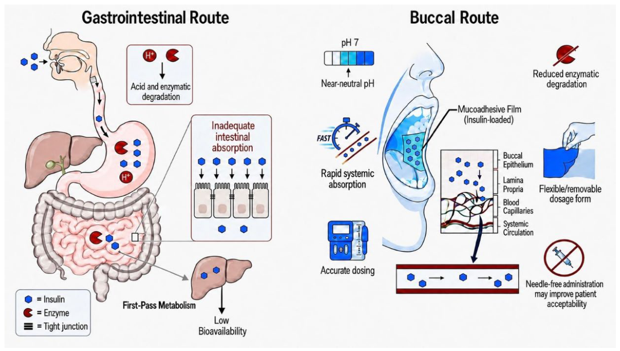
Figure 2.
Major physiological barriers to buccal insulin delivery. Source: Figure created using Figure Labs, AI Scientific Figure Illustrator (https://www.figurelabs.ai/, accessed 30 July 2026).
Figure 2.
Major physiological barriers to buccal insulin delivery. Source: Figure created using Figure Labs, AI Scientific Figure Illustrator (https://www.figurelabs.ai/, accessed 30 July 2026).
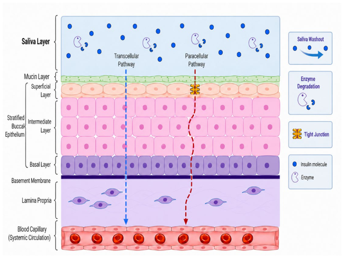
Figure 3.
Major formulation strategies for buccal insulin delivery. Source: Figure created using Figure Labs, AI Scientific Figure Illustrator (https://www.figurelabs.ai/, accessed 30 July 2026).
Figure 3.
Major formulation strategies for buccal insulin delivery. Source: Figure created using Figure Labs, AI Scientific Figure Illustrator (https://www.figurelabs.ai/, accessed 30 July 2026).
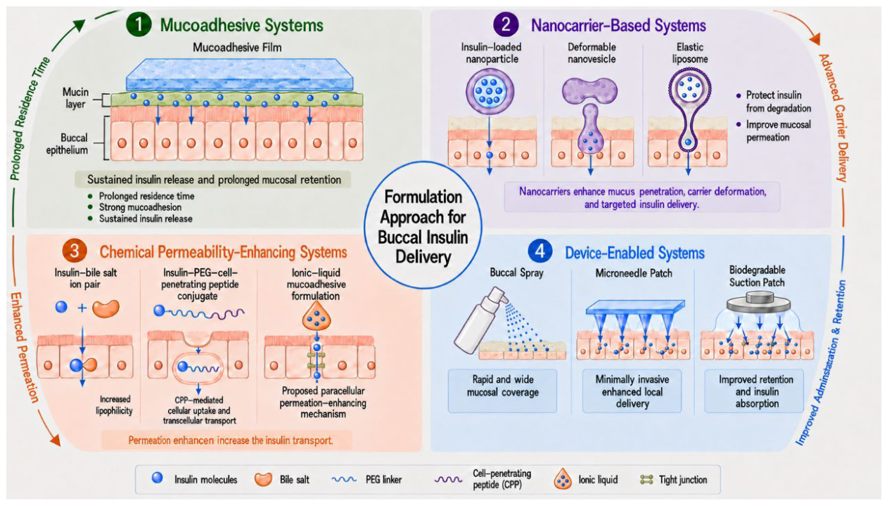
Figure 4.
Schematic overview of formulation- and storage-induced insulin degradation. Source: Figure created using Figure Labs, AI Scientific Figure Illustrator (https://www.figurelabs.ai/, accessed 30 July 2026).
Figure 4.
Schematic overview of formulation- and storage-induced insulin degradation. Source: Figure created using Figure Labs, AI Scientific Figure Illustrator (https://www.figurelabs.ai/, accessed 30 July 2026).
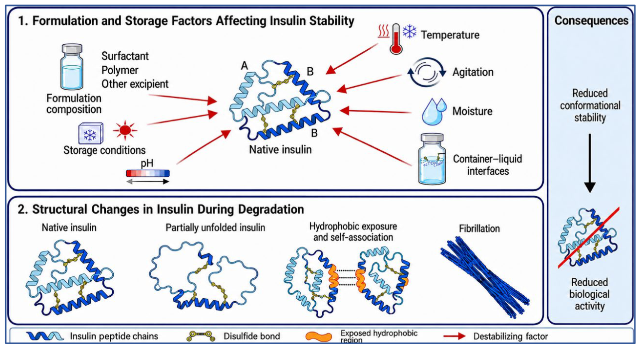

Table 1.
Overview of barriers to buccal insulin delivery.
| Barrier type | Barrier | Effect | Key feature | References |
|---|---|---|---|---|
| Physiochemical | High molecular weight and hydrophilicity | Limits absorption and permeation | Human insulin is a 51-amino acid peptide hormone with a molecular weight of approximately 5808 Da | [2,38] |
| Physical Physical Physical Physical |
Stratified buccal epithelium | Restricts diffusion | Human buccal epithelium is about 500 to 600 μm thick and consists of 40 to 50 cell layers | [39] |
| Intercellular lipid domains and tight junctions | Reduce transport | Buccal lipid domains and intercellular junctions restrict passive permeation of hydrophilic macromolecules | [6,16,39] | |
| Small effective pore radius | Limits paracellular passage | The estimated effective pore radius is about 1.5 to 3 nm (too small to readily permit passive transport of insulin) | [16] | |
| Low intrinsic permeability | Limits passive permeation | Log permeability coefficient is about −8.18 in porcine buccal tissue and −5.92 to −7.10 in TR146 models | [16] | |
| Physiological Physiological |
Salivary washout | Reduces residence time | Dilutes the drug and shortens the residence time | [6,9,39] |
| Mucus barrier | Restricts epithelial access | Mucin forms a gel barrier that can restrict diffusion and entrap insulin before epithelial contact | [6,39,40] | |
| Enzymatic | Peptidases in saliva and mucosa | Promotes insulin degradation | Includes aminopeptidases, carboxypeptidases, endopeptidases, and DPP-IV (Dipeptidyl peptidase) | [2,39] |
Table 2.
Mucoadhesive film and patch strategies for buccal insulin delivery.
| Experimental approach | Purpose of the study | Preparation method | Characterization methods | Main findings | Limitation | Reference |
|---|---|---|---|---|---|---|
| Double-layer mucoadhesive film with HPMC-PAA interpolymer complex, insulin, protamine, and ethyl cellulose backing | To improve adhesion, support unidirectional release, and enhance insulin penetration | Sequential coating with EC backing layer followed by IPC adhesive layer | IPC optimisation, viscosity and turbidity testing, adhesion force and adhesion time, release studies, pharmacokinetics, in vivo glucose evaluation (In vivo diabetic rat model) | Adhesion force 120.2 ± 20.3 N/m2, adhesion duration 491 ± 45 min, insulin AUC 2.24-fold higher than free insulin, glucose control maintained for about 8 h in diabetic rats | Preclinical evidence only, and translational dose efficiency remains uncertain | [18] |
| Chitosan-based mucoadhesive buccal film containing insulin, glycerin, and L-arginine | To provide a flexible, noninvasive film with acceptable insulin loading and buccal pharmacological activity | Film casting from a chitosan-based film-forming solution, followed by drying and unit-dose preparation | Film appearance and flexibility, insulin content by ELISA, spectroscopic analysis, and in vivo glucose-lowering study (Male Sprague-Dawley rats were used for in vivo evaluation) | Films retained at least 80% of the theoretical insulin load, reduced blood glucose by about 40%, and achieved about 57% of the subcutaneous insulin effect | Performance remained below subcutaneous insulin, and formulation properties were sensitive to composition | [19] |
| Triple-layer unidirectional mucoadhesive buccal tablet concept | To reduce salivary loss, improve directional delivery, and support a dry peptide dosage form | Conceptual multilayer design with insulin nanoparticles converted to dry powder and incorporated into an active layer with mucoadhesive and backing layers | Conceptual design, layer configuration, and polymer selection | Suggested the approach of combining nanoparticle loading, dry-state formulation, mucoadhesion, and unidirectional release in one system | Conceptual study with limited direct experimental validation for buccal insulin | [20] |
Table 3.
Nanocarrier-based strategies for buccal insulin delivery.
| Experimental approach | Composition | Method used | Key findings | Reference |
|---|---|---|---|---|
| Nanoparticle-loaded buccal film | Insulin-loaded PEG-b-PLA nanoparticles in chitosan film | Double emulsion solvent evaporation for nanoparticles, followed by solvent casting for films | Optimised nanoparticles showed a size of 215.5 ± 2.5 nm and encapsulation efficiency of about 71%; films showed homogeneous distribution and sustained release | [21] |
| Nanoparticle-based buccal film | Insulin-coated nanoparticles with D, L-valine core in ERL or ERL-HPMC film | Antisolvent co-precipitation for nanoparticles, followed by solvent casting | Optimised ERL film showed about 65% insulin release in 4 h and flux of 0.34 ± 0.05 μg/h/cm2 through human buccal mucosa model (Tridimensional human buccal tissue (EpiOral®) | [22] |
| Deformable vesicles | SPC and sodium deoxycholate deformable vesicles loaded with insulin | Reverse phase evaporation followed by hydration and sonication | Relative pharmacological bioavailability 15.59% and relative bioavailability 19.78%, higher than conventional vesicles (for in vivo study, a fasted male rabbit model was used) | [23] |
| Deformable nanovesicles | Insulin-phospholipid complex nanovesicles with lecithin, Tween 20, and sodium deoxycholate | Thin-film hydration | Optimised formulation showed particle size 85.84 ± 2.38 nm, encapsulation efficiency 77.47 ± 3.85%, and relative pharmacological bioavailability 15.53% (ex vivo porcine buccal mucosa mounted in Franz diffusion cells for insulin permeation) | [24] |
| Elastic liposomes | Insulin-loaded soy lecithin liposomes with SC or SGDC as edge activator | Thin-film hydration followed by extrusion | Optimised SGDC liposomes showed a size of 150.40 ± 3.50 nm, EE 79.41 ± 5.07%, and a 4.33-fold higher permeability coefficient than insulin solution. (ex vivo porcine buccal mucosa in Franz diffusion cells was used) | [25] |
| Gel-stabilised deformable vesicles | Insulin-phospholipid complex deformable nanovesicles in 2% gelatin matrix | Film hydration and high-pressure homogenization, followed by mixing into gelatin gel | Stable at 4 °C for 3 months with preserved delivery performance; relative pharmacological bioavailability 14.90 ± 3.12%. (TR146 human buccal epithelial cell layers model was used) | [26] |
| Elastic biosomes | Insulin-loaded elastic bilosomes modified with cholic acid derivatives | Vesicle preparation and TR146 cell permeation study | Showed facilitated insulin permeation across TR146 cells, supporting bile salt-modified flexible carriers as a transport-enhancing approach (TR146 human buccal epithelial cell layers model was used) | [41] |
Table 4.
Comparison of chemistry-led permeation strategies for buccal insulin delivery.
| Approach | Study rationale | Model used | Key permeability or bioavailability findings | Key limitation | Reference |
|---|---|---|---|---|---|
| Hydrophobic ion pairing | To increase insulin lipophilicity and improve membrane transport | TR146 cells and ex vivo porcine buccal tissue | Optimised complex produced about 3-fold higher permeability in TR146 cells and about 51.76-fold higher permeability ex vivo than insulin solution | No in vivo validation or integrated buccal dosage form | [27] |
| Ionic liquid system | To enhance buccal transport while also supporting insulin stability | Ex vivo porcine buccal tissue and in vivo rat buccal model | Choline geranate patch increased ex vivo transport about 7-fold and produced a relative bioavailability of about 5.4 ± 0.79% in vivo | Long-term safety and translational acceptability remain unclear | [29] |
| Ionic liquid mechanistic support | To stabilise insulin and reduce degradation while improving transport-related behaviour | In vitro model: Caco-2 human intestinal epithelial cell monolayers. Adult, non-diabetic male Wistar rat |
Choline geranate preserved insulin structure, reduced proteolysis, and improved delivery behaviour | Not a buccal study (oral study) |
[36] |
| Cell-penetrating peptide conjugate | To improve epithelial interaction and buccal uptake of insulin | Porcine buccal tissue, TR146 multilayers, and rabbit buccal model | Relative bioavailability up to 26.86% and pharmacological bioavailability about 21.34% | Greater molecular and formulation | [28] |
Table 5.
Device-enabled strategies relevant to buccal insulin delivery.
| Device-enabled technique | Main features of enhancing delivery | Relevance to buccal insulin delivery | Main translational challenge | References |
|---|---|---|---|---|
| Buccal spray | Human pharmacokinetic and pharmacodynamic proof of concept | Shows rapid prandial type insulin delivery in humans, with insulin  about 23 to 30 min about 23 to 30 min |
Low bioavailability, high dose burden, and variable performance across users | [13,14,15] |
| Buccal microneedle patch | Direct barrier bypass with strong acceptability data | Supports relatively rapid systemic insulin delivery by barrier bypass, with insulin  about 40 to 50 min in swine about 40 to 50 min in swine |
Stability during storage, manufacturing control, and repeat-use safety still need stronger finding | [30,32] |
| Suction or octopus-inspired patch | Noninvasive mechanical enhancement of mucosal transport | Shows that epithelial stretching and increased contact area may improve peptide delivery without needle penetration, which is attractive for future insulin systems | No insulin-specific in vivo evidence is available yet, and the required application time remains relatively long | [31,32] |
| Biodegradable suction patch | Adds sustainability to the suction-patch concept while preserving peptide delivery performance | This suggests that future buccal peptide devices may need to consider not only efficacy, but also material design suitable for repeated long-term use. |
Insulin-specific evidence is still lacking, and concerns remain about variability and storage stability. |
[43] |
Table 6.
Stability challenges and formulation implications in buccal insulin delivery.
| Stability aspect | Primary factors affecting stability | Impact on insulin or dosage form | Implications for buccal formulation performance | references |
|---|---|---|---|---|
| Post-administration biological instability | Salivary dilution, mucosal and salivary enzymes, short residence time, swallowing | Reduces the amount of structurally intact insulin available at the absorption site | This can limit absorption even when the formulation shows strong mucoadhesion or permeation enhancement. |
[2,6] |
| Native conformational instability | Heat, agitation, acidic pH, interfacial stress, and excipient-induced conformational disturbance | Promotes partial unfolding, exposure of hydrophobic regions, self-association, and fibrillation | Structural disruption reduces biological activity and compromises formulation performance | [33,34,35,44] |
| Processing-induced instability | Drying conditions, polymer-protein interactions, and thermal or mechanical stress during manufacturing | Can reduce insulin content or disrupt ordered secondary structure during dosage form preparation | Processing conditions affect whether insulin remains stable and usable in buccal films. |
[19] |
| Storage-related physical instability | Vesicle fusion, leakage, precipitation, matrix relaxation, and carrier destabilisation in hydrated systems | Alters particle size, morphology, pH, and retained insulin content during storage | Poor storage stability reduces shelf-life, reproducibility, and translational feasibility | [26] |
| Chemical instability associated with formulation environment | Nonoptimal pH, reactive hydrated microenvironments, prolonged water exposure, and excipient incompatibility | Can promote hydrolysis, aggregation, or loss of native helical structure | The formulation microenvironment is critical for preserving insulin bioactivity during storage and delivery | [33,36] |
Disclaimer/Publisher’s Note: The statements, opinions and data contained in all publications are solely those of the individual author(s) and contributor(s) and not of MDPI and/or the editor(s). MDPI and/or the editor(s) disclaim responsibility for any injury to people or property resulting from any ideas, methods, instructions or products referred to in the content. |
© 2026 by the authors. Licensee MDPI, Basel, Switzerland. This article is an open access article distributed under the terms and conditions of the Creative Commons Attribution (CC BY) license (http://creativecommons.org/licenses/by/4.0/).
Copyright: This open access article is published under a Creative Commons CC BY 4.0 license, which permit the free download, distribution, and reuse, provided that the author and preprint are cited in any reuse.