

Submitted:
10 August 2026
Posted:
11 August 2026
You are already at the latest version
Abstract
Biological stress and aging are traditionally understood primarily through stressor-specific molecular mechanisms, including osmoregulation, stress-signaling pathways, proteostasis impairment, mitochondrial dysfunction, membrane remodeling, and cellular senescence. Despite substantial differences in their initiating mechanisms, diverse stressors and age-related changes are often accompanied by a common functional consequence: a reduced capacity of the cell to reversibly reorganize its intracellular environment and restore a functionally competent state after stressor removal. Here, we propose a unified biophysical theory of aging and stress based on phase plasticity, which considers the capacity of cellular matter to undergo controlled and reversible physical reorganization as a potential integrative level linking molecular mechanisms with functional recovery. The central concept of the model is phase plasticity, defined as the capacity of the cytoplasm, membranes, biomolecular condensates, and intracellular compartments to transition between functionally distinct physical states while retaining the capacity for reversible restoration. On this basis, Phase Rigidity Syndrome (PRS) is introduced as a hypothetical integrative state that may emerge from the progressive and cumulative loss of phase plasticity. We propose that phase rigidity is associated with reduced molecular mobility, altered biomolecular condensate dynamics, impaired membrane fluidity, diminished proteostatic capacity, and delayed or incomplete recovery following stress. Under prolonged or repeated stress, these changes may mutually reinforce one another, promoting a transition from adaptive phase reorganization toward persistent phase rigidity and, under extreme conditions, phase collapse. The theoretical framework integrates the concepts of phase homeostasis, phase buffer, phase resilience, phase recovery, phase memory, phase threshold, and phase-transition window, and introduces a conceptual Phase Resilience Index (PRI) for characterizing the capacity of cells to maintain and restore phase organization. Importantly, PRS is not proposed as an established biological or clinical syndrome, nor is phase plasticity presented as a validated universal variable. Rather, the proposed framework represents a research hypothesis that requires experimental testing. The proposed theory does not replace existing molecular models of cellular stress and aging but introduces an additional biophysical level of integration, at which distinct primary perturbations may partially converge through changes in the capacity of cellular matter to undergo reversible physical reorganization. The scientific value of the framework will ultimately depend on whether measures of phase plasticity provide additional predictive information on cellular resilience and recovery beyond established molecular markers of stress and aging.
Keywords:
Phase Rigidity Syndrome
; phase homeostasis
; phase plasticity
; cellular stress
; aging
; liquid-liquid phase separation
; biomolecular condensates
; cytoplasmic viscosity
; proteostasis
; cellular resilience
; cellular biophysics
; stress recovery
1. Introduction
Cell biology has accumulated, over the past several decades, remarkably detailed mechanistic accounts of how cells respond to individual categories of stress. Drought and osmotic stress are understood largely through osmosensing, abscisic acid (ABA) signaling, and compatible-solute accumulation in plants [1,2]. Salinity stress adds ion-transport and compartmentalization mechanisms specific to Na+ and Cl- handling. Heat stress is understood through heat-shock factor activation and chaperone induction. Cold and freezing stress recruit membrane-remodeling and, in some organisms, ice-binding proteins [3]. Heavy-metal toxicity is handled through metallothionein- and phytochelatin-mediated chelation [4,5]. Oxidative stress is addressed through an extensive antioxidant and redox-signaling literature [6,7]. Chronic psychosocial and physiological stress in animals has its own literature built around the hypothalamic-pituitary-adrenal axis and the concept of allostatic load [8]. Biological aging, finally, has its own largely separate literature, currently organized around twelve interconnected hallmarks spanning genomic instability, epigenetic drift, loss of proteostasis, mitochondrial dysfunction, and cellular senescence, among others [9,10].
Each of these bodies of work is mechanistically well grounded within its own domain, and we do not intend to suggest otherwise. Our starting observation is instead about what happens at the boundaries between them. Several molecularly unrelated interventions — mild heat, sub-lethal osmotic stress, brief hypoxia, and caloric restriction among others — produce broadly overlapping downstream signatures: activation of stress granules and other biomolecular condensates, changes in chaperone and autophagic flux, altered membrane order, and shifts in the TOR/mTOR–SnRK1/AMPK energy-sensing axis [11,12,13]. This overlap is not total, and it would be a mistake to read it as evidence that all stressors converge on one molecular pathway; the primary lesions caused by desiccation, heat, and heavy-metal exposure are chemically and structurally distinct, and the literature is right to treat them as such. What the overlap does suggest, in our reading, is that beneath the pathway-specific molecular responses there may be a higher level of cellular organization — the physical, phase-organizational state of the cytoplasm and its compartments — on which these otherwise distinct responses converge, and at which the reversibility of the stress response is ultimately determined.
This raises a question that, to our knowledge, existing stress-specific and aging-specific frameworks do not directly address: is there a physical, rather than purely biochemical, variable that governs whether a given episode of cellular stress remains reversible or crosses into a state from which recovery is incomplete or absent? We propose phase plasticity — the capacity of cytoplasmic and membrane-associated material to undergo reversible transitions among physical organizational states — as a candidate for that variable. Building on this proposal, and on our own prior work developing a phase-homeostasis framework for stress adaptation [14], we introduce Phase Rigidity Syndrome (PRS) as a hypothesis: the proposal that progressive, cumulative loss of cytoplasmic phase plasticity constitutes a common, measurable integrating factor linking otherwise distinct stressors, impaired recovery, and age-related functional decline. From the outset, we emphasize that PRS is not an established syndrome, is not equivalent to liquid–liquid phase separation (LLPS) considered in isolation, is not simply another term for aging, and should not be equated with cell death. Rather, PRS is proposed as an integrative conceptual construct, whose scientific value depends entirely on whether it can be operationalized, experimentally tested, and ultimately subjected to falsification.
2. Why Existing Stress Models Do Not Fully Account for the Convergence of Stress Phenotypes
2.1. Aging
Aging is presently described through an expanding set of interconnected hallmarks — genomic instability, telomere attrition, epigenetic alteration, loss of proteostasis, disabled macroautophagy, deregulated nutrient sensing, mitochondrial dysfunction, cellular senescence, stem cell exhaustion, altered intercellular communication, chronic inflammation, and dysbiosis [9]. This framework is explicitly non-reductive: its authors do not claim that any single hallmark is causally primary, and the list itself has grown as new mechanisms have been incorporated. What the hallmarks framework does not provide is an account of why these particular processes cluster together mechanistically rather than being an arbitrary list of things that happen to change with age. Several of the hallmarks — loss of proteostasis, altered membrane lipid composition [15], and mitochondrial dysfunction — have a shared physical consequence that is rarely made explicit: each independently degrades the cell’s capacity to maintain and restore an ordered, functionally competent physical organization of its cytoplasm and membranes.
2.2. Drought and dehydration
The initiating disturbance in drought stress is a reduction in cellular water potential, which is sensed through mechanisms that remain incompletely characterized at the molecular level in plants but that converge on ABA biosynthesis and PYR/PYL/RCAR receptor signaling [1]. The primary molecular response — accumulation of compatible solutes, LEA-protein expression, stomatal closure — is well described [2,16]. The physical consequence of water loss, however, extends beyond any single signaling pathway: it directly increases macromolecular crowding and cytoplasmic viscosity as the same complement of macromolecules occupies a progressively smaller aqueous volume [17]. Treating dehydration purely as an osmotic-signaling problem risks missing this direct biophysical consequence, which is present even before any transcriptional response has occurred.
2.3. Salinity
Salt stress combines an osmotic component, mechanistically overlapping with drought, with ion-specific toxicity from Na+ and Cl- accumulation. The literature on ion transport (SOS pathway, tonoplast Na+/H+ antiporters) and on compatible-solute-mediated osmotic adjustment is extensive and well validated. High intracellular ionic strength additionally perturbs protein surface charge and hydration shells in ways that are physically distinct from, though co-occurring with, the crowding effects of drought — a distinction that a purely osmotic-signaling account tends to collapse.
2.4. Heat stress
Heat stress increases the kinetic energy of molecular motion, which would be expected to increase, not decrease, molecular mobility; the well-documented consequence for the proteome, however, is destabilization of native protein folds and a shift toward aggregation-prone conformations, countered by heat-shock-factor-driven chaperone induction. The apparent paradox — a stressor that increases thermal energy nonetheless being associated, at the proteomic level, with reduced functional mobility of client proteins once they aggregate — is a first illustration of why kinetic energy and phase-organizational mobility are not the same physical quantity, a distinction we return to in Section 4.
2.5. Cold and freezing
Cold stress reduces membrane fluidity directly, through decreased lipid acyl-chain motion, and organisms counteract this through homeoviscous adaptation — regulated desaturase activity and altered lipid headgroup composition [18,19]. Freezing adds the further, distinct physical problem of ice nucleation, countered in some organisms by ice-binding proteins that control ice crystal growth rather than preventing freezing outright [3]. Cold and freezing therefore illustrate two physically distinct mechanisms — reduced membrane fluidity and ice-crystal mechanics — that are often discussed together under a single stress category despite requiring different physical descriptions.
2.6. Heavy-metal toxicity
Heavy-metal exposure is chemically distinct from the preceding categories: its primary lesion is direct coordination of metal ions to protein thiol and other reactive groups, addressed through phytochelatin- and metallothionein-mediated chelation [4,5]. Unrepaired, this coordination chemistry can drive non-specific protein cross-linking and aggregation, a physical consequence that again converges, at the level of outcome, with the aggregation phenotypes seen under heat and oxidative stress, despite an entirely different initiating chemistry.
2.7. Oxidative stress
Reactive oxygen species (ROS) are increasingly understood as physiological signaling molecules operating within a normal concentration range, rather than as purely damaging agents to be minimized at all costs [6,7]. Excess ROS, however, oxidizes cysteine thiols, lipids, and nucleic acids, and oxidative modification of stress-granule and other condensate-resident proteins has been directly implicated in pathological liquid-to-solid transitions of biomolecular condensates [20,21]. Oxidative stress therefore provides one of the more direct mechanistic links between a specific, well-characterized chemical insult and a phase-organizational consequence, though we emphasize that this link has been demonstrated for specific proteins (notably TDP-43 within stress granules) and should not be generalized without qualification to all condensates or all oxidative conditions.
2.8. Chronic stress
Chronic, low-grade stress in animals is addressed by the allostatic-load framework, which explicitly models the cost of repeated or sustained physiological adjustment as distinct from the adjustment itself [8,22]. This framework already anticipates, at the level of organismal physiology, a central claim we will make at the cellular level: that the capacity to return to baseline after a perturbation, not the perturbation itself, is what determines whether a stress response remains adaptive or becomes pathological. We treat allostasis as a conceptual precedent for PRS at a different level of biological organization, not as an equivalent framework, since allostatic load is defined primarily through neuroendocrine and systemic physiological markers rather than through direct measurement of subcellular phase behavior.
None of the eight cases above shares a common initiating chemistry or a common primary molecular target. What they share, we argue, is a physical consequence for the cell as an organized material system: each perturbs, through a different route, the same small set of physical variables — crowding, viscosity, membrane order, condensate dynamics — that jointly determine whether the cell’s organization remains reversible.
3. Central Hypothesis: Phase Rigidity Syndrome
We propose that progressive loss of cytoplasmic phase plasticity may function as a shared, higher-order determinant of stress vulnerability, impaired recovery, and age-associated functional decline, operating alongside — not in place of — the pathway-specific mechanisms reviewed above. We call this proposed condition Phase Rigidity Syndrome. We want to be explicit about the epistemic status of this proposal: PRS is a hypothesis and an organizing construct, not a documented clinical or cell-biological syndrome with an agreed diagnostic definition. No study, to our knowledge, has measured the full set of variables we associate with PRS in a single system or has demonstrated that a composite phase-plasticity measure outperforms existing biochemical stress markers in predicting recovery.
The rationale for treating phase plasticity as a variable of a qualitatively different order than any individual molecular pathway rests on three observations. First, cellular biochemistry is diffusion-limited: reaction rates for encounter-limited processes depend directly on the translational and rotational mobility of the reactants, which is itself a function of cytoplasmic viscosity and crowding rather than of any single signaling pathway [23,24]. A change in bulk physical state can therefore alter the rate of many biochemical processes simultaneously, without any of those processes being individually perturbed at the level of enzyme structure or gene expression. Second, reversibility is not guaranteed by the removal of a stressor: several of the phase transitions discussed in this manuscript, once a critical threshold of protein concentration, oxidative modification, or aggregation is exceeded, are kinetically far more difficult to reverse than to initiate, an asymmetry with direct parallels in the physics of glass transitions and gelation. Third, and most speculatively, we suggest that loss of mobility may itself become self-reinforcing rather than remaining a passive readout of upstream damage: reduced diffusion slows the delivery of chaperones and quality-control machinery to sites of protein misfolding, which in turn favors further aggregation and further loss of local mobility. This third point is, at present, a mechanistic inference drawn from the general physics of diffusion-limited repair rather than a direct experimental demonstration within the specific context we are proposing, and we flag it accordingly (Figure 1).
4. Theory of Cellular Phase Plasticity
We propose the following terminological system to make the phase-organizational level of description explicit and, eventually, measurable. These terms are introduced as proposed constructs; several build on established physical concepts (phase transition, glass transition, hysteresis) applied here to a specific biological context, while others (Phase Resilience Index, Phase Rigidity Syndrome itself) are, to our knowledge, new to the literature.
Phase homeostasis denotes the capacity of a cell to maintain functionally appropriate, dynamically regulated physical states of its cytoplasm and compartments despite external perturbation. Phase plasticity denotes the capacity of cellular material to undergo reversible transitions between physical organizational states in response to changing conditions; it is the component capacity that makes phase homeostasis achievable. Phase buffer denotes the aggregate capacity of a cellular system to absorb a physical or chemical perturbation without loss of reversibility — operationally, the distance between the current cell state and the nearest threshold beyond which recovery becomes incomplete. Phase resilience denotes the capacity to maintain or restore phase organization following a perturbation, a concept related to but not identical with phase buffer: buffer capacity describes how much perturbation can be absorbed before a threshold is approached, while resilience describes how effectively the system returns once that threshold has been approached or crossed.
Phase rigidity denotes a state characterized by reduced molecular mobility, reduced reversibility of phase transitions, impaired adaptive transitions, and increased resistance to functional recovery. Phase recovery denotes the process by which a cell restores a prior or functionally competent phase organization after stress has been removed. Phase collapse denotes a transition beyond which the cell can no longer maintain reversible phase organization and enters a dysfunctional state; we treat phase collapse as continuous with, but not synonymous with, cell death, since a cell may in principle persist in a phase-collapsed but not yet dead state. Phase memory denotes persistent alterations in physical cellular organization that influence subsequent responses to perturbation, a construct with a partial precedent in the transcriptional- and chromatin-memory literature but extended here explicitly to physical, rather than purely regulatory, persistence. Phase threshold denotes a critical region beyond which phase changes become strongly nonlinear, poorly reversible, or functionally damaging. Phase transition window denotes the period during which a cell can still be returned from an adaptive toward a pathological phase trajectory, or vice versa, by intervention.
We further propose a Phase Resilience Index (PRI) as a conceptual, integrative measure intended to summarize the recovery kinetics, mobility, reversibility, and adaptive capacity of phase transitions within a single operational metric. We emphasize that PRI is currently proposed only as a conceptual target for the future development of appropriate measurement methods and tools; at present, no validated measurement protocol or normative range of values has been established.
These constructs relate to one another hierarchically, though we caution against treating the hierarchy as a strict linear pipeline. Under mild or transient perturbation, phase homeostasis is maintained through phase-buffer capacity acting within a regime of intact phase plasticity, and any departure from the baseline organizational state is corrected through phase recovery. Under sustained, severe, or repeated perturbation, phase-buffer capacity may become progressively depleted; the cell then enters a state of phase rigidity, in which recovery becomes slower, partial, or eventually absent, and in the most severe case the trajectory terminates in phase collapse. Phase memory, accumulated across repeated excursions toward phase rigidity, may in turn narrow the phase transition window available for subsequent stress episodes — a proposed mechanism by which repeated sub-threshold stress could, over time, lower the threshold for PRS onset, though we are not aware of direct experimental confirmation of this specific proposed link at the phase-organizational level, as distinct from the better-established transcriptional stress-memory literature (Table 1).
5. Biophysical Mechanisms Contributing to PRS
The mechanistic picture we propose is not a single linear signaling cascade, and we want to resist the natural tendency of a schematic diagram to imply strict sequential causation. Stress perturbs hydration and osmotic state, which alters macromolecular crowding [17]; crowding influences the propensity of proteins and RNA to undergo LLPS [25]; the physical state of biomolecular condensates and of the bulk cytosol influences, and is influenced by, membrane lipid order [18]; all of these variables jointly determine cytoplasmic viscosity and the anomalous diffusion coefficients of macromolecules [23,26]; diffusion in turn gates the efficiency of proteostatic quality control, since chaperone-client encounter is itself diffusion-dependent [27,28]; cytoskeletal organization both constrains and is constrained by local viscosity and crowding [29]; mitochondrial function depends on and contributes to local ATP availability, which feeds back onto the TOR/mTOR–SnRK1/AMPK axis [11,13], itself now known to directly tune cytoplasmic crowding and phase-separation propensity through mTORC1 activity [25]; and ROS/redox state both responds to and modulates several of the preceding nodes, including condensate aggregation propensity through cysteine oxidation [7,20]. In plants specifically, ABA signaling provides an additional, partially parallel integrating layer that couples osmotic perception to metabolic suppression [1,30], and we do not claim that this layer has a direct animal-cell counterpart.
We consider it important to distinguish, within this network, three categories of relationship. Some links are supported by direct causal evidence: mTORC1 activity has been shown experimentally to tune cytoplasmic crowding [25]; glucose starvation has been shown to reduce macromolecular mobility through an ATP-dependent mechanism [31]; cytosolic acidification has been shown to drive a fluid-to-solid cytoplasmic transition during dormancy entry [32]; and specific oxidative modifications of TDP-43 have been shown to drive pathological condensate hardening [20]. Other links are correlational, established by co-occurrence across many independent studies without a demonstrated causal mechanism connecting them directly — for example, the frequent co-occurrence of reduced membrane fluidity and altered condensate dynamics under a shared stressor, which to our knowledge has rarely been measured simultaneously in the same experimental system. Still other links, including the general proposition that reduced phase plasticity itself, rather than any single upstream lesion, is what predicts impaired recovery, are at present conceptual extrapolations from the physics of glassy and gel-forming systems rather than demonstrated biological facts, and we present them as the hypothesis under test rather than as background (Figure 2).
6. LLPS and Biomolecular Condensates
Liquid-liquid phase separation and the biomolecular condensates it produces have become one of the most active areas of cell biology over the past decade, and we do not attempt an exhaustive review here; several excellent treatments exist [33,34,35,36]. For the purposes of this framework, the central fact is that condensates are not a fixed material class but occupy a continuum of material states, from highly dynamic liquids with rapid component exchange, through more viscous liquid or gel-like states, to solid, poorly exchanging aggregates [34]. Reversible condensate formation under stress — stress granules being the best-characterized example [37] — is generally understood as adaptive, sequestering translationally silent mRNAs and signaling components in a manner that is rapidly reversible once stress is relieved [38]. The same condensate class, however, can undergo an aging process in which liquid-like material properties are progressively lost in favor of more solid, gel- or amyloid-like states, and this transition has been directly linked, in specific well-studied cases, to neurodegenerative pathology — the transformation of TDP-43-containing stress granules into pathological aggregates in ALS and frontotemporal dementia being the clearest documented example [20,21].
We do not present LLPS as a universally protective or universally pathological process; the evidence supports neither generalization. What we propose is more specific: that loss of condensate reversibility — the transition from a liquid or gel state that readily dissolves upon stress relief to one that does not — may be one measurable component contributing to phase rigidity, alongside membrane, cytoskeletal, and viscosity changes discussed in Section 7, rather than being, by itself, equivalent to phase rigidity. This distinction matters because a cell could in principle show entirely normal condensate dynamics while still exhibiting impaired membrane fluidity, mitochondrial function, or diffusive mobility, or vice versa; PRS, as we define it, requires convergent impairment across multiple physical layers, not a phenotype in any single one.
7. Membranes, Cytoplasmic Viscosity, Diffusion, and Proteostasis
Membrane lipid order and cytoplasmic viscosity are frequently treated as separate topics — one belonging to membrane biophysics, the other to cytosolic biochemistry — but they are physically coupled aspects of the same cell. Homeoviscous adaptation demonstrates that membrane fluidity is not a passive consequence of temperature but an actively regulated variable, defended through desaturase activity and headgroup remodeling across bacteria, plants, and animals [18,19]. Bulk cytoplasmic viscosity and the closely related quantity of macromolecular crowding are likewise actively regulated rather than passively determined: mTORC1 signaling tunes crowding directly [25], ATP depletion increases cytoplasmic solidity in a manner distinct from simple concentration effects [31,39], and the bacterial cytoplasm has been shown directly, using particle-tracking microrheology, to behave as a metabolically fluidized glass-forming material rather than a simple aqueous solution [40,41].
These physical variables are not downstream readouts of proteostasis; they are among its preconditions. Chaperone-client encounter, ubiquitin-proteasome substrate delivery, and autophagosome-lysosome fusion are all transport-dependent processes whose rates are set in part by the diffusive and viscoelastic properties of the surrounding cytoplasm [27,28,42]. Cytoskeletal organization both depends on and constrains local cytoplasmic mechanics, with recent work showing that cytoplasmic viscoelastic properties directly modulate microtubule polymerization kinetics [29], and organelle mobility, including that of mitochondria and autophagosomes, is similarly gated by the physical state of the surrounding medium. We propose, as the central conceptual claim of this section, that molecular mobility should be treated not merely as a passive consequence of stress-induced damage but as a variable that itself feeds back on the cell’s capacity for repair — a claim that follows naturally from the transport-dependence of the proteostasis and autophagy machineries just described, but that has not, to our knowledge, been tested directly as a general principle across stress types.
8. The Continuum of Cellular Phase States
We do not think PRS is well represented as a single threshold or a binary switch between an adaptive and a pathological state. A more appropriate representation, borrowed from the language of nonlinear dynamical systems [43], treats the cell as occupying a position within a multidimensional state space,
X(t) = {phase organization, viscosity, diffusion coefficient, membrane fluidity, condensate dynamics, proteostatic capacity, redox state, energy state, ...},
whose trajectory over time is shaped jointly by external perturbation and internal regulatory dynamics. Within this space, we propose that regions of relatively high phase plasticity constitute basins of attraction toward which the system returns after small perturbations, consistent with the general behavior of attractor dynamics in physiological systems, and that sufficiently large or sustained perturbations can push the trajectory across a separatrix into a different basin from which return to the original attractor is no longer the default outcome. This framing accommodates hysteresis — the empirical observation, well documented in ecological and climate systems undergoing critical transitions [44,45], that removing the perturbation which caused a transition does not necessarily restore the pre-transition state — as a structural feature of the model rather than an anomaly requiring separate explanation.
Under this framing, PRS corresponds not to a single point in state space but to a region or trajectory characterized by reduced local basin depth (weakened phase buffer), slowed or incomplete return kinetics following perturbation (reduced phase resilience), and progressive contraction of the phase transition window within which corrective intervention remains effective. We regard this representation as a genuinely useful organizing framework for generating hypotheses, and we note explicitly that it is, at present, descriptive rather than derived from a fitted dynamical model of any specific cellular system; the variables in X(t) are measurable in principle, using the techniques discussed in Section 13, but a jointly parameterized model spanning all of them has not, to our knowledge, been constructed for any single stress or aging paradigm (Figure 3).
9. Aging as a Slow Trajectory Toward Phase Rigidity
We propose that biological aging may involve, alongside the well-established hallmark processes, a gradual reduction in cytoplasmic phase plasticity, and we want to be careful about the scope of this claim. We are not proposing that PRS replaces or supersedes the hallmarks-of-aging framework [9]; we are proposing an additional, cross-cutting physical layer that may help explain why several hallmarks — loss of proteostasis, mitochondrial dysfunction, and altered membrane lipid composition among them — tend to co-occur and to interact synergistically rather than behaving as independent, additive processes. Age-related decline in autophagic flux [46,47], in chaperone-mediated autophagy specifically [48], and in overall proteostatic network capacity [27,42] each independently reduces the cell’s capacity to reverse protein misfolding and aggregation once it occurs. The mitochondrial free radical theory of aging, once treated as close to a complete explanation, has since been substantially qualified by evidence that ROS manipulation does not consistently track with longevity across experimental systems in the manner the theory originally predicted [49]; a more nuanced, network-level view of mitochondrial dysfunction as one contributor among several is now more consistent with the evidence [50,51,52], and we adopt that more cautious framing here rather than treating mitochondrial ROS output as a primary driver in its own right. Cellular senescence, with its associated senescence-associated secretory phenotype, represents a further, well-documented state in which cells persist in a viable but non-dividing condition with altered secretory behavior [53,54,55]; whether senescent cells represent an extreme, stable point on the phase-rigidity continuum we describe, or a mechanistically distinct cell-cycle-arrest phenomenon that happens to co-occur with reduced phase plasticity, is at present an open question that our framework does not resolve.
A specific, testable implication of treating aging as a trajectory toward reduced phase plasticity concerns the phase transition window introduced in Section 4. We propose that this window narrows with age — that is, that older cells retain less capacity to be returned, by a given corrective intervention, from an incipiently rigid state back toward an adaptive one — and that this narrowing may manifest as an asymmetry between young and aged cells in their post-stress recovery trajectories: a young, resilient cell subjected to stress is expected to show a pattern of perturbation followed by comparatively complete recovery, whereas an aged, less plastic cell subjected to a comparable stress is expected to show perturbation followed by incomplete recovery and persistence in a durably altered state, a pattern with the qualitative signature of hysteresis. This is, again, a prediction rather than a documented finding; we are not aware of a study that has directly compared post-stress phase-recovery kinetics, using the biophysical methods described in Section 13, between young and aged cells of the same type (Figure 4).
10. Convergence of Distinct Stressors on PRS
Table 2 summarizes the eight stress categories discussed in Section 2, alongside the primary biophysical challenge each poses, the early molecular or cellular response, the proposed disruption to phase homeostasis, and the proposed relationship to PRS. We use deliberately qualified language throughout the table — “may contribute to,” “is consistent with,” “may be associated with” — because the right-hand columns describe interpretation and hypothesis, not established fact, in every row. The point of the table is not to claim that these eight stressors act through a common molecular pathway; Section 2 argues explicitly against that reading. The point is to show that despite their molecular heterogeneity, each stressor plausibly intersects the same small set of physical variables — hydration, crowding, viscosity, membrane order, condensate dynamics — introduced in Sections 4 through 7, and that the proposed convergence occurs at this physical level rather than at the level of any shared upstream signal.
Thus, despite the diversity of their primary causes, different forms of stress converge toward a common biophysical outcome—the progressive loss of cellular phase plasticity. Within the Phase Rigidity Syndrome (PRS) framework, stressors are not considered independent pathological processes but rather as distinct inputs into a common phase transition from an adaptive state characterized by high phase flexibility toward a state of phase rigidity. This transition is characterized by reduced reversibility, impaired LLPS dynamics, decreased membrane plasticity, and constrained cellular resilience (Figure 5).
11. Biomarkers of Phase Rigidity Syndrome
A useful biomarker of PRS would need to distinguish cells or tissues with reduced phase plasticity from those with intact plasticity, ideally before functional failure becomes apparent through conventional means. Several existing biophysical measurements are directly relevant, though none was developed specifically for this purpose and none currently has an established normative range or validated threshold corresponding to a “rigid” versus “plastic” cellular state. Condensate dynamics and dissolution kinetics can be assessed through FRAP, which measures the recovery of fluorescence in a photobleached region as a function of unbleached-molecule diffusion and exchange, and which has been directly applied to distinguish liquid-like from more solid condensate states [37,56]. Bulk and local cytoplasmic diffusion coefficients can be measured through fluorescence correlation spectroscopy and through single-particle tracking microrheology, both of which have been used to characterize the glass-like behavior of bacterial and eukaryotic cytoplasm directly [23,40,41]. Membrane order can be assessed through fluorescence anisotropy of order-sensitive membrane probes, a well-established but not condensate-specific technique. Organelle and macromolecule mobility more broadly can be assessed through live-cell particle tracking. Proteostatic capacity has established, though indirect, readouts through reporter-substrate degradation assays and through direct measurement of chaperone and autophagic flux.
We propose, as a genuinely new element of this framework, that a PRS biomarker panel would need to combine several of these measurements — condensate dynamics, diffusion coefficient, membrane order, and a functional recovery-time measurement following a defined perturbation — into a composite readout, rather than relying on any single parameter, precisely because our central claim is that PRS is a convergent, multi-layer phenomenon rather than a single-parameter one. Table 3 lists candidate biomarkers, the biological property each addresses, the measurement method, and the principal advantages and limitations of each; we have deliberately not proposed numerical thresholds, since none currently exists and inventing one would misrepresent the state of the evidence.
12. A Conceptual Quantitative Framework
We offer the following as a conceptual scaffold for future quantitative work, not as a validated or experimentally calibrated model. Phase plasticity, P, can be represented as a function of the principal physical variables discussed above:
where D is a measure of molecular diffusion, eta is cytoplasmic viscosity, M is membrane fluidity, C is a measure of condensate dynamics (for example, FRAP recovery half-time or mobile fraction), R is proteostatic or repair capacity, and E is cellular energy state. We do not specify a functional form for f; doing so without data to constrain it would create a false impression of quantitative rigor. We further propose a Phase Resilience Index, PRI, conceived as a function of recovery kinetics, baseline mobility, and the reversibility of phase transitions following a defined perturbation — again presented as a conceptual target rather than a derived formula.
P(t) = f(D, eta, M, C, R, E)
For the temporal dynamics of phase rigidity, one can write, purely as a theoretical representation of the qualitative balance we have described throughout this manuscript,
where the two terms on the right-hand side are placeholders for the phase-buffer and repair mechanisms discussed in Sections 4 through 7 and for the aggregation-, oxidation-, and crowding-driven processes that oppose them, respectively. We want to be unambiguous that this equation has not been parameterized, fitted, or experimentally validated in any system; we include it only to make explicit, in compact form, the balance of processes the rest of the manuscript describes in prose, and we regard its calibration against real measurements as a distant, not an immediate, goal of the research program we outline in Section 18.
dP/dt = (adaptive restorative processes) - (rigidity-promoting processes),
13. Experimental Measurement of PRS
The methods relevant to measuring the components of PRS are, individually, well established in other contexts; what is missing is their systematic joint application to a single stress or aging paradigm. FRAP and fluorescence correlation spectroscopy (FCS) remain the standard tools for measuring molecular mobility and condensate exchange dynamics in living cells [56], though both require careful attention to photobleaching artifacts, fitting-model assumptions, and the distinction between diffusive and binding-limited recovery. Fluorescence anisotropy provides a complementary readout of local rotational mobility and membrane order. Microrheology, whether based on tracking of endogenous granules [40] or of introduced tracer particles, provides direct estimates of viscoelastic moduli and has been central to establishing the glass-like behavior of the cytoplasm. Confocal and live-cell imaging more generally allow direct visualization of condensate morphology, number, and coalescence behavior over time. Proteostatic capacity can be assessed through established reporter systems and through direct biochemical measurement of chaperone and autophagic flux. Mitochondrial function is accessible through standard respirometry and membrane-potential assays. Metabolomic, transcriptomic, and single-cell profiling provide complementary molecular context that would be needed to interpret any observed phase-plasticity changes mechanistically rather than merely descriptively.
Integrating these measurements into a composite index, as proposed in Section 11, is a data-analytic and instrumentation challenge rather than a conceptual one, and approaches from machine learning and dynamical systems modeling are plausible tools for that integration once sufficient multi-parameter datasets exist. We note explicitly, given the current enthusiasm for such approaches, that the use of machine learning or of “digital twin” concepts, already explored in other areas of physiology [57], would not itself constitute evidence for PRS; these are analytic tools that could in principle be applied to test the framework, not independent sources of biological validation. Longitudinal single-cell measurement, tracking the same cells through a stress-recovery cycle or across a substantial fraction of a replicative or organismal lifespan, would be needed to test the trajectory-based claims made in Section 8 and Section 9, and such longitudinal single-cell biophysical datasets, to our knowledge, do not yet exist at the scale this framework would require (Figure 6).
14. Experimental Predictions and Falsifiability
A conceptual framework that cannot be shown to be wrong is not, in our view, worth proposing as a scientific hypothesis rather than as a metaphor. We list below eight predictions that follow from the PRS framework as developed in this manuscript, together with the observation that would weaken or refute each one. Table 4 presents these in summary form.
Prediction 1. Different stressors, despite molecularly distinct primary lesions, should show partially convergent trajectories of declining phase plasticity when measured with a common biophysical panel (Section 13). Falsifying observation: if phase-plasticity trajectories under drought, heat, and heavy-metal stress are found to be uncorrelated or to diverge systematically rather than converge, this would weaken the case for a shared physical layer beneath these stressors.
Prediction 2. Changes in membrane fluidity and in intracellular molecular mobility should correlate with changes in condensate dynamics across stress types, even though the three variables are governed by partially distinct molecular machinery. Falsifying observation: consistent absence of correlation between membrane-order and condensate-dynamics measurements across multiple stress paradigms would argue against treating these as coupled components of a single phase-organizational system.
Prediction 3. Recovery of phase plasticity following stress removal should predict subsequent functional recovery (for example, resumption of growth or division) better than individual biochemical stress markers, at least in some experimental systems. Falsifying observation: if biochemical markers such as HSP induction or ROS titer consistently outperform composite phase-plasticity measures as predictors of functional recovery, the added value of the phase-organizational framework would be undermined.
Prediction 4. Cells with lower baseline phase plasticity should show greater vulnerability to a second, subsequent stressor than cells with higher baseline plasticity, independent of the specific identity of either stressor. Falsifying observation: absence of such cross-sensitization, or its full explanation by a shared biochemical pathway rather than by baseline phase-organizational state, would weaken this prediction.
Prediction 5. Aging should be associated with a measurable narrowing of the phase transition window, operationalized as reduced responsiveness to interventions that would restore phase plasticity in younger cells. Falsifying observation: if aged and young cells show equivalent responsiveness to such interventions once matched for the severity of the initiating insult, the proposed narrowing would not be supported.
Prediction 6. Interventions that preserve or restore phase plasticity should improve functional recovery even when they do not directly neutralize the primary stressor. Falsifying observation: if phase-plasticity-directed interventions fail to improve recovery independent of their effect on the primary stress signal, this would suggest that phase plasticity is a correlate rather than a determinant of recovery.
Prediction 7. Phase rigidity should exhibit hysteresis: removal of the primary stressor should not reliably or immediately restore the pre-stress phase-organizational state. Falsifying observation: if phase-organizational parameters consistently and rapidly return to baseline immediately upon stressor removal across the systems tested, the hysteresis prediction central to Section 4 and Section 8 would be undermined.
Prediction 8. Interventions acting through molecularly distinct mechanisms (for example, a chaperone inducer and a membrane-fluidity modulator) should converge on similar improvements in phase-recovery kinetics, even though they act on different molecular targets. Falsifying observation: if mechanistically distinct interventions produce phase-recovery outcomes that simply track their known, distinct molecular targets with no evidence of convergence at the phase-organizational level, this would argue against treating phase plasticity as an integrating variable (Figure 7).
15. Practical and Translational Directions
We discuss the following applications as motivating directions for future work, not as demonstrated or currently available interventions. In stress biology and agronomy, a phase-plasticity-informed approach could, in principle, complement existing biochemical stress diagnostics and inform the design of biostimulant or agronomic interventions targeted at specific components of the phase system (osmotic buffering, membrane stabilization, chaperone support) rather than at a single stressor in isolation, extending the “Regulatory Agronomy” perspective we have outlined elsewhere [58]. In geroscience, compounds or interventions already under investigation for their effects on proteostasis, autophagy, and mitochondrial function could in principle be re-evaluated for their effects on phase-plasticity measures specifically, which might reveal shared or divergent mechanisms of action not visible through biochemical endpoints alone. In precision monitoring more broadly, composite phase-plasticity biomarkers, if validated, could contribute to personalized assessment of cellular stress state or biological age, complementing existing transcriptomic and epigenetic clocks. We stress again that none of these applications is currently supported by direct evidence for PRS itself; they are directions that would become worth pursuing only once the predictions in Section 14 have been tested.
16. Relationship to Existing Theories
PRS is intended as an additional, cross-cutting layer of description, not as a replacement for the frameworks it draws on. Relative to the hallmarks-of-aging framework [9], PRS does not propose new molecular hallmarks but proposes a physical dimension along which several existing hallmarks — proteostasis loss, mitochondrial dysfunction, altered membrane composition — may be jointly organized and along which their interactions might become measurable as a single trajectory rather than as a list of co-occurring but separately quantified processes. Relative to the mitochondrial and oxidative-stress theories of aging, now substantially qualified by direct experimental evidence against a simple causal role for ROS accumulation [49], PRS treats redox state as one contributing node among several rather than as a primary driver, consistent with the more cautious current consensus. Relative to the LLPS and biomolecular-condensate literature, PRS treats condensate behavior as one measurable component of a broader phase-organizational system rather than as coextensive with it; this distinction, developed in Section 6, is one of the more important boundaries this manuscript tries to draw, since collapsing PRS into “LLPS applied to aging” would both overstate what is known about condensates specifically and understate the contribution of membrane, cytoskeletal, and mitochondrial physical state. Relative to hormesis [59,60] and allostasis [8,22], PRS operates at the subcellular, physical-organizational level rather than at the level of whole-organism dose-response curves or neuroendocrine load, but shares with both frameworks the core insight that the capacity to return to baseline, not the magnitude of the initial perturbation alone, determines whether a stress response remains adaptive. We regard PRS as a candidate physical substrate that could, if validated, help explain why allostatic load and hormetic responses show the temporal and dose-dependent patterns they do, though this connection is, again, a hypothesis rather than an established link.
17. Limitations
Several limitations bear directly on whether PRS, as presented here, can be considered a scientifically productive hypothesis. First, phase plasticity as we have defined it lacks a single, universally agreed operational definition; the working definition offered in Section 4 will need refinement, and different research groups measuring “phase plasticity” by different methods may not be measuring the same underlying quantity, which would undermine cross-study comparison. Second, cells and tissues are heterogeneous, and different organelles and cytoplasmic subregions plausibly show different phase behavior under the same global stressor; a bulk or whole-cell PRS measure risks averaging over biologically important heterogeneity, and the framework as presented does not yet specify how to handle this. Third, distinguishing cause from consequence is a persistent difficulty throughout this manuscript: reduced phase plasticity could be a driver of impaired recovery, a downstream marker of upstream molecular damage with no independent causal role, or both simultaneously depending on context, and the correlational nature of most existing evidence (Section 5) does not resolve this. Fourth, it remains entirely possible that no single, biologically meaningful “phase plasticity” variable exists — that the convergence we have described in Section 2 is better explained by a smaller number of shared upstream signals (for example, shared involvement of TOR/AMPK signaling) than by an emergent physical property in its own right, in which case PRS would collapse back into existing pathway-level explanations rather than adding anything beyond them. Fifth, cytoplasmic viscosity and related quantities remain genuinely difficult to measure quantitatively in vivo, with different probe types and measurement modalities sometimes yielding divergent estimates within the same cell type; this is a technical rather than conceptual limitation, but it directly constrains near-term testability. Sixth, composite indices such as the proposed PRI risk manufacturing an appearance of quantitative precision that the underlying component measurements do not support, and risk producing false-positive correlations if constructed post hoc from data rather than pre-specified. We regard the scientific value of PRS as contingent on whether it generates predictions, of the kind listed in Section 14, that outperform or add to existing stress- and aging-specific models; if it does not, the honest conclusion would be that the observed cross-stressor convergence is adequately explained by existing pathway-level frameworks without requiring a new integrating construct.
18. Future Research Program
We propose a staged program, recognizing that each stage depends on results from the preceding one and that the full program, as outlined, would require substantial time and resources beyond any single research group. Stage I would establish baseline measurements of phase plasticity, using the methods reviewed in Section 13, across the eight stress categories discussed in Section 2, within a single, well-characterized cell type. Stage II would compare phase-plasticity trajectories directly across stressors within that same system, testing Predictions 1 and 2. Stage III would extend measurement to a longitudinal, aging time course within the same or a comparable system, testing Prediction 5. Stage IV would introduce defined interventions targeting individual nodes of the network described in Section 5 (chaperone induction, membrane-fluidity modulators, autophagy inducers, TOR/AMPK-pathway modulators) to test Predictions 6 and 8. Stage V would use the resulting multi-parameter datasets to attempt a first empirical construction and validation of a Phase Resilience Index, moving the conceptual formulation in Section 12 toward an operational one. Stage VI would integrate single-cell multi-omic data with the biophysical measurements to connect phase-organizational state to underlying molecular and transcriptional context. Stage VII would explore predictive and dynamical modeling, including digital-twin-style approaches, once sufficient longitudinal single-cell data exist to constrain such models meaningfully.
19. A Unified Biophysical Theory of Aging and Stress: A Theoretical Model Based on Phase Plasticity
19.1. The Central Idea of the Theory
Modern biology of stress and aging largely approaches cellular dysfunction through distinct molecular mechanisms, including osmosensing and abscisic acid (ABA) signaling during drought, ion transport under salinity stress, heat-shock responses and chaperone systems during elevated temperature, antioxidant defenses under oxidative stress, and proteostasis, autophagy, mitochondrial dysfunction, and cellular senescence during aging.
Each of these concepts has a well-established experimental basis. However, they do not fully explain a fundamental question: Why do molecularly distinct stressors so often converge on a similar functional outcome—the progressive loss of the cell’s ability to return to its previous functional state after the stressor is removed?
The proposed unified theory is based on the premise that, above and beyond individual signaling pathways, there is a higher level of cellular organization represented by the physical state of the cytoplasm and its compartments. This level determines how readily a cell can transition between functional states and recover following the removal of a stressor.
We propose phase plasticity as a central parameter at this level of organization. Phase plasticity refers to the capacity of the cytoplasm, membranes, biomolecular condensates, and other intracellular structures to undergo controlled and reversible transitions between distinct physical states (Figure 8).
19.2. The Central Postulate
A cell is not merely a biochemical system of reactions but a dynamic physicochemical system capable of occupying distinct organizational phase states.
Within this framework, water, proteins, RNA, lipids, metabolites, membranes, biomolecular condensates, the cytoskeleton, mitochondria, and signaling systems form an interconnected material system. Therefore, cellular adaptation involves not only changes in gene expression or hormone concentrations but also remodeling of the physical state of the intracellular environment. In the previously proposed theory of cytoplasmic phase homeostasis, the preservation of the cytoplasm’s capacity to undergo reversible phase transitions was identified as a potential integrative principle of cellular adaptation.
19.3. Phase Homeostasis
Phase homeostasis is the capacity of a cell to maintain a functionally competent physical state of the cytoplasm, membranes, and intracellular compartments under changing external and internal conditions. It does not imply the maintenance of a single, unchanging physical state. Rather, cellular homeostasis is defined by the capacity to dynamically alter the phase state while preserving functionality and to return to a functionally competent state following the cessation of a stressor. In this sense, homeostasis should not be understood as physical immobility or the preservation of a fixed intracellular configuration, but as controlled dynamic reversibility. This concept represents a logical extension of the Cytoplasmic Phase Homeostasis Theory, in which phase plasticity is proposed as a physical basis of cellular resilience. Within this framework, maintenance of cellular function depends not on preserving a constant intracellular physical state, but on the capacity of the cell to dynamically and reversibly reorganize its internal material state in response to changing conditions and subsequently restore a functionally competent configuration.
19.4. Phase Plasticity as a Central Variable
Phase plasticity is defined as the capacity of the cellular material system to reversibly transition between distinct physical organizational states in response to changing conditions. It encompasses, at minimum, molecular diffusion, cytoplasmic viscosity, macromolecular crowding, membrane fluidity, liquid–liquid phase separation (LLPS) dynamics, exchange of components within biomolecular condensates, cytoskeletal organization, organelle mobility, proteostatic capacity, autophagic flux, mitochondrial function, cellular energy status, and redox state. Thus, phase plasticity should not be regarded as a single molecular process or as the activity of any individual signaling pathway. Rather, it represents an integrative property of the cellular material system that emerges from the coordinated interaction of multiple physical, biochemical, structural, and metabolic processes. This distinction is essential for the conceptual definition of Phase Rigidity Syndrome (PRS), which is therefore not equated with LLPS, aging, or cell death. PRS instead refers to a progressive reduction in the capacity of the cellular material system to undergo controlled and reversible physical reorganization, regardless of the specific molecular mechanisms responsible for its initiation.
19.5. A Unified Logic of Different Stressors
The unified theory does not propose that all stressors operate through a single molecular mechanism. On the contrary, different stressors initiate distinct primary forms of cellular perturbation and activate different signaling responses, but these molecularly diverse trajectories may converge, at least partially, at the level of the physical reorganization of the cell. Thus, different stressors can be conceptualized as following the general sequence: different stressors → different primary perturbations → different signaling responses → partially shared physical reorganization of the cell. The proposed convergence is therefore not expected to occur at the level of a single signaling molecule or pathway, but rather at the level of the physical state and material organization of the cell (Table 5).
Thus, although the initiating molecular mechanisms may differ substantially among stressors, their downstream effects can converge on common physical variables, including hydration, macromolecular crowding, viscosity, membrane fluidity, molecular mobility, condensate dynamics, protein organization, and intracellular structural integrity. The point of convergence proposed by the theory is therefore not a single signaling molecule but the physical state of the cell. This provides a possible biophysical basis for understanding why diverse stressors can ultimately produce partially overlapping functional outcomes, particularly when the capacity for reversible cellular reorganization progressively declines.
19.6. The Four-Level Framework of the Theory
The proposed theory can be organized into four interconnected levels that describe the progression from an initial perturbation to the final functional outcome of the cellular system. Level I represents the stressor, which may arise from an external or internal perturbation, including drought, salinity, temperature extremes, toxicants, reactive oxygen species (ROS), energy deficiency, or chronic physiological load. Level II represents the molecular response, involving the activation of signaling and regulatory systems such as abscisic acid (ABA), salicylic acid (SA), jasmonate–ethylene (JA–ET), TOR, SnRK1, AMPK, ROS, Ca²⁺, heat-shock proteins (HSPs), autophagy, and antioxidant systems. Level III represents the physical reorganization of the cellular material system, during which changes occur in hydration, macromolecular crowding, viscosity, molecular diffusion, membrane order and fluidity, liquid–liquid phase separation (LLPS) dynamics, cytoskeletal organization, and organelle mobility. Level IV represents the functional outcome of the cellular trajectory. Depending on the magnitude, duration, and reversibility of the preceding changes, the system may proceed toward adaptation, stabilization, and functional recovery, or alternatively toward phase rigidity, incomplete recovery, phase collapse, and ultimately loss of cellular viability. This four-level framework provides a conceptual bridge between molecular signaling, cellular organization, and the biophysical properties of the intracellular material system, thereby integrating molecular, cellular, and physical biology within a single theoretical model (Figure 9).
19.7. The Cellular Phase Trajectory
The cell should not be conceptualized simply as a system progressing from a normal state through stress to death. A more adequate representation is a dynamic phase trajectory in which the cellular material system can transition through a sequence of distinct physical organizational states: a functional liquid phase, a viscous adaptive phase, an adaptive gel, a protective glass-like state, and ultimately phase collapse. In the previously proposed Cytoplasmic Phase Homeostasis Theory, these five states were introduced as Dynamic Liquid, Viscous Liquid, Adaptive Gel, Protective Glass, and Phase Collapse, respectively. Importantly, however, this trajectory should not be considered necessarily unidirectional or irreversible. If the stressor is removed while sufficient phase plasticity is preserved, the cell may undergo adaptive reorganization followed by recovery and return to a functionally competent state. In contrast, if phase plasticity progressively declines, the trajectory may shift from stress-induced reorganization toward phase rigidity, reduced reversibility, and ultimately phase collapse. Thus, the proposed model describes cellular stress not as a simple linear progression toward damage and death, but as a dynamic and potentially reversible trajectory whose direction depends on the capacity of the cellular material system to preserve phase plasticity and restore its functional organization.
19.8. The Phase Buffer
Between a normally functioning cell and phase rigidity, there is a certain reserve of reversibility that can be conceptualized as the phase buffer. The phase buffer represents the capacity of a cell to absorb physical or chemical load without losing its ability to return to a functionally competent state. When the phase buffer is large, a stressor can induce temporary physical reorganization followed by relatively rapid recovery. When the phase buffer is reduced, the same stress may result in slower recovery and, eventually, incomplete restoration of the functional state. Repeated or prolonged exposure to stressors may progressively deplete this buffer, thereby reducing the capacity of the cellular material system to accommodate further perturbations while maintaining reversibility. This concept provides a potential explanation for why the same stress intensity may be relatively harmless to a young or well-adapted cell but may produce a pathological response in a cell that has experienced previous stress or undergone aging. In this framework, the phase buffer represents a dynamic reserve of cellular phase plasticity that determines the extent to which stress-induced physical reorganization remains reversible.
19.9. The Key Concept — Phase Rigidity
Phase rigidity is proposed as a cellular state characterized by reduced molecular mobility, decreased reversibility of phase transitions, reduced membrane fluidity, altered condensate dynamics, increased cytoplasmic viscosity or viscoelasticity, impaired proteostatic capacity, delayed recovery, and a reduced capacity to adaptively transition between functional states. These features constitute the physical core of the proposed Phase Rigidity Syndrome (PRS).
19.10. Phase Rigidity Syndrome
Phase Rigidity Syndrome (PRS) is proposed as an integrative cellular state characterized by a progressive loss of phase plasticity, in which different forms of cellular damage begin to mutually reinforce one another and progressively reduce the capacity of the system to recover. Repeated or chronic stress may initiate a cascade in which the loss of phase plasticity is followed by impaired molecular diffusion, disruption of intracellular transport and proteostasis, accumulation of damaged proteins, aggregation and altered condensate dynamics, deterioration of membrane and organelle function, and progressive energy deficiency. These changes may subsequently impair TOR–SnRK1/AMPK signaling and redox regulation, thereby further reducing the capacity of the cellular material system to maintain or restore phase plasticity. This sequence establishes a positive feedback loop in which declining phase plasticity promotes further structural, metabolic, and regulatory dysfunction, while these dysfunctions in turn accelerate the loss of phase plasticity. Within the proposed framework, PRS therefore represents not a single molecular lesion but an emergent state resulting from the progressive coupling of physical, structural, metabolic, and regulatory impairments within the cellular system.
19.11. Self-Reinforcing Mechanism
Phase rigidity is not merely a consequence of cellular damage; it may itself contribute to the persistence and progression of damage. For example, reduced molecular diffusion may slow the delivery of chaperones and other components required for protein maintenance, thereby impairing the removal of damaged proteins and promoting their accumulation and aggregation. Increased aggregation may further alter the physical properties of the intracellular environment, including cytoplasmic viscosity, which in turn may further restrict molecular diffusion. Thus, a potential self-reinforcing cycle can be conceptualized as damage → rigidity → impaired repair → further damage → greater rigidity. This mechanism may transform phase rigidity from a simple marker of cellular damage into a potential driver of progressive cellular dysfunction. At the same time, it is important to emphasize that the causal nature of this feedback loop remains a hypothesis rather than an established universal mechanism. Its proposed role therefore requires experimental validation across different cell types, stress conditions, and stages of cellular aging.
19.12. A Unified Explanation of Aging
Within this theory, aging is not regarded as a separate primary mechanism but rather as a slow, long-term trajectory characterized by the gradual decline of cellular phase plasticity. This distinction is conceptually important because aging should not be equated with Phase Rigidity Syndrome (PRS). Instead, aging may progressively promote conditions that increase the probability of phase rigidity through the accumulation of multiple forms of cellular and molecular perturbation, gradual depletion of the phase buffer, and subsequent reduction in phase plasticity. Thus, the proposed trajectory can be conceptualized as aging → accumulation of cellular stressors and damage → reduction of the phase buffer → decline in phase plasticity → increased probability of PRS. In this framework, aging represents one possible pathway toward phase rigidity rather than a synonym for PRS. This distinction allows the theory to separate the temporal process of aging from the biophysical state of phase rigidity while proposing a mechanistic relationship between them: progressive aging may gradually reduce the capacity of the cellular material system to accommodate and reversibly recover from perturbations, thereby increasing its susceptibility to entering a phase-rigid state.
19.13. How the Hallmarks of Aging Are Integrated into the Theory
The proposed framework provides a strong conceptual connection with contemporary gerontology without requiring replacement of the established hallmarks of aging. Instead, the hallmarks can be interpreted as molecular and cellular manifestations or mechanisms that influence the phase plasticity of the cellular material system. For example, loss of proteostasis may promote the accumulation of damaged proteins, thereby altering the physical properties of the cytoplasm and contributing to a reduction in phase plasticity. Mitochondrial dysfunction may decrease ATP availability, impair energy-dependent organization of the cellular system, compromise proteostasis, and consequently further reduce phase plasticity. Alterations in membrane composition may modify membrane order and fluidity, disrupting intracellular transport and signaling and thereby contributing to a decline in phase plasticity. Cellular senescence may establish a persistently altered functional state accompanied by changes in metabolism, proteostasis, and intercellular communication, potentially reinforcing phase rigidity. Thus, Phase Rigidity Syndrome (PRS) is not proposed as a competing framework to the hallmarks of aging, but rather as an additional physical and biophysical integrative level that may connect several established hallmarks within a common trajectory of progressive loss of cellular phase plasticity.
19.14. Why Aging Accelerates the Effects of Stress
The theory makes a specific prediction regarding the interaction between aging and cellular stress. A younger cell is expected to possess higher phase plasticity, a larger phase buffer, and a greater capacity for rapid recovery. In contrast, an aged cell is expected to exhibit reduced phase plasticity, a smaller phase buffer, and slower recovery following perturbation. Consequently, the same stressor may produce fundamentally different trajectories depending on the biological age and physical state of the cell. In a younger cell, the trajectory may be represented as stress → adaptation → recovery, whereas in an aged cell it may follow the trajectory stress → adaptation → incomplete recovery → accumulation of alterations. Repeated exposure to stress under conditions of reduced phase plasticity may therefore promote the progressive accumulation of structural and functional changes. This framework provides a conceptual bridge between stress and aging by proposing that aging progressively reduces the capacity of the cellular material system to absorb and reversibly recover from stress, thereby increasing the likelihood that otherwise manageable perturbations will produce persistent cellular alterations.
19.15. Repeated Stress and “Cellular Memory”
The proposed concept of Phase Memory can be integrated into this framework as a physical memory of previous stress exposure. Following an initial stress event, the phase state of the cell does not necessarily return precisely to its original condition. Residual changes may persist in membrane organization, protein aggregates, proteostasis, metabolism, cytoskeletal organization, and biomolecular condensates. Consequently, a subsequent stressor may act on a cellular system that has already undergone a partial alteration of its physical organization and therefore begins from a different initial state. This process can be conceptually represented as Stress 1 → incomplete recovery → Phase Memory → Stress 2 → further reduction of the phase buffer. In this framework, Phase Memory provides a potential physical explanation for phenomena that can be related to established concepts of stress memory, priming, and allostatic load, while remaining conceptually distinct from them. Rather than implying a specific molecular memory mechanism, Phase Memory refers to the persistence of structural and biophysical changes that alter the initial conditions under which the cell encounters subsequent stress.
19.16. The Central Feedback Loop of the Theory
The central feedback loop of the proposed theory describes the transition from an adaptive cellular response to a potentially self-reinforcing trajectory of progressive dysfunction. Under conditions in which phase plasticity is preserved, the cellular response can be conceptualized as a sequence of stress, phase perturbation, adaptive phase transition, and recovery. However, when stress is repeated or persistent and cellular damage progressively accumulates, the trajectory may shift toward reduced phase plasticity, phase rigidity, impaired recovery, further damage, and additional loss of phase plasticity. This second feedback loop is particularly important because it may transform an initially adaptive cellular response into a chronic trajectory of aging and dysfunction. Within this framework, repeated or persistent stress progressively reduces the phase plasticity of the cellular material system, thereby increasing phase rigidity and impairing the capacity of the cell to recover its function after perturbation. Impaired recovery, in turn, promotes the accumulation of additional structural and functional damage, which may further reduce phase plasticity and increase the tendency toward phase rigidity. Thus, the cellular system may progressively shift from a reversible adaptive trajectory toward a self-reinforcing trajectory characterized by increasing phase rigidity, impaired recovery, accumulation of cellular damage, and progressive dysfunction associated with aging.
19.17. Phase Aging
Phase aging of the cell refers to the gradual decline in the capacity of a cell to reversibly modify and restore the physical organization of its cytoplasm, membranes, and intracellular compartments. It does not necessarily imply an increase in any single specific molecular marker. Rather, phase aging is characterized by decreased molecular mobility, reduced reversibility, diminished recovery capacity, a smaller phase buffer, and a narrowing of the phase-transition window, accompanied by increased aggregation, greater phase rigidity, prolonged recovery time, and increased susceptibility to subsequent stress.
19.18. The Phase Transition Window
One of the most interesting concepts of the proposed theory is the Phase Transition Window, defined as a specific temporal or physical interval during which a cell can still be redirected from a pathological trajectory toward an adaptive trajectory. In a young cell, this recovery window is relatively broad, whereas in an aged or chronically stressed cell, the window becomes progressively narrower. Once a critical threshold is crossed, the system may transition from Phase Rigidity to Phase Collapse. This concept provides a framework for moving beyond the static notion of cellular “damage” toward a dynamic model of the progressive loss of reversibility and the emergence of cellular irreversibility.
19.19. The Phase Threshold
The theory proposes the existence of a Phase Threshold, which defines a boundary between predominantly reversible and potentially irreversible changes in the physical state of the cell. Below this threshold, stress-induced changes remain largely reversible, and the cellular system retains the capacity to return to a functionally competent state. Once the threshold is crossed, even relatively small additional changes may produce disproportionately large functional consequences, as the system approaches the limits of its phase plasticity and recovery capacity. Thus, the cellular response to stress may be nonlinear: the progressive accumulation of physical or molecular perturbations does not necessarily result in a proportional decline in cellular function; however, after a critical phase threshold has been reached, even a small additional burden may trigger an abrupt transition from an adaptive state to pronounced or potentially irreversible dysfunction. Within the proposed theory, the Phase Threshold therefore represents a conceptual boundary beyond which the preservation of phase plasticity becomes insufficient to maintain reversible cellular adaptation.
19.20. Hysteresis
The unified theory predicts the existence of hysteresis, meaning that the return of environmental or physiological conditions to their initial state does not necessarily guarantee the automatic return of the cell to its original phase state. For example, an increase in stress may promote the transition toward phase rigidity, whereas a subsequent reduction in stress does not necessarily result in an equivalent or immediate reduction in phase rigidity. In other words, Stress ↑ → Phase Rigidity, but Stress ↓ ≠ automatically Phase Rigidity ↓. This property represents one of the key distinctions between a simple stress-response model and the proposed phase-based framework. Hysteresis implies that the current physical state of the cell depends not only on the present intensity of the stressor but also on the previous trajectory of cellular states and the history of stress exposure. Consequently, the same external conditions may produce different cellular states depending on whether the system is approaching those conditions from a less stressed or a more rigid state. Within the proposed framework, hysteresis therefore represents a distinct and experimentally falsifiable characteristic of Phase Rigidity Syndrome (PRS).
19.21. Integration of TOR–SnRK1/AMPK Signaling
Energy status can be considered one of the major regulators of cellular phase plasticity. In a simplified model, when energy availability is sufficient, increased ATP and nutrient availability promote TOR/mTOR activity, supporting anabolism, protein synthesis, cellular growth, and the maintenance of functional cellular organization. Under conditions of energy deficiency, reduced ATP availability activates SnRK1/AMPK signaling and suppresses TOR activity, resulting in reduced anabolic activity and increased autophagy and stress-responsive programs. Thus, the TOR–SnRK1/AMPK axis may be viewed not as the theory itself, but as an important regulatory bridge linking the metabolic state of the cell to its physical and phase organization. Within the proposed framework, TOR and SnRK1/AMPK therefore represent regulatory nodes within a broader biophysical system that coordinates energy availability, cellular organization, adaptation, and recovery, rather than a single causal mechanism underlying Phase Rigidity Syndrome (PRS).
19.22. The Role of ABA and ROS
In plants, ABA is incorporated into this framework as a regulatory component that links the perception of water and osmotic stress to metabolic reorganization. Conceptually, drought-induced decreases in water potential promote an increase in ABA, which contributes to stomatal closure and metabolic suppression, thereby altering cellular hydration and energy status and ultimately affecting cytoplasmic phase organization. ROS, in turn, should not be regarded simply as “harmful molecules.” At physiological concentrations, ROS function as signaling molecules, whereas excessive ROS accumulation may contribute to the oxidation of proteins, lipids, and components of biomolecular condensates. Within the proposed framework, this distinction is essential because it avoids returning to the simplified interpretation of ROS as a direct cause of aging and instead considers ROS as context-dependent regulators and potential mediators of changes in cellular phase organization.
19.23. The Most Important Theoretical Transition
The proposed theory therefore shifts the central question from the conventional molecular perspective, which asks, “Which molecular pathway is responsible for this stress?”, toward a broader biophysical perspective: “How does this stress alter the phase organization of the cell, and can the cell return to its previous functional state?” This represents a fundamentally different level of description, in which molecular signaling pathways are not discarded but are considered components of a higher-order physical framework that determines the capacity of the cellular system to adapt, recover, and maintain functional organization.
19.24. The Central Equation of the Concept
The proposed theory can be formally represented by the following conceptual relationship:
where P denotes phase plasticity, D represents molecular diffusion, η represents cytoplasmic viscosity, M denotes membrane fluidity, C represents the dynamics of biomolecular condensates, R denotes proteostatic and repair capacity, and E represents the energetic state of the cell. This relationship is already present in the conceptual mathematical framework of the PRS theory, but it can be further developed as a central formalism of the unified theory. Importantly, the specific functional form of f should not be predefined in the absence of sufficient experimental data. Instead, the equation should initially be regarded as a conceptual representation indicating that phase plasticity emerges from the integrated interaction of multiple biophysical, structural, and energetic determinants. Future experimental studies may determine the relative contributions of these variables, their nonlinear interactions, threshold behavior, and potential feedback relationships, thereby allowing the conceptual function f to be progressively transformed into an empirically testable quantitative model.
P(t) = f[D, η, M, C, R, E]
On this basis, the Phase Resilience Index (PRI) can be defined as an integrative measure of a cell’s capacity to tolerate phase reorganization, preserve reversibility, restore its physical organization, and maintain functionality following stress.
Conceptually, PRI can be represented as:
where mobility represents molecular mobility, reversibility represents the reversibility of phase transitions, recovery rate denotes the rate of recovery, and phase buffer represents the capacity of the cell to absorb phase perturbations while preserving functional reversibility.
PRI ∼ f(mobility, reversibility, recovery rate, phase buffer)
At this stage, however, PRI should be regarded as a conceptual quantity rather than a validated biomarker.
19.25. What Is Fundamentally New About the Theory
In its most concise form, the novelty of the proposed theory lies not simply in the introduction of Phase Rigidity Syndrome (PRS), but in the proposition that aging and chronic stress may represent distinct trajectories that partially converge at a higher biophysical level, namely, the progressive loss of cellular phase plasticity. Thus, stress is not equivalent to aging, but stress may promote a loss of phase plasticity, whereas aging may be associated with a progressive decline in phase plasticity. As phase plasticity decreases, the cellular system may progressively approach a state of phase rigidity accompanied by impaired recovery. Within this framework, different initiating processes, despite their distinct molecular mechanisms, may therefore converge on a common biophysical limitation: the progressive reduction in the capacity of the cellular material system to undergo controlled and reversible phase transitions and to restore its functional organization following perturbation. This convergence provides the conceptual basis for a unified theory of cellular stress and aging rather than merely introducing a new descriptive term.
19.26. The Theory Makes Specific Predictions
This is critical for the scientific evaluation and potential experimental validation of the proposed theory. The theory predicts that different stressors should partially converge on similar trajectories of phase plasticity loss; membrane fluidity, molecular diffusion, and condensate dynamics should exhibit partially shared patterns of change; restoration of phase plasticity should predict functional recovery; low baseline phase plasticity should increase cellular sensitivity to subsequent stress; the phase recovery window should progressively narrow with aging; interventions aimed at preserving phase plasticity should improve cellular recovery; Phase Rigidity Syndrome (PRS) should exhibit hysteresis; and interventions acting through different molecular mechanisms may produce similar effects on phase recovery. Together, these predictions make the proposed framework fundamentally falsifiable and provide a basis for designing experiments that can distinguish the phase-plasticity model from conventional stress- and aging-based explanations.
19.27. The Strongest Formulation of the Theory
The unified theory of aging and stress based on phase plasticity proposes that diverse stressors and processes of biological aging, despite their distinct primary molecular mechanisms, may partially converge at a common biophysical level—the level of reversibility of the physical organization of the cytoplasm, membranes, and intracellular compartments. The central variable of this system is phase plasticity, whereas its progressive loss gives rise to a state of phase rigidity characterized by reduced molecular mobility, altered condensate dynamics, changes in membrane fluidity, impaired proteostasis, and delayed recovery. When this loss becomes cumulative and self-reinforcing, the cell may transition from adaptive phase reorganization toward incomplete recovery, phase rigidity, and, in extreme cases, phase collapse.
20. Conclusions
It remains unknown whether chronic abiotic stress and cellular senescence truly converge toward a common biophysical trajectory, as proposed by the Phase Rigidity Syndrome (PRS) hypothesis, or whether they merely produce superficially similar terminal phenotypes through distinct molecular mechanisms.
The unified theory of aging and stress based on phase plasticity proposes that different forms of stress and age-related changes, although initiated by distinct molecular mechanisms, may ultimately affect a common fundamental property of the cell—the capacity of its internal environment to dynamically alter its physical state and return to a functionally stable organization. Within this framework, phase plasticity is regarded as a key cellular property that enables the reversible remodeling of the cytoplasm, membranes, protein condensates, and intracellular compartments in response to external and internal perturbations.
Progressive loss of this plasticity limits the ability of the cell to reorganize its physical architecture in response to changing conditions. This decline is accompanied by reduced molecular mobility, altered condensate dynamics, changes in membrane properties, accumulation of proteostatic defects, and delayed recovery following stress. When such changes recur over prolonged periods, they may accumulate and reinforce one another, progressively shifting the cell from a reversible adaptive state toward phase rigidity. Once a critical level of plasticity loss is reached, this trajectory may culminate in incomplete recovery, persistent dysfunctional organization, or, in extreme cases, phase collapse of the cellular system.
Based on the Cytoplasmic Phase Homeostasis Theory, we propose a new conceptual term, Phase Rigidity Syndrome (PRS), defined as a syndrome of progressive loss of phase plasticity that may represent a common biophysical mechanism underlying cellular dysfunction during aging and under diverse forms of biotic and abiotic stress. In contrast to traditional concepts that treat drought, heat stress, salinity, heavy-metal toxicity, freezing, and aging as distinct processes driven by different molecular mechanisms, the PRS concept proposes that these diverse conditions may ultimately converge on a common functional outcome—the progressive loss of the cytoplasm’s capacity to maintain controlled and reversible phase transitions between structurally and functionally distinct states.
Under normal conditions, cells exhibit high phase plasticity, enabling rapid and reversible reprogramming of intracellular organization in response to changing environmental conditions. Liquid–liquid phase separation (LLPS), dynamic membrane organization, regulated cytoplasmic viscosity, cytoskeletal remodeling, the balance between anabolism and catabolism, and coordination among the TOR–SnRK1–ABA signaling networks may together form an integrated phase-homeostatic system. This system enables cells to transition between physiological states without compromising functional integrity.
However, prolonged or excessive exposure to adverse conditions may progressively exhaust the cell’s phase-buffering capacity. The cytoplasm may become less dynamic, its effective viscosity may increase, membranes may lose their optimal fluidity, and LLPS-derived condensates may shift from reversible liquid-like structures toward more persistent gel-like or aggregated states. At the same time, signaling networks may become increasingly dysregulated. As a consequence, the cell may lose the ability to rapidly reorganize its internal architecture in response to new perturbations, giving rise to a state of impaired phase plasticity.
Within the proposed framework, aging can likewise be viewed as a progressive development of Phase Rigidity Syndrome. With age, cells gradually accumulate molecular damage; membrane lipid composition changes; proteostasis becomes impaired; chaperone activity declines; mitochondrial function deteriorates; oxidative stress increases; and the behavior of biomolecular condensates may become altered. Despite the diversity of these processes, they may share a common biophysical consequence—the progressive reduction of cytoplasmic phase plasticity and a declining capacity to maintain phase homeostasis.
A similar mechanism may occur under many major forms of abiotic stress. During heat stress, membrane fluidity is altered, protein denaturation is promoted, and the dynamics of LLPS-derived condensates may be disrupted. Under low temperatures and freezing conditions, cytoplasmic viscosity can increase markedly, macromolecular mobility can decrease, and membrane organization can be disturbed. Drought causes cellular dehydration, macromolecular crowding, and a shift of the cytoplasm toward more rigid physical states. Salinity simultaneously imposes osmotic and ionic stress, leading to remodeling of the cytoplasmic environment and disruption of signaling processes. Heavy metals can induce excessive production of reactive oxygen species, alter the structural organization of proteins and membranes, and accelerate the loss of phase plasticity. Despite the different nature of these stressors, they may converge, at least in part, on a common terminal condition characterized by the development of Phase Rigidity Syndrome.
Within the Cytoplasmic Phase Homeostasis Theory, the degree of phase plasticity, rather than the level of any individual hormone, antioxidant, or signaling molecule, is proposed to determine the boundary between cellular adaptation and progressive, potentially irreversible dysfunction. PRS can therefore be viewed as a potentially universal integrative biophysical syndrome characterizing the transition from adaptive cellular functioning toward pathological loss of cellular flexibility, regardless of the nature of the initiating stressor.
In the proposed unified theory of aging and stress, loss of phase plasticity represents the central event, whereas established molecular mechanisms—including LLPS, membrane fluidity, proteostasis, TOR–SnRK1 signaling, ABA, ROS, and related processes—are interpreted as interconnected components of a broader dynamic system. If this model can be rigorously substantiated and translated into clear, experimentally testable predictions, it may possess substantially greater conceptual value than a framework that merely introduces a new terminology.
The unified theory of aging and stress also provides a basis for developing a new terminology for describing the dynamic physical state of the cell, including Phase Homeostasis, Phase Plasticity, Phase Buffer, Phase Resilience, Phase Recovery, Phase Rigidity, and Phase Collapse. These terms could potentially serve as universal conceptual categories for describing stress physiology, aging, and cellular adaptation not only in plants but also across diverse living systems.
Author Contributions
Conceptualization, S.H.K.; investigation and literature synthesis, S.H.K., L.M.B.; writing - original draft, S.H.K.; writing - review and editing, L.M.B., Y. V. K., V.M.S., Y.A.A.; supervision, S.H.K. All authors have read and agreed to the published version of the manuscript.
Funding
This research received no external funding.
Institutional Review Board Statement
Not applicable. This is a conceptual/theoretical manuscript and did not involve human or animal subjects.
Informed Consent Statement
Not applicable.
Data Availability Statement
No new data were generated or analyzed in this manuscript. All literature sources supporting the conceptual synthesis are cited in the References section.
Conflicts of Interest
The authors declare no conflict of interest.
Generative AI and AI-assisted Technologies Disclosure
During the preparation of this work, the authors used NotebookLM and Claude (Anthropic) (version/date: 2025–2026) for language refinement, grammar checking, and stylistic editing. The authors reviewed and edited all content, verified its accuracy, and take full responsibility for the integrity and originality of the final manuscript. No artificial intelligence tool is listed as an author.
References
- Cutler SR, Rodriguez PL, Finkelstein RR, Abrams SR. Abscisic acid: emergence of a core signaling network. Annu Rev Plant Biol. 2010;61:651. [CrossRef]
- Hare PD, Cress WA, Van Staden J. Dissecting the roles of osmolyte accumulation during stress. Plant Cell Environ. 1998;21(6):535. [CrossRef]
- Davies PL. Ice-binding proteins: a remarkable diversity of structures for stopping and starting ice growth. Trends Biochem Sci. 2014;39(11):548. [CrossRef]
- Cobbett C, Goldsbrough P. Phytochelatins and metallothioneins: roles in heavy metal detoxification and homeostasis. Annu Rev Plant Biol. 2002;53:159. [CrossRef]
- Hall JL. Cellular mechanisms for heavy metal detoxification and tolerance. J Exp Bot. 2002;53(366):1. [CrossRef]
- Mittler R. ROS are good. Trends Plant Sci. 2017;22(1):11. [CrossRef]
- Sies H, Jones DP. Reactive oxygen species (ROS) as pleiotropic physiological signalling agents. Nat Rev Mol Cell Biol. 2020;21(7):363. [CrossRef]
- McEwen BS, Wingfield JC. The concept of allostasis in biology and biomedicine. Horm Behav. 2003;43(1):2. [CrossRef]
- Lopez-Otin C, Blasco MA, Partridge L, Serrano M, Kroemer G. Hallmarks of aging: an expanding universe. Cell. 2023;186(2):243. [CrossRef]
- Lopez-Otin C, Blasco MA, Partridge L, Serrano M, Kroemer G. The hallmarks of aging. Cell. 2013;153(6):1194. [CrossRef]
- Baena-Gonzalez E, Rolland F, Thevelein JM, Sheen J. A central integrator of transcription networks in plant stress and energy signalling. Nature. 2007;448(7156):938. [CrossRef]
- Broeckx T, Hulsmans S, Rolland F. The plant energy sensor: evolutionary conservation and divergence of SnRK1 structure, regulation, and function. J Exp Bot. 2016;67(22):6215. [CrossRef]
- Khablak SH, Bondareva LM, Dolia MM, Spychak VM, Lykholat TY, Sklyar TV, Lykholat YV. Integrative model of plant immunity to pathogens and stresses: A multilevel signal-metabolic network. Regul Mech Biosyst. 2025;16(4):e. [CrossRef]
- Khablak SH, Bondareva LM, Kolomiiets YV, Lykholat TY, Sklyar TV, Lykholat YV, Spychak VM, Abdullaieva YA. The phase homeostasis theory of cellular survival: regulated cytoplasmic phase plasticity as a universal principle of stress adaptation in living organisms. Regul Mech Biosyst. 2026;17(9):e. [CrossRef]
- Skowronska-Krawczyk D, Budin I. Aging membranes: Unexplored functions for lipids in the lifespan of the central nervous system. Exp Gerontol. 2020;131:. [CrossRef]
- Hincha DK, Thalhammer A. LEA proteins: IDPs with versatile functions in cellular dehydration tolerance. Biochem Soc Trans. 2012;40(5):1000. [CrossRef]
- Rivas G, Minton AP. Macromolecular Crowding In Vitro, In Vivo, and In Between. Trends Biochem Sci. 2016;41(11):970–. [CrossRef]
- Ernst R, Ejsing CS, Antonny B. Homeoviscous adaptation and the regulation of membrane lipids. J Mol Biol. 2016;428(24):4776. [CrossRef]
- Mansilla MC, Cybulski LE, Albanesi D, de Mendoza D. Control of membrane lipid fluidity by molecular thermosensors. J Bacteriol. 2004;186(20):6681. [CrossRef]
- Yan X, Kuster D, Mohanty P, et al. Intra-condensate demixing of TDP-43 inside stress granules generates pathological aggregates. Cell. 2025;188(15):4123–4140.e. [CrossRef]
- Mateju D, Franzmann TM, Patel A, et al. An aberrant phase transition of stress granules triggered by misfolded protein and prevented by chaperone function. EMBO J. 2017;36(12):1669. [CrossRef]
- McEwen BS. Protective and damaging effects of stress mediators. N Engl J Med. 1998;338(3):171. [CrossRef]
- Tabaka M, Kalwarczyk T, Szymanski J, Hou S, Holyst R. The effect of macromolecular crowding on mobility of biomolecules, association kinetics, and gene expression in living cells. Front Phys. 2014;2:. [CrossRef]
- Elowitz MB, Surette MG, Wolf PE, Stock JB, Leibler S. Protein mobility in the cytoplasm of Escherichia coli. J Bacteriol. 1999;181(1):197. [CrossRef]
- Delarue M, Brittingham GP, Pfeffer S, et al. mTORC1 controls phase separation and the biophysical properties of the cytoplasm by tuning crowding. Cell. 2018;174(2):338. [CrossRef]
- Guigas G, Kalla C, Weiss M. The degree of macromolecular crowding in the cytoplasm and nucleoplasm of mammalian cells is conserved. FEBS Lett. 2007;581(26):5094. [CrossRef]
- Hipp MS, Kasturi P, Hartl FU. The proteostasis network and its decline in ageing. Nat Rev Mol Cell Biol. 2019;20(7):421. [CrossRef]
- Klaips CL, Jayaraj GG, Hartl FU. Pathways of cellular proteostasis in aging and disease. J Cell Biol. 2018;217(1):51. [CrossRef]
- Molines AT, Lemière J, Gazzola M, et al. Physical properties of the cytoplasm modulate the rates of microtubule polymerization and depolymerization. Dev Cell. 2022;57(4):466–479.e. [CrossRef]
- Wang P, Zhao Y, Li Z, et al. Reciprocal regulation of the TOR kinase and ABA receptor balances plant growth and stress response. Mol Cell. 2018;69(1):100. [CrossRef]
- Joyner RP, Tang JH, Helenius J, et al. A glucose-starvation response regulates the diffusion of macromolecules. eLife. 2016;5:e. [CrossRef]
- Munder MC, Midtvedt D, Franzmann T, et al. A pH-driven transition of the cytoplasm from a fluid- to a solid-like state promotes entry into dormancy. eLife. 2016;5:e. [CrossRef]
- Banani SF, Lee HO, Hyman AA, Rosen MK. Biomolecular condensates: organizers of cellular biochemistry. Nat Rev Mol Cell Biol. 2017;18(5):285. [CrossRef]
- Alberti S, Gladfelter A, Mittag T. Considerations and challenges in studying liquid-liquid phase separation and biomolecular condensates. Cell. 2019;176(3):419. [CrossRef]
- Mittag T, Pappu RV. A conceptual framework for understanding phase separation and addressing open questions and challenges. Mol Cell. 2022;82(12):2201. [CrossRef]
- Hyman AA, Weber CA, Julicher F. Liquid-liquid phase separation in biology. Annu Rev Cell Dev Biol. 2014;30:39. [CrossRef]
- Buchan JR, Parker R. Eukaryotic stress granules: the ins and outs of translation. Mol Cell. 2009;36(6):932. [CrossRef]
- Riback JA, Katanski CD, Kear-Scott JL, et al. Stress-triggered phase separation is an adaptive, evolutionarily tuned response. Cell. 2017;168(6):1028–1040.e. [CrossRef]
- Persson LB, Ambati VS, Brandman O. Cellular control of viscosity counters changes in temperature and energy availability. Cell. 2020;183(6):1572. [CrossRef]
- Parry BR, Surovtsev IV, Cabeen MT, O’Hern CS, Dufresne ER, Jacobs-Wagner C. The bacterial cytoplasm has glass-like properties and is fluidized by metabolic activity. Cell. 2014;156(1-2):183. [CrossRef]
- Nishizawa K, Fujiwara K, Ikenaga M, Nakajo N, Yanagisawa M, Mizuno D. Universal glass-forming behavior of in vitro and living cytoplasm. Sci Rep. 2017;7:. [CrossRef]
- Labbadia J, Morimoto RI. The biology of proteostasis in aging and disease. Annu Rev Biochem. 2015;84:435. [CrossRef]
- Strogatz SH. Nonlinear Dynamics and Chaos: With Applications to Physics, Biology, Chemistry, and Engineering. 2nd ed. Boca Raton: CRC Press; ISBN: 978-0-8133-4910-7.
- Scheffer M, Bascompte J, Brock WA, et al. Early-warning signals for critical transitions. Nature. 2009;461(7260):53. [CrossRef]
- Scheffer M, Carpenter SR, Lenton TM, et al. Anticipating critical transitions. Science. 2012;338(6105):344. [CrossRef]
- Kaushik S, Tasset I, Arias E, et al. Autophagy and the hallmarks of aging. Ageing Res Rev. 2021;72:. [CrossRef]
- Yamamoto H, Zhang S, Mizushima N. Autophagy genes in biology and disease. Nat Rev Genet. 2023;24(6):382. [CrossRef]
- Kaushik S, Cuervo AM. Chaperone-mediated autophagy: a unique way to enter the lysosome world. Trends Cell Biol. 2012;22(8):407. [CrossRef]
- Stuart JA, Maddalena LA, Merilovich M, Robb EL. A midlife crisis for the mitochondrial free radical theory of aging. Longev Healthspan. 2014;3:. [CrossRef]
- Sun N, Youle RJ, Finkel T. The mitochondrial basis of aging. Mol Cell. 2016;61(5):654. [CrossRef]
- Wallace DC. Mitochondrial DNA mutations in disease and aging. Environ Mol Mutagen. 2010;51(5):440. [CrossRef]
- Pamplona R, Barja G. An evolutionary comparative scan for longevity-related oxidative stress resistance mechanisms in homeotherms. Biogerontology. 2011;12(5):409. [CrossRef]
- Campisi J, d’Adda di Fagagna F. Cellular senescence: when bad things happen to good cells. Nat Rev Mol Cell Biol. 2007;8(9):729. [CrossRef]
- Gorgoulis V, Adams PD, Alimonti A, et al. Cellular senescence: defining a path forward. Cell. 2019;179(4):813. [CrossRef]
- Coppe JP, Desprez PY, Krtolica A, Campisi J. The senescence-associated secretory phenotype: the dark side of tumor suppression. Annu Rev Pathol. 2010;5:99. [CrossRef]
- Taylor NO, Wei MT, Stone HA, Brangwynne CP. Quantifying dynamics in phase-separated condensates using fluorescence recovery after photobleaching. Biophys J. 2019;117(7):1285. [CrossRef]
- Coorey G, Figtree GA, Fletcher DF, et al. The health digital twin to tackle cardiovascular disease-a review of an emerging interdisciplinary field. NPJ Digit Med. 2022;5(1):. [CrossRef]
- Khablak SH, Bondareva LM, Abdullaieva YA, Spychak VM. From stress to growth dominance: hormonal switches, energy sensing, and the concept of Regulatory Agronomy in plants. Plant Hormones. 2026;2:e. [CrossRef]
- Calabrese EJ. Hormesis: a fundamental concept in biology. Microb Cell. 2014;1(5):145. [CrossRef]
- Calabrese EJ, Baldwin LA. Defining hormesis. Hum Exp Toxicol. 2002;21(2):91. [CrossRef]
Figure 1.
Proposed convergent biophysical network contributing to Phase Rigidity Syndrome. Multiple molecularly distinct stressors (dehydration, salinity, heat, cold, heavy-metal exposure, oxidative stress, chronic stress, and aging) are proposed to converge, through distinct initiating mechanisms, on a shared set of interacting physical and biochemical variables, including macromolecular crowding, LLPS/condensate dynamics, membrane fluidity, cytoplasmic viscosity, proteostatic capacity, cytoskeletal organization, mitochondrial function, and TOR/mTOR–SnRK1/AMPK and ROS/redox signaling. These components are depicted as a reciprocally interacting network rather than a linear cascade, with bidirectional arrows indicating feedback and mutual interactions rather than unidirectional causation. The framework represents a conceptual hypothesis and does not imply that all stressors engage identical molecular mechanisms or that the proposed convergence has been experimentally established.
Figure 1.
Proposed convergent biophysical network contributing to Phase Rigidity Syndrome. Multiple molecularly distinct stressors (dehydration, salinity, heat, cold, heavy-metal exposure, oxidative stress, chronic stress, and aging) are proposed to converge, through distinct initiating mechanisms, on a shared set of interacting physical and biochemical variables, including macromolecular crowding, LLPS/condensate dynamics, membrane fluidity, cytoplasmic viscosity, proteostatic capacity, cytoskeletal organization, mitochondrial function, and TOR/mTOR–SnRK1/AMPK and ROS/redox signaling. These components are depicted as a reciprocally interacting network rather than a linear cascade, with bidirectional arrows indicating feedback and mutual interactions rather than unidirectional causation. The framework represents a conceptual hypothesis and does not imply that all stressors engage identical molecular mechanisms or that the proposed convergence has been experimentally established.
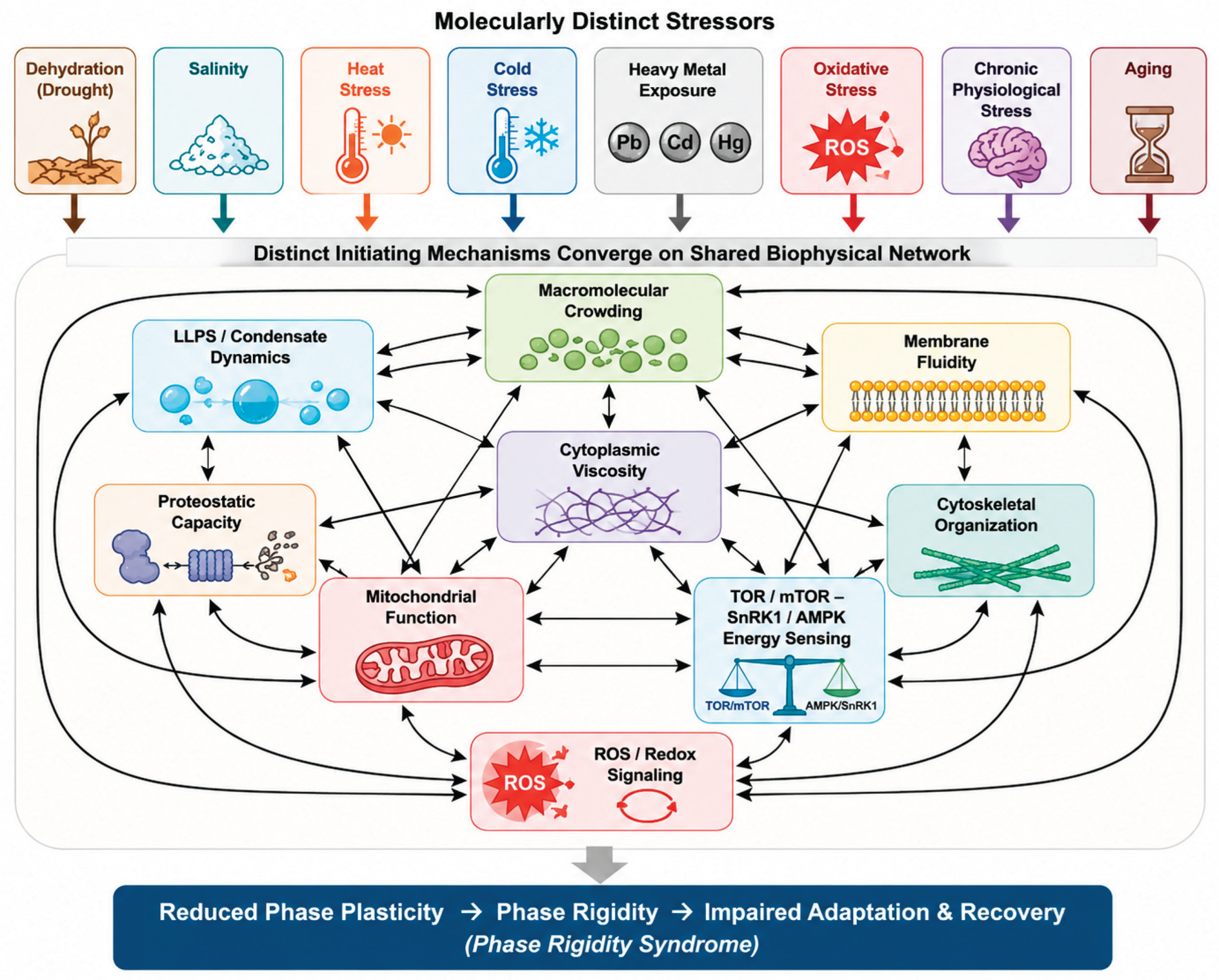
Figure 2.
Convergent biophysical pathway leading to the proposed Phase Rigidity Syndrome (PRS). Diverse environmental stressors trigger distinct molecular responses but converge on a common biophysical trajectory characterized by progressive loss of cytoplasmic phase plasticity. Perturbations of LLPS dynamics, membrane fluidity, cytoplasmic viscosity, proteostasis, cytoskeletal organization, mitochondrial function, TOR–SnRK1 signaling, ABA homeostasis, and ROS balance collectively promote the transition toward the proposed Phase Rigidity Syndrome (PRS). In the proposed framework, PRS represents a state of reduced reversibility of cytoplasmic phase organization, providing a unifying biophysical explanation for impaired stress recovery, metabolic dysfunction, and eventual cellular failure.
Figure 2.
Convergent biophysical pathway leading to the proposed Phase Rigidity Syndrome (PRS). Diverse environmental stressors trigger distinct molecular responses but converge on a common biophysical trajectory characterized by progressive loss of cytoplasmic phase plasticity. Perturbations of LLPS dynamics, membrane fluidity, cytoplasmic viscosity, proteostasis, cytoskeletal organization, mitochondrial function, TOR–SnRK1 signaling, ABA homeostasis, and ROS balance collectively promote the transition toward the proposed Phase Rigidity Syndrome (PRS). In the proposed framework, PRS represents a state of reduced reversibility of cytoplasmic phase organization, providing a unifying biophysical explanation for impaired stress recovery, metabolic dysfunction, and eventual cellular failure.
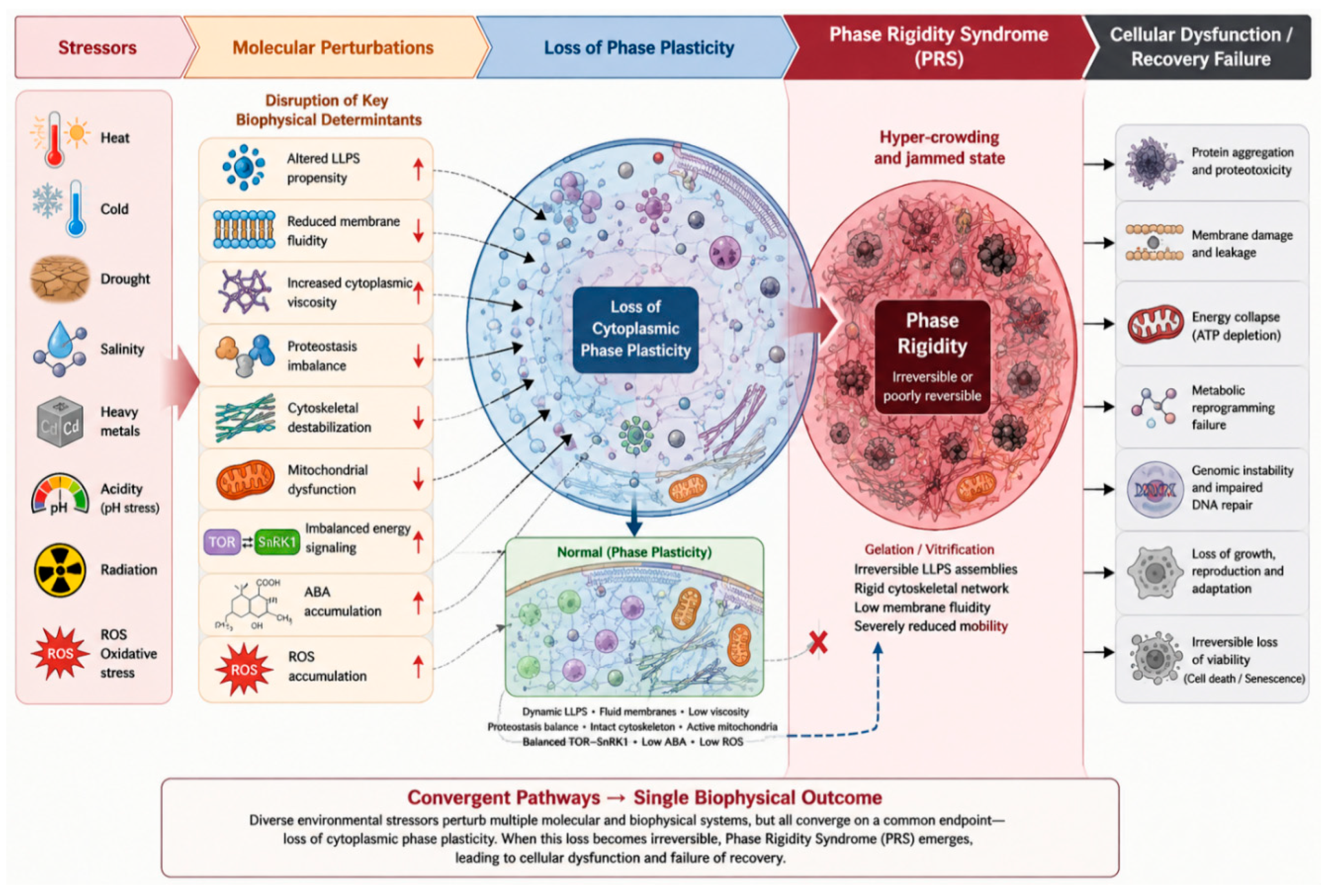
Figure 3.
Cellular phase-state landscape. A schematic energy-landscape representation of cellular state space showing an adaptive basin corresponding to phase homeostasis, trajectories representing stress-induced excursions and subsequent recovery, a phase threshold separating the adaptive basin from a region of phase rigidity, a phase collapse region beyond the rigidity threshold, and a phase transition window during which corrective intervention and return to the adaptive basin remain achievable.
Figure 3.
Cellular phase-state landscape. A schematic energy-landscape representation of cellular state space showing an adaptive basin corresponding to phase homeostasis, trajectories representing stress-induced excursions and subsequent recovery, a phase threshold separating the adaptive basin from a region of phase rigidity, a phase collapse region beyond the rigidity threshold, and a phase transition window during which corrective intervention and return to the adaptive basin remain achievable.
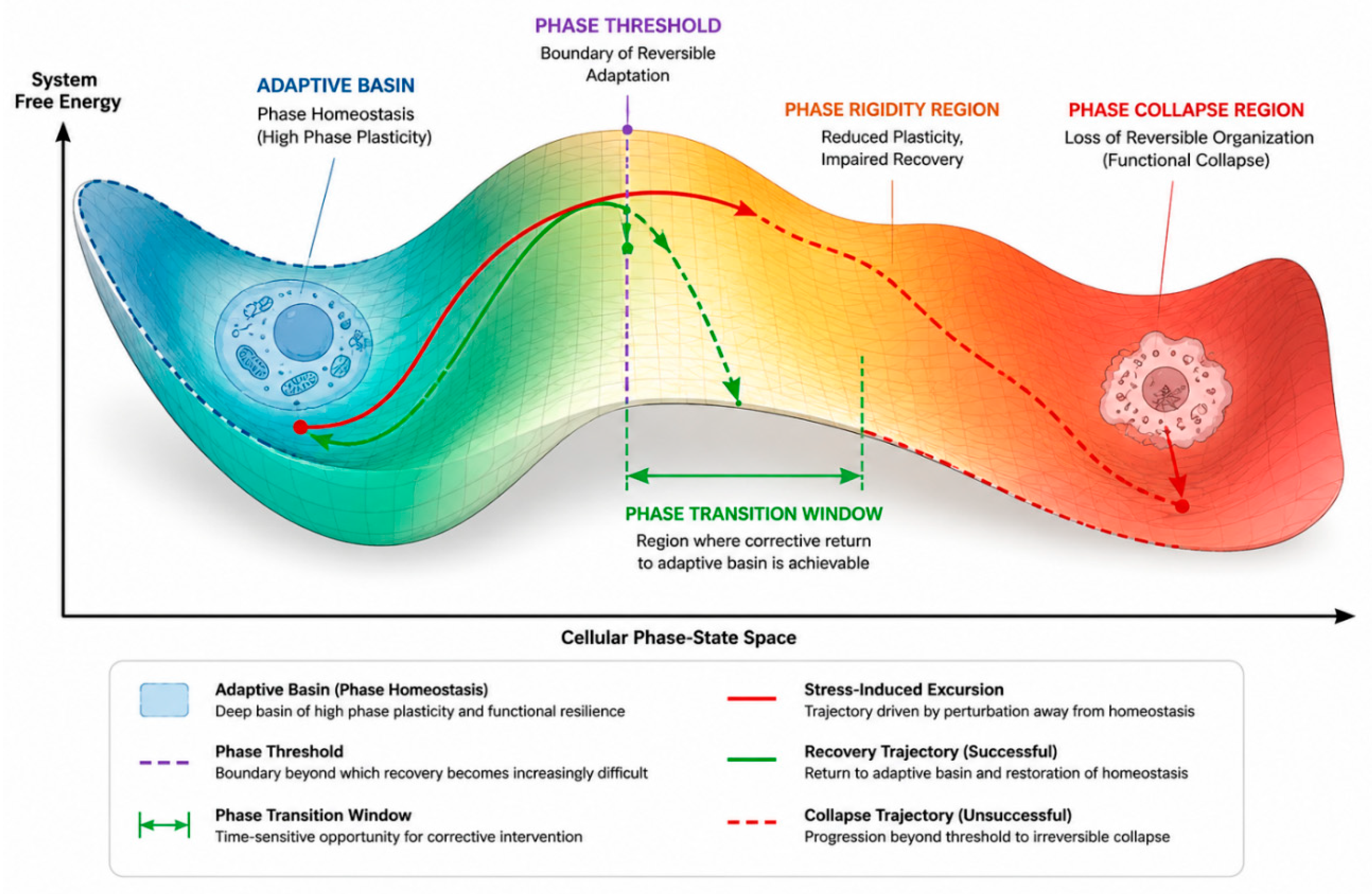
Figure 4.
Aging as a proposed trajectory toward reduced phase plasticity. Side-by-side comparison of a young, resilient cell and an aged, phase-rigid cell, illustrating the proposed differences in molecular diffusion, condensate exchange dynamics, membrane fluidity, proteostatic capacity, phase-recovery time following a standardized perturbation, and the width of the phase transition window.
Figure 4.
Aging as a proposed trajectory toward reduced phase plasticity. Side-by-side comparison of a young, resilient cell and an aged, phase-rigid cell, illustrating the proposed differences in molecular diffusion, condensate exchange dynamics, membrane fluidity, proteostatic capacity, phase-recovery time following a standardized perturbation, and the width of the phase transition window.
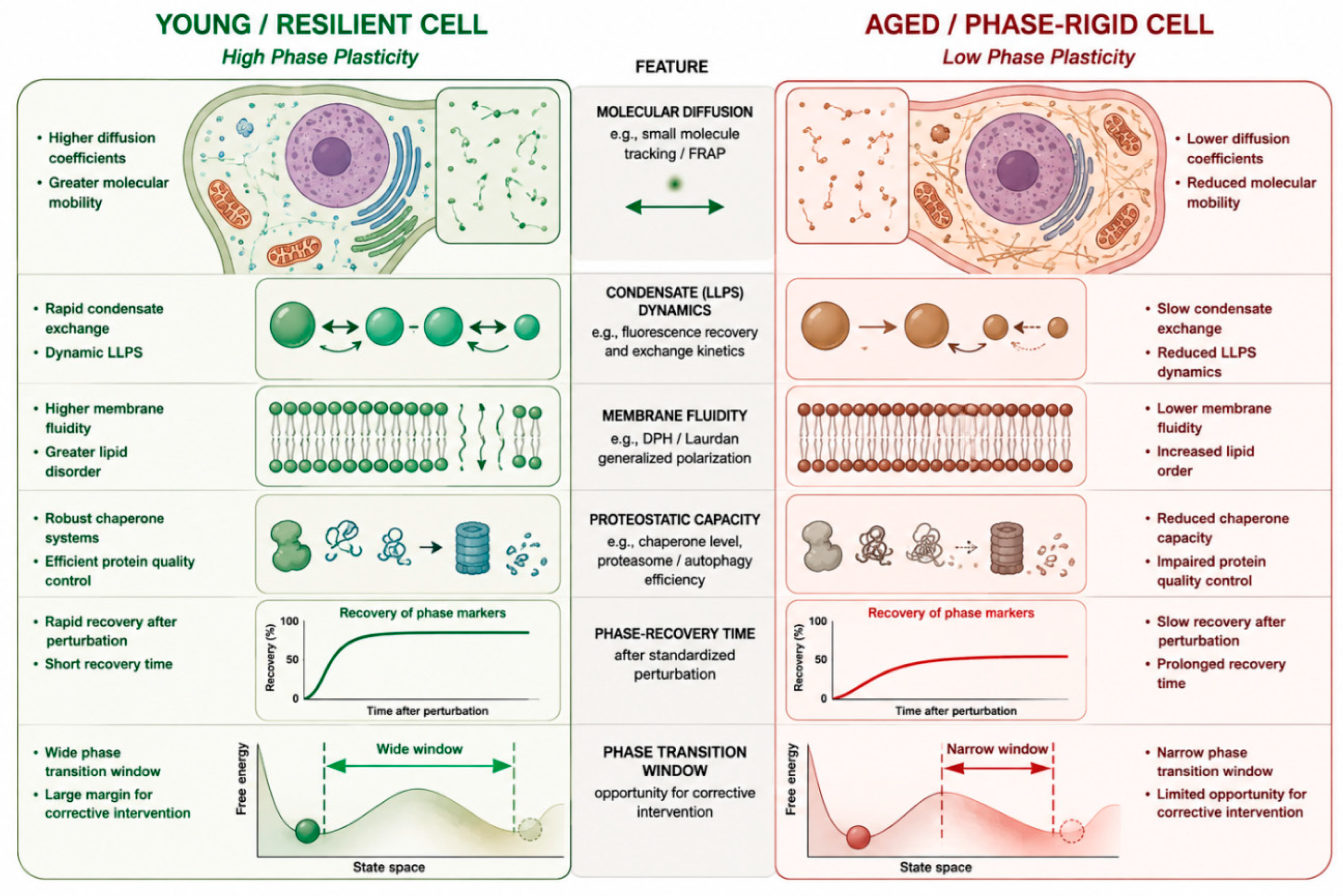
Figure 5.
Cross-stressor convergence. Schematic showing the eight stress categories discussed in Section 2, each characterized by distinct primary mechanisms and lesions, converging toward a shared region of reduced phase plasticity. .
Figure 5.
Cross-stressor convergence. Schematic showing the eight stress categories discussed in Section 2, each characterized by distinct primary mechanisms and lesions, converging toward a shared region of reduced phase plasticity. .
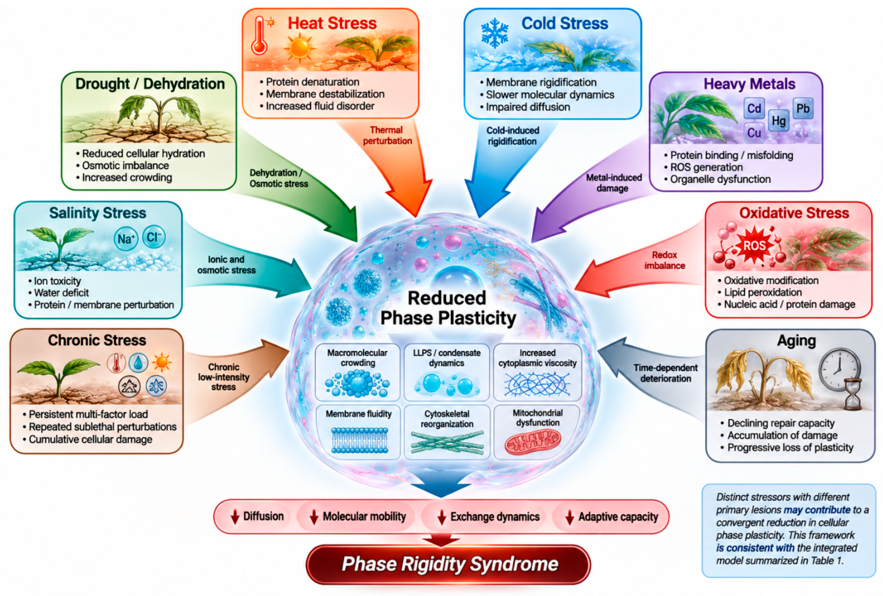
Figure 6.
Proposed experimental framework for measuring components of PRS. Integration diagram showing FRAP, fluorescence correlation spectroscopy, microrheology, fluorescence anisotropy, live-cell imaging, proteostasis assays, metabolomic and transcriptomic profiling, and single-cell analysis feeding into a proposed composite phase-plasticity assessment, with explicit indication that the integration step itself remains conceptual pending multi-parameter datasets.
Figure 6.
Proposed experimental framework for measuring components of PRS. Integration diagram showing FRAP, fluorescence correlation spectroscopy, microrheology, fluorescence anisotropy, live-cell imaging, proteostasis assays, metabolomic and transcriptomic profiling, and single-cell analysis feeding into a proposed composite phase-plasticity assessment, with explicit indication that the integration step itself remains conceptual pending multi-parameter datasets.
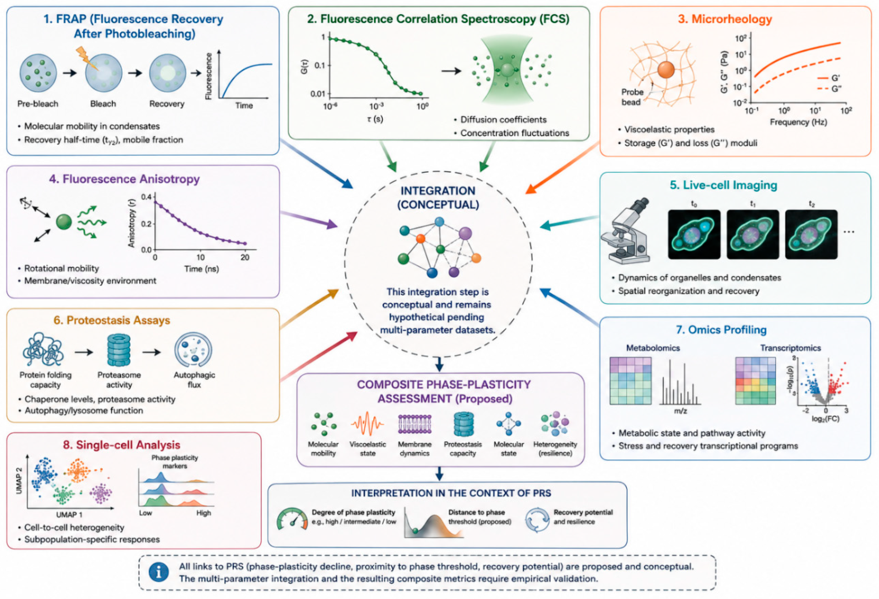
Figure 7.
Proposed validation and falsification framework. Flow diagram illustrating the proposed experimental sequence from stress exposure through phase-plasticity measurement and calculation of a candidate Phase Resilience Index, followed by targeted intervention and measurement of recovery, and concluding with explicit evaluation against the prediction and falsification criteria defined in Table 4 and Section 14.
Figure 7.
Proposed validation and falsification framework. Flow diagram illustrating the proposed experimental sequence from stress exposure through phase-plasticity measurement and calculation of a candidate Phase Resilience Index, followed by targeted intervention and measurement of recovery, and concluding with explicit evaluation against the prediction and falsification criteria defined in Table 4 and Section 14.
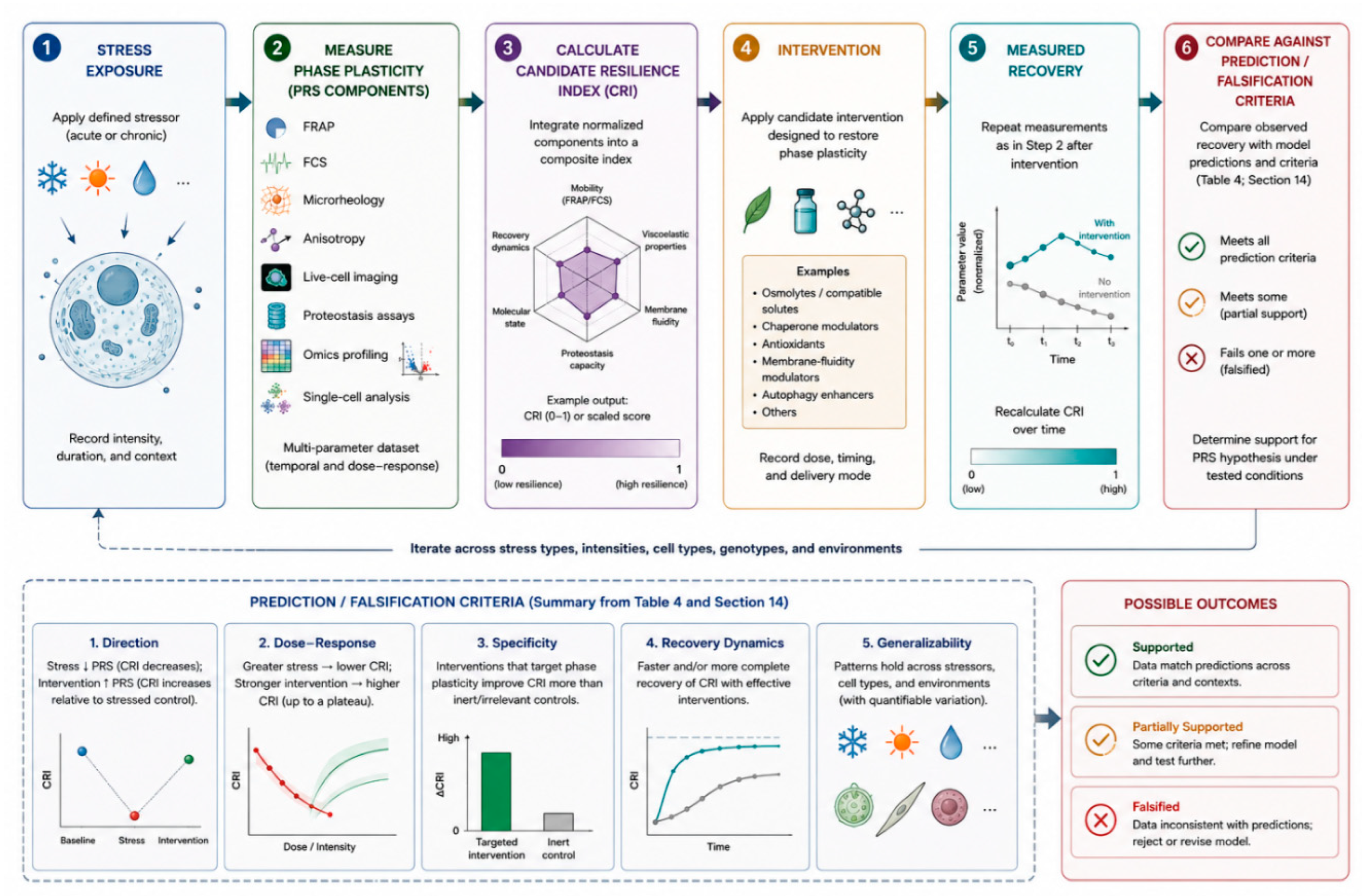
Figure 8.
Proposed unified biophysical framework of stress and aging based on phase plasticity. Multiple stressors, including dehydration, salinity, heat, cold, toxicants, oxidative stress, chronic stress, and aging, are proposed to initiate distinct molecular responses that converge on a shared set of biophysical processes, including hydration, macromolecular crowding, cytoplasmic viscosity, molecular diffusion, membrane fluidity, LLPS/condensate dynamics, and cytoskeletal and organelle mobility. These processes collectively influence cellular phase plasticity. When phase plasticity is preserved, phase homeostasis supports adaptation, phase recovery, and functional recovery. Progressive loss of phase plasticity is proposed to promote phase rigidity, impaired repair, incomplete recovery, phase memory, and transition toward phase collapse, ultimately contributing to cellular dysfunction, senescence, and loss of viability. The framework is conceptual and does not imply that all stressors act through identical molecular mechanisms or that the proposed convergence has been experimentally established.
Figure 8.
Proposed unified biophysical framework of stress and aging based on phase plasticity. Multiple stressors, including dehydration, salinity, heat, cold, toxicants, oxidative stress, chronic stress, and aging, are proposed to initiate distinct molecular responses that converge on a shared set of biophysical processes, including hydration, macromolecular crowding, cytoplasmic viscosity, molecular diffusion, membrane fluidity, LLPS/condensate dynamics, and cytoskeletal and organelle mobility. These processes collectively influence cellular phase plasticity. When phase plasticity is preserved, phase homeostasis supports adaptation, phase recovery, and functional recovery. Progressive loss of phase plasticity is proposed to promote phase rigidity, impaired repair, incomplete recovery, phase memory, and transition toward phase collapse, ultimately contributing to cellular dysfunction, senescence, and loss of viability. The framework is conceptual and does not imply that all stressors act through identical molecular mechanisms or that the proposed convergence has been experimentally established.
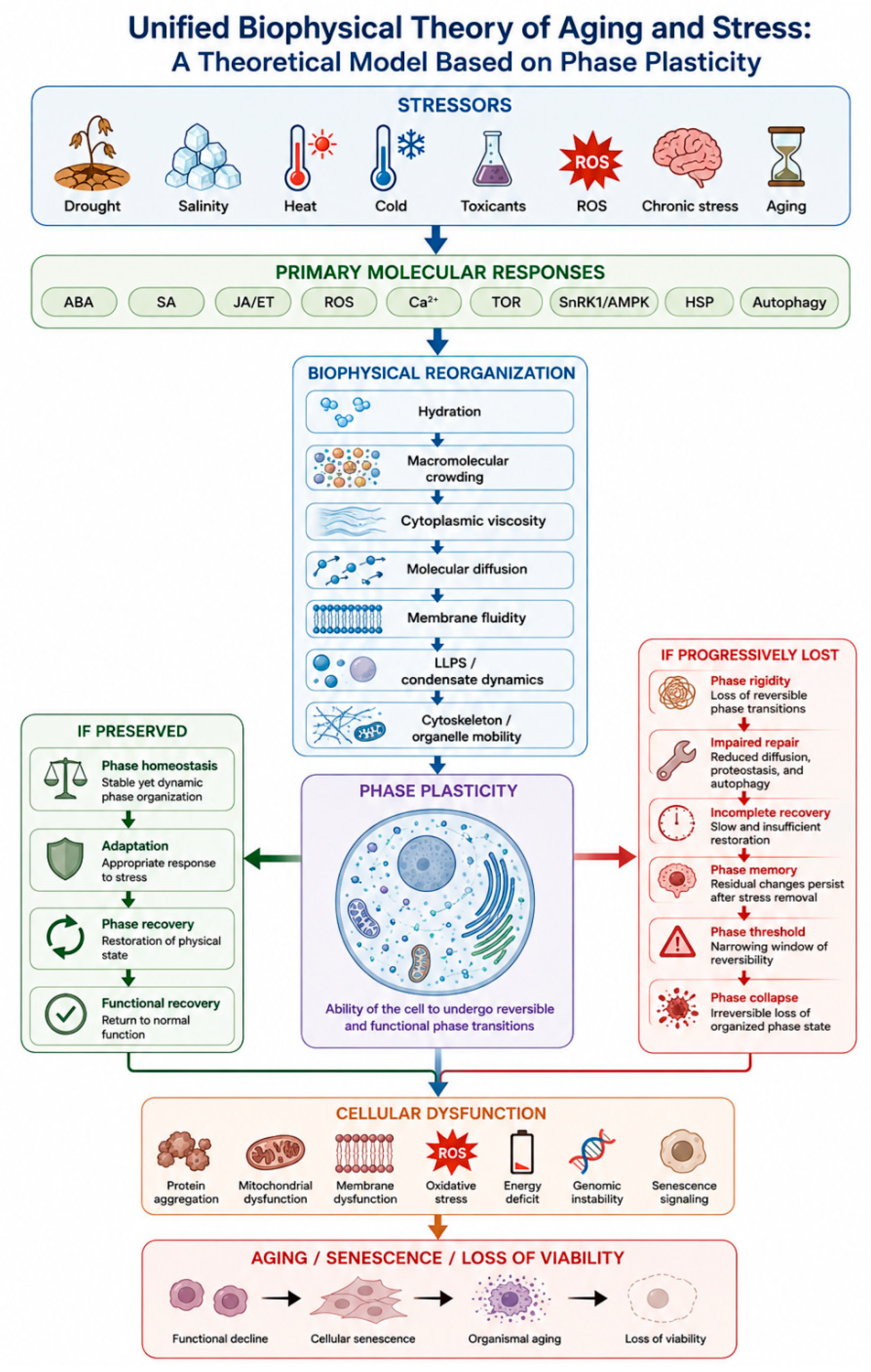
Figure 9.
The four-level framework of the unified biophysical theory of stress and aging. Diverse external and internal stressors initiate distinct molecular responses that converge on the physical reorganization of the cellular material system. Changes in hydration, macromolecular crowding, viscosity, molecular diffusion, membrane order, LLPS dynamics, cytoskeletal organization, and organelle mobility determine whether the system follows an adaptive trajectory toward stabilization and recovery or a maladaptive trajectory toward phase rigidity, incomplete recovery, phase collapse, and loss of viability.
Figure 9.
The four-level framework of the unified biophysical theory of stress and aging. Diverse external and internal stressors initiate distinct molecular responses that converge on the physical reorganization of the cellular material system. Changes in hydration, macromolecular crowding, viscosity, molecular diffusion, membrane order, LLPS dynamics, cytoskeletal organization, and organelle mobility determine whether the system follows an adaptive trajectory toward stabilization and recovery or a maladaptive trajectory toward phase rigidity, incomplete recovery, phase collapse, and loss of viability.
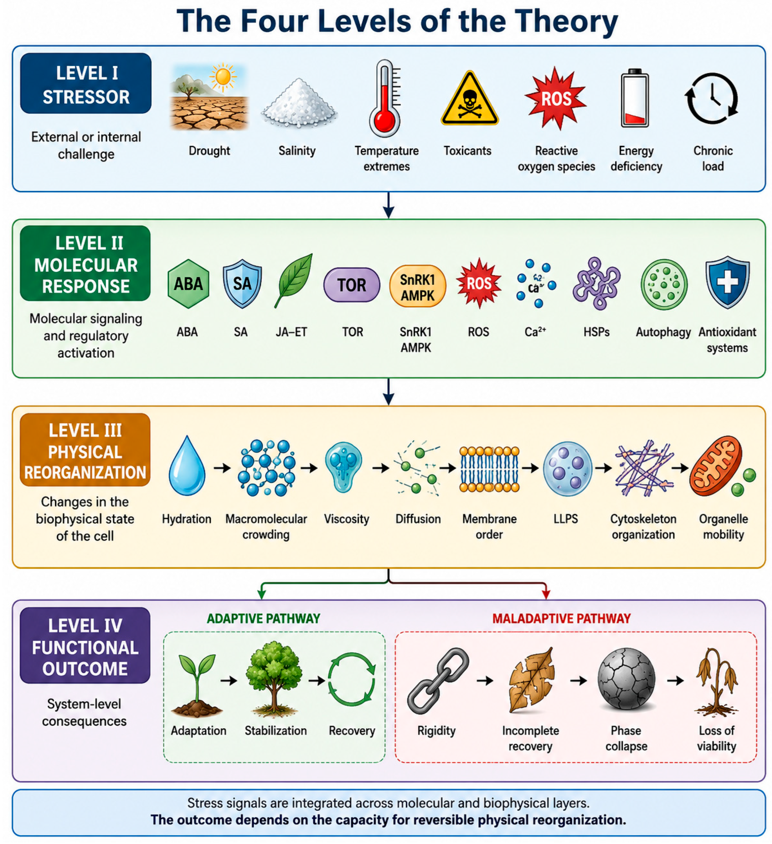

Table 1.
Proposed terminology of the Phase Rigidity Syndrome framework.
| Term | Definition | Potential measurable variable | Current evidentiary status |
|---|---|---|---|
| Phase homeostasis | Capacity to maintain functionally appropriate, dynamically regulated physical states of the cytoplasm and compartments despite perturbation | Composite of variables below, measured over time under stable conditions | Extension of an established general homeostasis concept to the phase-organizational level; not previously formalized in this form |
| Phase plasticity | Capacity of cellular material to undergo reversible transitions between physical organizational states | Reversibility of condensate, membrane, and viscosity changes following perturbation and recovery | Proposed construct; component physical transitions are individually documented, but not previously unified under this term |
| Phase buffer | Aggregate capacity of a cellular system to absorb perturbation without loss of reversibility | Distance (in measured physical parameters) from empirically defined thresholds of irreversibility | Proposed, not yet operationalized |
| Phase resilience | Capacity to maintain or restore phase organization following perturbation | Recovery kinetics and completeness following a standardized stress | Proposed; related to, but distinct from, phase buffer as defined here |
| Phase rigidity | State of reduced molecular mobility, reduced transition reversibility, and resistance to functional recovery | Reduced FRAP recovery, reduced diffusion coefficients, prolonged recovery time | Proposed integrative term; component phenomena (e.g., condensate hardening) are independently documented |
| Phase recovery | Process by which prior or functionally competent phase organization is restored after stress removal | Time course of the biomarkers listed in Table 3 following stress removal | Proposed as a named process; recovery of individual components (e.g., stress granule disassembly) is documented |
| Phase collapse | Transition beyond which reversible phase organization can no longer be maintained | Irreversible loss of condensate/membrane/viscosity recovery; not equated with cell death | Proposed; boundary with cell death requires empirical definition |
| Phase memory | Persistent alteration in physical cellular organization influencing subsequent responses to perturbation | Altered baseline biomarkers or altered response kinetics to a second stressor | Proposed; partial precedent in transcriptional/chromatin stress-memory literature, not previously extended to physical persistence |
| Phase threshold | Critical region beyond which phase changes become strongly nonlinear or poorly reversible | Empirically defined breakpoint in dose-response curves for the biomarkers in Table 3 | Proposed; analogous thresholds are established in glass-transition and gelation physics generally |
| Phase transition window | Period during which corrective intervention can still redirect a cell from a pathological toward an adaptive phase trajectory | Time-dependent responsiveness to a standardized restorative intervention | Proposed; not yet measured |
| Phase Resilience Index (PRI) | Proposed integrative quantitative measure of phase-organizational recovery capacity | Composite score derived from Table 3 biomarkers | Conceptual only; no validated protocol or normative range exists |
| Phase Rigidity Syndrome (PRS) | Proposed integrated condition in which cumulative loss of phase plasticity across multiple cellular layers reduces adaptive and recovery capacity | Composite of all variables above, tracked longitudinally | Hypothesis; not an established clinical or cell-biological syndrome |
Table 2.
Proposed convergence of distinct stress categories on Phase Rigidity Syndrome.
| Stress type | Primary biophysical challenge | Early molecular/cellular response | Proposed disruption to phase homeostasis | Proposed relationship to PRS |
|---|---|---|---|---|
| Drought/dehydration | Reduced water potential, increased crowding | ABA signaling, LEA protein induction, osmolyte accumulation | Increased crowding and viscosity are consistent with reduced diffusive mobility | May contribute to phase rigidity if crowding exceeds the range compatible with reversible recovery |
| Salinity | Combined osmotic stress and ionic toxicity | Na+/H+ antiporter activity, compatible-solute synthesis | Altered protein hydration shells alongside crowding effects | May be associated with impaired condensate and membrane behavior distinct from pure osmotic effects |
| Heat | Increased thermal motion destabilizing native protein folds | Heat-shock factor activation, chaperone induction | Paradoxical reduction in functional protein mobility despite increased kinetic energy, once aggregation occurs | Is consistent with phase rigidity if chaperone capacity is exceeded |
| Cold/freezing | Reduced membrane fluidity; ice nucleation | Desaturase-mediated homeoviscous adaptation; ice-binding proteins | Direct reduction in membrane-associated mobility | May contribute to phase rigidity primarily through the membrane layer of the system |
| Heavy metals | Direct metal-thiol coordination chemistry | Phytochelatin/metallothionein-mediated chelation | Unrepaired coordination may drive non-specific protein cross-linking | May converge with heat- and oxidative-stress aggregation phenotypes despite distinct chemistry |
| Oxidative stress | Reactive oxygen and nitrogen species | Antioxidant enzyme induction, redox signaling | Cysteine oxidation directly linked, in specific documented cases, to condensate hardening | Provides the most direct documented mechanistic link to a phase-organizational endpoint |
| Chronic stress | Repeated or sustained physiological demand | HPA-axis activation; allostatic response | Not directly measured at the phase-organizational level to date | Proposed as a systemic analogue of cellular phase rigidity; the connection is a hypothesis, not a demonstrated link |
| Aging | Cumulative, multi-hallmark functional decline | Declining autophagy, proteostasis, mitochondrial function | Proposed cumulative narrowing of the phase transition window | Proposed as one possible slow trajectory toward phase rigidity, not a synonym for it |
Table 3.
Candidate biomarkers relevant to measuring components of Phase Rigidity Syndrome.
| Parameter | Biological significance | Method | Advantages | Limitations |
|---|---|---|---|---|
| Condensate exchange dynamics | Reflects liquidity/solidity of biomolecular condensates | FRAP | Directly visualizes recovery kinetics in living cells | Model-dependent interpretation; photobleaching artifacts; not a validated PRS-specific threshold |
| Molecular diffusion coefficient | Reflects bulk and local cytoplasmic mobility | FCS, single-particle tracking microrheology | Quantitative, has been used to establish glass-like cytoplasmic behavior directly | Technically demanding; probe choice affects results; limited throughput |
| Membrane order/fluidity | Reflects lipid bilayer physical state | Fluorescence anisotropy of order-sensitive probes | Well-established, relatively accessible technique | Reports on labeled membrane region only; not condensate-specific |
| Cytoplasmic viscosity | Integrates crowding, hydration, and macromolecular content | Passive/active microrheology, tracer-particle tracking | Directly physical, connects to established glass-transition physics | In vivo measurement remains technically difficult; values vary by probe size |
| Organelle/protein mobility | Reflects transport-dependent proteostasis and repair capacity | Live-cell particle tracking | Captures functionally relevant transport, not just diffusion | Indirect; confounded by active transport mechanisms |
| Proteostatic capacity | Reflects folding/degradation/quality-control competence | Reporter-substrate degradation assays; chaperone/autophagic flux measurement | Directly functional readout | Does not by itself capture the physical/phase dimension proposed here |
| Phase recovery time | Proposed integrative readout of resilience | Time-resolved combination of the above following a defined perturbation | Directly tests the recovery-based definition of phase resilience used in this framework | Not yet standardized; no validated protocol currently exists |
Table 4.
Experimental predictions of the Phase Rigidity Syndrome framework and criteria for falsification.
Table 4.
Experimental predictions of the Phase Rigidity Syndrome framework and criteria for falsification.
| Prediction | Experimental test | Expected result if PRS is correct | Result that would falsify or substantially weaken the hypothesis |
|---|---|---|---|
| 1. Partial convergence of phase-plasticity trajectories across distinct stressors | Common biophysical panel (Section 13) applied across stress types in one cell system | Partial correlation despite distinct molecular lesions | Uncorrelated or divergent trajectories across stressors |
| 2. Coupling between membrane fluidity, diffusion, and condensate dynamics | Simultaneous measurement of all three under multiple stress types | Consistent co-variation across stressors | Absence of correlation across stress paradigms |
| 3. Phase-recovery kinetics predict functional recovery better than single biochemical markers | Compare predictive value of composite phase measure vs. individual markers (e.g., HSP levels) | Composite measure adds predictive value in at least some systems | Biochemical markers consistently outperform the composite measure |
| 4. Low baseline plasticity increases vulnerability to a second stressor | Sequential double-stress paradigm with baseline plasticity measurement | Cells with lower baseline plasticity show greater impairment after the second stressor | No cross-sensitization, or full explanation by a shared biochemical pathway |
| 5. Aging narrows the phase transition window | Compare responsiveness of young vs. aged cells to a standardized restorative intervention | Aged cells show reduced responsiveness at matched insult severity | Equivalent responsiveness across ages |
| 6. Plasticity-preserving interventions improve recovery independent of primary stressor neutralization | Apply phase-plasticity-directed intervention without neutralizing the primary stress signal | Improved recovery despite unresolved primary stress | No improvement in recovery from plasticity-directed intervention alone |
| 7. Phase rigidity shows hysteresis | Measure phase parameters immediately and at intervals after stressor removal | Incomplete or delayed return to baseline | Immediate, complete return to baseline upon stressor removal |
| 8. Mechanistically distinct interventions converge on similar phase-recovery improvement | Compare a chaperone-inducing and a membrane-fluidity-modulating intervention | Convergent improvement in phase-recovery kinetics despite distinct molecular targets | Outcomes track only the distinct known targets, with no evidence of convergence |
Table 5.
Convergence of diverse stressors on common biophysical changes in the cellular material state.
Table 5.
Convergence of diverse stressors on common biophysical changes in the cellular material state.
| Stressor | Primary mechanism | Physical consequence |
|---|---|---|
| Drought | Water loss, ABA signaling | ↑ Macromolecular crowding, ↑ viscosity |
| Salinity | Osmotic and ionic stress | Altered hydration, macromolecular crowding |
| High temperature | Protein denaturation | Aggregation, membrane alterations |
| Cold | Reduced lipid mobility | ↓ Membrane fluidity |
| Freezing | Ice formation | Mechanical reorganization of the cell |
| Heavy metals | Metal coordination with proteins | Aggregation, cross-linking |
| Oxidative stress | Oxidation of proteins and lipids | Altered condensate and membrane organization |
| Chronic stress | Prolonged activation of adaptive responses | Progressive depletion of cellular reserves |
| Aging | Accumulation of multiple forms of cellular damage | Progressive reduction in phase plasticity |
Disclaimer/Publisher’s Note: The statements, opinions and data contained in all publications are solely those of the individual author(s) and contributor(s) and not of MDPI and/or the editor(s). MDPI and/or the editor(s) disclaim responsibility for any injury to people or property resulting from any ideas, methods, instructions or products referred to in the content. |
© 2026 by the authors. Licensee MDPI, Basel, Switzerland. This article is an open access article distributed under the terms and conditions of the Creative Commons Attribution (CC BY) license.
Copyright: This open access article is published under a Creative Commons CC BY 4.0 license, which permit the free download, distribution, and reuse, provided that the author and preprint are cited in any reuse.


