

Submitted:
10 August 2026
Posted:
11 August 2026
You are already at the latest version
Abstract
Cervical cancer is a major cause of mortality worldwide and has been attributed to long-lasting (persistent) infections with the human papillomavirus (HPV). Although more than 50 types of HPVs are known the infect the genital tract, fewer than a dozen of them are regularly linked to cervical cancers leading to these types being designated as “high risk” HPVs. Worldwide, over 90% of cervical cancers arise from persistent high-risk human papillomavirus (HPV) infection and the infectious nature of the virus procures the need for preventative vaccines against cervical cancer. To address this, we have developed a live-attenuated herpes simplex virus type 1 (HSV-1) VC2-vectored vaccine expressing a catalytically inactive mutant of HPV-16 E7 (E7m) fused to the viral capsid protein VP26. Incorporation of E7m into the VC2 backbone resulted in stable incorporation of VP26-E7m fusion protein in cell free viral particles and detectable intracellular expression within infected cells. The recombinant virus (E7m VC-2) maintained growth kinetics comparable to parental VC2 in vitro, though plaque size was modestly reduced, suggesting a minimal fitness cost associated with transgene insertion. To evaluate prophylactic efficacy, mice received a one-dose vaccine schedule of E7m VC-2 prior to challenge with syngeneic HPV-16 E6/E7-expressing tumor cells. In mice that did develop tumors, growth kinetics were attenuated relative to unvaccinated animals. Importantly, neither E7m VC-2 nor adjuvanted E7m protein alone achieved statistically significant protection versus untreated controls. Immunohistochemical analysis of tumor tissue revealed markedly increased CD8+ T cell infiltration in E7m VC-2 vaccinated mice, demonstrating that vaccination promotes a cytotoxic immune microenvironment capable of restricting tumor progression. Three-dose vaccination of mice with E7m VC-2 induced a robust Th1-polarized immune response characterized by strong interferon-gamma (IFN-γ) secretion in response to HPV16 E7 expressing syngeneic cell lines, consistent with the induction of cell-mediated antitumor immunity and superior to responses elicited by adjuvanted E7m protein alone. With an extended three-dose vaccination schedule E7m VC-2 conferred significantly greater protection against tumor establishment relative to untreated controls (80% vs. 20% tumor-free). Collectively, these findings establish proof-of-concept for the HSV-1 VC2 platform as an effective vector for HPV-targeted cancer immunotherapy, delivering statistically significant prophylactic protection alongside potent induction of tumor-reactive CD8+ T cells. Future studies will incorporate both E6 and E7 antigens to broaden antigenic coverage, evaluate therapeutic efficacy in established tumor models, and optimize dosing regimens with the long-term goal of advancing this Louisiana-developed platform toward IND-enabling studies and clinical translation.
Keywords:
HPV vaccination
; HSV-1
; cervical cancer
; viral vectored vaccine
; VC2
1. Introduction
Human papillomavirus (HPV) is a DNA-virus that primarily infects the mucosal and subcutaneous epithelia, and causes persistent infections within basal keratinocytes [1]. HPV is associated with a wide range of diseases and is the causative agent of a wide range of cancers [2,3]. Over 90% of cervical cancer (CC) cases are associated with persistent HPV infections [4], and CC is the fourth most common cancer in females worldwide. CC can account for up to 25% of female cancer deaths worldwide and is the second leading cause of cancer associated deaths in females [5]. HPV genomes are composed of the structural L1 and L2 proteins and the E1, E2, E4, E5, E6 and E7 proteins, the latter of which contribute significantly to cellular transformation, uncontrolled growth and cancer formation [6,7,8]. In particular, the E6 and E7 proteins are the major oncogenic proteins of HPV and cause cellular transformation through disrupting p53 and pRb respectively [9,10,11]. E6 and E7 have been identified as major targets for the development of therapeutic vaccines against established HPV associated tumors. The proteins E6 and E7 present as ideal targets for future vaccines due to multiple reasons. Current vaccines target the L1 and L2 proteins and primarily stimulate humoral immunity [12]. However, following transformation of cells into cancerous cells, L1 and L2 expression is downregulated or at times completely lost during integration [13] and E6/E7 are not adequately controlled by the humoral immune response. As a result, focus has shifted to vaccines inducing cell mediated immune responses [14].
There have been several strategies taken to vaccinate against cervical cancer, including protein/peptide based, nucleic acid based, lipoprotein based and live-vectored vaccine approaches [15]. Among these, live vectored vaccines represent an attractive platform due to their ability to induce strong cell mediated immune responses and lead to an upregulation of Type-1 interferons (IFNs) and skew the immune response towards a Th1 response, also while upregulating E6/E7 specific CD8+ T-cell responses [16,17]. Herpesvirus vectored vaccines have been a platform utilized in recent years to vaccinate against a wide array of diseases and its utilization is rationalized due to FDA approval of T-Vec, which is the first oncolytic viral therapy approved for use in humans for the treatment of melanoma [18]. Herpesviruses are ideal vector candidates due to their ability to tolerate large genome inserts, relative safety of attenuated strains, ease of genetic manipulation, and vaccination is unaffected by pre-existing immunity [19].
VC2 is a live attenuated HSV-1 vector that contains mutations in the envelope proteins glycoprotein-K (gK) and UL-20, which renders the virus incapable of infecting neurons and establishing latency [20]. VC2 has demonstrated extended safety and efficacy in murine, guinea pig and non-human primate models [21,22]. Furthermore, vaccination with VC2 has been shown to induce effective cell mediated immunity and lead to increased T-cell infiltration into existing tumors [23]. Therefore, in this study we assessed the potential of VC2 as a vaccine vector to immunize against HPV associated cancers. To reiterate, E7 is a major target for CC and studies have shown that inactivating the pRb binding sites of E7 can render it non-transforming but allow it to retain its immunogenic properties [17]. Therefore, we have selected E7 as a likely target to immunize against HPV associated CC.
In this study, we have engineered VC2 to express the inactivated version of HPV-16 E7 (E7m VC2) as a fusion with VP-26. We characterized its immunogenicity in mice and found that vaccination with E7m VC2 leads to a robust upregulation in IFN-γ secretion and E7-specific CD8+ T-cells. We report here that single dose vaccination with E7m VC2 leads to a lower tumor burden in mice and increases CD8+ T-cell tumor infiltration. Additionally, our data demonstrate that three-dose vaccination with E7m VC2 protects mice against cervical cancer challenge. These results support the utilization of VC2 as a live attenuated vector for vaccination against HPV associated cervical cancer as well as its potential application against other HPV associated cancer types.
2. Results
2.1. Construction and Characterization of the E7m VC2 Virus
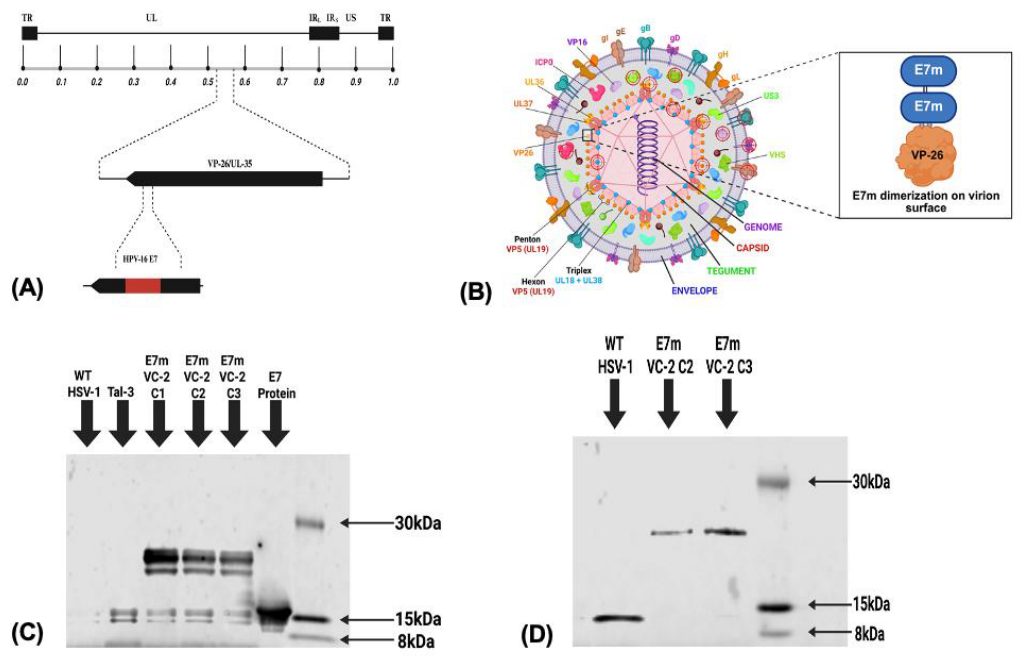
HPV-16 E7 is one of the major oncogenic proteins which contributes to cervical cancer and is a major antigenic target for HPV vaccination. To understand the potential of VC2 as a live attenuated vaccine to prevent cervical cancer we generated a recombinant VC2 expressing non-oncogenic HPV-16 E7. The E7m protein was cloned into the capsid protein VP-26 and expressed as a polyprotein (Figure-1A-B). The mutant virus carrying the mutated HPV-16 E7 was designated E7m VC2. The E7m expression was verified by western blot on purified virions and E7m VC2 was able to express the protein in Vero cells (Figure-1C). A western blot was also performed to blot against VP-26 to ensure the E7m protein was inserted into the correct position and expressed as a polyprotein with VP-26. The size differences in VP-26 between WT HSV-1 and E7m VC2 indicate that the protein was inserted into the correct position and expressed as a polyprotein (Figure-1D). Furthermore, to elucidate E7m expression efficiently within infected cells, a PCR was performed to detect E7m mRNA within infected cells as indicated in Supplemental Figure S1.
Figure 1.
Schematic representation of the E7m VC2 construct. (A) Construction of E7m VC2 inserted into the N-terminal of VP-26 using two-step red mediated recombination (B) Schematic representation of E7m dimerization on virion surface, leading to more than 1800 antigen copies/virion (C) Western blot on purified virions showing expression of the E7m protein on E7m VC2. Blot was stained with anti HPV-16 E7 antibody and WT HSV-1 was used as a negative control. (D) Western blot on purified virions showing expression of VP-26 and size difference between WT HSV-1 and E7m VC2. Blot was stained using anti VP-26 antibody.
2.2. Viral Spread and Growth Kinetics
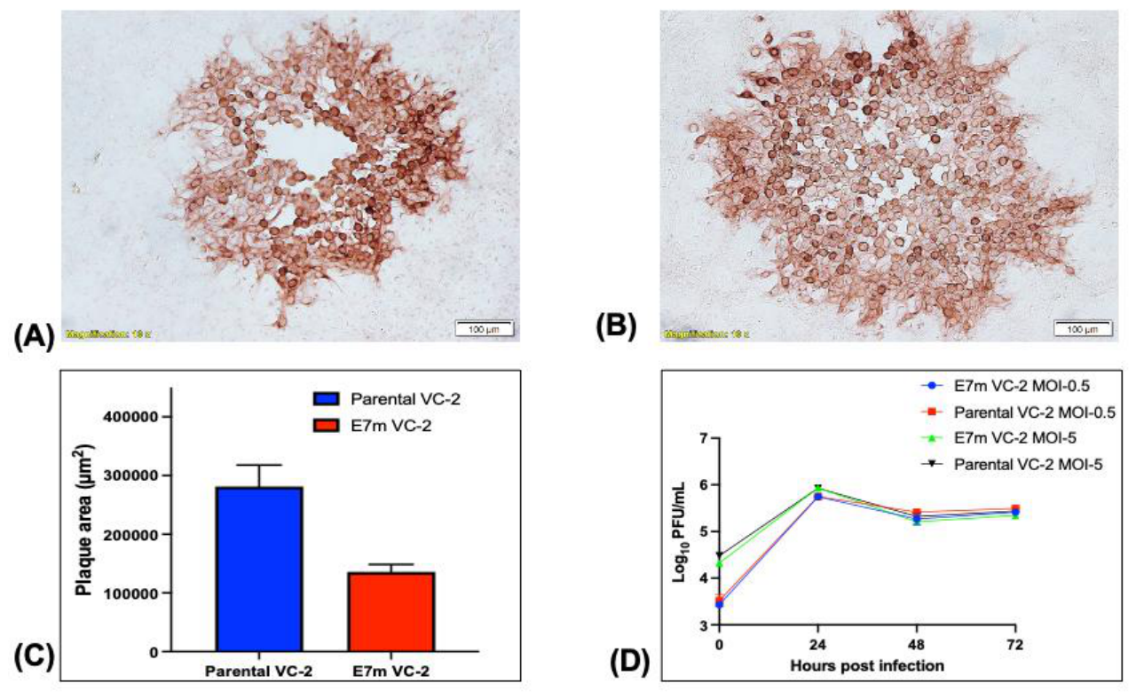
As reported previously, VC2 produces smaller plaques compared to its parental F strain [24]. The E7m mutation engineered into VC2 decreased the average plaque size of the virus in comparison to the parental VC2 (Figure-2C). However, we also investigated the impact of the E7m insertion on viral replication. Viruses were grown on Vero Cells at an MOI of 0.5 and 5 for 24, 48 and 72 hrs post-infection (p.i.) E7m VC2 and VC2 exhibited similar growth kinetics and E7m VC2 approached similar titers at 24, 48 and 72 hrs post-infection (Figure-2D), indicating that the intended mutations did not have any significant defect in viral growth.
Figure 2.
Replication kinetics and plaque morphology of E7m VC2. (A) Plaque morphology of E7m VC2 and (B) Parental VC2 visualized using IHC and developed using NovaRed substrate 48 hrs post infection on Vero cells at an MOI of 0.1 (C) Average plaque sizes of Parental VC2 vs E7m VC2 analyzed using ImageJ software (D) Growth curves of the replication kinetics of E7m VC2 vs Parental VC2 at an MOI of 0.5 (low) and 5 (high). Samples were collected at 24, 28 and 72 hrs post infection and titrated on Vero cells.
2.3. Single Dose Immunization with E7m VC2 Leads to Lower Tumor Burden in Mice
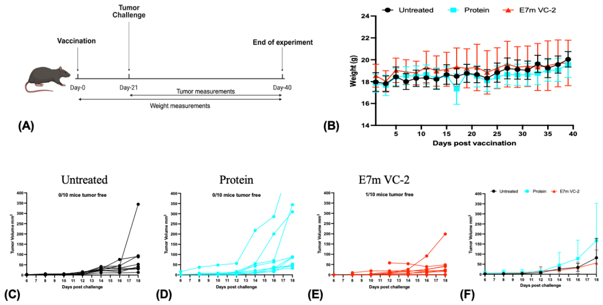
In order to evaluate the prophylactic efficacy of E7m VC2, we utilized a syngenic mouse model of cervical cancer. Specifically, 6-8 week old C57BL6/J mice (n=10) were vaccinated intramuscularly with one dose of 1x10⁶ PFU of E7m VC2 or 25μg of the E7m protein adjuvanted with 25μg of QS-21. For these experiments, we utilized a single dose vaccination regimen and during the vaccination period (Figure-3A), there were no adverse effects on the weight of the animals (Figure-3B). Three weeks following vaccination, the mice were challenged with 1x10⁶ Tal-3 cells per mice. The group of mice vaccinated with E7m VC2 exhibited lower tumor sizes in comparison to the Untreated or Protein groups (Figure-3C-E), indicating that E7m VC2 partially protected mice from a higher tumor burden (Figure-3F). Furthermore, 1/10 mice were tumor free in the E7m VC2 group (Figure-3E), whereas 10/10 mice formed tumors from the Untreated and the Protein groups (Figure-3C-D), indicating that additional vaccination dosages may augment tumor protection. However, 2 mice from the E7m VC2 group were found dead and postmortem analysis (data not shown) by a licensed veterinarian revealed that they succumbed to secondary bacterial infections within the lung and cause of death was not related to vaccination or tumor challenge. Furthermore, mice were seen to be extremely moribund 2 weeks post challenge and before tumor sizes could reach adequate endpoint criteria. Therefore, subsequent studies utilized the C3.43 tumor model which we found to be more well-tolerated within the mice.
Figure 3.
Vaccination of mice with E7m VC2 leads to lower tumor burden in animals. (A) 6-8 week-old C57BL6/J mice (n=10) were vaccinated with 1x10⁶ PFU of E7m VC2 or 25ug of E7m protein adjuvanted with 25ug of QS-21. As a negative control, mice were left unvaccinated. Average changes in (B) weight were recorded over the vaccination period. Three weeks post vaccination mice were challenged with syngenic E6/E7 positive 1x10⁶ Tal-3 cells and tumor sizes were measured for individual (C) Untreated, (D) E7m VC2 and (E) Protein groups. (F) Average tumor sizes were also recorded post challenge.
2.4. Single Dose Vaccination with E7m VC2 Leads to Higher CD8+ T-Cell Infiltration Into Tumors
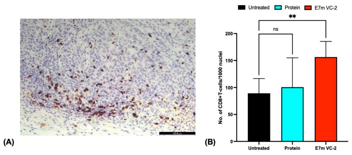
To determine the effects of E7m VC2 vaccination on intra tumoral T-cell infiltration, immunohistochemistry was performed on FFPE sections of the tumors. Mice were sacrificed following vaccination and tumor challenge, and tumors were excised and processed for immunohistochemistry. Tissue sections were stained for anti-CD8 to identify CD8+ T-cells in tumors following vaccination with E7m VC2. The analysis revealed that vaccination with E7m VC2 leads to significantly higher CD8+ T-cell infiltration into tumors (Figure-4B), indicating that vaccination with E7m VC2 leads to a greater anti-tumoral response compared to the protein or untreated groups.
Figure 4.
(A) Vaccination with E7m VC2 leads to an upregulation in CD8+ T-cell infiltration into tumors. (A) Representative immunohistological staining to identify CD8+ T-cell infiltrating cells within tumors. (B) Number of CD8+ T-cell infiltration within tumors. Statistical significance was determined using one-way ANOVA followed by Dunnett’s multiple comparisons test. *P < 0.05, **P < 0.01, ***P < 0.001.
2.5. Triple-Dose Immunization with E7m VC2 Leads to an Upregulation of IFN-γ Secretion
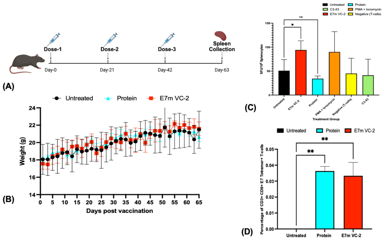
To evaluate the immunogenic properties of E7m VC2, we evaluated the secretion of IFN-γ in splenocytes of mice immunized with E7m VC2. Previous studies have shown that intramuscular immunization with VC2 is safe in murine models and elicits immune responses to targeted antigens [24]. For evaluation, 6-8 week old C57BL6/J mice (n=3) were vaccinated with three doses of 1x10⁶ PFU of E7m VC2 or 25μg of the E7m protein adjuvanted with 25μg of QS-21. There were no significant weight differences or clinical signs of disease (ruffled fur, difficulty breathing etc.) among groups following vaccination (Figure-5B). Following analysis, the level of IFN-γ secreted following vaccination with E7m VC2 was significantly higher compared to the protein alone (Figure-5C). Together, these results indicate that E7m VC2 is well tolerated in mice and results in a Th1-based immune response following vaccination.
Figure 5.
Vaccination of mice with E7m VC2 leads to an upregulation in Type-1 cytokine and E7-specific CD8+ T-cells. (A) Vaccination strategy, 6-8 week-old C57BL6/J mice (n=3) were vaccinated with 1x10⁶ PFU of E7m VC2 or 25ug of E7m protein adjuvanted with 25ug of QS-21. As a negative control, mice were left unvaccinated. (B) Mice were observed for weight loss over the period of vaccination. Post vaccination mice were sacrificed, and single cell suspensions of the spleen were stimulated with C3.43 cells at an E:T of 5:1 and number of spot forming units (SFU) were normalized to 106 splenocytes to quantify (C) IFN-gamma secreting cells. (D) Percentage of E7 tetramer positive CD8+ T-cells within splenocyte population following vaccination was assessed using E7-tetramer staining. Statistical significance was determined using one-way ANOVA followed by Dunnett’s multiple comparisons test. *P < 0.05, **P < 0.01, ***P < 0.001.
2.6. Triple- Dose Immunization with E7m VC2 Induces E7-Specific T-Cell Responses
To determine the effects of E7m VC2 vaccination on T-cell populations and evaluate E7 specific CD8+ T-cell responses, flow cytometry experiments were performed on splenocytes, and single-cell suspensions were stained with antibodies against CD3, CD8 and E7-tetramer. As a percentage of CD8+ T-cells (Supplemental Figure S2), E7-tetramer positive cells were found to be significantly higher in E7m VC2 treated group as compared to the controls (Figure-5D), indicating an increase in E7-specific CD8+ T-cells following vaccination with E7m VC2.
2.7. Triple Dose Vaccination with E7m VC2 Protects Mice Against Cervical Cancer
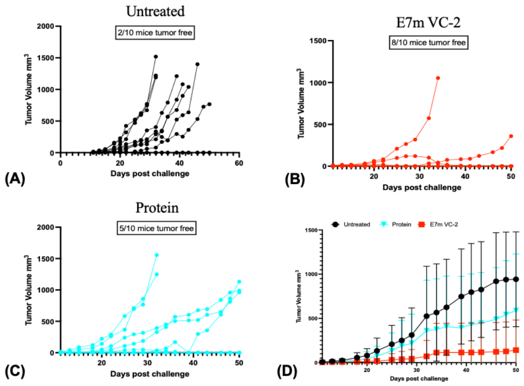
Based on preliminary results, our data suggests that one dose vaccination with E7m VC2 has a partial protective effect against cervical cancer and triple-dose vaccination induces a strong Type-1 immune response. We hypothesize that subsequent booster dosages may have additional protective efficacy. Therefore, 6-8 week old C57BL6/J mice (n=10) were vaccinated intramuscularly with one dose of 1x10⁶ PFU of E7m VC2 or 25μg of the E7m protein adjuvanted with 25μg of QS-21 followed by two booster doses spaced three weeks apart. There were no adverse effects during the vaccination period and three weeks post second booster dosage, the mice were challenged with 1x10⁵ C3.43 cells. The results from the study indicates that vaccination with E7m VC2 leads to lower tumor burden when compared to the Untreated or Protein controls (Figure-6D). Additionally, 8/10 mice were found to be tumor free in the E7m VC2 vaccinated group, whereas 2/10 and 5/10 mice were found to be tumor free in the Untreated and Protein groups respectively (Figure-6A-C). These results indicate that E7m VC2 is a potent stimulator of anti-E7 immune response and protects mice from cervical cancer formation.
Figure 6.
Triple dose vaccination of mice with E7m VC2 protects mice against cervical cancer challenge. 6-8 week-old C57BL6/J mice (n=10) were vaccinated with initial and two booster dosages with 1x10⁶ PFU of E7m VC2 or 25ug of E7m protein adjuvanted with 25ug of QS-21. As a negative control, mice were left unvaccinated. Three weeks post vaccination mice were challenged with syngenic E6/E7 positive 1x10⁵ C3.43 cells and tumor sizes were measured for individual (A) Untreated, (B) E7m VC2 and (C) Protein groups. (D) Average tumor sizes were also recorded post challenge.
3. Discussion
In previous studies, it has been well-established that HSV-1 based vectors are suitable candidates for vaccines due to their ability to trigger robust immune responses and their extended safety profiles [25]. Our group has demonstrated that VC2 expressing tumor associated antigens can effectively control tumor challenge in the murine model of metastatic melanoma [26]. Here, we have described the generation of a HSV-1 based vaccine to target HPV induced cervical cancer using the live attenuated HSV-1 strain VC2. In this study, we engineered VC2 to express attenuated HPV-16 E7 as a fusion with VP-26 to stimulate anti-tumor immunity. We found that triple-dose immunization in mice using E7m VC2 protects against HPV induced cervical cancer and leads to robust anti-E7 immune responses.
The selection of an appropriate vector for vaccine development requires the fulfillment of preset criteria including appropriate knowledge of the infectious agent, understanding the mode of transmission, replication mechanisms, mode of administration and thorough knowledge of the vector and associated immune responses [27]. HSV-1 VC2 contains mutations in gK and UL-20, which render the virus incapable of infecting neurons and thus establishing latency [28]. VC2 has been shown to be safe and immunogenic in various animal models and has been utilized as an oncolytic viral therapy and as a vaccine platform to immunize against influenza [19,22,23,29]. Furthermore, previous studies have shown that delivery of an antigen using VC-2 leads to upregulation of targeted immune responses against the protein associated with the cancer [30]. Consistent with this, our finding suggests that vaccination with E7m VC2 leads to adequate expression of the protein within infected cells leading to targeted immune responses against the E7 protein.
The route of vaccination plays a major role in the type of immune response stimulated and intramuscular injection is commonly recommended for most vaccines due to enhanced efficacy and stimulation of localized immune responses [31]. Similarly, single dose intramuscular immunization with E7m VC2 was found to lower tumor burden in mice. Additionally, IHC analysis revealed that infiltration of CD8+ T-cells was significantly higher in E7m VC2 vaccinated groups and previous studies have established the contributions of CD8+ T-cells in tumor elimination [32,33]. These results indicate that additional vaccinations with E7m VC2 can potentially augment the protective effects observed and lead to higher efficacy.
E7m VC2 immunization led to an increase in IFN-γ secretion by splenocytes stimulated with syngeneic E7 expressing cell lines, indicating the development of a robust Th1-based immune response to E7 expressing tumor cell lines. Additionally, vaccination with E7m VC2 stimulated an increase in E7-tetramer positive CD8+ T-cells confirming the presence of E7 reactive T cells in vaccinated animals. Previous studies have shown that although cytokine patterns vary within patients suffering from cervical cancer, a Th1 cytokine response, particularly IFN-γ secretion is associated with higher clearance of cervical HPV infection [34]. Additionally, the induction of cell mediated immunity against cervical cancer remains the primary immune response required for clearance of cancerous cells and regression of lesions [35]. Collectively, our results support the finding that E7m VC2 vaccination can potentially inhibit the formation of cervical cancer.
In previous studies, it has been observed that a triple dosage vaccination regimen against HPV is most effective and leads to a higher overall protective effect [36]. Akin to previous reports, we found that triple dosage vaccination with E7m VC2 had the highest overall protective effect and prevented the formation of CC in 80% of challenged animals. Similarly, despite the ability of purified E7 protein to stimulate E7 specific T cell responses via tetramer staining. These cells were not able to mount an IFN-γ specific functional response to E7 positive tumor cell lines (Figure 5). This functional distinction between CMI generated by adjuvanted E7 vaccination and vectored E7 vaccination drive the question as to what functionally regulates E7 specific Th1 mediated immunity. Insights into how to tailor effective immunity against HPV E7 will drive the success of future therapeutic CC vaccines.
Our study demonstrated that E7m VC2 elicited immune responses in animal models of CC and immune responses mounted were shown to be a Th1 response and antigen specific to the E7 protein. A triple dosage vaccination regimen with E7m VC2 was found to be most effective and immune infiltration with a single dosage vaccination regimen was found to lead to lower tumor burden. These findings build the foundation for future therapeutic applications for E7m VC2. While this study primarily focused on prophylactic efficacy, follow up studies exploring therapeutic application and targeting both E6 and E7 proteins are required to elucidate higher protective rates. In conclusion, E7m VC2 enhanced T-cell mediated immune responses and provided strong evidence of protective efficacy and laid the foundation for potential clinical applications of HSV-1 vectored vaccines against cervical cancer.
4. Materials and Methods
4.1. Animals
Female C57BL/6J mice between the age range of 6-8 weeks were purchased from the Jackson Laboratory (Bar Harbour, ME). All animals were housed under standard pathogen free conditions, in a temperature-controlled room with a 12h light/12h dark cycle with no more than 5 animals per cage. All procedures involving animals were conducted in accordance with the National Institutes of Health (NIH) guidelines and with prior approval by the Louisiana State University Institutional Animal Care and Use Committee (IACUC).
4.2. Construction of the Inactive HPV-16 E7 (E7m) Plasmid
The HPV-16 E7 protein is composed of three conserved regions- CR1, CR2 and CR3. CR1 and CR2, which are the N-terminal domains of E7, significantly contribute to cellular transformation. Based on previous studies, we have mutated the CR2 and CR3 region of the E7 protein to block binding of E7 to pRb and disrupt binding to the E2F transcription factors [17]. The mutations were executed using fast cloning as described previously [37], on the pGEX2T E7 plasmid (Plasmid #13634) purchased from Addgene (Watertown, MA). Oligonucleotides used for the construction of the mutated plasmid are listed in Supplementary Table S1.
4.3. Expression and Purification of Recombinant Proteins
The DNA fragment encoding HPV-16 E7m transforming protein (Integrated DNA Technologies, IDT) was amplified by PCR and cloned in-frame at the C-terminus of an N-terminal glutathione S-transferase (GST) fusion protein carrying an N-terminal His6 tag in the pHH239 expression vector using the HiFi DNA Assembly Master Mix (New England Biolabs, Ipswich, MA). All constructs were verified by Sanger sequencing.
For protein expression, Escherichia coli BL21(DE3) cells co-transformed with the GST-E7m expression plasmid and a plasmid encoding the Erv1p and DsbC folding chaperones were grown in 2YT medium supplemented with 100 μg/ml carbenicillin and 34 μg/ml chloramphenicol at 37 °C with shaking (200 rpm) to an OD600 of approximately 0.4. Expression of Erv1p and DsbC was induced by the addition of 0.5% (w/v) L-arabinose, after which cultures were incubated at 30 °C for 45 min with shaking (200 rpm). GST-E7m expression was subsequently induced with 0.1 mM IPTG, and cultures were grown for overnight at 18 °C with shaking [38].
Cells were harvested by centrifugation, lysed by standard methods, and the His6-GST-E7m fusion protein was purified by immobilized metal affinity chromatography (IMAC) using Ni-NTA resin (Qiagen). The eluted protein was buffer-exchanged into TEV cleavage buffer (50 mM Tris-HCl, pH 8.5, 0.5 mM Tris (2-carboxyethyl) phosphine (TCEP), 0.5 mM EDTA) and incubated with TEV protease at a 1:100 molar ratio (protease) for 18 h at 4 °C. Additional TEV protease was then added, and cleavage was allowed to proceed for a further 3 h. Following digestion, the sample was applied to Ni-NTA resin to remove the His6-GST tag, His-tagged TEV protease, and any uncleaved fusion protein. The unbound fraction containing E7m was collected, analyzed by SDS-PAGE, and concentrated using Amicon Ultra-4 centrifugal filters with a 10 kDa molecular weight cutoff (Millipore, Burlington, MA).
The protein was further purified by size-exclusion chromatography on an ÄKTA purification system (GE Healthcare, Chicago, IL) equipped with a HiLoad 16/60 Superdex 75 column (Cytiva, Marlborough, MA). Peak fractions corresponding to monomeric GST-E7m were pooled and analyzed by SDS-PAGE to confirm purity. Purified protein was concentrated to the desired concentration using Amicon Ultra centrifugal filters, and protein concentration was determined using a BCA protein assay (Thermo Fisher Scientific, Waltham, MA).
4.4. Construction of the E7m VC2 Virus
The construction and characterization of the bacterial artificial chromosome (BAC) containing the HSV-1 VC2 genome has been described previously [24]. E7m VC2 was constructed using two step red mediated recombination, a strategy that has been described in detail [39]. Briefly, the E7m gene was amplified using primers listed in Supplementary Table S1 with homology to the N-terminal of VP-26. The resulting amplicon was used as a universal transfer construct and inserted into the VC2 BAC, fusing VP-26 with the E7m gene. To confirm the correct insertion of the E7m gene, the E7m VC2 DNA was subjected to PCR confirmation. Recombinant HSV-1 was recovered following transfection of the BACs into Vero cells using Lipofectamine 3000 (Thermo Fisher, Waltham, MA) according to the manufacturer’s protocol, followed by Western Blot on purified virions to identify E7m expression.
4.5. Cell Culture and Virus Propagation
Tal-3, a syngeneic mouse epithelial cell line transformed with HPV-16 E6/E7 proteins and containing c-myc and Akt oncogenic mutations, was purchased from the John Hopkins Cell Repository and has been described previously [40]. Tal-3 cells were cultured in RPMI-1640 ATCC Modification (Thermo Fisher, Waltham, MA) supplemented with 10% fetal bovine serum (FBS) (Thermo Fisher, Waltham, MA) and 1% Penicillin/Streptomycin (Thermo Fisher, Waltham, MA). C3.43, a syngeneic mouse cell line transformed with the Ras oncogene and the complete HPV-16 genome was a kind gift from Dr. Martin Kast (University of Southern California) and were cultured in IMDM supplemented with 10% FBS (Thermo Fisher, Waltham, MA), 2mM L-glutamine, 50mM 2-mercaptoethanol and 1% Gentamicin [41,42]. Vero cells were obtained from ATCC and maintained in DMEM (Thermo Fisher, Waltham, MA) supplemented with 10% FBS and 1% Penicillin/Streptomycin. To prepare purified viruses, Vero cells were cultured in T-175 tissue culture flasks. After 24h, at 90% confluency, cells were infected with E7m VC2 at a multiplicity of infection (MOI) of 0.01 and incubated at 37°C in 5% CO2. At full cytopathic effect, cells were frozen and supernatant was harvested and subjected to freezing and thawing 3 times. Following freeze-thaw cycles, the cellular debris was removed by centrifugation at 400rpm for 10mins, followed by filtration using a 0.45μm filter (VWR, Radnor, PA). The resulting supernatant was aliquoted and stored at -80°C. Virus titer and growth curves were determined by calculating the plaque forming units in Vero cells and has been described previously [43].
4.6. Vaccination and Tumor Challenge
Six- to eight-week-old female C57BL/6J mice were vaccinated with either 1x10⁶ PFU of E7m VC2 or 25μg of the E7m protein adjuvanted with 25μg of QS-21 (Invivogen, San Diego, CA). The mice were administered the vaccine using intramuscular injections and two groups of mice (n=10 mice per group) received either one dose or three doses of the vaccination. The three immunizations were performed at three-week intervals. For appropriate controls, mice were also left unvaccinated. Three weeks post vaccination, mice were engrafted with either 1x10⁶ Tal-3 cells (for one dose study) or 1x10⁵ C3.43 cells (three-dose study) subcutaneously into the right flank. Following engraftment, tumors were measured every 2-3 days using a digital caliper. Tumor volumes were calculated using the formula- ½ (L X W2). Tumor bearing mice were euthanized when tumors reached a volume of 1000mm3 or when the mice were excessively moribund.
4.7. Immunohistochemistry (IHC)
Following euthanasia, tumor tissues were fixed in 10% neutral buffered formalin and processed at the LSU-LADDL histology core. For IHC, 4 μm sections of FFPE tissue were mounted on positively charged Superfrost® Plus slides and subjected to IHC using the automated BOND-MAX and the Polymer Refine Detection kit. Following automated deparaffinization and heat-induced epitope retrieval (HIER) with an EDTA-based buffer (pH 9.0; Leica Biosystems), sections were incubated with anti-CD8 (Cell Signaling D4W2Z) at 1:100 for 30 minutes, followed by a polymer-labeled goat anti-rabbit IgG coupled with horseradish peroxidase (8 min). 3,3’-diaminobenzidine tetrahydrochloride (DAB) was used as the chromogen (10 min), and counterstaining was performed with hematoxylin. Slides were mounted with a permanent mounting medium.
4.8. Western Blot Analysis
Vero cells were infected at an MOI of 1 with either VC2 or E7m VC2 for 48hrs in a T-175 cm2 tissue culture flask. The resulting supernatant following infection was collected and subjected to three freeze-thaw cycles followed by centrifugation at 400rpm for 10mins to remove cellular debris. The resulting supernatant was filtered using a 0.45μm filter (VWR, Radnor, PA) and subjected to centrifugation at 80,000g for 1hr at 4°C using an OptiPrepTM Density Gradient Medium (MilliporeSigma, Burlington, MA). Following centrifugation, the resulting concentrated virus was resuspended in complete RIPA lysis buffer (Thermo Fisher, Waltham, MA) containing protease/phosphatase inhibitors. Thirty microliters of viral lysate were then mixed with 4x NuPAGETM LDS Sample Buffer (Thermo Fisher, Waltham, MA) and 1μl of β-mercaptoethanol. The mixtures were boiled at 95° for 10mins and cooled on ice before loading into a 12% SurePAGETM (GenScript, Piscataway, NJ) precast gel and separated at 100V for 1h using TRIS-MOPS SDS Running Buffer (GenScript, Piscataway, NJ). The gel was then transferred to a PVDF membrane using the GenScript eBlotTM L1 protein transfer system (GenScript, Piscataway, NJ). The membrane was then blocked with 5% BSA in PBST for 1h, followed by overnight incubation with a VP-26 antibody which was a kind gift from Dr. Prashant Desai (John Hopkins University). Another membrane was blocked for HPV-16 E7 using the SC-65711 antibody (Santa Cruz Biotechnology, Dallas, TX). The membrane was then washed 3x with PBST and incubated with the Licor anti-mouse IgG Secondary Antibody (Licor Biosciences, Lincoln, NE) for 1h at room temperature using the GenScript eZwestTM Lite automated western machine (GenScript, Piscataway, NJ). The membrane was then washed 3x with PBST and visualized using the Licor Odyssey CLx imaging system (Licor Biosciences, Lincoln, NE).
4.9. Tissue Processing and Flow Cytometry
Mice were vaccinated thrice at intervals of three weeks and three weeks post last booster dosage; the whole spleen was collected in PBS following euthanasia. The spleens were then homogenized using mechanical dissociation and passed through a 70μm filter to prepare single cell suspensions. The splenocytes were incubated with a pre-titrated antibody and tetramer mixture for 30mins at 4°C, washed and analyzed using the BD LSR Fortessa Cell Analyzer (BD Biosciences, Franklin Lakes, NJ). The antimouse antibodies used for flow cytometry were: CD8a-APC, CD3-FITC and E7 RAHYNIVTF tetramer-PE.
4.10. ELISPOT Assay
IFN-γ secreting cells were analyzed using an ELISPOT assay as described previously [30]. Briefly, mice were vaccinated thrice at an interval of 3 weeks and post vaccination the mice were sacrificed and their spleens collected. The splenocytes were collected and cultured overnight with C3.43 cervical cancer cells at an effector:target (E:T) ratio of 5:1. IFN-γ producing splenocytes were quantified according to the manufacturers protocol using the Mouse IFN-γ Single Color ELISPOT kit (Immunospot, Shaker Heights, OH). The number of spot forming units were then quantified on the S6 Ultra M2 (Immunospot, Shaker Heights, OH).
4.11. Statistical Analysis
Statistical analyses were performed using GraphPad Prism Software (Graphpad Sofware Inc., San Diego, CA). To compare between vaccination groups, one-way ANOVA was used and groups were analyzed using multiple comparisons. A p-value of less than 0.05 was considered statistically significant.
5. Patents
KGK holds a patent on the VC2 vaccine vector technology.
Supplementary Materials
The following supporting information can be downloaded at the website of this paper posted on Preprints.org.
Author Contributions
Conceptualization, Mohammed T Hussain and Brent A Stanfield; Methodology, Mohammed T Hussain, Md Mostafijur Rahman, Vladimir N Chouljenko, Gianluca Veggiani, Mariano Carossino, Konstantin G Kousoulas and Brent A Stanfield; Validation, Mohammed T Hussain and Brent A Stanfield; Formal analysis, Mohammed T Hussain and Brent A Stanfield; Investigation, Mohammed T Hussain, Kiran Fida, Amber A Anderson, Md Mostafijur Rahman and Brent A Stanfield; Resources, Brent A Stanfield; Data curation, Mohammed T Hussain and Brent A Stanfield; Writing – original draft, Mohammed T Hussain and Brent A Stanfield; Writing – review & editing, Mohammed T Hussain and Brent A Stanfield; Visualization, Mohammed T Hussain and Brent A Stanfield; Supervision, Brent A Stanfield; Project administration, Brent A Stanfield; Funding acquisition, Brent A Stanfield.
Funding
This research received no external funding or This research was funded by The Louisiana Cancer Research Center.
Institutional Review Board Statement
All procedures involving animals were conducted in accordance with the National Institutes of Health (NIH) guidelines and with prior approval by the Louisiana State University Institutional Animal Care and Use Committee (IACUC No. IACUCAM-24-034).
Informed Consent Statement
Not Applicable.
Data Availability Statement
The original contributions presented in this study are included in the article/supplementary material. Further inquiries can be directed to the corresponding author.
Acknowledgments
We thank the NIH Tetramer Core Facility (NIH Contract 75N93020D00005 and RRID:SCR_026557) for providing the HPV-16 E7 tetramer. This work was supported by a New-Investigator Award provided to BAS from the Louisiana Cancer Research Center.
Conflicts of Interest
The authors declare no conflict of interest.
References
- Fields, B.N. Lippincott Williams & Wilkins; Fields’ virology, 2007. [Google Scholar]
- Malagon, T.; Franco, E.L.; Tejada, R.; Vaccarella, S. Epidemiology of HPV-associated cancers past, present and future: towards prevention and elimination. Nat. Rev. Clin. Oncol. 2024, 21(7), 522–38. [Google Scholar] [CrossRef] [PubMed]
- Amboree, T.L.; Damgacioglu, H.; Sonawane, K.; Adsul, P.; Montealegre, J.R.; Deshmukh, A.A. Recent trends in cervical cancer incidence, stage at diagnosis, and mortality according to county-level income in the United States, 2000–2019. Int. J. Cancer 2024, 154(9), 1549–55. [Google Scholar] [CrossRef] [PubMed]
- Okunade, K.S. Human papillomavirus and cervical cancer. J. Obstet. Gynaecol. 2020, 40(5), 602–8. [Google Scholar] [PubMed]
- Bray, F.; Ferlay, J.; Soerjomataram, I.; Siegel, R.L.; Torre, L.A.; Jemal, A. Global cancer statistics 2018: GLOBOCAN estimates of incidence and mortality worldwide for 36 cancers in 185 countries. CA A Cancer J. Clin. 2018, 68(6), 394–424. [Google Scholar] [CrossRef] [PubMed]
- Gong, X.; Chi, H.; Xia, Z.; Yang, G.; Tian, G. Advances in HPV-associated tumor management: therapeutic strategies and emerging insights. J. Med. Virol. 2023, 95(7), e28950. [Google Scholar] [CrossRef] [PubMed]
- Skelin, J.; Đukić, A.; Filić, V.; Hufbauer, M.; Akgül, B.; Thomas, M.; et al. MAML1-induced HPV E6 oncoprotein stability is required for cellular proliferation and migration of cervical tumor-derived cells. J. Med. Virol. 2023, 95(3), e28624. [Google Scholar] [CrossRef] [PubMed]
- Haręża, D.A.; Wilczyński, J.R.; Paradowska, E. Human papillomaviruses as infectious agents in gynecological cancers. Oncogenic properties of viral proteins. Int. J. Mol. Sci. 2022, 23(3), 1818. [Google Scholar] [CrossRef] [PubMed]
- Pal, A.; Kundu, R. Human papillomavirus E6 and E7: the cervical cancer hallmarks and targets for therapy. Front. Microbiol. 2020, 10, 3116. [Google Scholar] [CrossRef] [PubMed]
- Senapati, R.; Senapati, N.N.; Dwibedi, B. Molecular mechanisms of HPV mediated neoplastic progression. Infect. Agents Cancer 2016, 11(1), 59. [Google Scholar] [CrossRef] [PubMed]
- Bansal, A.; Singh, M.P.; Rai, B. Human papillomavirus-associated cancers: A growing global problem. Int. J. Appl. Basic Med. Res. 2016, 6(2), 84. [Google Scholar] [CrossRef] [PubMed]
- Jagu, S.; Kwak, K.; Garcea, R.L.; Roden, R.B.S. Vaccination with multimeric L2 fusion protein and L1 VLP or capsomeres to broaden protection against HPV infection. Vaccine 2010, 28(28), 4478–86. [Google Scholar] [CrossRef] [PubMed]
- Schichl, K.; Doorbar, J. Regulation and deregulation of viral gene expression during high-risk HPV infection. Viruses 2025, 17(7), 937. [Google Scholar] [CrossRef] [PubMed]
- Fiander, A.; Man, S.; Borysiewicz, L.K.; Wilkinson, G.W.G. Therapeutic vaccines for cervical cancer: concept and clinical results. BioDrugs 1997, 8(5), 331–8. [Google Scholar] [CrossRef] [PubMed]
- Amiri, S.; Rasekh, S.; Moezzi, S.M.I.; Seifi, N.; Fatemi, S.A.; Fathi, S.; et al. Prophylactic vaccines against HPV-caused cervical cancer: novel vaccines are still demanded. Infect. Agents Cancer 2025, 20(1), 16. [Google Scholar] [CrossRef] [PubMed]
- Wieking, B.G.; Vermeer, D.W.; Spanos, W.C.; Lee, K.M.; Vermeer, P.; Lee, W.T.; et al. A non-oncogenic HPV 16 E6/E7 vaccine enhances treatment of HPV expressing tumors. Cancer Gene Ther. 2012, 19(10), 667–74. [Google Scholar] [CrossRef] [PubMed]
- Huang, C.-Y.; Chen, J.J.W.; Shen, K.-Y.; Chang, L.-S.; Yeh, Y.-C.; Chen, I.H.; et al. Recombinant lipidated HPV E7 induces a Th-1-biased immune response and protective immunity against cervical cancer in a mouse model. PLoS ONE 2012, 7(7), e40970. [Google Scholar] [CrossRef] [PubMed]
- Conry, R.M.; Westbrook, B.; McKee, S.; Norwood, T.G. Talimogene laherparepvec: First in class oncolytic virotherapy. Hum. Vaccines Immunother. 2018, 14(4), 839–46. [Google Scholar] [CrossRef] [PubMed]
- Rider, P.J.F.; Dulin, H.; Uche, I.K.; McGee, M.C.; Huang, W.; Kousoulas, K.G.; et al. A Herpes Simplex Virus Type-1-Derived Influenza Vaccine Induces Balanced Adaptive Immune Responses and Protects Mice From Lethal Influenza Virus Challenge. J. Med. Virol. 2024, 96(11), e70067. [Google Scholar] [CrossRef] [PubMed]
- David, A.T.; Saied, A.; Charles, A.; Subramanian, R.; Chouljenko, V.N.; Kousoulas, K.G. A herpes simplex virus 1 (McKrae) mutant lacking the glycoprotein K gene is unable to infect via neuronal axons and egress from neuronal cell bodies. MBio 2012, 3(4), 10–1128. [Google Scholar] [CrossRef] [PubMed]
- Bernstein, D.I.; Pullum, D.A.; Cardin, R.D.; Bravo, F.J.; Dixon, D.A.; Kousoulas, K.G. The HSV-1 live attenuated VC2 vaccine provides protection against HSV-2 genital infection in the guinea pig model of genital herpes. Vaccine 2019, 37(1), 61–8. [Google Scholar] [CrossRef] [PubMed]
- Stanfield, B.A.; Pahar, B.; Chouljenko, V.N.; Veazey, R.; Kousoulas, K.G. Vaccination of rhesus macaques with the live-attenuated HSV-1 vaccine VC2 stimulates the proliferation of mucosal T cells and germinal center responses resulting in sustained production of highly neutralizing antibodies. Vaccine 2017, 35(4), 536–43. [Google Scholar] [CrossRef] [PubMed]
- Nabi, R.; Musarrat, F.; Menk, P.; Lima, J.C.; Langohr, I.M.; Chouljenko, V.N.; Kousoulas, K.G. The Oncolytic herpes simplex virus type-1 (HSV-1) vaccine strain VC2 causes intratumor infiltration of functionally active T cells and inhibition of tumor metastasis and pro-tumor genes VEGF and PDL1 expression in the 4T1/Balb/c mouse model of stage four breast cancer. Front. Mol. Biosci. 2023, 10, 1199068. [Google Scholar] [CrossRef] [PubMed]
- Stanfield, B.A.; Stahl, J.; Chouljenko, V.N.; Subramanian, R.; Charles, A.-S.; Saied, A.A.; et al. A single intramuscular vaccination of mice with the HSV-1 VC2 virus with mutations in the glycoprotein K and the membrane protein UL20 confers full protection against lethal intravaginal challenge with virulent HSV-1 and HSV-2 strains. PLoS ONE 2014, 9(10), e109890. [Google Scholar] [CrossRef] [PubMed]
- Bloom, D.C.; Tran, R.K.; Feller, J.; Voellmy, R. Immunization by replication-competent controlled herpesvirus vectors. J. Virol. 2018, 92(16), 10–1128. [Google Scholar] [CrossRef] [PubMed]
- Uche, I.K.; Stanfield, B.A.; Rudd, J.S.; Kousoulas, K.G.; Rider, P.J.F. Utility of a Recombinant HSV-1 Vaccine Vector for Personalized Cancer Vaccines. Front Mol. Biosci. 2022, 9, 832393. [Google Scholar] [CrossRef] [PubMed]
- Choi, Y.; Chang, J. Viral vectors for vaccine applications. Clin. Exp. Vaccine Res. 2013, 2(2), 97–105. [Google Scholar] [CrossRef] [PubMed]
- Jambunathan, N.; Charles, A.S.; Subramanian, R.; Saied, A.A.; Naderi, M.; Rider, P.; et al. Deletion of a Predicted beta-Sheet Domain within the Amino Terminus of Herpes Simplex Virus Glycoprotein K Conserved among Alphaherpesviruses Prevents Virus Entry into Neuronal Axons. J. Virol. 2015, 90(5), 2230–9. [Google Scholar] [CrossRef] [PubMed]
- Naidu, S.K.; Nabi, R.; Cheemarla, N.R.; Stanfield, B.A.; Rider, P.J.; Jambunathan, N.; et al. Intramuscular vaccination of mice with the human herpes simplex virus type-1 (HSV-1) VC2 vaccine, but not its parental strain HSV-1 (F) confers full protection against lethal ocular HSV-1 (McKrae) pathogenesis. PLoS ONE 2020, 15(2), e0228252. [Google Scholar] [CrossRef] [PubMed]
- Uche, I.K.; Stanfield, B.A.; Rudd, J.S.; Kousoulas, K.G.; Rider, P.J.F. Utility of a recombinant HSV-1 vaccine vector for personalized cancer vaccines. Front. Mol. Biosci. 2022, 9, 832393. [Google Scholar] [CrossRef] [PubMed]
- Thomas, K.S. Intramuscular injections for COVID-19 vaccinations. J. Nucl. Med. Technol. 2021, 49(1), 11–2. [Google Scholar] [CrossRef] [PubMed]
- Yang, M.-C.; Yang, A.; Qiu, J.; Yang, B.; He, L.; Tsai, Y.-C.; et al. Buccal injection of synthetic HPV long peptide vaccine induces local and systemic antigen-specific CD8+ T-cell immune responses and antitumor effects without adjuvant. Cell Biosci. 2016, 6(1), 17. [Google Scholar] [CrossRef] [PubMed]
- Westrich, J.A.; Vermeer, D.W.; Silva, A.; Bonney, S.; Berger, J.N.; Cicchini, L.; et al. CXCL14 suppresses human papillomavirus-associated head and neck cancer through antigen-specific CD8+ T-cell responses by upregulating MHC-I expression. Oncogene 2019, 38(46), 7166–80. [Google Scholar] [CrossRef] [PubMed]
- Scott, M.; Stites, D.P.; Moscicki, A.-B. Th1 cytokine patterns in cervical human papillomavirus infection. Clin. Diagn. Lab. Immunol. 1999, 6(5), 751–5. [Google Scholar] [CrossRef] [PubMed]
- Savage, D.; Hu, J.; Burgener, A.D.; Raouf, A.; Murooka, T.T. Cell-mediated immunity against human papillomavirus infection: From viral clearance to oncogenesis. Viruses 2026, 18(3), 362. [Google Scholar] [CrossRef] [PubMed]
- Johnson Jones, M.L.; Gargano, J.W.; Powell, M.; Park, I.U.; Niccolai, L.M.; Bennett, N.M.; et al. Effectiveness of 1, 2, and 3 doses of human papillomavirus vaccine against high-grade cervical lesions positive for human papillomavirus 16 or 18. Am. J. Epidemiol. 2020, 189(4), 265–76. [Google Scholar] [CrossRef] [PubMed]
- Li, C.; Wen, A.; Shen, B.; Lu, J.; Huang, Y.; Chang, Y. FastCloning: a highly simplified, purification-free, sequence-and ligation-independent PCR cloning method. BMC Biotechnol. 2011, 11(1), 92. [Google Scholar] [CrossRef] [PubMed]
- Veggiani, G.; Yates, B.P.; Martyn, G.D.; Manczyk, N.; Singer, A.U.; Kurinov, I.; et al. Panel of engineered ubiquitin variants targeting the family of human ubiquitin interacting motifs. ACS Chem. Biol. 2022, 17(4), 941–56. [Google Scholar] [CrossRef] [PubMed]
- Karstentischer, B.; von Einem, J.; Kaufer, B.; Osterrieder, N. Two-step red-mediated recombination for versatile high-efficiency markerless DNA manipulation in Escherichia coli. Biotechniques 2006, 40(2), 191–7. [Google Scholar] [CrossRef] [PubMed]
- Henkle, T.R.; Lam, B.; Kung, Y.J.; Lin, J.; Tseng, S.-H.; Ferrall, L.; et al. Development of a novel mouse model of spontaneous high-risk HPVE6/E7–expressing carcinoma in the cervicovaginal tract. Cancer Res. 2021, 81(17), 4560–9. [Google Scholar] [CrossRef] [PubMed]
- Feltkamp, M.C.W.; Smits, H.L.; Vierboom, M.P.M.; Minnaar, R.P.; De Jongh, B.M.; Drijfhout, J.W.; et al. Vaccination with cytotoxic T lymphocyte epitope-containing peptide protects against a tumor induced by human papillomavirus type 16-transformed cells. Eur. J. Immunol. 1993, 23(9), 2242–9. [Google Scholar] [CrossRef] [PubMed]
- Smith, K.A.; Meisenburg, B.L.; Tam, V.L.; Pagarigan, R.R.; Wong, R.; Joea, D.K.; et al. Lymph node–targeted immunotherapy mediates potent immunity resulting in regression of isolated or metastatic human papillomavirus–transformed tumors. Clin. Cancer Res. 2009, 15(19), 6167–76. [Google Scholar] [CrossRef] [PubMed]
- Grosche, L.; Döhner, K.; Düthorn, A.; Hickford-Martinez, A.; Steinkasserer, A.; Sodeik, B. Herpes simplex virus type 1 propagation, titration and single-step growth curves. Bio-protocol 2019, 9(23), e3441–e. [Google Scholar] [CrossRef] [PubMed]
Disclaimer/Publisher’s Note: The statements, opinions and data contained in all publications are solely those of the individual author(s) and contributor(s) and not of MDPI and/or the editor(s). MDPI and/or the editor(s) disclaim responsibility for any injury to people or property resulting from any ideas, methods, instructions or products referred to in the content. |
© 2026 by the authors. Licensee MDPI, Basel, Switzerland. This article is an open access article distributed under the terms and conditions of the Creative Commons Attribution (CC BY) license (http://creativecommons.org/licenses/by/4.0/).
Copyright: This open access article is published under a Creative Commons CC BY 4.0 license, which permit the free download, distribution, and reuse, provided that the author and preprint are cited in any reuse.