

Submitted:
07 August 2026
Posted:
11 August 2026
You are already at the latest version
Abstract
Gastroenteropancreatic (GEP) neuroendocrine tumors (NETs) are the most common subtype of NETs, and their incidence continues to increase worldwide. Many NETs express somatostatin receptors (SSTRs), enabling molecular imaging and treatment with radiolabeled somatostatin analogs (SSTAs). [177Lu]Lu-DOTATATE is an established treatment for advanced SSTR-positive NETs, but complete and durable responses remain uncommon. This review summarizes current clinical experience with [177Lu]Lu-DOTATATE and emerging SSTR-targeting radiopharmaceuticals and examines strategies to improve PRRT efficacy. Two complementary approaches are emphasized: increasing tumor uptake through modulation of SSTR expression and enhancing radiation response through radiosensitization. SSTR upregulation has been investigated using radiation priming, somatostatin analog pretreatment, epigenetic modifiers, and selected pharmacological agents. Radiosensitization strategies include inhibition of DNA repair, cellular stress-response and oncogenic signaling pathways, tumor metabolism, and immune checkpoint signaling. PARP, DNA-PK, and HSP90 inhibitors and chemotherapy-based combinations have shown particularly strong preclinical or clinical evidence, whereas mTOR inhibitors, tyrosine kinase inhibitors, Hedgehog inhibitors, immunotherapy, NAMPT inhibitors, and epigenetic modifiers remain under investigation. Ongoing development of novel radiopharmaceuticals, personalized dosimetry, retreatment strategies, and rational combinations is expected to refine PRRT and improve outcomes for patients with NETs.
Keywords:
lutetium-177
; radiosensitization
; somatostatin receptor upregulation
; Lutathera
; radiopharmaceuticals
; personalized dosimetry
; theranostics
; DNA damage repair
; treatment optimization
; targeted radionuclide therapy
1. Introduction
The incidence of neuroendocrine tumors (NETs) has increased substantially during recent decades and is currently estimated at approximately 6–7 new cases per 100,000 persons annually in North America and Europe [1,2,3]. Surgery remains the primary curative treatment option; however, many patients present with locally advanced or metastatic disease at diagnosis, precluding curative resection [2,4].
NETs are a heterogeneous group of neoplasms that can arise in multiple organs, most commonly in the gastroenteropancreatic (GEP) tract and lungs [3]. They vary considerably with respect to biological behavior, differentiation status, hormone production, and clinical presentation. NETs may be functioning, resulting in hormone-related syndromes and reduced quality of life, or non-functioning, where symptoms are primarily related to tumor burden and metastatic spread [5,6].
After diagnosis, patients with GEP-NETs are usually first treated with surgery when feasible. In patients with advanced or metastatic disease, other locoregional treatments such as radiofrequency ablation and hepatic intra-arterial interventions may be used together with systemic therapies including somatostatin analogs (SSTAs), chemotherapy, targeted therapies such as the mTOR inhibitor everolimus and the tyrosine kinase inhibitor sunitinib, and peptide receptor radionuclide therapy (PRRT) using radiolabeled SSTAs [5,6,7].
SSTRs are key targets for treatment with radiolabeled SSTAs. [177Lu]Lu-DOTATATE therapy is a systemic molecular radionuclide treatment that delivers radiation selectively to SSTR-expressing tumor cells. The radionuclide 177Lu emits β-particles with a relatively short tissue penetration range, enabling targeted irradiation of tumor tissue, while concomitant γ-emissions allow post-treatment imaging and dosimetric evaluation. These properties make [177Lu]Lu-DOTATATE well suited for theranostic applications, combining targeted therapy with molecular imaging and treatment monitoring [8,9,10]. During recent years, [177Lu]Lu-DOTATATE has become an established treatment option for patients with advanced metastatic NETs. Clinical studies have demonstrated prolonged progression-free survival (PFS), durable disease control, and favorable long-term outcomes, leading to regulatory approval by both the EMA and FDA in 2018 [11,12,13]. The clinical outcome of [177Lu]Lu-DOTATATE therapy has been relatively successful, with prolonged overall survival (OS) and PFS, although complete responses and cures remain uncommon [3,14]. Adverse effects have been observed in a limited number of patients and are primarily related to bone marrow and kidney toxicity [9,15].
In parallel with the clinical implementation of [177Lu]Lu-DOTATATE, several new SSTR-targeting radiopharmaceuticals have been developed, including alternative agonists, receptor antagonists, albumin-binding radiopharmaceuticals, and alpha-emitting compounds [16,17,18,19,20,21,22,23,24]. Ongoing clinical studies are also evaluating new treatment indications, personalized dosimetry approaches, retreatment strategies, and novel radiopharmaceutical platforms [19,25,26,27,28,29,30,31,32].
To further optimize [177Lu]Lu-DOTATATE therapy, new strategies to increase the therapeutic effect in tumor tissue while preserving normal tissues need to be developed and evaluated. Broadly, these approaches can be divided into two complementary categories: strategies that increase tumor uptake of radiolabeled SSTAs through modulation of SSTR expression, and strategies that enhance the biological response to radiation through radiosensitization [3,7,33]. Several radiosensitizing approaches have been investigated, including inhibition of DNA damage repair pathways, cellular stress-response pathways, oncogenic signaling pathways, tumor metabolism, and immune checkpoint signaling [3,7]. The principal strategies discussed in this review and their proposed mechanisms for enhancing SSTR-targeting PRRT are summarized in Figure 1.
The aims of this work are to summarize the current clinical and preclinical knowledge regarding PRRT in NETs and to review strategies aimed at improving the efficacy of [177Lu]Lu-DOTATATE therapy. We discuss clinical experience with currently available radiopharmaceuticals, ongoing clinical studies, and approaches to increase tumor uptake of radiolabeled SSTAs through SSTR upregulation. Furthermore, we review preclinical and clinical studies investigating radiosensitization strategies, including HSP90 inhibitors, PARP inhibitors, DNA-PK inhibitors, epigenetic modifiers, receptor tyrosine kinase inhibitors, Hedgehog inhibitors, immunotherapy, NAMPT inhibitors, chemotherapeutic agents, and mTOR inhibitors. Finally, we discuss emerging targets and future directions that may contribute to the next generation of PRRT for patients with NETs.
The primary focus of this review is the optimization of treatment of NETs using 177Lu-labeled SSTAs. However, when relevant, studies involving other radionuclides, alternative forms of radiation, or other tumors with neuroendocrine properties or SSTR expression are also included to provide biological and translational context.
2. Somatostatin and Somatostatin Receptors
Somatostatin (SST) is a peptide hormone that inhibits the secretion of several endocrine and exocrine hormones, including growth hormone (somatotropin). SST exists in two biologically active forms consisting of 14 and 28 amino acids and is widely expressed throughout the neuroendocrine and central nervous systems. In addition to its endocrine functions, SST influences neurotransmission, cellular proliferation, and apoptosis [6,34]. SST exerts its biological effects through somatostatin receptors (SSTRs), which are G protein-coupled receptors expressed on the cell membrane [8]. Five SSTR subtypes (SSTR1-5) have been identified in humans, of which SSTR2 is the most frequently expressed subtype in NETs and the principal target for imaging and therapy with radiolabeled SST analogs [35]. Approximately 80% of NETs express SSTRs at levels suitable for diagnostic imaging and targeted radionuclide therapy [8].
Because endogenous SST has a plasma half-life of only a few minutes, more stable SSTAs have been developed for clinical use. Octreotide (TOC), an octapeptide analog of SST, was approved by the FDA in 1988 for symptom control in patients with functional NETs through suppression of hormone secretion [6]. Other clinically approved SSTAs include lanreotide and pasireotide. Octreotate (TATE) is a closely related octapeptide analog that differs from TOC by a terminal threonine residue, resulting in higher affinity for SSTR2 [36]. Both TOC and TATE derivatives have been extensively used for molecular imaging and peptide receptor radionuclide therapy, although TATE-based compounds generally exhibit higher affinity for SSTR2 [35].
3. Radiolabeled Somatostatin Analogs
A radiolabeled SSTA consists of three main components: a receptor-binding peptide, a radionuclide, and a chelator that links the two. Commonly used chelators include DOTA (tetraazacyclododecane tetraacetic acid) and DTPA (diethylenetriaminepentaacetic acid), while the radionuclide is often a radiometal [5]. Following radiolabeling, it is important that the affinity of the peptide for SSTRs, particularly SSTR2, is preserved.
Several radiolabeled SSTAs have been developed and evaluated both preclinically and clinically for diagnostic imaging and therapeutic purposes, primarily in patients with SSTR-expressing NETs. After systemic administration, radiolabeled SSTAs bind to SSTRs expressed on the tumor-cell surface. The receptor-ligand complex is subsequently internalized through receptor-mediated endocytosis, resulting in intracellular retention of the radionuclide, primarily within lysosomes, thereby increasing retention of radioactivity in tumor tissue [2,5].
The radiolabeled SSTA [177Lu]Lu-DOTATATE (177Lu-oxodotreotide, 177Lu-octreotate) is today routinely used for treatment of patients with disseminated tumor disease [5]. Another clinically established radiopharmaceutical is [177Lu]Lu-DOTATOC (177Lu-edotreotide, 177Lu-octreotide), which has demonstrated efficacy in patients with advanced NETs and is currently being evaluated in several phase III clinical trials [17]. Both compounds target SSTR2, although DOTATATE exhibits higher affinity for SSTR2 and has therefore become the most widely used peptide for PRRT [35,36]. 177Lu has suitable physical properties, with high emission of β-particles of relatively low energy (approximately 2 mm maximum tissue penetration), resulting in a higher absorbed fraction in tumors and lower renal toxicity compared with [90Y]Y-DOTATATE [10]. Additionally, 177Lu emits γ-photons useful for scintigraphy, enabling dosimetric evaluation and post-treatment follow-up [8].
[Lu]Lu-DOTATATE (trademarked Lutathera®) was approved by both the EMA and FDA in 2018 for treatment of patients with advanced SSTR-positive gastroenteropancreatic NETs. The currently approved treatment protocol consists of four administrations of 7.4 GBq separated by approximately 8 weeks (which may be extended if toxicity occurs), together with concomitant administration of amino acids for renal protection and long-acting (LA) SSTAs [11,12].
4. Clinical Experience of Therapy with 177Lu-Labeled SSTAs
4.1. Clinical Experience of [177Lu]Lu-DOTATATE Therapy
[177Lu]Lu-DOTATATE has been used clinically since the early 2000s, primarily for treatment of patients with GEP-NETs. Overall, the clinical outcome has been relatively successful, with prolonged PFS and OS, although complete responses and cures remain uncommon [14]. The first clinical studies demonstrated encouraging tumor responses, prolonged disease stabilization, and acceptable toxicity profiles [37,38]. Subsequent prospective and retrospective studies consistently confirmed objective response rates of approximately 20–40%, disease control rates exceeding 70% in many cohorts, and median PFS commonly ranging between 20 and 40 months [11,37,38,39,40,41,42,43,44,45,46,47,48,49] (Table 1). Clinical benefit has also been demonstrated in specific patient populations, including patients with functioning pancreatic NETs, paragangliomas, pheochromocytomas, heavily pretreated pancreatic NETs, and lung NETs [50,51,52,53].
Across studies, a substantial proportion of patients achieve partial response or durable stable disease. The NETTER-1 phase III clinical study, involving patients with advanced midgut NETs, showed initially promising results: after 20 months the progression-free survival was 65% in patients who received [177Lu]Lu-DOTATATE together with octreotide long-acting repeatable (LAR), compared with 11% in the control group receiving octreotide LAR alone [7,12,13]. However, at long-term follow-up from the same study (after a median follow-up of 76 months), the difference in overall survival was not statistically significant [13]. The phase III NETTER-2 study (NCT03972488) evaluated [177Lu]Lu-DOTATATE as first-line treatment in patients with newly diagnosed advanced grade 2 or grade 3 well-differentiated SSTR-positive GEP-NETs. Patients received [177Lu]Lu-DOTATATE in combination with octreotide LAR and were compared with a control group receiving octreotide LAR alone [54]. Preliminary results demonstrated a median progression-free survival of 23 months in the [177Lu]Lu-DOTATATE group compared with 8 months in the control group, supporting the role of PRRT in earlier treatment lines.
Clinical experience accumulated over more than two decades indicates that [177Lu]Lu-DOTATATE therapy is generally well tolerated and that kidneys and bone marrow are the principal dose-limiting organs (Table 1) [9,38]. Nausea and vomiting are common acute, but usually mild, side effects and are primarily related to co-administration of positively charged amino acids used to reduce renal uptake and retention of 177Lu and thereby decrease the risk of late nephrotoxicity [9,46]. Bone marrow suppression is a subacute side effect that typically occurs 4–6 weeks after treatment and may manifest as reductions in hemoglobin levels, platelet counts, and/or white blood cell counts [4,9].
Hematological toxicity of grade 3 or higher has been reported in a minority of patients across studies [38,42,47]. While severe long-term hematological complications such as leukemia or myelodysplastic syndrome (MDS) occur in approximately 1–2% of treated patients [9,11,38]. Long-term follow-up studies and dosimetry-guided treatment protocols have generally demonstrated acceptable renal safety and favorable long-term tolerability, particularly when amino acid nephroprotection is used and absorbed dose constraints are respected [43,49]. These findings support the favorable therapeutic index of [177Lu]Lu-DOTATATE and have contributed to its establishment as a standard treatment for patients with advanced SSTR-positive NETs.
4.2. Clinical Experience of Therapy with Other 177Lu-Labeled SSTAs
New 177Lu-labeled SSTAs are continuously being developed with the aim of improving tumor uptake, prolonging tumor retention, increasing absorbed dose to tumor tissue, or reducing uptake in normal organs. Although [177Lu]Lu-DOTATATE remains the most widely used radiopharmaceutical for PRRT, several alternative agonists and antagonists have demonstrated encouraging preclinical and clinical results.
One clinically established alternative is [177Lu]Lu-DOTATOC, which differs from DOTATATE in receptor affinity profile and exhibits somewhat lower affinity for SSTR2. Nevertheless, clinical studies have demonstrated favorable efficacy and safety in patients with advanced NETs, with objective response rates and progression-free survival comparable to those reported for [177Lu]Lu-DOTATATE in select patient populations [17]. The phase III COMPETE trial compared [177Lu]Lu-DOTATOC with everolimus in patients with progressive grade 1–2 GEP-NETs [55]. The study demonstrated significantly prolonged progression-free survival, higher objective response rates, and fewer grade 3–4 treatment-related adverse events in patients treated with [177Lu]Lu-DOTATOC, providing the first phase III evidence establishing the superiority of DOTATOC-based PRRT over targeted therapy with everolimus. These results contributed to the FDA acceptance of a new drug application for [177Lu]Lu-DOTATOC (under nonproprietary name [177Lu]Lu-edotreotide (ITM-11)) for treatment of GEP-NETs [56].
A more LA SSTA using Evans blue (EB) as an additional linker (EB-TATE) was developed to improve pharmacokinetics and increase tumor uptake through reversible albumin binding. Preclinical studies in mice bearing AR42J tumors demonstrated high uptake of [86Y]Y-DOTA-EB-TATE and therapeutic effects of [90Y]Y-DOTA-EB-TATE [23]. The first clinical evaluation in patients with advanced metastatic NETs demonstrated higher tumor uptake, prolonged retention, and increased absorbed dose to tumor tissue after administration of [177Lu]Lu-DOTA-EB-TATE compared with [177Lu]Lu-DOTATATE [24]. However, uptake and retention in normal tissues were also increased. In an ongoing phase I study (NCT03478358), dose escalation demonstrated increased hematological and hepatic toxicity at higher administered activities [22]. Subsequent evaluation of multiple treatment cycles suggested improved tumor control at higher administered activities, although tolerability limited treatment intensity in a proportion of patients [57].
Another strategy has been to target SSTRs using receptor antagonists rather than agonists. In a first-in-human study, the SSTR antagonist [177Lu]Lu-DOTA-LM3 demonstrated substantially higher tumor uptake than [177Lu]Lu-DOTATATE in patients with metastatic NETs, albeit at the expense of increased uptake in several normal tissues, including kidneys and liver [18]. This evidence supports the concept that SSTR antagonists may further improve tumor targeting and absorbed dose delivery compared with conventional agonist-based radiopharmaceuticals.
Collectively, these studies demonstrate that modifications of peptide structure, pharmacokinetics, and receptor-binding characteristics can substantially alter tumor and normal-organ dosimetry. While [177Lu]Lu-DOTATATE remains the current standard of care, newer agonists and antagonists have the potential to improve PRRT efficacy and broaden future treatment options for patients with SSTR-positive NETs.
4.3. Ongoing Clinical Studies
Several ongoing clinical studies are expected to define the future role of PRRT in NETs and other SSTR-positive malignancies. Recent reviews and large clinical series confirm that PRRT has become an established treatment modality for advanced NETs, while also highlighting several remaining questions regarding optimal patient selection, treatment sequencing, dosimetry, retreatment, and the development of novel radiopharmaceuticals (Figure 1) [25,29,32].
The NETTER-3 phase III study (NCT06784752) is evaluating [177Lu]Lu-DOTATATE in patients with advanced grade 1–2 GEP-NETs. This study broadens the clinical development program beyond the populations evaluated in NETTER-1 and NETTER-2 and may clarify the role of PRRT earlier in the disease course. In parallel, the Italian multicenter observational REAL-LU study (NCT04727723) is evaluating long-term effectiveness, safety, and treatment patterns of [177Lu]Lu-DOTATATE in routine clinical practice [30]. Complementing these prospective studies, a recent retrospective analysis of 173 patients evaluated dose delays and dose reductions during routine [177Lu]Lu-DOTATATE therapy. The study demonstrated that treatment modifications are relatively common in clinical practice, primarily because of hematological toxicity or intercurrent clinical factors, highlighting the importance of individualized treatment scheduling while maintaining favorable overall tolerability [58].
Alternative radiolabeled SSTAs are also being investigated. The phase III COMPOSE study (NCT04919226) is currently comparing [177Lu]Lu-DOTATOC with standard systemic therapy in patients with aggressive grade 2–3 GEP-NETs [20]. Additional studies are exploring the use of PRRT in earlier treatment lines and alternative treatment sequences (NCT05064150, NCT05894486). Recent reviews suggest that the results of these studies may substantially influence future treatment algorithms and the positioning of PRRT within NET management.
Current clinical studies are exploring optimization of PRRT delivery and patient selection. Personalized dosimetry-guided treatment approaches are under investigation (NCT05387603, NCT04903899, NCT02743741, NCT06395402, NCT06256705, NCT04917484, NCT03454763), reflecting increasing interest in individualized activity administration rather than fixed treatment schedules. These studies seek to determine whether patient-specific absorbed-dose planning can improve efficacy while maintaining acceptable toxicity. Recent clinical dosimetry studies have highlighted the importance of longitudinal absorbed-dose assessment. Kayal et al. demonstrated that tumor absorbed dose generally decreased over successive treatment cycles, whereas kidney absorbed dose remained relatively stable, resulting in declining tumor-to-kidney absorbed dose ratios during therapy [59]. Cumulative renal absorbed dose was associated with subsequent decline in renal function, supporting the rationale for multicycle dosimetry rather than relying solely on first-cycle measurements. This approach has emerged as one of the major priorities in contemporary PRRT research and is increasingly emphasized in recent reviews of the field [26,27,29,31,59]. Other studies are evaluating retreatment strategies following prior radionuclide therapy (NCT04954820, NCT05773274, NCT01876771), an increasingly important clinical question as more patients receive PRRT earlier in the disease course.
The development of SSTR antagonists represents another important area of research. Antagonists such as satoreotide-based radiopharmaceuticals have demonstrated higher tumor uptake than conventional agonists in early studies and are currently being evaluated clinically, including in patients with meningioma (NCT04997317). Similarly, [177Lu]Lu-DOTA-LM3 and related compounds continue to be investigated as potential alternatives to agonist-based PRRT [18]. The growing interest in antagonists reflects accumulating evidence that receptor antagonists may target a larger number of receptor-binding sites and thereby increase tumor radiation delivery compared with conventional agonists.
Beyond conventional β-emitting radionuclides, α-particle therapy is attracting increasing interest [19]. Ongoing studies are evaluating alpha-emitting SSTR-targeted radiopharmaceuticals (NCT05477576, NCT05636618, NCT05153772, NCT06427798). Because α-particles deposit substantially higher linear energy transfer over a shorter path length than β-particles, these agents may offer advantages in resistant disease and micrometastatic tumor deposits while potentially reducing irradiation of surrounding normal tissues. Although clinical experience remains limited, alpha therapy is widely considered one of the most actively investigated next-generation developments in radionuclide therapy. A recent retrospective comparative study evaluating salvage [225Ac]Ac-DOTATATE versus repeat [177Lu]Lu-DOTATATE in patients progressing after an initial response to PRRT suggested improved disease control following α-emitter therapy, supporting the potential role of targeted α therapy for patients with acquired resistance to β-emitting radionuclides [60]. However, prospective randomized studies are still lacking. Recent studies have highlighted α-emitter PRRT as a potential strategy for overcoming resistance to conventional β-emitting radiopharmaceuticals and for improving responses in patients with advanced disease [16,19,21].
Ongoing studies are evaluating SSTR-targeted radionuclide therapy in other neuroendocrine and non-neuroendocrine malignancies, reflecting increasing interest in increasing the clinical applications of theranostics beyond conventional GEP-NET indications. (NCT04529044, NCT06045260, NCT06991738, NCT03971461, NCT05918302, NCT04949282, NCT05691465, NCT07538791, NCT04665739, NCT06121271). Furthermore, PRRT is being explored in pediatric and adolescent patients with SSTR-positive tumors, reflecting the gradual expansion of radionuclide therapy beyond traditional adult NET populations (NCT06607692, NCT06441331).
In parallel with the development of new treatment strategies, increasing attention is being directed toward optimization of response assessment following PRRT. Conventional radiographic assessment using RECIST 1.1 may underestimate treatment response in slowly growing NETs, where changes in tumor size often occur late despite substantial biological response. Satapathy et al. proposed the Response Evaluation Criteria in Neuroendocrine Tumors (RECIN), an SSTR PET/CT-based framework incorporating changes in summed SULpeak following [177Lu]Lu-DOTATATE therapy [61]. In a post hoc analysis of the phase II LuCAP trial, RECIN identified a substantially larger proportion of responding patients than RECIST and demonstrated improved prediction of progression-free survival [61,62]. The ENETS SSTR-PET response assessment framework (SSTR-PeRForm) emphasizes the need for standardized PET-based response evaluation in patients treated with PRRT [63]. Although these approaches require prospective validation, they illustrate the growing recognition that molecular imaging may complement or improve conventional anatomical response assessment in NETs.
Taken together, current clinical development is moving in several directions, including expansion of PRRT to new patient populations, optimization of treatment delivery through personalized dosimetry, evaluation of retreatment strategies, development of novel agonists and antagonists, and introduction of alpha-emitting radionuclides. Results from these studies are expected to define the future role of PRRT and may lead to substantial changes in radionuclide therapy protocols over the coming years. Recent reviews suggest that the next decade of PRRT development will likely be characterized by increasing personalization, broader clinical indications, and the introduction of new radiopharmaceutical platforms beyond conventional [177Lu]Lu-DOTATATE.
5. Strategies to Improve Therapy with 177Lu-Labeled SSTAs
Several strategies to optimize treatment of NETs were previously suggested. There are three major ways: treatment individualization, methods to increase the effect on tumor tissue, and methods to reduce the toxic effect on normal tissues [33,64,65]. Furthermore, there are several options to increase tumor uptake and radiobiological effect on tumor tissue, including SSTR upregulation, radiosensitization and combination therapy (Figure 1) [33]. Below is a summary of results from preclinical and clinical studies that have addressed the options to increase the therapeutic effect on tumor tissue (Table 2, Table 3, Table 4 and Table 5). To facilitate the understanding, a summary of cell types and cell lines mentioned are given in Table 6. It should be noted that not all cell lines historically used as neuroendocrine tumor models are considered authentic NET cell lines. KRJ-I, L-STS and H-STS were subsequently shown to be lymphoblastoid rather than neuroendocrine in origin and therefore do not represent bona fide NET cell lines. Nevertheless, H-STS and KRJ-I have been reported to retain aspects of GEP-NET regulatory networks and drug-response profiles and have therefore continued to be used in selected mechanistic and drug-screening studies. Findings obtained using these models should be interpreted with appropriate caution [66,67].
5.1. Upregulation of SSTR Expression
High expression of SSTRs in NETs is a key determinant of the efficacy of [177Lu]Lu-DOTATATE treatment, and a prerequisite for higher tumor uptake of the radiopharmaceutical [3]. However, when high amounts of the radiopharmaceutical are used, saturation of SSTRs may occur, which decreases the binding and uptake in the tumor cells, leading to a lower absorbed dose to the tumor and a lower tumor-to-organ absorbed dose ratio [68]. One way to reduce this limitation is to increase SSTR expression in tumor cells (Figure 1). Several approaches to upregulate SSTR expression have been explored in vitro and in vivo in animal models, and to a limited extent in patients, including pre-stimulation with radiation, unlabeled SSTAs, or other compounds (Table 2, Table 3, Table 4 and Table 5) [33].
5.1.1. Radiation-Induced SSTR Upregulation
One approach to increase SSTR expression is exposure to ionizing radiation. This phenomenon has been studied both in vitro and in animal models, and is summarized as a potential SSTR-upregulation strategy in Table 7. Several of the available studies suggest that radiation-induced receptor modulation may increase subsequent uptake of radiolabeled SSTAs, although the effect appears to depend on absorbed dose and timing.
An in vitro study on human small cell lung cancer H69 cells demonstrated a twofold higher expression of SSTR2 mRNA 3 days after external beam radiation (EBR) exposure 2–8 Gy with 100 keV (max) X-rays, resulting in 1.5–3 times higher uptake of [177Lu]Lu-DOTATATE in the cells [69]. In a similar study, when the H69 cells were irradiated to lower absorbed doses (0.12–6.0 Gy) with 100 keV (max) X-rays, the SSTR1, SSTR2 and SSTR5 mRNA expression increased 1.5–2 times at the higher absorbed doses, and the binding of [177Lu]Lu-DOTATATE was 2–3 times higher for almost all absorbed doses compared with nonirradiated cells [70].
Similar findings were reported in vivo in the GOT1 human siNET xenograft model, where injections with a low amount of [177Lu]Lu-DOTATATE 3–13 days prior to injection of another radiolabeled SSTA, [111In]In-DOTATOC, caused up to twofold higher activity concentration of 111In compared with 177Lu in the tumors [71]. In the same GOT1 animal model, this priming/fractionation schedule was tested using a first injection with low activity of [177Lu]Lu-DOTATATE followed by a second injection with higher activity of [177Lu]Lu-DOTATATE given 24 h later. The results showed almost twofold higher mean absorbed dose to the tumor, and also lower absorbed dose to kidneys, than if the total amount of [177Lu]Lu-DOTATATE was given at the same time [72]. Gene expression analyses also showed that priming increased effects on e.g., cell death and cell cycle regulation [72,73].
In the same GOT1 animal model, therapeutic effects were evaluated after 1–3 injections with higher amounts of [177Lu]Lu-DOTATATE, given 24 h, 1 week or 2 weeks apart [74]. The study demonstrated that fractionation resulted in higher tumor volume reduction, delayed tumor regrowth and prolonged overall survival. This is also reflected in Table 7, where priming/fractionation of [177Lu]Lu-DOTATATE is listed among tested strategies for SSTR upregulation, and in Table 4, where GOT1-based preclinical studies are included among in vivo strategies to enhance [177Lu]Lu-DOTATATE therapy. Higher tumor volume reduction was also reported in a small cell lung cancer (SCLC) H69 mouse model after [177Lu]Lu-DOTATATE fractionation [75]. Fractionation of [177Lu]Lu-DOTATATE or [67Cu]Cu-Sar-octreotate resulted in higher therapeutic effects in mice carrying the rat exocrine pancreatic tumor AR42J [76]. Taken together, this supports the idea that fractionation may improve therapeutic efficacy across several SSTR-expressing tumor models, although the underlying mechanisms may include both SSTR upregulation and reduced receptor saturation.
In two studies on rats with rat pancreatic CA20948 tumors, exposure to either [177Lu]Lu-DOTATATE or [111In]In-DOTATOC initially resulted in lower expression of SSTR2, but in regrowing tumors the SSTR2 expression was 2–5 times higher, indicating a radiation induced effect [77,78]. These results suggest that SSTR regulation may be time-dependent, with early receptor downregulation followed by increased expression during tumor regrowth.
In a case study on a patient with siNET, higher uptake of [111In]In-DOTATOC in tumors was obtained when the radiopharmaceutical was given as two fractions 24 h apart compared with one administration [79]. This effect could be related to SSTR upregulation, but also to reduced receptor saturation, receptor recycling, or altered pharmacokinetics. Clinical evidence for this strategy remains limited, and Table 7 therefore mainly supports this as a tested preclinical strategy rather than an established clinical approach.
Altogether, the available evidence suggests that the higher binding and uptake of radiolabeled SSTAs and the improved therapeutic effects observed after fractionation result from a combination of increased SSTR expression, reduced receptor saturation, and altered receptor dynamics. Priming irradiation should preferably be performed with a radiolabeled SSTA, most likely the same compound used for therapy, rather than with EBR, since radiolabeled SSTAs can target all SSTR-positive tumor lesions, including unknown micrometastases, whereas whole-body or large-field EBR would unnecessarily increase the absorbed dose to normal tissues.
5.1.2. SSTR Upregulation by SSTAs
Exposure to SSTAs may result in down- or upregulation or saturation of SSTRs in vitro and in vivo, dependent on the time and dose of SSTAs given. These effects are relevant for treatment optimization strategies listed in Table 7, where SSTA priming is included as a potential approach to modulate receptor availability.
In vitro data on AR42J cells showed that a short incubation with high amounts of TOC resulted in downregulation of SSTR2, and the level was restored after 24 h and required de novo SSTR2 synthesis [80]. The downregulation was not seen with low amounts of TOC. The same study showed similar downregulation in vivo after a single injection of TOC in SCID mice transplanted with AR42J cells, while continuous exposure to low amounts of TOC resulted in upregulation of SSTR2 after 7 days.
In CA20948 tumor-bearing rats, acute injection with very high amounts of TOC (175 µg/kg) resulted in reduced uptake of 68Ga-DOTATATE if given simultaneously, while no effects were found after 4 h–1 d, or when given 20 min prior to 68Ga-DOTATATE, indicating a competition for receptor binding [81]. In male Wistar rats, priming with TOC 0, 10 or 20 min prior to injection of [111In]In-DOTATOC decreased the uptake in SSTR-expressing normal tissues (adrenals and pancreas), but not when given 30 min after [111In]In-DOTATOC injection [82]. However, no data on tumor tissue was available, and there may be differences in receptor expression and kinetics between tumor and different normal tissues.
In patients with metastatic NETs undergoing treatment with LA SSTAs, pretreatment with lanreotide 24 h in advance resulted in higher tumor uptake of [68Ga]Ga-DOTATATE, while uptake in several normal tissues was somewhat reduced [83]. In another study comparing patients receiving different LA SSTA regimens, no clear effect on tumor uptake of [68Ga]Ga-DOTATATE was observed between groups, while liver uptake was reduced after initiation of LA SSTA treatment [84]. A longitudinal PET study evaluating serial [68Ga]Ga-DOTATOC imaging during PRRT demonstrated dynamic changes in physiologic SSTR expression in normal organs over the course of treatment. The study also highlighted that different LA SSTA regimens may influence normal-organ tracer uptake and suggested that serial SSTR PET imaging may provide insight into treatment response and organ dosimetry during PRRT [85].
Overall, SSTA-induced modulation of SSTR expression appears to depend strongly on dose, timing, and treatment schedule. Available data suggest that high-dose acute exposure may lead to receptor saturation or transient downregulation, whereas lower or prolonged exposure may result in receptor upregulation. Together, these findings support careful scheduling of SSTA administration in relation to radiolabeled SSTA therapy, as summarized in Table 7, although clinical evidence for consistent improvement in therapeutic efficacy remains limited.
5.1.3. SSTR Upregulation by Other Hormones
Multiple studies demonstrate that different hormones may lead to modulation of SSTR expression, mainly in preclinical models, as summarized in Table 7. In addition, interactions between SSTR signaling and endocrine pathways have been described in physiological and tumor contexts, suggesting that hormonal regulation may influence receptor expression and function [34,35,86].
In a study on hypothyroid mice bearing murine TtT-97 thyrotropic tumors, administration of thyroid hormone (T4) resulted in upregulation of SSTR1 and SSTR5 mRNA and increased cell surface expression of high affinity SSTR receptors [87].
Exposure of AR42J cells to epidermal growth factor (EGF) and gastrin showed increased SSTR2 mRNA expression, while expression of SSTR1 and SSTR3 was reduced, with no alteration in receptor affinity [88].
Studies on the effect of estrogen and the estrogen receptor antagonist tamoxifen on breast cancer cell lines (ZR-75-1 and T47D) demonstrated that tamoxifen increased SSTR1 mRNA levels, while SSTR2 expression was upregulated after treatment with either tamoxifen or estradiol [89].
Collectively, hormonal regulation can influence SSTR expression, although the effects appear to be receptor subtype-specific and dependent on the signaling context. It should be noted that these studies are limited to in vitro and animal models, and no clinical data currently demonstrate hormone-induced SSTR upregulation in the context of PRRT (Table 7).
5.1.4. SSTR Upregulation by Epigenetic-Modifying Drugs
The role of epigenetics has received increasing attention in cancer research, and several epigenetic modifiers have been investigated in NETs. Modification of the epigenome, including DNA methylation (e.g., hypermethylation of CpG islands in promoter regions) and chromatin remodeling (e.g., histone modifications) are well-known mechanisms involved in regulation of gene expression [90,91]. Epigenetic alterations may lead to silencing of tumor suppressor genes or activation of oncogenes. SSTR expression may be increased by epigenetic-modifying drugs (epidrugs), such as DNA methyltransferase inhibitors (DNMTis) and histone deacetylase inhibitors (HDACis), as summarized in Table 7.
Preclinical studies of DNMTis and HDACis have demonstrated upregulation of SSTR2 expression. Methylation at the upstream promoter region of SSTR2 has been shown to be inversely associated with mRNA expression levels in several cell lines, while epidrug treatment resulted in a significant increase in SSTR2 expression [92]. Another study on human pancreatic NET cell lines BON and QGP-1 showed increased SSTR2 mRNA expression and increased uptake of [125I]I-octreotide after treatment with the epidrugs 5-aza-2’-deoxycytidine (5-aza-dC) and HDACi valproic acid (VPA) [93]. When one low (QGP-1), and two high (BON and CM) methylated pNET cells were incubated with the DNMTi guadecitabine, SSTR2 expression was increased, demonstrated by higher binding of the radiolabeled SSTA 18F-FET-bAG-TOCA ([18F]F-FETO) [94]. Corresponding in vivo studies in nude mice bearing QGP-1 and BON tumors showed higher uptake of [18F]F-FETO in BON, but not in QGP-1 tumors after treatment with guadecitabine. However, in a therapeutic experiment in mice with BON tumors, treatment with [177Lu]Lu-DOTATATE in combination with guadecitabine did not result in an improved therapeutic effect despite increased receptor expression [94].
Incubation with HDAC inhibitors such as thailandepsin A (TDP-A), romidepsin (FK228), suberoylanilide hydroxamic acid (SAHA), AB3, and VPA resulted in increased SSTR2 mRNA and protein expression in several NET cell lines, including TT and MZ-CRC-1 (medullary thyroid carcinoma), BON, and H727 (pulmonary carcinoid), with the most pronounced effect observed in H727 [95,96]. The functionality of SSTR2 was confirmed by increased uptake of [68Ga]Ga-DOTATATE after HDACi exposure [95,96]. In a subsequent study, increased gene expression of SSTR2 was observed in both BON and QGP-1 cells after exposure to these HDAC inhibitors, although increased protein expression was primarily seen in BON cells [96]. However, increased expression of SSTR2 protein was only obtained in BON cells with lower SSTR2 expression than QGP-1 cells after exposure to TDP-A, FK228, SAHA, VPA, and AB3. In vivo, mice bearing BON tumors showed increased uptake of [68Ga]Ga-DOTATATE after pretreatment with FK228 [96]. Auernhammer et al. expanded this concept by evaluating combined epigenetic modulation using the class I HDAC inhibitor entinostat together with the LSD1 inhibitor CC-90011 [97]. Combined treatment markedly increased SSTR2 expression and enhanced uptake of [18F]SiTATE in BON, H727 and QGP-1 cells compared with either treatment alone, suggesting that simultaneous targeting of multiple epigenetic regulatory pathways may provide a more effective strategy for increasing SSTR expression than HDAC inhibition alone. Although reports remain limited to in vitro models, they support epigenetic modulation as a candidate strategy for improving molecular imaging and potentially PRRT in tumors with low baseline SSTR2 expression. In vitro studies on three NET cell lines (GOT1, KRJ-I, and BON) demonstrated a dose-dependent increase in apoptosis after incubation with VPA, accompanied by increased SSTR2 expression [98]. Similarly, immunohistochemical staining showed increased expression of membrane-bound SSTR2 in HEKsst2 cells (SSTR2-transfected human embryonic kidney cells), after exposure to 5-aza-dC and VPA [99].
Klomp et al. highlighted the complexity of translating HDACi-induced SSTR2 upregulation into improved PRRT efficacy in H69 cells. VPA increased SSTR2 expression in vitro and enhanced tumor uptake of [177Lu]Lu-DOTATATE in vivo; however, the increased uptake appeared to be related primarily to altered pharmacokinetics and prolonged radiotracer circulation rather than increased receptor expression in tumor tissue [100]. Increased renal tubular damage was observed, suggesting that epigenetic priming strategies may also influence the therapeutic window of PRRT. In a subsequent study, Klomp et al. screened several HDAC inhibitors for their ability to increase SSTR2 expression and radiolabeled DOTATATE uptake. Although robust increases in receptor expression were observed in vitro, corresponding increases in radiotracer uptake in vivo were more limited, emphasizing the challenges associated with translating epigenetic receptor modulation into improved PRRT efficacy [101].
Similarly, Sharma et al. demonstrated that pharmacological upregulation of SSTR2 in receptor-deficient pancreatic neuroendocrine tumor models resulted in increased uptake of [177Lu]Lu-DOTATATE and improved therapeutic efficacy [102]. This provides further support for the concept that increasing SSTR expression can enhance PRRT responses, particularly in tumors with low baseline receptor expression, although the relative contributions of receptor upregulation, altered pharmacokinetics, and radiosensitization remain incompletely understood.
Clinical studies investigating epigenetic priming to improve imaging and therapy of NETs have been initiated. The LANTana phase Ib study is evaluating whether treatment with the DNMTi ASTX727 (decitabine + cedazuridine) can increase SSTR2 expression (assessed by [68Ga]Ga-DOTATATE PET) in NET patients with low baseline uptake, with the aim of enabling subsequent treatment with [177Lu]Lu-DOTATATE [103].
5.1.5. SSTR Upregulation by Other Pharmacological Agents
Several chemotherapy drugs have been shown to modulate SSTR expression, mainly in preclinical models, as summarized in Table 7. These effects are often time- and dose-dependent and may involve both direct regulation of receptor expression and indirect effects on cellular stress and differentiation pathways.
In vitro studies on BON and H727 cells showed that incubation with chemotherapy drugs such as temozolomide resulted in higher SSTR2 mRNA expression after 3–7 days, accompanied by increased uptake of [177Lu]Lu-DOTATATE and decreased cell proliferation rate [105].
Gemcitabine is a nucleoside analog of deoxycytidine that inhibits DNA synthesis and is clinically used to treat several types of cancer, including pancreatic and lung cancer. Pretreatment of Capan-2, AR42J and H69 cells with gemcitabine initially caused downregulation, but after 4 days resulted in upregulation of SSTR expression, with increased binding and uptake of [177Lu]In-DOTATOC [106].
Glucocorticoids (GCs) may induce downregulation of SSTR2 expression. However, the GC receptor antagonist relacorilant inhibited GC-mediated suppression of SSTR2 and increased SSTR2 expression in vitro in the murine At-T20 cell line (pituitary cancer) [107]. High concentrations of relacorilant also increased SSTR2 mRNA expression above basal levels. In a patient with ACTH-secreting bronchial NET, no uptake of 68Ga-labeled SSTA was observed in the pituitary gland at baseline, whereas treatment with relacorilant restored SSTR2 expression and tracer uptake [108].
Similar effects of chemotherapy-induced modulation of receptor expression and radiosensitivity have been described in other tumor models, although direct evidence linking these effects to improved PRRT efficacy remains limited [109,110].
Overall, chemotherapy-induced modulation of SSTR expression appears to vary according to treatment schedule and tumor type. Although several agents have increased receptor expression and radiotracer uptake in preclinical models, clinical evidence supporting improved therapeutic efficacy in combination with [177Lu]Lu-DOTATATE remains limited (Table 2, Table 5 and Table 7).
5.2. Radiosensitization
An interesting approach to enhance the therapeutic effect on tumor tissue is to combine radiolabeled SSTAs with drugs that can potentiate radiation-induced effects. Ionizing radiation induces cellular damage through direct ionization of biomolecules and indirectly through generation of reactive oxygen species (ROS), resulting in DNA damage, oxidative stress, mitochondrial dysfunction, and activation of stress-response signaling pathways [111,112]. Among these effects, DNA double-strand breaks are generally considered the most lethal form of radiation-induced damage [113]. Cellular responses to radiation are regulated through multiple pathways, including DNA damage sensing and repair mechanisms, apoptosis signaling, cell cycle checkpoint regulation, and survival pathways such as PI3K/Akt and MAPK/ERK signaling [114,115,116,117]. These pathways may therefore represent exploitable targets for radiosensitization strategies [3]. Additive, or preferably synergistic, effects of such combinations may increase the probability of tumor cell death, either by co-targeting resistance mechanisms within response signaling pathways or by addressing heterogeneity in therapeutic sensitivity among tumor cell populations [33]. The radiosensitizers described in this review have demonstrated varying degrees of preclinical and clinical evidence for improving tumor responses in NETs and are summarized in Figure 1 and Table 2, Table 3, Table 4, Table 5 and Table 7.
Combining radionuclide therapy with these agents may enhance treatment efficacy through several mechanisms, including inhibition of DNA damage repair, increased oxidative stress, modulation of cell cycle checkpoints, and alteration of immune or stromal responses that support tumor survival [118]. However, radiosensitization strategies may also increase radiation-induced toxicity in normal tissues. Since kidneys and bone marrow are the principal dose-limiting organs in [177Lu]Lu-DOTATATE therapy, combination approaches that interfere with DNA repair, apoptosis regulation, or cellular stress responses may narrow the therapeutic window and increase the risk of hematological or renal toxicity [3,119,120,121,122]. Patient-related factors such as age, comorbidities, prior treatments, and baseline organ function may influence both treatment efficacy and combination-related toxicity [123,124,125,126,127,128,129,130,131,132]. As PRRT is increasingly being evaluated in broader patient populations, including pediatric and adolescent patients, careful optimization of treatment scheduling, dosing, and therapeutic combinations will therefore be important in future preclinical and clinical studies.
Most evidence for these radiosensitizing effects is currently derived from in vitro and preclinical studies, and clinical data remain limited. The different classes of radiosensitizers and their potential mechanisms are described below.
5.2.1. HSP90 Inhibitors
Heat shock protein 90 (HSP90) is a molecular chaperone involved in protein folding and stabilization, maintaining the function of numerous signaling proteins. In cancer, HSP90 supports the stability of oncogenic proteins and enables tumor cells to tolerate cellular stress and maintain signaling pathways that promote survival and proliferation [133]. HSP90 is overexpressed in several tumor types, including NETs, although no clear association with overall survival has been demonstrated [134].
Several HSP90 inhibitors (HSP90is) have been developed and evaluated in different tumor types, with variable outcomes [135]. In NET models, Hofving et al. demonstrated high HSP90 expression in the GOT1 siNET cell line and showed synergistic effects when combining HSP90 inhibitors with EBR in GOT1 cells, although similar effects were not observed in the P-STS cell line [134]. Corresponding experiments in primary siNET cells from patients confirmed enhanced responses when ganetespib was combined with EBR [134].
The radiosensitizing effects of HSP90 inhibition have also been demonstrated in the context of PRRT. In SSTR-expressing NET models, HSP90 inhibitors such as ganetespib and onalespib enhance the tumor-killing effect of [177Lu]Lu-DOTATATE, resulting in increased apoptosis and reduced tumor growth in both in vitro and in vivo models [134,136,137,138]. In vivo, combination treatment with [177Lu]Lu-DOTATATE and ganetespib resulted in greater tumor volume reduction in GOT1 xenografts compared with monotherapy [134]. Similarly, onalespib increased tumor doubling time when combined with [177Lu]Lu-DOTATATE in BON xenograft models, without altering radiopharmaceutical uptake or spatial distribution, indicating that the effect is mediated through radiosensitization rather than changes in tumor targeting [137].
Recent mechanistic work has clarified how HSP90 inhibition enhances PRRT efficacy. Engbers et al. demonstrated that ganetespib significantly increases radiation-induced cytotoxicity in NET cell models (GOT1 and BON1-SSTR2) when combined with both EBR and PRRT [139]. Notably, this radiosensitizing effect was not primarily driven by increased DNA damage or persistent double-strand breaks. Although homologous recombination was partially impaired, the overall contribution of DNA repair inhibition appeared modest. Instead, transcriptomic analyses revealed that HSP90 inhibition predominantly activates stress-related pathways, including protein folding and heat shock responses, consistent with a broader disruption of proteostasis. These findings suggest that the radiosensitizing effect of HSP90 inhibition is not mediated by targeting a single pathway, but rather by a pleiotropic effect on multiple cellular processes.
On the whole, HSP90 inhibition enhances the efficacy of [177Lu]Lu-DOTATATE in preclinical models through multiple radiosensitization mechanisms, primarily driven by disruption of protein homeostasis and cellular stress responses rather than direct effects on DNA repair. Despite consistent preclinical evidence, no clinical studies evaluating HSP90 inhibitors in combination with PRRT have yet been reported (Table 3, Table 4, Table 5 and Table 7).
5.2.2. PARP Inhibitors
Poly (ADP-ribose) polymerase (PARP) is a family of proteins involved in several cellular processes, including DNA damage repair, chromatin remodeling, and programmed cell death. PARP-1 is one of the earliest proteins recruited to sites of DNA damage, where it binds to DNA breaks and facilitates repair by recruiting downstream repair proteins [140,141]. PARP is primarily involved in the repair of single-strand breaks through base excision repair, but also contributes to maintenance of genomic stability during repair of double-strand breaks (DSBs) through interactions with homologous recombination (HR) and non-homologous end joining (NHEJ) pathways. Inhibition of PARP results in accumulation of unrepaired DNA damage and enhanced sensitivity to ionizing radiation, making PARP inhibitors (PARPis) attractive radiosensitizing agents [142,143]. In contrast to HSP90 inhibition, which appears to radiosensitize cells primarily through disruption of proteostasis and cellular stress responses, PARP inhibition acts directly on DNA damage response pathways. By preventing repair of radiation-induced DNA lesions, PARPis prolong and amplify DNA damage, thereby increasing the probability of tumor cell death. The magnitude of this effect is likely influenced by factors such as proliferation rate, DNA repair capacity, and cell cycle distribution.
The potential of PARP inhibition to enhance PRRT has been demonstrated in several preclinical studies (Table 4 and Table 5). An in vitro investigation on SSTR2-expressing rat pancreatic adenocarcinoma CA20948 cells and human osteosarcoma U2OS cells showed a potentiating effect of [177Lu]Lu-DOTATATE therapy when combined with the PARPi olaparib [144]. Combined treatment resulted in complete growth arrest compared with reduced proliferation after [177Lu]Lu-DOTATATE monotherapy. DNA DSBs persisted for up to six days after combination treatment, whereas DNA damage was largely repaired within four days after treatment with [177Lu]Lu-DOTATATE alone. The number of micronuclei, indicative of genomic instability and apoptosis, was also increased after combination treatment [144]. Similarly, Purohit et al. demonstrated that the cytotoxic effect of [177Lu]Lu-DOTATATE in BON and bronchopulmonary H727 cells was enhanced by the PARP inhibitors 1,5-dihydroxyisoquinoline (DHQ), PJ-34 and ABT-888 (veliparib). Co-treatment increased apoptosis, induced cell cycle arrest, and shifted cells into the sub-G1 phase, indicating enhanced cell death and impaired proliferation [145].
Additional support for PARP inhibition as a PRRT radiosensitization strategy was provided by Cullinane et al. using the PARPi talazoparib. In vitro, talazoparib significantly increased the number of DNA DSBs after [177Lu]Lu-DOTATATE treatment. In vivo, combination therapy in AR42J xenograft-bearing mice resulted in prolonged tumor growth inhibition, increased tumor necrosis, reduced tumor cell proliferation, and improved survival compared with [177Lu]Lu-DOTATATE alone (Table 4) [146]. Rauch et al. extended this concept to SSTR2-expressing small cell lung cancer models, combining [177Lu]Lu-DOTATOC with the PARPis olaparib or rucaparib [147]. Despite relatively low SSTR2 expression, PARP inhibition markedly increased PRRT potency in vitro, increased persistent DNA damage, delayed tumor growth, and improved survival in vivo, supporting the broader relevance of PARP inhibition as a radiosensitization strategy in SSTR-expressing neuroendocrine tumors.
Early clinical evidence supporting this strategy has emerged (Table 2). In the LuPARP phase I trial, Hallqvist et al. demonstrated that treatment with [177Lu]Lu-DOTATATE in combination with olaparib was feasible in patients with SSTR-positive tumors [148]. The most common adverse events were hematological toxicities, including lymphocytopenia, while non-hematological toxicities were generally mild and manageable. These results support clinical development of PARPi-PRRT combinations, although efficacy data remain limited.
The encouraging safety results from LuPARP have led to several ongoing clinical studies evaluating PARP inhibition in combination with PRRT (Table 3). Multiple phase I and I/II trials are currently investigating [177Lu]Lu-DOTATATE together with olaparib in patients with GEP-NETs and other SSTR-positive malignancies (NCT04086485, NCT04375267, NCT05870423). In parallel, the PARP inhibitor talazoparib is being evaluated in combination with [177Lu]Lu-DOTATATE in patients with advanced grade 2 GEP-NETs (NCT05053854). These studies are expected to provide important information regarding optimal scheduling, safety, and preliminary efficacy of PARP inhibition in combination with PRRT. A pediatric phase II study is evaluating [177Lu]Lu-DOTATATE in combination with olaparib in children and adolescents with recurrent or relapsed SSTR-expressing solid tumors (NCT06607692), highlighting the growing interest in extending radiosensitization strategies beyond adult NET populations.
Altogether, PARP inhibition represents one of the most well-characterized radiosensitization strategies for PRRT (Table 2, Table 3, Table 4, Table 5 and Table 7). Preclinical studies consistently demonstrate enhanced DNA damage, apoptosis, and tumor control, and early clinical data indicate that the combination is feasible. Several ongoing phase I, I/II, and phase II trials are evaluating combinations of [177Lu]Lu-DOTATATE with olaparib or talazoparib across both adult and pediatric patient populations (Table 3), making PARP inhibition one of the most clinically advanced radiosensitization approaches under investigation. These studies will determine whether the encouraging preclinical findings translate into meaningful improvements in patient outcomes.
5.2.3. DNA-PK Inhibitors
DNA-dependent protein kinase (DNA-PK) is a serine/threonine protein kinase that plays a central role in the repair of DNA DSBs through the NHEJ pathway. Following radiation-induced DNA damage, DNA-PK is rapidly recruited to DSBs through interaction with the Ku70/Ku80 heterodimer, facilitating DNA end processing and ligation [149]. Because DSBs are considered one of the most lethal forms of radiation-induced damage, inhibition of DNA-PK has emerged as an attractive radiosensitization strategy [150]. In contrast to PARP inhibitors, which primarily interfere with repair of single-strand breaks, DNA-PK inhibitors directly target one of the major pathways responsible for DSB repair.
The potential of DNA-PK inhibition as a strategy to enhance PRRT has been demonstrated by Waldeck et al., who performed a genome-wide CRISPR/Cas9 screen to identify determinants of sensitivity and resistance to [177Lu]Lu-DOTATATE therapy [151]. Among several DNA damage response pathways identified, genes involved in NHEJ were the most significantly enriched, with PRKDC, encoding DNA-PK, emerging as the strongest sensitization target. Genetic disruption of DNA-PK markedly increased sensitivity to [177Lu]Lu-DOTATATE, while pharmacological inhibition using the DNA-PK inhibitors nedisertib (peposertib) and AZD7648 enhanced the cytotoxic effects of PRRT in both SSTR2-overexpressing H1299 and AR42J cells. In vivo, combination treatment resulted in improved tumor control and prolonged survival compared with [177Lu]Lu-DOTATATE alone in SSTR-expressing xenograft models. The combination was effective even in models exhibiting relative resistance to PRRT, suggesting a potential role in overcoming treatment resistance.
Similar findings were reported by Reuvers et al., who demonstrated that DNA-PKcs inhibition sensitized NET cells to PRRT both in vitro and in vivo [152]. Treatment with DNA-PK inhibitors enhanced radiation-induced DNA damage and reduced tumor-cell survival, supporting the concept that NHEJ represents a critical repair pathway following exposure to β-emitting radionuclides. No major acute hematological or renal toxicity was observed in the evaluated models, suggesting that a therapeutic window may exist for this strategy.
Mechanistically, inhibition of DNA-PK is expected to increase the persistence of radiation-induced DSBs, thereby promoting genomic instability, mitotic catastrophe, and apoptosis [150]. Since β-particle irradiation from [177Lu]Lu-DOTATATE continuously generates DNA damage over an extended period, suppression of DSB repair may be particularly effective for enhancing PRRT responses [153]. Furthermore, the identification of DNA-PK through an unbiased genome-wide screen suggests that NHEJ may be one of the dominant determinants of intrinsic PRRT sensitivity and resistance [151].
Clinical translation of this strategy has already begun. The phase Ib clinical trial NCT04750954 is currently evaluating the DNA-PK inhibitor peposertib (nedisertib) in combination with [177Lu]Lu-DOTATATE in patients with SSTR-positive pancreatic NETs (Table 3). Results from this study will provide important information regarding safety, tolerability, and preliminary efficacy of DNA-PK inhibition in combination with PRRT.
The identification of DNA-PK through genome-wide CRISPR screening places NHEJ among the most compelling therapeutic targets currently under investigation. Preclinical studies consistently demonstrate enhanced tumor responses through inhibition of DSB repair, and ongoing clinical evaluation will determine whether these findings translate into improved outcomes for patients with NETs (Table 3, Table 4, Table 5 and Table 7).
5.2.4. Epigenetic Modifiers
In epigenetic therapy, drugs targeting epigenetic regulators are used to normalize aberrant DNA methylation patterns and histone modifications that arise during tumor development [154]. Epigenetic alterations are frequently observed in NETs and may contribute to dysregulated signaling, altered differentiation, and therapy resistance, making these pathways attractive therapeutic targets [155]. In addition to their effects on tumor biology, epigenetic modifiers may influence cellular responses to radiation by altering chromatin accessibility, DNA repair capacity, apoptosis signaling, and expression of stress-response genes. Consequently, epigenetic-modifying drugs have been investigated as potential radiosensitizers in NETs and other tumor types (Table 3, Table 4, Table 5 and Table 7).
DNMTis and HDACis are the most extensively studied epigenetic agents in NET models. The cytidine analog zebularine, a DNMT inhibitor, has been shown to enhance sensitivity to both external irradiation and radiopharmaceutical therapy in vitro, although these studies were performed in non-NET models [156]. Similarly, the DNMTi decitabine increased apoptosis, reduced proliferation, and enhanced radiosensitivity in BON and QGP-1 cells when combined with 5-fluorouracil [157]. The combination was also associated with increased SSTR2 expression and radioligand binding, suggesting that epigenetic modifiers may simultaneously influence both cellular radiosensitivity and responsiveness to receptor-targeted radionuclide therapy.
HDAC inhibitors represent a particularly interesting class of epigenetic radiosensitizers. Histone deacetylase overexpression results in chromatin condensation and reduced gene transcription, whereas HDAC inhibition promotes a more open chromatin structure and altered gene expression [158]. In addition, HDAC inhibitors can induce DNA damage directly and impair cellular DNA repair mechanisms, thereby increasing sensitivity to radiation-induced damage [159,160]. These findings suggest that epigenetic modifiers may enhance the effects of radiation both by increasing apoptotic susceptibility and by reducing the cellular capacity to repair radiation-induced DNA damage.
Previous investigations in GOT1 cells demonstrated that treatment with VPA induced growth inhibition, activation of death receptor signaling, and mitochondria-mediated apoptosis. VPA treatment also induced dose-dependent activation of caspase-3/7 and altered expression of multiple apoptosis-related genes, including upregulation of TNFSF10 (TRAIL), TRADD, RIPK2, and CASP5, and downregulation of cFLAR [98]. A large-scale inhibitor screen of authentic GEPNET cell lines demonstrated that small-intestinal NET models were considerably more sensitive to HDAC inhibition than pancreatic NET models, suggesting that the efficacy of epigenetic radiosensitization strategies may differ between NET subtypes [66].
Several studies have also demonstrated modulation of signaling pathways associated with NET biology after epigenetic treatment. VPA increased expression of TGF-β in multiple NET cell lines and altered NOTCH signaling, including increased expression of NOTCH2 and, after prolonged exposure, NOTCH1 [98,161]. Since both TGF-β and NOTCH signaling influence cellular differentiation, proliferation, and treatment response, these observations suggest additional mechanisms by which epigenetic modifiers may affect radiosensitivity.
In addition to DNMT and HDAC inhibitors, other epigenetic regulators such as bromodomain and extra-terminal domain (BET) inhibitors and enhancer of zeste homolog 2 (EZH2) inhibitors have been investigated in NETs and other malignancies [162,163,164,165]. These agents regulate chromatin accessibility and transcriptional programs associated with tumor growth and differentiation, although their potential role as radiosensitizers in PRRT remains largely unexplored. In a large-scale radiosensitizer screen, the BET inhibitor I-BET151 synergized with external radiation in both GOT1 and P-STS cells, suggesting that BET inhibition may warrant investigation as a potential radiosensitization strategy, although no PRRT-combination studies have yet been reported [134].
Clinical evidence remains limited. The ongoing LANTana study evaluates epigenetic priming with ASTX727 prior to PRRT, although this strategy is primarily intended to increase SSTR2 expression rather than radiosensitization, as mentioned above [103].
Taken together, epigenetic modifiers represent promising radiosensitization candidates through multiple mechanisms, including impaired DNA repair, altered chromatin accessibility, activation of apoptotic pathways, and modulation of cellular stress responses. However, evidence supporting improved PRRT efficacy remains largely preclinical, and additional mechanistic and clinical studies are required to determine whether these findings translate into meaningful clinical benefit for patients treated with [177Lu]Lu-DOTATATE (Table 2, Table 3, Table 4, Table 5 and Table 7).
5.2.5. Multi-Target Receptor Tyrosine Kinase Inhibitors
Receptor tyrosine kinases (RTKs) regulate cellular proliferation, survival, angiogenesis, invasion, and metastasis. Dysregulation of RTK signaling contributes to tumor progression in many cancers, including NETs. Most receptor tyrosine kinase inhibitors (TKIs) investigated in NETs target vascular endothelial growth factor receptor (VEGFR) signaling either alone or together with kinases such as RET, MET, KIT, PDGFR, FGFR, and CSF1R. Their anti-tumor effects are therefore likely mediated through a combination of antiangiogenic, antiproliferative, and microenvironment-modulating mechanisms [7,166,167,168]. This is particularly relevant for newer agents such as surufatinib, which targets VEGFR1-3, FGFR1, and CSF1R, thereby influencing both angiogenesis and immune-cell populations within the tumor microenvironment [169].
Several multi-target RTK inhibitors have demonstrated clinical efficacy as monotherapies in NETs. Sunitinib is approved for treatment of pancreatic NETs, while cabozantinib, vandetanib, lenvatinib, surufatinib, pazopanib, and axitinib have shown anti-tumor activity in different NET subtypes [166,170,171]. The phase III CABINET trial demonstrated improved progression-free survival with cabozantinib in patients with advanced NETs, supporting RTK inhibition as a clinically relevant therapeutic strategy. Likewise, surufatinib and lenvatinib have shown promising activity in advanced NETs in phase II and III studies [169,172,173,174].
The strongest evidence supporting a radiosensitizing role of RTK inhibition in NETs comes from studies combining TKIs with EBR. In a patient-derived medullary thyroid carcinoma xenograft model (GOT2), combination treatment with vandetanib and EBR resulted in greater tumor volume reduction and prolonged time to tumor regrowth compared with either treatment alone. Similar results were observed for cabozantinib combined with EBR, leading to sustained tumor control throughout the follow-up period and a reduced proportion of animals reaching tumor progression [175] (Table 5). These findings suggest that inhibition of RTK signaling can enhance radiation responses in NET-related tumor models. In addition to receptor tyrosine kinase inhibition, downstream MAPK pathway inhibition may also be relevant in NETs. MEK inhibitors demonstrated preferential activity in pancreatic NET models in large-scale inhibitor screens and may therefore represent potential future combination partners for PRRT [134].
Potential mechanisms of radiosensitization include inhibition of pro-survival signaling pathways activated after radiation exposure, suppression of angiogenesis, vascular normalization leading to improved drug delivery, modulation of DNA damage responses, and alterations of the tumor immune microenvironment. However, despite strong biological rationale, direct evidence supporting combinations of TKIs with [177Lu]Lu-DOTATATE remains limited.
To date, no published preclinical studies have systematically evaluated [177Lu]Lu-DOTATATE in combination with clinically relevant TKIs such as sunitinib, lenvatinib, surufatinib, pazopanib, or axitinib. Likewise, no completed clinical studies have reported efficacy data for PRRT-TKI combinations (Table 2 and Table 4). Nevertheless, ongoing clinical trials are currently evaluating [177Lu]Lu-DOTATATE in combination with cabozantinib (NCT05249114) and sunitinib (NCT05687123) in patients with SSTR-positive NETs (Table 3).
Multi-target receptor tyrosine kinase inhibitors are attractive combination partners for [177Lu]Lu-DOTATATE due to their effects on angiogenesis, tumor growth signaling, and the tumor microenvironment. However, current evidence supporting PRRT-specific radiosensitization remains indirect and is largely derived from EBR-based studies. Additional preclinical and clinical investigations are therefore needed to determine whether the effects observed with RTK inhibition translate into improved outcomes after PRRT (Table 3, Table 4 and Table 7).
5.2.6. Hedgehog Inhibitors
The Hedgehog (Hh) signaling pathway is an evolutionarily conserved developmental pathway involved in cellular differentiation, proliferation, stem-cell maintenance, and tissue regeneration. Aberrant activation of Hh signaling has been implicated in several malignancies, where it contributes to tumor growth, treatment resistance, and disease progression. In NETs, activation of Hh signaling has been associated with tumor regrowth, increased proliferative capacity, and radioresistance [176]. Consequently, pharmacological inhibition of this pathway has emerged as a potential strategy to enhance the efficacy of radiation-based therapies.
Several Hh inhibitors (Hhis) have been developed, including vismodegib (GDC-0449) and sonidegib (LDE225), both of which target the Smoothened (SMO) receptor, a key regulator of Hh signaling. In human medullary thyroid carcinoma tissue and MTC cell models, inhibition of Hh signaling using vismodegib demonstrated anti-tumor effects, supporting a functional role of the pathway in neuroendocrine malignancies [177]. Additional support for Hh inhibition in NETs was obtained in transgenic Rip1Tag2 mice bearing pancreatic islet-cell neoplasms. Treatment with sonidegib resulted in marked tumor growth inhibition, reducing tumor volume by approximately 95% and significantly prolonging survival compared with untreated controls [178]. These findings suggest that Hh signaling contributes to NET progression and may represent a therapeutically relevant target.
The only published study evaluating Hh inhibition in combination with PRRT in NETs was performed in the human small-intestinal NET xenograft model GOT1 [179] (Table 4). In this study, sonidegib monotherapy reduced tumor growth, while combination treatment with sonidegib and [177Lu]Lu-DOTATATE resulted in further reduction of tumor burden and prolonged time to tumor regrowth compared with either treatment alone. Gene expression analyses demonstrated significant modulation of several signaling pathways, including NF-κB signaling. Furthermore, expression of PDGFRA was increased after sonidegib treatment, whereas expression of PDGFA was reduced after [177Lu]Lu-DOTATATE and combination treatment [179]. These findings suggest that Hh inhibition may enhance PRRT responses through modulation of growth factor signaling, stromal interactions, and cellular stress-response pathways.
Potential radiosensitization mechanisms of Hh inhibition include suppression of tumor-cell survival signaling, inhibition of cancer stem-cell populations, reduction of tumor repopulation after irradiation, and modulation of the tumor microenvironment [180,181]. Similar concepts have been explored clinically in neuroendocrine-related malignancies such as small cell lung cancer, where sonidegib has been combined with standard cytotoxic therapy [182], although corresponding PRRT studies remain lacking.
In summary, Hedgehog inhibition is a biologically attractive but still underexplored strategy for enhancing [177Lu]Lu-DOTATATE therapy. While preclinical data demonstrate anti-tumor activity and suggest a radiosensitizing effect, evidence remains limited to a small number of studies, and no clinical trials evaluating Hh inhibitors in combination with PRRT have yet been reported (Table 3, Table 4, Table 5 and Table 7). Additional mechanistic and translational studies are required to determine whether inhibition of Hh signaling can improve outcomes in patients treated with [177Lu]Lu-DOTATATE.
5.2.7. Immunotherapy
Immune checkpoint inhibitors (ICIs) have transformed the treatment of several malignancies by restoring anti-tumor immune responses through blockade of inhibitory pathways such as programmed cell death protein 1 (PD-1), programmed death ligand 1 (PD-L1), and cytotoxic T-lymphocyte-associated protein 4 (CTLA-4) [183]. However, the efficacy of ICIs in NETs has generally been modest, likely due to low tumor mutational burden, limited baseline immune infiltration, and a relatively immunosuppressive tumor microenvironment [184,185].
Radiation has long been recognized to modulate anti-tumor immunity, and combinations of EBR with ICIs have demonstrated synergistic effects in several tumor types [186], more recently, attention has turned to targeted radionuclide therapies such as [177Lu]Lu-DOTATATE, which may similarly promote anti-tumor immune responses while simultaneously delivering tumor-selective radiation [187,188,189]. Potential mechanisms include increased tumor antigen release, enhanced antigen presentation, activation of inflammatory signaling pathways, modulation of immune-cell infiltration, and induction of immunogenic cell death. Together, these effects may increase susceptibility to immune checkpoint blockade and thereby improve therapeutic efficacy.
Preclinical evidence supporting this concept remains limited. The only published study directly evaluating PRRT-ICI combinations in NETs was performed by Esfahani et al. using the human pancreatic NET cell line QGP-1 implanted in immunodeficient mice reconstituted with human peripheral blood mononuclear cells [190]. Combination treatment with [177Lu]Lu-DOTATATE followed by pembrolizumab resulted in increased recruitment of cytotoxic immune cells, enhanced anti-tumor responses, and improved therapeutic efficacy compared with either monotherapy alone. These findings provide proof-of-principle that PRRT may sensitize NETs to immune checkpoint inhibition, although more representative immunocompetent NET models are still needed (Table 4).
Clinical experience with PRRT-ICI combinations remains limited but is growing (Table 2). Kim et al. reported the first phase I study combining [177Lu]Lu-DOTATATE with nivolumab in patients with lung neuroendocrine tumors, demonstrating that the combination was feasible with manageable toxicity, predominantly lymphopenia and other hematological adverse events [191]. Additional clinical experience has been reported in Merkel cell carcinoma, where combinations of PRRT and PD-1 blockade have induced durable responses in heavily pretreated patients [192,193]. Although Merkel cell carcinoma differs biologically from NETs, these studies support the feasibility of combining radionuclide therapy with immune checkpoint inhibition in SSTR-expressing malignancies.
Interest in this therapeutic strategy is reflected by several ongoing clinical trials (Table 3). Current studies are evaluating combinations of [177Lu]Lu-DOTATATE with nivolumab (NCT04525638), pembrolizumab (NCT03457948 and NCT05583708), avelumab (NCT04261855), and atezolizumab-containing regimens (NCT05142696) in patients with NETs, Merkel cell carcinoma, and small cell lung cancer. These trials will provide important information regarding safety, optimal treatment scheduling, and potential efficacy.
A key unanswered question is whether PRRT can convert immunologically “cold” NETs into more inflamed tumors that are susceptible to immune checkpoint blockade. In addition, the optimal sequencing of PRRT and immunotherapy remains unknown. Since PRRT may induce both immune stimulation and lymphocyte depletion, treatment timing may be critical for maximizing therapeutic benefit while minimizing immunosuppressive effects.
Collectively, immunotherapy represents one of the most actively investigated combination strategies for [177Lu]Lu-DOTATATE. Preclinical studies suggest that PRRT may enhance anti-tumor immunity and improve responses to checkpoint inhibition, while early clinical studies demonstrate feasibility and acceptable toxicity. However, efficacy data remain limited, and ongoing clinical trials will determine whether these combinations improve outcomes in patients with NETs and other SSTR-expressing tumors (Table 2, Table 3, Table 4 and Table 7).
5.2.8. Nicotinamide Phosphoribosyltransferase (NAMPT) Inhibitors
NAMPT is the rate-limiting enzyme in the NAD+ salvage pathway and is important for maintaining cellular energy metabolism, redox balance, and DNA repair capacity [194]. Since tumor cells often have high metabolic demands and depend on NAD+ availability to tolerate cellular stress, NAMPT inhibition has been explored as a strategy to increase sensitivity to DNA-damaging therapies, including radiation.
The only published study evaluating NAMPT inhibition in combination with [177Lu]Lu-DOTATATE in NETs was performed in the human small-intestinal NET GOT1 model [195]. In vitro, the NAMPT inhibitor GMX1778 showed strong anti-tumor effects in GOT1 cells. In vivo, GMX1778 monotherapy inhibited tumor growth, while combined treatment with GMX1778 and [177Lu]Lu-DOTATATE resulted in a clearly improved anti-tumor response and markedly prolonged progression-free survival compared with either treatment alone [195].
Mechanistically, the effect of NAMPT inhibition is likely related to depletion of intracellular NAD+, resulting in impaired energy metabolism, reduced capacity to respond to radiation-induced stress, and compromised DNA repair [194,196]. This makes NAMPT inhibition conceptually attractive as a radiosensitization strategy. However, NAMPT also has important functions in normal tissues, and systemic NAD+ depletion may narrow the therapeutic window. Therefore, toxicity and scheduling will be important considerations if this strategy is developed further.
At present, NAMPT inhibition remains an underexplored approach for enhancing [177Lu]Lu-DOTATATE therapy. The available preclinical data demonstrate improved tumor control in a NET xenograft model, but no clinical studies or ongoing clinical trials evaluating NAMPT inhibitors in combination with PRRT have yet been reported. Additional mechanistic, toxicity, and translational studies are required before this strategy can be considered for clinical application.
5.2.9. Chemotherapeutic Agents
Conventional chemotherapeutic agents are among the most extensively investigated combination partners for PRRT. Several cytotoxic drugs can enhance radiation-induced cell death through inhibition of DNA synthesis, interference with DNA repair, modulation of cell-cycle progression, depletion of nucleotide pools, or direct radiosensitization [110]. Because many of these agents are already used in the clinical management of NETs, combinations with [177Lu]Lu-DOTATATE represent one of the most clinically advanced strategies for enhancing PRRT efficacy [3] (Table 2, Table 3, Table 4, Table 5 and Table 7).
Preclinical studies have demonstrated several mechanisms by which chemotherapeutic agents may enhance PRRT responses. Temozolomide increased SSTR2 expression and uptake of [177Lu]Lu-DOTATATE in BON and H727 cells while simultaneously reducing cell proliferation [105]. Similarly, pretreatment with gemcitabine initially reduced SSTR expression but subsequently induced receptor upregulation and increased uptake of radiolabeled SSTAs in Capan-2, AR42J and H69 cells [106]. In a patient-derived medullary thyroid carcinoma xenograft model (GOT2), combination treatment with gemcitabine and [177Lu]Lu-DOTATATE prolonged time to progression compared with either treatment alone, although tumor growth inhibition was similar to that observed with gemcitabine monotherapy. Gemcitabine also enhanced responses to EBR in the same model, supporting its role as a radiosensitizer [197].
More recently, Cheng et al. investigated whether metronomic chemotherapeutics could enhance the efficacy of [177Lu]Lu-DOTATATE in vitro. Pretreatment with the ribonucleotide reductase inhibitors hydroxyurea, gemcitabine, and triapine increased SSTR2A expression, enhanced [177Lu]Lu-DOTATATE uptake, induced accumulation of cells in S-phase, and significantly reduced metabolic viability after PRRT compared with [177Lu]Lu-DOTATATE monotherapy. These findings suggest that modulation of nucleotide synthesis and cell-cycle progression may represent an effective strategy for enhancing PRRT responses and provide additional rationale for ongoing clinical evaluation of triapine in combination with PRRT [198].
The strongest evidence supporting chemotherapy-PRRT combinations comes from clinical studies (Table 2). Capecitabine, a prodrug of 5-fluorouracil, has been the most extensively investigated radiosensitizing agent. Multiple phase I and II studies demonstrated that combinations of [177Lu]Lu-DOTATATE or [177Lu]Lu-DOTATOC with capecitabine are feasible and generally well tolerated, with encouraging response rates and prolonged progression-free survival [199,200,201,202,203,204]. Combination treatment with capecitabine and temozolomide (CAPTEM) has shown particularly promising activity. Objective response rates exceeding those typically reported for PRRT alone have been observed in several studies, particularly in pancreatic NETs and FDG-avid tumors [203,205,206].
Additional support for PRRT-based chemoradionuclide therapy was provided by Kong et al. and Kashyap et al., who combined [177Lu]Lu-DOTATATE with 5-fluorouracil and reported favorable long-term outcomes and manageable toxicity profiles [207,208]. Prospective studies evaluating CAPTEM combined with PRRT have continued to demonstrate encouraging efficacy [209,210]. These findings were subsequently supported by the randomized phase II AGITG CONTROL NETs trial, which evaluated concurrent CAPTEM and [177Lu]Lu-DOTATATE in patients with advanced pancreatic and small bowel NETs [211]. While no progression-free survival benefit was observed in patients with small bowel NETs compared with PRRT alone, encouraging activity was observed in pancreatic NETs, providing the first randomized prospective evidence supporting further evaluation of PRRT-based chemoradionuclide therapy in this subgroup. However, not all studies have demonstrated a clear clinical benefit from chemotherapy-based radiosensitization. In the multicenter randomized phase II trial reported by Becx et al., the addition of capecitabine to [177Lu]Lu-DOTATATE did not significantly improve objective response rate, progression-free survival, or overall survival compared with PRRT alone, although the study was closed prematurely due to slow recruitment [212]. Collectively, these findings suggest that the benefit of chemotherapy-based radiosensitization may depend on both the chemotherapeutic regimen and the underlying NET subtype, emphasizing the need for adequately powered phase III studies.
Several ongoing clinical trials reflect the continued interest in chemotherapy-based radiosensitization strategies (Table 3). Current studies are evaluating combinations of [177Lu]Lu-DOTATATE or [177Lu]Lu-DOTATOC with capecitabine, temozolomide, or CAPTEM (NCT04194125, NCT05247905, NCT07185672, and NCT05387603). Inhibition of ribonucleotide reductase has emerged as a novel approach. Triapine impairs deoxyribonucleotide synthesis and may enhance radiation-induced DNA damage by limiting DNA repair capacity. Clinical trials evaluating triapine in combination with [177Lu]Lu-DOTATATE are currently ongoing (NCT04234568 and NCT05724108). The ongoing LANTana study (NCT05178693) investigates ASTX727 (decitabine plus cedazuridine) prior to PRRT, primarily as an epigenetic priming strategy to increase SSTR2 expression and facilitate subsequent [177Lu]Lu-DOTATATE treatment [103].
Among the strategies reviewed, chemotherapeutic agents are the most clinically mature combination approach for PRRT. Substantial clinical experience supports the feasibility of combining PRRT with capecitabine, 5-fluorouracil, temozolomide, and CAPTEM, while newer approaches involving ribonucleotide reductase inhibitors such as triapine may provide additional radiosensitizing effects. Although the optimal drug, dose, and treatment schedule remain uncertain, available evidence suggests that chemotherapy can enhance the therapeutic effect of [177Lu]Lu-DOTATATE through both radiosensitization and modulation of tumor-cell biology. Results from ongoing randomized trials will clarify the future role of PRRT-based chemoradionuclide therapy in NETs.
5.2.10. mTOR Inhibitors
The mammalian target of rapamycin (mTOR) is a central regulator of cellular growth, metabolism, proliferation, and survival. mTOR signaling is activated downstream of several growth factor receptors, primarily through the phosphatidylinositol 3-kinase (PI3K)/AKT pathway. Under physiological conditions, this pathway is negatively regulated by phosphatase and tensin homolog (PTEN), whereas dysregulation of PI3K/AKT/mTOR signaling contributes to tumor development and progression in several malignancies, including NETs [213]. Everolimus and temsirolimus are clinically available mTOR inhibitors (mTORis) that exert anti-proliferative and anti-angiogenic effects. Everolimus is approved for treatment of advanced NETs and has become an established component of systemic therapy in selected patients. In addition to direct effects on tumor-cell proliferation, mTOR inhibition may influence tumor vasculature, cellular metabolism, and responses to radiation-induced stress, making mTORis attractive candidates for combination with PRRT [7].
Preclinical studies have demonstrated anti-tumor effects of mTOR inhibition in NET models. In BON cells, treatment with the mTOR inhibitor RAD001 (everolimus) reduced proliferation and inhibited tumor-cell growth [214,215]. In other tumor models, mTOR inhibition has also been shown to enhance responses to external beam radiation. For example, combining everolimus with EBR significantly inhibited tumor growth in a bladder cancer xenograft model compared with either treatment alone [216]. These findings suggest that mTOR inhibition may act as a radiosensitizing strategy, although earlier studies investigating mTOR inhibition in combination with PRRT yielded mixed results. In the CA20948 rat pancreatic NET model, Pool et al. and Bison et al. evaluated combinations of everolimus and [177Lu]Lu-DOTATATE and found no clear improvement compared with PRRT alone. Furthermore, metastatic progression was observed in animals that failed to achieve complete remission, suggesting complex interactions between mTOR inhibition and tumor biology in this model [217,218]. Zellmer et al. evaluated the combination of everolimus and [177Lu]Lu-DOTATATE in AR42J pancreatic NET xenografts [219]. While PRRT significantly inhibited tumor growth, the addition of everolimus did not enhance the therapeutic response. The combination was generally well tolerated, with no major increase in hematological toxicity, although renal damage remained primarily associated with PRRT. These findings suggest that, despite a strong biological rationale, mTOR inhibition does not necessarily translate into improved PRRT efficacy in vivo.
Unlike several other radiosensitization strategies discussed in this review, mTOR inhibition has already entered clinical evaluation together with PRRT (Table 2 and Table 3). In the phase I NETTLE study, Claringbold and Turner combined [177Lu]Lu-DOTATATE with everolimus in patients with advanced NETs [220]. The combination demonstrated encouraging anti-tumor activity, although hematological toxicity and nephrotoxicity required careful monitoring. Aljubran et al. reported additional clinical experience with everolimus and [177Lu]Lu-DOTATATE, demonstrating that the combination was feasible but associated with a relatively high frequency of grade ≥ 3 adverse events, including infection and neutropenia [221].
Interest in modulation of the mTOR pathway has been reflected both by comparative and combination-based clinical studies. While the phase III COMPETE trial demonstrated superior efficacy of [177Lu]Lu-DOTATOC compared with everolimus monotherapy in patients with advanced GEP-NETs [55], ongoing clinical studies are investigating whether concomitant mTOR inhibition with everolimus can enhance the efficacy of [177Lu]Lu-DOTATATE in NET patients (NCT04665739 and NCT05773274) (Table 3).
Potential mechanisms by which mTOR inhibition may enhance PRRT include suppression of pro-survival signaling, inhibition of tumor-cell proliferation, modulation of angiogenesis, and interference with cellular responses to radiation-induced stress [7,215,216]. However, since both PRRT and mTOR inhibition may contribute to hematological toxicity, careful optimization of treatment schedules and dosing will likely be required.
In contrast to several other strategies discussed above, mTOR inhibitors have already reached relatively advanced clinical evaluation in combination with PRRT. However, available preclinical and clinical studies have produced mixed results. While clinical feasibility has been demonstrated, neither the CA20948 studies nor the AR42J xenograft study by Zellmer et al. have provided convincing evidence of synergistic anti-tumor efficacy. Ongoing trials will therefore determine whether selected patient populations may benefit from this combination (Table 2, Table 3, Table 4 and Table 7).
6. Conclusions and Outlook
[177Lu]Lu-DOTATATE has become an established and highly effective treatment for patients with SSTR-positive NETs [11,12,13]. Nevertheless, complete and durable responses remain relatively uncommon, highlighting the need for strategies that improve therapeutic efficacy while preserving the favorable safety profile of PRRT [3,11,15]. The studies reviewed here demonstrate that two principal approaches have emerged for enhancing PRRT: increasing tumor uptake of radiolabeled SSTAs through upregulation of SSTR expression and increasing the biological response to radiation through radiosensitization (Figure 1) [3,33].
Several strategies have successfully increased SSTR expression and radiopharmaceutical uptake in preclinical models, including radiation priming, SSTA pretreatment, epigenetic modifiers, and selected chemotherapeutic agents [33,72,93,105]. These approaches may enhance tumor targeting, improve absorbed dose delivery, and potentially expand the population of patients eligible for PRRT [33,94,102]. Recent studies suggest that receptor upregulation can, under selected conditions, translate into improved therapeutic efficacy, although the relationship between increased receptor expression, radiopharmaceutical uptake, and treatment response remains incompletely understood [94,100,101,102].
In parallel, numerous radiosensitization strategies have been investigated with the aim of enhancing the biological effects of radiation after radiopharmaceutical uptake has occurred [3,7,118]. Among the approaches reviewed, inhibition of DNA damage repair pathways using PARP inhibitors and DNA-PK inhibitors has demonstrated particularly encouraging preclinical results and has already entered early clinical evaluation [144,146,148,151,152]. HSP90 inhibition has likewise shown synergistic enhancement of PRRT in multiple preclinical models [134,137,139], while chemotherapy-based approaches, particularly capecitabine, temozolomide, and CAPTEM, have generated the largest body of clinical evidence [201,202,203,204,205,206,207,208,209,210,212]. Other strategies, including mTOR inhibition, Hedgehog inhibition, NAMPT inhibition, immunotherapy, and multi-target receptor tyrosine kinase inhibitors, remain under active investigation [175,179,190,195,219,220,221].
An important observation is that several of the most promising combination partners are already clinically available and routinely used in oncology, potentially facilitating rapid clinical translation [3,7]. Clinical trials evaluating combinations of PRRT with PARP inhibitors, DNA-PK inhibitors, immune checkpoint inhibitors, chemotherapeutic agents, mTOR inhibitors, and tyrosine kinase inhibitors are currently ongoing and will help define their future role in NET management (Table 3).
In addition to the strategies already evaluated in combination with PRRT, several other candidate radiosensitization targets deserve future investigation. DNA damage response pathways involving ATM, ATR, CHK1, and other cell-cycle checkpoint regulators are attractive candidates because of their established roles in radiation sensitivity, DNA damage signaling, and DNA repair [153]. Nuclear export regulators such as XPO1/CRM1 may similarly enhance radiation responses through modulation of DNA damage response and stress-response pathways [241,242]. Although direct evidence in NET models and PRRT remains limited, these approaches are currently being investigated in combination with external beam radiotherapy and other DNA-damaging therapies and may represent promising future PRRT combination partners. Additional opportunities may arise from targeting mitotic checkpoint regulation. Inhibitors of Aurora kinases, PLK1, MPS1, CENP-E, and kinesin spindle protein (KSP) can disrupt chromosome segregation and mitotic progression, thereby promoting mitotic catastrophe following radiation-induced DNA damage [243]. These agents have demonstrated radiosensitizing properties in several tumor types and may therefore warrant evaluation in combination with PRRT [244]. Modulation of apoptotic signaling pathways also represents an attractive strategy. Since many well-differentiated NETs retain wild-type TP53, resistance to therapy may in part reflect impaired execution of apoptosis rather than defective DNA damage sensing [155,245]. Agents that lower the apoptotic threshold, including BH3 mimetics targeting BCL-2 family proteins, SMAC mimetics, death receptor agonists, and inhibitors of c-FLIP-mediated survival signaling, may therefore increase susceptibility to PRRT-induced cell death by facilitating apoptosis in response to radiation-induced stress [246,247,248]. Supporting this concept, pharmacological stabilization of p53 enhanced the efficacy of [177Lu]Lu-DOTATATE and increased apoptosis in neuroblastoma spheroid models [224]. Similarly, emerging epigenetic regulators such as BET inhibitors, as well as other compounds identified in large-scale inhibitor screens of NET models, may reveal opportunities for rational combination therapies [134,164]. Collectively, these approaches highlight the broad range of molecular pathways that remain to be explored for enhancement of [177Lu]Lu-DOTATATE therapy.
Taken together, the available evidence supports the concept that PRRT efficacy can be improved through both enhanced tumor targeting and radiosensitization. Rather than being competing approaches, these strategies are likely complementary. Future treatment optimization may therefore involve combinations that simultaneously increase radiopharmaceutical uptake, improve tumor dosimetry, and enhance the biological response to radiation. Continued integration of molecular biology, radiobiology, and translational research will be essential for identifying the most effective combination strategies and determining how they can be implemented safely in patients. The growing number of ongoing clinical trials suggests that the next generation of PRRT may increasingly rely on rational combination therapies to improve outcomes for patients with NETs [3,7,33].
Author Contributions
Conceptualization, E.F.A. and J.K.E.S.; methodology, H.B., A.A., J.K.E.S. and E.F.A.; literature search, H.B., A.A., J.K.E.S. and E.F.A.; writing: original draft preparation, H.B., A.A., J.K.E.S. and E.F.A.; writing: review and editing, H.B., A.A., J.K.E.S. and E.F.A.; visualization, J.K.E.S.; supervision, J.K.E.S. and E.F.A. All authors have read and agreed to the final version of the manuscript.
Funding
This study was supported by grants from the Swedish Research Council (grants no. 2021-02636), the Swedish Cancer Society (grants no. 23 2975, 25 4774), the Swedish state under the agreement between the Swedish government and the county councils—the ALF-agreement (ALFGBG-1005656), the King Gustav V Jubilee Clinic Cancer Research Foundation, the Sahlgrenska University Hospital Research Funds, the Assar Gabrielsson Cancer Research Foundation, the Herbert & Karin Jacobsson Foundation, and the Royal Society of Arts and Sciences Fund in Gothenburg (KVVS).
Conflicts of Interest
The authors declare no conflicts of interest.
References
- Takayanagi, D.; Cho, H.; Machida, E.; Kawamura, A.; Takashima, A.; Wada, S.; Tsunoda, T.; Kohno, T.; Shiraishi, K. Update on Epidemiology, Diagnosis, and Biomarkers in Gastroenteropancreatic Neuroendocrine Neoplasms. Cancers 2022, 14, 1119. [Google Scholar] [CrossRef] [PubMed]
- Maqsood, M.H.; Tameez Ud Din, A.; Khan, A.H. Neuroendocrine Tumor Therapy with Lutetium-177: A Literature Review. Cureus 2019, 11, e3986. [Google Scholar] [CrossRef] [PubMed]
- Adant, S.; Shah, G.M.; Beauregard, J.M. Combination treatments to enhance peptide receptor radionuclide therapy of neuroendocrine tumours. Eur. J. Nucl. Med. Mol. Imaging 2020, 47, 907–921. [Google Scholar] [CrossRef] [PubMed]
- Kim, K.; Kim, S.J. Lu-177-Based Peptide Receptor Radionuclide Therapy for Advanced Neuroendocrine Tumors. Nucl. Med. Mol. Imaging 2018, 52, 208–215. [Google Scholar] [CrossRef] [PubMed]
- Hirmas, N.; Jadaan, R.; Al-Ibraheem, A. Peptide Receptor Radionuclide Therapy and the Treatment of Gastroentero-pancreatic Neuroendocrine Tumors: Current Findings and Future Perspectives. Nucl. Med. Mol. Imaging 2018, 52, 190–199. [Google Scholar] [CrossRef] [PubMed]
- Mittra, E.S. Neuroendocrine Tumor Therapy: (177)Lu-DOTATATE. AJR Am. J. Roentgenol. 2018, 211, 278–285. [Google Scholar] [CrossRef] [PubMed]
- Yordanova, A.; Ahmadzadehfar, H. Combination Therapies with PRRT. Pharmaceuticals 2021, 14, 1005. [Google Scholar] [CrossRef] [PubMed]
- Basu, S.; Parghane, R.V.; Kamaldeep; Chakrabarty, S. Peptide Receptor Radionuclide Therapy of Neuroendocrine Tumors. Semin Nucl. Med. 2020, 50, 447–464. [Google Scholar] [CrossRef] [PubMed]
- Brabander, T.; Teunissen, J.J.; Van Eijck, C.H.; Franssen, G.J.; Feelders, R.A.; de Herder, W.W.; Kwekkeboom, D.J. Peptide receptor radionuclide therapy of neuroendocrine tumours. Best Pract. Res. Clin. Endocrinol. Metab. 2016, 30, 103–114. [Google Scholar] [CrossRef] [PubMed]
- Vegt, E.; de Jong, M.; Wetzels, J.F.; Masereeuw, R.; Melis, M.; Oyen, W.J.; Gotthardt, M.; Boerman, O.C. Renal toxicity of radiolabeled peptides and antibody fragments: Mechanisms, impact on radionuclide therapy, and strategies for prevention. J. Nucl. Med. 2010, 51, 1049–1058. [Google Scholar] [CrossRef] [PubMed]
- Brabander, T.; van der Zwan, W.A.; Teunissen, J.J.M.; Kam, B.L.R.; Feelders, R.A.; de Herder, W.W.; van Eijck, C.H.J.; Franssen, G.J.H.; Krenning, E.P.; Kwekkeboom, D.J. Long-Term Efficacy, Survival, and Safety of [(177)Lu-DOTA(0),Tyr(3)]octreotate in Patients with Gastroenteropancreatic and Bronchial Neuroendocrine Tumors. Clin. Cancer Res. 2017, 23, 4617–4624. [Google Scholar] [CrossRef] [PubMed]
- Strosberg, J.; El-Haddad, G.; Wolin, E.; Hendifar, A.; Yao, J.; Chasen, B.; Mittra, E.; Kunz, P.L.; Kulke, M.H.; Jacene, H.; et al. Phase 3 Trial of (177)Lu-Dotatate for Midgut Neuroendocrine Tumors. N. Engl. J. Med. 2017, 376, 125–135. [Google Scholar] [CrossRef] [PubMed]
- Strosberg, J.R.; Caplin, M.E.; Kunz, P.L.; Ruszniewski, P.B.; Bodei, L.; Hendifar, A.E.; Mittra, E.; Wolin, E.M.; Yao, J.C.; Pavel, M.E.; et al. Final overall survival in the phase 3 NETTER-1 study of lutetium-177-DOTATATE in patients with midgut neuroendocrine tumors. J. Clin. Oncol. 2021, 39, 4112. [Google Scholar] [CrossRef]
- Zhang, J.; Song, Q.; Cai, L.; Xie, Y.; Chen, Y. The efficacy of (177)Lu-DOTATATE peptide receptor radionuclide therapy (PRRT) in patients with metastatic neuroendocrine tumours: A systematic review and meta-analysis. J. Cancer Res. Clin. Oncol. 2020, 146, 1533–1543. [Google Scholar] [CrossRef] [PubMed]
- Brabander, T.; Nonnekens, J.; Hofland, J. The next generation of peptide receptor radionuclide therapy. Endocr. Relat. Cancer 2019, 26, C7–C11. [Google Scholar] [CrossRef] [PubMed]
- Ballal, S.; Yadav, M.P.; Bal, C.; Sahoo, R.K.; Tripathi, M. Broadening horizons with (225)Ac-DOTATATE targeted alpha therapy for gastroenteropancreatic neuroendocrine tumour patients stable or refractory to (177)Lu-DOTATATE PRRT: First clinical experience on the efficacy and safety. Eur. J. Nucl. Med. Mol. Imaging 2020, 47, 934–946. [Google Scholar] [CrossRef] [PubMed]
- Baum, R.P.; Kluge, A.W.; Kulkarni, H.; Schorr-Neufing, U.; Niepsch, K.; Bitterlich, N.; van Echteld, C.J. [(177)Lu-DOTA](0)-D-Phe(1)-Tyr(3)-Octreotide ((177)Lu-DOTATOC) For Peptide Receptor Radiotherapy in Patients with Advanced Neuroendocrine Tumours: A Phase-II Study. Theranostics 2016, 6, 501–510. [Google Scholar] [CrossRef] [PubMed]
- Baum, R.P.; Zhang, J.; Schuchardt, C.; Muller, D.; Macke, H. First-in-Humans Study of the SSTR Antagonist (177)Lu-DOTA-LM3 for Peptide Receptor Radionuclide Therapy in Patients with Metastatic Neuroendocrine Neoplasms: Dosimetry, Safety, and Efficacy. J. Nucl. Med. 2021, 62, 1571–1581. [Google Scholar] [CrossRef] [PubMed]
- Delpassand, E.S.; Tworowska, I.; Esfandiari, R.; Torgue, J.; Hurt, J.; Shafie, A.; Nunez, R. Targeted alpha-Emitter Therapy with (212)Pb-DOTAMTATE for the Treatment of Metastatic SSTR-Expressing Neuroendocrine Tumors: First-in-Humans Dose-Escalation Clinical Trial. J. Nucl. Med. 2022, 63, 1326–1333. [Google Scholar] [CrossRef] [PubMed]
- Halfdanarson, T.R.; Reidy, D.L.; Vijayvergia, N.; Daniel, M.; Halperin, G.G.; Kong, G.; Michael, M.; Leyden, S.; Grozinsky-Glasberg, S.; Sorbye, H.; et al. Pivotal phase III COMPOSE trial will compare 177Lu-edotreotide with best standard of care for well-differentiated aggressive grade 2 and grade 3 gastroenteropancreatic neuroendocrine tumors. In Proceedings of the 2022 ASCO Gastrointestinal Cancers Symposium, San Fransisco, CA, USA, 20–22 January 2022. [Google Scholar]
- Koh, T.T.; Bezak, E.; Chan, D.; Cehic, G. Targeted alpha-particle therapy in neuroendocrine neoplasms: A systematic review. World J. Nucl. Med. 2021, 20, 329–335. [Google Scholar] [CrossRef] [PubMed]
- Liu, Q.; Cheng, Y.; Zang, J.; Sui, H.; Wang, H.; Jacobson, O.; Zhu, Z.; Chen, X. Dose escalation of an Evans blue-modified radiolabeled somatostatin analog (177)Lu-DOTA-EB-TATE in the treatment of metastatic neuroendocrine tumors. Eur. J. Nucl. Med. Mol. Imaging 2020, 47, 947–957. [Google Scholar] [CrossRef] [PubMed]
- Tian, R.; Jacobson, O.; Niu, G.; Kiesewetter, D.O.; Wang, Z.; Zhu, G.; Ma, Y.; Liu, G.; Chen, X. Evans Blue Attachment Enhances Somatostatin Receptor Subtype-2 Imaging and Radiotherapy. Theranostics 2018, 8, 735–745. [Google Scholar] [CrossRef] [PubMed]
- Zhang, J.; Wang, H.; Jacobson, O.; Cheng, Y.; Niu, G.; Li, F.; Bai, C.; Zhu, Z.; Chen, X. Safety, Pharmacokinetics, and Dosimetry of a Long-Acting Radiolabeled Somatostatin Analog (177)Lu-DOTA-EB-TATE in Patients with Advanced Metastatic Neuroendocrine Tumors. J. Nucl. Med. 2018, 59, 1699–1705. [Google Scholar] [CrossRef] [PubMed]
- Alsadik, S.; Gnanasegaran, G.; Chen, L.; Quigley, A.M.; Mandair, D.; Toumpanakis, C.; Caplin, M.; Navalkissoor, S. Single centre retrospective review of outcome of (177) Lu-DOTATATE peptide receptor radionuclide therapy in the treatment of progressive metastatic neuroendocrine tumours: Survival, toxicity, and prognostic factors. J. Neuroendocrinol. 2022, 34, e13210. [Google Scholar] [CrossRef] [PubMed]
- Deshayes, E.; Karfis, I.; Santoro, L.; Mileva, M.; Danieli, R.; Hebert, K.; Maccauro, M.; Chiesa, C.; Bardies, M. Patient-Specific Dosimetry-Driven PRRT: Time to Move Forward! J. Nucl. Med. 2025, 66, 983. [Google Scholar] [CrossRef] [PubMed]
- Dia, A.D.; Ferrari, M.E.; Travaini, L.L.; Grappeja, L.; Guglielmo, P.; Barone, A.; Mei, R.; Papi, S.; Ceci, F.; Grana, C.M. The value of dosimetry in Peptide Receptor Radionuclide Therapy (PRRT) for neuroendocrine tumors: Literature and practical aspects. Clin. Transl. Imaging 2025, 13, 121–129. [Google Scholar] [CrossRef]
- Grassi, I.; Nicolini, S.; Marini, I.; Matteucci, F.; Ranallo, N.; Di Iorio, V.; Sarnelli, A.; Foca, F.; Monti, M.; Fabbri, L.; et al. Lu-PRRT Used More Intensively on Advanced Gastro-Entero-Pancreatic and Lung Neuroendocrine Neoplasms: Preliminary Results on Toxicity from a Randomized Study. Neuroendocrinology 2025, 115, 434–445. [Google Scholar] [CrossRef] [PubMed]
- Kuiper, J.; Zoetelief, E.; Brabander, T.; de Herder, W.W.; Hofland, J. Current status of peptide receptor radionuclide therapy in grade 1 and 2 gastroenteropancreatic neuroendocrine tumours. J. Neuroendocrinol. 2025, 37, e13469. [Google Scholar] [CrossRef] [PubMed]
- Lastoria, S.; Rodari, M.; Sansovini, M.; Baldari, S.; D’Agostini, A.; Cervino, A.R.; Filice, A.; Salgarello, M.; Perotti, G.; Nieri, A.; et al. Lutetium [(177)Lu]-DOTA-TATE in gastroenteropancreatic-neuroendocrine tumours: Rationale, design and baseline characteristics of the Italian prospective observational (REAL-LU) study. Eur. J. Nucl. Med. Mol. Imaging 2024, 51, 3417–3427. [Google Scholar] [CrossRef] [PubMed]
- Virgolini, I.J.; Di Santo, G.; Santo, G. Peptide receptor radionuclide therapy in neuroendocrine tumours: Advances, combination strategies, and future directions. Eur. J. Nucl. Med. Mol. Imaging 2026, 53, 4335–4347. [Google Scholar] [CrossRef] [PubMed]
- Yusuf, S.; Alsadik, A.; AL-Nahhas, A. Peptide receptor radionuclide therapy for neuroendocrine tumours. Clin. Transl. Imaging 2018, 6, 101–111. [Google Scholar] [CrossRef]
- Forssell-Aronsson, E.; Spetz, J.K.E.; Ahlman, H. Radionuclide therapy via SSTR: Future aspects from experimental animal studies. Neuroendocrinology 2013, 97, 86–98. [Google Scholar] [CrossRef] [PubMed]
- Patel, Y.C. Somatostatin and its receptor family. Front Neuroendocrinol. 1999, 20, 157–198. [Google Scholar] [CrossRef] [PubMed]
- Reubi, J.C.; Waser, B. Concomitant expression of several peptide receptors in neuroendocrine tumours: Molecular basis for in vivo multireceptor tumour targeting. Eur. J. Nucl. Med. Mol. Imaging 2003, 30, 781–793. [Google Scholar] [CrossRef] [PubMed]
- Gomes-Porras, M.; Cárdenas-Salas, J.; Álvarez-Escolá, C. Somatostatin Analogs in Clinical Practice: A Review. Int. J. Mol. Sci. 2020, 21, 1682. [Google Scholar] [CrossRef] [PubMed]
- Kwekkeboom, D.J.; Bakker, W.H.; Kam, B.L.; Teunissen, J.J.; Kooij, P.P.; de Herder, W.W.; Feelders, R.A.; van Eijck, C.H.; de Jong, M.; Srinivasan, A.; et al. Treatment of patients with gastro-entero-pancreatic (GEP) tumours with the novel radiolabelled somatostatin analogue [177Lu-DOTA(0),Tyr3]octreotate. Eur. J. Nucl. Med. Mol. Imaging 2003, 30, 417–422. [Google Scholar] [CrossRef] [PubMed]
- Kwekkeboom, D.J.; de Herder, W.W.; Kam, B.L.; van Eijck, C.H.; van Essen, M.; Kooij, P.P.; Feelders, R.A.; van Aken, M.O.; Krenning, E.P. Treatment with the radiolabeled somatostatin analog [177 Lu-DOTA 0,Tyr3]octreotate: Toxicity, efficacy, and survival. J. Clin. Oncol. 2008, 26, 2124–2130. [Google Scholar] [CrossRef] [PubMed]
- Bodei, L.; Cremonesi, M.; Grana, C.M.; Fazio, N.; Iodice, S.; Baio, S.M.; Bartolomei, M.; Lombardo, D.; Ferrari, M.E.; Sansovini, M.; et al. Peptide receptor radionuclide therapy with 177Lu-DOTATATE: The IEO phase I-II study. Eur. J. Nucl. Med. Mol. Imaging 2011, 38, 2125–2135. [Google Scholar] [CrossRef] [PubMed]
- Del Prete, M.; Buteau, F.A.; Arsenault, F.; Saighi, N.; Bouchard, L.O.; Beaulieu, A.; Beauregard, J.M. Personalized (177)Lu-octreotate peptide receptor radionuclide therapy of neuroendocrine tumours: Initial results from the P-PRRT trial. Eur. J. Nucl. Med. Mol. Imaging 2019, 46, 728–742. [Google Scholar] [CrossRef] [PubMed]
- Ezziddin, S.; Attassi, M.; Yong-Hing, C.J.; Ahmadzadehfar, H.; Willinek, W.; Grünwald, F.; Guhlke, S.; Biersack, H.J.; Sabet, A. Predictors of long-term outcome in patients with well-differentiated gastroenteropancreatic neuroendocrine tumors after peptide receptor radionuclide therapy with 177Lu-octreotate. J. Nucl. Med. 2014, 55, 183–190. [Google Scholar] [CrossRef] [PubMed]
- Ezziddin, S.; Khalaf, F.; Vanezi, M.; Haslerud, T.; Mayer, K.; Al Zreiqat, A.; Willinek, W.; Biersack, H.J.; Sabet, A. Outcome of peptide receptor radionuclide therapy with 177Lu-octreotate in advanced grade 1/2 pancreatic neuroendocrine tumours. Eur. J. Nucl. Med. Mol. Imaging 2014, 41, 925–933. [Google Scholar] [CrossRef] [PubMed]
- Garske-Román, U.; Sandström, M.; Fröss Baron, K.; Lundin, L.; Hellman, P.; Welin, S.; Johansson, S.; Khan, T.; Lundqvist, H.; Eriksson, B.; et al. Prospective observational study of (177)Lu-DOTA-octreotate therapy in 200 patients with advanced metastasized neuroendocrine tumours (NETs): Feasibility and impact of a dosimetry-guided study protocol on outcome and toxicity. Eur. J. Nucl. Med. Mol. Imaging 2018, 45, 970–988. [Google Scholar] [CrossRef] [PubMed]
- Hamiditabar, M.; Ali, M.; Roys, J.; Wolin, E.M.; OʼDorisio, T.M.; Ranganathan, D.; Tworowska, I.; Strosberg, J.R.; Delpassand, E.S. Peptide Receptor Radionuclide Therapy With 177Lu-Octreotate in Patients With Somatostatin Receptor Expressing Neuroendocrine Tumors: Six Years’ Assessment. Clin. Nucl. Med. 2017, 42, 436–443. [Google Scholar] [CrossRef] [PubMed]
- Ianniello, A.; Sansovini, M.; Severi, S.; Nicolini, S.; Grana, C.M.; Massri, K.; Bongiovanni, A.; Antonuzzo, L.; Di Iorio, V.; Sarnelli, A.; et al. Peptide receptor radionuclide therapy with (177)Lu-DOTATATE in advanced bronchial carcinoids: Prognostic role of thyroid transcription factor 1 and (18)F-FDG PET. Eur. J. Nucl. Med. Mol. Imaging 2016, 43, 1040–1046. [Google Scholar] [CrossRef] [PubMed]
- Paganelli, G.; Sansovini, M.; Ambrosetti, A.; Severi, S.; Monti, M.; Scarpi, E.; Donati, C.; Ianniello, A.; Matteucci, F.; Amadori, D. 177 Lu-Dota-octreotate radionuclide therapy of advanced gastrointestinal neuroendocrine tumors: Results from a phase II study. Eur. J. Nucl. Med. Mol. Imaging 2014, 41, 1845–1851. [Google Scholar] [CrossRef] [PubMed]
- Sabet, A.; Dautzenberg, K.; Haslerud, T.; Aouf, A.; Sabet, A.; Simon, B.; Mayer, K.; Biersack, H.J.; Ezziddin, S. Specific efficacy of peptide receptor radionuclide therapy with (177)Lu-octreotate in advanced neuroendocrine tumours of the small intestine. Eur. J. Nucl. Med. Mol. Imaging 2015, 42, 1238–1246. [Google Scholar] [CrossRef] [PubMed]
- Sansovini, M.; Severi, S.; Ianniello, A.; Nicolini, S.; Fantini, L.; Mezzenga, E.; Ferroni, F.; Scarpi, E.; Monti, M.; Bongiovanni, A.; et al. Long-term follow-up and role of FDG PET in advanced pancreatic neuroendocrine patients treated with (177)Lu-D OTATATE. Eur. J. Nucl. Med. Mol. Imaging 2017, 44, 490–499. [Google Scholar] [CrossRef] [PubMed]
- Swärd, C.; Bernhardt, P.; Ahlman, H.; Wängberg, B.; Forssell-Aronsson, E.; Larsson, M.; Svensson, J.; Rossi-Norrlund, R.; Kölby, L. [177Lu-DOTA 0-Tyr 3]-octreotate treatment in patients with disseminated gastroenteropancreatic neuroendocrine tumors: The value of measuring absorbed dose to the kidney. World J. Surg. 2010, 34, 1368–1372. [Google Scholar] [CrossRef] [PubMed]
- Fröss-Baron, K.; Garske-Roman, U.; Welin, S.; Granberg, D.; Eriksson, B.; Khan, T.; Sandström, M.; Sundin, A. 177Lu-DOTATATE Therapy of Advanced Pancreatic Neuroendocrine Tumors Heavily Pretreated with Chemotherapy: Analysis of Outcome, Safety, and Their Determinants. Neuroendocrinology 2021, 111, 330–343. [Google Scholar] [CrossRef] [PubMed]
- Zandee, W.T.; Brabander, T.; Blažević, A.; Kam, B.L.R.; Teunissen, J.J.M.; Feelders, R.A.; Hofland, J.; de Herder, W.W. Symptomatic and Radiological Response to 177Lu-DOTATATE for the Treatment of Functioning Pancreatic Neuroendocrine Tumors. J. Clin. Endocrinol. Metab. 2019, 104, 1336–1344. [Google Scholar] [CrossRef] [PubMed]
- Zandee, W.T.; Feelders, R.A.; Smit Duijzentkunst, D.A.; Hofland, J.; Metselaar, R.M.; Oldenburg, R.A.; van Linge, A.; Kam, B.L.R.; Teunissen, J.J.M.; Korpershoek, E.; et al. Treatment of inoperable or metastatic paragangliomas and pheochromocytomas with peptide receptor radionuclide therapy using 177Lu-DOTATATE. Eur. J. Endocrinol. 2019, 181, 45–53. [Google Scholar] [CrossRef] [PubMed]
- Zidan, L.; Iravani, A.; Oleinikov, K.; Ben-Haim, S.; Gross, D.J.; Meirovitz, A.; Maimon, O.; Akhurst, T.; Michael, M.; Hicks, R.J.; et al. Efficacy and Safety of (177)Lu-DOTATATE in Lung Neuroendocrine Tumors: A Bicenter study. J. Nucl. Med. 2022, 63, 218–225. [Google Scholar] [CrossRef] [PubMed]
- Singh, S.; Halperin, D.; Myrehaug, S.; Herrmann, K.; Pavel, M.; Kunz, P.L.; Chasen, B.; Tafuto, S.; Lastoria, S.; Capdevila, J.; et al. [(177)Lu]Lu-DOTA-TATE plus long-acting octreotide versus high-dose long-acting octreotide for the treatment of newly diagnosed, advanced grade 2-3, well-differentiated, gastroenteropancreatic neuroendocrine tumours (NETTER-2): An open-label, randomised, phase 3 study. Lancet 2024, 403, 2807–2817. [Google Scholar] [CrossRef] [PubMed]
- Walter, T.; Jann, H.; Ansquer, C.; Deshayes, E.; Garcia-Carbonero, R.; Teule, A.; Baum, R.P.; Verberne, H.J.; Cwikla, J.B.; Srirajaskanthan, R.; et al. [(177)Lu]Lu-edotreotide versus everolimus for gastroenteropancreatic neuroendocrine tumours (COMPETE): A phase 3, multicentre, randomised, open-label, superiority trial. Lancet 2026, 408, 234–247. [Google Scholar] [CrossRef] [PubMed]
- ITM. FDA Acceptance of New Drug Application (NDA) and PDUFA Date for n.c.a. 177Lu-edotreotide (ITM-11) in Gastroenteropancreatic Neuroendocrine Tumors (GEP-NETs). 2025.
- Liu, Q.; Zang, J.; Sui, H.; Ren, J.; Guo, H.; Wang, H.; Wang, R.; Jacobson, O.; Zhang, J.; Cheng, Y.; et al. Peptide Receptor Radionuclide Therapy of Late-Stage Neuroendocrine Tumor Patients with Multiple Cycles of (177)Lu-DOTA-EB-TATE. J. Nucl. Med. 2021, 62, 386–392. [Google Scholar] [CrossRef] [PubMed]
- Wang, I.E.; Patterson, J.; Akhavanallaf, A.; Lu, Z.; Viglianti, B.L.; Roseland, M.; Muhleman, M.; Frey, K.A.; Scott, P.J.H.; Dewaraja, Y.K.; et al. Posology for 177Lu-DOTATATE Therapy in Neuroendocrine Tumor: Effectiveness of Dose Reduction and Cycle Delay for Improving Therapy Tolerability. Clin. Nucl. Med. 2025, 50, 1144–1151. [Google Scholar] [CrossRef] [PubMed]
- Kayal, G.; Roseland, M.E.; Wang, C.; Fitzpatrick, K.; Mirando, D.; Suresh, K.; Wong, K.K.; Dewaraja, Y.K. Multicycle Dosimetric Behavior and Dose-Effect Relationships in [(177)Lu]Lu-DOTATATE Peptide Receptor Radionuclide Therapy. J. Nucl. Med. 2025, 66, 900–908. [Google Scholar] [CrossRef] [PubMed]
- Agrawal, R.; Parghane, R.V.; Basu, S. Targeted Alpha Therapy With [225Ac]Ac-DOTATATE Versus Salvage Retreatment PRRT With [177Lu]Lu-DOTATATE in Progressive Metastatic Neuroendocrine Tumor Patients After Initial Course of [177Lu]Lu-DOTATATE Treatment: An Observational Study Comparing the Therapeutic Response, Survival, and Clinical Toxicity. Clin. Nucl. Med. 2026. [Google Scholar] [CrossRef] [PubMed]
- Satapathy, S.; Aggarwal, P.; Sood, A.; Chandekar, K.R.; Das, C.K.; Gupta, R.; Khosla, D.; Das, N.; Kapoor, R.; Kumar, R.; et al. Novel framework for response evaluation criteria in grade 1/2 neuroendocrine tumors (RECIN) following [(177)Lu]Lu-DOTATATE therapy: Post-hoc analysis of the phase 2 LuCAP trial. Eur. J. Nucl. Med. Mol. Imaging 2025, 52, 4902–4912. [Google Scholar] [CrossRef] [PubMed]
- Satapathy, S.; Aggarwal, P.; Sood, A.; Chandekar, K.R.; Das, C.K.; Gupta, R.; Khosla, D.; Das, N.; Kapoor, R.; Kumar, R.; et al. (177)Lu-DOTATATE Plus Capecitabine Versus (177)Lu-DOTATATE Alone in Patients with Advanced Grade 1/2 Gastroenteropancreatic Neuroendocrine Tumors (LuCAP): A Randomized, Phase 2 Trial. J. Nucl. Med. 2025, 66, 238–244. [Google Scholar] [CrossRef] [PubMed]
- Deroose, C.M.; Leupe, H.; Hicks, R.J.; Virgolini, I.; Braat, A.; Mittra, E.S.; Albert, N.L.; Bailey, D.L.; Bodei, L.; Chan, D.L.; et al. Somatostatin receptor PET response assessment framework for patients with neuroendocrine tumours (V1.0): A modified Delphi consensus from the European Neuroendocrine Tumor Society (endorsed by EANM and NANETS). Lancet Oncol. 2026, 27, e392–e402. [Google Scholar] [CrossRef] [PubMed]
- Gacon, R.; Chapuis, R.; Fabris-Davet, L.; Ranjit, N.; Goutelle, S.; Bourguignon, L.; Flaus, A.; Bolot, C.; Levigoureux, E.; Gallet, M.; et al. Drug interactions involving therapeutic radiopharmaceuticals: A systematic review. EJNMMI Radiopharm. Chem. 2026. [Google Scholar] [CrossRef] [PubMed]
- Sung, M.J.; Park, N. Medical Management of Well-Differentiated Pancreatic Neuroendocrine Tumors: From Conventional Therapies to Emerging Strategies. J. Clin. Med. 2026, 15, 1713. [Google Scholar] [CrossRef] [PubMed]
- Hofving, T.; Arvidsson, Y.; Almobarak, B.; Inge, L.; Pfragner, R.; Persson, M.; Stenman, G.; Kristiansson, E.; Johanson, V.; Nilsson, O. The neuroendocrine phenotype, genomic profile and therapeutic sensitivity of GEPNET cell lines. Endocr. Relat. Cancer 2018, 25, 367–380. [Google Scholar] [CrossRef] [PubMed]
- Alvarez, M.J.; Yan, P.; Alpaugh, M.L.; Bowden, M.; Sicinska, E.; Zhou, C.W.; Karan, C.; Realubit, R.B.; Mundi, P.S.; Grunn, A.; et al. Reply to ‘H-STS, L-STS and KRJ-I are not authentic GEPNET cell lines’. Nat. Genet 2019, 51, 1427–1428. [Google Scholar] [CrossRef] [PubMed]
- Ilan, E.; Sandstrom, M.; Wassberg, C.; Sundin, A.; Garske-Roman, U.; Eriksson, B.; Granberg, D.; Lubberink, M. Dose response of pancreatic neuroendocrine tumors treated with peptide receptor radionuclide therapy using 177Lu-DOTATATE. J. Nucl. Med. 2015, 56, 177–182. [Google Scholar] [CrossRef] [PubMed]
- Oddstig, J.; Bernhardt, P.; Nilsson, O.; Ahlman, H.; Forssell-Aronsson, E. Radiation-induced up-regulation of somatostatin receptor expression in small cell lung cancer in vitro. Nucl. Med. Biol. 2006, 33, 841–846. [Google Scholar] [CrossRef] [PubMed]
- Oddstig, J.; Bernhardt, P.; Nilsson, O.; Ahlman, H.; Forssell-Aronsson, E. Radiation induces up-regulation of somatostatin receptors 1, 2, and 5 in small cell lung cancer in vitro also at low absorbed doses. Cancer Biother. Radiopharm. 2011, 26, 759–765. [Google Scholar] [CrossRef] [PubMed]
- Bernhardt, P.; Oddstig, J.; Kölby, L.; Nilsson, O.; Ahlman, H.; Forssell-Aronsson, E. Effects of treatment with (177)Lu-DOTA-Tyr(3)-octreotate on uptake of subsequent injection in carcinoid-bearing nude mice. Cancer Biother. Radiopharm. 2007, 22, 644–653. [Google Scholar] [CrossRef] [PubMed]
- Dalmo, J.; Spetz, J.K.E.; Montelius, M.; Langen, B.; Arvidsson, Y.; Johansson, H.; Parris, T.Z.; Helou, K.; Wängberg, B.; Nilsson, O.; et al. Priming increases the anti-tumor effect and therapeutic window of (177)Lu-octreotate in nude mice bearing human small intestine neuroendocrine tumor GOT1. EJNMMI Res. 2017, 7, 6. [Google Scholar] [CrossRef] [PubMed]
- Spetz, J.K.E.; Langen, B.; Rudqvist, N.P.; Parris, T.Z.; Shubbar, E.; Dalmo, J.; Wängberg, B.; Nilsson, O.; Helou, K.; Forssell-Aronsson, E. Transcriptional effects of (177)Lu-octreotate therapy using a priming treatment schedule on GOT1 tumor in nude mice. EJNMMI Res. 2019, 9, 28. [Google Scholar] [CrossRef] [PubMed]
- Elvborn, M.; Shubbar, E.; Forssell-Aronsson, E. Hyperfractionated Treatment with (177)Lu-Octreotate Increases Tumor Response in Human Small-Intestine Neuroendocrine GOT1 Tumor Model. Cancers 2022, 14, 235. [Google Scholar] [CrossRef] [PubMed]
- Schmitt, A.; Bernhardt, P.; Nilsson, O.; Ahlman, H.; Kölby, L.; Maecke, H.R.; Forssell-Aronsson, E. Radiation therapy of small cell lung cancer with 177Lu-DOTA-Tyr3-octreotate in an animal model. J. Nucl. Med. 2004, 45, 1542–1548. [Google Scholar] [PubMed]
- Cullinane, C.; Jeffery, C.M.; Roselt, P.D.; van Dam, E.M.; Jackson, S.; Kuan, K.; Jackson, P.; Binns, D.; van Zuylekom, J.; Harris, M.J.; et al. Peptide Receptor Radionuclide Therapy with (67)Cu-CuSarTATE Is Highly Efficacious Against a Somatostatin-Positive Neuroendocrine Tumor Model. J. Nucl. Med. 2020, 61, 1800–1805. [Google Scholar] [CrossRef] [PubMed]
- Capello, A.; Krenning, E.; Bernard, B.; Reubi, J.C.; Breeman, W.; de Jong, M. 111In-labelled somatostatin analogues in a rat tumour model: Somatostatin receptor status and effects of peptide receptor radionuclide therapy. Eur. J. Nucl. Med. Mol. Imaging 2005, 32, 1288–1295. [Google Scholar] [CrossRef] [PubMed]
- Melis, M.; Forrer, F.; Capello, A.; Bijster, M.; Bernard, B.F.; Reubi, J.C.; Krenning, E.P.; De Jong, M. Up-regulation of somatostatin receptor density on rat CA20948 tumors escaped from low dose [177Lu-DOTA0,Tyr3]octreotate therapy. Q. J. Nucl. Med. Mol. Imaging 2007, 51, 324–333. [Google Scholar] [PubMed]
- Andersson, P.; Forssell-Aronsson, E.; Grétarsdóttir, J.; Johanson, V.; Wängberg, B.; Nilsson, O.; Fjälling, M.; Ahlman, H. Biokinetics and dosimetry after repeated injections of 111In-DTPA-D-Phe1-octreotide. Proc. Sixth Int. Radiopharm. Dosim. Symp. ORISE 99-0164 1996, 1, 126–136. [Google Scholar]
- Froidevaux, S.; Hintermann, E.; Torok, M.; Macke, H.R.; Beglinger, C.; Eberle, A.N. Differential regulation of somatostatin receptor type 2 (sst 2) expression in AR4-2J tumor cells implanted into mice during octreotide treatment. Cancer Res. 1999, 59, 3652–3657. [Google Scholar] [PubMed]
- Hanin, F.X.; Pauwels, S.; Bol, A.; Breeman, W.; de Jong, M.; Jamar, F. Tumor uptake of 68Ga-DOTA-Tyr3-octreotate: Animal PET studies of tumor flow and acute somatostatin receptor modulation in the CA20948 rat model. Nucl. Med. Biol. 2010, 37, 157–165. [Google Scholar] [CrossRef] [PubMed]
- Breeman, W.A.; de Jong, M.; Bernard, B.F.; Bakker, W.H.; Rolleman, E.J.; Kwekkeboom, D.J.; Visser, T.J.; Krenning, E.P. Effects of ligand priming and multiple-dose injection on tissue uptake of 111In-pentetreotide in rats. Nucl. Med. Biol. 1997, 24, 749–753. [Google Scholar] [CrossRef] [PubMed]
- Aalbersberg, E.A.; de Wit-van der Veen, B.J.; Versleijen, M.W.J.; Saveur, L.J.; Valk, G.D.; Tesselaar, M.E.T.; Stokkel, M.P.M. Influence of lanreotide on uptake of (68)Ga-DOTATATE in patients with neuroendocrine tumours: A prospective intra-patient evaluation. Eur. J. Nucl. Med. Mol. Imaging 2019, 46, 696–703. [Google Scholar] [CrossRef] [PubMed]
- Galne, A.; Almquist, H.; Almquist, M.; Hindorf, C.; Ohlsson, T.; Nordenstrom, E.; Sundlov, A.; Tragardh, E. A Prospective Observational Study to Evaluate the Effects of Long-Acting Somatostatin Analogs on (68)Ga-DOTATATE Uptake in Patients with Neuroendocrine Tumors. J. Nucl. Med. 2019, 60, 1717–1723. [Google Scholar] [CrossRef] [PubMed]
- Ait Talhiq, H.; Baltzinger, P.; Frega, J.; Collen, C.; Colin Valentin, M.; Malouf, G.G.; Porot, C.; Abdelghani, M.B.; Addeo, P.; Goichot, B.; et al. Normal organ [(68)Ga]DOTATOC uptake during PRRT: Challenging the assumption of stable background shaped by cold somatostatin analogues treatment. Eur. J. Nucl. Med. Mol. Imaging 2026, 53, 2478–2488. [Google Scholar] [CrossRef] [PubMed]
- Kumar, U. Cross-talk and modulation of signaling between somatostatin and growth factor receptors. Endocrine 2011, 40, 168–180. [Google Scholar] [CrossRef] [PubMed]
- James, R.A.; Sarapura, V.D.; Bruns, C.; Raulf, F.; Dowding, J.M.; Gordon, D.F.; Wood, W.M.; Ridgway, E.C. Thyroid hormone-induced expression of specific somatostatin receptor subtypes correlates with involution of the TtT-97 murine thyrotrope tumor. Endocrinology 1997, 138, 719–724. [Google Scholar] [CrossRef] [PubMed]
- Vidal, C.; Rauly, I.; Zeggari, M.; Delesque, N.; Esteve, J.P.; Saint-Laurent, N.; Vaysse, N.; Susini, C. Up-regulation of somatostatin receptors by epidermal growth factor and gastrin in pancreatic cancer cells. Mol. Pharmacol. 1994, 46, 97–104. [Google Scholar] [CrossRef]
- Rivera, J.A.; Alturaihi, H.; Kumar, U. Differential regulation of somatostatin receptors 1 and 2 mRNA and protein expression by tamoxifen and estradiol in breast cancer cells. J. Carcinog. 2005, 4, 10. [Google Scholar] [CrossRef] [PubMed]
- Berdasco, M.; Esteller, M. Clinical epigenetics: Seizing opportunities for translation. Nat. Rev. Genet 2019, 20, 109–127. [Google Scholar] [CrossRef] [PubMed]
- Di Domenico, A.; Wiedmer, T.; Marinoni, I.; Perren, A. Genetic and epigenetic drivers of neuroendocrine tumours (NET). Endocr. Relat. Cancer 2017, 24, R315–R334. [Google Scholar] [CrossRef] [PubMed]
- Torrisani, J.; Hanoun, N.; Laurell, H.; Lopez, F.; Maoret, J.J.; Souque, A.; Susini, C.; Cordelier, P.; Buscail, L. Identification of an upstream promoter of the human somatostatin receptor, hSSTR2, which is controlled by epigenetic modifications. Endocrinology 2008, 149, 3137–3147. [Google Scholar] [CrossRef] [PubMed]
- Veenstra, M.J.; van Koetsveld, P.M.; Dogan, F.; Farrell, W.E.; Feelders, R.A.; Lamberts, S.W.J.; de Herder, W.W.; Vitale, G.; Hofland, L.J. Epidrug-induced upregulation of functional somatostatin type 2 receptors in human pancreatic neuroendocrine tumor cells. Oncotarget 2018, 9, 14791–14802. [Google Scholar] [CrossRef] [PubMed]
- Evans, J.S.; Beaumont, J.; Braga, M.; Masrour, N.; Mauri, F.; Beckley, A.; Butt, S.; Karali, C.S.; Cawthorne, C.; Archibald, S.; et al. Epigenetic potentiation of somatostatin-2 by guadecitabine in neuroendocrine neoplasias as a novel method to allow delivery of peptide receptor radiotherapy. Eur. J. Cancer 2022, 176, 110–120. [Google Scholar] [CrossRef] [PubMed]
- Guenter, R.E.; Aweda, T.; Carmona Matos, D.M.; Whitt, J.; Chang, A.W.; Cheng, E.Y.; Liu, X.M.; Chen, H.; Lapi, S.E.; Jaskula-Sztul, R. Pulmonary Carcinoid Surface Receptor Modulation Using Histone Deacetylase Inhibitors. Cancers 2019, 11, 767. [Google Scholar] [CrossRef] [PubMed]
- Guenter, R.; Aweda, T.; Carmona Matos, D.M.; Jang, S.; Whitt, J.; Cheng, Y.Q.; Liu, X.M.; Chen, H.; Lapi, S.E.; Jaskula-Sztul, R. Overexpression of somatostatin receptor type 2 in neuroendocrine tumors for improved Ga68-DOTATATE imaging and treatment. Surgery 2020, 167, 189–196. [Google Scholar] [CrossRef] [PubMed]
- Auernhammer, C.J.; Zitzmann, K.; Lindner, S.; Ilhan, H.; Bartenstein, P.; Maccio, U.; Orth, M.; Peischer, L.; Maurer, J.; Spoettl, G.; et al. Upregulation of SSTR2 Expression and Radioligand Binding of [18F]SiTATE in Neuroendocrine Tumour Cells with Combined Inhibition of Class I HDACs and LSD1. Neuroendocrinology 2025, 115, 618–631. [Google Scholar] [CrossRef] [PubMed]
- Arvidsson, Y.; Johanson, V.; Pfragner, R.; Wängberg, B.; Nilsson, O. Cytotoxic Effects of Valproic Acid on Neuroendocrine Tumour Cells. Neuroendocrinology 2016, 103, 578–591. [Google Scholar] [CrossRef] [PubMed]
- Kotzerke, J.; Buesser, D.; Naumann, A.; Runge, R.; Huebinger, L.; Kliewer, A.; Freudenberg, R.; Brogsitter, C. Epigenetic-Like Stimulation of Receptor Expression in SSTR2 Transfected HEK293 Cells as a New Therapeutic Strategy. Cancers 2022, 14, 2513. [Google Scholar] [CrossRef] [PubMed]
- Klomp, M.J.; Hofland, L.J.; van den Brink, L.; van Koetsveld, P.M.; Dogan, F.; de Ridder, C.M.A.; Stuurman, D.C.; Clahsen-van Groningen, M.C.; de Jong, M.; Dalm, S.U. The Effect of VPA Treatment on Radiolabeled DOTATATE Uptake: Differences Observed In Vitro and In Vivo. Pharmaceutics 2022, 14, 173. [Google Scholar] [CrossRef] [PubMed]
- Klomp, M.J.; van den Brink, L.; van Koetsveld, P.M.; de Ridder, C.M.A.; Stuurman, D.C.; Lowik, C.; Hofland, L.J.; Dalm, S.U. Applying HDACis to increase SSTR2 expression and radiolabeled DOTA-TATE uptake: From cells to mice. Life Sci. 2023, 334, 122173. [Google Scholar] [CrossRef] [PubMed]
- Sharma, R.; Earla, B.; Baidoo, K.E.; Zeiger, M.A.; Madigan, J.P.; Escorcia, F.E.; Sadowski, S.M. Upregulation of Somatostatin Receptor Type 2 Improves 177Lu-DOTATATE Therapy in Receptor-Deficient Pancreatic Neuroendocrine Tumor Model. Mol. Cancer Ther. 2023, 22, 1052–1062. [Google Scholar] [CrossRef] [PubMed]
- Murphy, R.; Chander, G.; Martinez, M.; Ward, C.; Khan, S.R.; Naik, M.; Barwick, T.; Aboagye, E.; Sharma, R. Study protocol of LANTana: A phase Ib study to investigate epigenetic modification of somatostatin receptor-2 with ASTX727 to improve therapeutic outcome with [177Lu]Lu-DOTA-TATE in patients with metastatic neuroendocrine tumours, UK. BMJ Open 2023, 13, e075221. [Google Scholar] [CrossRef] [PubMed]
- Meyers, M.; Karfis, I.; Hendlisz, A. Are epigenetic-targeting approaches ready for prime time in neuroendocrine neoplasms? Curr. Opin. Oncol. 2025, 37, 377–382. [Google Scholar] [CrossRef] [PubMed]
- Shah, R.G.; Merlin, M.A.; Adant, S.; Zine-Eddine, F.; Beauregard, J.M.; Shah, G.M. Chemotherapy-Induced Upregulation of Somatostatin Receptor-2 Increases the Uptake and Efficacy of (177)Lu-DOTA-Octreotate in Neuroendocrine Tumor Cells. Cancers 2021, 13, 232. [Google Scholar] [CrossRef] [PubMed]
- Nayak, T.K.; Atcher, R.W.; Prossnitz, E.R.; Norenberg, J.P. Enhancement of somatostatin-receptor-targeted (177)Lu-[DOTA(0)-Tyr(3)]-octreotide therapy by gemcitabine pretreatment-mediated receptor uptake, up-regulation and cell cycle modulation. Nucl. Med. Biol. 2008, 35, 673–678. [Google Scholar] [CrossRef] [PubMed]
- Pivonello, R.; Munster, P.N.; Terzolo, M.; Ferrigno, R.; Simeoli, C.; Puglisi, S.; Bali, U.; Moraitis, A.G. Glucocorticoid Receptor Antagonism Upregulates Somatostatin Receptor Subtype 2 Expression in ACTH-Producing Neuroendocrine Tumors: New Insight Based on the Selective Glucocorticoid Receptor Modulator Relacorilant. Front. Endocrinol. 2021, 12, 793262. [Google Scholar] [CrossRef] [PubMed]
- Ceccato, F.; Cecchin, D.; Gregianin, M.; Ricci, G.; Campi, C.; Crimi, F.; Bergamo, M.; Versari, A.; Lacognata, C.; Rea, F.; et al. The role of 68Ga-DOTA derivatives PET-CT in patients with ectopic ACTH syndrome. Endocr. Connect 2020, 9, 337–345. [Google Scholar] [CrossRef] [PubMed]
- Liu, Y.P.; Zheng, C.C.; Huang, Y.N.; He, M.L.; Xu, W.W.; Li, B. Molecular mechanisms of chemo- and radiotherapy resistance and the potential implications for cancer treatment. MedComm (2020) 2021, 2, 315–340. [Google Scholar] [CrossRef] [PubMed]
- Lawrence, T.S.; Blackstock, A.W.; McGinn, C. The mechanism of action of radiosensitization of conventional chemotherapeutic agents. Semin Radiat. Oncol. 2003, 13, 13–21. [Google Scholar] [CrossRef] [PubMed]
- Pouget, J.P.; Lozza, C.; Deshayes, E.; Boudousq, V.; Navarro-Teulon, I. Introduction to radiobiology of targeted radionuclide therapy. Front. Med. 2015, 2, 12. [Google Scholar] [CrossRef] [PubMed]
- Spetz, J.K.E.; Rudqvist, N.; Langen, B.; Parris, T.Z.; Dalmo, J.; Schuler, E.; Wängberg, B.; Nilsson, O.; Helou, K.; Forssell-Aronsson, E. Time-dependent transcriptional response of GOT1 human small intestine neuroendocrine tumor after (177)Lu[Lu]-octreotate therapy. Nucl. Med. Biol. 2018, 60, 11–18. [Google Scholar] [CrossRef] [PubMed]
- Jeggo, P.A.; Lobrich, M. DNA double-strand breaks: Their cellular and clinical impact? Oncogene 2007, 26, 7717–7719. [Google Scholar] [CrossRef] [PubMed]
- Huang, R.X.; Zhou, P.K. DNA damage response signaling pathways and targets for radiotherapy sensitization in cancer. Signal Transduct. Target Ther. 2020, 5, 60. [Google Scholar] [CrossRef] [PubMed]
- Aksamitiene, E.; Kiyatkin, A.; Kholodenko, B.N. Cross-talk between mitogenic Ras/MAPK and survival PI3K/Akt pathways: A fine balance. Biochem Soc. Trans. 2012, 40, 139–146. [Google Scholar] [CrossRef] [PubMed]
- Kastan, M.B.; Bartek, J. Cell-cycle checkpoints and cancer. Nature 2004, 432, 316–323. [Google Scholar] [CrossRef] [PubMed]
- Pawlik, T.M.; Keyomarsi, K. Role of cell cycle in mediating sensitivity to radiotherapy. Int. J. Radiat. Oncol. Biol. Phys. 2004, 59, 928–942. [Google Scholar] [CrossRef] [PubMed]
- Maier, P.; Hartmann, L.; Wenz, F.; Herskind, C. Cellular Pathways in Response to Ionizing Radiation and Their Targetability for Tumor Radiosensitization. Int. J. Mol. Sci. 2016, 17, 102. [Google Scholar] [CrossRef] [PubMed]
- Bodei, L.; Kidd, M.; Paganelli, G.; Grana, C.M.; Drozdov, I.; Cremonesi, M.; Lepensky, C.; Kwekkeboom, D.J.; Baum, R.P.; Krenning, E.P.; et al. Long-term tolerability of PRRT in 807 patients with neuroendocrine tumours: The value and limitations of clinical factors. Eur. J. Nucl. Med. Mol. Imaging 2015, 42, 5–19. [Google Scholar] [CrossRef] [PubMed]
- Spetz, J.K.E.; Florido, M.H.C.; Fraser, C.S.; Qin, X.; Choiniere, J.; Yu, S.J.; Singh, R.; Friesen, M.; Rubin, L.L.; Salem, J.E.; et al. Heightened apoptotic priming of vascular cells across tissues and life span predisposes them to cancer therapy-induced toxicities. Sci. Adv. 2022, 8, eabn6579. [Google Scholar] [CrossRef] [PubMed]
- Schuler, E.; Rudqvist, N.; Parris, T.Z.; Langen, B.; Spetz, J.K.E.; Helou, K.; Forssell-Aronsson, E. Time- and dose rate-related effects of internal (177)Lu exposure on gene expression in mouse kidney tissue. Nucl. Med. Biol. 2014, 41, 825–832. [Google Scholar] [CrossRef] [PubMed]
- Grozovsky, R.; Fraser, C.S.; Qin, X.; Spetz, J.K.E.; Sarosiek, K.A. Platelet aging and desialylation increase apoptotic priming and BCL-X(L) dependence. Cell Death Dis. 2026, 17, 13. [Google Scholar] [CrossRef] [PubMed]
- Paal, K.; Stranz, B.; Thurner, E.M.; Langsenlehner, U.; Renner, W.; Brunner, T.B.; Langsenlehner, T. Comprehensive geriatric assessment predicts radiation-induced acute toxicity in prostate cancer patients. Strahlenther. Onkol. 2024, 200, 208–218. [Google Scholar] [CrossRef] [PubMed]
- Wang, K.; Tepper, J.E. Radiation therapy-associated toxicity: Etiology, management, and prevention. CA Cancer J. Clin. 2021, 71, 437–454. [Google Scholar] [CrossRef] [PubMed]
- Chen, W.; Altshuler, R.D.; Daschner, P.; Salvador Morales, C.; St Germain, D.C.; Guida, J.; Prasanna, P.G.S.; Buchsbaum, J.C. Older adults with cancer and common comorbidities-challenges and opportunities in improving their cancer treatment outcomes. J. Natl. Cancer Inst. 2024, 116, 1730–1738. [Google Scholar] [CrossRef] [PubMed]
- Constine, L.S.; Ronckers, C.M.; Hua, C.H.; Olch, A.; Kremer, L.C.M.; Jackson, A.; Bentzen, S.M. Pediatric Normal Tissue Effects in the Clinic (PENTEC): An International Collaboration to Analyse Normal Tissue Radiation Dose-Volume Response Relationships for Paediatric Cancer Patients. Clin. Oncol. (R Coll Radiol) 2019, 31, 199–207. [Google Scholar] [CrossRef] [PubMed]
- Spetz, J.K.E.; Moslehi, J.; Sarosiek, K. Radiation-Induced Cardiovascular Toxicity: Mechanisms, Prevention, and Treatment. Curr. Treat. Options Cardiovasc. Med. 2018, 20, 31. [Google Scholar] [CrossRef] [PubMed]
- Pearson, A.D.J.; Federico, S.; Gatz, S.A.; Ortiz, M.; Lesa, G.; Scobie, N.; Gounaris, I.; Weiner, S.L.; Weigel, B.; Unger, T.J.; et al. Paediatric Strategy Forum for medicinal product development of DNA damage response pathway inhibitors in children and adolescents with cancer: ACCELERATE in collaboration with the European Medicines Agency with participation of the Food and Drug Administration. Eur. J. Cancer 2023, 190, 112950. [Google Scholar] [CrossRef] [PubMed]
- Zhao, S.J.; Prior, D.; Heske, C.M.; Vasquez, J.C. Therapeutic Targeting of DNA Repair Pathways in Pediatric Extracranial Solid Tumors: Current State and Implications for Immunotherapy. Cancers 2024, 16, 1648. [Google Scholar] [CrossRef] [PubMed]
- Singh, R.; Yu, S.; Osman, M.; Inde, Z.; Fraser, C.; Cleveland, A.H.; Almanzar, N.; Lim, C.B.; Joshi, G.N.; Spetz, J.K.E.; et al. Radiotherapy-Induced Neurocognitive Impairment Is Driven by Heightened Apoptotic Priming in Early Life and Prevented by Blocking BAX. Cancer Res. 2023, 83, 3442–3461. [Google Scholar] [CrossRef] [PubMed]
- Keller, K.M.; Koetsier, J.; Schild, L.; Amo-Addae, V.; Eising, S.; van den Handel, K.; Ober, K.; Koopmans, B.; Essing, A.; van den Boogaard, M.L.; et al. The potential of PARP as a therapeutic target across pediatric solid malignancies. BMC Cancer 2023, 23, 310. [Google Scholar] [CrossRef] [PubMed]
- Sarosiek, K.A.; Fraser, C.; Muthalagu, N.; Bhola, P.D.; Chang, W.; McBrayer, S.K.; Cantlon, A.; Fisch, S.; Golomb-Mello, G.; Ryan, J.A.; et al. Developmental Regulation of Mitochondrial Apoptosis by c-Myc Governs Age- and Tissue-Specific Sensitivity to Cancer Therapeutics. Cancer Cell 2017, 31, 142–156. [Google Scholar] [CrossRef] [PubMed]
- Kumar, A.; Rai, A.; Rangra, N.K.; Singh, T.G.; Ugwuagbo, S.C.; Bhatia, R. Recent progress in the development of HSP90 inhibitors: Structure-activity relationship and biological evaluation studies. Mol. Divers 2025, 30, 3073–3089. [Google Scholar] [CrossRef] [PubMed]
- Hofving, T.; Sandblom, V.; Arvidsson, Y.; Shubbar, E.; Altiparmak, G.; Swanpalmer, J.; Almobarak, B.; Elf, A.K.; Johanson, V.; Elias, E.; et al. 177Lu-octreotate therapy for neuroendocrine tumours is enhanced by Hsp90 inhibition. Endocr. Relat. Cancer 2019, 26, 437–449. [Google Scholar] [CrossRef] [PubMed]
- Singh, A.; Maity, S.; Devi, P.; Rai, A.; Asati, V. Recent progress and structural insights of potential Hsp90 inhibitors as anticancer agents. Mol. Divers 2025, 29, 5335–5366. [Google Scholar] [CrossRef] [PubMed]
- Spiegelberg, D.; Abramenkovs, A.; Mortensen, A.C.L.; Lundsten, S.; Nestor, M.; Stenerlow, B. The HSP90 inhibitor Onalespib exerts synergistic anti-cancer effects when combined with radiotherapy: An in vitro and in vivo approach. Sci. Rep. 2020, 10, 5923. [Google Scholar] [CrossRef] [PubMed]
- Lundsten, S.; Spiegelberg, D.; Raval, N.R.; Nestor, M. The radiosensitizer Onalespib increases complete remission in (177)Lu-DOTATATE-treated mice bearing neuroendocrine tumor xenografts. Eur. J. Nucl. Med. Mol. Imaging 2020, 47, 980–990. [Google Scholar] [CrossRef] [PubMed]
- Lundsten, S.; Spiegelberg, D.; Stenerlow, B.; Nestor, M. The HSP90 inhibitor onalespib potentiates 177Lu-DOTATATE therapy in neuroendocrine tumor cells. Int. J. Oncol. 2019, 55, 1287–1295. [Google Scholar] [CrossRef] [PubMed]
- Engbers, P.A.M.; Reuvers, T.G.A.; Heredia-Genestar, J.M.; Chang, J.; Verkaik, N.S.; Sabatella, M.; Nonnekens, J. Radiosensitization of NET cells by HSP90 inhibitor ganetespib is mediated through pleiotropic stress responses. EJNMMI Res. 2025, 15, 146. [Google Scholar] [CrossRef] [PubMed]
- Ray Chaudhuri, A.; Nussenzweig, A. The multifaceted roles of PARP1 in DNA repair and chromatin remodelling. Nat. Rev. Mol. Cell Biol. 2017, 18, 610–621. [Google Scholar] [CrossRef] [PubMed]
- De Vos, M.; Schreiber, V.; Dantzer, F. The diverse roles and clinical relevance of PARPs in DNA damage repair: Current state of the art. Biochem Pharmacol. 2012, 84, 137–146. [Google Scholar] [CrossRef] [PubMed]
- Scott, C.L.; Swisher, E.M.; Kaufmann, S.H. Poly (ADP-ribose) polymerase inhibitors: Recent advances and future development. J. Clin. Oncol. 2015, 33, 1397–1406. [Google Scholar] [CrossRef] [PubMed]
- Nile, D.L.; Rae, C.; Hyndman, I.J.; Gaze, M.N.; Mairs, R.J. An evaluation in vitro of PARP-1 inhibitors, rucaparib and olaparib, as radiosensitisers for the treatment of neuroblastoma. BMC Cancer 2016, 16, 621. [Google Scholar] [CrossRef] [PubMed]
- Nonnekens, J.; van Kranenburg, M.; Beerens, C.E.; Suker, M.; Doukas, M.; van Eijck, C.H.; de Jong, M.; van Gent, D.C. Potentiation of Peptide Receptor Radionuclide Therapy by the PARP Inhibitor Olaparib. Theranostics 2016, 6, 1821–1832. [Google Scholar] [CrossRef] [PubMed]
- Purohit, N.K.; Shah, R.G.; Adant, S.; Hoepfner, M.; Shah, G.M.; Beauregard, J.M. Potentiation of (177)Lu-octreotate peptide receptor radionuclide therapy of human neuroendocrine tumor cells by PARP inhibitor. Oncotarget 2018, 9, 24693–24706. [Google Scholar] [CrossRef] [PubMed]
- Cullinane, C.; Waldeck, K.; Kirby, L.; Rogers, B.E.; Eu, P.; Tothill, R.W.; Hicks, R.J. Enhancing the anti-tumour activity of (177)Lu-DOTA-octreotate radionuclide therapy in somatostatin receptor-2 expressing tumour models by targeting PARP. Sci. Rep. 2020, 10, 10196. [Google Scholar] [CrossRef] [PubMed]
- Rauch, H.; Kitzberger, C.; Janghu, K.; Hawarihewa, P.; Nguyen, N.T.; Min, Y.; Ballke, S.; Steiger, K.; Weber, W.A.; Kossatz, S. Combining [(177)Lu]Lu-DOTA-TOC PRRT with PARP inhibitors to enhance treatment efficacy in small cell lung cancer. Eur. J. Nucl. Med. Mol. Imaging 2024, 51, 4099–4110. [Google Scholar] [CrossRef] [PubMed]
- Hallqvist, A.; Brynjarsdottir, E.; Krantz, T.; Sjögren, M.; Svensson, J.; Bernhardt, P. (177)Lu-DOTATATE in Combination with PARP Inhibitor Olaparib Is Feasible in Patients with Somatostatin-Positive Tumors: Results from the LuPARP Phase I Trial. J. Nucl. Med. 2025, 66, 707–712. [Google Scholar] [CrossRef] [PubMed]
- Mohiuddin, I.S.; Kang, M.H. DNA-PK as an Emerging Therapeutic Target in Cancer. Front. Oncol. 2019, 9, 635. [Google Scholar] [CrossRef] [PubMed]
- Brown, J.S.; O’Carrigan, B.; Jackson, S.P.; Yap, T.A. Targeting DNA Repair in Cancer: Beyond PARP Inhibitors. Cancer Discov. 2017, 7, 20–37. [Google Scholar] [CrossRef] [PubMed]
- Waldeck, K.; Van Zuylekom, J.; Cullinane, C.; Gulati, T.; Simpson, K.J.; Tothill, R.W.; Blyth, B.; Hicks, R.J. A genome-wide CRISPR/Cas9 screen identifies DNA-PK as a sensitiser to (177)Lutetium-DOTA-octreotate radionuclide therapy. Theranostics 2023, 13, 4745–4761. [Google Scholar] [CrossRef] [PubMed]
- Reuvers, T.G.A.; Verkaik, N.S.; Stuurman, D.; de Ridder, C.; Groningen, M.C.C.; de Blois, E.; Nonnekens, J. DNA-PKcs inhibitors sensitize neuroendocrine tumor cells to peptide receptor radionuclide therapy in vitro and in vivo. Theranostics 2023, 13, 3117–3130. [Google Scholar] [CrossRef] [PubMed]
- Obata, H.; Ogawa, M.; Zalutsky, M.R. DNA Repair Inhibitors: Potential Targets and Partners for Targeted Radionuclide Therapy. Pharmaceutics 2023, 15, 1926. [Google Scholar] [CrossRef] [PubMed]
- Cheng, Y.; He, C.; Wang, M.; Ma, X.; Mo, F.; Yang, S.; Han, J.; Wei, X. Targeting epigenetic regulators for cancer therapy: Mechanisms and advances in clinical trials. Signal Transduct. Target Ther. 2019, 4, 62. [Google Scholar] [CrossRef] [PubMed]
- Oberg, K.; Casanovas, O.; Castano, J.P.; Chung, D.; Delle Fave, G.; Denefle, P.; Harris, P.; Khan, M.S.; Kulke, M.H.; Scarpa, A.; et al. Molecular pathogenesis of neuroendocrine tumors: Implications for current and future therapeutic approaches. Clin. Cancer Res. 2013, 19, 2842–2849. [Google Scholar] [CrossRef] [PubMed]
- Bryan, J.N.; Kumar, S.R.; Jia, F.; Balkin, E.R.; Lewis, M.R. Zebularine significantly sensitises MEC1 cells to external irradiation and radiopharmaceutical therapy when administered sequentially in vitro. Cell Biol. Int. 2014, 38, 187–197. [Google Scholar] [CrossRef] [PubMed]
- Jin, X.F.; Auernhammer, C.J.; Ilhan, H.; Lindner, S.; Nolting, S.; Maurer, J.; Spottl, G.; Orth, M. Combination of 5-Fluorouracil with Epigenetic Modifiers Induces Radiosensitization, Somatostatin Receptor 2 Expression, and Radioligand Binding in Neuroendocrine Tumor Cells In Vitro. J. Nucl. Med. 2019, 60, 1240–1246. [Google Scholar] [CrossRef] [PubMed]
- Bates, S.E. Epigenetic Therapies for Cancer. N. Engl. J. Med. 2020, 383, 650–663. [Google Scholar] [CrossRef] [PubMed]
- Bose, P.; Dai, Y.; Grant, S. Histone deacetylase inhibitor (HDACI) mechanisms of action: Emerging insights. Pharmacol. Ther. 2014, 143, 323–336. [Google Scholar] [CrossRef] [PubMed]
- Lee, J.H.; Choy, M.L.; Ngo, L.; Foster, S.S.; Marks, P.A. Histone deacetylase inhibitor induces DNA damage, which normal but not transformed cells can repair. Proc. Natl. Acad. Sci. USA 2010, 107, 14639–14644. [Google Scholar] [CrossRef] [PubMed]
- Sun, L.; Qian, Q.; Sun, G.; Mackey, L.V.; Fuselier, J.A.; Coy, D.H.; Yu, C.Y. Valproic acid induces NET cell growth arrest and enhances tumor suppression of the receptor-targeted peptide-drug conjugate via activating somatostatin receptor type II. J. Drug Target 2016, 24, 169–177. [Google Scholar] [CrossRef] [PubMed]
- Mapelli, P.; Aboagye, E.O.; Stebbing, J.; Sharma, R. Epigenetic changes in gastroenteropancreatic neuroendocrine tumours. Oncogene 2015, 34, 4439–4447. [Google Scholar] [CrossRef] [PubMed]
- Karpathakis, A.; Dibra, H.; Thirlwell, C. Neuroendocrine tumours: Cracking the epigenetic code. Endocr. Relat. Cancer 2013, 20, R65–R82. [Google Scholar] [CrossRef] [PubMed]
- Dawson, M.A.; Kouzarides, T. Cancer epigenetics: From mechanism to therapy. Cell 2012, 150, 12–27. [Google Scholar] [CrossRef] [PubMed]
- Kim, K.H.; Roberts, C.W. Targeting EZH2 in cancer. Nat. Med. 2016, 22, 128–134. [Google Scholar] [CrossRef] [PubMed]
- Das, S.; Dasari, A. Novel therapeutics for patients with well-differentiated gastroenteropancreatic neuroendocrine tumors. Ther. Adv. Med. Oncol. 2021, 13, 17588359211018047. [Google Scholar] [CrossRef] [PubMed]
- Yakes, F.M.; Chen, J.; Tan, J.; Yamaguchi, K.; Shi, Y.; Yu, P.; Qian, F.; Chu, F.; Bentzien, F.; Cancilla, B.; et al. Cabozantinib (XL184), a novel MET and VEGFR2 inhibitor, simultaneously suppresses metastasis, angiogenesis, and tumor growth. Mol. Cancer Ther. 2011, 10, 2298–2308. [Google Scholar] [CrossRef] [PubMed]
- Bentzien, F.; Zuzow, M.; Heald, N.; Gibson, A.; Shi, Y.; Goon, L.; Yu, P.; Engst, S.; Zhang, W.; Huang, D.; et al. In vitro and in vivo activity of cabozantinib (XL184), an inhibitor of RET, MET, and VEGFR2, in a model of medullary thyroid cancer. Thyroid 2013, 23, 1569–1577. [Google Scholar] [CrossRef] [PubMed]
- Xu, J.; Shen, L.; Zhou, Z.; Li, J.; Bai, C.; Chi, Y.; Li, Z.; Xu, N.; Li, E.; Liu, T.; et al. Surufatinib in advanced extrapancreatic neuroendocrine tumours (SANET-ep): A randomised, double-blind, placebo-controlled, phase 3 study. Lancet Oncol. 2020, 21, 1500–1512. [Google Scholar] [CrossRef] [PubMed]
- Wells, S.A., Jr.; Robinson, B.G.; Gagel, R.F.; Dralle, H.; Fagin, J.A.; Santoro, M.; Baudin, E.; Elisei, R.; Jarzab, B.; Vasselli, J.R.; et al. Vandetanib in patients with locally advanced or metastatic medullary thyroid cancer: A randomized, double-blind phase III trial. J. Clin. Oncol. 2012, 30, 134–141. [Google Scholar] [CrossRef] [PubMed]
- Kurzrock, R.; Sherman, S.I.; Ball, D.W.; Forastiere, A.A.; Cohen, R.B.; Mehra, R.; Pfister, D.G.; Cohen, E.E.; Janisch, L.; Nauling, F.; et al. Activity of XL184 (Cabozantinib), an oral tyrosine kinase inhibitor, in patients with medullary thyroid cancer. J. Clin. Oncol. 2011, 29, 2660–2666. [Google Scholar] [CrossRef] [PubMed]
- Chan, J.A.; Geyer, S.; Zemla, T.; Knopp, M.V.; Behr, S.; Pulsipher, S.; Ou, F.S.; Dueck, A.C.; Acoba, J.; Shergill, A.; et al. Phase 3 Trial of Cabozantinib to Treat Advanced Neuroendocrine Tumors. N. Engl. J. Med. 2025, 392, 653–665. [Google Scholar] [CrossRef] [PubMed]
- Xu, J.; Shen, L.; Li, J.; Zhou, Z.; Bai, C.; Li, Z.; Chi, Y.; Li, E.; Yu, X.; Xu, N.; et al. Surufatinib in advanced neuroendocrine tumours: Final overall survival from two randomised, double-blind, placebo-controlled phase 3 studies (SANET-ep and SANET-p). Eur. J. Cancer 2025, 222, 115398. [Google Scholar] [CrossRef] [PubMed]
- Capdevila, J.; Fazio, N.; Lopez, C.; Teule, A.; Valle, J.W.; Tafuto, S.; Custodio, A.; Reed, N.; Raderer, M.; Grande, E.; et al. Lenvatinib in Patients With Advanced Grade 1/2 Pancreatic and Gastrointestinal Neuroendocrine Tumors: Results of the Phase II TALENT Trial (GETNE1509). J. Clin. Oncol. 2021, 39, 2304–2312. [Google Scholar] [CrossRef] [PubMed]
- Sandblom, V.; Spetz, J.K.E.; Shubbar, E.; Montelius, M.; Stahl, I.; Swanpalmer, J.; Nilsson, O.; Forssell-Aronsson, E. Increased therapeutic effect on medullary thyroid cancer using a combination of radiation and tyrosine kinase inhibitors. PLoS ONE 2020, 15, e0233720. [Google Scholar] [CrossRef] [PubMed]
- Fendrich, V.; Waldmann, J.; Esni, F.; Ramaswamy, A.; Mullendore, M.; Buchholz, M.; Maitra, A.; Feldmann, G. Snail and Sonic Hedgehog activation in neuroendocrine tumors of the ileum. Endocr. Relat. Cancer 2007, 14, 865–874. [Google Scholar] [CrossRef] [PubMed]
- Bohinc, B.; Michelotti, G.; Diehl, A.M. Hedgehog signaling in human medullary thyroid carcinoma: A novel signaling pathway. Thyroid 2013, 23, 1119–1126. [Google Scholar] [CrossRef] [PubMed]
- Fendrich, V.; Wiese, D.; Waldmann, J.; Lauth, M.; Heverhagen, A.E.; Rehm, J.; Bartsch, D.K. Hedgehog inhibition with the orally bioavailable Smo antagonist LDE225 represses tumor growth and prolongs survival in a transgenic mouse model of islet cell neoplasms. Ann. Surg. 2011, 254, 818–823, Discussion 823. [Google Scholar] [CrossRef] [PubMed]
- Spetz, J.K.E.; Langen, B.; Rudqvist, N.; Parris, T.Z.; Helou, K.; Nilsson, O.; Forssell-Aronsson, E. Hedgehog inhibitor sonidegib potentiates (177)Lu-octreotate therapy of GOT1 human small intestine neuroendocrine tumors in nude mice. BMC Cancer 2017, 17, 528. [Google Scholar] [CrossRef] [PubMed]
- Zhang, L.; Zhang, Y.; Li, K.; Xue, S. Hedgehog signaling and the glioma-associated oncogene in cancer radioresistance. Front. Cell Dev. Biol. 2023, 11, 1257173. [Google Scholar] [CrossRef] [PubMed]
- Gan, G.N.; Eagles, J.; Keysar, S.B.; Wang, G.; Glogowska, M.J.; Altunbas, C.; Anderson, R.T.; Le, P.N.; Morton, J.J.; Frederick, B.; et al. Hedgehog signaling drives radioresistance and stroma-driven tumor repopulation in head and neck squamous cancers. Cancer Res. 2014, 74, 7024–7036. [Google Scholar] [CrossRef] [PubMed]
- Pietanza, M.C.; Litvak, A.M.; Varghese, A.M.; Krug, L.M.; Fleisher, M.; Teitcher, J.B.; Holodny, A.I.; Sima, C.S.; Woo, K.M.; Ng, K.K.; et al. A phase I trial of the Hedgehog inhibitor, sonidegib (LDE225), in combination with etoposide and cisplatin for the initial treatment of extensive stage small cell lung cancer. Lung Cancer 2016, 99, 23–30. [Google Scholar] [CrossRef] [PubMed]
- Sharma, P.; Goswami, S.; Raychaudhuri, D.; Siddiqui, B.A.; Singh, P.; Nagarajan, A.; Liu, J.; Subudhi, S.K.; Poon, C.; Gant, K.L.; et al. Immune checkpoint therapy-current perspectives and future directions. Cell 2023, 186, 1652–1669. [Google Scholar] [CrossRef] [PubMed]
- Gile, J.J.; Liu, A.J.; McGarrah, P.W.; Eiring, R.A.; Hobday, T.J.; Starr, J.S.; Sonbol, M.B.; Halfdanarson, T.R. Efficacy of Checkpoint Inhibitors in Neuroendocrine Neoplasms: Mayo Clinic Experience. Pancreas 2021, 50, 500–505. [Google Scholar] [CrossRef] [PubMed]
- Jimenez, C.; Subbiah, V.; Stephen, B.; Ma, J.; Milton, D.; Xu, M.; Zarifa, A.; Akhmedzhanov, F.O.; Tsimberidou, A.; Habra, M.A.; et al. Phase II Clinical Trial of Pembrolizumab in Patients with Progressive Metastatic Pheochromocytomas and Paragangliomas. Cancers 2020, 12, 2307. [Google Scholar] [CrossRef] [PubMed]
- Sun, Q.; Hong, Z.; Zhang, C.; Wang, L.; Han, Z.; Ma, D. Immune checkpoint therapy for solid tumours: Clinical dilemmas and future trends. Signal Transduct. Target Ther. 2023, 8, 320. [Google Scholar] [CrossRef] [PubMed]
- Kleinendorst, S.C.; Oosterwijk, E.; Bussink, J.; Westdorp, H.; Konijnenberg, M.W.; Heskamp, S. Combining Targeted Radionuclide Therapy and Immune Checkpoint Inhibition for Cancer Treatment. Clin. Cancer Res. 2022, 28, 3652–3657. [Google Scholar] [CrossRef] [PubMed]
- Albertelli, M.; Dotto, A.; Nista, F.; Veresani, A.; Patti, L.; Gay, S.; Sciallero, S.; Boschetti, M.; Ferone, D. “Present and future of immunotherapy in Neuroendocrine Tumors”. Rev. Endocr. Metab. Disord. 2021, 22, 615–636. [Google Scholar] [CrossRef] [PubMed]
- Sun, Q.; Li, J.; Ding, Z.; Liu, Z. Radiopharmaceuticals heat anti-tumor immunity. Theranostics 2023, 13, 767–786. [Google Scholar] [CrossRef] [PubMed]
- Esfahani, S.A.; De Aguiar Ferreira, C.; Summer, P.; Mahmood, U.; Heidari, P. Addition of Peptide Receptor Radiotherapy to Immune Checkpoint Inhibition Therapy Improves Outcomes in Neuroendocrine Tumors. J. Nucl. Med. 2023, 64, 1056–1061. [Google Scholar] [CrossRef] [PubMed]
- Kim, C.; Liu, S.V.; Subramaniam, D.S.; Torres, T.; Loda, M.; Esposito, G.; Giaccone, G. Phase I study of the (177)Lu-DOTA(0)-Tyr(3)-Octreotate (lutathera) in combination with nivolumab in patients with neuroendocrine tumors of the lung. J. Immunother. Cancer 2020, 8, e000980. [Google Scholar] [CrossRef] [PubMed]
- Aicher, A.; Sindrilaru, A.; Crisan, D.; Thaiss, W.; Steinacker, J.; Beer, M.; Wiegel, T.; Scharffetter-Kochanek, K.; Beer, A.J.; Prasad, V. Short-Interval, Low-Dose Peptide Receptor Radionuclide Therapy in Combination with PD-1 Checkpoint Immunotherapy Induces Remission in Immunocompromised Patients with Metastatic Merkel Cell Carcinoma. Pharmaceutics 2022, 14, 1466. [Google Scholar] [CrossRef] [PubMed]
- Ferdinandus, J.; Fendler, W.P.; Lueckerath, K.; Berliner, C.; Kurzidem, S.; Hadaschik, E.; Klode, J.; Zimmer, L.; Livingstone, E.; Schadendorf, D.; et al. Response to Combined Peptide Receptor Radionuclide Therapy and Checkpoint Immunotherapy with Ipilimumab Plus Nivolumab in Metastatic Merkel Cell Carcinoma. J. Nucl. Med. 2022, 63, 396–398. [Google Scholar] [CrossRef] [PubMed]
- Watson, M.; Roulston, A.; Belec, L.; Billot, X.; Marcellus, R.; Bedard, D.; Bernier, C.; Branchaud, S.; Chan, H.; Dairi, K.; et al. The small molecule GMX1778 is a potent inhibitor of NAD+ biosynthesis: Strategy for enhanced therapy in nicotinic acid phosphoribosyltransferase 1-deficient tumors. Mol. Cell Biol. 2009, 29, 5872–5888. [Google Scholar] [CrossRef] [PubMed]
- Elf, A.K.; Bernhardt, P.; Hofving, T.; Arvidsson, Y.; Forssell-Aronsson, E.; Wängberg, B.; Nilsson, O.; Johanson, V. NAMPT Inhibitor GMX1778 Enhances the Efficacy of 177Lu-DOTATATE Treatment of Neuroendocrine Tumors. J. Nucl. Med. 2017, 58, 288–292. [Google Scholar] [CrossRef] [PubMed]
- Ghanem, M.S.; Caffa, I.; Monacelli, F.; Nencioni, A. Inhibitors of NAD(+) Production in Cancer Treatment: State of the Art and Perspectives. Int. J. Mol. Sci. 2024, 25, 2092. [Google Scholar] [CrossRef] [PubMed]
- Sandblom, V.; Spetz, J.K.E.; Shubbar, E.; Montelius, M.; Stahl, I.; Swanpalmer, J.; Nilsson, O.; Forssell-Aronsson, E. Gemcitabine potentiates the anti-tumour effect of radiation on medullary thyroid cancer. PLoS ONE 2019, 14, e0225260. [Google Scholar] [CrossRef] [PubMed]
- Cheng, J.; Zink, J.; O’Neill, E.; Cornelissen, B.; Nonnekens, J.; Livieratos, L.; Terry, S.Y.A. Enhancing [(177)Lu]Lu-DOTA-TATE therapeutic efficacy in vitro by combining it with metronomic chemotherapeutics. EJNMMI Res. 2024, 14, 73. [Google Scholar] [CrossRef] [PubMed]
- Ballal, S.; Yadav, M.P.; Damle, N.A.; Sahoo, R.K.; Bal, C. Concomitant 177Lu-DOTATATE and Capecitabine Therapy in Patients With Advanced Neuroendocrine Tumors: A Long-term-Outcome, Toxicity, Survival, and Quality-of-Life Study. Clin. Nucl. Med. 2017, 42, e457–e466. [Google Scholar] [CrossRef] [PubMed]
- van Essen, M.; Krenning, E.P.; Kam, B.L.; de Herder, W.W.; van Aken, M.O.; Kwekkeboom, D.J. Report on short-term side effects of treatments with 177Lu-octreotate in combination with capecitabine in seven patients with gastroenteropancreatic neuroendocrine tumours. Eur. J. Nucl. Med. Mol. Imaging 2008, 35, 743–748. [Google Scholar] [CrossRef] [PubMed]
- Hubble, D.; Kong, G.; Michael, M.; Johnson, V.; Ramdave, S.; Hicks, R.J. 177Lu-octreotate, alone or with radiosensitising chemotherapy, is safe in neuroendocrine tumour patients previously treated with high-activity 111In-octreotide. Eur. J. Nucl. Med. Mol. Imaging 2010, 37, 1869–1875. [Google Scholar] [CrossRef] [PubMed]
- Claringbold, P.G.; Brayshaw, P.A.; Price, R.A.; Turner, J.H. Phase II study of radiopeptide 177Lu-octreotate and capecitabine therapy of progressive disseminated neuroendocrine tumours. Eur. J. Nucl. Med. Mol. Imaging 2011, 38, 302–311. [Google Scholar] [CrossRef] [PubMed]
- Claringbold, P.G.; Price, R.A.; Turner, J.H. Phase I-II study of radiopeptide 177Lu-octreotate in combination with capecitabine and temozolomide in advanced low-grade neuroendocrine tumors. Cancer Biother. Radiopharm. 2012, 27, 561–569. [Google Scholar] [CrossRef] [PubMed]
- Nicolini, S.; Bodei, L.; Bongiovanni, A.; Sansovini, M.; Grassi, I.; Ibrahim, T.; Monti, M.; Caroli, P.; Sarnelli, A.; Diano, D.; et al. Combined use of 177Lu-DOTATATE and metronomic capecitabine (Lu-X) in FDG-positive gastro-entero-pancreatic neuroendocrine tumors. Eur. J. Nucl. Med. Mol. Imaging 2021, 48, 3260–3267. [Google Scholar] [CrossRef] [PubMed]
- Claringbold, P.G.; Turner, J.H. Pancreatic Neuroendocrine Tumor Control: Durable Objective Response to Combination 177Lu-Octreotate-Capecitabine-Temozolomide Radiopeptide Chemotherapy. Neuroendocrinology 2016, 103, 432–439. [Google Scholar] [CrossRef] [PubMed]
- Yordanova, A.; Ahrens, H.; Feldmann, G.; Brossart, P.; Gaertner, F.C.; Fottner, C.; Weber, M.M.; Ahmadzadehfar, H.; Schreckenberger, M.; Miederer, M.; et al. Peptide Receptor Radionuclide Therapy Combined With Chemotherapy in Patients With Neuroendocrine Tumors. Clin. Nucl. Med. 2019, 44, e329–e335. [Google Scholar] [CrossRef] [PubMed]
- Kong, G.; Thompson, M.; Collins, M.; Herschtal, A.; Hofman, M.S.; Johnston, V.; Eu, P.; Michael, M.; Hicks, R.J. Assessment of predictors of response and long-term survival of patients with neuroendocrine tumour treated with peptide receptor chemoradionuclide therapy (PRCRT). Eur. J. Nucl. Med. Mol. Imaging 2014, 41, 1831–1844. [Google Scholar] [CrossRef] [PubMed]
- Kashyap, R.; Hofman, M.S.; Michael, M.; Kong, G.; Akhurst, T.; Eu, P.; Zannino, D.; Hicks, R.J. Favourable outcomes of (177)Lu-octreotate peptide receptor chemoradionuclide therapy in patients with FDG-avid neuroendocrine tumours. Eur. J. Nucl. Med. Mol. Imaging 2015, 42, 176–185. [Google Scholar] [CrossRef] [PubMed]
- Puranik, A.D.; Dev, I.D.; Yadav, S.; Rangarajan, V.; Agrawal, A.; Basu, S.; Chaudhari, V.A.; Ramaswamy, A.; Ostwal, V.; Bhandare, M.S.; et al. PReCedeNT trial: Phase III randomized-controlled trial of Lutetium—177 DOTATATE Peptide Receptor Radionuclide Therapy (PRRT) plus Chemotherapy versus PRRT alone in FDG-avid, Well-differentiated Gastroenteropancreatic neuroendocrine tumors (GEP-NETs). BMC Cancer 2025, 25, 1659. [Google Scholar] [CrossRef] [PubMed]
- di Santo, G.; Santo, G.; Wirth, L.; Kronthaler, A.; Gastl, G.; Djanani, A.; Virgolini, I.J. Peptide receptor radionuclide therapy alone or in combination with temozolomide plus/minus capecitabine in [(18)F]FDG-positive metastatic neuroendocrine tumors. Eur. J. Nucl. Med. Mol. Imaging 2026, 53, 1927–1938. [Google Scholar] [CrossRef] [PubMed]
- Chan, D.L.; Sjoquist, K.M.; Ransom, D.T.; Wyld, D.; Wilson, K.; Gebski, V.; Murray, J.; Kiberu, A.D.; Burge, M.E.; Macdonald, W.; et al. [(177)Lu]Lu-DOTATATE PRRT and CAPTEM chemotherapy for pancreas and small bowel neuroendocrine tumours: The AGITG CONTROL NETS Multi-centre randomized trial. Eur. J. Cancer 2026, 241, 116781. [Google Scholar] [CrossRef] [PubMed]
- Becx, M.N.; Hofland, J.; Krenning, E.P.; Wyld, D.K.; Nonnekens, J.; Verburg, F.A.; Brabander, T.; van Busschbach, J.J.V.; de Herder, W.W. A multicenter phase II randomized controlled trial comparing 177Lu-Dotatate/capecitabine combination treatment with 177Lu-Dotatate monotherapy in patients with neuroendocrine tumors. Clin. Cancer Res. 2026, 32, 1424–1433. [Google Scholar] [CrossRef] [PubMed]
- Wang, L.; Ignat, A.; Axiotis, C.A. Differential expression of the PTEN tumor suppressor protein in fetal and adult neuroendocrine tissues and tumors: Progressive loss of PTEN expression in poorly differentiated neuroendocrine neoplasms. Appl. Immunohistochem. Mol. Morphol. 2002, 10, 139–146. [Google Scholar] [CrossRef] [PubMed]
- Nilsson, O.; Arvidsson, Y.; Johanson, V.; Forssell-Aronsson, E.; Ahlman, H. New medical strategies for midgut carcinoids. Anticancer Agents Med. Chem. 2010, 10, 250–269. [Google Scholar] [CrossRef] [PubMed]
- Zitzmann, K.; De Toni, E.N.; Brand, S.; Goke, B.; Meinecke, J.; Spottl, G.; Meyer, H.H.; Auernhammer, C.J. The novel mTOR inhibitor RAD001 (everolimus) induces antiproliferative effects in human pancreatic neuroendocrine tumor cells. Neuroendocrinology 2007, 85, 54–60. [Google Scholar] [CrossRef] [PubMed]
- Nassim, R.; Mansure, J.J.; Chevalier, S.; Cury, F.; Kassouf, W. Combining mTOR inhibition with radiation improves antitumor activity in bladder cancer cells in vitro and in vivo: A novel strategy for treatment. PLoS ONE 2013, 8, e65257. [Google Scholar] [CrossRef] [PubMed]
- Pool, S.E.; Bison, S.; Koelewijn, S.J.; van der Graaf, L.M.; Melis, M.; Krenning, E.P.; de Jong, M. mTOR inhibitor RAD001 promotes metastasis in a rat model of pancreatic neuroendocrine cancer. Cancer Res. 2013, 73, 12–18. [Google Scholar] [CrossRef] [PubMed]
- Bison, S.M.; Pool, S.E.; Koelewijn, S.J.; van der Graaf, L.M.; Groen, H.C.; Melis, M.; de Jong, M. Peptide receptor radionuclide therapy (PRRT) with [(177)Lu-DOTA(0),Tyr(3)]octreotate in combination with RAD001 treatment: Further investigations on tumor metastasis and response in the rat pancreatic CA20948 tumor model. EJNMMI Res. 2014, 4, 21. [Google Scholar] [CrossRef] [PubMed]
- Zellmer, J.; Yen, H.Y.; Kaiser, L.; Gildehaus, F.J.; Boning, G.; Steiger, K.; Hacker, M.; Bartenstein, P.; Todica, A.; Haug, A.R.; et al. Evaluation of the Efficacy of a Combined Treatment Using the mTOR-Inhibitor Everolimus and [177Lu]Lu-DOTA-TATE in Nude CD1 Mice with SSTR-Expressing Pancreatic AR42J Xenograft Tumors. Biomedicines 2022, 10, 3102. [Google Scholar] [CrossRef] [PubMed]
- Claringbold, P.G.; Turner, J.H. NeuroEndocrine Tumor Therapy with Lutetium-177-octreotate and Everolimus (NETTLE): A Phase I Study. Cancer Biother. Radiopharm. 2015, 30, 261–269. [Google Scholar] [CrossRef] [PubMed]
- Aljubran, A.; Badran, A.; Alrowaily, M.; Raef, H.; Alzahrani, A.M.; Almuhaideb, A.; Almanea, H.; El-Dali, A.; Tuli, M.; Bazarbashi, S. Efficacy of Everolimus Combined with (177)Lu-Dotatate in the Treatment of Neuroendocrine Tumors. Cancer Biother. Radiopharm. 2024, 39, 164–168. [Google Scholar] [CrossRef] [PubMed]
- Kobayashi, N.; Ono, H.; Okubo, N.; Akahoshi, K.; Kudo, A.; Ban, D.; Ichikawa, Y. Long-term efficacy and safety of peptide receptor radionuclide therapy in Japanese patients with unresectable neuroendocrine tumor: Extension of the Japanese phase I and phase I/II study. Ann. Nucl. Med. 2026, 40, 687–694. [Google Scholar] [CrossRef] [PubMed]
- Ma, G.; Dong, Y.; Sun, S.; Wang, Y.; Ji, H.; Xu, X.; Yang, Q.; Sun, Y.; Zhou, H.; Zhang, X.; et al. Pharmacokinetics and Dosimetry of (177)Lu-DOTATATE in Patients with Advanced Well-Differentiated, Gastroenteropancreatic Neuroendocrine Tumors: Results from a Substudy of the Phase 3 XT-XTR008-3-01 Trial. Mol. Pharm. 2026, 23, 3688–3696. [Google Scholar] [CrossRef] [PubMed]
- Lundsten, S.; Berglund, H.; Jha, P.; Krona, C.; Hariri, M.; Nelander, S.; Lane, D.P.; Nestor, M. p53-Mediated Radiosensitization of (177)Lu-DOTATATE in Neuroblastoma Tumor Spheroids. Biomolecules 2021, 11, 1695. [Google Scholar] [CrossRef] [PubMed]
- Christophe, J. Pancreatic tumoral cell line AR42J: An amphicrine model. Am. J. Physiol. 1994, 266, G963–G971. [Google Scholar] [CrossRef] [PubMed]
- Buonassisi, V.; Sato, G.; Cohen, A.I. Hormone-producing cultures of adrenal and pituitary tumor origin. Proc. Natl. Acad. Sci. USA 1962, 48, 1184–1190. [Google Scholar] [CrossRef] [PubMed]
- Evers, B.M.; Townsend, C.M., Jr.; Upp, J.R.; Allen, E.; Hurlbut, S.C.; Kim, S.W.; Rajaraman, S.; Singh, P.; Reubi, J.C.; Thompson, J.C. Establishment and characterization of a human carcinoid in nude mice and effect of various agents on tumor growth. Gastroenterology 1991, 101, 303–311. [Google Scholar] [CrossRef] [PubMed]
- Kyriazis, A.A.; Kyriazis, A.P.; Sternberg, C.N.; Sloane, N.H.; Loveless, J.D. Morphological, biological, biochemical, and karyotypic characteristics of human pancreatic ductal adenocarcinoma Capan-2 in tissue culture and the nude mouse. Cancer Res. 1986, 46, 5810–5815. [Google Scholar] [PubMed]
- Longnecker, D.S.; Lilja, H.S.; French, J.; Kuhlmann, E.; Noll, W. Transplantation of azaserine-induced carcinomas of pancreas in rats. Cancer Lett. 1979, 7, 197–202. [Google Scholar] [CrossRef] [PubMed]
- Gueli, N.T.A.; Palmieri, G.; Carmenini, G.; Delpino, A.; Ferrini, U. In vitro growth of a cell line originated from a human insulinoma. J. Exp. Clin. Cancer Res. 1987, 6, 281–285. [Google Scholar]
- Kölby, L.; Bernhardt, P.; Ahlman, H.; Wängberg, B.; Johanson, V.; Wigander, A.; Forssell-Aronsson, E.; Karlsson, S.; Ahren, B.; Stenman, G.; et al. A transplantable human carcinoid as model for somatostatin receptor-mediated and amine transporter-mediated radionuclide uptake. Am. J. Pathol. 2001, 158, 745–755. [Google Scholar] [CrossRef] [PubMed]
- Johanson, V.; Ahlman, H.; Bernhardt, P.; Jansson, S.; Kölby, L.; Persson, F.; Stenman, G.; Swärd, C.; Wängberg, B.; Stridsberg, M.; et al. A transplantable human medullary thyroid carcinoma as a model for RET tyrosine kinase-driven tumorigenesis. Endocr. Relat. Cancer 2007, 14, 433–444. [Google Scholar] [CrossRef] [PubMed]
- Gazdar, A.F.; Carney, D.N.; Russell, E.K.; Sims, H.L.; Baylin, S.B.; Bunn, P.A., Jr.; Guccion, J.G.; Minna, J.D. Establishment of continuous, clonable cultures of small-cell carcinoma of lung which have amine precursor uptake and decarboxylation cell properties. Cancer Res. 1980, 40, 3502–3507. [Google Scholar] [PubMed]
- Hegedus, T.J.; Falzon, M.; Margaretten, N.; Gazdar, A.F.; Schuller, H.M. Inhibition of N-diethylnitrosamine metabolism by human lung cancer cell lines with features of well differentiated pulmonary endocrine cells. Biochem Pharmacol. 1987, 36, 3339–3343. [Google Scholar] [CrossRef] [PubMed]
- Pfragner, R.; Behmel, A.; Hoger, H.; Beham, A.; Ingolic, E.; Stelzer, I.; Svejda, B.; Moser, V.A.; Obenauf, A.C.; Siegl, V.; et al. Establishment and characterization of three novel cell lines—P-STS, L-STS, H-STS—Derived from a human metastatic midgut carcinoid. Anticancer Res. 2009, 29, 1951–1961. [Google Scholar] [PubMed]
- Pfragner, R.; Wirnsberger, G.; Niederle, B.; Behmel, A.; Rinner, I.; Mandl, A.; Wawrina, F.; Luo, J.; Adamiker, D.; Hoger, H.; et al. Establishment of a continuous cell line from a human carcinoid of the small intestine (KRJ-I). Int. J. Oncol. 1996, 8, 513–520. [Google Scholar] [CrossRef] [PubMed]
- Cooley, L.D.; Elder, F.F.; Knuth, A.; Gagel, R.F. Cytogenetic characterization of three human and three rat medullary thyroid carcinoma cell lines. Cancer Genet. Cytogenet. 1995, 80, 138–149. [Google Scholar] [CrossRef] [PubMed]
- Kaku, M.; Nishiyama, T.; Yagawa, K.; Abe, M. Establishment of a carcinoembryonic antigen-producing cell line from human pancreatic carcinoma. Gan 1980, 71, 596–601. [Google Scholar] [PubMed]
- Leong, S.S.; Horoszewicz, J.S.; Shimaoka, K.; Friedman, M.; Kawinski, E.; Song, M.J.; Ziegel, Z.; Chu, T.M.; Baylin, S.B.; Mirand, E.A. A new cell line for study of human medullary carcinoma. In Proceedings of the Advances in Thyroid Neoplasia, Rome, Italy, 1981; pp. 95–108. [Google Scholar]
- Ridgway, E.C.; Kieffer, J.D.; Ross, D.S.; Downing, M.; Mover, H.; Chin, W.W. Mouse pituitary tumor line secreting only the alpha-subunit of the glycoprotein hormones: Development from a thyrotropic tumor. Endocrinology 1983, 113, 1587–1591. [Google Scholar] [CrossRef] [PubMed]
- Ferreiro-Neira, I.; Torres, N.E.; Liesenfeld, L.F.; Chan, C.H.; Penson, T.; Landesman, Y.; Senapedis, W.; Shacham, S.; Hong, T.S.; Cusack, J.C. XPO1 Inhibition Enhances Radiation Response in Preclinical Models of Rectal Cancer. Clin. Cancer Res. 2016, 22, 1663–1673. [Google Scholar] [CrossRef] [PubMed]
- Azmi, A.S.; Uddin, M.H.; Mohammad, R.M. The nuclear export protein XPO1—From biology to targeted therapy. Nat. Rev. Clin. Oncol. 2021, 18, 152–169. [Google Scholar] [CrossRef] [PubMed]
- Vitale, I.; Galluzzi, L.; Castedo, M.; Kroemer, G. Mitotic catastrophe: A mechanism for avoiding genomic instability. Nat. Rev. Mol. Cell Biol. 2011, 12, 385–392. [Google Scholar] [CrossRef] [PubMed]
- Dominguez-Brauer, C.; Thu, K.L.; Mason, J.M.; Blaser, H.; Bray, M.R.; Mak, T.W. Targeting Mitosis in Cancer: Emerging Strategies. Mol. Cell 2015, 60, 524–536. [Google Scholar] [CrossRef] [PubMed]
- Scarpa, A.; Chang, D.K.; Nones, K.; Corbo, V.; Patch, A.M.; Bailey, P.; Lawlor, R.T.; Johns, A.L.; Miller, D.K.; Mafficini, A.; et al. Whole-genome landscape of pancreatic neuroendocrine tumours. Nature 2017, 543, 65–71. [Google Scholar] [CrossRef] [PubMed]
- Fulda, S. Targeting apoptosis for anticancer therapy. Semin Cancer Biol. 2015, 31, 84–88. [Google Scholar] [CrossRef] [PubMed]
- Fulda, S. Smac mimetics as IAP antagonists. Semin Cell Dev. Biol. 2015, 39, 132–138. [Google Scholar] [CrossRef] [PubMed]
- Montero, J.; Letai, A. Why do BCL-2 inhibitors work and where should we use them in the clinic? Cell Death Differ. 2018, 25, 56–64. [Google Scholar] [CrossRef] [PubMed]
Figure 1.
Overview of strategies to enhance SSTR-targeting peptide receptor radionuclide therapy (PRRT) in neuroendocrine tumors. Strategies to improve PRRT can broadly be divided into two complementary approaches: increasing tumor uptake of radiolabeled SSTAs through upregulation of SSTR expression, and enhancing the biological response to radiation through radiosensitization. The figure also highlights emerging developments in PRRT. Together, these approaches aim to improve therapeutic efficacy while maintaining an acceptable safety profile. Image generated using material from BioRender (www.biorender.com).
Figure 1.
Overview of strategies to enhance SSTR-targeting peptide receptor radionuclide therapy (PRRT) in neuroendocrine tumors. Strategies to improve PRRT can broadly be divided into two complementary approaches: increasing tumor uptake of radiolabeled SSTAs through upregulation of SSTR expression, and enhancing the biological response to radiation through radiosensitization. The figure also highlights emerging developments in PRRT. Together, these approaches aim to improve therapeutic efficacy while maintaining an acceptable safety profile. Image generated using material from BioRender (www.biorender.com).
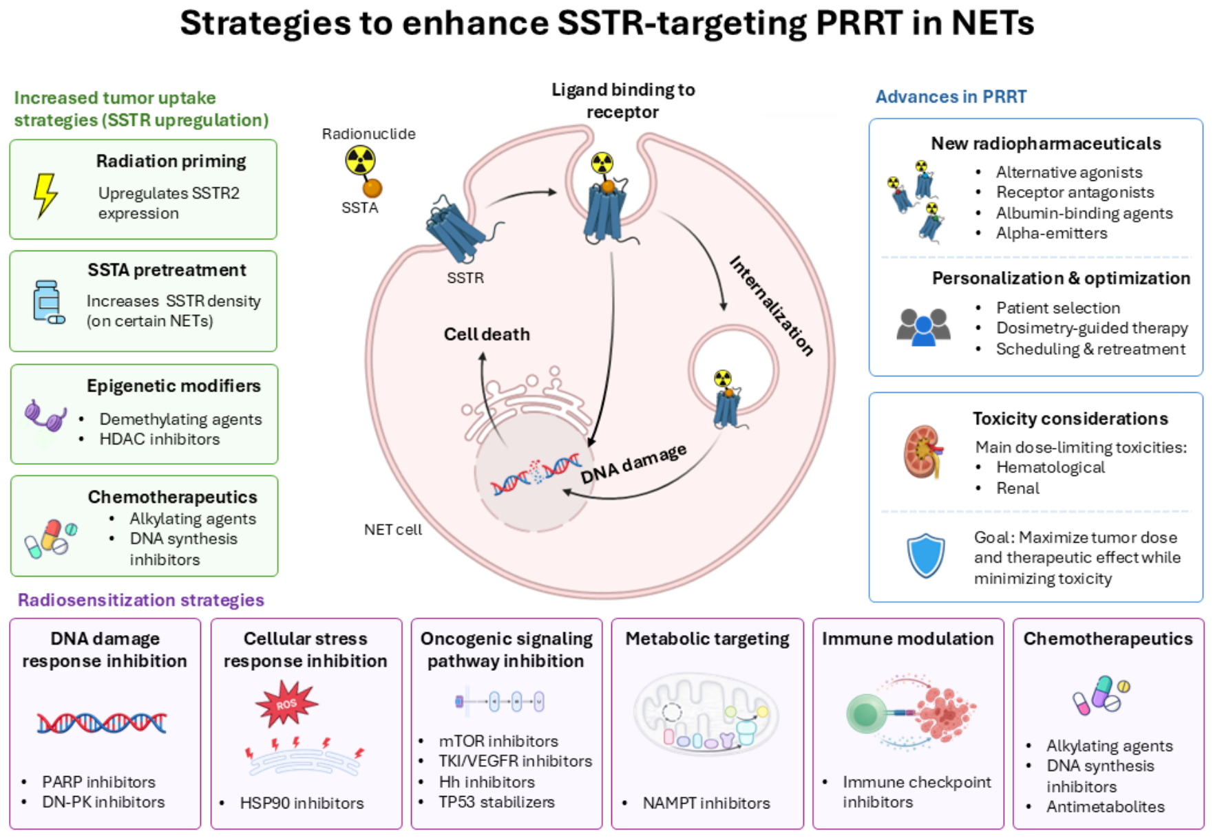

Table 1.
Clinical experience with [177Lu]Lu-DOTATATE therapy in NETs.
| Administered Activity (GBq/Cycle) (Number of Cycles) | No. of Patients | Toxicity/Side Effects | Reference | ||||||
|---|---|---|---|---|---|---|---|---|---|
| CR (%) |
PR (%) |
MR (%) |
SD (%) |
OS (Months) |
PFS (Months) |
||||
| 3.7–7.4 (7 cycles) DOTATATE |
35 | 3 | 35 | 0 | 41 | NA | NA | 0% anemia (grade 3), 1% leukocytopenia, 1% thrombocytopenia | Kwekkeboom et al. 2003 [37] |
| 3.7–7.4 (4 cycles) DOTATATE |
504 (toxicity analysis), 310 (efficacy analysis) |
2 | 28 | 16 | 35 | 46 | 33 | 3.6% hematologic toxicity (≥grade 3), myelodysplastic syndrome (three patients), liver toxicity (two patients) | Kwekkeboom et al. 2008 [38] |
| 8.0 (1–5 cycles) DOTATATE |
26 | 0 | 38 | NA | 50 | NA | NA | 12% hematologic toxicity (grade 3) | Swärd et al. 2010 [49] |
| 3.7–7.4 (6 cycles) DOTATATE |
51 | 2 | 27 | 21 | 27 | NA | 36 | No major acute or delayed renal or hematologic toxicity leukopenia (grade 3), thrombocytopenia | Bodei et al. 2011 [39] |
| 8.0 (6–9 cycles) DOTATATE |
68 | 0 | 60 | 28 | 13 | 53 | 34 | 5.9% hematologic toxicity (grade 3), 1.7% leucopenia, 2.2% thrombocytopenia, 0.9% anemia | Ezziddin et al. 2014 [42] |
| 7.9 (4 cycles) DOTATATE |
74 | 0 | 36 | 18 | 35 | 55 | 26 | NA | Ezziddin et al. 2014 [41] |
| 7.9 (4 cycles) DOTATATE |
61 | 0 | 13 | 31 | 48 | 61 | 33 | 8.2% hematological toxicity (≥grade 3) | Sabet et al. 2015 [47] |
| 3.7–5.5 (5 cycles) DOTATATE |
43 | 7 | NA | NA | 77 | NA | 36 | Nausea (max grade 2), asthenia, mild alopecia | Paganelli et al. 2014 [46] |
| 7.0 (1–4 cycles) DOTATOC |
56 | 16 | 18 | NA | 32 | 34 | 17 | 1.7% grade 3 myelotoxicity, 20% mild renal toxicity | Baum et al. 2016 [17] |
| 3.7–5.5 (4–5) DOTATATE |
34 | 3 | 12 | NA | 47 | 49 | 18 | No acute or delayed hematologic toxicity | Ianniello et al. 2016 [45] |
| 7.4 (4 cycles) DOTATATE |
116 | 1 | 17 | 64 | 48 | 20 | 1% neutropenia (≥grade 3), 2% thrombocytopenia, No cases of MDS or acute leukemia | Strosberg et al. 2017 and 2021 [12,13] | |
| 5.6–7.4 (4 cycles) DOTATATE |
143 | 0 | 8 | NA | 46 | NA | NA | 1% MDS and liver toxicities | Hamiditabar et al. 2017 [44] |
| 3.7 (1–6 cycles) DOTATAT |
443 | 2 | 37 | NA | 43 | 63 | 29 | 0.7% leukemia, 1.5% MDS |
Brabander et al. 2017 [11] |
| 7.4 (4 cycles) DOTATATE |
79 | NA | 6 | NA | 61 | 48 | 48 | Mild decreases in platelets, leukocytes, hemoglobin; no grade 3–4 events No significant nephrotoxicity Fatigue and diarrhea in a few patients |
Ballal et al. 2017 [199] |
| 3.7 (5 cycles) 5.5 (5 cycles) DOTATATE |
28 32 |
11 3 |
24 16 |
NA NA |
43 59 |
NR 64 |
53 22 |
In 3.7 group, 39.2% patients had anemia, 10.7% thrombocytopenia, 21.4% leucopenia. In 5.5 Group 31.3% patients had anemia, 28.1% thrombocytopenia, 21.9% leucopenia, no grade 3 or 4 hematological |
Sansovini et al. 2017 [48] |
| 7.4 (4 cycles) DOTATATE |
200 | 1 | 24 | NA | 68 | 43 | 27 | 1.5% acute leukemia, 0.5% chronic leukemia 15% grade 3 or 4 bone marrow toxicity. 4% grade 2 kidney toxicity 0.5% grade 4 kidney toxicity | Garske-Román et al. 2018 [43] |
| 7.4 (4 cycles) DOTATATE |
52 | 0 | 23 | 36 | 33 | NR | 16 | 10.0% subacute grade 3 or 4 toxicities, 52% lymphocytopenia, 33% nausea, 30.8% fatigue | Del Prete et al. 2019 [40] |
| 7.4 (4 cycles) DOTATATE |
34 | 3 | 56 | NA | 24 | NA | 18 | 12% Subacute hematological toxicity, grade 3 or 4.9% hormonal crisis | Zandee et al. 2019 [51] |
| 7.4 (4 cycles) DOTATATE |
30 | NA | 23 | NA | 67 | NR | 30 | 20% hematological toxicity, grade 3 or 4.7% catecholamine release | Zandee et al. 2019 [52] |
| 7.4 (1–10 cycles) DOTATATE |
102 | 4 | 45 | NA | 44 | 24 | 42 | 10.8% Bone marrow toxicity grade 3–4 occurred, 1.0% acute myeloid leukemia | Fröss-Baron et al. 2021 [50] |
| 7.4 (1–4 cycles) DOTATATE |
48 | NA | 44 | NA | 44 | 59 | 23 | 14% lymphopenia, no incidence of MDS or leukemia | Zidan et al. 2022 [53] |
| 7.4 (4 cycles) DOTATATE |
36 | 0 | 31 | NA | NA | NR | 31 | Generally well tolerated. Grade ≥ 3 adverse events were mainly hematological. No grade ≥ 3 nephrotoxicity, myelodysplastic syndrome, or leukemia were observed. | Satapathy et al. 2025 [62] |
| 7.4 (4 cycles) DOTATATE |
162 | NA | NA | NA | NA | NA | NA | NA | Puranik et al. 2025 [209] |
| 7.4 (3–5 cycles) DOTATATE |
10 | 7 | 0 | NA | 29 | 21 | 10 | One patient showed transient grade 3 anemia, grade 4 thrombocytopenia, another patient showed only transient G4anemia and G3 thrombocytopenia | di Santo et al. 2026 [210] |
| 7.4 (4 cycles) DOTATATE |
13 | 0 | 23 | NA | 69 | NR | 51 | Grade 3–4 adverse events were mainly lymphopenia. Long-term hematologic toxicity occurred in 31% of patients. One case of treatment-related acute leukemia was reported. No late renal toxicity was observed. | Chan et al. 2026 [211] |
| 7.4 (4 cycles) DOTATATE |
61 | 2 | 44 | NA | 49 | 61 | 32 | Grade ≥ 3 adverse events occurred in 18% of patients, mainly hematological and gastrointestinal toxicities. | Becx et al. 2026 [212] |
| 7.4 (4 cycles) DOTATATE |
21 | 2 | 12 | NA | 6 | NR | 37 | Generally well tolerated. Grade 3 hematological toxicity was uncommon (primarily lymphopenia and anemia). No severe renal toxicity or treatment-related deaths were observed. One case of CML occurred during long-term follow-up. | Kobayashi et al. 2026 [222] |
| 7.4 (4 cycles) DOTATATE |
10 | 0 | 30 | NA | 70 | NR | NR | Generally well tolerated. Grade ≥ 3 treatment-related adverse events were primarily hematological, most commonly lymphopenia and thrombocytopenia. No cases of AML, MDS, severe infections, or other secondary malignancies were reported. | Ma et al. 2026 [223] |
| 5.5–6.3 (4–6 cycles) DOTATATE |
20 | 0 | 10 | NA | 50 | NR | 12 | Generally well tolerated. No grade ≥ 3 hematological toxicity. Mild transient gastrointestinal symptoms and fatigue were common. One patient developed grade 1 nephrotoxicity. No hepatotoxicity was observed. | Agrawal et al. 2026 [60] |
| Abbreviations: NA = not available, NR = not reached CR = complete response, PR = partial response, MR = minor response, PFS = progression-free survival, SD = stable disease, OS = overall survival, MDS = myelodysplastic syndrome. | |||||||||
Table 2.
Clinical studies evaluating combination strategies for enhancement of [177Lu]Lu-DOTATATE therapy in NETs.
Table 2.
Clinical studies evaluating combination strategies for enhancement of [177Lu]Lu-DOTATATE therapy in NETs.
| Strategy Combination |
Treatment Combination Administered Activity (GBq per Cycle) (Number of Cycles) |
No. of Patients | Reported Tumor Response | Toxicity/Side Effects | Reference | |||||
|---|---|---|---|---|---|---|---|---|---|---|
| CR (%) |
PR (%) | MR (%) |
SD (%) |
OS (Months) |
PFS (Months) |
|||||
| Chemotherapy drugs | ||||||||||
| 7.4 (4 cycles) DOTATATE + Capecitabine |
7 | 0 | 14 | NA | NA | NA | NA | 1 × grade-3 anemia; one case of grade-3 thrombocytopenia; one occurrence of grade-2 thrombocytopenia; no grade-4 hematologic events were observed. | van Essen et al. 2008 [200] | |
| 7.4 (4 cycles) DOTATATE + Capecitabine |
27 | NA | NA | NA | NA | 36 | NA | No hematologic toxicity, no MDS, leukemia, or late renal failure | Hubble et al. 2010 [201] | |
| 7.8 (4 cycles) DOTATATE + Capecitabine |
33 | 0 | 24 | NA | 70 | NR | NR | 1 × Grade-3 thrombocytopenia; no neutropenia; no nephrotoxicity | Claringbold et al. 2011 [202] | |
| 7.8 (4 cycles) DOTATATE + Capecitabine and Temozolomide |
35 | 13 | 70 | NA | 38 | NR | 48 | 18% of cases had moderate nausea, and 3% had severe nausea. 24% had moderate low platelet levels, and 6% had severe low white blood cell levels. There were no very severe side effects, and no kidney or liver problems were reported. | Claringbold et al. 2012 [203] | |
| 7.4 (3–6 cycles) DOTATATE + 5-FU |
63 | 0 | 30 | 9 | 29 | >60 | NA | NA | Kong et al. 2014 [207] | |
| 6.0–10 (3–5 cycles) DOTATATE + 5-FU |
52 | 2 | 28 | NR | 68 | NR | 48 | well tolerated with negligible grade 3/4 toxicities | Kashyap et al. 2015 [208] | |
| 7.9 (4 cycles) DOTATATE + Capecitabine and Temozolomide |
30 | 13 | 70 | NA | 20 | NR | 48 | transient nausea of grade 2, grade 3 Hematological toxicity thrombocytopenia (10%) and anemia (10%). | Claringbold et al. 2016 [205] | |
| 5.5–7.4 (6–8 cycles) DOTATATE + Capecitabine |
88 | 0 | 34 | 9.1 | 50 | NR | NR | No clinical thrombocytopenia; no grade 3–4 events. Two patients had mild insufficiency; no significant nephrotoxicity. Diarrhea and anorexia occurred in a few patients. | Ballal et al. 2017 [199] | |
| 7.1 (3 cycles) DOTATATE + Temozolomide +/− Capecitabine |
15 (3 + 12) | NA | NA | NA | NA | 25 | 7 | Grade 4 hepatic failure: 1 patient Grade 3 hepatic toxicity (liver enzymes): 4 patients (27%) |
Yordanova et al. 2019 [206] | |
| 5.5 (8 cycles) DOTATATE + Capecitabine |
33 | 0 | 30 | NA | 55 | NR | 31 | Grade 3 (G3) or 4 (G4) hematological toxicity occurred in 16.2% of patients. diarrhea in 5.4% of cases and asthenia in 5.4%. No renal toxicity was observed for the duration of follow-up | Nicolini et al. 2021 [204] | |
| 7.4 (4 cycles) DOTATATE + Capecitabine |
36 | 0 | 33 | NA | NA | NR | 29 | Generally well tolerated. Grade ≥ 3 adverse events were mainly hematological. No grade ≥ 3 nephrotoxicity, myelodysplastic syndrome, or leukemia were observed. | Satapathy et al. 2025 [62] | |
| 7.4 (4 cycles) DOTATATE + Temozolomide and Capecitabine |
162 | NA | NA | NA | NA | NA | NA | NA | Puranik et al. 2025 [209] | |
| 7.4 (3–5 cycles) DOTATATE + Temozolomide +/− Capecitabine |
7 7 |
00 | 71 14 |
NA NA |
029 | NR 21 |
42 13 |
In the temozolomide group, some grade 3–4 hematological and renal toxicities were reported, whereas no severe (grade 3–4) hematological or renal toxicities were observed in the CAPTEM group. | di Santo et al. 2026 [210] | |
| 7.4 (4 cycles) DOTATATE + Temozolomide and Capecitabine (pNET and siNET) |
18 32 |
00 | 72 34 |
NA NA |
28 63 |
NR NR |
59 46 |
Grade ≥ 3 adverse events were primarily hematological and consistent with the known safety profile of CAPTEM and PRRT. | Chan et al. 2026 [211] | |
| 7.4 (4 cycles) DOTATATE + Capecitabine |
50 | 2 | 30 | NA | 60 | 76 | 46 | Grade ≥ 3 adverse events occurred in 26% of patients, mainly hematological and gastrointestinal toxicities. | Becx et al. 2026 [212] | |
| PARP inhibitors | ||||||||||
| 7.4 (4 cycles) DOTATATE + Olaparib |
18 | 3 | NA | 8 | NA | NA | NA | Hematological toxicity (including grade 3–4 lymphocytopenia) was most common; non-hematological effects were mainly mild (nausea, fatigue), with occasional liver enzyme elevations. | Hallqvist et al. 2025 [148] | |
| mTOR inhibitors | ||||||||||
| 7.8 (1–4 cycles) DOTATATE + Everolimus |
16 | 0 | 44 | NA | 56 | NR | NR | grade 2 or 3 neutropenia and thrombocytopenia grade 2 nephrotoxicity |
Claringbold et al. 2015 [220] | |
| 3.7–7.4 (4 cycles) DOTATATE + Everolimus |
11 | NA | 9 | NA | 82 | NA | 23.3 | grade ≥ 3 events in ~36% of patients (including infection and neutropenia), occasional dose reductions, and one serious thrombotic event. | Aljubran et al. 2024 [221] | |
| Checkpoint Inhibitors | ||||||||||
| 3.7–7.4 (4 cycles) DOTATATE + Nivolumab |
9 | 14 | NA | NA | NA | NA | NA | lymphopenia (n = 7), thrombocytopenia (n = 4), anemia (n = 3), and nausea (n = 3). The most common grade 3 TRAE was lymphopenia (n = 4). | Kim et al. 2020 [191] | |
| 3.5 (1–6 cycles) DOTATATE + PD-1 |
2 | NA | NA | NA | NA | NA | NA | lymphocyte counts decreased | Aicher et al. 2022 [192] | |
| 7.4 (4 cycles) DOTATATE + Ipilimumab and nivolumab |
1 | NA | NA | NA | NA | NA | NA | Transient hemtoxicity and mild nausea | Ferdinandus et al. 2022 [193] | |
| Abbreviations: NA = not available, NR = not reached, CR = complete response, PR = partial response, MR = minor response, PFS = progression-free survival, SD = stable disease, OS = overall survival, MDS = myelodysplastic syndrome. | ||||||||||
Table 3.
Ongoing clinical trials evaluating [177Lu]Lu-DOTATATE or [177Lu]Lu-DOTATOC in combination with other agents in patients with SSTR-expressing tumors.
Table 3.
Ongoing clinical trials evaluating [177Lu]Lu-DOTATATE or [177Lu]Lu-DOTATOC in combination with other agents in patients with SSTR-expressing tumors.
| Class of Combined Agent | Trial Number | Treatment Combination | n | Patients | Study Phase | Status |
|---|---|---|---|---|---|---|
| PARP inhibitors | NCT04086485 | [177Lu]Lu-DOTATATE + Olaparib | 56 | GEP-NET | 1/2 | Recruiting |
| NCT05870423 | [177Lu]Lu-DOTATATE + Olaparib | 24 | mNET | 1 | Recruiting | |
| NCT04375267 | [177Lu]Lu-DOTATATE + Olaparib | 18 | SSRT+ | 1 | Active, not recruiting | |
| NCT05053854 | [177Lu]Lu-DOTATATE + Talazoparib | 24 | GEPNET, G 2 |
1 | Recruiting | |
| NCT06607692 | [177Lu]Lu-DOTATATE + Olaparib | 25 | Children and adolescents with recurrent or relapsed solid tumor expressing SSTR | 2 | Recruiting | |
| Immune checkpoint inhibitors | NCT04525638 | [177Lu]Lu-DOTATATE + Nivolumab | 30 | NET G3 or NEC | 2 | Unknown status |
| NCT04261855 | [177Lu]Lu-DOTATATE + Avelumab | 19 | mMCC | 1/2 | Recruiting | |
| NCT03457948 | [177Lu]Lu-DOTATATE + Pembrolizumab | 32 | NET G2 and G3 |
2 | Active, not recruiting | |
| NCT05583708 | [177Lu]Lu-DOTATATE + Pembrolizumab | 18 | Merkel Cell Cancer | 2 | Recruiting |
|
| NCT05142696 | [177Lu]Lu-DOTATATE + Carboplatin, Etoposide and Atezolizumab | 24 | Extensive Stage SCLC | 1/2 | Active, not recruiting | |
| TK inhibitors | NCT05249114 | [177Lu]Lu-DOTATATE + Cabozantinib | 6 | SSTR+ NET |
1 | Active, not recruiting |
| NCT05687123 | [177Lu]Lu-DOTATATE + Sunitinib malate | 24 | pNET | 1 | Recruiting | |
| DNA-PK inhibitors | NCT04750954 | [177Lu]Lu-DOTATATE + Peposertib | 29 | pNET | 1 | Active, not recruiting |
| Chemotherapy drugs | NCT04194125 | [177Lu]Lu-DOTATOC + Capecitabine and Temozolomide |
25 | non-resectable and/or GEP-NET G1and G2 | 2 | Unknown |
| NCT02736500 | [177Lu]Lu-DOTATATE + Capecitabine | 39 | GEP-NET | 1/2 | Completed | |
| NCT05247905 | [177Lu]Lu-DOTATOC + Capecitabine & temozolomide | 31 | pNET | 2 | Active, not recruiting | |
| NCT02358356 | [177Lu]Lu-DOTATOC + Capecitabine & temozolomide |
75 | mNET | 2 | Completed | |
| NCT07185672 | [177Lu]Lu-DOTATOC + Capecitabine & temozolomide | 162 | GEP-NET | 3 | Recruiting | |
| NCT05387603 | [177Lu]Lu-DOTATOC + Capecitabine | 300 | GEP-NEN | 3 | Recruiting | |
| NCT04234568 | [177Lu]Lu-DOTATATE + Triapine | 33 | NET | 1 | Active, not recruiting | |
| NCT05724108 | [177Lu]Lu-DOTATATE + Triapine | 94 | mNET | 2 | Recruiting | |
| Endocrine therapeutic | NCT05884255 | [177Lu]Lu-DOTATATE + LA TOC/High-dose LA TOC | 220 | GEP-NET | 3 | Recruiting |
| NCT06855095 | [177Lu]Lu-DOTATATE + LA SSTAs | 39 | NET G1 and G2 |
2/3 | Recruiting | |
| NCT06663072 | [177Lu]Lu-DOTATATE + Fulvestrant | 25 | pNET | 1 | Recruiting | |
| Epigenetic drug | NCT05178693 | [177Lu]Lu-DOTATATE + ASTX727 | 27 | NET | 1 | Recruiting |
| mTOR inhibitors | NCT03049189 | [177Lu]Lu-DOTATOC + Everolimus | 309 | SSTR+/GEP-NET | 3 | Active. not recruiting |
| NCT04665739 | [177Lu]Lu-DOTATATE + Everolimus | 70 | bronchial NET | 2 | Recruiting | |
| NCT05773274 | [177Lu]Lu-DOTATATE + Everolimus | 100 | NET | 2 | Recruiting | |
| External beam radiation (EBR) | NCT07150546 | [177Lu]Lu-DOTATATE + EBR | 15 | Large GI NET | 1 | Recruiting |
| NCT05109728 | [177Lu]Lu-DOTATATE + EBR and Temozolomide |
65 | Recurrent Glioblastoma | 1 | Active, not recruiting |
Table 4.
Preclinical in vitro studies evaluating enhancement of [177Lu]Lu-DOTATATE therapy.
| Class | Mechanism of Action | Drug | Animal Model | Results | Reference |
|---|---|---|---|---|---|
| HSP90 inhibitors | Inhibit HSP90 chaperone function, leading to degradation of client proteins and impaired cellular stress responses | Ganetespib | GOT1 human small intestine NET xenograft model | Reduction in tumor volume | Hofving et al. 2019 [134] |
| Onalespib | BON xenograft mouse model | Delayed tumor doubling time; increased SSTR2 expression | Lundsten et al. 2020. [137] | ||
| PARP inhibitors | Inhibit DNA repair mechanisms, leading to accumulation of DNA damage | Talazoparib | AR42J xenograft mouse model | Prolonged tumor growth inhibition; improved survival | Cullinane et al. 2020 [146] |
| DNA-PK inhibitors | Inhibit NHEJ-mediated DSB repair | Nedisertib | H1299-SSTR2 and AR42J xenografts | Improved tumor control and prolonged survival compared with PRRT alone | Waldeck et al. 2023 [151] |
| AZD7648 | BON and H69 xenografts | Enhanced radiosensitivity and improved therapeutic efficacy | Reuvers et al. 2023 [152] | ||
| Tyrosine kinase inhibitors * | Inhibit receptor tyrosine kinase signaling pathways involved in tumor growth and angiogenesis | Vandetanib (+EBR) | GOT2 human MTC xenograft | Reduction in tumor volume; prolonged time to progression | Sandblom et al. 2020 [175] |
| Cabozantinib (+EBR) | GOT2 human MTC xenograft | Reduction in tumor volume; delayed tumor regrowth | Sandblom et al. 2020 [175] | ||
| Hedgehog inhibitors | Inhibit Hedgehog signaling pathway involved in tumor growth and progression | Sonidegib | GOT1 human small intestine NET xenograft | Reduction in tumor volume; prolonged time to progression; altered gene expression (e.g., PDGFRA) |
Spetz et al. 2017 [179] |
| NAMPT inhibitors | Deplete intracellular NAD+, impair energy metabolism and cellular stress responses | GMX1778 (CHS 828) | GOT1 human small-intestinal NET xenograft | Enhanced anti-tumor effect of [177Lu]Lu-DOTATATE; prolonged progression-free survival compared with monotherapy | Elf et al. 2017 [195] |
| Chemotherapeutic agents | Inhibition of DNA synthesis | Gemcitabine | GOT2 medullary thyroid carcinoma xenograft | Prolonged time to progression compared with monotherapy; gemcitabine enhanced radiation response | Sandblom et al. 2019 [197] |
| mTOR inhibitors | Inhibit PI3K/AKT/mTOR signaling, reducing proliferation and survival signaling | Everolimus (RAD001) | CA20948 rat pancreatic NET xenograft | No anti-tumor benefit; increased metastatic progression observed in non-cured animals | Pool et al. 2013 [217] |
| Everolimus (RAD001) | CA20948 rat pancreatic NET xenograft | No improvement compared with PRRT alone; metastatic progression observed in animals not achieving complete remission | Bison et al. 2014 [218] | ||
| Everolimus (RAD001) | AR42J rat pancreatic NET xenograft | No additional anti-tumor effect beyond PRRT alone; acceptable toxicity profile | Zellmer et al. 2022 [219] |
* Evidence derived from EBR, not [177Lu]Lu-DOTATATE.
Table 5.
Results from in vitro studies on combination strategies to enhance [177Lu]Lu-DOTATATE therapy in NET.
Table 5.
Results from in vitro studies on combination strategies to enhance [177Lu]Lu-DOTATATE therapy in NET.
| Class | Mechanism of Action |
Drug | Cell Type | Results | Reference |
|---|---|---|---|---|---|
| HSP90 inhibitors | Inhibit HSP90 chaperone function, leading to degradation of client proteins and impaired cellular stress responses | Onalespib | BON, H727, H460 (monolayer and spheroids) | Synergistic effects with increased apoptosis in BON and H727 spheroids | Lundsten et al. 2019 [138] |
| Onalespib | BON (ex vivo autoradiography) | No change in uptake or spatial distribution of [177Lu]Lu-DOTATATE | Lundsten et al. 2020 [137] | ||
| Ganetespib | GOT1, BON-SSTR2 NET models | Enhanced radiation-induced cytotoxicity; radiosensitization mediated by proteotoxic stress rather than increased DNA damage | Engbers et al., 2025 [139] | ||
| PARP inhibitors | Inhibit DNA repair mechanisms, leading to accumulation of DNA damage (DSB) | Olaparib | CA20948, U2OS | Increased uptake of [177Lu]Lu-DOTATATE; reduced proliferation; increased DNA damage | Nonnekens et al. 2016 [144] |
| DHQ, PJ-34, ABT-888 (Veliparib) | BON, H727 | Increased cytotoxicity; increased apoptosis; cell cycle arrest | Purohit et al. 2018 [145] | ||
| Talazoparib | AR42J | Increased DNA double-strand breaks | Cullinane et al. 2020 [146] | ||
| Olaparib, rucaparib | H69, H446 | Increased DNA damage and PRRT potency | Rauch et al. 2024 [147]. | ||
| DNA-PK inhibitors | Inhibit NHEJ-mediated DSB repair | Nedisertib, AZD7648 | H1299-SSTR2, AR42J | Increased sensitivity to [177Lu]Lu-DOTATATE and reduced cell survival | Waldeck et al. 2023 [151] |
| AZD7648 | BON, H69, GOT1 | Enhanced PRRT-induced cytotoxicity | Reuvers et al. 2023 [152] | ||
| DNA methyltransferase inhibitors (DNMTis) | Modify epigenetic regulation and gene expression, including increased SSTR expression and altered DNA repair | Decitabine + 5-FU | BON, QGP-1 | Increased radiosensitivity, apoptosis, SSTR2 expression and radioligand binding | Jin et al. 2019 [157] |
| 5-aza2’-deoxycytidine (5-aza-dC) | SSTR2-transfected HEK293 | Increased uptake and therapeutic effect of [177Lu]Lu-DOTATATE | Kotzerke et al. 2022 [99] | ||
| HDAC inhibitors | Modify epigenetic regulation and gene expression, including increased SSTR expression and altered DNA repair. | VPA | SSTR2-transfected HEK293 | Increased uptake and therapeutic effect of [177Lu]Lu-DOTATATE | Kotzerke et al. 2022 [99] |
| BET inhibitors * | Epigenetic/transcriptional regulation | I-BET151 | GOT1, P-STS | Synergistic reduction in cell viability in both siNET cell lines | Hofving et al. 2019 [134] |
| Chemotherapeutic agents | Ribonucleotide reductase inhibition; S-phase accumulation | Hydroxyurea, gemcitabine, triapine | U2OS + SSTR2A, BON1 | Increased SSTR2A expression, increased [177Lu]Lu-DOTATATE uptake, enhanced PRRT-induced cytotoxicity | Cheng et al. 2024 [198] |
| p53 stabilizers | Stabilize p53 by inhibiting its degradation, enhancing DNA damage response and apoptosis | VIP116 | IMR-32, SK-N-AS, N2a, LU-NB-1, LU-NB-2 (monolayer and spheroids) | Synergistic effects with increased apoptosis in IMR-32 spheroids | Lundsten et al. 2021 [224] |
| mTOR inhibitors | Inhibit PI3K/AKT/mTOR signaling and cellular proliferation | Everolimus (RAD001) | BON | Anti-proliferative effects and growth inhibition | Zitzmann et al. 2007 [215] |
DHQ = 1,5-dihydroxyisoquinoline. * Evidence derived from EBR, not [177Lu]Lu-DOTATATE.
Table 6.
Characteristics of tumor cell lines and experimental models used in NET-related PRRT research.
Table 6.
Characteristics of tumor cell lines and experimental models used in NET-related PRRT research.
| Cell Line | Origin (Species) | Tumor Type | SSTR Expression | Exocrine/Endocrine | References |
|---|---|---|---|---|---|
| AR42J | Rat | Pancreatic tumor | SSTR1,2,3,5 | Exocrine | [225] |
| At-T20 | Mouse | Pituitary tumor | SSTR2,5 | Endocrine | [226] |
| BON/BON-1 | Human | Pancreatic NET | SSTR2,3,5 | Endocrine | [227] |
| Capan-2 | Human | Pancreatic adenocarcinoma | SSTR2,5 | Exocrine | [228] |
| CA20948 | Rat | Pancreatic tumor | SSTR2 | Exocrine | [229] |
| CM | Human | Pancreatic insulinoma | SSTR2 | Endocrine | [230] |
| GOT1 | Human | Small intestinal NET (siNET) | SSTR2 | Endocrine | [231] |
| GOT2 | Human | Medullary thyroid carcinoma (MTC) | SSTR2 | Endocrine | [232] |
| H69 | Human | Small cell lung cancer | SSTR1,2 | - | [233] |
| H727 | Human | Bronchopulmonary NET | Low SSTR2 expression | Endocrine | [234] |
| H-STS | Human | Small intestinal NET (siNET) | Low SSTR2,3 expression | Endocrine | [235] |
| KRJ-I | Human | Small intestinal NET (siNET) | Low SSTR2,3 expression | Endocrine | [236] |
| L-STS | Human | Small intestinal NET (siNET) | Low SSTR2,3 expression | Endocrine | [235] |
| MZ-CRC-1 | Human | Medullary Thyroid Cancer | SSTR2 | Endocrine | [237] |
| P-STS | Human | Small intestinal NET (siNET) | Low SSTR2,3 expression | Endocrine | [235] |
| QGP-1 | Human | Pancreatic NET | Low SSTR2 expression | Endocrine | [238] |
| TT | Human | Medullary Thyroid Cancer | SSTR2 | Endocrine | [239] |
| TtT-97 | Mouse | Pituitary tumor | Not reported | Endocrine | [240] |
Table 7.
Overview of strategies for enhancement of [177Lu]Lu-DOTATATE therapy.
| Strategy | Method | In Vitro | Preclinical In Vivo | Clinical |
|---|---|---|---|---|
| SSTR upregulation | ||||
| EBR-induced upregulation | EBR | Increased SSTR expression and uptake in several models | Increased uptake after EBR in tumor models | NA |
| PRRT-induced upregulation | PRRT priming/fractionation | Fractionation enhances radiobiological effects | Higher tumor uptake and absorbed dose, delayed tumor growth | Limited clinical observations (case reports); no controlled clinical studies |
| SSTA-mediated modulation | TOC, pasireotide (dose- and time-dependent) | Down- or upregulation depending on dose/exposure | Increased binding under certain conditions | Variable effects on tumor uptake; no consistent improvement demonstrated |
| Epigenetic modifiers | DNMTi, HDACi (e.g., 5-aza-dC, VPA) | Increased SSTR expression and uptake | Increased uptake in some models | Early-phase clinical trials ongoing for SSTR upregulation strategies (no efficacy data yet) |
| Hormonal regulation | Thyroid hormone, EGF, tamoxifen | Increased SSTR expression in several cell lines | NA | NA |
| Chemotherapy-induced modulation | Temozolomide, gemcitabine, triapine | Increased SSTR expression and uptake in some models | NA | NA |
| Radiosensitization | ||||
| HSP90 inhibitors | Ganetespib, Onalespib | Enhanced apoptosis and radiosensitivity; radiosensitization linked to proteotoxic stress and pleiotropic pathway disruption | Reduced tumor growth and increased doubling time | No clinical studies with PRRT combinations reported |
| PARP inhibitors |
Olaparib, Talazoparib, Veliparib | Increased DNA damage and apoptosis | Enhanced tumor control and survival | Phase I clinical data demonstrate feasibility and manageable toxicity |
| DNA-PK inhibition | Nedisertib (peposertib), AZD7648 | Increased sensitivity to PRRT through inhibition of NHEJ-mediated DNA repair | Improved tumor control and survival in NET xenograft models | Phase Ib clinical trial ongoing |
| Epigenetic modifiers | DNMTi, HDACi | Increased apoptosis and radiosensitivity | NA | NA |
| TK inhibitors |
Vandetanib, cabozantinib | NA | Improved tumor control in combination with EBR | TKIs effective as monotherapy in NETs; no clinical PRRT combination data available |
| Hh inhibitors | Sonidegib | NA | Reduced tumor growth, delayed regrowth, altered NF-κB and PDGF signaling | NA |
| Immunotherapy | PD-1/PD-L1 and CTLA-4 inhibitors | Enhanced antigen presentation, immune activation, and tumor-cell recognition after radiation exposure | Increased cytotoxic immune-cell recruitment and improved therapeutic efficacy when combined with [177Lu]Lu-DOTATATE in NET models | Early clinical studies demonstrate feasibility; multiple ongoing phase I-II trials |
| NAMPT inhibitors | GMX1778 | Reduced tumor cell viability through NAD+ depletion and impaired stress-response capacity | Improved anti-tumor response and prolonged progression-free survival in GOT1 xenografts | NA |
| Chemotherapeutic agents | Gemcitabine + [177Lu]Lu-DOTATATE/EBR | NA | Improved tumor control and time to progression; stronger effects with EBR | No clinical studies combining with PRRT; gemcitabine used clinically in NETs |
| Capecitabine, 5-FU, temozolomide, gemcitabine, triapine | Increased SSTR expression and radioligand uptake; radiosensitization through DNA synthesis inhibition, S-phase accumulation, and impaired DNA repair | Improved tumor control and prolonged time to progression in selected models | Most clinically mature combination strategy; multiple positive studies and ongoing randomized trials, although not all studies have demonstrated superiority over PRRT alone | |
| mTOR inhibition | Everolimus, temsirolimus | Reduced proliferation and tumor-cell growth | Mixed results; no clear enhancement of PRRT efficacy in CA20948 or AR42J NET models | Clinical feasibility demonstrated; ongoing trials |
NA = Not available, TK = tyrosine kinase, Hh = Hedgehog, NAMPT = Nicotinamide phosphoribosyltransferase.
Disclaimer/Publisher’s Note: The statements, opinions and data contained in all publications are solely those of the individual author(s) and contributor(s) and not of MDPI and/or the editor(s). MDPI and/or the editor(s) disclaim responsibility for any injury to people or property resulting from any ideas, methods, instructions or products referred to in the content. |
© 2026 by the authors. Licensee MDPI, Basel, Switzerland. This article is an open access article distributed under the terms and conditions of the Creative Commons Attribution (CC BY) license (http://creativecommons.org/licenses/by/4.0/).
Copyright: This open access article is published under a Creative Commons CC BY 4.0 license, which permit the free download, distribution, and reuse, provided that the author and preprint are cited in any reuse.