

Submitted:
24 July 2026
Posted:
11 August 2026
You are already at the latest version
Abstract
Dendrodoris arborescens (Mollusca: Gastropoda) is a dorid nudibranch species for which internal anatomical details have remained undocumented. In September 2024, a total of 20 specimens were collected from the low intertidal zone at Heishijiao, Dalian, Liaoning, China (38.865°N, 121.548°E), representing the first record of this species from the area. Here we present a comprehensive description of both external mor-phology and internal anatomy, based on external observation, anatomical dissection, histological sectioning, and scanning electron microscopy (SEM). The body is elon-gate-elliptical and uniformly black; the mantle surface is smooth without tubercles, bordered by a narrow orange-red marginal band. The rhinophores are clavate, bearing densely arranged, obliquely oriented lamellae on their surface. The branchial plumes are dendritic and encircle the anus. The digestive system features a suctorial oral ap-paratus lacking radula. The reproductive system exhibits a tri-duct hermaphroditic configuration, with the hermaphroditic gonad embedded within the digestive gland. These findings provide foundational morphological data for understanding the repro-ductive biology of Dendrodoris and offer a reference baseline for future studies on spe-cies identification and cultivation of this genus.
Keywords:
Dendrodoris arborescens
; external morphology
; internal anatomy
; histology
; scanning electron microscopy (SEM)
1. Introduction
Dendrodoris arborescens (Mollusca: Gastropoda: Doridida: Dendrodorididae: Dendrodoris) was first described from Chinese waters by Collingwood in 1881 and subsequently reported from Japan, Australia, and other regions [1]. Although previously known from southern China, the present study marks the first record of this species from northern China(preprint). This species holds considerable significance in evolutionary and applied biology: its "simultaneous hermaphroditic" reproductive structure makes it a valuable model for understanding reproductive strategies and adaptive evolution [2], while its rich endowment of sesquiterpene metabolites has attracted growing interest in pharmacological research [3].
The Dendrodorididae and Phyllidiidae were initially placed within Porostomata [4,5] owing to their shared absence of jaws and a radula, which are replaced by a highly specialized suctorial buccal apparatus adapted for feeding on various prey. However, recent molecular phylogenetic studies have demonstrated that the two families are not monophyletic but rather paraphyletic assemblages; the loss of the radula represents an independently evolved character, and the similar suctorial feeding mode is homoplasious [6,7]. Currently, both families, together with Forjacellidae and Mandeliidae, are classified within the superfamily Phyllidioidea [8].
For many years, D. arborescens was frequently confused with two closely related species, D. fumata and D. nigra, due to their overall morphological similarity. Brodie and Calado [2] resolved this taxonomic ambiguity by analyzing larval development characteristics, thereby establishing the species' distinct status. Two diagnostic larval features serve to differentiate D. arborescens from its congeners: the presence of an enlarged head-foot that compensates for the absence of a retractable operculum, and the early shedding of the larval shell during the planktotrophic trochophore stage [2,9,10]. Although preliminary studies have addressed aspects of its reproductive biology and culture [10], the post-settlement mechanism remains poorly understood.
Despite these advances, critical knowledge gaps persist: the larval stage ends with mortality following shell detachment, and virtually nothing is known about the feeding mechanism or internal reproductive anatomy of juvenile and adult stages [10]. These gaps have substantially hindered the development of reliable culture protocols for this species.
In September 2024, specimens of D. arborescens were collected from the intertidal zone at Heishijiao, Dalian, Liaoning Province, China (38.865°N, 121.548°E). In this study, we present a comprehensive description of both external morphology and internal anatomy of this species based on morphological observation, histological analysis, and scanning electron microscopy. These results provide foundational morphological data for species identification and taxonomic revision of the genus Dendrodoris, and offer a basis for future efforts toward captive breeding and culture.
2. Materials and Methods
Specimen collection
A total of 20 mature individuals of Dendrodoris arborescens (length: 40–80 mm) were collected from the intertidal zone of Heishijiao, Dalian, Liaoning Province, China (38.865° N, 121.548° E) in September 2024. All samples were anaesthetized in 10% magnesium chloride (MgCl₂) solution prior to fixation. A portion of the specimens was fixed in 4% paraformaldehyde (PFA) for histological section preparation, whereas fresh tissue fragments from other individuals were preserved in 2.5% glutaraldehyde for scanning electron microscopy (SEM). All voucher specimens are deposited in the specimen collection of Dalian Ocean University.
Following anaesthesia in MgCl2 solution, each specimen was dissected longitudinally along the dorsal midline of the mantle to fully expose the visceral mass and internal organ complexes. The gross morphology, spatial distribution and topological associations of all organ systems were examined and documented photographically under an Olympus SZ61 stereomicroscope (Olympus, Japan). Special dissections were performed to characterize the anatomical structures of the digestive, reproductive and nervous systems, with detailed morphological descriptions provided for each system.
Preparation of tissue sections for light microscopy
Tissues from the rhinophores, branchial plumes, and reproductive system were dehydrated through a graded ethanol series (70%, 80%, 90%, 95%, 100%; 1h each), cleared in xylene (2 × 15 min), and embedded in paraffin. Serial sections at 5–6μm thickness were prepared using a rotary microtome (Leica RM2235, Germany). Sections were stained with hematoxylin and eosin (H&E) following standard protocols: hematoxylin for 5 min, rinsed in running water, differentiated in 1% HCl-ethanol for 5 s, blued under running water, counterstained with eosin for 1 min, and mounted with neutral balsam. Slides were examined and photographed under a light microscope (Nikon Eclipse E100).
SEM sample preparation and observation
For scanning electron microscopy (SEM), fresh tissues were fixed in 2.5% glutaraldehyde (prepared in 0.1 M phosphate buffer) at 4℃ for 24 h, rinsed in 0.1 M phosphate buffer (3 × 10 min), and post-fixed in 1% osmium tetroxide solution for 1–2 h at room temperature in the dark. Samples were then rinsed three times, dehydrated through a graded ethanol series (30%, 50%, 70%, 80%, 90%, 95% for 10 min each; 100% ethanol for 15 min×2), and replaced with isoamyl acetate (2×10-15 min). After dehydration, specimens were critical-point dried with liquid CO2 using a Hitachi HCP-2 critical point dryer. Dried samples were mounted on SEM stubs with conductive adhesive, sputter-coated with a thin gold layer (~10–15 nm) for approximately 30 s in a gold sputter coater, and examined under a Hitachi SU8100 field-emission SEM at an accelerating voltage of 10 kV.
3. Results
3.1. External Morphology
The body of D. arborescens is elongate-elliptical, soft, and highly extensible. Mature specimens range from 40 to 80 mm in length (n = 20) and are uniformly black. The dorsal mantle is smooth and devoid of tubercles, covering the entire dorsal surface. A distinct orange-red marginal band runs along the edge of the mantle. The body surface secretes abundant transparent mucus, forming a thin, even mucous film. Upon stimulation, the foot rolled inward, while the mantle everted and expanded radially, resembling a flower in bloom. (Figure 1).
The head bears two pairs of tentacles: the oral tentacles anteriorly and the rhinophores posteriorly. The oral tentacles are small and situated on either side of the mouth. The rhinophores are clavate, with a relatively thick base and a bluntly rounded apex, and are positioned at approximately the anterior one-third of the body length. Their surface bears densely arranged, obliquely oriented lamellae-the characteristic chemosensory structures of nudibranch rhinophores. The rhinophores can be fully retracted into rhinophoral sheaths embedded in the body wall.
The branchial plumes are situated on the posterodorsal surface, arranged in a circular pattern around the anus. Each plume is dendritic, with slender, leaf-like branchial lamellae that are basally attached to the body wall with free distal ends. Dense cilia are visible on the lamellar surface under light microscopy, whose coordinated beating generates water currents for gas exchange.
The foot is broad and flattened, situated on the ventral side of the body; its anterior end is rounded and the posterior end slightly pointed. The mantle and foot enclose a mantle cavity, within which both the anus and the genital aperture open (Figure 2).
3.2. Digestive System
The digestive system comprises the mouth, buccal cavity, oesophagus, stomach, digestive gland, intestine, and anus. The mouth is situated on the ventral surface of the head. Internally, the oral apparatus is suctorial and devoid of a radula; the buccal cavity is short and wide, with a smooth inner wall. The oesophagus is slender and connects the buccal cavity to the stomach, which is sac-like and located in the anterior portion of the visceral mass. The digestive gland consists of a pair of dark-brown, branching glandular structures situated on either side of the stomach. In dissected specimens, the digestive gland varies in colour from dark brown to black. Notably, the hermaphroditic gonad of D. arborescens is embedded within the digestive gland tissue. The intestine originates from the stomach and coils between the lobes of the digestive gland; its wall is relatively thin and ultimately opens into the anus within the mantle cavity (Figure 3).
3.3. Respiratory System
The respiratory organ consists of a branchial plume situated on the dorsal surface. The gill is composed of three principal components: the branchial sac, the primary branchiae, and the secondary branchiae. Each primary branchium consists of two robust main axes containing abundant connective tissue; from these axes arise multiple secondary branchial axes, which further ramify into finer tertiary branches, collectively exhibiting a typical triply pinnate branching pattern (Figure 4(b)). The branchial sac, located at the base of the plume, provides a protective housing for the gill. The inner wall of the sac is lined with numerous branchial glands, the glandular cells of which are columnar or goblet-shaped and contain abundant secretory granules in the cytoplasm (Figure 4(c)). Sparse smooth muscle fibers are observed within the wall of the branchial sac (Figure 4(d)); occasional branchial glands are also present in the secondary branchiae, though at a markedly lower density than in the sac region (Figure 4(e)). The surface of the branchial lamellae is densely covered with cilia, and the epidermis also contains numerous goblet cells and large mucus-secreting cells (Figure 4(f), Figure 5).
3.4. Sensory Structures
The rhinophores are the principal chemosensory organs of Dendrodoris arborescens. Their surface bears densely arranged, obliquely oriented lamellae distributed symmetrically around the rhinophoral axis, extending from the base to the apical region. Each lamella is uniform in thickness with a smooth margin and is densely covered with regularly arranged cilia (Figure 6 (d–f)).
Histological observation reveals that the rhinophore comprises three distinct layers (Figure 7): (1) the epidermal layer, composed of a single layer of columnar epithelial cells with basally positioned nuclei and cytoplasm containing abundant pigment granules; flask-shaped cells are scattered among the epithelial cells; (2) the connective tissue layer, situated beneath the epidermis, consisting of loose connective tissue that contains blood vessels, nerves, and chromatophores; and (3) the central muscular layer, occupying the core of the rhinophore, composed of both longitudinal and circular smooth muscle fibers, through which the rhinophoral nerve passes; nerve fibers are distributed beneath the sensory cells of the epithelial layer. The muscular layer enables the rhinophore to retract fully into the rhinophoral sheath. Scanning electron microscopy reveals that the lamellar surface is densely covered with cilia approximately 3–5 μm in length, arranged in a helical pattern.
3.5. Reproductive System
Dendrodoris arborescens is a simultaneous hermaphrodite [10], with a reproductive system comprising three independent duct systems that radiate from the sperm vesicle (SV): the large hermaphroditic duct, the small hermaphroditic duct, and the copulatory bursa. This tri-duct configuration is a characteristic feature of Dendrodorididae and of nudibranchs in general, reflecting a shared evolutionary trait in the reproductive system of this group. Anatomical observation (Figure 8) reveals that the reproductive system of D. arborescens consists primarily of the hermaphroditic gonad (HG), sperm vesicle (SV), albumen gland (AG), mucous gland (MG), and their associated duct systems.
The hermaphroditic gonad is the central organ of the reproductive system, responsible for the simultaneous production of both sperm and oocytes. Rather than existing as an independent organ within the body cavity, it is situated adjacent to and partially embedded within the digestive gland (DG). The gonad connects to other reproductive structures via the large hermaphroditic duct (LHD) and the small hermaphroditic duct (SHD), while the mucous gland links to the sperm vesicle through the mucous gland duct.
The copulatory bursa is lined by an epithelial layer composed of columnar cells (COC) and goblet cells (GOC). The columnar cells are regularly arranged with basally positioned nuclei, while the goblet cells are interspersed among them; the apical portion of each goblet cell is enlarged and cup-shaped, containing abundant mucus granules. Beneath the epithelium lies a layer of smooth muscle fibers (SMF) and connective tissue (CT) (Figure 9(a)). The sperm vesicle contains mature oocytes and sperm, and fertilized eggs (Fe) can be observed within it; a distinct egg membrane is visible surrounding each fertilized egg (Figure 9(b)). The hermaphroditic gonad is embedded within the digestive gland and simultaneously contains developing oocytes (MO) and sperm (SZ) (Figure 9(c-d)). The mucous gland and albumen gland are composed of large cells with abundant eosinophilic protein granules in the cytoplasm and large oval nuclei at the base (Figure 9(e-f)).
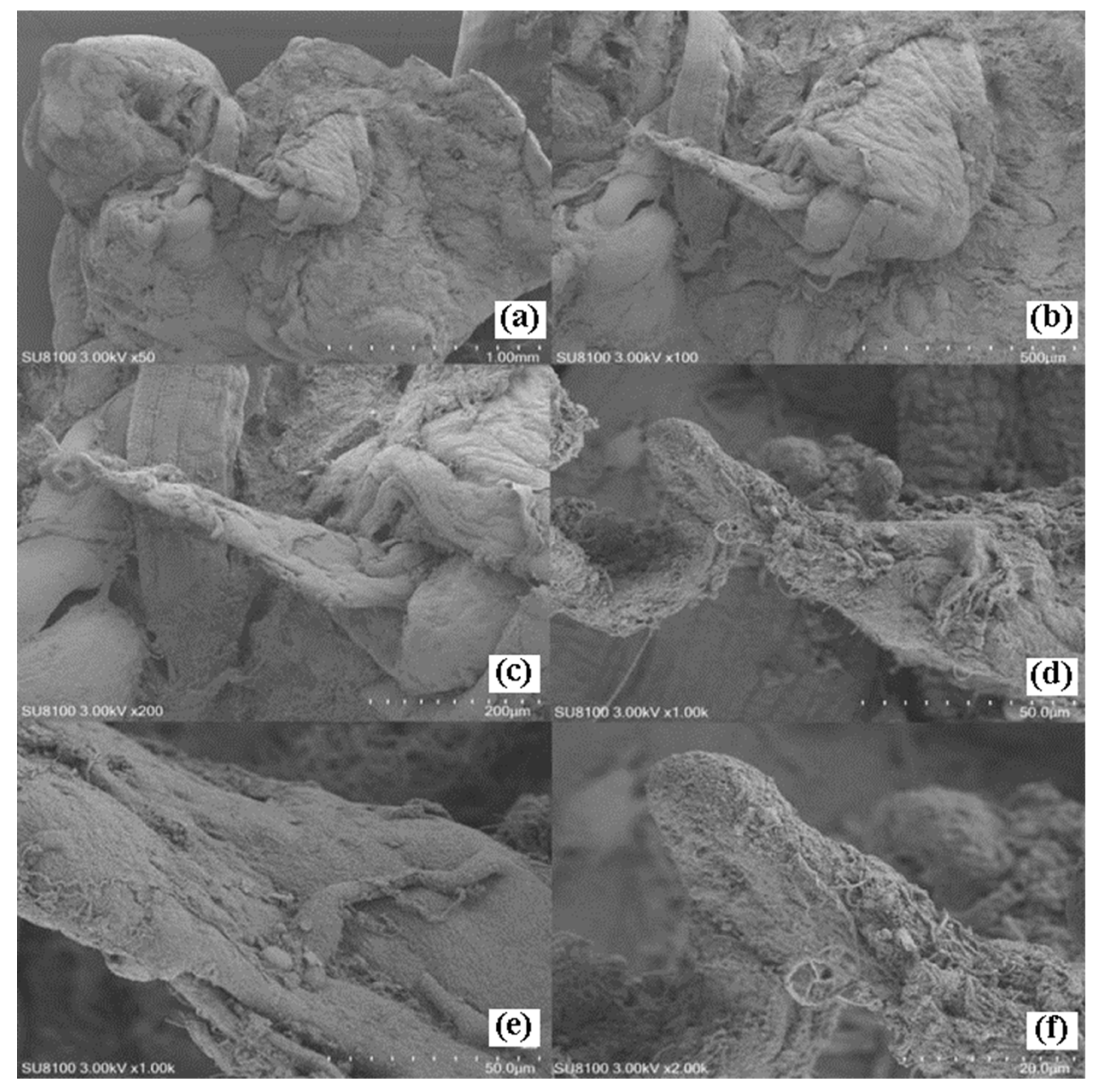
The copulatory bursa is sac-like in morphology, connected to the mucous gland via a duct (Figure 10(a-b)). The penis extends from the junction of the copulatory bursa; it is slender and cylindrical, tapering gradually from base to apex, and the distal portion is elongated and curved (Figure 10(c-d)). The basal surface of the penis bears ridges and blood vessels, while the curved apical region contains numerous irregular small cavities (Figure 10(e-f)).
4. Discussion
The organ systems of Dendrodoris arborescens exhibit morphological specializations closely aligned with its particular ecological niche, differing markedly from those of other nudibranchs and molluscs. Notably, the oral apparatus has completely lost the radula and oral tentacles—structures characteristic of most nudibranch feeding organs—and has been replaced by a highly specialize suctorial feeding device. This morphological specialization diverges from the condition in the majority of nudibranchs, which retain a well-developed radula and feed by scraping algae, coral, or other invertebrates [11,12]. Although this specialized feeding mode restricts the breadth of the species' diet, it enables D. arborescens to exploit sponges—prey items rich in indigestible spicules that are largely inaccessible to most other predators—thereby occupying a comparatively unique ecological niche [13].
As the principal chemosensory organs of nudibranchs, the rhinophores play a critical role in foraging, mate location, and predator avoidance, with their morphological and functional specialization directly influencing these behaviors. In D. arborescens, the lamellar structures on the rhinophoral surface increase the contact area with the surrounding water column, thereby enhancing chemosensory efficiency. Histological analysis reveals that the epidermal layer of the rhinophoral lamellae is markedly thickened and densely populated with goblet cells, while the densely arranged surface cilia represent specialized apical structures of chemosensory cells that are directly involved in chemoreception [14,15]. These features indicate that the rhinophores of D. arborescens are highly specialized for active detection of chemical cues from specific food sources. Compared with other nudibranchs, the number and packing density of rhinophoral lamellae in D. arborescens are moderate, a condition that may be correlated with its comparatively narrow dietary range (sponge specialist). In contrast, members of the family Dendronotidae, which feed on a broader range of prey, possess more complexly branched rhinophoral structures to accommodate the recognition of diverse chemical signals [16]. Furthermore, the rhinophores of D. arborescens are fully retractable; coordinated contraction of the circular muscles in the rhinophoral sheath wall retracts the rhinophore into a protective sheath—a feature shared with the majority of nudibranchs. The rhinophoral sheath glands are distributed circumferentially around the sheath lumen; their secretory function remains unclear, though they are speculated to facilitate lubrication during retraction or to serve a chemical defense role [15].
As simultaneous hermaphrodites, D. arborescens possesses a characteristic tri-duct reproductive system, a structural pattern that is widespread among nudibranchs and is considered a diagnostic feature of the group, yet differs markedly from the hermaphroditic reproductive systems of another molluscs [17]. In this study, we systematically described both the gross anatomical features and histological structures of the reproductive system of D. arborescens. Our observations revealed that the hermaphroditic gonad simultaneously contains developing oocytes and sperm; tissue sections demonstrated that gametes at various developmental stages coexist within the same acinus. This structural arrangement ensures that the species is capable of producing both male and female gametes simultaneously at any given time.
Author Contributions
Conceptualization, W.L. and Y.T.; methodology, Y.T. and H. G.; investigation, W.L., W.S., B.Z., L.C. and Y.S.; writing—original draft, W.L.; writing—review & editing, Y.T. and W.L.; supervision, Y.T.; funding acquisition, Y.T. and H. G. All authors have read and agreed to the published version of the manuscript.
Funding
Research and Application of Liaoning Provincial Science and Technology Plan Project (2025JH2/101330186); Liaoning Provincial Science and Technology Major Project (2025JH1/11700001); the Joint Research Project of Liaoning Provincial Science and Technology Program (2024JH2/102600076); Liaoning Provincial “Xingliao Talent Program” Science and Technology Innovation Team Project; Fundamental Research Funds for Innovative Teams, Dalian Ocean University.
Data Availability Statement
The data presented in this study are available on request from the corresponding authors.
Conflicts of Interest
The authors declare no conflicts of interest.
References
- MolluscaBase. Dendrodoris arborescens . (Collingwood, 1881). Available online: https://www.molluscabase.org/aphia.php?p=taxdetails&id=533877 (accessed on 25 June 2026).
- Brodie, G.; Calado, G. Dendrodoris arborescens (Collingwood, 1881) (Mollusca: Nudibranchia): Larval characteristics reveal a masked porostome species. Rec. West. Aust. Mus. Suppl. 2006, 69, 119–126. [Google Scholar] [CrossRef]
- Gris, L.; Prinsep, M.R. The fascinating chemistry of nudibranchs and their potential for new drug discovery. Chem. N. Z. 2021, 85. [Google Scholar]
- Bergh, L.S.R. Die Nudibranchiata holohepatica Porostomata. Verhandlungen Der Zool.-Bot. Ges. 1892, 42, 1–16. [Google Scholar] [CrossRef]
- Thompson, T.E. Biology of Opisthobranch Molluscs; The Ray Society: London, UK, 1976; Volume 1. [Google Scholar]
- Brunckhorst, D.J. The systematics and phylogeny of phyllidiid nudibranchs (Doridoidea). Rec. Aust. Mus. Suppl. 1993, 16, 1–107. [Google Scholar] [CrossRef]
- Li, W.; Li, F.; Liu, M.; Wang, A.; Liu, C.; Yang, Y. Characterization of 11 complete mitochondrial genomes in Nudibranchia (Mollusca, Gastropoda). ZooKeys 2025, 1244, 61–86. [Google Scholar] [CrossRef] [PubMed]
- MolluscaBase. Phyllidioidea Rafinesque, 1814. Available online: https://www.molluscabase.org/aphia.php?p=taxdetails&id=412563 (accessed on 25 June 2026).
- Rose, R.A. The spawn and embryonic development of colour variants of Dendrodoris nigra Stimpson (Mollusca: Nudibranchia). J. Malacol. Soc. Aust. 1985, 7, 75–88. [Google Scholar] [CrossRef]
- Cai, L.; Han, Y.; Jia, M.; Li, W.; Hao, Z.; Tian, Y. The spawn and embryonic development of Dendrodoris arborescens (Collingwood, 1881) (Mollusca: Nudibranchia). Isr. J. Aquac.-Bamidgeh 2025, 77. [Google Scholar] [CrossRef]
- Goodheart, J.A.; Bazinet, A.L.; Valdés, Á.; Collins, A.G.; Cummings, M.P. Prey preference follows phylogeny: Evolutionary dietary patterns within the marine gastropod group Cladobranchia (Gastropoda: Heterobranchia: Nudibranchia). BMC Evol. Biol. 2017, 17, 221. [Google Scholar] [CrossRef] [PubMed]
- Mehrotra, R.; Caballer Gutierrez, M.; Chavanich, S. On the genus Armina (Gastropoda: Heterobranchia: Nudibranchia) in Thailand. In Marine Biodiversity; 2017; Volume *47*, pp. 549–559. [Google Scholar] [CrossRef]
- Todd, C.D. The ecology of nudibranch molluscs. Oceanogr. Mar. Biol. An. Annu. Rev. 1981, 19, 141–234. [Google Scholar]
- Chia, F.S.; Koss, R. Fine structure of the larval rhinophores of the nudibranch, Rostanga pulchra, with emphasis on the sensory receptor cells. Cell Tissue Res. 1982, 225, 235–248. [Google Scholar] [CrossRef] [PubMed]
- Wertz, A.; Rössler, W.; Obermayer, M.; Bicker, G. Functional neuroanatomy of the rhinophore of Archidoris pseudoargus. Helgol. Mar. Res. 2007, 61, 135–142. [Google Scholar] [CrossRef]
- Korshunova, T.; Martynov, A.; Lundin, K.; Malmberg, K. A synoptic review of the family Dendronotidae (Mollusca, Nudibranchia): A multilevel organismal diversity approach. Zool. Anz. 2021, 291, 161–187. [Google Scholar] [CrossRef]
- Karlsson, A.; Haase, M. The enigmatic mating behaviour and reproduction of a simultaneous hermaphrodite: The nudibranch Aeolidiella glauca (Gastropoda, Opisthobranchia). Can. J. Zool. 2002, 80, 260–270. [Google Scholar] [CrossRef]
Figure 1.
Posture of Dendrodoris arborescens upon external stimulation.
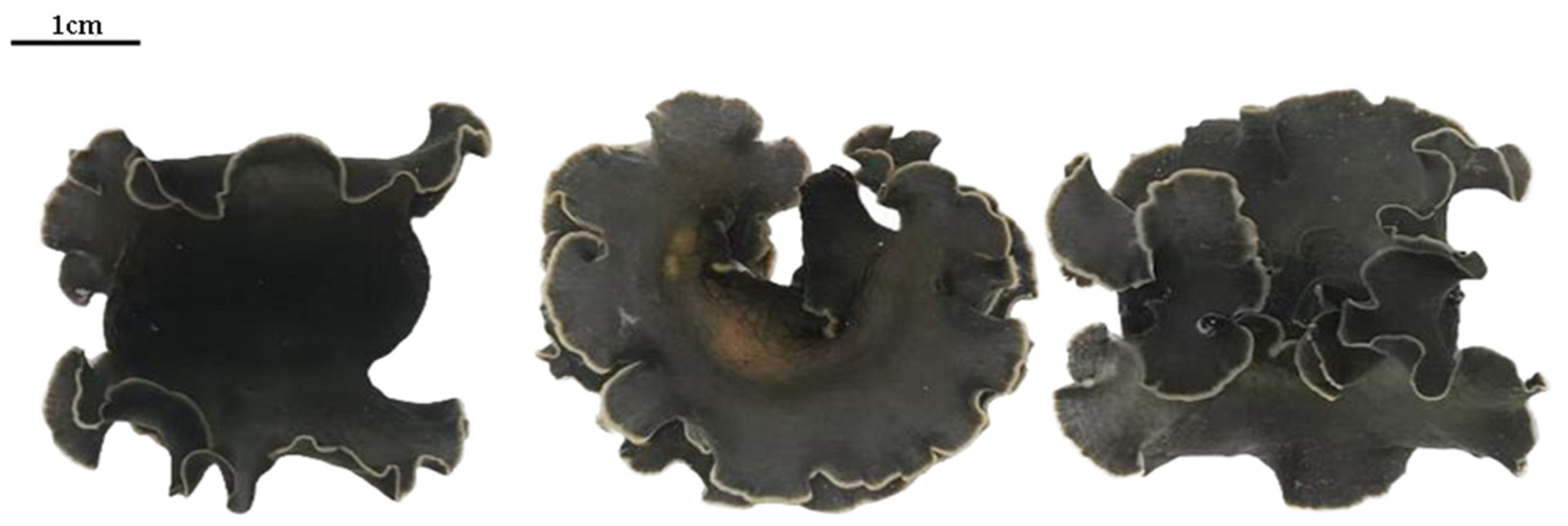
Figure 2.
External morphology of D. arborescens: (a) Anterior (dorsal) view; (b) Ventral view; (c) Rhinophore view; (d) Gill view; (e) Bursa copulatrix view. Rh: Rhinophore; Ma: Mantle; Gi: Gill; Mo: Mouth; Mgr: Mucus groove; Fo: Foot; RP: Rhinophoral apex; PL: Lamellae; RA: Rhinophoral axis; An: Anus; GA: Gill axis; BC: Bursa copulatrix.
Figure 2.
External morphology of D. arborescens: (a) Anterior (dorsal) view; (b) Ventral view; (c) Rhinophore view; (d) Gill view; (e) Bursa copulatrix view. Rh: Rhinophore; Ma: Mantle; Gi: Gill; Mo: Mouth; Mgr: Mucus groove; Fo: Foot; RP: Rhinophoral apex; PL: Lamellae; RA: Rhinophoral axis; An: Anus; GA: Gill axis; BC: Bursa copulatrix.
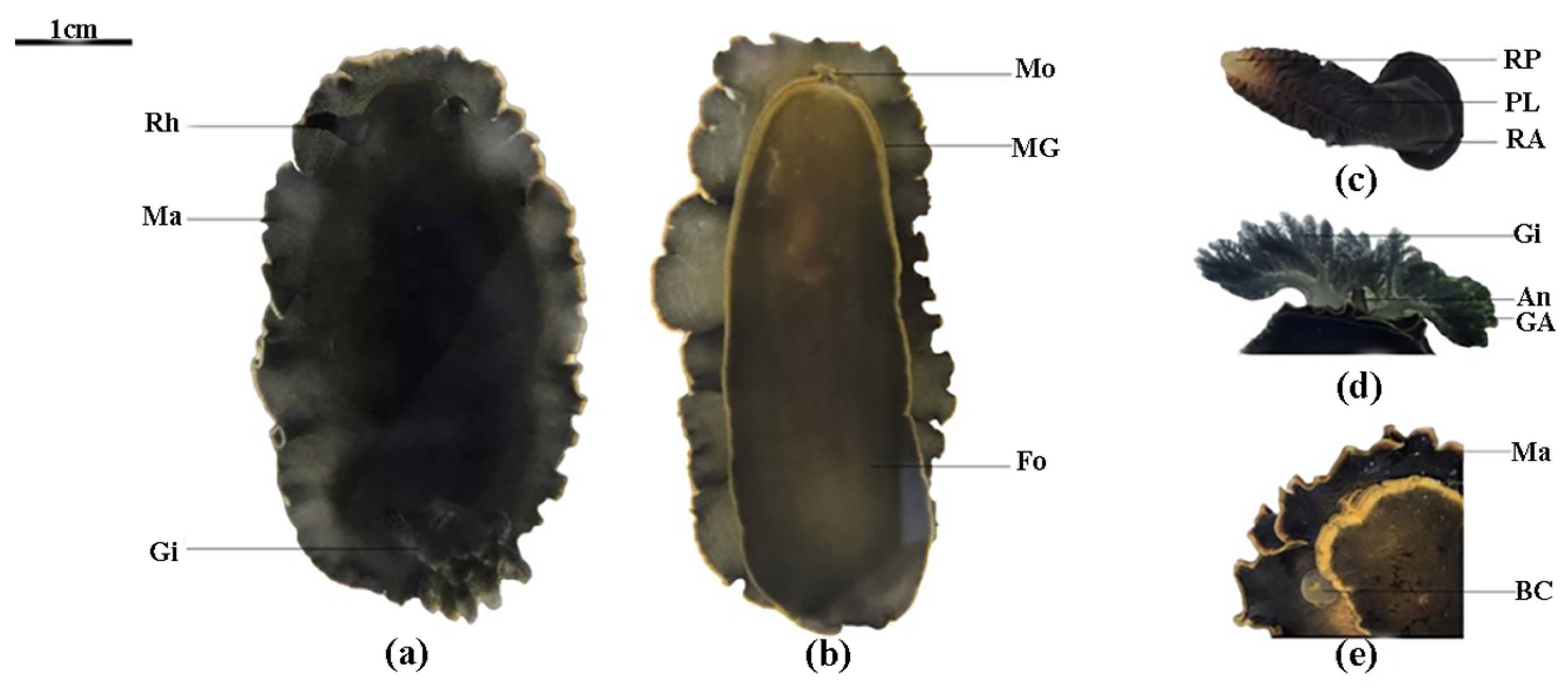
Figure 3.
Internal structure of D. arborescens (dorsal view): PM:Pharyngeal muscles; LHD: Large hermaphroditic duct; DG: Digestive gland; RC: Renal capsule; Mo: Mouth; AG: Albumen gland; MG: Mucous gland; BCI: Bursa copulatrix interface; SHD: Small hermaphroditic duct; SV: Seminal vesicle; HG: Hermaphroditic gland; Pe: Pericardium.
Figure 3.
Internal structure of D. arborescens (dorsal view): PM:Pharyngeal muscles; LHD: Large hermaphroditic duct; DG: Digestive gland; RC: Renal capsule; Mo: Mouth; AG: Albumen gland; MG: Mucous gland; BCI: Bursa copulatrix interface; SHD: Small hermaphroditic duct; SV: Seminal vesicle; HG: Hermaphroditic gland; Pe: Pericardium.
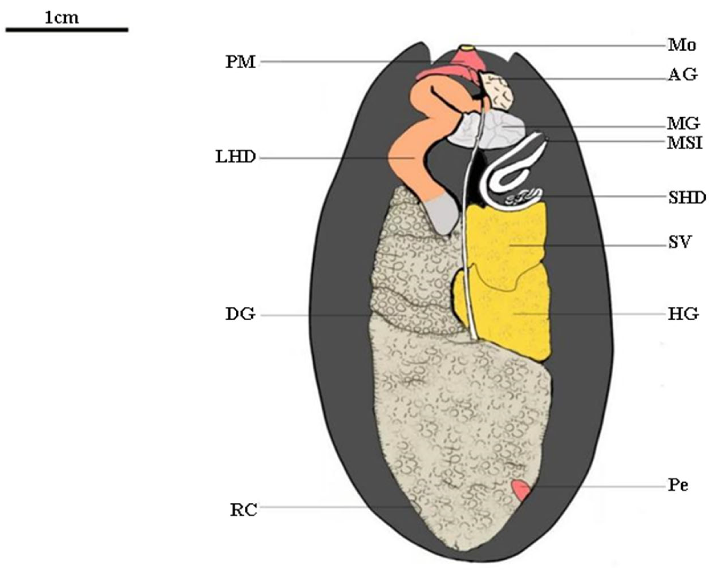
Figure 4.
Longitudinal section of gill of D. arborescens (H.E staining): (a) Overall longitudinal section of gill (×5); (b) Tripinnate branching structure of gill leaf (×10); (c) Partial enlargement of gill gland (×40); (d) Partial enlargement of gill axis (×10); (e) Partial enlargement of gill leaf (×20); (f) Partial enlargement of gill leaf (×70). GD: Gill chamber; MGi: Main gill; BG: Branched gill; MGA: Main gill axis; BGA: Branched gill axis; GG: Gill gland; CT: Connective tissue; SMF: Smooth muscle fiber; MC: Mucous cell; Epi: Epidermis; Ci: Cilia.
Figure 4.
Longitudinal section of gill of D. arborescens (H.E staining): (a) Overall longitudinal section of gill (×5); (b) Tripinnate branching structure of gill leaf (×10); (c) Partial enlargement of gill gland (×40); (d) Partial enlargement of gill axis (×10); (e) Partial enlargement of gill leaf (×20); (f) Partial enlargement of gill leaf (×70). GD: Gill chamber; MGi: Main gill; BG: Branched gill; MGA: Main gill axis; BGA: Branched gill axis; GG: Gill gland; CT: Connective tissue; SMF: Smooth muscle fiber; MC: Mucous cell; Epi: Epidermis; Ci: Cilia.
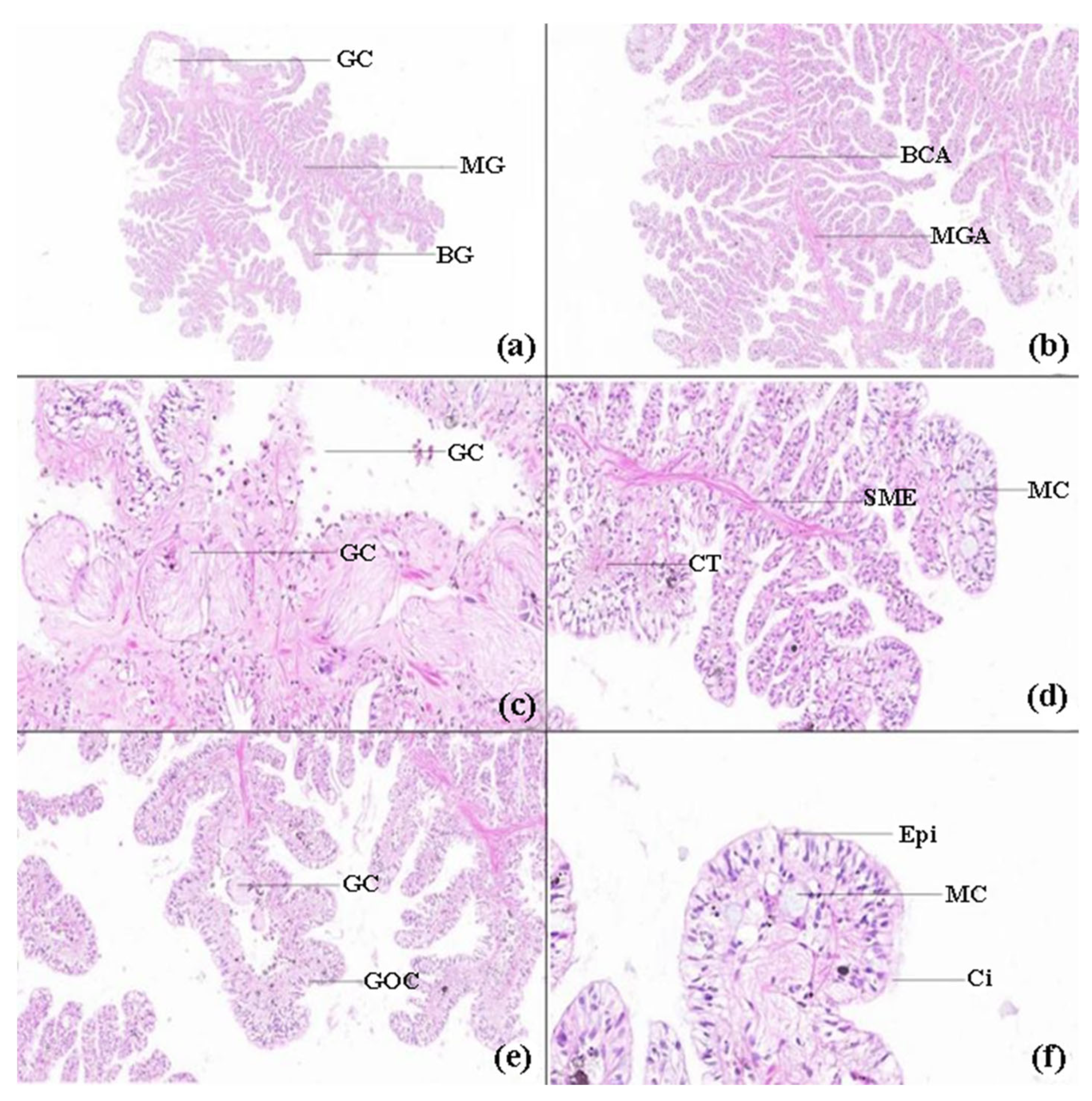
Figure 5.
Scanning electron microscopy of gill of D. arborescens: (a) Overall morphology of gill (×30); (b) Partial enlargement of gill (×50); (c) Partial enlargement of main gill (×200); (d) Partial enlargement of branched gill (×500); (e) Cilia on gill (×1000); (f) Cilia on gill (×2000).
Figure 5.
Scanning electron microscopy of gill of D. arborescens: (a) Overall morphology of gill (×30); (b) Partial enlargement of gill (×50); (c) Partial enlargement of main gill (×200); (d) Partial enlargement of branched gill (×500); (e) Cilia on gill (×1000); (f) Cilia on gill (×2000).
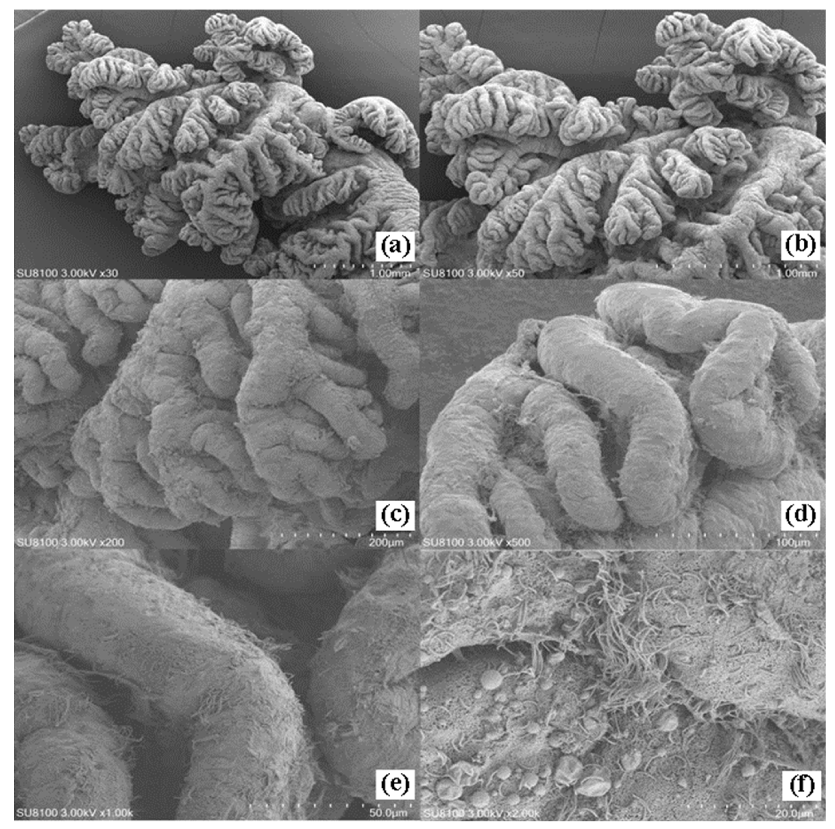
Figure 6.
Scanning electron microscopy of rhinophore of D. arborescens: (a) Overall morphology of rhinophore (×50); (b) Rhinophoral apex (×200); (c) Lamellae on rhinophore surface (×200); (d) Cilia on lamellae (×500); (e) Cilia on lamellae (×1000); (f) Cilia on lamellae (×2000).
Figure 6.
Scanning electron microscopy of rhinophore of D. arborescens: (a) Overall morphology of rhinophore (×50); (b) Rhinophoral apex (×200); (c) Lamellae on rhinophore surface (×200); (d) Cilia on lamellae (×500); (e) Cilia on lamellae (×1000); (f) Cilia on lamellae (×2000).
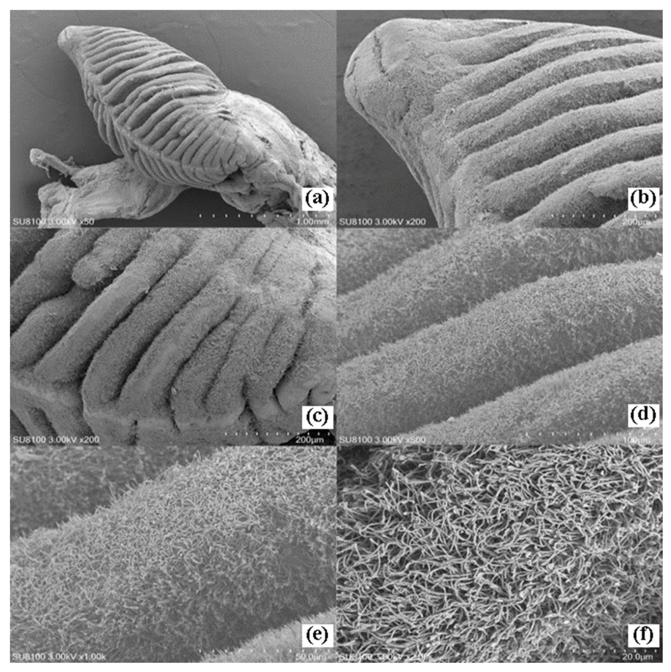
Figure 7.
Longitudinal section of rhinophore of D. arborescens (H.E staining): (a) Overall longitudinal section of rhinophore (×5); (b) Partial enlargement of rhinophore (×10); (c) Partial enlargement of rhinophoral sheath (×10); (d) Partial enlargement of lamellae (×10); (e) Partial enlargement of rhinophoral axis (×40); (f) Partial enlargement of rhinophoral sheath (×40) Lamellae. RA: Rhinophoral axis; RS: Rhinophoral sheath; BPL: Big lamellae; LPL: Little lamellae; RCG: Rhinophoral chamber gland; RCC: Rhinophoral chamber cavity; Epi: Epidermis; GOC: Goblet cells; CT: Connective tissue; Ci: Cilia; Va: Vacuole; LM: Longitudinal muscle; CM: Circular muscle.
Figure 7.
Longitudinal section of rhinophore of D. arborescens (H.E staining): (a) Overall longitudinal section of rhinophore (×5); (b) Partial enlargement of rhinophore (×10); (c) Partial enlargement of rhinophoral sheath (×10); (d) Partial enlargement of lamellae (×10); (e) Partial enlargement of rhinophoral axis (×40); (f) Partial enlargement of rhinophoral sheath (×40) Lamellae. RA: Rhinophoral axis; RS: Rhinophoral sheath; BPL: Big lamellae; LPL: Little lamellae; RCG: Rhinophoral chamber gland; RCC: Rhinophoral chamber cavity; Epi: Epidermis; GOC: Goblet cells; CT: Connective tissue; Ci: Cilia; Va: Vacuole; LM: Longitudinal muscle; CM: Circular muscle.
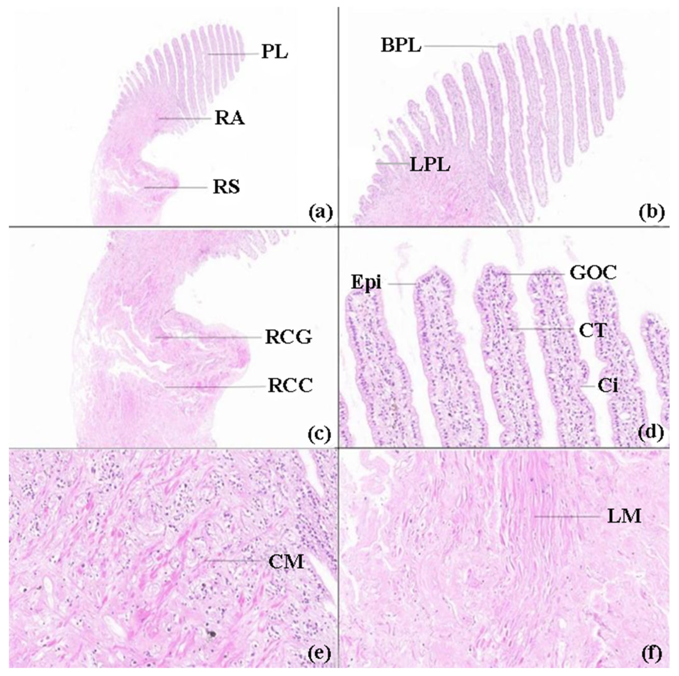
Figure 8.
Anatomy of the reproductive system of D. arborescens: MG: mucus gland; BCI: copulatory bursa opening (or bursal interface); SV: spermatheca (seminal receptacle); AG: albumen gland; HG: hermaphroditic gonad (ovotestis); DG: digestive gland; MGD: albumen gland duct; LHD: large hermaphroditic duct; SHD: small hermaphroditic duct; Mo: mouth (oral opening).
Figure 8.
Anatomy of the reproductive system of D. arborescens: MG: mucus gland; BCI: copulatory bursa opening (or bursal interface); SV: spermatheca (seminal receptacle); AG: albumen gland; HG: hermaphroditic gonad (ovotestis); DG: digestive gland; MGD: albumen gland duct; LHD: large hermaphroditic duct; SHD: small hermaphroditic duct; Mo: mouth (oral opening).
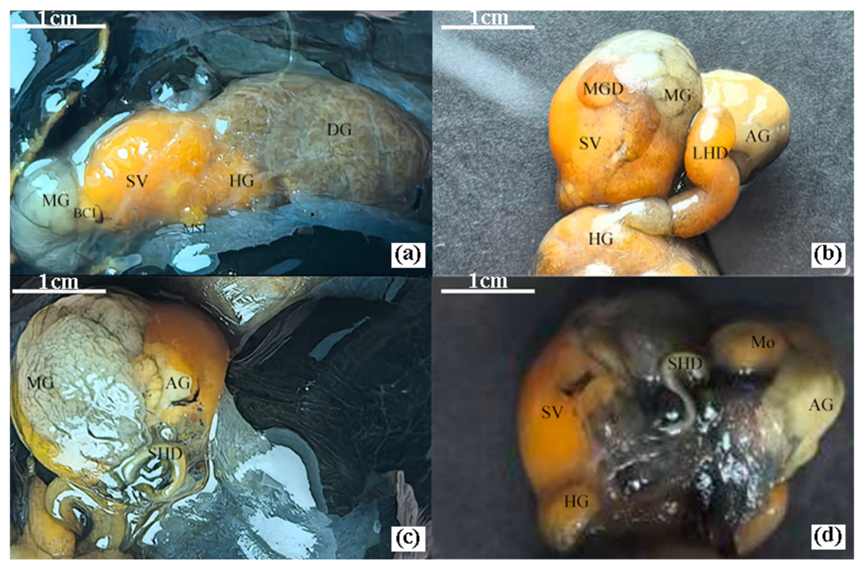
Figure 9.
Longitudinal section of reproductive system of D. arborescens (H.E staining): (a) Bursa copulatrix interface (×10); (b) Fertilization chamber (×20); (c) Hermaphroditic gland (×2); (d) Partial enlargement of hermaphroditic gland (×5); (e) Mucous gland (×10); (f) Albumen gland (×70). COC: Columnar cell; GOC: Goblet cell; SMF: Smooth muscle fiber; CT: Connective tissue; MO: Maturing oocyte; SZ: Spermatozoa; Fe: Fertilized egg; DG: Digestive gland.
Figure 9.
Longitudinal section of reproductive system of D. arborescens (H.E staining): (a) Bursa copulatrix interface (×10); (b) Fertilization chamber (×20); (c) Hermaphroditic gland (×2); (d) Partial enlargement of hermaphroditic gland (×5); (e) Mucous gland (×10); (f) Albumen gland (×70). COC: Columnar cell; GOC: Goblet cell; SMF: Smooth muscle fiber; CT: Connective tissue; MO: Maturing oocyte; SZ: Spermatozoa; Fe: Fertilized egg; DG: Digestive gland.
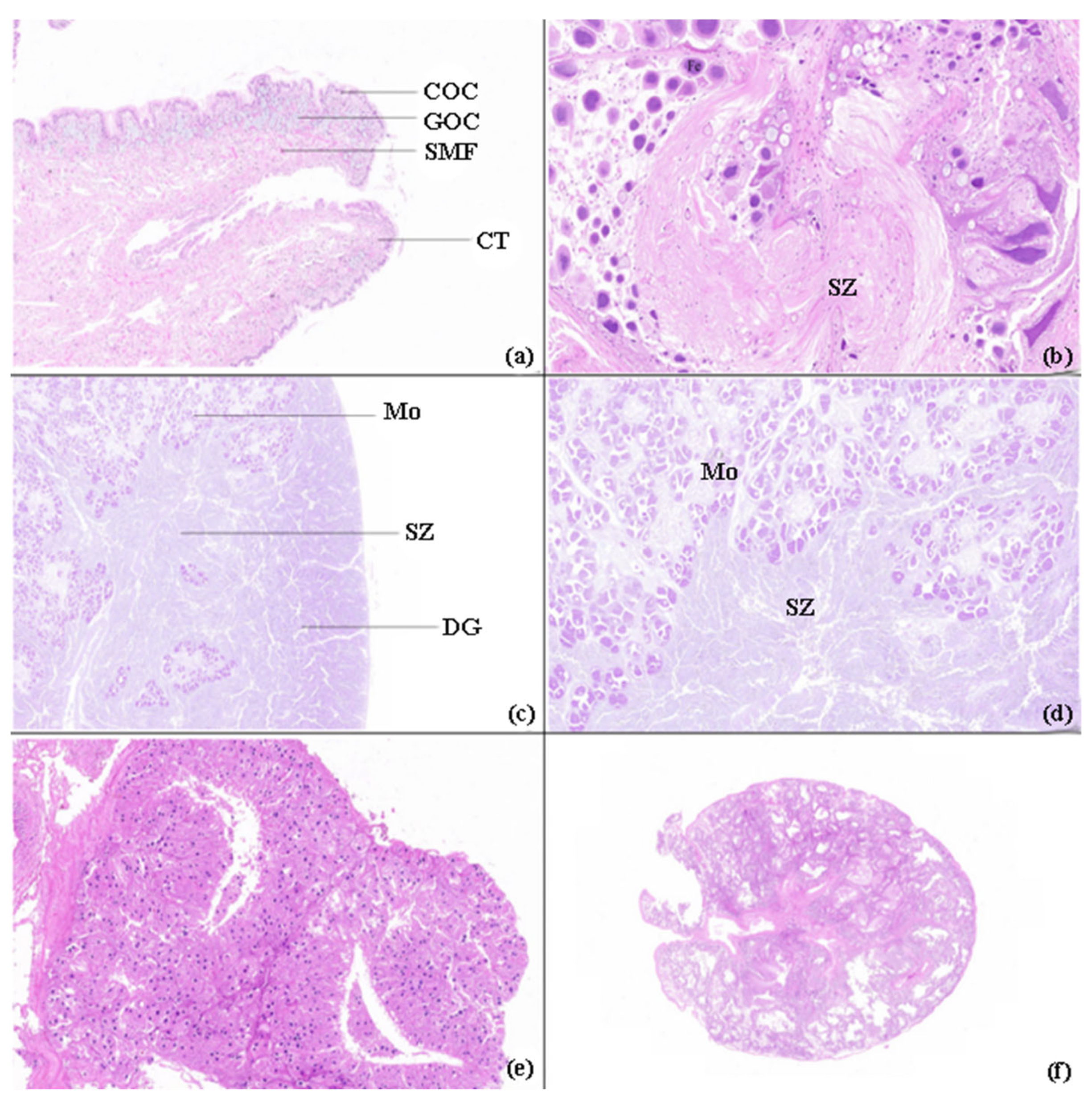
Figure 10.
Scanning electron microscopy of bursa copulatrix of D. arborescens: (a) Overall morphology of bursa copulatrix (×50); (b) Penis (×100); (c) Partial enlargement of penis (×200); (d) Partial enlargement of penis (×1000); (e) Partial enlargement of penis (×1000); (f) Partial enlargement of penis (×2000).
Figure 10.
Scanning electron microscopy of bursa copulatrix of D. arborescens: (a) Overall morphology of bursa copulatrix (×50); (b) Penis (×100); (c) Partial enlargement of penis (×200); (d) Partial enlargement of penis (×1000); (e) Partial enlargement of penis (×1000); (f) Partial enlargement of penis (×2000).
Disclaimer/Publisher’s Note: The statements, opinions and data contained in all publications are solely those of the individual author(s) and contributor(s) and not of MDPI and/or the editor(s). MDPI and/or the editor(s) disclaim responsibility for any injury to people or property resulting from any ideas, methods, instructions or products referred to in the content. |
© 2026 by the authors. Licensee MDPI, Basel, Switzerland. This article is an open access article distributed under the terms and conditions of the Creative Commons Attribution (CC BY) license (http://creativecommons.org/licenses/by/4.0/).
Copyright: This open access article is published under a Creative Commons CC BY 4.0 license, which permit the free download, distribution, and reuse, provided that the author and preprint are cited in any reuse.