

Submitted:
09 August 2026
Posted:
11 August 2026
You are already at the latest version
Abstract
ctDNA is evolving as an important biomarker for ICI treatment response assessment in advanced melanoma. However, the utility of ctDNA monitoring during concurrent ICI with RT remains incompletely explored. In this multicenter, retrospective study, patients with unresectable stage III/IV melanoma treated with ICI and palliative RT were identified. Patients had retrospectively collected, tumor-informed, exome-based, ctDNA monitoring within 3 months following RT. 50 patients treated with ICI and palliative RT were analyzed. Patients with ctDNA clearance had significantly longer OS (HR 2.10, 95% CI 1.16-3.80, p=0.014) compared to those with increasing or decreasing and detectable ctDNA following RT. One-year OS was 85.9% with ctDNA clearance, 30.6% with decreasing ctDNA, and 22.0% for increasing ctDNA. In a multivariable analysis, both decreasing and increasing ctDNA were associated with worse OS compared with ctDNA clearance (HR 5.08 [95% CI, 1.26–20.48, p=0.022] and HR 9.83 [95% CI, 2.69–35.94, p< 0.001], respectively). Among patients with advanced melanoma treated with ICI and palliative RT, ctDNA clearance is associated with significantly improved OS compared to patients with detectable ctDNA within 3 months following RT. Ongoing studies are needed to understand the role of ctDNA in this population.
Keywords:
ctDNA
; melanoma
; radiation
; checkpoint inhibition
Introduction
Although incidence of melanoma remains high, survival rates have dramatically increased in recent years [1]. This improvement in survival outcomes is largely attributed to the development and implementation of treatment with immune checkpoint inhibition [2,3]. Anti-PD-1-based therapy is now established as the frontline treatment of unresectable or metastatic melanoma [4]. Radiation therapy (RT) can also be incorporated into the treatment of metastatic melanoma with definitive or palliative intent, most often in patients with oligometastatic disease or brain metastases. Although melanoma is typically radioresistant, RT can be used in the adjuvant or palliative setting, but has not been shown to improve survival outcomes [5]. Despite advances in systemic therapy leading to better outcomes, mortality rates for patients with metastatic and unresectable stage III/IV melanoma remain high [1,2].
Circulating tumor DNA (ctDNA) is tumor-derived, fragmented DNA molecules shed by malignant cells into plasma. ctDNA dynamics have been shown to have prognostic value among patients with metastatic melanoma treated with ICI. Decreasing ctDNA is associated with improved overall survival, progression-free survival, and odds of disease control [6]. In another cohort of patients with metastatic melanoma, all patients with ctDNA clearance were progression-free at a median follow-up of 14.7 months, while patients with detectable ctDNA were more likely to have disease progression [7]. ctDNA has also been shown to differentiate pseudoprogression from true progression [8,9]. Trends in ctDNA may be useful in multiple clinical scenarios including determining prognosis, prognosticating the likelihood of recurrence, and distinguishing pseudoprogression.
The utility of ctDNA monitoring following radiation to sites of metastatic melanoma remains incompletely explored. Here, we describe the clinical characteristics and survival outcomes of patients with advanced-stage melanoma treated with ICI and integration of palliative RT based on ctDNA dynamics.
Methods
Patients with unresectable stage III/IV melanoma treated with ICI and palliative radiation therapy between October 2021 and June 2025 at five institutions were identified via retrospective chart review. Patients with uveal melanoma were excluded. Palliative radiation was defined as radiation to metastatic melanoma with the primary goal of improving symptom management. There were no restrictions on site of disease or modality of radiation. Data was extracted from the electronic medical record and stored in a REDCap database. All patients were monitored with longitudinal, tumor-informed, exome-based ctDNA testing (Signatera). Those with a baseline ctDNA level within 0 to 90 days prior to incorporating radiation and follow-up ctDNA testing obtained within 90 days following RT were included in this analysis.
To evaluate the prognostic impact of circulating tumor DNA (ctDNA) dynamics, patients were separated into 3 cohorts based on nadir ctDNA dynamics within 90 days after RT. Those with undetectable ctDNA were categorized as “ctDNA cleared.” Patients with a positive fold ctDNA change were “increasing ctDNA” and those with a negative fold ctDNA change were “decreasing ctDNA.”
A multivariable Cox proportional hazards regression was utilized to estimate the association between ctDNA trends and overall survival, adjusting for predefined clinical covariates including age (≥65 vs. <65 years), disease stage (IV vs. III), presence of brain or leptomeningeal metastases (yes vs. no), and melanoma subtype (cutaneous vs. other). To account for the limited sample size relative to the number of covariates, which introduced numerical instability and complete separation in standard unpenalized models, a penalized Cox model (Ridge/L2 regularization) was employed as an additional analytical strategy.
In a planned subset analysis of patients with detectable baseline ctDNA, the depth of molecular response was quantified as a continuous variable using a log10 reduction framework. A stabilizing constant (c = 0.01) was added to accommodate zero values in the logarithmic transformation. The proportional hazards assumption was verified for all covariates using Schoenfeld residuals. All statistical analyses and visualizations were performed using Python version 3.6, utilizing the lifelines library for survival modeling.
Results
Sixty-one patients with irradiated, advanced-stage melanoma and longitudinal ctDNA monitoring were identified. Of these 61 patients, 4 did not have a baseline ctDNA level within 90 days prior to initiation of RT, and 7 patients did not have follow-up ctDNA testing drawn within 90 days after RT. These 11 patients were excluded, and the remaining 50 patients that met criteria were included in this analysis. Twenty patients had clearance of ctDNA following irradiation of melanoma, while 18 patients had increasing ctDNA levels, and 12 patients were found to have decreasing circulating tumor DNA quantification [Figure 1].
Patient characteristics are described in Table 1. Median age was 65 (24-86), and median baseline ctDNA level was 8.9 MTM/mL (0-11,390 MTM/mL). Median follow up was 8.0 months from RT start date. Primary sites of melanoma included 41 patients with cutaneous melanoma (82%), 5 with mucosal (10%), and 4 patients with melanoma of unknown primary (8%). The majority (92%) of patients had stage IV disease, including 5 (10.0%) each with M1a and M1b stage disease, 12 (24.0%) with M1c and 24 patients (48.0%) with M1d staging. Three patients (6.0%) had stage IIIC melanoma and 1 (2.0%) had stage IIID disease. ICI regimens included 70% (n=35) anti-PD-1/anti-CTLA-4, 16% (n=8) anti-PD-1/anti-LAG-3, 2% (n=1) anti-PD-1/investigational ICI, and 12% (n=6) anti-PD-1 monotherapy. Thirteen (26.0%) patients had surgery while receiving checkpoint inhibitor therapy. The most common RT sites were the central nervous system (50%, n=25) and skin/soft tissue (24%, n=12), followed by lymph nodes and bone (each 14%, n=7), and lung and liver (each 6%, n=3).
Overall survival was significantly longer among patients with clearance of ctDNA compared to those with either increasing or decreasing ctDNA levels following RT. One-year overall survival was 85.9% among patients with ctDNA clearance, 30.6% with decreasing ctDNA, and 22.0% for those with increasing ctDNA [Figure 2A]. Compared to clearance of ctDNA within 90 days following RT, increasing ctDNA levels was associated with worse overall survival with a hazard ratio of 9.83 (p=0.001, 95% CI 2.69-35.94) [Table 2]. The cohort of patients with decreasing ctDNA concentrations also had worse overall survival than those with ctDNA clearance, with a hazard ratio of 5.08 (0.022, 95% CI 1.26-20.48). Comparing ctDNA clearance with all patients with detectable ctDNA, including those with increasing and decreasing ctDNA levels, resulted in a statistically significant difference in overall survival. Detectable ctDNA was associated with worse overall survival with a hazard ratio of 2.10 (95% CI 1.16-3.80, p=0.014) [Figure 2B].
Patients with clearance of ctDNA were more likely to be alive at the time of follow-up, and those with increasing or decreasing, yet detectable ctDNA were more likely to be deceased. Individual patients had variable duration of follow-up and frequency of ctDNA monitoring. Additionally, logarithmic ctDNA values were higher among deceased patients than alive [Figure 3].
In a multivariate analysis of overall survival, both increasing and decreasing ctDNA following RT were associated with worse overall survival than ctDNA clearance. There was no statistically significant difference between patients with age greater than 64 versus patients 64 and younger (p=0.755, HR 1.15, 95% CI 0.48-2.75). There was also no significant difference between those with radiation to the brain or leptomeninges compared to patients who received radiation to other sites of disease (p=0.608, HR 1.25, 95% CI 0.53-2.93). Differences between stage IV versus stage III disease were insignificant (p=0.996, HR 1.48 x 10⁷, 95% CI 0-∞) as well as cutaneous melanoma in comparison to mucosal or unknown primary site of melanoma (p=0.625, HR 0.75, 95% CI 0.23-2.39) [Table 2].
Discussion
Circulating tumor DNA has emerged as a robust prognostic indicator in patients with melanoma [10]. Detectable ctDNA levels both before and after any systemic therapy have been associated with diminished progression-free and overall survival [11]. In patients receiving checkpoint inhibitors, persistently detectable ctDNA following treatment initiation similarly correlates with worse PFS and OS [12]. Similarly, early ctDNA dynamics within only 3-4 weeks after starting ICI therapy have proven to be predictive of both OS and PFS as well [6]. By contrast, less is understood about the prognostic implications of ctDNA following radiation in patients with advanced-stage melanoma. Here, we demonstrate ctDNA clearance is associated with significantly improved OS compared to both increasing and decreasing ctDNA levels within 3 months following initiation of RT. Based on these results, ctDNA should be considered in clinical practice and clinical trials to help prognosticate patient outcomes.
These results support the integration of ctDNA monitoring into clinical practice for patients with advanced melanoma following treatment with RT. Serial ctDNA assessment within 3 months after initiating radiation may be an accessible and informative tool to prognosticate and guide goals of care discussions with patients, offering clinicians and patients earlier, actionable insight into treatment response. For patients with persistently detectable ctDNA within this window, more intensive or frequent imaging may be warranted to identify recurrent or progressive disease and to determine candidacy for targeted therapies in cases of oligometastatic disease. Patients with continually detectable ctDNA represent a clinically distinct, higher-risk population who may be particularly well suited for enrollment in prospective clinical trials investigating treatment adaptation based on ctDNA dynamics. This approach could potentially improve both overall and progression-free survival in this group of patients with especially poor prognosis.
This subgroup of patients receiving radiation in combination with immunotherapy may represent a biologically unique population, which could help explain why ctDNA detection is particularly prognostically useful in this setting. Radiation-induced tumor cell death can lead to immunogenic cell death, releasing tumor antigens and increasing damage associated molecular pattern molecules (DAMPs). This upregulation of tumor antigens and DAMPs leads to further enhancement of the immune system within the tumor microenvironment, which can prime patients for checkpoint inhibitor therapy [13]. Persistent ctDNA detection in this context may therefore reflect not only residual tumor burden, but also an underlying failure of the immune response in achieving durable systemic tumor control despite priming of the immune system through radiation.
In addition to RT improving immune activation within the melanoma tumor microenvironment, it may also increase ctDNA shedding into the systemic circulation and could produce an abscopal effect. In other cancers, radiation has been associated with an initial increase in cell free DNA in the blood [14]. This could lead to systemic immune system exposure to tumor neoantigens. Multiple studies have demonstrated size reduction of non-irradiated metastases following local radiotherapy, especially in patients with ICI refractory disease [15,16,17]. When ctDNA remains detectable despite this combined local and systemic immune activation, it may indicate tumor evasion of both radiation induced and checkpoint inhibitor mediated immune control, identifying a more aggressive disease biology associated with worse survival outcomes. Further studies are necessary to clarify the extent to which radiation enhances ICI efficacy in patients with advanced-stage melanoma and how ctDNA dynamics might function as a biomarker in this setting.
The sensitivity of circulating tumor DNA for detecting recurrent disease has been shown to be highly variable based on site of metastatic melanoma, and worse for detecting brain metastases than other sites of disease [18]. In the multivariate analysis presented here, there was no significant difference between patients with brain or leptomeningeal radiation compared to other sites of disease. This may be due to the increased shedding of ctDNA following radiation. Further investigation comparing ctDNA levels in patients with radiated versus non-radiated central nervous system disease is warranted.
Limitations of this analysis include a small sample size leading to reduced power of statistical analysis, potential introduction of immortal time bias, short duration of ctDNA monitoring, and variable intervals of ctDNA monitoring and follow-up duration. In the multivariable analysis, unpenalized model, estimations for stage IV disease were unstable with extremely large HR and infinite confidence intervals, reflecting sparse events among stage III patients. Given the final ctDNA measurement was selected retrospectively across variable follow up durations, this approach introduces the potential for immortal-time and look-ahead biases, which should be considered when interpreting the survival outcomes associated with these groupings. Furthermore, patients with undetectable ctDNA at both baseline and final follow-up were classified alongside those who actively cleared their disease, reflecting an absence of detectable molecular burden at the conclusion of observation, but limiting the ability to isolate the specific effect of active clearance.
Larger, prospective trials with longer duration of follow up are warranted to further characterize the clinical utility of ctDNA for patients with advanced-stage melanoma after radiation therapy. In addition to overall survival analysis, calculating progression-free survival as well as objective response rates in this population would also be valuable. It would also be useful to better understand how the durability of ctDNA clearance beyond 3 months after radiation impacts survival outcomes. Furthermore, clarifying whether the degree of change in ctDNA would also impact prognostication in many patient scenarios. Collectively, these findings identify ctDNA as a promising biomarker in melanoma necessitating further investigation in prospective studies.
Conclusions
The prognostic value of ctDNA in melanoma has been well described, however, its role after RT in advanced disease is less understood. Here, ctDNA clearance within 3 months of RT was associated with significantly better OS than either increasing or decreasing ctDNA, supporting serial ctDNA monitoring to help guide prognosis. Further research with prospective analysis, longer follow-up duration, and assessment of PFS and response-rates are needed to validate ctDNA as a biomarker in this setting.
Funding
The project described was supported by: the Clinical and Translational Science Award (CTSA) program, through the NIH National Center for Advancing Translational Sciences (NCATS), grant UL1TR002373; and the 1R37CA300434-01A1 grant supported by the National Cancer Merit Award.
Conflicts of interest
VTM serves as a consulting or advisory role for Immunocore, Regeneron Pharmaceuticals, Replimune, Bristol Myers Squibb, Partner Therapeutics, Delcath, Y-mAbs Therapeutics, Ideaya Biosciences, Castle Biosciences, Sun Pharmaceuticals, and Pfizer. VTM receives research funding (inst) from: Jounce Therapeutics, Seagen, Natera, Fujifilm, Y-mAbs Therapeutics, Top Alliance BioScience, Immunocore, AIQ Solutions, Innate Pharma, C4 Therapeutics, Pfizer, Astellas Pharma, Immuneering, Regeneron, Merck, Krystal Biotech, Marengo Therapeutics, TuHura Biosciences, Aminex Therapeutics, Replimune, Delcath, Incyte, Taiho. GKI: has served in a consulting or advisory role for BMS, Merck, Regeneron, Sanofi, Replimune, Novartis, Sun Pharma and Pfizer. GKI receives research funding (inst) from: Regeneron, Idera, Replimune, Xencor, InstilBio, Pfizer, Obsidian, Bicara, Georgiamune, Immunocore, Tuhura, Iovance, Linnaeus Therapeutics, and Checkpoint Pharmaceuticals. JMA is a founder of Visynia Biotechnologies LLC.
References
- Siegel RL, Kratzer TB, Wagle NS, Sung H, Jemal A. Cancer statistics, 2026. CA Cancer J Clin. 2026;76(1):e70043. [CrossRef]
- Wolchok JD, Chiarion-Sileni V, Rutkowski P, et al. Final, 10-Year Outcomes with Nivolumab plus Ipilimumab in Advanced Melanoma. N Engl J Med. 2025;392(1):11-22. [CrossRef]
- Kahlon N, Doddi S, Yousif R, et al. Melanoma Treatments and Mortality Rate Trends in the US, 1975 to 2019. JAMA Netw Open. 2022;5(12):e2245269. Published 2022 Dec 1. [CrossRef]
- Long GV, Swetter SM, Menzies AM, Gershenwald JE, Scolyer RA. Cutaneous melanoma. Lancet. 2023;402(10400):485-502. [CrossRef]
- Bliley R, Avant A, Medina TM, Lanning RM. Radiation and Melanoma: Where Are We Now?. Curr Oncol Rep. 2024;26(8):904-914. [CrossRef]
- Ma VT, Zhou AY, Forati A, et al. Multi-Institutional Study Evaluating the Role of Early Circulating Tumor DNA Dynamics During Treatment With Immune Checkpoint Inhibitors in Patients With Advanced-Stage Melanoma. JCO Precis Oncol. 2026;10:e2500254. [CrossRef]
- Eroglu Z, Krinshpun S, Kalashnikova E, et al. Circulating tumor DNA-based molecular residual disease detection for treatment monitoring in advanced melanoma patients. Cancer. 2023;129(11):1723-1734. [CrossRef]
- Steimle AK, Cho SY, Menon N, et al. ctDNA Dynamics Identifies Pseudoprogression in a Metastatic Melanoma Patient Treated With Nivolumab/Relatlimab. J Immunother. 2025;48(8):325-328. Published 2025 Oct 1. [CrossRef]
- Lee JH, Long GV, Menzies AM, et al. Association Between Circulating Tumor DNA and Pseudoprogression in Patients With Metastatic Melanoma Treated With Anti-Programmed Cell Death 1 Antibodies. JAMA Oncol. 2018;4(5):717-721. [CrossRef]
- Gracie L, Pan Y, Atenafu EG, et al. Circulating tumour DNA (ctDNA) in metastatic melanoma, a systematic review and meta-analysis. Eur J Cancer. 2021;158:191-207. [CrossRef]
- Gandini S, Zanna I, De Angelis SP, et al. Circulating tumour DNA and melanoma survival: A systematic literature review and meta-analysis. Crit Rev Oncol Hematol. 2021;157:103187. [CrossRef]
- Liu L, Hou S, Zhu A, Yan B, Li L, Song D. The prognostic value of circulating tumor DNA in malignant melanoma patients treated with immune checkpoint inhibitors: a systematic review and meta-analysis. Front Immunol. 2025;15:1520441. Published 2025 Jan 17. [CrossRef]
- Tagliaferri L, Lancellotta V, Fionda B, et al. Immunotherapy and radiotherapy in melanoma: a multidisciplinary comprehensive review. Hum Vaccin Immunother. 2022;18(3):1903827. [CrossRef]
- Kageyama, S. I., Nihei, K., Karasawa, K., Sawada, T., Koizumi, F., Yamaguchi, S., Kato, S., Hojo, H., Motegi, A., Tsuchihara, K., & Akimoto, T. (2018). Radiotherapy increases plasma levels of tumoral cell-free DNA in non-small cell lung cancer patients. Oncotarget, 9(27), 19368–19378. [CrossRef]
- Ferdinandus S, Rühle A, Lamrani A, et al. Abscopal effects in patients with malignant melanoma treated with radiotherapy and immune checkpoint inhibition: analysis of a large observational multicenter study. J Immunother Cancer. 2025;13(10):e012717. Published 2025 Oct 2. [CrossRef]
- Trommer M, Rühle A, Lamrani A, et al. Radiotherapy-induced abscopal effects in immune checkpoint inhibitor-refractory metastatic disease: results from a large multicenter real-world cohort study. Oncoimmunology. 2026;15(1):2610529. [CrossRef]
- Backlund E, Grozman V, Egyhazi Brage S, Lewensohn R, Lindberg K, Helgadottir H. Radiotherapy with or without immunotherapy in metastatic melanoma: efficacy and tolerability. Acta Oncol. 2023;62(12):1921-1930. [CrossRef]
- Steimle AK, Harris MS, Forati A, Hollnagel F, Birbrair A & Ma VT. Sensitivity of Circulating Tumor DNA for Detection of Minimal Residual Disease Recurrence or Relapse in Patients With Melanoma. JCO Oncol Adv 3, e2500119(2026). [CrossRef]
Figure 1.
Consort diagram describing the inclusion criteria and final number of patients included in each group based on ctDNA trends.
Figure 1.
Consort diagram describing the inclusion criteria and final number of patients included in each group based on ctDNA trends.
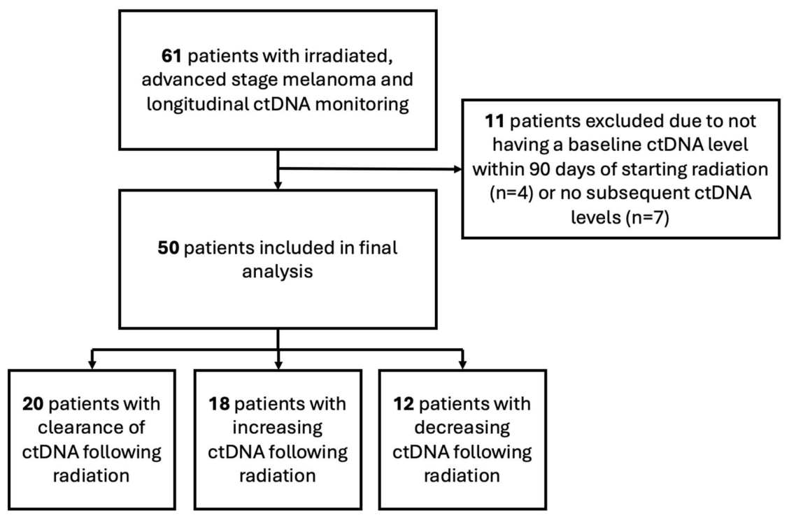
Figure 2.
Kaplan-Meier plots for overall survival (OS) for patients with advanced, irradiated melanoma and cleared ctDNA compared to detectable and increasing ctDNA levels as well as detectable and decreasing ctDNA levels (2A). Kaplan-Meier plots for OS comparing patients with cleared ctDNA to persistently detectable ctDNA (2B).
Figure 2.
Kaplan-Meier plots for overall survival (OS) for patients with advanced, irradiated melanoma and cleared ctDNA compared to detectable and increasing ctDNA levels as well as detectable and decreasing ctDNA levels (2A). Kaplan-Meier plots for OS comparing patients with cleared ctDNA to persistently detectable ctDNA (2B).
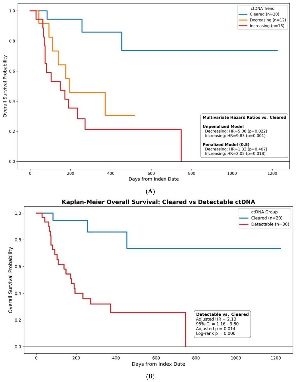
Figure 3.
Spider plots of each patient’s ctDNA levels based on survival at follow up.
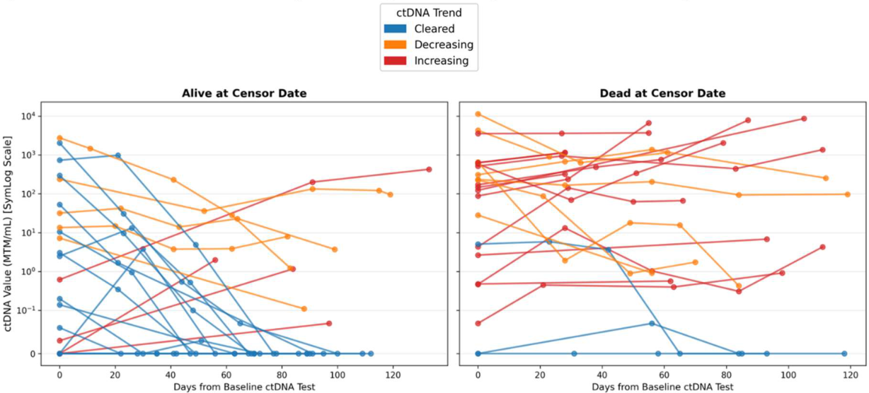

Table 1.
Baseline Characteristics.
| Characteristic | All (n=50) |
ctDNA Clearance (n=20) |
ctDNA Detectable and Decreasing (n=12) |
ctDNA Detectable and Increasing (n=18) |
|---|---|---|---|---|
| Age — Median (Range) | ||||
| years | 65 (24-86) | 59 (34-78) | 67 (41-76) | 65 (24-86) |
| Melanoma Type — No. (%) | ||||
| Cutaneous | 41 (82.0) | 16 (80.0) | 9 (75.0) | 16 (88.9) |
| Mucosal | 5 (10.0) | 1 (5.0) | 2 (16.7) | 2 (11.1) |
| Unknown Primary |
4 (8.0) | 3 (15.0) | 1 (8.3) | 0 (0.0) |
| AJCC Stage — No. (%) | ||||
| IIIC | 3 (6.0) | 2 (10.0) | 0 (0.0) | 1 (5.6) |
| IIID | 1 (2.0) | 0 (0.0) | 0 (0.0) | 1 (5.6) |
| IV | 46 (92.0) | 18 (90.0) | 12 (100.0) | 16 (88.9) |
| M1a | 5 (10.0) | 1 (5.0) | 2 (17.0) | 2 (11.0) |
| M1b | 5 (10.0) | 1 (5.0) | 2 (17.0) | 2 (11.0) |
| M1c | 12 (24.0) | 3 (15.0) | 3 (25.0) | 6 (33.0) |
| M1d | 24 (48.0) | 13 (65.0) | 5 (42.0) | 6 (33.0) |
| Immunotherapy Type — No. (%) | ||||
| Ipi + Nivo | 35 (70.0) | 13 (65.0) | 10 (83.3) | 12 (66.7) |
| Nivo + Rela | 8 (16.0) | 4 (20.0) | 2 (16.7) | 2 (11.1) |
| Anti-PD-1 monotherapy | 6 (12.0) | 3 (15.0) | 0 (0.0) | 3 (16.7) |
| Anti-PD-1 + anti-tMUC1 | 1 (2.0) | 0 (0.0) | 0 (0.0) | 1 (5.6) |
| Surgery During IO — No. (%) | ||||
| 13 (26.0) | 7 (35.0) | 3 (25.0) | 3 (16.7) | |
| Radiation Site — No. (%) | ||||
| Skin/Muscle | 12 (24.0) | 4 (20.0) | 3 (25.0) | 5 (27.8) |
| Lymph Node | 7 (14.0) | 2 (10.0) | 1 (8.3) | 4 (22.2) |
| Lung/Pleura | 3 (6.0) | 0 (0.0) | 2 (16.7) | 1 (5.6) |
| Liver | 3 (6.0) | 0 (0.0) | 2 (16.7) | 1 (5.6) |
| Bone | 7 (14.0) | 2 (10.0) | 1 (8.3) | 4 (22.2) |
| Brain | 25 (50.0) | 13 (65.0) | 5 (41.7) | 7 (38.9) |
AJCC: American Joint Committee on Cancer; ctDNA: circulating tumor DNA; IO: immunotherapy; Ipi: ipilimumab; Nivo: Nivolumab; Rela: Relatlimab.
Table 2.
Multivariable analysis for overall survival.
| Variable | Hazard Ratio (HR) | 95% Confidence Interval | p-value |
|---|---|---|---|
| Age ≥ 65 | 1.15 | 0.48-2.75 | 0.755 |
| Brain/leptomeninges RT (vs other site of RT) | 1.25 | 0.53-2.93 | 0.608 |
| Stage IV (vs III) | 1.48 x 10⁷ | 0-∞ | 0.996 |
| Cutaneous melanoma (vs other) | 0.75 | 0.23-2.39 | 0.625 |
| ctDNA decreasing (vs cleared) | 5.08 | 1.26-20.48 | 0.022* |
| ctDNA increasing (vs cleared) | 9.83 | 2.69-35.94 | <0.001* |
*p < 0.05 = statistically significant.
Disclaimer/Publisher’s Note: The statements, opinions and data contained in all publications are solely those of the individual author(s) and contributor(s) and not of MDPI and/or the editor(s). MDPI and/or the editor(s) disclaim responsibility for any injury to people or property resulting from any ideas, methods, instructions or products referred to in the content. |
© 2026 by the authors. Licensee MDPI, Basel, Switzerland. This article is an open access article distributed under the terms and conditions of the Creative Commons Attribution (CC BY) license (http://creativecommons.org/licenses/by/4.0/).
Copyright: This open access article is published under a Creative Commons CC BY 4.0 license, which permit the free download, distribution, and reuse, provided that the author and preprint are cited in any reuse.