

Submitted:
09 August 2026
Posted:
11 August 2026
You are already at the latest version
Abstract
Salmonella enterica is an important zoonotic and foodborne pathogen requiring integrated molecular and antimicrobial surveillance. This study characterized 20 selected S. enterica strains preserved in the National Collection of Microorganisms in Kazakhstan using serotyping, whole-genome sequencing (WGS)-based genotyping, multilocus sequence typing (MLST), and disk-diffusion antimicrobial susceptibility testing. Ten serovars and 11 sequence types (STs) were identified. All five Enteritidis strains belonged to ST11; three of four Typhimurium strains belonged to ST19 and one to ST3718, while Muenchen and Choleraesuis strains were assigned to ST82 and ST68, respectively. Phenotypic susceptibility to 13 antimicrobial agents was assessed using 260 strain–antimicrobial measurements. Inhibition-zone diameters ranged from 0 to 30 mm and varied considerably among strains, including those sharing the same serovar and ST. No measurable inhibition zones were most frequently observed for ampicillin and trimethoprim–sulfamethoxazole (10/20 strains each), whereas cefepime and lomefloxacin produced measurable inhibition zones in all strains. Exploratory principal component analysis showed that the first two components explained 65.35% of the total variance. Considerable within-group heterogeneity indicated that serovar and seven-locus MLST classification alone did not predict phenotypic antimicrobial susceptibility profiles. These findings provide a molecular and phenotypic baseline for selected S. enterica strains preserved in Kazakhstan and support further comprehensive genomic investigation of antimicrobial resistance determinants and population structure.
Keywords:
Salmonella enterica
; antimicrobial susceptibility
; antimicrobial resistance
; whole-genome sequencing
; MLST
; serotyping
; population structure
; Kazakhstan
1. Introduction
Salmonella enterica is a major bacterial pathogen at the human–animal–food interface and remains an important cause of foodborne and zoonotic infections worldwide. Its epidemiological importance is reinforced by the extensive diversity of serovars associated with different hosts, ecological niches, transmission pathways, and disease manifestations [1,2,3]. Livestock, poultry, and foods of animal origin represent important reservoirs and vehicles of transmission, making Salmonella surveillance particularly relevant within a One Health framework [4,5].
The increasing occurrence of antimicrobial resistance (AMR) among S. enterica populations further complicates the control of salmonellosis. Resistance to clinically important antimicrobial classes can reduce therapeutic options and facilitate the persistence and dissemination of resistant lineages across human, animal, food-production, and environmental compartments [6,7,8,9]. Consequently, antimicrobial susceptibility testing remains an important component of Salmonella surveillance. However, phenotypic susceptibility profiles are most informative when interpreted together with robust strain-characterization approaches that can distinguish epidemiologically and genetically diverse lineages [10,11,12].
Serotyping has historically provided a fundamental framework for the classification and epidemiological tracking of Salmonella [13,14]. Molecular typing approaches have subsequently increased the resolution with which population structure can be investigated [15,16]. Multilocus sequence typing (MLST), based on allelic variation in conserved housekeeping genes, provides standardized and reproducible sequence types that can be compared across laboratories and geographical regions [17,18,19]. Whole-genome sequencing (WGS) now provides substantially greater discriminatory resolution and enables genome-based serotyping, sequence-type assignment, phylogenomic analysis, and characterization of antimicrobial resistance determinants [20,21,22]. Nevertheless, seven-locus MLST remains useful for placing isolates within internationally comparable population structures, particularly when integrated with phenotypic and epidemiological information.
Archived microbial collections represent valuable resources for retrospective investigation because they preserve strains obtained across different periods, hosts, and epidemiological contexts [17,23]. Their characterization can reveal molecular and phenotypic diversity that may otherwise remain undocumented and can provide reference material for subsequent comparative and genomic investigations [24,25]. Nevertheless, collection-based studies require careful interpretation: strains retained in a repository constitute a selected set rather than a systematically sampled population. Consequently, observations from such collections should not be interpreted as estimates of population prevalence, national serovar distribution, antimicrobial resistance prevalence, or temporal trends unless supported by an appropriate sampling framework [26,27,28,29].
Information integrating serovar identity, molecular population structure, and phenotypic antimicrobial susceptibility characteristics of S. enterica strains preserved in microbial collections in Kazakhstan remains limited [30,31]. Characterization of such strains using complementary phenotypic and genome-based approaches can provide a structured baseline for comparison with contemporary isolates and for subsequent comprehensive investigation of antimicrobial resistance determinants and population structure. Therefore, the present study aimed to characterize 20 selected S. enterica strains preserved in the National Collection of Microorganisms in Kazakhstan using serotyping, WGS-based genotyping, MLST, and phenotypic antimicrobial susceptibility testing by disk diffusion. Specifically, we assessed serovar and MLST diversity and characterized phenotypic susceptibility profiles against a panel of antimicrobial agents. WGS data were used in the present study for genome-based serovar confirmation and in silico MLST assignment, whereas comprehensive analysis of other genomic features was beyond the scope of the current investigation. This retrospective characterization was designed to describe molecular and phenotypic diversity within the selected collection rather than to estimate the prevalence, national distribution, antimicrobial resistance prevalence, or temporal dynamics of S. enterica in Kazakhstan.
2. Materials and Methods
2.1. Study Design and Strain Collection
This retrospective study included 20 selected Salmonella enterica strains preserved in the National Collection of Microorganisms of the M. Aikimbayev National Scientific Center for Especially Dangerous Infections, Almaty, Kazakhstan. The strains were selected to represent the available serovar and sequence-type diversity within the collection and therefore constituted a selected retrospective collection rather than a systematically sampled or nationally representative population.
According to the available collection records, the strains originated from human and animal sources and were accessioned into the National Collection of Microorganisms between 1977 and 2025. The year reported for each strain represents the year of accession to the collection and not necessarily the year of primary isolation. These dates were therefore used only for descriptive characterization of the collection and not for temporal trend analysis.
All strains were retrieved from the collection on 9 February 2026 for retrospective laboratory characterization. Before analysis, the strains were recovered and cultured using standard bacteriological procedures for Enterobacterales. Culture purity was verified using microscopic and bacteriological methods before serotyping, genome sequencing and genotyping, and antimicrobial susceptibility testing.
2.2. Ethics Statement
This retrospective study was conducted using bacterial strains previously deposited and maintained in the National Collection of Microorganisms of the M. Aikimbayev National Scientific Center for Especially Dangerous Infections, Almaty, Kazakhstan. The study protocol was reviewed and approved by the Bioethics Committee of the M. Aikimbayev National Scientific Center for Especially Dangerous Infections (Protocol No. 2, approved 12 February 2024).
The study involved only previously collected and archived bacterial strains of human and animal origin. No participants or animals were prospectively recruited or subjected to additional sampling or interventions for the purposes of the present study, and no new clinical or veterinary specimens were collected specifically for this research. Any information associated with the archived strains was handled in a de-identified manner, and no personally identifiable information was used in the analysis.
2.3. Serotyping
Serological characterization of the S. enterica strains was performed according to the White–Kauffmann–Le Minor scheme based on the determination of somatic (O) and flagellar (H) antigens. Serotyping was performed using monovalent and polyvalent O- and H-agglutinating diagnostic antisera (PETSAL®, St. Petersburg Research Institute of Vaccines and Sera, FMBA of Russia, St. Petersburg, Russia). Antigenic formulae were determined from the observed O- and H-antigen profiles and used to assign the corresponding serovars according to the Salmonella serotyping scheme.
2.4. Genome Sequencing and Genotyping
Genomic DNA was extracted using the QIAamp DNA Mini Kit (QIAGEN, Hilden, Germany) according to the manufacturer’s instructions. Purified DNA was eluted in Tris-EDTA (AE) buffer and stored at −20 °C until further use.
Whole-genome sequencing (WGS) was performed using both short- and long-read sequencing technologies. Short-read sequencing libraries were prepared using the Illumina DNA Prep (M) Tagmentation Kit (Illumina, San Diego, CA, USA) and indexed with the IDT for Illumina DNA/RNA UD Indexes Set A. Sequencing was performed on the Illumina MiSeq platform using the MiSeq Reagent Kit v3 (600-cycle). Long-read libraries were prepared using the Native Barcoding Kit 24 V14 (Oxford Nanopore Technologies, Oxford, UK) together with the NEBNext Companion Module (New England Biolabs, Ipswich, MA, USA), and sequencing was performed on a MinION Mk1C device using an R10.4.1 flow cell.
Illumina reads were quality-filtered and trimmed using Trimmomatic v0.36 [32] and assembled de novo using SPAdes v3.15.0 [33]. Read mapping and sequence-processing procedures were performed using Bowtie2 v2.4.1 [34] and BCFtools v1.19 [35], while scaffolding of the assembled contigs was performed using RagTag v2.1.0 [36]. A custom computational pipeline for short-read genome assembly (LLAMINA) was additionally used as previously described and validated [37,38].
For Oxford Nanopore data, raw electrical signals were basecalled into nucleotide sequences in FASTQ format using Dorado (Oxford Nanopore Technologies). Long reads were assembled de novo using Flye [39], followed by sequence polishing and error correction with Medaka (Oxford Nanopore Technologies).
In silico genotyping was performed using the assembled genome sequences. Serovar identity and antigenic formulae were predicted using SeqSero2 [40]. Multilocus sequence typing (MLST) was performed according to the seven-locus Salmonella scheme (aroC, dnaN, hemD, hisD, purE, sucA, and thrA). Allele numbers and sequence types (STs) were assigned by comparison with the Salmonella MLST database available through PubMLST [41].
For the purposes of the present study, WGS data were used for genome-based serovar confirmation and in silico MLST assignment. The analysis was specifically designed to compare phenotypic antimicrobial susceptibility profiles with broad population-structure markers represented by serovar and seven-locus MLST. Comprehensive resistome characterization, including the identification of acquired antimicrobial resistance genes and resistance-associated chromosomal mutations, was beyond the scope of the present study and was not included in the current analysis. Such investigation will require a dedicated genotype–phenotype analysis.
2.5. Antimicrobial Susceptibility Testing
Phenotypic antimicrobial susceptibility was assessed using the Kirby–Bauer disk-diffusion method on Mueller–Hinton agar following the standardized methodology described in CLSI M02, 14th ed. Applicable interpretive criteria were derived from CLSI M100, 36th ed.
The antimicrobial panel comprised 13 agents: ampicillin (AMP; 10 µg), aztreonam (ATM; 30 µg), ceftazidime (CAZ; 30 µg), ceftriaxone (CRO; 30 µg), cefotaxime (CTX; 30 µg), cefepime (FEP; 30 µg), lomefloxacin (LOM; 10 µg), levofloxacin (LVX; 5 µg), pefloxacin (PEF; 5 µg), tetracycline (TET; 30 µg), ciprofloxacin (CIP; 5 µg), trimethoprim–sulfamethoxazole (SXT; 25 µg), and chloramphenicol (CHL; 30 µg).
Bacterial inocula were standardized to a turbidity equivalent to a 0.5 McFarland standard and inoculated onto Mueller–Hinton agar. Antimicrobial disks (HiMedia Laboratories Pvt. Ltd., Mumbai, India) were applied according to the standardized disk-diffusion procedure. Plates were incubated at 35–37 °C for 18 h, after which inhibition-zone diameters were measured in millimeters.
Quality control was performed using Escherichia coli ATCC 25922 as the reference strain. The primary quantitative data analyzed were the measured inhibition-zone diameters (mm) for each strain–antimicrobial combination. Categorical susceptibility interpretations were applied only where validated organism- and antimicrobial-specific interpretive criteria were available according to CLSI M100, 36th ed. Antimicrobial agents for which no applicable CLSI interpretive breakpoints were available were retained for descriptive analysis of inhibition-zone diameters and were not assigned categorical susceptible, intermediate, or resistant classifications.
2.6. Data Analysis and Visualization
Descriptive statistics were used to summarize the serovar and MLST composition of the selected strain collection. The numbers and proportions of strains assigned to each serovar and sequence type were calculated relative to the 20 strains included in the study.
Antimicrobial susceptibility data were analyzed at the individual-strain level using inhibition-zone diameters measured in millimeters. The complete dataset comprised 260 measurements representing 20 strains tested against 13 antimicrobial agents. For each antimicrobial agent, the range, mean, standard deviation, and median inhibition-zone diameter were calculated, together with the number and proportion of strains showing no measurable inhibition zone (0 mm). A value of 0 mm was treated descriptively as the absence of a measurable inhibition zone and was not, by itself, used as a universal criterion for categorical resistance.
Phenotypic inhibition-zone profiles were visualized using a heatmap. Exploratory hierarchical clustering was applied to visualize similarities among strains based on their inhibition-zone profiles. Clustering was used solely for descriptive visualization and was not interpreted as evidence of phylogenetic relatedness or epidemiological linkage.
Principal component analysis (PCA) was performed on the complete 20 × 13 matrix of inhibition-zone diameters to explore the major sources of variation in phenotypic profiles among strains. Because all variables were expressed in the same unit (mm), the data were mean-centered without variance scaling before PCA. The proportion of total variance explained by each principal component was calculated, and the first two principal components were used for two-dimensional visualization.
Comparisons of inhibition-zone profiles within repeatedly represented serovar–ST combinations were descriptive and exploratory because of the small and unequal numbers of strains within individual groups. No population-level prevalence estimates, antimicrobial resistance prevalence estimates, or temporal trend analyses were performed because the study was based on a selected retrospective collection rather than a prospectively or systematically sampled population.
3. Results
3.1. Serovar Diversity of the Selected Salmonella enterica Strains
All 20 strains included in the study were identified as Salmonella enterica. Ten serovars were represented in the selected collection: Enteritidis, Typhimurium, Muenchen, Choleraesuis, Newport, Infantis, Dublin, Kottbus, Typhi, and Litchfield (Table 1).
Enteritidis was the most frequently represented serovar, accounting for 5 of the 20 strains (25.0%), followed by Typhimurium with 4 strains (20.0%), Muenchen with 3 strains (15.0%), and Choleraesuis with 2 strains (10.0%). Newport, Infantis, Dublin, Kottbus, Typhi, and Litchfield were each represented by a single strain (5.0% each).
Overall, the selected collection showed substantial serovar diversity, with 10 serovars represented among the 20 strains. Because the strains constituted a selected retrospective collection, these proportions describe only the composition of the analyzed dataset and should not be interpreted as estimates of serovar prevalence in Kazakhstan.
3.2. MLST-Based Sequence Type Diversity
MLST analysis identified 11 STs among the 20 S. enterica strains (Table 1, Figure 1). The most frequently represented sequence type was ST11, which included all five Enteritidis strains. ST19 was identified in three of the four Typhimurium strains, whereas the remaining Typhimurium strain was assigned to ST3718.
All three Muenchen strains belonged to ST82, and both Choleraesuis strains belonged to ST68. The remaining sequence types were each represented by a single strain: ST86 (Newport), ST32 (Infantis), ST10 (Dublin), ST582 (Kottbus), ST1 (Typhi), and ST214 (Litchfield).
Overall, the 20 selected strains comprised multiple serovar-associated sequence types, with repeated representation of several serovar–ST combinations, including Enteritidis–ST11, Typhimurium–ST19, Muenchen–ST82, and Choleraesuis–ST68. Within the selected Typhimurium subset, two sequence types were observed, ST19 and ST3718, whereas each of the other represented serovars was associated with a single ST in this dataset.
Taken together, the MLST results demonstrated substantial sequence-type diversity within the selected collection, with 11 STs identified among 20 strains. The observed correspondence between most serovars and specific STs provided a population-structure framework for subsequent comparison with the phenotypic antimicrobial susceptibility profiles of the individual strains.
3.3. Phenotypic Antimicrobial Susceptibility
Phenotypic antimicrobial susceptibility was assessed for all 20 Salmonella enterica strains against 13 antimicrobial agents representing seven pharmacological classes. A complete dataset comprising 260 strain–antimicrobial measurements was obtained, with inhibition-zone diameters ranging from 0 to 30 mm across the tested panel.
Substantial variation in inhibition-zone diameters was observed among the tested antimicrobial agents (Table 2). The widest overall range was recorded for lomefloxacin (10–30 mm), whereas several other agents showed inhibition-zone diameters extending from 0 mm to maximum values of 21–26 mm. Cefepime and lomefloxacin were the only antimicrobial agents for which measurable inhibition zones were observed for all 20 strains, with ranges of 10–26 mm and 10–30 mm, respectively. Aztreonam and ciprofloxacin each showed no measurable inhibition zone in two strains.
Absence of a measurable inhibition zone was most frequently observed for ampicillin and trimethoprim–sulfamethoxazole, each in 10 of 20 strains (50%). Zero-millimeter inhibition zones were also recorded for cefotaxime, tetracycline, and chloramphenicol in six strains each (30%), ceftriaxone in five strains (25%), ceftazidime and pefloxacin in four strains each (20%), and levofloxacin in three strains (15%). No zero-millimeter inhibition zones were observed for cefepime or lomefloxacin.
Overall, the distribution of inhibition-zone diameters demonstrated substantial variation in phenotypic antimicrobial response across the tested panel. These measurements were interpreted primarily as quantitative phenotypic data; categorical susceptibility or resistance classifications were applied only where validated organism- and antimicrobial-specific interpretive criteria were available.
Representative disk-diffusion assays are shown in Figure 2.
3.4. Distribution of Antimicrobial Susceptibility Profiles among Individual Strains
The distribution of inhibition-zone diameters across individual strains and antimicrobial agents was visualized using a heatmap with hierarchical clustering (Figure 3). Considerable heterogeneity in phenotypic profiles was observed across the 20 strains. Six strains (2S, 5S, 6S, 10S, 13S, and 15S) showed measurable inhibition zones for all 13 antimicrobial agents, whereas the remaining strains showed no measurable inhibition zones for one or more agents.
The greatest numbers of antimicrobial agents with no measurable inhibition zone were observed for strains 27S (9/13), 30S (8/13), 24S (7/13), and 41S (7/13). Strains 23S, 31S, and 32S each showed no measurable inhibition zones for five agents (5/13), strain 36S for four agents (4/13), and strain 26S for three agents (3/13). Strains 17S, 28S, 29S, 37S, and 40S each showed no measurable inhibition zone for one of the 13 tested agents. Thus, the number of antimicrobial agents producing no measurable inhibition zone ranged from 0 to 9 among individual strains.
Exploratory hierarchical clustering based on inhibition-zone diameters identified groups of strains with broadly similar phenotypic profiles. However, strains sharing the same serovar and sequence type did not invariably cluster together. This within-group heterogeneity was particularly evident among Enteritidis–ST11, Typhimurium, and Muenchen strains. Accordingly, the observed clustering was interpreted solely as a representation of similarity in phenotypic inhibition-zone profiles within the present dataset and not as evidence of phylogenetic relatedness or epidemiological linkage.
Overall, the strain-level analysis demonstrated that phenotypic antimicrobial response varied considerably even among strains sharing the same serovar or sequence type, indicating that serovar and seven-locus MLST classification alone did not account for the observed diversity of inhibition-zone profiles.
3.5. Integrated Relationship between Serovar, Sequence Type, and Antimicrobial Phenotype
To explore the relationship between molecular type and phenotypic antimicrobial response, inhibition-zone profiles were compared descriptively within the serovar–ST combinations represented by multiple strains: Enteritidis–ST11, Typhimurium–ST19, Muenchen–ST82, and Choleraesuis–ST68. Considerable within-group heterogeneity was observed, indicating that strains sharing the same serovar and sequence type did not necessarily exhibit similar inhibition-zone profiles.
Among the five Enteritidis–ST11 strains, strain 6S showed measurable inhibition zones for all 13 antimicrobial agents, whereas strain 41S showed no measurable inhibition zone for 7 of the 13 agents. The remaining Enteritidis–ST11 strains (17S, 37S, and 40S) each showed no measurable inhibition zone for one tested agent. Similarly, among the three Typhimurium–ST19 strains, strains 28S and 29S each showed no measurable inhibition zone for one antimicrobial agent, whereas strain 32S showed no measurable inhibition zones for five agents.
Considerable variation was also observed among the three Muenchen–ST82 strains. Strains 30S, 31S, and 36S showed no measurable inhibition zones for eight, five, and four of the 13 tested antimicrobial agents, respectively. The two Choleraesuis–ST68 strains also differed in their phenotypic profiles, with strains 23S and 24S showing no measurable inhibition zones for five and seven antimicrobial agents, respectively.
Overall, the observed within-group variability demonstrates that serovar and seven-locus MLST classification alone did not account for the diversity of phenotypic inhibition-zone profiles observed in this selected strain collection. These comparisons were considered exploratory because of the small and unequal numbers of strains within individual serovar–ST groups; accordingly, no population-level, phylogenetic, or epidemiological inferences were drawn from these patterns.
3.6. Principal Component Analysis of Phenotypic Antimicrobial Profiles
Principal component analysis (PCA) was used to further explore the overall structure of phenotypic variation in inhibition-zone diameters across the 20 Salmonella enterica strains. The first two principal components accounted for 65.35% of the total variance, with PC1 explaining 44.19% and PC2 explaining 21.16% (Figure 4).
The PCA score plot revealed substantial dispersion among the strains, indicating pronounced heterogeneity in their phenotypic antimicrobial profiles. Importantly, strains assigned to the same serovar–sequence type combination did not always occupy closely adjacent positions in the PCA space. This pattern was particularly evident for the Enteritidis–ST11, Typhimurium–ST19, Muenchen–ST82, and Choleraesuis–ST68 groups, which showed varying degrees of within-group separation. Thus, although some strains with shared serovar–ST backgrounds exhibited partial clustering, the overall distribution demonstrated that phenotypic similarity was not determined solely by serovar or sequence type.
These findings complement the hierarchical clustering analysis and further support the presence of within-group phenotypic heterogeneity in antimicrobial susceptibility profiles among the examined strains. Given the selected retrospective nature and limited size of the strain collection, the PCA results are interpreted as descriptive patterns within the studied dataset rather than as evidence of population-level structure.
4. Discussion
The present study provides an integrated retrospective characterization of 20 selected Salmonella enterica strains preserved in the National Collection of Microorganisms in Kazakhstan using serotyping, WGS-based genotyping, MLST, and phenotypic antimicrobial susceptibility testing. The collection comprised 10 serovars and 11 STs, demonstrating substantial antigenic and sequence-type diversity within the selected strain set. Enteritidis and Typhimurium were the most frequently represented serovars, while Muenchen and Choleraesuis were also represented by multiple strains. Because the strains were selected from an archival collection rather than obtained through systematic population-based sampling, their relative frequencies characterize only the composition of the analyzed dataset and should not be interpreted as estimates of serovar prevalence in Kazakhstan.
The representation of Enteritidis and Typhimurium is epidemiologically relevant because these serovars are among the major S. enterica lineages associated with human salmonellosis and animal reservoirs worldwide. The inclusion of eight additional serovars—Muenchen, Choleraesuis, Newport, Infantis, Dublin, Kottbus, Typhi, and Litchfield—further illustrates the taxonomic breadth of the archived collection and highlights the value of national microbial repositories for retrospective characterization. MLST identified 11 STs. All five Enteritidis strains belonged to ST11, consistent with the well-established association between S. Enteritidis and this globally distributed lineage [42,43,44]. Among the four Typhimurium strains, three belonged to ST19, another widely reported lineage, whereas one was assigned to ST3718 [45,46,47]. Muenchen and Choleraesuis were associated with ST82 and ST68, respectively, while the remaining serovars were each represented by a single ST in the present dataset. These findings place the selected strains within internationally comparable MLST-defined lineages while demonstrating sequence-type diversity within the collection. However, seven-locus MLST provides limited genomic resolution, and assignment to the same ST does not establish close phylogenetic relatedness, clonality, or epidemiological linkage.
Phenotypic testing against 13 antimicrobial agents revealed marked heterogeneity in inhibition-zone profiles. Across the complete 20 × 13 dataset comprising 260 measurements, inhibition-zone diameters ranged from 0 to 30 mm. Absence of a measurable inhibition zone was most frequently observed for ampicillin and trimethoprim–sulfamethoxazole, each in 10 of 20 strains. In contrast, measurable inhibition zones were observed for all 20 strains with cefepime and lomefloxacin. These findings illustrate substantial variation in phenotypic antimicrobial response across both individual strains and antimicrobial agents within the selected collection.
Particularly noteworthy was the phenotypic variability observed among strains sharing the same serovar and ST. Among the five Enteritidis–ST11 strains, strain 6S showed measurable inhibition zones for all 13 antimicrobial agents, whereas strain 41S showed no measurable inhibition zone for seven agents. Similarly, substantial within-group variation was observed among Typhimurium–ST19, Muenchen–ST82, and Choleraesuis–ST68 strains. Thus, serovar and seven-locus MLST classification alone did not account for the diversity of antimicrobial inhibition-zone profiles observed in this collection. Such discordance is biologically plausible because antimicrobial phenotypes may be influenced by acquired resistance genes, resistance-associated chromosomal mutations, plasmids and other mobile genetic elements, changes in membrane permeability, and efflux-related mechanisms [8,48,49,50]. A dedicated genomic resistome analysis integrated with phenotypic AST would therefore be required to determine the genetic basis of these differences and assess genotype–phenotype concordance.
Interpretation of the inhibition-zone data requires particular caution. A small or absent inhibition zone does not, by itself, constitute a universally applicable definition of antimicrobial resistance, because categorical interpretation depends on validated organism- and antimicrobial-specific breakpoints or interpretive criteria. Accordingly, the present study emphasizes measured inhibition-zone diameters as quantitative phenotypic characteristics and avoids applying a uniform interpretive threshold across the entire antimicrobial panel. The observed patterns should therefore be interpreted as comparative phenotypic profiles of the selected strains rather than as estimates of antimicrobial resistance prevalence in Kazakhstan. This distinction is particularly important given the retrospective and non-population-based nature of the collection.
The combined use of conventional serotyping, WGS-based serovar confirmation, MLST, and phenotypic AST provided complementary information on antigenic, molecular, and phenotypic diversity. In the present study, WGS data were intentionally used for genome-based serovar confirmation and in silico MLST assignment rather than comprehensive resistome characterization. Nevertheless, WGS has substantially greater discriminatory potential than seven-locus MLST and can support investigation of phylogenetic relationships, acquired antimicrobial resistance genes, resistance-associated chromosomal mutations, virulence-associated determinants, plasmids, and other mobile genetic elements [51,52,53]. The phenotypic heterogeneity observed among strains sharing identical serovar–ST combinations therefore provides a clear rationale for subsequent dedicated genotype–phenotype analysis of this collection.
Several limitations should be considered when interpreting these findings. First, the dataset comprised only 20 selected archival strains with unequal representation of serovars and STs and was not derived from systematic population-based sampling; consequently, the observed distributions cannot be extrapolated to the prevalence of particular serovars, sequence types, or antimicrobial phenotypes in Kazakhstan. Second, the recorded year represents the year of accession to the National Collection of Microorganisms and does not necessarily correspond to the year of primary isolation, precluding temporal trend analysis. Third, phenotypic disk-diffusion testing alone cannot identify the genetic mechanisms underlying the observed antimicrobial profiles, and comprehensive WGS-based resistome analysis was outside the scope of the present study. Finally, seven-locus MLST lacks the resolution required for fine-scale phylogenetic reconstruction, assessment of clonality, or inference of transmission relationships.
Despite these limitations, the study provides a structured molecular and phenotypic baseline for selected S. enterica strains preserved in Kazakhstan. The combined analysis demonstrates substantial serovar, sequence-type, and antimicrobial phenotypic diversity and, importantly, reveals considerable phenotypic heterogeneity even among strains sharing the same serovar and ST. Future studies incorporating larger and systematically sampled strain collections, detailed epidemiological metadata, and comprehensive WGS-based analysis will be important for resolving population structure, antimicrobial resistance determinants, genotype–phenotype relationships, and potential transmission patterns in Kazakhstan and the broader Central Asian region.
5. Conclusions
This study provides an integrated retrospective characterization of 20 selected Salmonella enterica strains preserved in the National Collection of Microorganisms in Kazakhstan. Serotyping and MLST identified 10 serovars and 11 sequence types, with Enteritidis–ST11 as the most frequently represented serovar–ST combination. Typhimurium included both ST19 and ST3718, while Muenchen and Choleraesuis were associated with ST82 and ST68, respectively. Phenotypic testing against 13 antimicrobial agents revealed marked heterogeneity in inhibition-zone profiles, including substantial variation among strains sharing the same serovar and sequence type. Absence of a measurable inhibition zone was most frequently observed for ampicillin and trimethoprim–sulfamethoxazole, whereas cefepime and lomefloxacin produced measurable inhibition zones in all tested strains. Hierarchical clustering and principal component analysis further demonstrated substantial phenotypic dispersion, including within major serovar–ST groups. The first two principal components accounted for 65.35% of the total variance, supporting the observation that serovar and seven-locus MLST classification alone do not fully account for the diversity of phenotypic antimicrobial susceptibility profiles in this selected collection.
These findings establish a structured molecular and phenotypic baseline for the selected strain collection but should not be extrapolated to national Salmonella prevalence, serovar distribution, antimicrobial resistance prevalence, or temporal trends in Kazakhstan. Future studies incorporating larger, systematically sampled strain collections, detailed epidemiological metadata, standardized antimicrobial susceptibility interpretation, and comprehensive WGS-based genomic analysis will be important for resolving fine-scale population structure and phylogenetic relationships, identifying antimicrobial resistance determinants, assessing genotype–phenotype concordance, and investigating potential transmission relationships among S. enterica populations in Kazakhstan and the broader Central Asian region.
Supplementary Materials
Not applicable.
Author Contributions
Conceptualization, D.Y. and A.A.; methodology, D.Y., B.A., Z.A., A.R., A.K. and O.R.; software, O.R.; validation, B.A., Z.A., A.K. and O.R.; formal analysis, D.Y., O.R. and A.A.; investigation, D.Y., B.A., Z.A., A.R., A.K., E.B., S.A., G.A., A.N. and R.N.; resources, E.B., G.A., A.N., R.N. and A.A.; data curation, D.Y., B.A., Z.A. and A.K.; writing—original draft preparation, D.Y.; writing—review and editing, D.Y., O.R. and A.A.; visualization, D.Y. and O.R.; supervision, O.R. and A.A.; project administration, A.A.; funding acquisition, A.A. All authors have read and agreed to the published version of the manuscript.
Funding
This research was funded by the 2024–2026 grant project IRN AP23490794, “Investigation of the Distribution of Salmonellosis and Enteroviral Infection Pathogens Using PCR-Based Diagnostic Assays Developed to Improve Epidemiological Surveillance.”.
Institutional Review Board Statement
The study was conducted in accordance with the principles of the Declaration of Helsinki and was approved by the Local Bioethics Committee of the M. Aikimbayev National Scientific Center for Especially Dangerous Infections (Protocol No. 2, dated 12 February 2024).
Informed Consent Statement
Informed consent was not required because the study used previously collected, archived, and fully de-identified bacterial strains, and no personally identifiable information was accessed.
Data Availability Statement
The data supporting the findings of this study are available from the corresponding author upon reasonable request. The bacterial strains analyzed in this study are preserved in the National Collection of Microorganisms of the M. Aikimbayev National Scientific Center for Especially Dangerous Infections, Almaty, Kazakhstan.
Conflicts of Interest
The authors declare no conflicts of interest.
Acknowledgments
Not applicable.
Abbreviations
The following abbreviations are used in this manuscript:
| Abbreviation | Definition |
| AMR | Antimicrobial resistance |
| AST | Antimicrobial susceptibility testing |
| CLSI | Clinical and Laboratory Standards Institute |
| MLST | Multilocus sequence typing |
| ST | Sequence type |
| WGS | Whole-genome sequencing |
| PCA | Principal component analysis |
References
- Teklemariam, A.D.; Al-Hindi, R.R.; Albiheyri, R.S.; Alharbi, M.G.; Alghamdi, M.A.; Filimban, A.A.R.; Mutiri, A.S.A.; Al-Alyani, A.M.; Alseghayer, M.S.; Almaneea, A.M.; et al. Human Salmonellosis: A Continuous Global Threat in the Farm-to-Fork Food Safety Continuum. Foods 2023, 12. [Google Scholar] [CrossRef]
- Han, J.; Aljahdali, N.; Zhao, S.; Tang, H.; Harbottle, H.; Hoffmann, M.; Frye, J.G.; Foley, S.L. Infection Biology of Salmonella Enterica. EcoSal Plus 2024, 12, eesp-0001-2023. [Google Scholar] [CrossRef] [PubMed]
- Stevens, M.P.; Kingsley, R.A. Salmonella Pathogenesis and Host-Adaptation in Farmed Animals. Curr. Opin. Microbiol. 2021, 63, 52–58. [Google Scholar] [CrossRef] [PubMed]
- Tang, B.; Siddique, A.; Jia, C.; Ed-Dra, A.; Wu, J.; Lin, H.; Yue, M. Genome-Based Risk Assessment for Foodborne Salmonella Enterica from Food Animals in China: A One Health Perspective. Int. J. Food Microbiol. 2023, 390, 110120. [Google Scholar] [CrossRef] [PubMed]
- Liu, B.; Zhang, X.; Ding, X.; Bin, P.; Zhu, G. The Vertical Transmission of Salmonella Enteritidis in a One-Health Context. One Health 2023, 16, 100469. [Google Scholar] [CrossRef] [PubMed]
- Zheng, G.; Chen, L.; Dong, G.; Ye, K.; Liu, Z.; Wang, L. Epidemiology, Antimicrobial Resistance Transmission, and One Health–Based Prevention of Non-Typhoidal Salmonella and Campylobacter Infections: An Updated Literature Review. Front. Microbiol. 2026, 17. [Google Scholar] [CrossRef] [PubMed]
- Du, P.; Liu, X.; Liu, Y.; Li, R.; Lu, X.; Cui, S.; Wu, Y.; Fanning, S.; Bai, L. Dynamics of Antimicrobial Resistance and Genomic Epidemiology of Multidrug-Resistant Salmonella Enterica Serovar Indiana ST17 from 2006 to 2017 in China. mSystems 2022, 7, e00253-22. [Google Scholar] [CrossRef] [PubMed]
- Wang, Y.; Xu, X.; Jia, S.; Qu, M.; Pei, Y.; Qiu, S.; Zhang, J.; Liu, Y.; Ma, S.; Lyu, N.; et al. A Global Atlas and Drivers of Antimicrobial Resistance in Salmonella during 1900-2023. Nat. Commun. 2025, 16, 4611. [Google Scholar] [CrossRef] [PubMed]
- Algarni, S.; Ricke, S.C.; Foley, S.L.; Han, J. The Dynamics of the Antimicrobial Resistance Mobilome of Salmonella Enterica and Related Enteric Bacteria. Front. Microbiol. 2022, 13. [Google Scholar] [CrossRef] [PubMed]
- Day, M.R.; Doumith, M.; Do Nascimento, V.; Nair, S.; Ashton, P.M.; Jenkins, C.; Dallman, T.J.; Stevens, F.J.; Freedman, J.; Hopkins, K.L.; et al. Comparison of Phenotypic and WGS-Derived Antimicrobial Resistance Profiles of Salmonella Enterica Serovars Typhi and Paratyphi. J. Antimicrob. Chemother. 2018, 73, 365–372. [Google Scholar] [CrossRef] [PubMed]
- Zankari, E.; Hasman, H.; Kaas, R.S.; Seyfarth, A.M.; Agersø, Y.; Lund, O.; Larsen, M.V.; Aarestrup, F.M. Genotyping Using Whole-Genome Sequencing Is a Realistic Alternative to Surveillance Based on Phenotypic Antimicrobial Susceptibility Testing. J. Antimicrob. Chemother. 2013, 68, 771–777. [Google Scholar] [CrossRef] [PubMed]
- Mensah, N.; Tang, Y.; Cawthraw, S.; AbuOun, M.; Fenner, J.; Thomson, N.R.; Mather, A.E.; Petrovska-Holmes, L. Determining Antimicrobial Susceptibility in Salmonella Enterica Serovar Typhimurium through Whole Genome Sequencing: A Comparison against Multiple Phenotypic Susceptibility Testing Methods. BMC Microbiol. 2019, 19, 148. [Google Scholar] [CrossRef] [PubMed]
- Lamichhane, B.; Mawad, A.M.M.; Saleh, M.; Kelley, W.G.; Ii, P.J.H.; Lovestad, C.W.; Amezcua, J.; Sarhan, M.M.; Zowalaty, M.E.E.; Ramadan, H.; et al. Salmonellosis: An Overview of Epidemiology, Pathogenesis, and Innovative Approaches to Mitigate the Antimicrobial Resistant Infections. Antibiotics 2024, 13. [Google Scholar] [CrossRef] [PubMed]
- Chattaway, M.A.; Langridge, G.C.; Wain, J. Salmonella Nomenclature in the Genomic Era: A Time for Change. Sci. Rep. 2021, 11, 7494. [Google Scholar] [CrossRef] [PubMed]
- Liu, C.C.; Hsiao, W.W.L. Large-Scale Comparative Genomics to Refine the Organization of the Global Salmonella Enterica Population Structure. Microb. Genom. 2022, 8, 000906. [Google Scholar] [CrossRef] [PubMed]
- Medina-Santana, J.L.; Ortega-Paredes, D.; de Janon, S.; Burnett, E.; Ishida, M.; Sauders, B.; Stevens, M.; Vinueza-Burgos, C. Investigating the Dynamics of Salmonella Contamination in Integrated Poultry Companies Using a Whole Genome Sequencing Approach. Poult. Sci. 2022, 101, 101611. [Google Scholar] [CrossRef] [PubMed]
- Yan, S.; Zhang, W.; Li, C.; Liu, X.; Zhu, L.; Chen, L.; Yang, B. Serotyping, MLST, and Core Genome MLST Analysis of Salmonella Enterica From Different Sources in China During 2004–2019. Front. Microbiol. 2021, 12. [Google Scholar] [CrossRef] [PubMed]
- Priya T, T.; Jacob, J.J.; M, Y.; Karthik, R.; Iyadurai, R.; G, K.; Devi B, Y.; Walia, K.; Veeraraghavan, B. Is It Time to Move on to Gene-Based SAlmonella Typing: Evidence and Implications. Indian J. Med. Microbiol. 2023, 44, 100359. [Google Scholar] [CrossRef] [PubMed]
- Sultana, K.F.; Saha, O.; Hoque, M.N.; Sultana, M.; Hossain, M.A. Multilocus Sequence Typing of Multidrug-Resistant Salmonella Strains Circulating in Poultry Farms of Bangladesh. Braz. J. Microbiol. 2021, 52, 2385–2399. [Google Scholar] [CrossRef] [PubMed]
- Tang, S.; Orsi, R.H.; Luo, H.; Ge, C.; Zhang, G.; Baker, R.C.; Stevenson, A.; Wiedmann, M. Assessment and Comparison of Molecular Subtyping and Characterization Methods for Salmonella. Front. Microbiol. 2019, 10. [Google Scholar] [CrossRef] [PubMed]
- Trees, E.; Carleton, H.A.; Folster, J.P.; Gieraltowski, L.; Hise, K.; Leeper, M.; Nguyen, T.-A.; Poates, A.; Sabol, A.; Tagg, K.A.; et al. Genetic Diversity in Salmonella Enterica in Outbreaks of Foodborne and Zoonotic Origin in the USA in 2006–2017. Microorganisms 2024, 12. [Google Scholar] [CrossRef] [PubMed]
- Payne, M.; Kaur, S.; Wang, Q.; Hennessy, D.; Luo, L.; Octavia, S.; Tanaka, M.M.; Sintchenko, V.; Lan, R. Multilevel Genome Typing: Genomics-Guided Scalable Resolution Typing of Microbial Pathogens. Eurosurveillance 2020, 25, 1900519. [Google Scholar] [CrossRef] [PubMed]
- Genomic Diversity of Salmonella Enterica... | Wellcome Open Research. Available online: https://wellcomeopenresearch.org/articles/5-223/v2 (accessed on 22 July 2026).
- Criscuolo, A.; Issenhuth-Jeanjean, S.; Didelot, X.; Thorell, K.; Hale, J.; Parkhill, J.; Thomson, N.R.; Weill, F.-X.; Falush, D.; Brisse, S. The Speciation and Hybridization History of the Genus Salmonella. Microb. Genom. 2019, 5, e000284. [Google Scholar] [CrossRef] [PubMed]
- Hayden, H.S.; Matamouros, S.; Hager, K.R.; Brittnacher, M.J.; Rohmer, L.; Radey, M.C.; Weiss, E.J.; Kim, K.B.; Jacobs, M.A.; Sims-Day, E.H.; et al. Genomic Analysis of Salmonella Enterica Serovar Typhimurium Characterizes Strain Diversity for Recent U.S. Salmonellosis Cases and Identifies Mutations Linked to Loss of Fitness under Nitrosative and Oxidative Stress. mBio 2016, 7. [Google Scholar] [CrossRef] [PubMed]
- Getzzg One Health Approach Probes Zoonotic Non-Typhoidal Salmonella Infections in China: A Systematic Review and Meta-Analysis. JOGH 2024. [CrossRef] [PubMed]
- Joshi, S.S.; Wiedmann, M.; Cohn, A.R. Comparison of Serovar Reporting Frequency in the United States Department of Agriculture Food Safety and Inspection Service and the National Center for Biotechnology Information Pathogen Detection Databases of Salmonella Strains Isolated from Livestock. Food Prot. Trends 2025, 45, 103–122. [Google Scholar] [CrossRef]
- Diaz, D.; Hernandez-Carreño, P.E.; Velazquez, D.Z.; Chaidez-Ibarra, M.A.; Montero-Pardo, A.; Martinez-Villa, F.A.; Canizalez-Roman, A.; Ortiz-Navarrete, V.F.; Rosiles, R.; Gaxiola, S.M.; et al. Prevalence, Main Serovars, and Antimicrobial Resistance Profiles of Non-Typhoidal Salmonella in Poultry Samples from the Americas: A Systematic Review and Meta-Analysis. Authorea 2021. [Google Scholar] [CrossRef] [PubMed]
- Deaven, A.M.; Ferreira, C.M.; Reed, E.A.; Chen See, J.R.; Lee, N.A.; Almaraz, E.; Rios, P.C.; Marogi, J.G.; Lamendella, R.; Zheng, J.; et al. Salmonella Genomics and Population Analyses Reveal High Inter- and Intraserovar Diversity in Freshwater. Appl. Environ. Microbiol. 2021, 87, e02594-20. [Google Scholar] [CrossRef] [PubMed]
- Yespembetov, B.; Kirkimbayeva, Z.; Abdykalyk, A.; Akhmetova, A.; Shevtsov, A.; Syrym, N.; Alpysbayeva, S.; Sarmykova, M.; Abdimukhtar, A.; Anarbekova, A.; et al. Detection and Preliminary Genomic Characterization of Poultry-Derived Salmonella Enterica from Southern Kazakhstan. Antibiotics 2025, 14. [Google Scholar] [CrossRef] [PubMed]
- Prevalence and Resistance to Antibacterial Agents in Salmonella Enterica Strains Isolated from Poultry Products in Northern Kazakhstan. Available online: https://www.veterinaryworld.org/Vol.16/March-2023/28.html (accessed on 22 July 2026).
- Bolger, A.M.; Lohse, M.; Usadel, B. Trimmomatic: A flexible trimmer for Illumina sequence data. Bioinformatics 2014, 30, 2114–2120. [Google Scholar] [CrossRef] [PubMed]
- Bankevich, A.; Nurk, S.; Antipov, D.; Gurevich, A.A.; Dvorkin, M.; Kulikov, A.S.; et al. SPAdes: A new genome assembly algorithm and its applications to single-cell sequencing. J. Comput. Biol. 2012, 19, 455–477. [Google Scholar] [CrossRef] [PubMed]
- Langmead, B.; Salzberg, S.L. Fast gapped-read alignment with Bowtie 2. Nat. Methods 2012, 9, 357–359. [Google Scholar] [CrossRef] [PubMed]
- Danecek, P.; Bonfield, J.K.; Liddle, J.; Marshall, J.; Ohan, V.; Pollard, M.O.; et al. Twelve years of SAMtools and BCFtools. GigaScience 2021, 10, giab008. [Google Scholar] [CrossRef] [PubMed]
- Alonge, M.; Soyk, S.; Ramakrishnan, S.; Wang, X.; Goodwin, S.; Sedlazeck, F.J.; et al. RagTag: Fast and accurate reference-guided scaffolding of draft genomes. Genome Biol. 2019, 20, 1–17. [Google Scholar] [CrossRef] [PubMed]
- Abdirassilova, A.A.; Yessimseit, D.T.; Kassenova, A.K.; Abdeliyev, B.Z.; Zhumadilova, Z.B.; Tokmurziyeva, G.Z.; Kovaleva, G.G.; Abdel, Z.Z.; Meka-Mechenko, T.V.; Umarova, S.K.; Begimbayeva, E.Z. Whole genome sequencing of Yersinia pestis isolates from Central Asian natural plague foci revealed the role of adaptation to different hosts and environmental conditions in shaping specific genotypes. PLoS Neglected Trop. Dis. 2025, 19(9), e0013533. [Google Scholar] [CrossRef] [PubMed]
- Abdel, Z.; Zhumadilova, Z.; Mussagalieva, R.; Abdirassilova, A.; Baitursyn, B.; Abdeliyev, B.; Dalibayev, Z.; Otebay, D.; Shaki, N.; Issaeva, S. Integrated Phenotypic, Molecular, and Genomic Analysis of Antimicrobial Resistance in Yersinia pestis Isolates from Natural Plague Foci of Kazakhstan. Bacteria 2026, 5, 37. [Google Scholar] [CrossRef]
- Kolmogorov, M.; Yuan, J.; Lin, Y.; Pevzner, P.A. Assembly of long, error-prone reads using repeat graphs. Nat. Biotechnol. 2019, 37, 540–546. [Google Scholar] [CrossRef] [PubMed]
- Zhang, S.; Den Bakker, H.C.; Li, S.; Chen, J.; Dinsmore, B.A.; Lane, C.; et al. SeqSero2: Rapid and concurrent genotyping of Salmonella enterica serotypes from whole-genome sequencing data. Appl. Environ. Microbiol. 2019, 85, e01746-19. [Google Scholar] [CrossRef] [PubMed]
- Jolley, K.A.; Maiden, M.C. BIGSdb: Scalable analysis of bacterial genome variation at the population level. BMC Bioinform. 2010, 11, 595. [Google Scholar] [CrossRef] [PubMed]
- Papadopoulos, T.; Petridou, E.; Zdragas, A.; Mandilara, G.; Nair, S.; Peters, T.; Chattaway, M.; de Pinna, E.; Passiotou, M.; Vatopoulos, A. Comparative Study of All Salmonella Enterica Serovar Enteritidis Strains Isolated from Food and Food Animals in Greece from 2008 to 2010 with Clinical Isolates. Eur. J. Clin. Microbiol. Infect. Dis. 2016, 35, 741–746. [Google Scholar] [CrossRef] [PubMed]
- D’Alessandro, B.; Pérez Escanda, V.; Balestrazzi, L.; Grattarola, F.; Iriarte, A.; Pickard, D.; Yim, L.; Chabalgoity, J.A.; Betancor, L. Comparative Genomics of Salmonella Enterica Serovar Enteritidis ST-11 Isolated in Uruguay Reveals Lineages Associated with Particular Epidemiological Traits. Sci. Rep. 2020, 10, 3638. [Google Scholar] [CrossRef] [PubMed]
- Aung, K.T.; Khor, W.C.; Ong, K.H.; Tan, W.L.; Wong, Z.N.; Oh, J.Q.; Wong, W.K.; Tan, B.Z.Y.; Maiwald, M.; Tee, N.W.S.; et al. Characterisation of Salmonella Enteritidis ST11 and ST1925 Associated with Human Intestinal and Extra-Intestinal Infections in Singapore. Int. J. Environ. Res. Public Health 2022, 19. [Google Scholar] [CrossRef] [PubMed]
- Yan, S.; Jiang, Z.; Zhang, W.; Liu, Z.; Dong, X.; Li, D.; Liu, Z.; Li, C.; Liu, X.; Zhu, L. Genomes-Based MLST, cgMLST, wgMLST and SNP Analysis of Salmonella Typhimurium from Animals and Humans. Comp. Immunol. Microbiol. Infect. Dis. 2023, 96, 101973. [Google Scholar] [CrossRef] [PubMed]
- Lim, Y.C.; Ong, K.H.; Khor, W.C.; Chua, F.Y.X.; Lim, J.Q.; Tan, L.K.; Chen, S.L.; Wong, W.K.; Maiwald, M.; Barkham, T.; et al. Sequence Types and Antimicrobial Resistance Profiles of Salmonella Typhimurium in the Food Chain in Singapore. Microorganisms 2024, 12. [Google Scholar] [CrossRef] [PubMed]
- Zhang, K.; Ge, H.; He, J.; Hu, M.; Xu, Z.; Jiao, X.; Chen, X. Salmonella Typhimurium ST34 Isolate Was More Resistant than the ST19 Isolate in China, 2007 − 2019. Foodborne Pathog. Dis. 2022, 19, 62–69. [Google Scholar] [CrossRef] [PubMed]
- Bharat, A.; Petkau, A.; Avery, B.P.; Chen, J.C.; Folster, J.P.; Carson, C.A.; Kearney, A.; Nadon, C.; Mabon, P.; Thiessen, J.; et al. Correlation between Phenotypic and In Silico Detection of Antimicrobial Resistance in Salmonella Enterica in Canada Using Staramr. Microorganisms 2022, 10. [Google Scholar] [CrossRef] [PubMed]
- Petrin, S.; Orsini, M.; Massaro, A.; Olsen, J.E.; Barco, L.; Losasso, C. Phenotypic and Genotypic Antimicrobial Resistance Correlation and Plasmid Characterization in Salmonella Spp. Isolates from Italy Reveal High Heterogeneity among Serovars. Front. Public Health 2023, 11. [Google Scholar] [CrossRef] [PubMed]
- Chang, M.-X.; Zhang, J.-F.; Sun, Y.-H.; Li, R.-S.; Lin, X.-L.; Yang, L.; Webber, M.A.; Jiang, H.-X. Contribution of Different Mechanisms to Ciprofloxacin Resistance in Salmonella Spp. Front. Microbiol. 2021, 12. [Google Scholar] [CrossRef] [PubMed]
- Ibrahim, G.M.; Morin, P.M. Salmonella Serotyping Using Whole Genome Sequencing. Front. Microbiol. 2018, 9. [Google Scholar] [CrossRef] [PubMed]
- Elnekave, E.; Hong, S.L.; Lim, S.; Johnson, T.J.; Perez, A.; Alvarez, J. Comparing Serotyping with Whole-Genome Sequencing for Subtyping of Non-Typhoidal Salmonella Enterica: A Large-Scale Analysis of 37 Serotypes with a Public Health Impact in the USA. Microb. Genom. 2020, 6, e000425. [Google Scholar] [CrossRef] [PubMed]
- Hong, W.; Yang, Z.; Wu, G.; Liu, C.; Wang, Y.; Liao, N. Integrating Serotyping, MLST, and Phenotypic Data: Decoding the Evolutionary Drivers of Salmonella Pathogenicity and Drug Resistance. Appl. Environ. Microbiol. 2025, 91, e01511-25. [Google Scholar] [CrossRef] [PubMed]
Figure 1.
Serovar–sequence type relationships among the 20 selected Salmonella enterica strains. (A) Distribution of the 10 identified serovars. (B) Distribution of the 11 STs assigned by multilocus sequence typing (MLST). Connecting bands indicate the correspondence between serovars and STs, with band width proportional to the number of strains.
Figure 1.
Serovar–sequence type relationships among the 20 selected Salmonella enterica strains. (A) Distribution of the 10 identified serovars. (B) Distribution of the 11 STs assigned by multilocus sequence typing (MLST). Connecting bands indicate the correspondence between serovars and STs, with band width proportional to the number of strains.
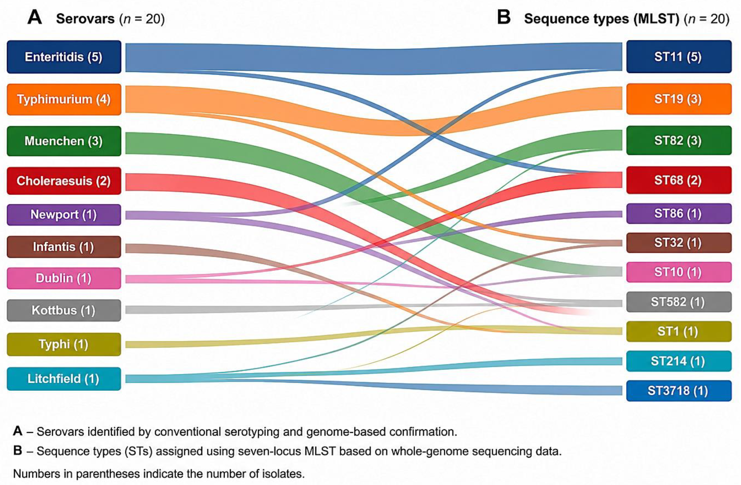
Figure 2.
Representative Kirby–Bauer disk-diffusion assays used for phenotypic antimicrobial susceptibility testing of Salmonella enterica.
Figure 2.
Representative Kirby–Bauer disk-diffusion assays used for phenotypic antimicrobial susceptibility testing of Salmonella enterica.
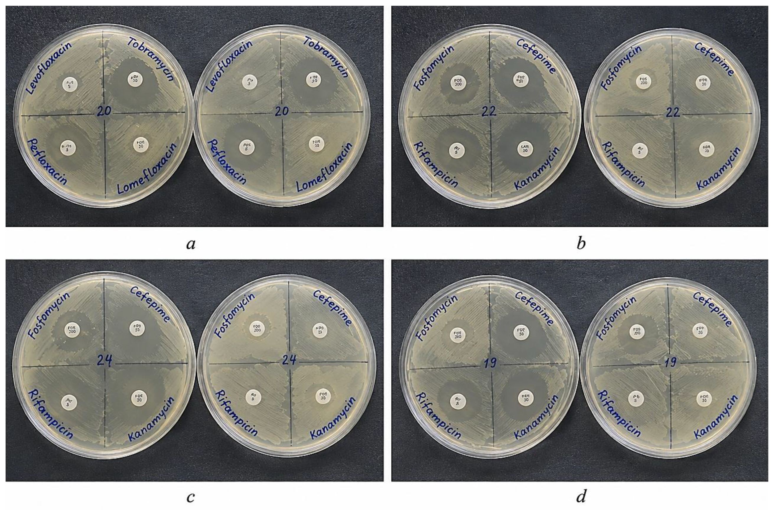
Figure 3.
Heatmap and hierarchical clustering of inhibition-zone diameters for 20 selected Salmonella enterica strains tested against 13 antimicrobial agents. Values within cells represent inhibition-zone diameters (mm). The dendrogram illustrates exploratory hierarchical clustering based on similarities in phenotypic inhibition-zone profiles and should not be interpreted as evidence of phylogenetic or epidemiological relatedness.
Figure 3.
Heatmap and hierarchical clustering of inhibition-zone diameters for 20 selected Salmonella enterica strains tested against 13 antimicrobial agents. Values within cells represent inhibition-zone diameters (mm). The dendrogram illustrates exploratory hierarchical clustering based on similarities in phenotypic inhibition-zone profiles and should not be interpreted as evidence of phylogenetic or epidemiological relatedness.
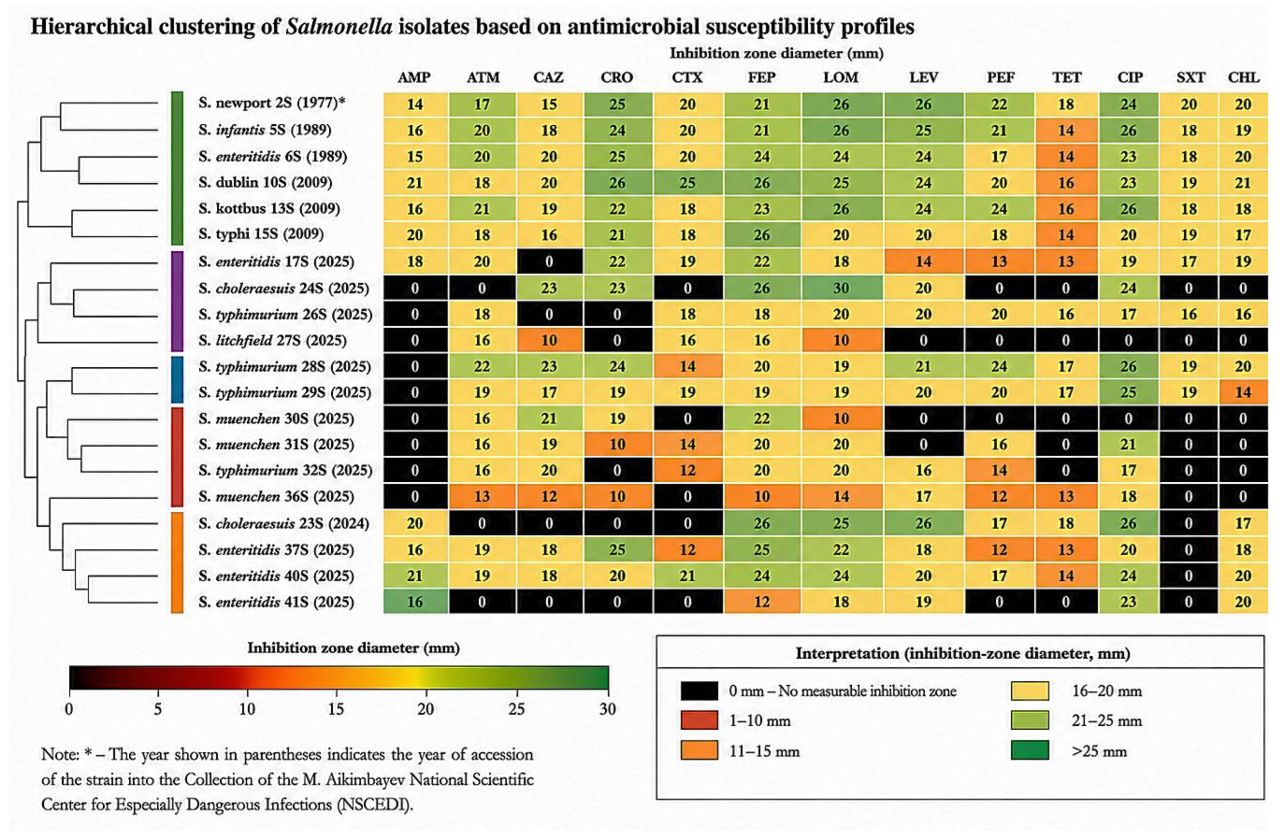
Figure 4.
Principal component analysis (PCA) score plot based on inhibition-zone diameters for 13 antimicrobial agents across 20 Salmonella enterica strains. Each point represents an individual strain and is labeled by isolate ID. Colors and symbols indicate serovar–sequence type groups, and dashed ellipses highlight selected major serovar–ST combinations. PC1 and PC2 explain 44.19% and 21.16% of the total variance, respectively (65.35% combined).
Figure 4.
Principal component analysis (PCA) score plot based on inhibition-zone diameters for 13 antimicrobial agents across 20 Salmonella enterica strains. Each point represents an individual strain and is labeled by isolate ID. Colors and symbols indicate serovar–sequence type groups, and dashed ellipses highlight selected major serovar–ST combinations. PC1 and PC2 explain 44.19% and 21.16% of the total variance, respectively (65.35% combined).
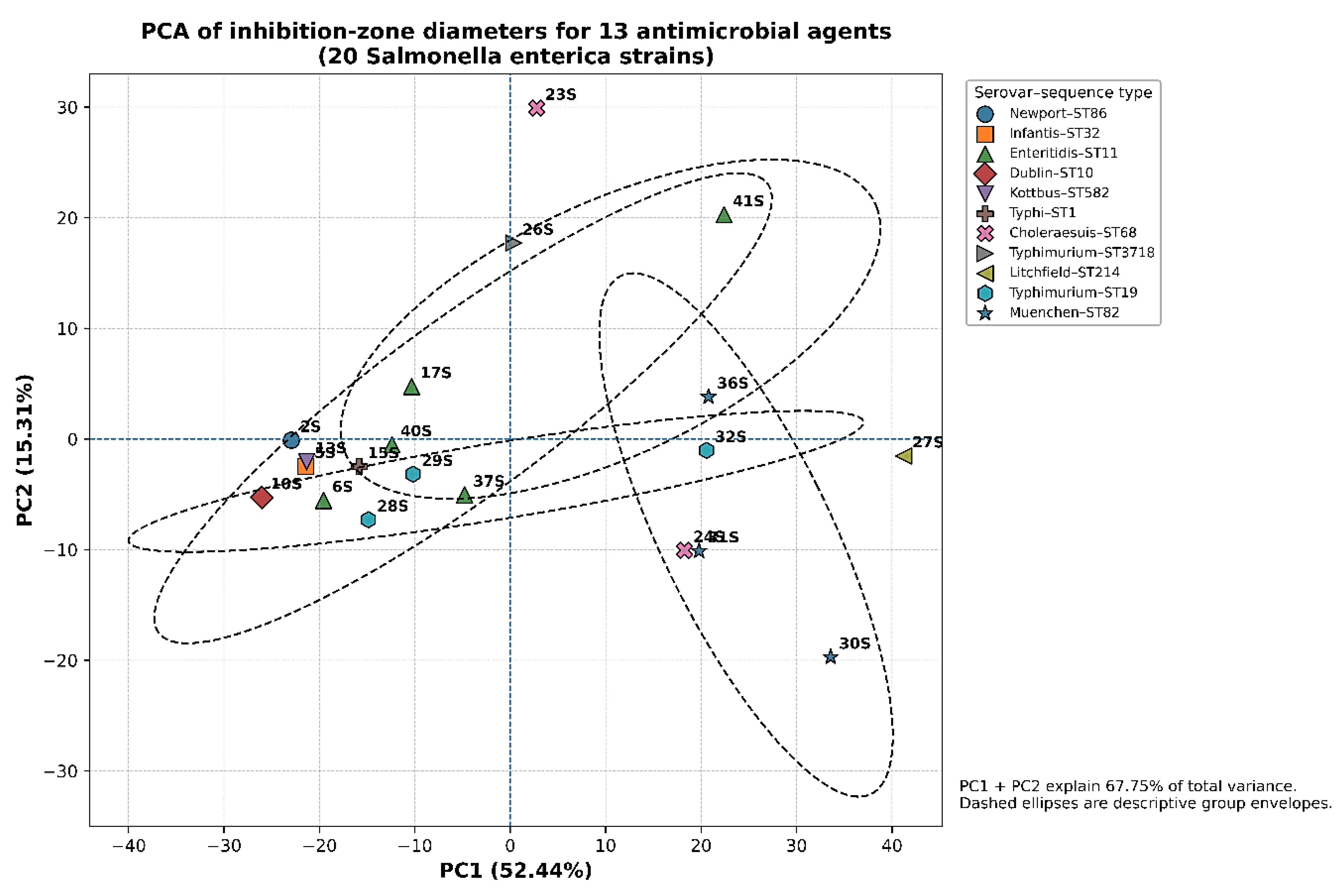

Table 1.
Characteristics of the selected Salmonella enterica strains included in the study.
| Strain | Year* | Serovar | Antigenic Formula | Sequence Type (ST) | Source | Location (Region) |
|---|---|---|---|---|---|---|
| 2S | 1977 | Newport | 4:b:1,2 | ST86 | Animal | Almaty |
| 5S | 1989 | Infantis | 7:r:1,5 | ST32 | Animal | Almaty |
| 6S | 1989 | Enteritidis | 9:g,m:- | ST11 | Animal | Almaty |
| 10S | 2009 | Dublin | 1,9,12:g,p:- | ST10 | Human | Shymkent |
| 13S | 2009 | Kottbus | 6,8:e,h:1,5 | ST582 | Human | Shymkent |
| 15S | 2009 | Typhi | 9,12[Vi]:d:- | ST1 | Human | Almaty |
| 17S | 2025 | Enteritidis | 9:g,m:- | ST11 | Animal | Karaganda |
| 23S | 2024 | Choleraesuis | 6,7:c:1,5 | ST68 | Animal | Almaty |
| 24S | 2025 | Choleraesuis | 6,7:c:1,5 | ST68 | Human | Almaty |
| 26S | 2025 | Typhimurium | 4:i:1,2 | ST3718 | Human | Almaty |
| 27S | 2025 | Litchfield | 6,8:l,v:1,2 | ST214 | Human | Almaty |
| 28S | 2025 | Typhimurium | 4:i:1,2 | ST19 | Human | Almaty |
| 29S | 2025 | Typhimurium | 4:i:1,2 | ST19 | Human | Almaty |
| 30S | 2025 | Muenchen | 6,8:d:1,2 | ST82 | Human | Almaty |
| 31S | 2025 | Muenchen | 6,8:d:1,2 | ST82 | Human | Almaty |
| 32S | 2025 | Typhimurium | 4:i:1,2 | ST19 | Human | Almaty |
| 36S | 2025 | Muenchen | 6,8:d:1,2 | ST82 | Human | Almaty |
| 37S | 2025 | Enteritidis | 9:g,m:- | ST11 | Human | Almaty |
| 40S | 2025 | Enteritidis | 9:g,m:- | ST11 | Human | Almaty |
| 41S | 2025 | Enteritidis | 9:g,m:- | ST11 | Animal | Almaty |
Note: * Year indicates the year of accession of the strain to the National Collection of Microorganisms and does not necessarily represent the year of primary isolation.
Table 2.
Distribution of inhibition-zone diameters among 20 selected Salmonella enterica strains tested against 13 antimicrobial agents.
Table 2.
Distribution of inhibition-zone diameters among 20 selected Salmonella enterica strains tested against 13 antimicrobial agents.
| Antimicrobial agent | Abbreviation | Disk content (µg) | Range (mm) | Mean ± SD (mm) | Median (mm) | No measurable inhibition zone, n/20 (%) |
|---|---|---|---|---|---|---|
| Ampicillin | AMP | 10 | 0–21 | 8.85 ± 9.26 | 7.0 | 10/20 (50%) |
| Aztreonam | ATM | 30 | 0–22 | 16.20 ± 5.93 | 18.0 | 2/20 (10%) |
| Ceftazidime | CAZ | 30 | 0–23 | 14.45 ± 8.05 | 18.0 | 4/20 (20%) |
| Ceftriaxone | CRO | 30 | 0–26 | 15.75 ± 10.26 | 20.5 | 5/20 (25%) |
| Cefotaxime | CTX | 30 | 0–25 | 12.50 ± 8.93 | 16.0 | 6/20 (30%) |
| Cefepime | FEP | 30 | 10–26 | 21.05 ± 4.49 | 21.5 | 0/20 (0%) |
| Lomefloxacin | LOM | 10 | 10–30 | 20.80 ± 5.27 | 20.0 | 0/20 (0%) |
| Levofloxacin | LVX | 5 | 0–26 | 17.70 ± 8.29 | 20.0 | 3/20 (15%) |
| Pefloxacin | PEF | 5 | 0–24 | 14.35 ± 8.14 | 17.0 | 4/20 (20%) |
| Tetracycline | TET | 30 | 0–18 | 10.65 ± 7.31 | 14.0 | 6/20 (30%) |
| Ciprofloxacin | CIP | 5 | 0–26 | 20.10 ± 7.50 | 23.0 | 2/20 (10%) |
| Trimethoprim–sulfamethoxazole | SXT | 25 | 0–20 | 9.15 ± 9.42 | 8.0 | 10/20 (50%) |
| Chloramphenicol | CHL | 30 | 0–21 | 12.95 ± 8.85 | 17.5 | 6/20 (30%) |
Note: Values represent inhibition-zone diameters obtained by the disk-diffusion method. A value of 0 mm indicates the absence of a measurable inhibition zone around the antimicrobial disk and should not, by itself, be interpreted as categorical resistance. Susceptible, intermediate, or resistant classifications require application of organism- and antimicrobial-specific interpretive criteria.
Disclaimer/Publisher’s Note: The statements, opinions and data contained in all publications are solely those of the individual author(s) and contributor(s) and not of MDPI and/or the editor(s). MDPI and/or the editor(s) disclaim responsibility for any injury to people or property resulting from any ideas, methods, instructions or products referred to in the content. |
© 2026 by the authors. Licensee MDPI, Basel, Switzerland. This article is an open access article distributed under the terms and conditions of the Creative Commons Attribution (CC BY) license (http://creativecommons.org/licenses/by/4.0/).
Copyright: This open access article is published under a Creative Commons CC BY 4.0 license, which permit the free download, distribution, and reuse, provided that the author and preprint are cited in any reuse.