

Submitted:
08 August 2026
Posted:
11 August 2026
You are already at the latest version
Abstract
Single-stranded DNA-binding protein (SSB) coats single-stranded DNA (ssDNA) and recruits downstream repair factors through its acidic C-terminal tip, yet this same coating must be cleared for RecA – a central protein in homologous recombination, to load during the process. RecO, a recombination mediator protein, is thought to regulate this handoff, but whether and how it drives release of full-length, ssDNA-bound SSB, rather than simply docking onto an isolated SSB peptide as captured in the only available crystal structure, has not been shown directly. Using single-molecule atomic force microscopy, we show that RecO actively displaces full-length wild-type SSB from a defined ssDNA substrate, reducing occupancy from 67% to 29%. Displacement efficiency decreases after deleting the SSB C-terminal tip (ΔC8) or extending it by a single residue (A179); the latter instead increases ssDNA occupancy and complex volume. Molecular dynamics simula-tions show that A179 retains its native RecO contact but additionally engages a non-native surface, producing a specific, over-engaged complex rather than non-specific aggregation suggesting that these interprotein interactions can affect SSB-ssDNA contacts. Together, these results suggest that RecO-driven SSB clearance is governed by the precise geometry of a single protein-protein interface rather than by affinity alone: too little engagement (ΔC8) or too much (A179) both abolish displacement, acting as a checkpoint against in-discriminate SSB removal during genome maintenance.
Keywords:
E. coli SSB
; E. coli RecO
; AFM
; single molecule
1. Introduction
Single-stranded DNA-binding protein (SSB) is an indispensable participant in virtually all aspects of DNA metabolism in Escherichia coli and across the bacterial kingdom. SSB functions mostly as a stable homo-tetramer [1]. Each subunit containing an N-terminal oligonucleotide/oligosaccharide binding (OB) fold domain that directly binds to the single-stranded DNA (ssDNA) and the C-terminal region of the protein containing an intrinsically disordered linker (IDL) and a highly conserved acidic tip [1,2]. The four OB-fold domains cooperate to wrap ssDNA partially or completely, occluding approximately 35 nucleotides per tetramer in the partial form and 65 nucleotides per tetramer in the fully bound state [3,4,5]. By coating the intermediates that arise during replication, repair, and recombination, SSB protects them from nucleolytic degradation and inhibits secondary structure formation, thereby enabling downstream enzymatic processing to proceed efficiently. Beyond the protective role, SSB has emerged as a master organizational hub of genome maintenance [6]. The C-terminal apparatus serves as an interaction interface for a large and diverse cohort of SSB-interacting proteins (SIPs), including the chi subunit of DNA polymerase III, PriA helicase, ExoI, RecQ, RecO, and RecG, which are key factors in DNA replication, repair, and recombination [6,7,8,9,10,11] . Through these interactions, SSB actively recruits and coordinates the downstream machinery that processes ssDNA intermediates, hence establishing its role also as a dynamic platform for genomic maintenance [12].
Understanding how SIPs precisely recognize and engage the SSB C-terminal domain has been the subject of active investigation and ongoing debate. Two principal models have emerged from structural, thermodynamic, and mutational studies. The first model, built on the work of Kozlov and Lohman groups, proposes that the highly conserved short acidic tip is the primary and sufficient determinant for SIP binding [13,14]. Isothermal titration calorimetry (ITC) studies demonstrated that synthetic peptides corresponding to the last nine residues of the SSB C-terminus recapitulate the full binding affinity of multiple SIPs, and that varying the length of IDL segment attached to the tip peptide produced no significant change in binding affinity [13]. The crystal structure of E. Coli. RecO bound to the SSB C-terminal peptide (PDB ID: 3Q8D) provided direct structural evidence, showing that the terminal Pro-Phe residues of the tip inserted into a hydrophobic pocket on RecO’s central alpha-helical domain [11]. Together, these data support a model in which the IDL serves primarily as a flexible tether that presents the tip to its binding partners, with the tip itself providing all the thermodynamically significant contacts. A second model, from Bianco and colleagues, holds that the IDL is an active participant rather than a passive linker: disrupting its three PXXP motifs abolishes RecG binding even when the tip itself is left intact [12,13,15,16,17]. Which model applies, and the consequences of the complex formation may depend on the specific SIP and on the structural context of full-length, DNA-bound SSB.
RecO functions not merely as a passive partner that docks onto SSB but as a regulatory protein that actively reshapes how SSB engages ssDNA, and the specific outcome of that regulation varies with the reaction and the organism [18]. At its core, this regulatory switch is governed by a single protein-protein interface – the contact between RecO and the SSB tail – whose precise geometry, and not simply its affinity, determines whether SSB is released, retained, or trapped in a non-productive complex. RecO is central to the RecFOR pathway [19,20,21], which relieves the barrier SSB poses to RecA loading during homologous recombination : Umezu and colleagues first showed biochemically that RecO and RecR together overcome SSB's inhibition of strand exchange [22], and Morimatsu and Kowalczykowski established RecFOR as a structure-specific mediator that loads RecA onto SSB-coated gapped DNA [23]. Kantake and colleagues showed that E. coli RecO anneals ssDNA while it remains complexed with its cognate SSB, forming a specific 1:1 RecO-SSB complex in a species-specific reaction that neither T4 gp32 nor yeast RPA can support – evidence that RecO can productively remodel an SSB-ssDNA complex without necessarily evicting SSB outright [24]. Ryzhikov and colleagues solved the RecO-SSB peptide crystal structure showing the SSB C-terminal tip docking into RecO's hydrophobic pocket [11], and further biochemical work from the Inoue and colleagues showed that SSB is displaced from ssDNA upon RecO binding yet remains indirectly tethered to the DNA through its continued interaction with RecO, with RecR subsequently modulating RecO's ssDNA-binding mode and suppressing its annealing activity [25]. NMR studies of the Thermus thermophilus RecO-SSB pair identified a chemical basis for this regulation: a positively charged surface on RecO's C-terminal region, centered on Arg-127, engages the acidic SSB tail, and this interaction alone is sufficient to weaken SSB's affinity for ssDNA without any requirement for ATP hydrolysis [26]. RecO's regulatory repertoire extends further still: in Mycobacterium smegmatis, RecO engages ssDNA through two structurally distinct and independently regulated binding modes – a zinc-stimulated mode that supports annealing and a RecR-regulated mode that supports recombinase loading – neither of which depends on the SSB-Ct interaction that dominates the E. coli mechanism [27,28]. And in Deinococcus radiodurans, single-molecule fluorescence imaging by Hwang and colleagues showed RecO competing SSB directly off ssDNA in an ATP-independent manner, fully dissociating a protein that binds ssDNA roughly 300-fold more tightly than RecO itself [29].
Taken together, these studies establish RecO as a bona fide regulator of the SSB-ssDNA interaction, capable of altering SSB's grip on the DNA through several distinct routes: allosteric weakening of its intrinsic affinity [14], competitive eviction [29], and remodeling of the SSB-ssDNA complex into an annealing-competent intermediate [11,24,25]. Yet for E. coli – the organism in which the RecO-SSB interface is structurally best defined – which of these regulatory outcomes applies to full-length, ssDNA-bound SSB has not been established directly. The available crystal structure is a static snapshot of an isolated nine-residue SSB peptide bound to RecO in solution, not a full-length SSB tetramer engaged with ssDNA: it shows that RecO can dock onto the tip, but not whether, or how, that docking translates into altered SSB occupancy of the DNA. Bulk biochemical assays average over populations of complexes and cannot distinguish whether SSB is displaced outright, remains indirectly tethered as proposed for the E. coli system, or persists as a mixture of intermediate states. The one single-molecule demonstration of active, RecO-driven displacement comes from D. radiodurans, whose SSB is a structurally distinct homodimer and whose RecO shares only limited homology with its E. coli counterpart [29]. Whether E. coli RecO likewise drives physical dissociation of full-length SSB from ssDNA – and, if so, which structural feature of SSB, the acidic tip or the IDL, gates that regulatory action – has not been tested directly at the single-molecule level.
Atomic force microscopy (AFM) closes this gap: it visualizes individual protein-DNA complexes directly, without the ensemble averaging inherent to bulk assays, and quantifies both whether a complex is present and its physical dimensions. Using a structurally defined ssDNA substrate designed to predominantly accommodate a single SSB tetramer, we show that RecO actively drives dissociation of full-length, DNA-bound wtSSB, and that this dissociation is gated strictly by the acidic tip: it is abolished by either removing the tip entirely (ΔC8) or extending it by a single residue (A179), while the IDL fully intact in both variants cannot substitute for it. These results extend Hwang and colleagues' single-molecule visualization of RecO-driven SSB displacement to the mechanistically distinct E. coli system, and, together with the finding that a single added residue is sufficient to abolish the reaction, establish the precisely terminated acidic tip as the necessary and sufficient determinant of RecO-driven SSB dissociation rather than one contributor among several.
2. Results
2.1. DNA Substrate
In our AFM studies we used hybrid DNA approach in which ssDNA segment (~70 residues) is attached to the end of the long DNA duplex (~300 bp) allowing visual recognition of complexes of ssDNA with single-stranded DNA binding proteins [30,31,32,33,34,35,36,37,38]. The current DNA construct with 68 ssDNA tail. The designed DNA substrate was purified using a gel extraction kit as described in the Materials and Methods section. Figure 1A shows the migration difference between naked dsDNA and dsDNA with an attached ssDNA tail. Figure 1B presents a schematic representation of the DNA substrate used throughout this study. The height distribution of dsDNA showed a mean value of approximately 1.0 nm, consistent with previously reported values for this substrate.
2.2. SSB Variants Used in This Study
To map the structural requirements of the SSB C-terminal domain for RecO-mediated dissociation, we used three SSB constructs throughout this study (Figure 1C). Wild-type SSB (wtSSB) carries the complete intrinsically disordered linker (IDL), including all three PXXP motifs, together with the intact eight-residue acidic tip (DFDDDIPF). ΔC8 retains the full IDL and all three PXXP motifs but lacks the eight C-terminal residues that make up the acidic tip. A179 retains the complete IDL and the native tip sequence unchanged, but carries a single alanine appended after the terminal Phe. Together, these variants separate the contributions of the tip and the IDL: ΔC8 tests whether an intact IDL is sufficient for RecO-driven binding and dissociation in the complete absence of the tip, while A179 tests whether the tip must terminate at its native last residue, or whether extending it by even one residue is enough to disrupt RecO engagement.
2.3. RecO Binds Weakly to the ssDNA
We used surface plasmon resonance (1:1 Langmuir kinetic fits) to test the ability of RecO and wtSSB to bind to the oligonucleotide used as an ssDNA tail in the study. wtSSB bound with the oligonucleotide with an estimated KD value of 15 nM. Whereas RecO bound with an estimated KD value of ~90 nM, reflecting a lower affinity to the DNA substrate compared to the wtSSB. RecO's tendency to aggregate limited its titration to a narrow concentration range (500–1000 nM), so this value should be regarded as an estimate rather than a precise measurement; nonetheless, this difference in estimated KD underlies our conclusion that RecO binds the oligo weaklier than wtSSB (Supplementary Figure 2).
2.4. Design and Validation of the ssDNA-Tailed AFM Substrate
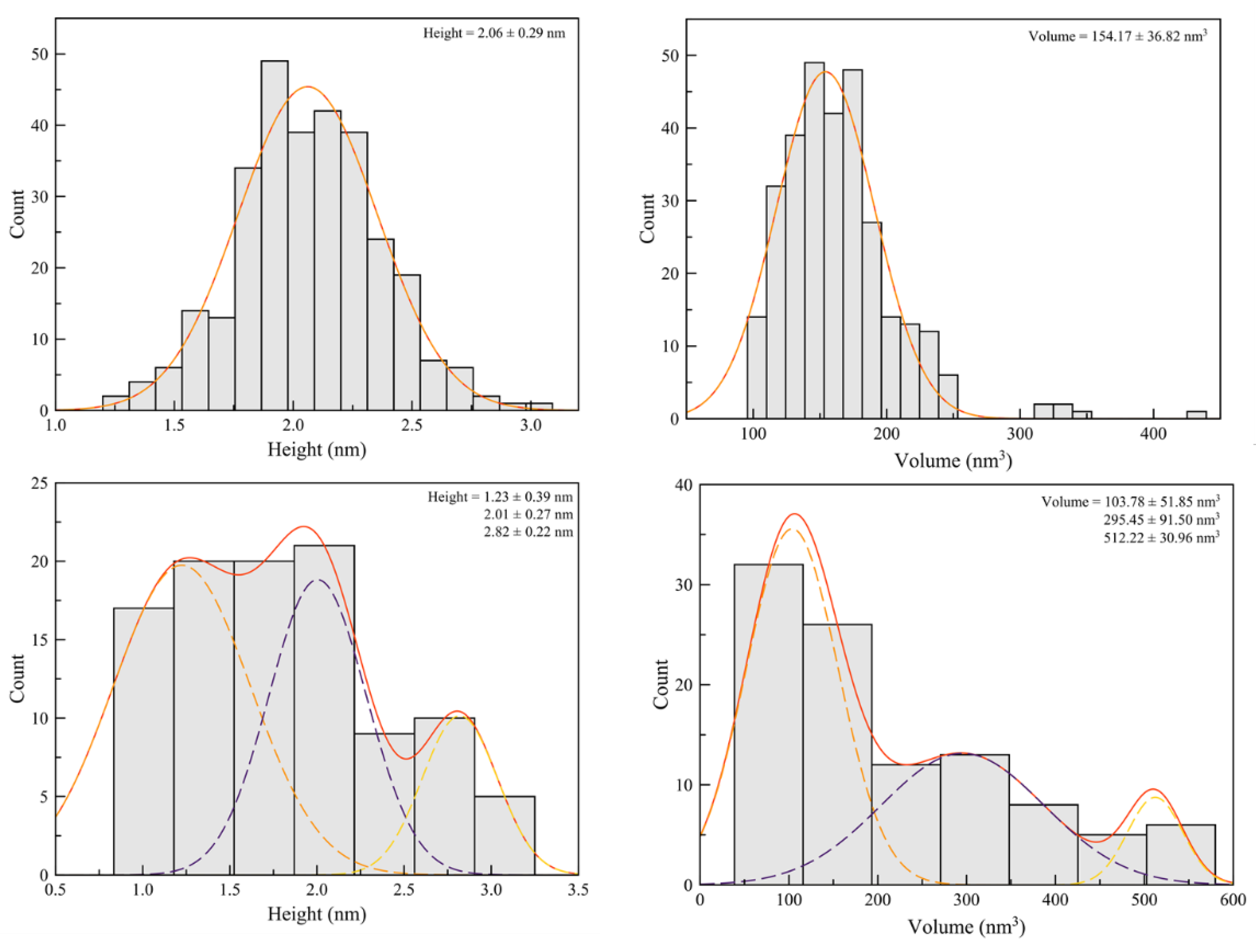
A structurally defined DNA substrate was prepared for AFM imaging to provide a controlled single-site ssDNA platform for SSB and SSB variant binding. The substrate consists of a 389-bp double-stranded DNA (dsDNA) body bearing a 68-nucleotide single-stranded tail at one end. The dsDNA body was designed to serve two complementary purposes. First, it acts as a visual marker: its defined, uniform contour under AFM makes it immediately distinguishable from protein-bound or aggregated species, so that substrate molecules can be unambiguously identified in every image. Second, because a dsDNA segment of this length has a well-defined, reproducible height under AFM, it also functions as an internal standard within every field of view. Measuring the dsDNA height in each experiment and confirming that it remains constant across sessions and samples verifies tip and instrument performance, establishing internal consistency between experiments before any biological signal is interpreted. To this marker, we appended a randomized, non-complementary 68-nucleotide ssDNA segment at one end, providing a single, spatially defined binding site for SSB or an SSB variant. To validate the substrate, we first imaged it in the absence of any protein. AFM images showed the expected morphology: a rod-like dsDNA body with a flexible single-stranded extension at one end, which may or may not be visible on the AFM image (supplementary Figure 1). Addition of wtSSB to the substrate resulted in the appearance of a discrete globular feature co-localized at one end of the rod-like structure, the ssDNA tail, confirming specific binding. In the absence of RecO, 66.7% of ssDNA tails were occupied by wtSSB (N ≥ 321 (occupied molecules); Figure 2A and Figure 3-top), with protein-free dsDNA body regions in all cases, confirming that SSB binding is specific for the single-stranded region. Occupied complexes displayed a mean height of 2.06 ± 0.29 nm and a volume of 154.17 ± 36.82 nm3, consistent with values reported for single SSB tetramers bound to ssDNA in prior AFM studies [39,40]. These measurements validate the substrate and establish baseline parameters for the RecO binding and displacement experiments.
Figure 2.
AFM imaging of wtSSB on the DNA substrate ± RecO. (A) Representative field and zoomed-in insets, DNA + wtSSB alone. (B) Representative field and zoomed-in insets, DNA + wtSSB + RecO. Scale bars, 200 nm. (C) Fraction of DNA molecules scored as SSB-bound vs. free, wtSSB alone vs. wtSSB + RecO.
Figure 2.
AFM imaging of wtSSB on the DNA substrate ± RecO. (A) Representative field and zoomed-in insets, DNA + wtSSB alone. (B) Representative field and zoomed-in insets, DNA + wtSSB + RecO. Scale bars, 200 nm. (C) Fraction of DNA molecules scored as SSB-bound vs. free, wtSSB alone vs. wtSSB + RecO.
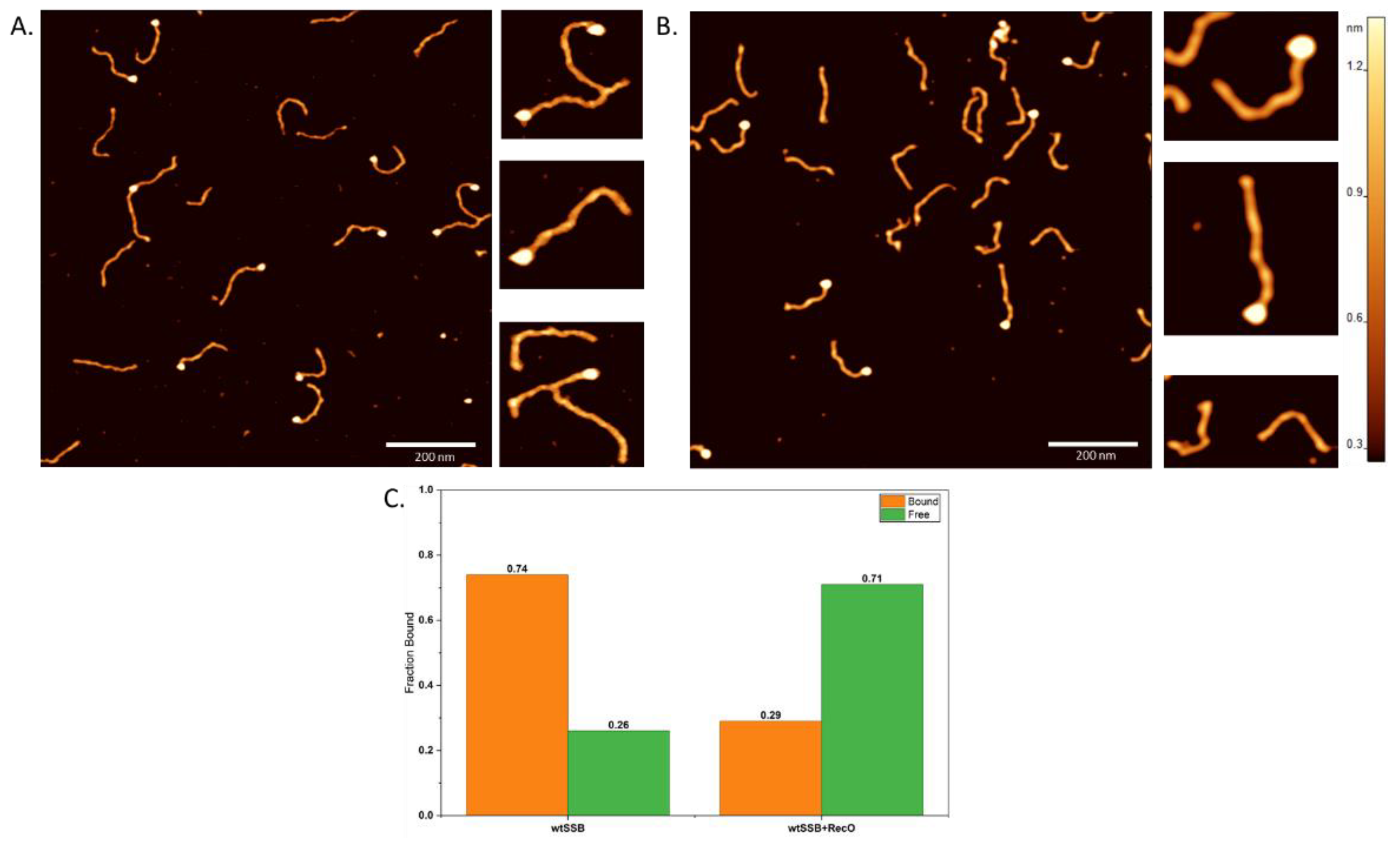
Figure 3.
Height and volume distributions of wtSSB particles measured by AFM, without (top) and with (bottom) RecO. Data were fit to one and three-population Gaussian distribution. Standard deviation was calculated as σ = dx/√(2·ln2). Data are reported as mean ± SD. The R2 for all the fits was ≥ 0.95.
Figure 3.
Height and volume distributions of wtSSB particles measured by AFM, without (top) and with (bottom) RecO. Data were fit to one and three-population Gaussian distribution. Standard deviation was calculated as σ = dx/√(2·ln2). Data are reported as mean ± SD. The R2 for all the fits was ≥ 0.95.
2.5. RecO Efficiently Displaces wtSSB from the ssDNA Tail
Throughout this manuscript, we use "displacement" and "dissociation" interchangeably to refer to the same observable: loss of an AFM-visible SSB/variant complex from the ssDNA tail following RecO addition. To test whether RecO can dissociate wtSSB from the ssDNA substrate, pre-formed wtSSB-ssDNA complexes were incubated with RecO and imaged by AFM. A striking reduction in ssDNA occupancy was observed upon RecO addition: the fraction of tails bearing SSB complexes decreased from 66.7% to 29.0% (N ≥ 105 (occupied molecules); Figure 2B and 3-bottom; Table 1), representing an approximately 2.3-fold reduction relative to the RecO-free condition Fischer’s exact test: p = 9.28*10-28, indicating a highly significant displacement (Table 2). In the presence of RecO, the majority of ssDNA tails appeared completely protein-free, with no alteration in substrate morphology (Figure 2B). However, the residual 29% protein-DNA complex was found to be highly heterogeneous and split into three sub-populations with the heights of 1.23 ± 0.39 nm, 2.01 ± 0.27 nm, and 2.82 ± 0.22 nm. The volume distribution was also found to be highly heterogeneous and was split into three distinct populations with the values ranging approximately between 103 and 512 nm3 (Table 1). The height and volume distributions were found to be significantly broadened across a wide range of values, suggesting the possible presence of either co-localized RecO/SSB complexes or a transition intermediate. These data suggest that RecO addition produces an essentially binary outcome; the ssDNA tail is either occupied or fully clear. The complexes that do remain bound could reflect several non-exclusive scenarios: molecules captured at different stages of the multi-step dissociation process, a subpopulation not yet engaged by RecO under these incubation conditions, or stable co-localized RecO/SSB complexes. Since the images are end-point rather than real-time recordings, our data cannot distinguish between these scenarios.
Nevertheless, the predominance of fully cleared tails indicates that RecO drives efficient and largely complete SSB removal from the defined ssDNA substrate.
2.6. Deletion of the Acidic Tip Abolishes RecO-Mediated Displacement: ΔC8
To determine whether the C-terminal acidic tip is required for RecO-driven displacement, we examined the ΔC8 variant, which lacks the tip entirely. In the absence of RecO, 92% of ssDNA tails were occupied by ΔC8 and formed protein complexes (N ≥ 543 (occupied molecules); Figure 4A; Table 1), confirming that ΔC8 is fully competent for ssDNA binding. Strikingly, ΔC8-ssDNA complexes displayed substantially different physical properties compared to wtSSB complexes.
The population distribution was bimodal, and the mean complex heights were 1.95 ± 0.32nm and 2.78 ± 0.33 nm (compared to 2.06 ± 0.29 nm for wtSSB), and the apparent volumes were 301.62 ± 135.07nm3 and 675.29 ± 113.01 nm3 (Figure 5 top). The subpopulation with smaller height and volume may be comparable with wild type, but the subpopulation with higher height approximates a volume four times larger than wtSSB complexes. These altered dimensions indicate that loss of the acidic tip changes the geometry of SSB-ssDNA engagement. This likely reflects a shift in the wrapping mode equilibrium toward the less compactly wrapped (SSB)35 configuration [41], or the assembly of multiple SSB tetramers on the 68-nt tail, either of which would produce complexes with elevated apparent volume in AFM.
Upon addition of RecO to ΔC8-loaded substrates, the ssDNA occupancy remained essentially unchanged: 89% of tails remained occupied (N ≥ 339 (occupied molecules); Figure 4B; 5 bottom; Table 1), Fischer’s exact test; p = 0.072 (Table 2), indicating no significant change and closing matching the RecO-free condition. The volumes also remain comparable to the values in the absence of RecO (Figure 5, bottom; Table 1). Within the sensitivity of this assay, RecO does not produce a detectable change in the ΔC8’s occupancy or volume, despite ΔC8 carrying the full IDL with all three PXXP motifs intact. Taken together with the intrinsic heterogeneity if ΔC8, this indicates that IDL alone is not sufficient to drive the displacement under these conditions. Though, a weak IDL-mediated interaction that does not produce a detectable displacement signal cannot be fully excluded.
2.7. A Single Amino Acid Addition to the Tip Abolishes Displacement: A179
To test the role of the precise tip termination in RecO engagement, we examined A179, which bears a single alanine addition at the end of the protein chain, with the remainder of the tip and the full IDL intact. In the absence of RecO, 64% of ssDNA tails were occupied by A179 (N ≥ 241 (occupied molecules); Figure 6A; Table 1), confirming competent ssDNA binding.
The height of the protein yielded a bimodal distribution, in contrast to that of the wtSSB, and the volume distribution was unimodal and comparable to the wild-type conditions (Figure 7-top and Table 1).
It should be noted that the R2 values for height distributions, both in the presence and absence of RecO, were ≥ 0.85 under Freedman-Diaconis (FD) binning. Deviating from the FD rule and adjusting the bin size yielded a unimodal distribution in the absence of RecO and improved the goodness of fit in the presence of RecO to ≥0.95. However, to maintain uniform binning methodology, FD binning was followed, and the resulting lower R2 fit was used for comparisons. The modestly lower baseline occupancy and the change in height distribution compared to wtSSB may reflect a subtle effect of the mutation on ssDNA affinity or binding mode.
Interestingly, the addition of RecO produced an increase in A179’s ssDNA occupancy to 80% Fisher’s exact p = 1.5*10-5 indicating a statistically significant increase in occupancy, in contrast to the effect of wtSSB’s displacement. (N ≥ 211 (occupied molecules), Figure 6B; Table 1). A single amino acid addition at the end of the tip is therefore sufficient to affect RecO-mediated displacement. In addition, this variant now accumulates more protein at the ssDNA end. The scale of A179 volume increase (roughly 2.5- to 5-fold) initially raised non-specific aggregation of RecO, A179, or both as an explanation at least as plausible as a stable, specific, non-functional co-complex; the molecular dynamics simulations presented below resolve this ambiguity in favor of a specific, structured interface. Both ΔC8 and A179 converge on the same functional outcome – no displacement, through two independent tip perturbations, indicates a genuine requirement for a correctly configured tip and argues against a trivial explanation based on gross structural disruption by the eight-residue truncation alone.
2.8. Molecular Dynamics Simulations Reveal a Structural Basis for A179’s Non-Productive RecO Engagement
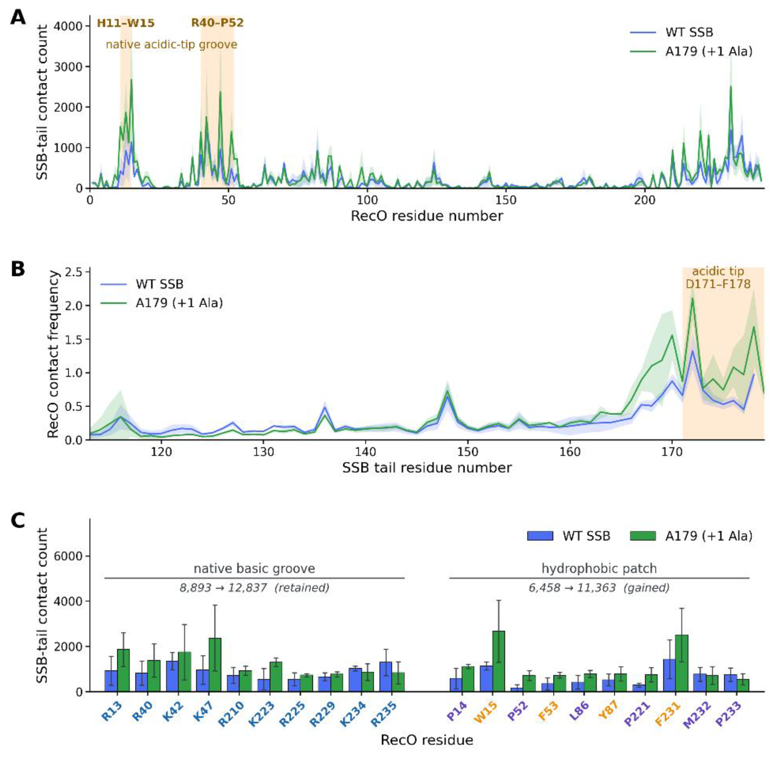
The outcome of RecO's engagement with SSB is set by the protein-protein interface between them rather than by ssDNA binding per se. We performed coarse-grained molecular dynamics simulations of the RecO-SSB tail interaction for wtSSB and A179 (n = 3 independent replicates per variant; see Materials and Methods). We analyzed these trajectories from two complementary vantage points: which residues on RecO's surface are contacted by the SSB tail (Figure 8A), and which residues along the SSB tail make those contacts (Figure 8B).
Figure 8A maps contact frequency across all 242 RecO residues, comparing wtSSB (blue) with A179 (purple), with shaded bands showing the spread across the three trajectories. The marked basic-groove residues (R210, R225, R229, K234) correspond closely to the positively charged rim of the crystallographically defined SSB-Ct pocket (Ryzhikov et al., PDB 3Q8D: Arg-210, Arg-225, Arg-229, Lys-206, Arg-203, [11]), and engagement here tracks closely between the two variants. The marked hydrophobic-patch residues span a wider range (W15, F53, V66, L86, L90); of these, L86 and L90 lie immediately adjacent to the hydrophobic residues that line the floor of that same crystallographic pocket (Ile-84, Tyr-91), while W15, F53, and V66 fall well outside it, in RecO's N-terminal OB-fold domain. A179 rises above wild type across the whole patch, most conspicuously at W15, where the peak roughly doubles, indicating both deeper engagement of the authentic pocket floor and genuinely novel contacts further from it.
Figure 8C collapses Panels A-B into the two functional groups. Engagement of the basic-groove rim is statistically indistinguishable between the variants (wtSSB 2965 ± 559 vs. A179 3307 ± 173). Engagement of the hydrophobic-patch group roughly doubles in A179 overall (wtSSB 2295 ± 497 vs. A179 5009 ± 849), driven mainly by the F172–W15 aromatic contact, the single strongest A179 pair, reproducing in two of the three runs, and located outside the known crystallographic pocket. The contribution from L86/L90, immediately adjacent to the pocket's native hydrophobic floor, cannot be separated from deeper engagement of the authentic site at this level of grouping and would need a per-residue (rather than per-group) comparison to resolve.
Taken together, the panels show that appending a single alanine at position 179 does not cost A179 the native RecO basic-groove rim. Instead, the tail's extra length is accommodated by increased engagement of the hydrophobic region in and around the same pocket: part of this (L86, L90) may reflect deeper penetration of the authentic hydrophobic floor (Ile-84, Tyr-91), while part, most notably W15, lies well outside the known pocket, in RecO's N-terminal OB-fold domain, and represents a genuinely novel contact. Rather than a clean switch from a specific native interface to an entirely separate non-native one, the data are more consistent with a single interface that becomes both deeper and more extensive in A179, retained at its native rim, but larger and less well-registered around its hydrophobic core. This still provides a structural rationale for the AFM observation that A179 accumulates more protein at the ssDNA end yet is not displaced by RecO, and still argues against non-specific aggregation as the explanation.
3. Discussion
This study provides the first direct nanoscale evidence for E. coli SSB displacement from a structurally defined ssDNA substrate by RecO protein. Using single-molecule AFM imaging, we demonstrate (Table 1 and Table 2) that RecO efficiently displaces wtSSB from a defined ssDNA tail with a 2.3-fold reduction in ssDNA occupancy (Supplementary Table 1 and Table 2) and a predominance of fully protein-free tails, indicating an efficient and largely stoichiometric displacement reaction. The predominance of fully displaced tails, with a few partially occupied or morphologically altered intermediates, suggests that once the RecO-SSB contact is established, the remodeling reaction proceeds rapidly and irreversibly to completion. This behavior is functionally appropriate RecA requires a contiguous ssDNA platform for filament nucleation, and even partial SSB coverage inhibits RecA polymerization [42]. The efficient, complete displacement we observe is therefore fully consistent with the biological role of RecO in clearing ssDNA gaps for RecA loading in the RecFOR pathway [18,42,43].
Displacement of the protein from the ssDNA end of the substrate cannot be explained by direct competition between SSB and RecO for ssDNA, since RecO's intrinsic affinity for ssDNA is substantially lower than that of SSB [18,27,28]. Instead, the determining factor is the specific interprotein interaction between RecO and SSB, specifically the C-terminal tip. Our variant data localizes this interaction with striking precision: ΔC8, which lacks the terminal tip entirely, shows no RecO-induced displacement, whereas A179, which retains the full-length tip but carries a single additional residue beyond the native C-terminal Pro-Phe (P-F) terminus and is equally unable to displace. This result is striking because A179 preserves the tip's length, charge, and sequence entirely. The sole modification is a one-residue extension beyond the terminal proline. Such a minimal change prevented the displacement activity completely, indicating that RecO does not recognize the SSB-C-terminal tail through the electrostatic interactions, but through a structurally precise, sequence-specific docking interactions. The tip must terminate exactly at the native Pro-Phe residues for a productive engagement and outcome. This is consistent with the RecO-SSB-Ct co-crystal structure, in which terminal tip residues insert into a defined pocket on RecO's surface [11]. The A179 result refines this structural requirement to single-residue precision: a one residue addition is sufficient to severely impair displacement, revealing that RecO engagement requires the tip to terminate at exactly the native C-terminal residues.
Upon RecO addition, the residual bound population of wtSSB (Supplementary Table 1 and Table 2) resolved into a trimodal distribution (a series of intermediates) (103, 295, and 512 nm³) rather than a single alternative state. This heterogeneity suggests that displacement proceeds through intermediates rather than direct, single-step release (Supplementary Table 1). This trimodal distribution is consistent with SSB transitioning from fully wrapped (SSB)₆₅-like (mean volume 154 nm³) toward the more extended (SSB)₃₅ mode during productive displacement; the changes in volume during this transition are reported previously [40]. However, SSB tetramer dissociation and transient RecO-SSB complex formation remain plausible explanations. By contrast, ΔC8's exhibits substantially altered baseline volumes and heights relative to wtSSB (Supplementary Table 1 and Table 2), reflecting a ~4-fold increase in volume. Critically, RecO addition does not alter the ΔC8 volume distribution, suggesting that RecO fails to establish productive contacts with the truncated variant. Deletion of the acidic tip eliminates the negative charge cluster that serves as the electrostatic anchor for RecO's basic-groove rim [11]. Without this acidic dipole, RecO cannot establish precise electrostatic contact required to destabilize the ΔC8-ssDNA complex [14]. Although the RecO-binding pocket remains structurally intact, the absence of the complementary negative charges on the SSB C-terminus prevents RecO in engaging SSB in the native electrostatic geometry - the interaction that couples tip occupancy to weakening of SSB's ssDNA binding affinity. These results isolate the electrostatic requirement for the efficient detachment of SSB from ssDNA [41].
By contrast, A179 remains essentially wtSSB-like at baseline (unimodal volume, comparable occupancy), but becomes bimodal and enlarged upon RecO addition indicating that its altered architecture is induced specifically by the non-productive RecO interaction. The three variants can thus map to three distinct points on the (SSB)₃₅/(SSB)₆₅ landscape: wtSSB transitions from fully wrapped toward productive unwrapping and release; ΔC8 sits constitutively in an intermediate state regardless of RecO; and A179 remains (SSB)₆₅-like until RecO traps it in an enlarged, non-productive state.
These results establish that RecO actively removes SSB – doing so only when the SSB C-terminal tip is intact and correctly positioned. This resolves a long-standing tension between the static, peptide-bound crystal structure of the SSB-RecO interface and the dynamic requirement, central to the RecFOR pathway, that SSB ultimately vacate the ssDNA it protects.
A key question emerges: what molecular interactions in A179 prevent productive displacement? Molecular dynamics simulations reveal the answer. MD simulation of RecO-SSB in the absence of ssDNA, at the tail interface reveal the mechanistic basis. Engagement of the basic-groove rim residues of the crystallographically defined SSB-C terminal binding pocket (R210, R225, R229, K234) [11] is statistically indistinguishable between wtSSB and A179, indicating that steric disruption of the native tip-pocket contact is not the primary failure mode of the A179 variant. Instead, the added alanine allows extended tail engagement with RecO, which is accommodated by the increased engagement of hydrophobic region in and around the pocket: part of this increase (L86, L90) lies immediately adjacent to the wild type pocket's own hydrophobic floor (Ile-84, Tyr-91), while most prominently W15 lies in RecO's N-terminal OB-fold domain, representing a novel contact.
Rather than replacing the native interface, the simulations reveal the native rim geometry while expanding the interface depth and breadth. The hydrophobic core contacts are retained but become less precisely aligned, allowing additional non-native conatcts. Taken together, ΔC8 and A179 represent opposite failure modes of the same interface: ΔC8 is a loss-of-interaction variant, in which the contact cannot form at all, whereas A179 is a gain-of-interaction variant, in which the native contact persists but is supplemented by additional, non-native contacts that enlarge the interface. Only the wild-type interface, complete, not expanded beyond its native boundaries supports productive displacement, underscoring that it is the specificity of this protein-protein contact, not merely its strength, that governs the outcome.
We propose that RecO recognition of the SSB C-terminal tip decreases SSB's intrinsic affinity for ssDNA, promoting complex dissociation. This mechanism is supported by Inoue et al., who demonstrated experimentally that SSB-Ct peptide alone decreases SSB’s ssDNA binding affinity, a finding consistent with the facilitated displacement we observe [26]. Furthermore, single-molecule studies in Deinococcus radiodurans show that RecO efficiently displaces SSB from ssDNA without consuming ATP, despite SSB binding ~300-fold more tightly than RecO. This kinetic advantage is only possible if RecO allosterically reduces SSB's binding stability [29]. The strict C-terminal tip dependence of SSB displacement in our AFM experiments directly supports this model.
Correct insertion of the native C-terminus into the RecO pocket is thus sufficient to trigger this effect, whereas any deviation from the exact terminal geometry, whether by truncation (ΔC8) or extension (A179) prevents proper docking and, with it, destabilization of the protein-ssDNA complex.
The single-residue precision observed at the SSB tip serves a physiological role beyond enabling RecO recognition: it functions as a fidelity checkpoint [13,14,44]. SSB coats ssDNA throughout the genome – at replication forks, resected double-strand breaks, and repair intermediates and its protective coating must be preserved at these sites even as it is cleared specifically at RecFOR-processed gaps to permit RecA loading [6,42,45,46] . By requiring the C-terminal tip to terminate at its native sequence, the cell restricts productive engagement to a single, precisely registered interface, so that SSB is released only where RecO has bound correctly rather than wherever it can bind at all. This specificity-over-affinity strategy may also explain divergence across species: D. radiodurans RecO achieves SSB displacement through direct competitive eviction rather than tip-gated engagement [29]. Different bacteria have thus evolved distinct molecular solutions to the same functional requirement, clearing SSB at the right place and time, with E. coli exploiting protein-protein interaction specificity rather than competitive affinity advantage.
4. Materials and Methods
Protein Expressions and Purification
All SSB variants (wtSSB, ΔC8, and A179) were expressed in Escherichia coli and purified according to established protocols from the Bianco laboratory [47,48]. RecO was purified using a strategy specifically designed to address its tendency to accumulate in the insoluble fraction upon overexpression. A construct encoding RecO with an N-terminal His-tag and SUMO solubility tag was co-expressed with SSB in E. coli to improve partitioning of RecO into the soluble fraction. Following detergent-assisted cell lysis and clarification by centrifugation, the soluble lysate was applied to a Ni-Sepharose affinity column. Bound His-tagged material was eluted and dialyzed against high-salt buffer (600 mM NaCl) in the presence of His-tagged SenP2 protease to cleave the SUMO tag. The digest was reloaded onto a Ni-Sepharose column; His-tagged components (uncleaved fusion, His-SenP2, free His-SUMO) were retained, and the flow-through containing untagged RecO was collected. RecO was further purified on a Mono S cation-exchange column, where it bound strongly and was separated from co-purifying SSB by a low-salt wash. RecO was eluted with a linear NaCl gradient (200 mM to 1 M NaCl over 10 column volumes) followed by a 2 M NaCl wash to recover tightly bound fractions. Pure RecO fractions were pooled, dialyzed into storage buffer (20 mM Tris-HCl pH 7.5, 200 mM NaCl, 1 mM DTT, 60% v/v glycerol), and stored at -80°C. 60% glycerol was specifically chosen based on the available literature for RecO purification and storage [49]. Purity was confirmed by SDS-PAGE and protein concentration was determined by absorbance at 280 nm.
DNA Substrate Preparation
Double-stranded DNA (dsDNA) was prepared as described previously [50,51] and digested with SapI. The digested dsDNA was purified using a Qiagen gel extraction kit and quantified by a nanodrop spectrophotometer. To generate the single-stranded DNA (ssDNA) tail, a random 91-nucleotide oligonucleotide (5'-/5Phos/GCT CCA GAC CGA GTC GTA ATC TTT AAC ATA GTT AGC TCT TCA GAA TTA TCC CTA AGA ACG ACC TGA ACA CTA ATA GTG TAT TCC ATA CTT T-3') was annealed to a 20-nucleotide adapter (5'-AAG ATT ACG ACT CGG TCT GG-3') by rapid heating to 95°C followed by gradual cooling to 4°C overnight. The adapter was designed to leave 3 nucleotides at the 5' end of the 91-nt oligonucleotide that were complementary to the SapI-digested dsDNA segment. The annealed oligonucleotides were then hybridized to the SapI-digested dsDNA by increasing the temperature to 37°C and gradually cooling to 16°C. The dsDNA:ssDNA tail molar ratio during annealing was maintained at 1:10. T4 DNA ligase and T4 ligation buffer were added to ligate the annealed DNA constructs, and the mixture was incubated at 16°C overnight. The ligated DNA was separated by 2% agarose gel electrophoresis, and the band of interest was excised and purified using a Qiagen gel extraction kit. The purified DNA was quantified by a nanodrop spectrophotometer (ND-1000, Thermo Fisher, Waltham, MA, USA). Both oligonucleotides were ordered from Integrated DNA Technologies (IDT).
Surface Plasmon Resonance
SPR experiments were performed on an OpenSPR instrument (Nicoya Lifesciences) using streptavidin/biotin/streptavidin sensor chips prepared according to the manufacturer's instructions. Streptavidin was first immobilized on the sensor surface at 0.5 μM; 200 nM biotinylated oligonucleotide was then captured on the streptavidin layer to present a ssDNA substrate. SSB/variants (200 nM) were flowed over the oligo-coated chip to achieve stable SSB immobilization on the ssDNA, and unbound SSB was removed by washing to a stable baseline. RecO was subsequently injected as the analyte at a series of concentrations to monitor its interaction with the surface-tethered SSB-ssDNA complex. Association and dissociation phases were recorded and sensorgrams were analyzed using TraceDrawer software. Due to limitations in fitting quality and reproducibility inherent to aggregation of RecO, quantitative kinetic parameters could not be reliably extracted; SPR data are discussed only as a qualitative indication of the effect of acidic tip deletion on RecO binding.
AFM Sample Preparation and Imaging
All experiments were performed in AFM buffer: 10 mM Tris-HCl (pH 7.4), 50 mM NaCl, 10 mM MgCl2, 1 mM DTT (DTT added freshly before each experiment) [39]. DNA substrate and SSB/ variant were mixed at a 1:2 molar ratio (1 uL of each stock) and incubated for 10 min at room temperature to form SSB-DNA complexes. For RecO displacement assays, 1 uL of RecO was added to the preformed SSB/variant-DNA mixture to achieve a final 1:2:2 molar ratio (DNA:SSB/varinat:RecO) and incubated at room temperature for an additional 10 min. The reaction was diluted with AFM buffer to a final DNA concentration of 2 nM.
50 μl of the diluted sample was deposited onto freshly cleaved bare mica and allowed to adsorb for 2 min. The surface was rinsed thoroughly with Milli-Q water to remove unbound material, then dried under a gentle stream of nitrogen gas. AFM images were acquired in tapping mode in air using a MultiMode AFM with a NanoScope VIII controller (Bruker) equipped with TESP-V2 probes (nominal spring constant 42 N/m, resonance frequency ~320 kHz). Scan areas of 3 x 3 μm were collected at 1536 x 1536-pixel resolution at 1.0 Hz scan rate. Images were processed in FemtoScan Analysis software with first-order line-by-line flattening only; no additional filtering was applied.
AFM Data Analysis
Protein-DNA complexes were identified by the presence of a globular feature co-localized with the ssDNA tail of the substrate. ssDNA occupancy was calculated as the fraction of substrate molecules carrying a bound complex on the ssDNA tail. Differences in ssDNA occupancy (the percentage of tails bound by a protein complex) between the RecO-free and RecO-added conditions were assessed for each SSB variant using a two-tailed Fisher's exact test applied to the underlying 2 × 2 contingency table of occupied and unoccupied molecule counts. Fisher's exact test was used rather than a chi-square approximation because it computes an exact p-value from the hypergeometric distribution rather than relying on a large-sample approximation. A threshold of P < 0.05 was used to define statistical significance
Complex height and apparent volume were measured using the particle-analysis module of FemtoScan Analysis. For statistical particle analysis, the grain size distributions were visualized as histograms with bin widths determined by the Freedman-Diaconis rules [52] (h = 2 * IQR * n -1/3), where h is the bin size, IQR is the interquartile range of the data, and n is the sample size. This rule is robust to outliers and does not assume normality. Histograms were then constructed using this calculated bin width, and Gaussian models were fit to the resulting distributions using MagicPlot v2.8.1 to extract peak parameters including mean grain size, standard deviation (σ), and relative peak area. Bin size sensitivity was systematically tested for all samples to confirm that Gaussian model parameters remained stable across reasonable binning ranges; exceptions to this robustness are indicated where they occur.
Coarse-Grained Molecular Dynamics Simulations
Full-length E. coli SSB was modelled by combining the crystallographic OB-fold core (PDB 1QVC) [53] with the disordered C-terminal region (residues 113-178) from the AlphaFold [54] model of UniProt P0AGE0, assembled as the physiological homotetramer; E. coli RecO (242 residues) was modelled likewise. Two constructs were simulated: wild type and A179. Systems were coarse-grained with martinize2 using the MARTINI 3 force field [55,56], with an elastic network on the folded OB-fold core only, solvated with explicit MARTINI water and counter-ions in a 24.6 nm cubic box, and propagated with GROMACS 2025.3 [57]. SSB-RecO trajectories of 25 microseconds each were produced (triplicates for wild type and A179), together with one 25-microsecond trajectory per variant of the SSB tetramer alone. Contacts were scored at a 0.6 nm bead-bead cut-off, and a frame was counted as bound when the minimum SSB-RecO distance fell below this threshold.
Supplementary Materials
The following supporting information can be downloaded at the website of this paper posted on Preprints.org.
Author Contributions
Conceptualization, Y.L.L and S.I.; methodology, S.I and S.F; software, S.I. and S.F.; validation, S.I. and S.F.; formal analysis, S.I.; investigation, S.I.; resources, Y.L.L.; data curation, S.I.; writing - original draft preparation, S.I and S.F.; writing - review and editing, S.I. and S.F.; visualization, S.I. and S.F.; supervision, Y.L.L.; project administration, Y.L.L.; funding acquisition, Y.L.L. All authors have read and agreed to the published version of the manuscript.
Funding
The research was funded by NIH grant GM144414 to YLL.
Institutional review board statement
Not applicable.
Informed consent statement
Not applicable.
Data Availability Statement
The original contributions presented in this study are included in the article/supplementary material. Further inquiries can be directed to the corresponding author.
Conflicts of Interest
The authors declare no conflict of interest.
Acknowledgments
Authors would like to thank Piero R Bianco for plasmids expressing SSB/SSB variants and RecO and also for his valuable assistance in optimizing RecO purification process. Authors also acknowledge Claude (Anthropic AI) for assistance with statistical analysis verification and for improving manuscripts clarity through grammatical corrections and sentence reconstruction.
Abbreviations
The following abbreviations are used in this manuscript:
| AFM | Atomic Force Microscopy |
| SSB | Single Stranded-DNA Binding Protein |
| RecO | Recombination Protein O |
| RecA | Recombinase Protein A |
| SD | Standard Deviation |
| dsDNA | Double-Stranded Deoxyribonucleic acid |
| ssDNA | Single-Stranded Deoxyribonucleic acid |
References
- Sancar, A.; Williams, K.R.; Chase, J.W.; Rupp, W.D. Sequences of the ssb gene and protein. Proc. Natl. Acad. Sci. 1981, 78, 4274–4278. [Google Scholar] [CrossRef] [PubMed]
- Raghunathan, S.; Kozlov, A.G.; Lohman, T.M.; Waksman, G. Structure of the DNA binding domain of E. coli SSB bound to ssDNA. Nat. Struct. Mol. Biol. 2000, 7, 648–652. [Google Scholar] [CrossRef]
- Lohman, T.M.; Ferrari, M.E. ESCHERICHIA COLI SINGLE-STRANDED DNA-BINDING PROTEIN: Multiple DNA-Binding Modes and Cooperativities. Annu. Rev. Biochem. 1994, 63, 527–570. [Google Scholar] [CrossRef]
- Ferrari, M.E.; Bujalowski, W.; Lohman, T.M. Co-operative Binding of Escherichia coli SSB Tetramers to Single-stranded DNA in the (SSB)35 Binding Mode. J. Mol. Biol. 1994, 236, 106–123. [Google Scholar] [CrossRef] [PubMed]
- Overman, L.B.; Bujalowski, W.; Lohman, T.M. Equilibrium binding of Escherichia coli single-strand binding protein to single-stranded nucleic acids in the (SSB)65 binding mode. Cation and anion effects and polynucleotide specificity. Biochemistry 1988, 27, 456–471. [Google Scholar] [CrossRef] [PubMed]
- Shereda, R.D.; Kozlov, A.G.; Lohman, T.M.; Cox, M.M.; Keck, J.L. SSB as an Organizer/Mobilizer of Genome Maintenance Complexes. Crit. Rev. Biochem. Mol. Biol. 2008, 43, 289–318. [Google Scholar] [CrossRef] [PubMed]
- Bhattacharyya, B.; George, N.P.; Thurmes, T.M.; Zhou, R.; Jani, N.; Wessel, S.R.; Sandler, S.J.; Ha, T.; Keck, J.L. Structural mechanisms of PriA-mediated DNA replication restart. Proc. Natl. Acad. Sci. 2013, 111, 1373–1378. [Google Scholar] [CrossRef] [PubMed]
- Shereda, R.D.; Bernstein, D.A.; Keck, J.L. A Central Role for SSB in Escherichia coli RecQ DNA Helicase Function. J. Biol. Chem. 2007, 282, 19247–19258. [Google Scholar] [CrossRef] [PubMed]
- Buss, J.A.; Kimura, Y.; Bianco, P.R. RecG interacts directly with SSB: implications for stalled replication fork regression. Nucleic Acids Res. 2008, 36, 7029–7042. [Google Scholar] [CrossRef] [PubMed]
- Bonde, N.J.; Henry, C.; A Wood, E.; Cox, M.M.; Keck, J.L. Interaction with the carboxy-terminal tip of SSB is critical for RecG function in E. coli. Nucleic Acids Res. 2023, 51, 3735–3753. [Google Scholar] [CrossRef] [PubMed]
- Ryzhikov, M.; Koroleva, O.; Postnov, D.; Tran, A.; Korolev, S. Mechanism of RecO recruitment to DNA by single-stranded DNA binding protein. Nucleic Acids Res. 2011, 39, 6305–6314. [Google Scholar] [CrossRef] [PubMed]
- Bianco, P.R. OB-fold Families of Genome Guardians: A Universal Theme Constructed From the Small β-barrel Building Block. Front. Mol. Biosci. 2022, 9, 784451. [Google Scholar] [CrossRef] [PubMed]
- Shinn, M.K.; Kozlov, A.G.; Nguyen, B.; Bujalowski, W.M.; Lohman, T.M. Are the intrinsically disordered linkers involved in SSB binding to accessory proteins? Nucleic Acids Res. 2019, 47, 8581–8594. [Google Scholar] [CrossRef] [PubMed]
- Shinn, M.K.; Chaturvedi, S.K.; Kozlov, A.G.; Lohman, T.M. Allosteric effects ofE. coliSSB and RecR proteins on RecO protein binding to DNA. Nucleic Acids Res. 2023, 51, 2284–2297. [Google Scholar] [CrossRef] [PubMed]
- Ding, W.; Tan, H.Y.; Zhang, J.X.; Wilczek, L.A.; Hsieh, K.R.; Mulkin, J.A.; Bianco, P.R. The mechanism of Single strand binding protein–RecG binding: Implications for SSB interactome function. Protein Sci. 2020, 29, 1211–1227. [Google Scholar] [CrossRef] [PubMed]
- Bianco, P.R.; Pottinger, S.; Tan, H.Y.; Nguyenduc, T.; Rex, K.; Varshney, U. The IDL of E. coli SSB links ssDNA and protein binding by mediating protein–protein interactions. Protein Sci. 2017, 26, 227–241. [Google Scholar] [CrossRef] [PubMed]
- Bianco, P.R. The mechanism of action of the SSB interactome reveals it is the first OB-fold family of genome guardians in prokaryotes. Protein Sci. 2021, 30, 1757–1775. [Google Scholar] [CrossRef] [PubMed]
- Bianco, P.R. The Role of the RecFOR Complex in Genome Stability. Int. J. Mol. Sci. 2025, 26, 5441. [Google Scholar] [CrossRef] [PubMed]
- Horii, Z.-I.; Clark, A.J. Genetic analysis of the recF pathway to genetic recombination in Escherichia coli k12: Isolation and characterization of mutants. J. Mol. Biol. 1973, 80, 327–344. [Google Scholar] [CrossRef] [PubMed]
- Kolodner, R.; A Fishel, R.; Howard, M. Genetic recombination of bacterial plasmid DNA: effect of RecF pathway mutations on plasmid recombination in Escherichia coli. J. Bacteriol. 1985, 163, 1060–1066. [Google Scholar] [CrossRef] [PubMed]
- Kidane, D.; Sanchez, H.; Alonso, J.C.; Graumann, P.L. Visualization of DNA double-strand break repair in live bacteria reveals dynamic recruitment of Bacillus subtilis RecF, RecO and RecN proteins to distinct sites on the nucleoids. Mol. Microbiol. 2004, 52, 1627–1639. [Google Scholar] [CrossRef] [PubMed]
- Umezu, K.; Kolodner, R.D. Protein interactions in genetic recombination in Escherichia coli. Interactions involving RecO and RecR overcome the inhibition of RecA by single-stranded DNA-binding protein. J. Biol. Chem. 1994, 269, 30005–30013. [Google Scholar] [CrossRef]
- Morimatsu, K.; Kowalczykowski, S.C. RecFOR Proteins Load RecA Protein onto Gapped DNA to Accelerate DNA Strand Exchange. Mol. Cell 2003, 11, 1337–1347. [Google Scholar] [CrossRef] [PubMed]
- Kantake, N.; Madiraju, M.V.V.M.; Sugiyama, T.; Kowalczykowski, S.C. Escherichia coli RecO protein anneals ssDNA complexed with its cognate ssDNA-binding protein: A common step in genetic recombination. Proc. Natl. Acad. Sci. 2002, 99, 15327–15332. [Google Scholar] [CrossRef] [PubMed]
- Inoue, J.; Honda, M.; Ikawa, S.; Shibata, T.; Mikawa, T. The process of displacing the single-stranded DNA-binding protein from single-stranded DNA by RecO and RecR proteins. Nucleic Acids Res. 2007, 36, 94–109. [Google Scholar] [CrossRef] [PubMed]
- Inoue, J.; Nagae, T.; Mishima, M.; Ito, Y.; Shibata, T.; Mikawa, T. A Mechanism for Single-stranded DNA-binding Protein (SSB) Displacement from Single-stranded DNA upon SSB-RecO Interaction. J. Biol. Chem. 2011, 286, 6720–6732. [Google Scholar] [CrossRef] [PubMed]
- Ryzhikov, M.; Gupta, R.; Glickman, M.; Korolev, S. RecO Protein Initiates DNA Recombination and Strand Annealing through Two Alternative DNA Binding Mechanisms. J. Biol. Chem. 2014, 289, 28846–28855. [Google Scholar] [CrossRef] [PubMed]
- Gupta, R.; Ryzhikov, M.; Koroleva, O.; Unciuleac, M.; Shuman, S.; Korolev, S.; Glickman, M.S. A dual role for mycobacterial RecO in RecA-dependent homologous recombination and RecA-independent single-strand annealing. Nucleic Acids Res. 2013, 41, 2284–2295. [Google Scholar] [CrossRef] [PubMed]
- Hwang, J.; Kim, J.-Y.; Kim, C.; Park, S.; Joo, S.; Kim, S.K.; Lee, N.K. Single-molecule observation of ATP-independent SSB displacement by RecO in Deinococcus radiodurans. eLife 2020, 9. [Google Scholar] [CrossRef] [PubMed]
- Shlyakhtenko, L.S.; Lushnikov, A.Y.; Miyagi, A.; Li, M.; Harris, R.S.; Lyubchenko, Y.L. Atomic force microscopy studies of APOBEC3G oligomerization and dynamics. J. Struct. Biol. 2013, 184, 217–225. [Google Scholar] [CrossRef] [PubMed]
- Lyubchenko, L.Y.L.; Gall, A.A.; Shlyakhtenko, L.S. Visualization of DNA and Protein–DNA Complexes with Atomic Force Microscopy BT - Electron Microscopy: Methods and Protocols; Kuo, J., Ed.; Humana Press: Totowa, NJ, 2014; pp. 367–384. [Google Scholar] [CrossRef] [PubMed]
- Shlyakhtenko, L.S.; Lushnikov, A.J.; Li, M.; Harris, R.S.; Lyubchenko, Y.L. Interaction of APOBEC3A with DNA Assessed by Atomic Force Microscopy. PLoS ONE 2014, 9, e99354. [Google Scholar] [CrossRef] [PubMed]
- Shlyakhtenko, L.S.; Dutta, S.; Banga, J.; Li, M.; Harris, R.S.; Lyubchenko, Y.L. APOBEC3G Interacts with ssDNA by Two Modes: AFM Studies. Sci. Rep. 2015, 5, 15648. [Google Scholar] [CrossRef] [PubMed]
- Lyubchenko, Y.L.; Shlyakhtenko, L.S. Imaging of DNA and Protein-DNA Complexes with Atomic Force Microscopy. Crit. Rev. Eukaryot. Gene Expr. 2016, 26, 63–96. [Google Scholar] [CrossRef] [PubMed]
- Pan, Y.; Sun, Z.; Maiti, A.; Kanai, T.; Matsuo, H.; Li, M.; Harris, R.S.; Shlyakhtenko, L.S.; Lyubchenko, Y.L. Nanoscale Characterization of Interaction of APOBEC3G with RNA. Biochemistry 2017, 56, 1473–1481. [Google Scholar] [CrossRef] [PubMed]
- Pan, Y.; Shlyakhtenko, L.S.; Lyubchenko, Y.L. Insight into the dynamics of APOBEC3G protein in complexes with DNA assessed by high speed AFM. Nanoscale Adv. 2019, 1, 4016–4024. [Google Scholar] [CrossRef] [PubMed]
- Sun, Z.; Tan, H.Y.; Bianco, P.R.; Lyubchenko, Y.L. Remodeling of RecG Helicase at the DNA Replication Fork by SSB Protein. Sci. Rep. 2015, 5, 9625. [Google Scholar] [CrossRef] [PubMed]
- Bianco, P.R.; Lyubchenko, Y.L. SSB and the RecG DNA helicase: an intimate association to rescue a stalled replication fork. Protein Sci. 2017, 26, 638–649. [Google Scholar] [CrossRef] [PubMed]
- Shlyakhtenko, L.S.; Lushnikov, A.Y.; Miyagi, A.; Lyubchenko, Y.L. Specificity of Binding of Single-Stranded DNA-Binding Protein to Its Target. Biochemistry 2012, 51, 1500–1509. [Google Scholar] [CrossRef] [PubMed]
- Hamon, L.; Pastré, D.; Dupaigne, P.; Breton, C.L.; Cam, E.L.; Piétrement, O. High-resolution AFM imaging of single-stranded DNA-binding (SSB) protein--DNA complexes. Nucleic Acids Res. 2007, 35, e58–e58. [Google Scholar] [CrossRef] [PubMed]
- Kozlov, A.G.; Weiland, E.; Mittal, A.; Waldman, V.; Antony, E.; Fazio, N.; Pappu, R.V.; Lohman, T.M. Intrinsically Disordered C-Terminal Tails of E. coli Single-Stranded DNA Binding Protein Regulate Cooperative Binding to Single-Stranded DNA. J. Mol. Biol. 2015, 427, 763–774. [Google Scholar] [CrossRef] [PubMed]
- P.R. Bianco, RecA Protein, in: ELS, n.d.: pp. 1–12. https://doi.org/10.1002/9780470015902.a0000584.pub3. [CrossRef]
- Sandler, S.J.; Clark, A.J. RecOR suppression of recF mutant phenotypes in Escherichia coli K-12. J. Bacteriol. 1994, 176, 3661–3672. [Google Scholar] [CrossRef] [PubMed]
- Bonde, N.J.; Kozlov, A.G.; Cox, M.M.; Lohman, T.M.; Keck, J.L. Molecular insights into the prototypical single-stranded DNA-binding protein from E. coli. Crit. Rev. Biochem. Mol. Biol. 2024, 59, 99–127. [Google Scholar] [CrossRef] [PubMed]
- Costes, A.; Lecointe, F.; McGovern, S.; Quevillon-Cheruel, S.; Polard, P. The C-Terminal Domain of the Bacterial SSB Protein Acts as a DNA Maintenance Hub at Active Chromosome Replication Forks. PLoS Genet. 2010, 6, e1001238. [Google Scholar] [CrossRef] [PubMed]
- Bell, J.C.; Plank, J.L.; Dombrowski, C.C.; Kowalczykowski, S.C. Direct imaging of RecA nucleation and growth on single molecules of SSB-coated ssDNA. Nature 2012, 491, 274–278. [Google Scholar] [CrossRef] [PubMed]
- Bianco, P.R.; Pottinger, S.; Tan, H.Y.; Nguyenduc, T.; Rex, K.; Varshney, U. The IDL of E. coli SSB links ssDNA and protein binding by mediating protein–protein interactions. Protein Sci. 2017, 26, 227–241. [Google Scholar] [CrossRef] [PubMed]
- Tan, H.Y.; Wilczek, L.A.; Pottinger, S.; Manosas, M.; Yu, C.; Nguyenduc, T.; Bianco, P.R. The intrinsically disordered linker of E. coliSSB is critical for the release from single-stranded DNA. Protein Sci. 2017, 26, 700–717. [Google Scholar] [CrossRef] [PubMed]
- Luisi-DeLuca; Kolodner, R.D. Purification and characterization of the Escherichia coli RecO protein. Renaturation of complementary single-stranded DNA molecules catalyzed by the RecO protein. J. Mol. Biol. 1994, 236, 124–138. [Google Scholar] [PubMed]
- Stormberg, T.; Stumme-Diers, M.; Lyubchenko, Y.L. Sequence-dependent nucleosome nanoscale structure characterized by atomic force microscopy. FASEB J. 2019, 33, 10916–10923. [Google Scholar] [CrossRef] [PubMed]
- Rafa, A.Y.; Filliaux, S.; Lyubchenko, Y.L. Nanoscale Characterization of Interaction of Nucleosomes with H1 Linker Histone. Int. J. Mol. Sci. 2024, 26, 303. [Google Scholar] [CrossRef] [PubMed]
- Freedman, D.; Diaconis, P. On the histogram as a density estimator:L 2 theory. Probab. Theory Relat. Fields 1981, 57, 453–476. [Google Scholar] [CrossRef]
- Matsumoto, T.; Morimoto, Y.; Shibata, N.; Kinebuchi, T.; Shimamoto, N.; Tsukihara, T.; Yasuoka, N. Roles of Functional Loops and the CD-Terminal Segment of a Single-Stranded DNA Binding Protein Elucidated by X-Ray Structure Analysis. J. Biochem. 2000, 127, 329–335. [Google Scholar] [CrossRef] [PubMed]
- Bertoni, D.; Tsenkov, M.; Magana, P.; Nair, S.; Pidruchna, I.; Afonso, M.Q.L.; Midlik, A.; Paramval, U.; Lawal, D.; Tanweer, A.; et al. AlphaFold Protein Structure Database 2025: a redesigned interface and updated structural coverage. Nucleic Acids Res. 2025, 54, D358–D362. [Google Scholar] [CrossRef] [PubMed]
- Souza, P.C.T.; Alessandri, R.; Barnoud, J.; Thallmair, S.; Faustino, I.; Grünewald, F.; Patmanidis, I.; Abdizadeh, H.; Bruininks, B.M.H.; Wassenaar, T.A.; et al. Martini 3: a general purpose force field for coarse-grained molecular dynamics. Nat. Methods 2021, 18, 382–388. [Google Scholar] [CrossRef] [PubMed]
- Monticelli, L.; Kandasamy, S.K.; Periole, X.; Larson, R.G.; Tieleman, D.P.; Marrink, S.-J. The MARTINI Coarse-Grained Force Field: Extension to Proteins. J. Chem. Theory Comput. 2008, 4, 819–834. [Google Scholar] [CrossRef] [PubMed]
- Abraham, M.J.; Murtola, T.; Schulz, R.; Páll, S.; Smith, J.C.; Hess, B.; Lindahl, E. GROMACS: High performance molecular simulations through multi-level parallelism from laptops to supercomputers. SoftwareX 2015, 1, 19–25. [Google Scholar] [CrossRef]
Figure 1.
DNA substrate design. (A): agarose gel electrophoresis of the substrate preparation (lane 1, DNA ladder, sizes in bp; lanes 2–15, substrate samples), resolving the ~400–500 bp duplex product. (B): Schematic of the DNA- substrate — 389 bp dsDNA joined to a 68-nt ssDNA tail (91-nt oligo + 20-nt adaptor with 3-nt SapI site). (C) Schematic protein domain representation of wtSSB and SSB variants.
Figure 1.
DNA substrate design. (A): agarose gel electrophoresis of the substrate preparation (lane 1, DNA ladder, sizes in bp; lanes 2–15, substrate samples), resolving the ~400–500 bp duplex product. (B): Schematic of the DNA- substrate — 389 bp dsDNA joined to a 68-nt ssDNA tail (91-nt oligo + 20-nt adaptor with 3-nt SapI site). (C) Schematic protein domain representation of wtSSB and SSB variants.
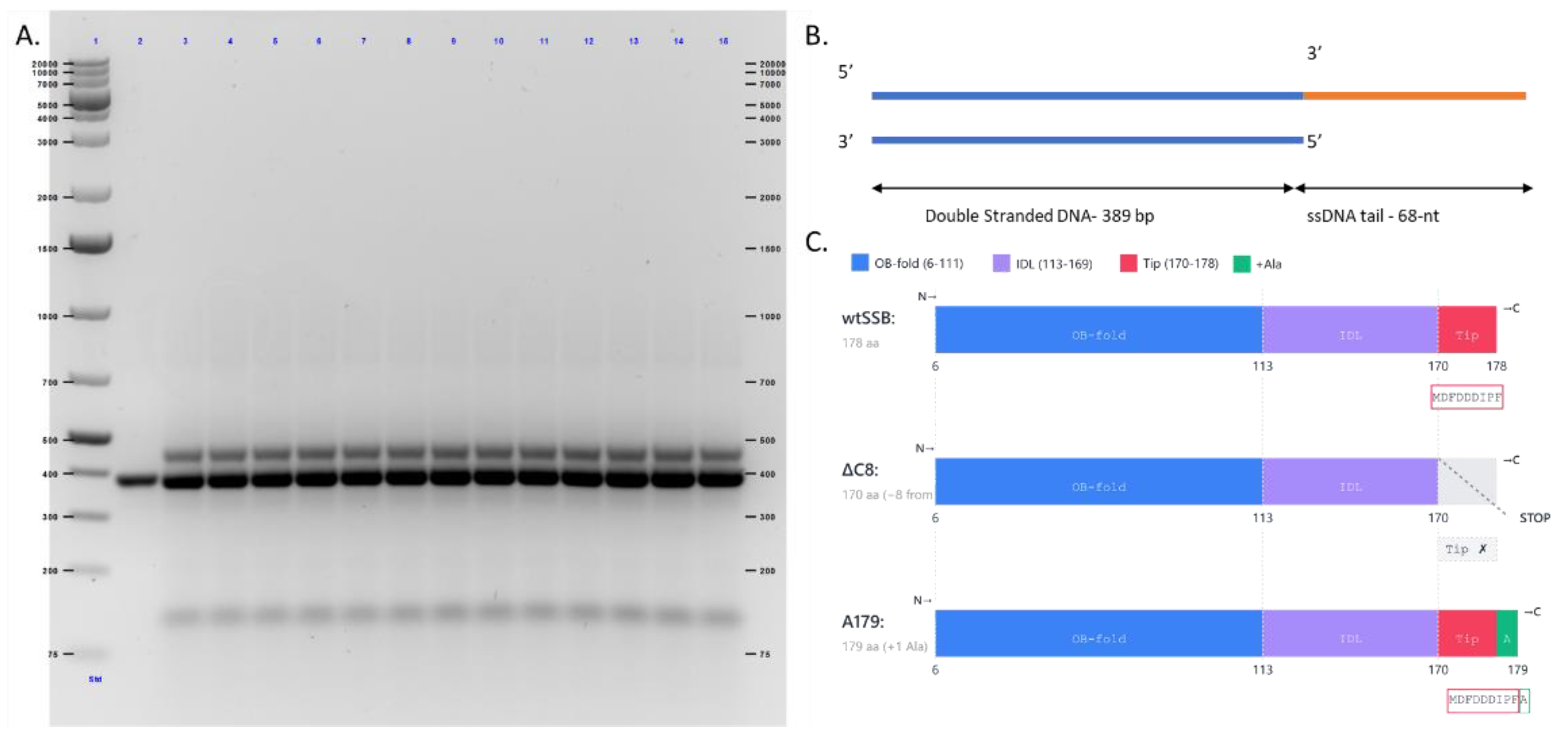
Figure 4.
AFM imaging of ΔC8 on the DNA substrate ± RecO. (A) Representative field and zoomed-in insets, DNA + ΔC8 alone. (B) Representative field and zoomed-in insets, DNA + ΔC8 + RecO. Scale bars, 200 nm. (C) Fraction of DNA molecules scored as SSB-bound vs. free, ΔC8 alone vs. ΔC8 + RecO.
Figure 4.
AFM imaging of ΔC8 on the DNA substrate ± RecO. (A) Representative field and zoomed-in insets, DNA + ΔC8 alone. (B) Representative field and zoomed-in insets, DNA + ΔC8 + RecO. Scale bars, 200 nm. (C) Fraction of DNA molecules scored as SSB-bound vs. free, ΔC8 alone vs. ΔC8 + RecO.
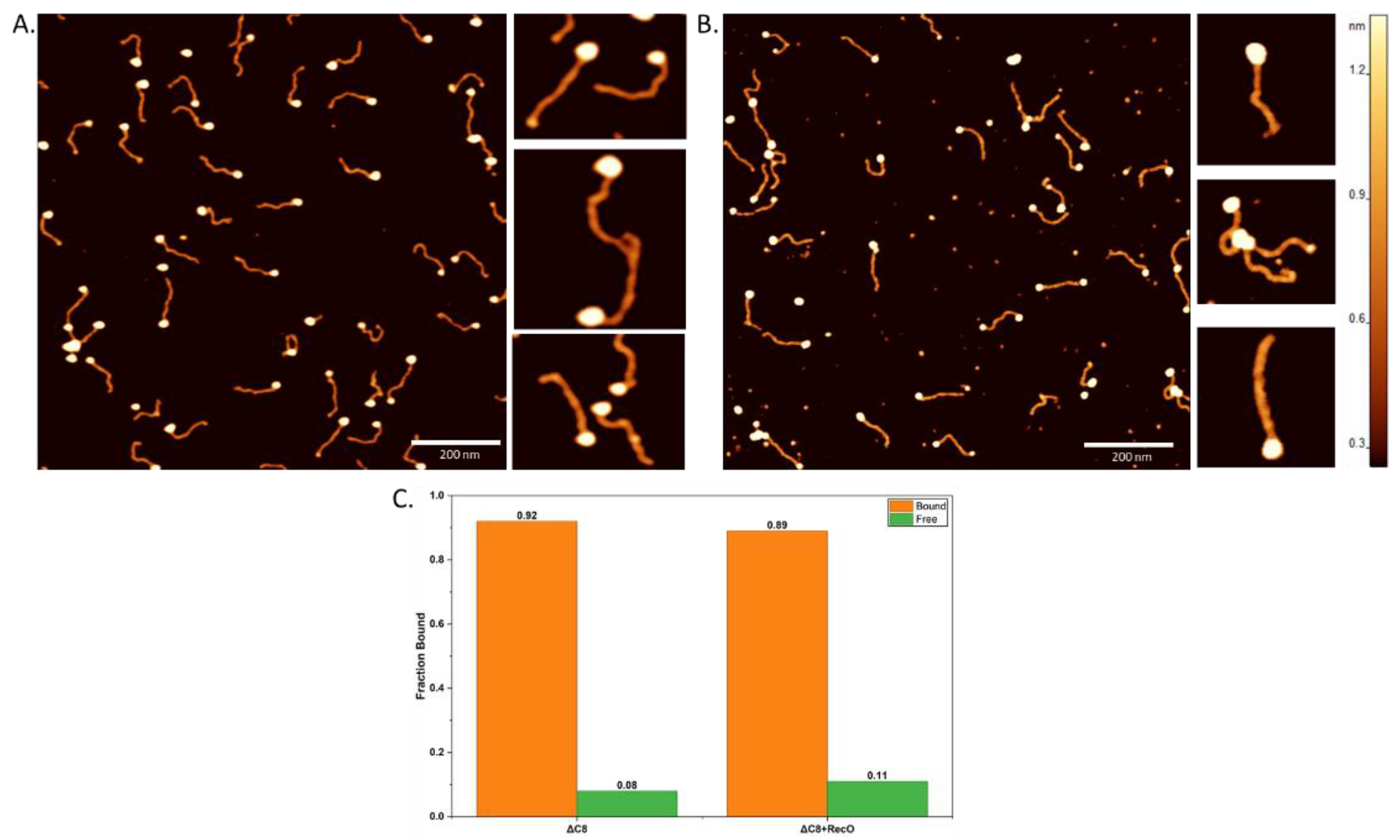
Figure 5.
Height and volume distributions of ΔC8 particles measured by AFM, without (top) and with (bottom) RecO. Data were fit to a two-population Gaussian distribution. Standard deviation was calculated as σ = dx/√(2·ln2). Data are reported as mean ± SD. The R2 for all the fits was ≥ 0.95.
Figure 5.
Height and volume distributions of ΔC8 particles measured by AFM, without (top) and with (bottom) RecO. Data were fit to a two-population Gaussian distribution. Standard deviation was calculated as σ = dx/√(2·ln2). Data are reported as mean ± SD. The R2 for all the fits was ≥ 0.95.
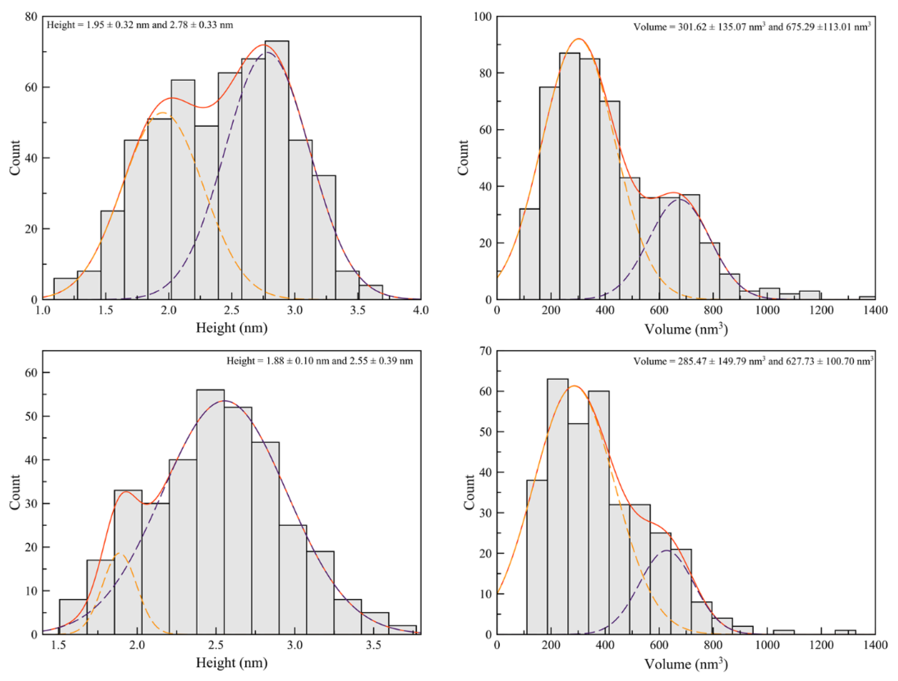
Figure 6.
AFM imaging of A179 on the DNA substrate ± RecO. (A) Representative field and zoomed-in insets, DNA + A179 alone. (B) Representative field and zoomed-in insets, DNA + A179 + RecO. Scale bars, 200 nm. (C) Fraction of DNA molecules scored as SSB-bound vs. free, A179 alone vs. A179 + RecO.
Figure 6.
AFM imaging of A179 on the DNA substrate ± RecO. (A) Representative field and zoomed-in insets, DNA + A179 alone. (B) Representative field and zoomed-in insets, DNA + A179 + RecO. Scale bars, 200 nm. (C) Fraction of DNA molecules scored as SSB-bound vs. free, A179 alone vs. A179 + RecO.
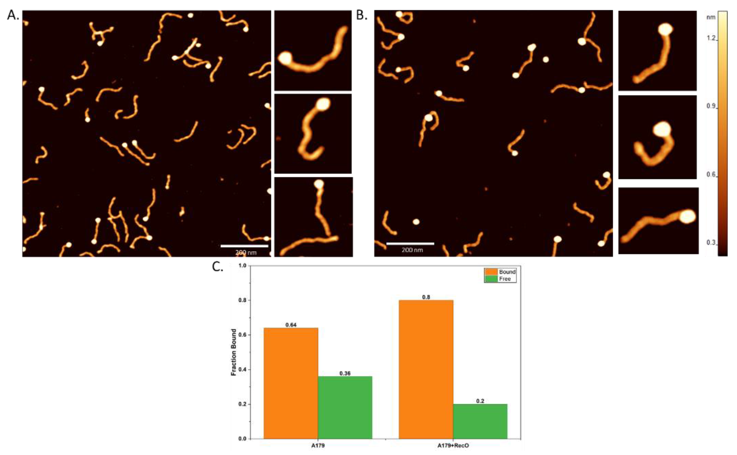
Figure 7.
Height and volume distributions of A179 particles measured by AFM, without (top) and with (bottom) RecO. Data were fit to one or two-population Gaussian distribution. Standard deviation was calculated as σ = dx/√(2·ln2). Data are reported as mean ± SD. The R2 for all the fits was ≥ 0.95, except for the height plots with and without RecO, where R2 ≥ 0.85.
Figure 7.
Height and volume distributions of A179 particles measured by AFM, without (top) and with (bottom) RecO. Data were fit to one or two-population Gaussian distribution. Standard deviation was calculated as σ = dx/√(2·ln2). Data are reported as mean ± SD. The R2 for all the fits was ≥ 0.95, except for the height plots with and without RecO, where R2 ≥ 0.85.
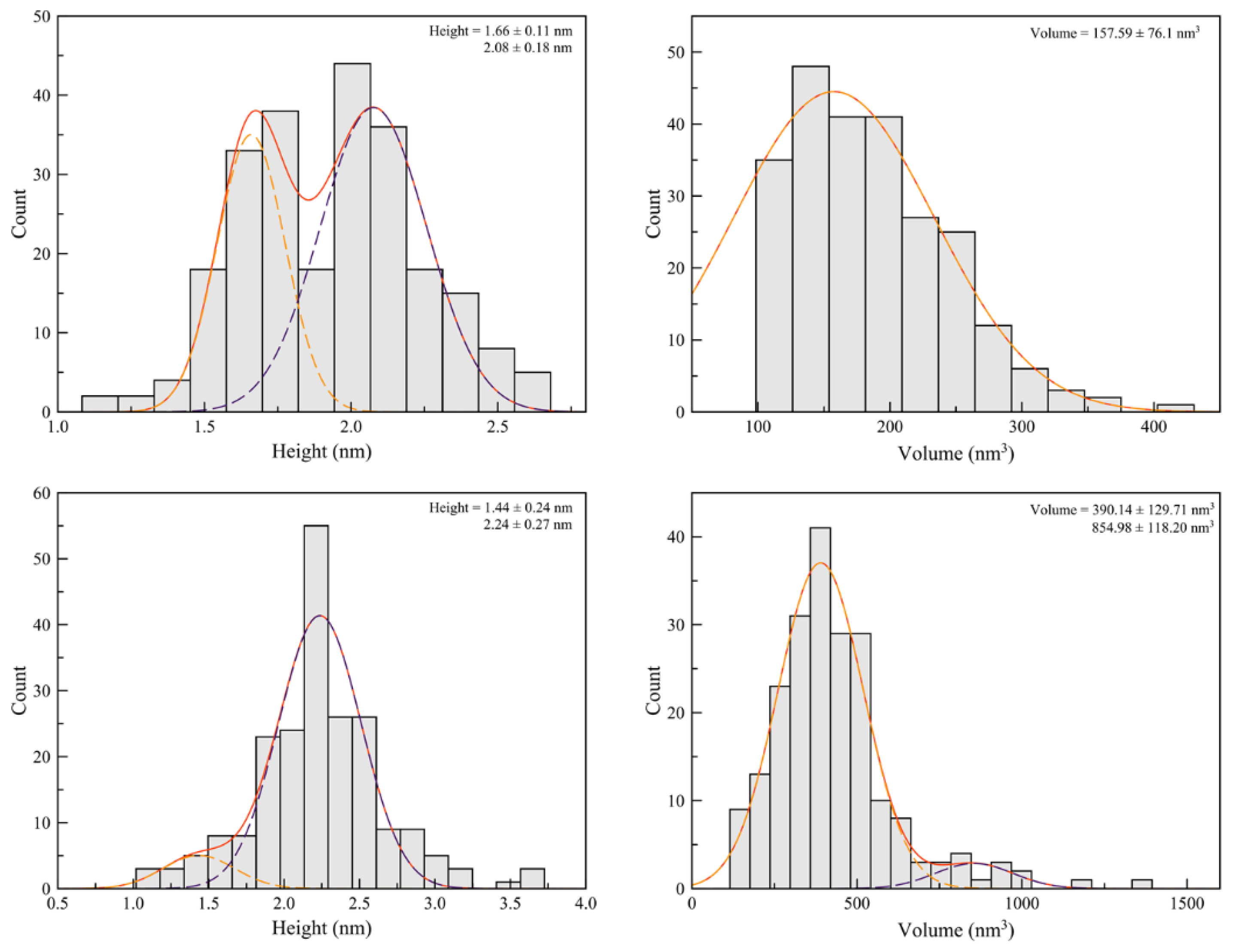
Figure 8.
Molecular dynamics simulations of the RecO-SSB for wtSSB and A179 (n = 3 replicates per variant). (A) RecO surface engaged by the SSB tail: per-residue contact counts (mean ± SEM, shaded bands) across all 242 RecO residues, wtSSB (blue) vs. A179 (purple). Red dotted lines mark native basic-groove residues (R210, R225, R229, K234); blue dotted lines mark hydrophobic-patch residues (W15, F53, V66, L86, L90). (B) SSB tail residues (110–179) contacting RecO, plotted as contact frequency; the acidic tip (residues 171–178) is shaded, and the appended alanine (position 179, A179 only) is marked. (C) Summed contact counts for the native basic-groove residues and the hydrophobic-patch residues, wtSSB vs. A179; bars show mean ± SEM with individual replicate values overlaid as points (n = 3 per variant).
Figure 8.
Molecular dynamics simulations of the RecO-SSB for wtSSB and A179 (n = 3 replicates per variant). (A) RecO surface engaged by the SSB tail: per-residue contact counts (mean ± SEM, shaded bands) across all 242 RecO residues, wtSSB (blue) vs. A179 (purple). Red dotted lines mark native basic-groove residues (R210, R225, R229, K234); blue dotted lines mark hydrophobic-patch residues (W15, F53, V66, L86, L90). (B) SSB tail residues (110–179) contacting RecO, plotted as contact frequency; the acidic tip (residues 171–178) is shaded, and the appended alanine (position 179, A179 only) is marked. (C) Summed contact counts for the native basic-groove residues and the hydrophobic-patch residues, wtSSB vs. A179; bars show mean ± SEM with individual replicate values overlaid as points (n = 3 per variant).
Disclaimer/Publisher’s Note: The statements, opinions and data contained in all publications are solely those of the individual author(s) and contributor(s) and not of MDPI and/or the editor(s). MDPI and/or the editor(s) disclaim responsibility for any injury to people or property resulting from any ideas, methods, instructions or products referred to in the content. |
© 2026 by the authors. Licensee MDPI, Basel, Switzerland. This article is an open access article distributed under the terms and conditions of the Creative Commons Attribution (CC BY) license (http://creativecommons.org/licenses/by/4.0/).
Copyright: This open access article is published under a Creative Commons CC BY 4.0 license, which permit the free download, distribution, and reuse, provided that the author and preprint are cited in any reuse.