
Submitted:
08 August 2026
Posted:
10 August 2026
You are already at the latest version
Abstract
Chiral plasmonic nanostructures have attracted considerable attention due to their remarkable optical behaviors, which vary significantly across spatial dimensions. By integrating plasmonic effects with complementary functionalities, researchers are uncovering entirely new classes of chiroptical materials with capabilities beyond conventional systems. These materials hold immense promise in diverse domains such as biosensing, asymmetric catalysis, and medical technologies. Recent progress in tailoring structural geometries and fine-tuning optical responses has further expanded the application scope of intrinsically chiral plasmonic systems. A wide range of designs has already been fabricated, enabling scientists to probe novel physical effects and practical uses. In this context, the present review highlights the different architectures of chiral plasmonic nanomaterials, explores the mechanisms underlying chirality generation, and surveys their roles in enantioselective detection, catalytic transformations, and biomedical applications. Additionally, we address current obstacles in synthesis and utilization while outlining strategic approaches and future directions for advancing these multifunctional materials.
Keywords:
chirality
; chiral nanoparticles
; biosensing
; enantioselective detection
; bioimaging
; therapy
1. Introduction
Chirality is a fundamental characteristic of matter that is widely observed in both natural and synthetic systems [1,2,3]. A structure is considered chiral when it cannot be superimposed onto its mirror image. This phenomenon plays a central role in biological processes, as many essential biomolecules including DNA, proteins, amino acids, and polysaccharides possess inherent chirality. Molecular handedness governs numerous biological interactions, influencing molecular recognition, catalytic activity, and physiological function [4,5]. Consequently, understanding and controlling chirality has become increasingly important across chemistry, materials science, and biomedicine.
The emergence of nanotechnology has expanded the significance of chirality beyond molecular systems to nanoscale materials [6,7]. Compared with their molecular counterparts, chiral nanomaterials often exhibit amplified chiroptical responses arising from their unique size- and shape-dependent optical properties. Advances in nanoparticle synthesis have enabled precise control over composition, morphology, and hierarchical organization, allowing the fabrication of chiral nanostructures with tunable optical characteristics [8,9,10]. Chiral behavior has now been demonstrated in a wide range of inorganic nanomaterials, including noble metals [11,12], semiconductors [13,14], metal oxides [15,16], and their assembled architectures [17,18]. These materials display strong circular dichroism and related chiroptical phenomena, creating opportunities in enantiomeric recognition [19,20], asymmetric catalysis [21,22], biosensing [23,24], molecular imaging [25,26], polarization optics [27,28], and photonic technologies [29,30].
Among the various inorganic nanomaterials, noble metal nanostructures have attracted particular attention because of their exceptional plasmonic properties [31]. Gold, silver, platinum, palladium, rhodium, ruthenium, iridium, and osmium support localized surface plasmon resonance (LSPR), which originates from the collective oscillation of conduction electrons upon interaction with incident light. The wavelength, intensity, and spectral characteristics of LSPR are highly sensitive to nanoparticle size, geometry, composition, and the surrounding dielectric environment. This tunability provides a powerful platform for engineering optical responses with high sensitivity and selectivity. The strong plasmonic characteristics of noble metal nanomaterials have enabled numerous applications in analytical science and biomedicine [32]. Local electromagnetic field enhancement associated with LSPR significantly improves optical signal generation, facilitating highly sensitive and label-free detection of biomolecular interactions while preserving the native state of biological targets. In addition, efficient absorption of near-infrared light allows these nanomaterials to convert optical energy into heat with remarkable efficiency, making them attractive candidates for photothermal therapy, multimodal imaging, and image-guided therapeutic interventions [33,34]. The combination of plasmonic enhancement and structural chirality further broadens their potential by enabling polarization-dependent optical responses and improved interactions with chiral biological systems [35].
This review provides a comprehensive overview of the design principles, chiroptical properties, and biomedical applications of chiral noble metal nanomaterials. We first discuss the fundamental mechanisms responsible for their optical activity and then systematically classify the origins of chirality into three structural levels. The first category, atomic-level chirality, arises from chiral ligands, asymmetric surface coordination, or intrinsic stereochemical arrangements within metal nanoclusters, where ligand–metal orbital interactions generate chiroptical activity. The second, nanoscale geometric chirality, originates from asymmetric nanoparticle morphologies, including helical, twisted, and anisotropically grown structures, which exhibit pronounced plasmonic circular dichroism and circularly polarized luminescence. The third category, hierarchical assembly-induced chirality, emerges from the organization of nanoparticles into larger chiral superstructures directed by templates such as DNA origami, chiral polymers, peptides, or cellulose nanocrystals, where collective plasmonic coupling produces enhanced chiral optical responses.
Unlike previous reviews that primarily emphasize synthetic methodologies or individual chiroptical phenomena, this article integrates the structural origin of chirality with plasmonic behavior and biomedical function within a unified framework. By correlating chirality across atomic, nanoscale, and hierarchical levels with their optical and biological performance, we provide a systematic perspective for the rational design of chiral plasmonic nanomaterials. We further summarize recent advances in biosensing, enantioselective recognition and separation, bioimaging, photothermal therapy, and cancer theranostics, while critically discussing the remaining challenges and future opportunities for translating these materials into practical biomedical technologies.
2. Optical properties of Chiral NPs
2.1. Localized Surface Plasmon Resonance
Localized surface plasmon resonance (LSPR) is a defining optical property of noble metal nanoparticles, arising from the collective oscillation of conduction electrons under incident electromagnetic radiation. This phenomenon produces characteristic absorption bands in the UV–visible–NIR region, whose spectral position and intensity are governed by nanoparticle size, shape, composition, and the surrounding dielectric environment. Consequently, precise control over these structural parameters enables systematic tuning of plasmonic responses (Figure 1A,B) [36].
Advances in nanofabrication have enabled the synthesis of highly monodisperse noble metal nanostructures with diverse morphologies, including nanospheres, nanorods, nanosheets, nanocones, cubes, and octahedra, each exhibiting distinct LSPR characteristics [37]. Among these, gold nanoparticles are the most extensively investigated for biomedical applications. Increasing particle size shifts the LSPR band toward longer wavelengths while enhancing plasmonic intensity; for example, the LSPR peak of gold nanoparticles red shifts from approximately 521 to 806 nm as the particle diameter increases from 24 to 221 nm. Interactions between noble metal nanoparticles and chiral molecules generate chiroptical responses at the LSPR wavelength, with signal intensity generally increasing as particle size increases (Figure 1C) [37]. Nanoparticle morphology provides an additional level of control over plasmonic behavior. In gold nanorods (GNRs), increasing the aspect ratio progressively red-shifts the longitudinal LSPR into the near-infrared region while simultaneously strengthening the circular dichroism (CD) response. As a result, the CD signal can be tuned from approximately 550 nm to beyond 900 nm (Figure 1D,E) [38]. This enhancement has been attributed to two synergistic effects: stronger longitudinal plasmon oscillations that promote more pronounced chiral currents within individual nanorods and enhanced near-field coupling between adjacent GNRs, which creates intense electromagnetic “hot spots” that further amplify the chiroptical response.
2.2. Circular dichroism properties
Chirality is a fundamental structural property in which an object or molecule cannot be superimposed on its mirror image. This asymmetry gives rise to unique optical phenomena, including circular dichroism (CD) and circularly polarized luminescence (CPL), which are widely employed to characterize chiral materials [39]. Among these techniques, CD spectroscopy is the most used method for probing molecular and nanoscale chirality. CD measures the differential absorption of left- and right-handed circularly polarized light (LCP and RCP), which can be expressed as:
where and denote the absorbance of left- and right-handed circularly polarized light, respectively.
To quantitatively evaluate the intrinsic chiral response of a material, the dissymmetry factor (g-factor) is commonly employed:
Unlike the raw CD signal, the g-factor normalizes the differential absorption to the average absorbance, eliminating the influence of sample concentration and optical path length. Consequently, it provides a direct measure of the inherent chiral asymmetry of a material. The magnitude of the g-factor varies significantly among different classes of chiral systems. Small organic molecules, including amino acids and peptides, typically exhibit relatively weak chiroptical responses with g-factors in the range of 10⁻⁴–10⁻³ [40]. However, hierarchical self-assembly into supramolecular architectures such as helical fibers or twisted ribbons can substantially amplify the chiral response, increasing g-factors to approximately 10⁻² [41]. Similarly, inorganic nanomaterials functionalized with chiral ligands generally exhibit g-factors ranging from 10⁻⁴ to 10⁻², whereas plasmonic nanoparticle assemblies possessing collective chiral arrangements frequently achieve values on the order of 10⁻² due to enhanced electromagnetic coupling [42].
Chiral emission is characterized by the luminescence dissymmetry factor () [43], which describes the difference in left- and right-handed circularly polarized emission:
where and represent the emission intensities of left- and right-handed circularly polarized light, respectively. Although provides a quantitative measure of CPL performance, it should be interpreted together with the photoluminescence quantum yield [44]. A high alone does not necessarily indicate an efficient chiral emitter, particularly for artificial inorganic chiral nanostructures where emission efficiency is equally critical for practical applications.
3. Origin of Plasmonic Chirality
Chirality describes the geometric property of an object that cannot be superimposed onto its mirror image. Although molecular chirality commonly arises from asymmetric carbon centers, it can also originate from helical molecular conformations or supramolecular arrangements. In plasmonic nanomaterials, chirality extends beyond individual molecules and can emerge through interactions between metallic nanostructures and chiral ligands, ordered nanoparticle assemblies, or intrinsically asymmetric nanostructures. These nanoscale architectures generate much stronger chiroptical responses than most isolated organic molecules because of the unique optical properties associated with localized surface plasmon resonance (LSPR).
Upon light irradiation, conduction electrons within metallic nanoparticles undergo collective oscillations, producing localized surface plasmon resonances that concentrate electromagnetic energy near the nanoparticle surface. These enhanced near fields dramatically strengthen light–matter interactions and amplify the optical response of nearby chiral molecules. As illustrated in Figure 2A, a chiral molecule positioned close to a plasmonic nanoparticle experiences strong electromagnetic coupling, resulting in an enhanced circular dichroism (CD) response compared with the molecule alone [45]. The intensity of this enhancement depends on several factors, including nanoparticle composition, size, morphology, and particularly the separation distance between the molecule and the nanoparticle surface. The enhancement becomes even more pronounced when chiral molecules are located within plasmonic hotspots, which are highly confined electromagnetic fields generated in the nanogaps between adjacent nanoparticles. As schematically shown in Figure 2B, nanoparticle dimers create intense, localized fields that significantly amplify the interaction between circularly polarized light and the chiral molecule [46]. Consequently, hotspot engineering has become an effective strategy for improving the sensitivity of plasmonic chiral sensors and spectroscopic platforms.
Besides molecule-mediated chirality, plasmonic nanoparticles can also acquire optical activity through their spatial organization. Self-assembly of metallic nanoparticles into left- or right-handed helices, twisted chains, or other asymmetric architectures generates collective plasmonic coupling that produces characteristic CD spectra. Figure 2C conceptually illustrates this mechanism, where the three-dimensional arrangement of nanoparticles dictates the handedness and magnitude of the optical response [46]. Unlike molecular chirality, these collective plasmonic modes can be readily tuned by controlling interparticle spacing, assembly geometry, and nanoparticle dimensions. An alternative approach involves fabricating nanoparticles with intrinsically chiral morphologies. Twisted, helical, or asymmetrically grown metallic nanostructures exhibit inherent optical activity without requiring chiral molecular ligands or templates. As represented in Figure 2D, the geometric asymmetry of these nanoparticles directly determines their interaction with circularly polarized light, leading to strong and tunable chiroptical signals [47]. Advances in synthetic methodologies have enabled precise control over nanoparticle morphology, allowing the design of chiral plasmonic systems with tailored optical properties for biosensing, imaging, catalysis, and photonic applications [48].
Because the optical response of complex plasmonic architectures is governed by intricate electromagnetic interactions, computational modeling has become an indispensable tool for understanding and optimizing chiral nanostructures [49]. Numerical approaches, including finite-difference time-domain (FDTD), finite-element method (FEM), boundary element method (BEM), and discrete dipole approximation (DDA), are widely employed to predict electromagnetic field distributions, plasmonic coupling, and CD spectra [50,51]. These simulations complement experimental investigations by revealing how structural parameters influence chiroptical activity and by guiding the rational design of next generation plasmonic nanomaterials. Overall, plasmonic chirality originates primarily from three complementary mechanisms: (i) electromagnetic coupling between plasmonic nanoparticles and chiral molecules, (ii) collective plasmonic interactions within chiral nanoparticle assemblies, and (iii) intrinsically chiral nanoparticle geometries [48]. Together, these mechanisms provide versatile routes for amplifying chiroptical responses and have established plasmonic nanostructures as promising platforms for molecular recognition, biosensing, biomedical imaging, and light-mediated therapeutic applications.
4. Chiral Plasmonic Nanoparticles Design Strategies
Chiral plasmonic nanoparticles (NPs), particularly those composed of noble metals such as gold (Au), silver (Ag), and copper (Cu), have emerged as an important class of nanomaterials owing to their unique optical responses arising LSPR [25,52]. Unlike conventional plasmonic nanomaterials, chiral plasmonic nanoparticles interact differently with left- and right-handed circularly polarized light, giving rise to measurable circular dichroism (CD) signals. This differential optical response results from asymmetric light–matter interactions and serves as the primary signature of plasmonic chirality. Owing to strong electromagnetic field confinement at the nanoparticle surface, plasmonic systems often exhibit chiroptical responses that are significantly stronger than those observed in small chiral molecules, making them attractive platforms for optical sensing, imaging, and photonic applications.
The origin of plasmonic chirality has been extensively investigated through theoretical and experimental studies. Electromagnetic simulations have demonstrated that adsorption of chiral molecules onto plasmonic nanoparticles induces asymmetric electric fields around the metal surface. These asymmetric fields generate chiral plasmonic currents that modify the localized electromagnetic environment, ultimately producing plasmon-enhanced circular dichroism. The resulting CD intensity is closely associated with the strength of the localized plasmon resonance and is highly sensitive to the orientation, spatial arrangement, and coupling of chiral dipoles near the nanoparticle surface. Consequently, structural modifications that strengthen plasmonic coupling generally lead to enhanced chiroptical responses. Among the various structural parameters, particle geometry plays a dominant role in determining plasmonic chirality. Helical or twisted nanoparticle architectures produce stronger optical dissymmetry than symmetric nanostructures because their three-dimensional asymmetry promotes efficient coupling with circularly polarized light. Increasing the degree of helicity or reducing the spacing between neighboring structural features enhances electromagnetic coupling, leading to stronger CD signals and, in many cases, a red shift of the plasmon resonance. Furthermore, left- and right-handed nanostructures exhibit mirror-image CD spectra, reflecting their opposite chiral configurations.
The optical behavior of chiral plasmonic nanoparticles can be precisely tailored by controlling their composition, size, morphology, and surface chemistry. Considerable progress has been achieved in synthesizing AuNPs with well-defined geometries, including nanospheres, nanorods, nanocubes, nanoprisms, nanotriangles, nanodiscs, octahedra, decahedra, and bipyramids. In parallel, a wide variety of chiral ligands, including amino acids [53,54], peptides [55], glutathione [53], and nucleic acids [56,57], have been employed to direct asymmetric nanoparticle growth or to transfer molecular chirality to plasmonic surfaces (Table 1). These synthetic approaches enable precise control over chiroptical activity across the visible and near-infrared spectral regions.
Current strategies for generating plasmonic chirality generally fall into two major categories [18,65]. The first relies on (i) surface-induced chirality, where chiral biomolecules or organic ligands are immobilized on otherwise achiral plasmonic nanoparticles. The interaction between the chiral ligand and the localized plasmonic field generates asymmetric electromagnetic distributions and produces characteristic CD signals. Although this strategy is straightforward and versatile, the optical activity often depends on the stability of the adsorbed ligands, which may detach under physiological or chemical conditions, reducing long-term chiral performance. The second and increasingly important strategy is the fabrication of (ii) intrinsically chiral plasmonic nanostructures, in which chirality originates directly from the nanoparticle architecture rather than from surface-bound molecules. Seed-mediated growth has become one of the most effective methods for producing such structures. By combining anisotropic metal seeds with chiral directing agents, controlled asymmetric crystal growth can be achieved, resulting in helicoidal, twisted, or other chiral morphologies [66]. Differences in growth kinetics at crystallographic facets with high Miller indices are believed to drive the formation of these enantiomeric structures. Recent advances have further expanded the synthetic toolbox for chiral plasmonic nanomaterials [67]. Circularly polarized light has been successfully employed as an external stimulus to direct the formation of plasmonic nanoparticles with controllable handedness and exceptionally large optical dissymmetry factors [68]. Likewise, hybrid plasmonic–dielectric architectures have demonstrated remarkable chiroptical performance in the near-infrared region through synergistic plasmonic coupling. These developments highlight the versatility of nanoscale engineering for producing chiral plasmonic systems with enhanced optical activity and broaden their potential applications in biosensing, bioimaging, asymmetric catalysis, photothermal therapy, and photonic devices [23,52]. Overall, the rapid evolution of synthetic methodologies and the growing understanding of plasmon–chirality interactions have established chiral plasmonic nanoparticles as a highly tunable class of functional nanomaterials. Continued advances in structural engineering and optical design are expected to further improve their chiroptical performance and accelerate their translation into advanced biomedical and nanophotonic applications.
4.1. Chiral nanomaterials by surface induced chirality/Chiral arrangement
The development of ligand-directed chiral metal nanoclusters has provided an important strategy for generating optical activity in otherwise achiral metallic systems. Early studies demonstrated that coating gold nanoclusters with optically active glutathione (GSH) could successfully transfer molecular chirality to the inorganic framework, establishing a foundation for the design of chiral noble-metal nanomaterials [69]. Since then, this approach has been widely extended to a variety of metal nanoclusters, including gold, silver, copper and palladium, using enantiomerically pure surface ligands. Naturally occurring and synthetic chiral molecules, such as cysteine [53,54], glutathione [70,71], penicillamine [72], N-acetylcysteine [73,74,75], and other tailor-made ligands [76,77], have all been employed to regulate the chiroptical properties of these nanostructures.
The origin of chirality in ligand-protected nanoclusters is generally attributed to strong electronic interactions at the metal–ligand interface. Upon binding to the nanoparticle surface, chiral ligands create an asymmetric local electronic environment that perturbs the electronic states of the otherwise symmetric metal core, resulting in characteristic CD responses. A representative example was reported by Zhu and co-workers, who synthesized atomically precise Au₅₂₅(SR)₁₈ nanoclusters using both R- and S-configured thiol ligands (Figure 3A) [78]. The resulting enantiomeric nanoclusters exhibited mirror-image CD spectra, demonstrating that the observed optical activity originated from electronic coupling between the surface-bound ligands and the outer gold atoms rather than from the metallic core alone (Figure 3B). This ligand-mediated chiral induction is commonly interpreted using the dissymmetric field model, in which the asymmetric electrostatic field generated by the adsorbed chiral molecules perturbs the electronic structure of the metal nanocluster and removes its intrinsic symmetry, thereby producing measurable chiroptical activity. Importantly, the magnitude and even the sign of the CD response are highly dependent on ligand identity, surface binding geometry, and ligand packing. Consequently, modifying the surface chemistry offers a versatile means of tuning nanoparticle chirality. For example, Cathcart and colleagues demonstrated that incorporating mixtures of different chiral ligands on silver nanoclusters enabled controllable modulation of their chiroptical properties, illustrating that ligand composition provides an effective handle for engineering and optimizing optical chirality (Figure 3C) [79]. Use of chiral reagents such as R/S-BINAP can induce chirality in Au and Pd NPs which in turn used for the chiral catalytic application such as hydrosilylation of styrene (Figure 4D) [80]. These NPs showed excellent enantioselective hydrosilylation under mild conditions.
Recent investigations have highlighted the growing potential of chiral metallic nanoparticles in biomedical and biosensing applications. For example, Zhang and co-workers demonstrated that D- and L-cysteine-functionalized gold nanoparticles can serve as efficient nanocarriers for immunoadjuvant delivery [81]. Interestingly, nanoparticles modified with L-cysteine elicited a significantly stronger immune response than their D-cysteine counterparts, producing more than a 66-fold enhancement in anti-ovalbumin (OVA) IgG antibody levels, thereby underscoring the critical role of nanoparticle chirality in regulating immune activation. In another study, Maniappan et al. synthesized L- and D-cysteine-stabilized copper nanoparticles and investigated their ability to discriminate chiral amino acids [82]. Among the analytes examined, L-histidine exhibited a remarkable inversion of the circular dichroism signal upon interaction with the chiral copper nanoparticles, demonstrating their potential as highly sensitive platforms for enantioselective molecular recognition. Chiral palladium nanoparticles were also prepared using such strategy where chiral amino acids such as proline was used as a chiral modifier to make dendritic or cubic palladium nanocrystals. These nanocrystals were used for asymmetric hydrogenation reactions [83]. Binaphthyl-derived chiral phosphoramidite ligands have been employed to stabilize palladium nanoparticles, producing highly enantioselective heterogeneous catalysts for asymmetric Suzuki C–C cross-coupling reactions [84]. The chiral ligand shell creates an asymmetric catalytic environment at the nanoparticle surface, enabling efficient stereo control while maintaining the high catalytic activity and recyclability associated with nanoparticle-based catalysts. This work demonstrates that ligand-engineered metal nanoparticles can combine the advantages of homogeneous chiral catalysts with the robustness of heterogeneous nanocatalysts, highlighting an effective strategy for developing enantioselective nanocatalytic systems.
Direct synthetic approaches offer a straightforward route for producing chiral gold nanoparticles because they generally involve fewer processing steps and relatively short reaction times. These methods enable the simultaneous formation of the nanoparticle core and its chiral characteristics within a single synthesis process, making them attractive for rapid and scalable preparation. Nevertheless, achieving consistent chirality requires precise regulation of reaction parameters, including precursor concentration, ligand chemistry, temperature, and growth kinetics. The complex interplay of these factors often limits reproducibility and makes it challenging to finely tune nanoparticle size, morphology, and chiroptical activity. In addition, directly synthesized chiral nanoparticles may exhibit broader size distributions and reduced structural stability, which can affect the uniformity and long-term performance of their chiral properties.
4.2. Chiral nanomaterials by seed mediated method
Studies have demonstrated that incorporating enantiomerically pure ligands during nanoparticle formation can effectively transfer molecular chirality to plasmonic materials, resulting in significant optical activity [85]. Among these approaches, Lee et al. introduced an amino acid- and peptide-assisted synthetic strategy in which selective adsorption of chiral biomolecules onto specific crystallographic facets guided anisotropic nanoparticle growth [53]. The differential interactions between the ligands and the evolving gold surface promoted the formation of highly twisted helicoidal nanostructures exhibiting intense plasmonic chiroptical properties. The resulting nanoparticles displayed strong CD signals, reaching approximately 0.5 mdeg, with a corresponding dissymmetry factor of nearly 0.2. In this methodology, small gold nanocrystals serve as seeds that are subsequently grown in the presence of cysteine or other chiral molecular precursors, enabling controlled development of well-defined chiral architectures. This synthetic route is commonly referred to as the seed-mediated growth strategy. The formation of these chiral plasmonic nanostructures is governed by the enantioselective interaction between chiral ligands and high-index gold crystal facets, which directs asymmetric crystal growth during a seed-mediated synthesis. Typically, cubic gold nanoparticles enclosed by low-index {100} facets are used as seeds and subsequently overgrown in the presence of chiral amino acids or peptides. Selective adsorption of ligands through thiol, amino, and carboxyl groups modifies the growth kinetics of specific crystallographic planes, resulting in twisted helicoidal nanostructures with pronounced plasmonic circular dichroism. In the absence of chiral ligands, seed overgrowth produces achiral stellated octahedra composed of alternating right- and left-handed {321} facets (Figure 4A). However, the addition of L- or D-cysteine selectively promotes the growth of one chiral domain over the other, generating 432 Helicoid I nanoparticles with twisted edges and nanoscale gaps (Figure 4B-C), exhibiting a dissymmetry factor (g) of approximately 0.03. Replacing cysteine with glutathione alters the crystal growth pathway, leading to a fourfold symmetric pinwheel-like 432 Helicoid II morphology with an improved g-factor of ~0.05 (Figure 4D). Further enhancement was achieved by employing octahedral gold seeds together with glutathione, producing 432 Helicoid III nanoparticles characterized by highly twisted pinwheel architectures and deeper radial nanogaps (Figure 4E). These nanostructures exhibited a significantly stronger chiroptical response, with g-factors reaching ~0.20, which were subsequently increased to 0.31 through optimized multistep growth and improved morphological uniformity. Use of cysteine with octahedral seeds resulted in Helicoid IV type structure (Figure 4F). Extending this Kumar et al. showed the use of various amino acids for the generation of various Helicoid structures and used them for enantioselective recognition of chiral analytes using Surface Enhanced Raman Spectroscopy (SERS) [54].
Despite significant advances in nanoparticle synthesis, the controlled preparation of intrinsically chiral silver (Ag) nanoparticles remains considerably more challenging than that of their gold counterparts [86]. One of the primary obstacles is the formation of chiral high-index crystal facets, characterized by Miller indices where h, k, and l are all different and nonzero. These crystallographic planes are widely regarded as essential structural motifs for generating nanoscale chirality because they provide the asymmetric atomic arrangements required for helical or enantiomorphic surface architectures. However, such facets are rarely observed in silver nanocrystals due to the high surface reactivity of Ag and the limited availability of chiral ligands capable of selectively stabilizing these energetically unfavorable surfaces during crystal growth.
To address these limitations, researchers have explored the use of chiral amino acids and biomolecular ligands, particularly L- and D-cysteine, as structure-directing agents [87,88]. Since the nucleation of silver typically requires rapid reduction, whereas controlled growth of chiral facets favors slower kinetic conditions, seed-mediated growth has become the preferred synthetic strategy. In this approach, preformed metallic seeds most commonly gold nanoparticles—serve as crystallographic templates that enable the gradual epitaxial deposition of silver under milder reaction conditions. Beyond facilitating controlled shell growth, the use of Au seeds also provides a convenient platform for monitoring the evolution of silver deposition and understanding the underlying growth mechanism.
Several studies have demonstrated that cysteine-assisted seed-mediated synthesis can generate Au@Ag core–shell nanostructures exhibiting transient chiral morphologies. A representative example involves the formation of truncated octahedral Au@Ag nanoparticles, where detailed mechanistic investigations revealed that chirality emerges during intermediate growth stages rather than in the final equilibrium morphology [89]. Initially, reduced Ag atoms nucleate heterogeneously on the seed surface, producing irregular protrusions and anisotropic surface features (Figure 5A). As silver deposition proceeds and the precursor concentration gradually decreases, particle growth slows and ultimately terminates when the available Ag ions are exhausted (Figure 5B,C). During this evolution, selective adsorption of cysteine onto specific crystallographic planes alters the relative growth rates of different facets, temporarily stabilizing asymmetric truncated geometries before the system evolves toward more thermodynamically favored structures (Figure 5C). Although the fully developed particles often approach highly symmetric octahedral morphologies, these intermediate truncated nanostructures retain distinct chiral characteristics arising from enantioselective ligand–surface interactions. The mechanistic insights obtained from Au@Ag systems also suggest that similar growth pathways could be realized using silver seeds, enabling the direct synthesis of entirely silver-based chiral nanoparticles without a gold core. Furthermore, the degree of structural asymmetry appears to depend strongly on the geometric characteristics of the initial seed. Seeds possessing larger surface areas provide more nucleation sites and allow localized growth anisotropy to persist under a finite precursor supply, while elongated seeds with higher aspect ratios inherently promote asymmetric deposition. Consequently, the magnitude of nanoparticle dissymmetry is expected to increase with both the available seed surface area and the seed aspect ratio. Experimental observations have consistently supported this relationship, demonstrating that careful control of seed geometry provides an effective strategy for tuning the chirality and optical activity of silver-based nanostructures (Figure 5D).
Seed-mediated synthesis is a powerful strategy for producing chiral nanomaterials with precise control over size, morphology, and surface structure. By separating nucleation from growth, it enables the formation of well-defined asymmetric nanostructures with tunable optical and catalytic properties. However, the method remains highly sensitive to reaction conditions, seed quality, and ligand chemistry, making reproducibility and large-scale production challenging. Future efforts should focus on improving synthetic robustness, scalability, and enantiomeric control to accelerate practical applications.
4.3. Chiral nanomaterials by template assisted synthesize or assemblies
Chiral soft templates, including self-assembled micelles [90], DNA [91], and biopolymer scaffolds [92], provide an effective platform for directing the growth of well-defined helical and superlattice nanostructures. Their structural organization enables the fabrication of plasmonic nanomaterials with strong collective chiroptical responses, making them attractive for applications in surface-enhanced Raman scattering (SERS), optical metamaterials, and chiral bioimaging. A notable example is the work by Liz-Marzán and co-workers, who employed chiral surfactant-derived micelles as growth-directing templates for seed-mediated synthesis of anisotropic gold nanostructures [58]. The helical micellar assemblies guided asymmetric metal deposition, producing grooved chiral morphologies with exceptionally strong circular dichroism, reaching g-factors of approximately 0.2 in the near-infrared region. The pronounced optical activity originated from the twisted plasmonic architecture of the nanoparticles. Furthermore, modulation of the concentration of chiral ligands and other growth parameters during seed-mediated synthesis enabled control over, and even reversal of, the plasmonic chiral response [93].
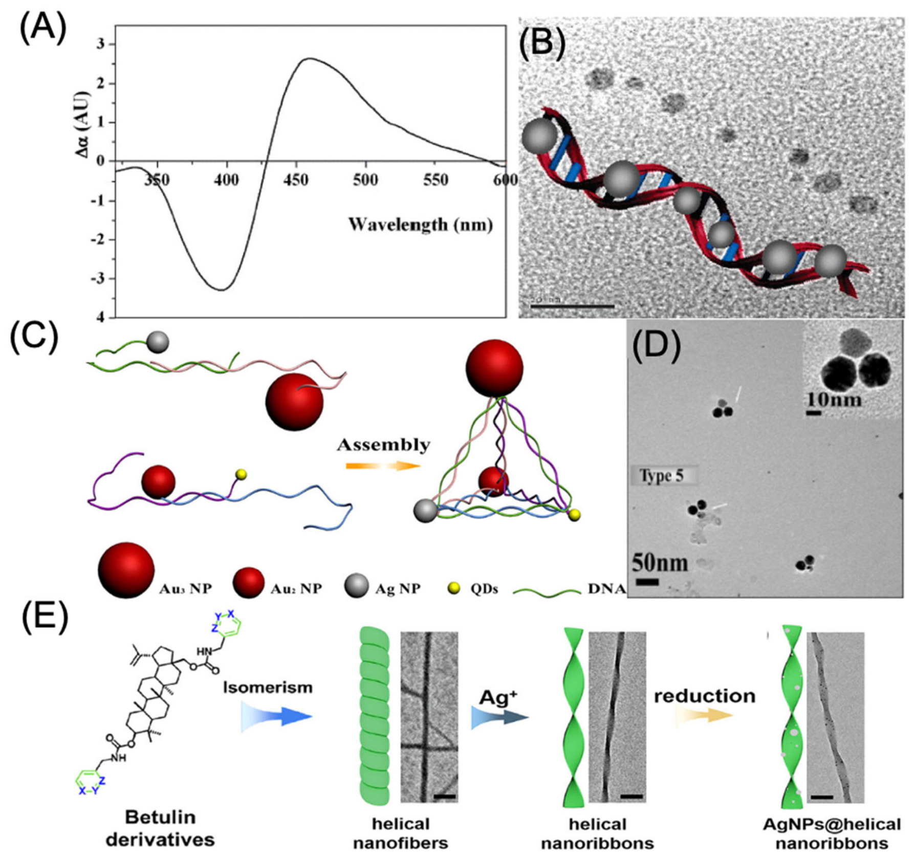
Biological molecules have emerged as versatile templates for the fabrication of chiral nanomaterials because of their intrinsic chirality, biocompatibility, and ability to direct nanoparticle assembly under mild conditions. Among these, DNA has emerged as one of the most versatile scaffolds because of its predictable helical structure and programmable molecular recognition [94]. While DNA-directed synthesis of chiral Au nanoparticles was first demonstrated in the late 1990s, the preparation of DNA-templated chiral Ag nanoparticles was achieved several years later owing to the greater synthetic complexity associated with silver. In a typical approach, Ag⁺ ions are coordinated along double-stranded DNA and subsequently reduced to generate ~5 nm Ag nanoparticles uniformly distributed along the DNA helix (Figure 6A) [95]. The periodic arrangement of Ag nanoparticles within the helical framework promotes strong plasmonic coupling, producing characteristic bisignate circular dichroism signals in the visible region (Figure 6B). Beyond serving as a structural template, DNA also functions as a programmable assembly ligand that enables the hierarchical organization of Ag nanoparticles into complex three-dimensional chiral architectures. DNA-functionalized nanoparticles can undergo sequence-specific hybridization and thermal annealing to form ordered superstructures, including chiral pyramids and other enantiomeric assemblies with precisely controlled handedness (Figure 6C-D) [96]. This strategy provides exceptional spatial precision and enhanced chiroptical activity, with anisotropy (g) factors approaching 10⁻², significantly exceeding those typically observed for small chiral molecules. Despite these advantages, DNA-directed assembly remains limited by the high cost of synthetic DNA, sensitivity to temperature and sample purity, and the need for carefully controlled experimental conditions. Besides DNA, naturally derived chiral biomolecules have also been employed as templates for silver nanostructure growth. Chiral triterpenoid-based ligands can self-assemble with Ag⁺ ions into helical supramolecular frameworks that serve as templates for nanoparticle formation (Figure 6E) [97]. Upon reduction, Ag nanoparticles nucleate along these helical scaffolds while preserving the underlying chiral organization, producing stable plasmonic nanostructures with pronounced CD responses. This biomolecule-assisted strategy provides a simple and mild synthetic route with good structural control; however, precise optimization of ligand concentration, metal-ion ratio, and irradiation conditions is essential to achieve reproducible chirality and optical performance.
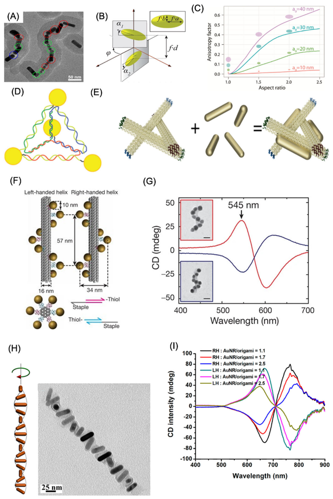
Template-directed assembly has emerged as an effective strategy for constructing plasmonic nanostructures with well-defined chirality [98]. Liu et al. demonstrated that chiral templates can direct the formation of end-to-end crossed (EEX) nanoparticle assemblies, producing pronounced CD responses (Figure 7A) [99]. Likewise, Liz-Marzán and co-workers fabricated crossed dimers of gold nanorods (Figure 7B-C), where the asymmetric CD spectra originated from plasmonic coupling between the closely positioned nanorods [100] . Computational simulations closely matched the experimental CD profiles, confirming the role of electromagnetic interactions in generating the observed optical activity. DNA has become one of the most versatile scaffolding materials for organizing plasmonic nanoparticles with nanometer-scale precision. By exploiting the inherent tetrahedral symmetry of DNA frameworks, researchers assembled pyramidal nanoparticle architectures in which the spatial arrangement and orientation of nanocrystals could be precisely controlled, leading to well-defined chiral geometries (Figure 7D) [101]. Beyond structural organization, DNA-mediated assembly has also been extended to biosensing applications. For example, Xu et al. employed antibody–antigen interactions to bridge silver and gold nanoparticles into chiral AgNP–AuNP dimers, enabling sensitive detection of environmental contaminants such as microcystin-LR as well as cancer-associated biomarkers [102].
Recent advances in DNA origami have further expanded the ability to engineer sophisticated chiral plasmonic systems by enabling programmable placement of nanoparticles with exceptional spatial accuracy [103]. Using a left-handed DNA origami template composed of three interconnected 14-helix bundles (14HBs), gold nanorods were assembled into chiral architectures through hybridization between A19-functionalized DNA strands and poly-T-modified nanorods (Figure 7E) [104]. Connector oligonucleotides linked individual origami units into larger plasmonic assemblies with controlled handedness. Building on this concept, Kuzyk and co-workers employed DNA origami to fold long single-stranded DNA scaffolds into predefined two- and three-dimensional architectures that served as templates for helical gold nanoparticle assemblies (Figure 7F) [105]. These structures exhibited strong circular dichroism and optical rotatory dispersion throughout the visible spectrum arising from collective plasmonic interactions among the helically arranged nanoparticles (Figure 7G), The helical geometry promotes directional propagation of coupled plasmon modes, resulting in preferential interaction with circularly polarized light that matches the handedness of the assembly. Importantly, theoretical calculations accurately reproduced both the spectral positions and intensities observed experimentally, confirming the structural origin of the chiroptical response. Wang et al. further demonstrated programmable assembly of anisotropic helical superstructures by positioning gold nanorods on two-dimensional DNA origami templates decorated with strategically arranged 15-nucleotide capture strands in an X-shaped configuration [106]. The resulting nanorod helices displayed exceptionally strong chiroptical activity, with anisotropy factors reaching approximately 0.02 (Figure 7H-I). Beyond helical architectures, DNA origami has also enabled the fabrication of numerous other chiral plasmonic nanostructures, including gold nanoparticle tetramers [107], bipyramidal assemblies [108], gold nanorod dimers [109], and a variety of other programmable nanoscale configurations.
Figure 6.
DNA- and biomolecule-assisted fabrication of chiral silver nanostructures. (A) Circular dichroism (CD) spectra of Ag nanoparticles synthesized directly on DNA templates, demonstrating their chiroptical response. (B) Representative TEM image showing the linear arrangement of Ag nanocrystals along the DNA scaffold. (C) Schematic illustration of the DNA-guided assembly process used to construct chiral nanoparticle pyramids, together with a representative TEM image. (D) TEM images of self-assembled pyramidal nanostructures composed of the S-enantiomer of Au₂, Au₃, Ag, and quantum dots (QDs). (E) Schematic representation of the biomolecule-templated synthesis of AgNP-decorated helical nanoribbons (AgNPs@helical nanoribbons). Panels (A, B) reproduced with permission from [95]; Copyright 2006 American Chemical Society. Panels (C, D) reproduced with permission from [96];Copyright 2012 American Chemical Society. Panel (E) reproduced with permission from [97]; Copyright 2022 American Chemical Society.
Figure 6.
DNA- and biomolecule-assisted fabrication of chiral silver nanostructures. (A) Circular dichroism (CD) spectra of Ag nanoparticles synthesized directly on DNA templates, demonstrating their chiroptical response. (B) Representative TEM image showing the linear arrangement of Ag nanocrystals along the DNA scaffold. (C) Schematic illustration of the DNA-guided assembly process used to construct chiral nanoparticle pyramids, together with a representative TEM image. (D) TEM images of self-assembled pyramidal nanostructures composed of the S-enantiomer of Au₂, Au₃, Ag, and quantum dots (QDs). (E) Schematic representation of the biomolecule-templated synthesis of AgNP-decorated helical nanoribbons (AgNPs@helical nanoribbons). Panels (A, B) reproduced with permission from [95]; Copyright 2006 American Chemical Society. Panels (C, D) reproduced with permission from [96];Copyright 2012 American Chemical Society. Panel (E) reproduced with permission from [97]; Copyright 2022 American Chemical Society.
Beyond molecular templates, inorganic chiral templates provide an effective route for constructing plasmonic nanostructures with well-defined three-dimensional architectures. For example, Correa-Duarte and co-workers developed chiral silica nanoribbons as scaffolds for assembling TiO₂ and Au nanoparticles into hybrid photocatalytic systems [110]. Following amine functionalization, spherical Au nanoparticles were covalently anchored onto the twisted silica surface, generating chiral plasmonic assemblies. The helical arrangement of Au nanoparticles promoted strong plasmonic coupling and polarization-dependent photocatalytic activity under circularly polarized light, where hot-carrier generation was governed by the light helicity. Cheng et al. fabricated three-dimensional helical plasmonic nanostructures by assembling AuNPs on silica nanohelices [111]. The chiroptical response was strongly influenced by both the size and spatial arrangement of the AuNPs, yielding an anisotropy factor (g-factor) of ~1 × 10⁻⁴ at the SPR wavelength, approximately ten times higher than previously reported values. Another widely adopted strategy is in situ nucleation and growth, where metal precursors are adsorbed onto preformed chiral templates and subsequently reduced to generate chiral nanostructures. Using this approach, Che et al. synthesized chiral mesoporous silica templates that directed the growth of Ag nanoparticles with tunable CD responses, producing Ag–silica composites exhibiting strong circular dichroism in the visible region [112].
Figure 7.
Template-assisted assembly strategies for chiral plasmonic nanostructures. (A) TEM image of the end-to-end crossed (EEX) gold nanostructure. (B) Schematic of a gold nanorod (AuNR) dimer. (C) Coupled dipole simulations showing the anisotropy factor (g) of AuNR dimers as a function of particle size and aspect ratio. (D) DNA nanocrystal pyramid used as a template for chiral plasmonic assembly. (E) Assembly of AuNRs on monomeric DNA origami structures. (F) Schematic of left- and right-handed DNA origami-templated AuNR nanohelices. (G) Enhanced circular dichroism (CD) response resulting from collective plasmonic coupling in nanoparticle assemblies. (H) Structural models, TEM images, and (I) CD spectra of right- and left-handed AuNR helices with different AuNR/origami ratios, demonstrating the influence of nanoparticle loading on chiroptical activity. Panel A reproduced with permission from [99]; Copyright 2017, American Chemical Society. Panel B-C reproduced with permission from [100]; Copyright 2011, American Chemical Society. Panel D, reproduced with permission from [101]; Copyright 2009, American Chemical Society. Panel E reproduced with permission from [104]; Copyright 2025, American Chemical Society. Panel F-G reproduced with permission from [105]; Copyright 2013, Nature Publishing group. Panel H-I is reproduced from [106]; Copyright 2015, American Chemical Society.
Figure 7.
Template-assisted assembly strategies for chiral plasmonic nanostructures. (A) TEM image of the end-to-end crossed (EEX) gold nanostructure. (B) Schematic of a gold nanorod (AuNR) dimer. (C) Coupled dipole simulations showing the anisotropy factor (g) of AuNR dimers as a function of particle size and aspect ratio. (D) DNA nanocrystal pyramid used as a template for chiral plasmonic assembly. (E) Assembly of AuNRs on monomeric DNA origami structures. (F) Schematic of left- and right-handed DNA origami-templated AuNR nanohelices. (G) Enhanced circular dichroism (CD) response resulting from collective plasmonic coupling in nanoparticle assemblies. (H) Structural models, TEM images, and (I) CD spectra of right- and left-handed AuNR helices with different AuNR/origami ratios, demonstrating the influence of nanoparticle loading on chiroptical activity. Panel A reproduced with permission from [99]; Copyright 2017, American Chemical Society. Panel B-C reproduced with permission from [100]; Copyright 2011, American Chemical Society. Panel D, reproduced with permission from [101]; Copyright 2009, American Chemical Society. Panel E reproduced with permission from [104]; Copyright 2025, American Chemical Society. Panel F-G reproduced with permission from [105]; Copyright 2013, Nature Publishing group. Panel H-I is reproduced from [106]; Copyright 2015, American Chemical Society.
Template-assisted strategies offer exceptional control over chiral nanoparticle assembly and reproducible optical properties but are often limited by complex fabrication procedures, poor scalability, and high production costs.
4.4. Chiral nanomaterials by nanofilms
Compared with individual chiral plasmonic nanoparticles, chiral nanofilms exhibit enhanced collective optical responses due to long-range nanoparticle ordering, leading to stronger CD and improved light–matter interactions [11]. These unique properties make them attractive for optical sensing, polarization control, and photonic devices. Fabrication strategies are generally classified into bottom-up and top-down approaches. Bottom-up assembly relies on solution-based methods such as interfacial self-assembly and the Langmuir–Schaefer technique, enabling scalable fabrication of ordered chiral films. For example, Xu et al. assembled L/D-phenylalanine-functionalized Au nanoparticles into chiral monolayers at a liquid–liquid interface using the Langmuir–Schaefer method (Figure 8A-B) [113]. The resulting nanofilms exhibited significantly enhanced CD signals compared with dispersed nanoparticles, demonstrating that collective nanoparticle organization amplifies chiroptical activity (Figure 8C). Similarly, Nam and co-workers employed helicoidal Au nanoparticles as building blocks to fabricate highly ordered two-dimensional helical crystals through dip-coating and directed assembly (Figure 8D–F) [114]. The resulting superstructures showed approximately 2.5-fold enhancement in CD intensity due to collective plasmonic coupling and were further extended into multilayer chiral Au nanofilms. Top-down strategies provide superior structural precision through techniques such as in situ growth [115], electron-beam lithography [116], and holographic lithography [117]. Che and co-workers synthesized chiral Au, Ag, and Cu nanofilms by directing the helical assembly of metal nanoparticles using N-acetyl-L/D-cysteine, producing inorganic chiral films with morphology-dependent plasmonic and scattering-induced optical activity [118]. In another study, Kadodwala and co-workers fabricated periodic fylfot-shaped Au nanostructures using electron-beam lithography [116]. These intrinsically chiral nanostructures exhibited strong CD responses arising from structural asymmetry and localized surface plasmon resonance, although practical implementation is limited by fabrication complexity and high production cost. Overall, bottom-up approaches offer scalable and cost-effective fabrication of large-area chiral nanofilms, whereas top-down techniques provide exceptional structural precision and reproducibility. Nevertheless, improving scalability while maintaining precise chiral architectures remains a key challenge for translating chiral nanofilms into practical optical and biomedical applications.
4.5. Circularly Polarized Light (CPL)-Induced Chiral Growth and Chirality Switching
Circularly polarized light (CPL) has emerged as a powerful external chiral stimulus for directing the synthesis and dynamic regulation of plasmonic nanostructures. Unlike conventional molecular templating approaches, CPL transfers optical chirality directly to metallic nanomaterials, enabling asymmetric nucleation, anisotropic crystal growth, and controlled chiroptical responses. Early studies by Kotov and co-workers demonstrated that irradiation of Au³⁺ precursor solutions with CPL produced gold nanoparticles that spontaneously assembled into chiral superstructures [119]. Subsequently, Xu and co-workers employed CPL-assisted seed-mediated growth to fabricate chiral Au nanoparticles with exceptionally high optical anisotropy factors (g ≈ 0.4), highlighting the ability of polarized light to precisely control nanoscale chirality [120]. Beyond directing nanoparticle growth, CPL also enables site-selective morphological evolution through localized plasmonic excitation. Lee and co-workers showed that the combination of CPL and chiral amino acids selectively modulates active growth sites on Au nanoparticles, resulting in enhanced or suppressed circular dichroism (CD) depending on the handedness of the incident light [121]. Moreover, alternating between left- and right-handed CPL allows reversible chirality switching, accompanied by inversion of the corresponding optical signals, demonstrating dynamic control over chiral plasmonic systems. Recent advances have further expanded CPL-mediated synthesis to hybrid nanostructures. Tatsuma and co-workers exploited CPL-generated plasmonic hot carriers to achieve asymmetric deposition of PbO₂ on Au nanorods supported on TiO₂, producing mirror-image chiral architectures through handedness-dependent localization of charge carriers [122,123]. Complementary theoretical studies by Govorov and co-workers revealed that the spatial distribution of plasmon-induced hot electrons governs non-uniform crystal growth under CPL, providing a mechanistic framework for light-driven chirality generation [124]. More recently, the synergistic combination of CPL irradiation with chiral molecular ligands has enabled the fabrication of plasmonic nanostructures with even stronger chiroptical responses (g values up to 0.44), offering a promising strategy for designing multifunctional chiral hybrid nanomaterials with precisely tunable optical properties [125].
4.6. Chirality induced by magnetic field
Chirality and magnetism are intrinsically connected, yet magnetically induced chirality in discrete nanoparticles remains relatively unexplored because of the weak interaction between magnetic moments and incident light. Developing nanomaterials that integrate strong magnetic responses with optical chirality is therefore an important objective. One promising approach involves the regioselective incorporation of magnetic domains into semiconductor nanostructures, although lattice mismatch between magnetic and semiconducting materials presents a significant synthetic challenge [126]. A notable example was reported by Yu, Tang, Sargent, and co-workers, who fabricated hybrid semiconductor nanorods with Fe₃O₄ magnetic nanodomains selectively grown at one end through an Au/Ag₂S buffer-layer strategy [127]. The intermediate layers reduced interfacial incompatibility, enabling controlled assembly of the multicomponent heterostructure. Despite lacking an inherently chiral morphology, the resulting hybrid exhibited pronounced ultraviolet optical chirality that was further enhanced under an external magnetic field. This behavior was attributed to magnetic-field-induced splitting of electronic states in the Fe₃O₄ domains, leading to differential absorption of circularly polarized light. These findings demonstrate that selective integration of magnetic and semiconducting components can generate magnetically controlled chiroptical responses without relying on structural chirality. Future development of plasmonic–magnetic hybrid nanomaterials is expected to provide new opportunities for investigating magneto-chiral interactions and advancing spin-dependent photonic and catalytic applications [128].
4.7. Chirality induced by galvanic displacement reactions
Galvanic replacement reaction (GRR) is a versatile strategy for fabricating multicomponent chiral plasmonic nanomaterials by exploiting differences in the electrochemical potentials of two metals. During the reaction, a sacrificial metallic template is partially replaced by a second metal, producing alloyed or porous nanostructures while preserving the original morphology. This approach enables precise control over composition, architecture, and chiroptical properties [129]. Qin and co-workers demonstrated site-selective GRR by converting Ag nanocubes into concave Au–Ag alloy nanoframes through controlled HAuCl₄ addition [130]. In another study, Huang and co-workers combined glancing angle deposition (GLAD) with GRR to transfer chirality from sacrificial Ag nanoparticles to porous Au–Ag alloy nanostructures [131]. The resulting nanoparticles exhibited enhanced and red-shifted circular dichroism, confirming efficient retention of chiral characteristics after alloy formation. The same strategy was extended to other alloy systems, including Ag–Pt, Ag–Pd, and Cu–Ag, and further adapted to fabricate ternary chiral nanostructures using Au as an intermediate scaffold [132,133]. Overall, GRR provides a robust route for producing porous multicomponent chiral alloys with tunable composition and optical activity. The combination of structural chirality, large surface area, and compositional versatility makes these nanomaterials attractive for applications in plasmonics, catalysis, sensing, and enantioselective reactions.
Various methods available for the fabrication of chiral plasmonic materials, their advantage and limitations with applications are represented in Table 2.
5. Applications of Chiral nanoparticles
5.1. Chiral sensing application
Chiral sensing is one of the most important applications of chiral nanoparticles. In recent years, a wide range of analytical techniques, including optical spectroscopy, CD, SERS, and reflectance spectroscopy, have been employed for the enantioselective detection of diverse chiral analytes using these nanostructures.
5.1.1. Biosensing using Optical spectroscopy studies
Chiral plasmonic nanoparticles, particularly those composed of gold (Au) and silver (Ag), have emerged as powerful platforms for enantioselective sensing because of their strong localized surface plasmon resonance (LSPR), high optical extinction, and exceptional chiroptical responses. These properties make them attractive for constructing highly sensitive sensing systems based on circular dichroism (CD). For instance, chiral plasmonic nanostructures have been employed as CD-active probes for the selective quantification of trace levels of Hg²⁺ ions in aqueous media [134]. Beyond single nanoparticles, coupled plasmonic architectures such as Au–Ag and Au–Au dimers exhibit distinct dual CD features corresponding to the plasmon resonances of both metals (approximately 399 nm for Ag and 526 nm for Au) [135]. Because these optical signatures are highly dependent on nanoscale geometry and interparticle coupling, they provide an effective strategy for detecting biomolecules including nucleic acids and proteins.
A notable advancement was reported by Xu and co-workers, who introduced the first chiroplasmonic nanoassembly capable of monitoring intracellular microRNA (miRNA) in real time [136]. Their sensing platform relied on programmable self-assembly between gold nanoparticles and upconversion nanoparticles (UCNPs) directed by complementary DNA strands (Figure 9A-B) [137]. Hybridization of the target miRNA initiated the formation of well-defined pyramidal chiral nanostructures, bringing the Au and UCNP building blocks into proximity. This structural rearrangement simultaneously generated an intensified CD response and enhanced upconversion luminescence, allowing dual-mode detection of intracellular miRNA with excellent sensitivity and selectivity (Figure 9C-D). The DNA-templated pyramids exhibited a pronounced plasmonic CD signal centered near 521 nm together with strong luminescence in the 500–600 nm spectral region.
Importantly, both optical outputs increased linearly with miRNA concentration, enabling reliable quantitative analysis in living cells (Figure 9E). Among the two detection modes, the CD signal showed superior sensitivity, which was attributed to enhanced light–matter interactions within the chiral plasmonic framework and plasmon-assisted amplification of the intrinsic chirality of the DNA scaffold. Kwang-Tae Nam and co-workers reported a collective circular dichroism (CD)-based chiral sensing platform capable of highly sensitive enantioselective detection (Figure 9F) [138]. By exploiting pronounced variations in the collective CD response, the system enabled direct colorimetric visualization and quantitative analysis of molecular chirality. Beyond enantiomer discrimination, the platform was demonstrated for monitoring biologically relevant systems, including miR-21 and soluble N-ethylmaleimide-sensitive factor attachment protein receptor (SNARE) complexes. It also successfully tracked conformational changes associated with the interactions of neutravidin (NTV), vesicle proteins (Ves), and soluble VAMP2 (sVAMP2), highlighting its versatility for biosensing applications. For enantiomer recognition, two-dimensional helical crystal arrays were exposed to solutions of L- and D-proline using a colorimetric cell with an optical path length of 8.9 mm. The collective CD spectra exhibited distinct wavelength shifts and intensity variations depending on the molecular handedness, with D-proline producing a more pronounced red shift and stronger CD response than L-proline (Figure 9G-H). Furthermore, the sensor enabled simultaneous determination of both the L:D enantiomeric ratio and analyte concentration through experimentally established calibration relationships, allowing accurate quantification of molecular optical activity. Owing to its sensitivity toward structural changes, this collective CD sensing strategy holds considerable promise for investigating membrane protein folding and conformational dynamics, with future potential for probing structural changes of individual membrane proteins in situ.
Engineering the nanoparticle architecture further enhances chiroptical performance. Zhao and co-workers demonstrated that depositing additional Au or Ag shells onto plasmonic heterodimers significantly strengthened their optical activity while allowing precise tuning of the chiroptical response across the visible spectrum (400–600 nm) [139]. When combined with the exponential amplification provided by polymerase chain reaction (PCR), these core–shell chiral nanostructures enabled ultrasensitive DNA analysis with detection limits reaching the zeptomolar regime, offering considerable potential for early diagnosis of cancer, infectious diseases, and genetic mutations. The sensing platform achieved a linear analytical range from 160 zM to 1.6 pM, with a detection limit of approximately 11 pg mL⁻¹. The remarkable sensitivity resulted from more efficient DNA hybridization in the nanoparticle assemblies together with enhanced plasmon–photon coupling. In addition to CD-based approaches, aggregation-induced colorimetric assays utilizing Au or Ag nanoparticles provide a simple and rapid alternative for real-time molecular detection. In these systems, analyte-induced nanoparticle aggregation causes distinct color changes that can be observed without sophisticated instrumentation [140]. As an example, Erhan Zor and colleagues developed a gold nanoparticle-based lab-in-a-syringe (LIS) platform for determining L-alanine levels in human serum [141]. The assay exhibited a detection limit of 0.77 mM, demonstrating the practicality of plasmonic colorimetric sensors for point-of-care and clinical diagnostic applications.
Lu et al. developed a simple and cost-effective polarization-directed growth strategy to fabricate highly ordered plasmonic chiral Au metamaterials with tunable handedness [142]. The self-aligned near-field guided anisotropic growth, producing nanostructures with strong circular differential scattering responses. By adjusting the alignment angle, the chiroptical response was optimized, enabling sensitive discrimination of molecular chirality. The platform detected L-cysteine at concentrations as low as 0.01 mg mL⁻¹, while achiral molecules produced negligible dissymmetric signals, demonstrating excellent selectivity and reusability for chiral sensing. Beyond sensing, chiral Au nanomaterials have also been explored for enantioselective separation. Liu et al. incorporated L-cysteine-modified Au nanoparticles into graphene oxide membranes, where the nanoparticles acted as stable spacers to prevent sheet collapse while L-cysteine regulated the interlayer spacing [143]. The resulting membrane exhibited efficient size-selective transport and high enantioselectivity toward penicillamine racemates, achieving a chiral separation factor of 1.83 with excellent permeability.
5.1.2. SERS based enantioselective sensors
Beyond monitoring bulk biomolecular interactions, future developments in chiral sensing may enable the direct observation of conformational transitions and folding dynamics of individual membrane proteins. In addition to CD, surface-enhanced Raman scattering (SERS) has emerged as another powerful analytical approach because it combines the molecular fingerprinting capability of Raman spectroscopy with the signal amplification provided by plasmonic nanostructures [144,145]. Exploiting this concept, Che and co-workers developed a surface-enhanced Raman scattering–chiral anisotropy (SERS-ChA) platform using chiral nanoparticle assemblies as plasmonic substrates (Figure 10A) [146]. Unlike conventional chiroptical techniques that rely on circularly polarized light, this method operates with linearly polarized illumination while still providing reliable discrimination between molecular enantiomers through differences in SERS intensity (Figure 10B). Calibration curves generated from the SERS-ChA response enabled quantitative determination of both enantiomer concentration and enantiomeric excess (ee) (Figure 10C). A notable feature of this sensing strategy is its consistently high anisotropy factor (g), with values typically ranging from 1.34 to 1.99 for a broad spectrum of chiral compounds. Evaluation of one hundred commercially available enantiomeric pairs demonstrated that the platform accurately differentiated molecules spanning diverse molecular weights, functional groups, and polarities (Figure 10D). Except for compounds possessing equivalent numbers of stereogenic centers, nearly all analytes could be quantitatively distinguished using the SERS-ChA response. Furthermore, the sensing performance remained stable under harsh experimental environments, including high ionic strength as well as strongly acidic and alkaline conditions. The platform also retained excellent selectivity in complex samples containing mixtures of chiral and achiral species, highlighting its potential for practical chemical and biological analyses.
Kumar et al. studied the influence of amino acid identity on the evolution of plasmonic chirality has also been systematically investigated using gold nanocube seeds as templates. Different amino acids directed the formation of distinct chiral morphologies, with tryptophan and cysteine producing the Helicoid I (432) architecture, whereas tyrosine favored the formation of Helicoid IV structures. In contrast, phenylalanine, valine, and leucine were unable to induce well-defined chiral nanostructures, highlighting the critical role of amino acid chemistry in directing chirality evolution (Figure 10E) [54]. The resulting tryptophan-mediated Helicoid I nanoparticles exhibited strong chiroptical activity and excellent enantioselective sensing toward L- and D-cysteine through both colorimetric and surface-enhanced Raman scattering (SERS) analyses (Figure 10F-H). Furthermore, the precisely engineered nanogaps (~24 nm) enabled highly sensitive discrimination of small chiral molecules, including epichlorohydrin, limonene, and 2-butanol, under non-polarized light illumination. These findings demonstrate that amino acid-directed growth provides an effective strategy for tailoring plasmonic chirality while expanding the application of helicoid nanostructures in chiral sensing and spectroscopic analysis. Wu et al. introduced a surface topographical engineering strategy to precisely regulate the surface morphology of intrinsically chiral gold nanocrystals by tuning the growth conditions with L- or D-cystine [147]. By controlling the degree of surface wrinkling, they generated nanocrystals with highly ordered, disordered, and less ordered chiral topographies, establishing a direct relationship between surface architecture and chiroptical performance. Notably, the ordered wrinkled surfaces created intense asymmetric electromagnetic “chiral hot spots,” which significantly enhanced plasmon-assisted enantioselective recognition. The engineered nanocrystals enabled highly sensitive discrimination of L- and D-penicillamine through plasmon-enhanced chiral sensing, demonstrating that nanoscale surface roughness is a critical structural parameter for amplifying localized chiral electromagnetic fields. This work highlights surface topographical engineering as an effective approach for designing next generation plasmonic nanomaterials with improved chiral sensing capabilities.
Beyond small molecules, chiral plasmonic nanostructures have shown considerable promise for biomedical sensing. Wang et al. engineered Pt@Au triangular nanorings using L/D-glutathione-directed growth to generate plasmonic nanostructures with strong chiroptical activity [147]. Owing to chirality-dependent plasmonic coupling, these nanorings selectively interacted with different aggregation states of amyloid-β (Aβ), enabling ultrasensitive discrimination of Aβ monomers and fibrils at femtomolar concentrations. Their ability to quantify Aβ directly in cerebrospinal fluid highlights the potential of chiral SERS platforms for the early diagnosis of Alzheimer’s disease and other protein aggregation-related neurodegenerative disorders. Alternative substrate designs have further expanded the scope of chiral SERS sensing. For example, asymmetric nanoporous gold bowls (NPGBs) integrated with electrochemical SERS generated intrinsic chiral surface sites that promoted stereoselective adsorption of analytes [148]. The application of an external electrochemical potential strengthened analyte–surface interactions, producing distinct Raman fingerprints for amino acid enantiomers such as tryptophan. Combined with density functional theory (DFT) calculations and chemometric analysis, this platform enabled accurate quantification of enantiomeric compositions while maintaining excellent reproducibility and label-free operation. More recently, Niu et al. developed chiral gold nanocrystals with precisely engineered chiral electromagnetic fields and embedded internal Raman standards to improve both signal stability and enantioselective sensitivity [149]. Theoretical analysis demonstrated that linearly polarized light could generate localized chiral near fields around the nanocrystals, producing significantly different Raman enhancement for opposite enantiomers. This design achieved stable discrimination of L- and D-phenylalanine and provides a robust framework for next-generation plasmonic SERS sensors with improved reliability and broader applicability in chiral molecular analysis. Overall, these studies demonstrate that engineering the morphology, surface chirality, and plasmonic near-fields of gold nanostructures substantially enhances SERS-based enantiomer recognition, offering highly sensitive and label-free platforms for applications ranging from biomarker detection to pharmaceutical and biomedical analysis. Although remarkable progress has been achieved, the sensing capabilities of chiral plasmonic nanomaterials continue to evolve. Future research should focus on designing nanostructures with stronger chiroptical amplification, higher signal-to-noise ratios, and improved selectivity in complex biological environments. Addressing these challenges will accelerate the development of highly sensitive and reliable chiral sensing platforms for applications ranging from biomedical diagnostics and pharmaceutical analysis to environmental monitoring.
5.1.3. Chiral nanomaterials as electrochemical sensors
Electrochemical sensing has emerged as a complementary strategy for chiral discrimination owing to its high sensitivity, rapid response, low cost, and simple instrumentation. Incorporating chiral nanomaterials into electrochemical platforms enhances enantioselective recognition by providing stereospecific adsorption sites, increasing the electroactive surface area, and facilitating efficient electron transfer. Chiral plasmonic nanostructures, metal nanoparticles, and hybrid nanocomposites have been successfully integrated with voltammetric and amperometric techniques for the selective detection of amino acids, pharmaceuticals, and other biologically relevant chiral molecules [150]. In particular, the combination of chiral surface engineering with electrochemical signal amplification has significantly improved detection sensitivity, enabling quantitative enantiomer analysis in complex samples. These advances highlight the growing potential of chiral nanomaterial-based electrochemical sensors for biomedical diagnostics, pharmaceutical quality control, environmental monitoring, and food safety.
Early demonstrations of chiral gold nanoparticle-based electrochemical sensors showed that penicillamine-functionalized Au nanoparticles could selectively recognize the enantiomers of 3,4-dihydroxyphenylalanine (DOPA) through chirality-dependent electron-transfer behavior, establishing the feasibility of electrochemical enantioselective sensing using intrinsically chiral plasmonic nanomaterials [151]. More recently, systematic investigations revealed that the enantioselective electrochemical performance of chiral Au nanoparticles is governed by key structural descriptors, including the chiroptical g-factor, surface geometry, and nanoparticle anisotropy. Establishing these structure–property relationships provide important design principles for optimizing chiral electrochemical sensors with enhanced selectivity and sensitivity [152]. Expanding this concept further, hierarchical step-rich Pt@Au nanostructures possessing abundant chiral surface motifs and intensified plasmonic near-fields were developed for plasmon-enhanced electrochemical recognition of penicillamine enantiomers and accurate determination of enantiomeric purity [153]. The same research group has demonstrated an enantioselective sensing platform for L- and D-penicillamine based on differential pulse voltammetry combined with chiral gold nanocrystals exhibiting controlled degrees of surface wrinkling [154]. By engineering highly disordered and plasmonically active surface features, the nanocrystals generated abundant chiral electromagnetic hot spots that significantly enhanced the discrimination between the two enantiomers. This study demonstrated that rational control over nanoscale surface morphology can markedly improve the sensitivity and selectivity of plasmon-assisted electrochemical chiral sensing.
Beyond conventional ligand-functionalized chiral electrodes, intrinsically chiral metallic nanostructures have recently emerged as a promising platform for enantioselective electrochemical biosensing. Unlike surface-modified systems, intrinsic chirality is embedded within the crystal architecture, providing enhanced structural stability, long-term chiroptical activity, and improved electrochemical performance. In this regard, Xu and co-workers developed intrinsically chiral Pd@AuPd alloy nanoparticles with a helical architecture for electrochemical biosensing [155]. The Pd-enriched surface significantly enhanced electrocatalytic activity while preserving the intrinsic chirality of the alloy, enabling highly sensitive detection of ascorbic acid and the simultaneous determination of dopamine and uric acid in complex biological samples. Compared with conventional chiral ligand-based systems, this strategy minimizes ligand desorption while improving both stability and sensing performance, highlighting the potential of intrinsically chiral metallic nanoarchitectures for next-generation electrochemical biosensors and multiplex bioanalytical applications.
These studies collectively demonstrate that precise engineering of chiral surface architecture and electromagnetic near-fields plays a decisive role in improving electrochemical enantioselective recognition and provides a versatile strategy for next-generation chiral biosensing platforms.
5.2. Chiral nanoparticles in Photocatalysis
The convergence of plasmonic chirality with photocatalytic functionality has emerged as a promising direction in nanomaterials research [156]. By integrating structurally chiral plasmonic architectures with catalytic components, these hybrid systems generate polarization-dependent optical near fields that regulate light-driven charge separation and transfer. Such unique light–matter interactions provide an effective strategy for improving photocatalytic efficiency while enabling selective activation under circularly polarized light (CPL).
Xu and colleagues developed helical Au@CeO₂ nanorods through a facile wet-chemical synthesis, in which CeO₂ was selectively deposited onto chiral gold nanorods, creating a spatially separated plasmonic–semiconductor interface (Figure 11A) [157]. This architecture significantly enhanced photocatalytic nitrogen fixation, achieving an activity approximately 50-fold higher than that of achiral Au nanorods (Figure 11B). Moreover, the photocatalytic response was strongly dependent on the handedness of both the nanostructure and the incident CPL. L-handed Au@CeO₂ nanorods exhibited superior performance under left-handed CPL, whereas D-handed counterparts showed maximum activity under right-handed CPL, with nearly a threefold enhancement compared with irradiation using the opposite polarization (Figure 11C). The enhanced performance was attributed to chirality-dependent generation and separation of plasmon-induced hot charge carriers, highlighting the potential of chiral plasmonic heterostructures for polarization-controlled photocatalysis. In another study, Correa-Duarte and co-workers fabricated chiral plasmonic photocatalysts by directing the self-assembly of Au nanoparticles onto inorganic chiral TiO₂ templates (Figure 11D-E) [158]. The resulting hybrid materials displayed pronounced polarization-sensitive photocatalytic behavior. Maximum catalytic activity was observed when the helicity of the incident CPL matched the intrinsic chirality of the plasmonic assembly (Figure 11F-G). This selective response originates from vortex plasmon modes that promote asymmetric excitation of hot electrons and holes, thereby enhancing charge-carrier generation and utilization. These findings demonstrate that coupling plasmonic chirality with semiconductor photocatalysts provides an effective route for manipulating photocatalytic reactions using circularly polarized light and opens new opportunities for enantioselective photochemistry and advanced chiral photocatalytic applications.
Surface engineering has enabled chiral gold nanomaterials to replicate the stereoselective catalytic behavior of natural enzymes, providing artificial nanozymes with high enantioselectivity. In one example, a chiral nanozyme consisting of cysteine-functionalized Au nanoparticles supported on expanded mesoporous silica (D-/L-Cys@AuNPs-EMSN) was developed, where AuNPs served as the catalytic sites, chiral cysteine imparted molecular recognition, and the mesoporous silica framework provided structural support [159]. The catalytic activity was evaluated using the enantiomeric substrate 3,4-dihydroxyphenylalanine (DOPA), revealing pronounced chirality-dependent substrate preference. Specifically, D-Cys@AuNPs-EMSN preferentially catalyzed the oxidation of L-DOPA, whereas L-Cys@AuNPs-EMSN exhibited enhanced activity toward D-DOPA. Kinetic analysis, activation energy measurements, and molecular dynamics simulations collectively indicated that selective hydrogen-bonding interactions between surface-bound cysteine and DOPA govern the observed enantioselectivity. These findings demonstrate the potential of chiral Au-based nanozymes as enzyme mimetics for asymmetric catalysis, with promising applications in the synthesis of chiral pharmaceuticals, agrochemicals, and other enantiomerically pure compounds.In a related study, Xu and co-workers designed chiral photocatalytic superparticles by first synthesizing quinidine-stabilized ZnS nanoparticles and subsequently directing their self-assembly into chiral architectures [160]. These assemblies were further integrated with glutathione-functionalized Au nanoparticles to produce hierarchical ZnS–Au hybrid superstructures. The resulting nanostructures displayed pronounced circular dichroism across the 200–550 nm spectral range, confirming their strong chiroptical characteristics. Their photocatalytic performance was evaluated using the enantiomers of tyrosine (Tyr), where L-ZnS–Au superparticles exhibited a clear preference for the photooxidation of L-Tyr over its D-counterpart. This enantioselective behavior highlights the influence of chiral plasmonic–semiconductor hybrid architectures on asymmetric photocatalysis and suggests that selective adsorption and spatial organization of substrate enantiomers at the catalyst surface play a key role in governing stereospecific reaction pathways. In addition to chiral plasmonic photocatalysts, chirally engineered metallic nanocrystals have also demonstrated significant potential in asymmetric heterogeneous photocatalysis. For example, chiral Pd nanocrystals synthesized using cinchonidine or S-proline as both shape-directing and chiral-inducing agents exhibited enhanced catalytic activity and enantioselectivity by eliminating conventional surface-capping ligands that often block active sites [161]. The in situ generated chiral Pd nanodendrites and nanocubes showed excellent stereoselective performance in asymmetric hydrogenation reactions, highlighting the importance of integrating intrinsic surface chirality with accessible catalytic interfaces. This strategy provides a versatile route for designing highly efficient chiral nanocatalysts with improved activity, selectivity, and stability, expanding the scope of chiral nanomaterials for asymmetric catalytic transformations.
5.3. Biomedical applications of chiral nanoparticles
5.3.1. Tumor Bioimaging Applications
The integration of chirality into nanomaterial design has created new opportunities for improving the precision and sensitivity of tumor imaging [162]. Unlike conventional imaging probes, chiral nanomaterials exploit stereospecific interactions with biological systems to achieve selective tumor recognition while simultaneously enhancing the intrinsic imaging performance of the nanoplatform. The overall strategy relies on two complementary features: enantiomer-dependent interactions with the chiral biological microenvironment that promote preferential tumor accumulation, and chirality-induced modulation of the physicochemical properties of nanomaterials to amplify imaging signals [11]. Together, these characteristics enable higher imaging contrast, improved target specificity, and superior signal-to-background ratios for cancer diagnosis [23].
5.3.2. Principles of Chiral Nanomaterial-Based Tumor Imaging
The effectiveness of chiral nanomaterials as imaging probes originates from their ability to recognize and respond to the stereochemical nature of biological systems [163]. Since biomolecules such as proteins, enzymes, membrane lipids, carbohydrates, and receptors possess intrinsic chirality, they interact differently with nanomaterials of opposite handedness. These stereoselective interactions influence cellular uptake, biodistribution, receptor affinity, and tumor retention, ultimately determining imaging performance.
One of the principal mechanisms underlying chiral tumor imaging is enantiomer-selective targeting. Through molecular “chiral matching,” nanomaterials with a specific handedness preferentially bind to tumor-associated receptors, resulting in enhanced accumulation within malignant tissues. For example, D-configured Fe₃O₄ superparticles exhibited stronger binding toward the CD47 receptor than their L-enantiomers, leading to improved tumor localization [164]. Likewise, R-configured Ag₂Se/Yb/Er nanoparticles displayed dramatically higher affinity for the CD44 receptor than the corresponding S-enantiomers, demonstrating that subtle differences in nanoparticle chirality can substantially alter receptor recognition and in vivo targeting efficiency [165]. Besides improving tumor localization, chirality also enhances imaging performance by modifying the optical, electronic, and magnetic properties of nanomaterials. Rational control of nanoparticle chirality can regulate charge transfer, electronic energy levels, fluorescence emission, and magnetic relaxation behavior, resulting in stronger imaging signals and higher contrast. Consequently, chiral engineering provides a versatile platform that combines molecular recognition with signal amplification for highly sensitive tumor imaging.
5.3.3. Magnetic Resonance Imaging Based on Chiral Nanomaterials
Magnetic resonance imaging (MRI) remains one of the most powerful diagnostic techniques owing to its excellent soft-tissue contrast, deep tissue penetration, and absence of ionizing radiation. Recent advances have demonstrated that introducing chirality into MRI contrast agents not only improves magnetic relaxation properties but also enhances tumor-targeting efficiency through stereoselective biological interactions. A representative example is the development of chiral Fe₃O₄ superparticles by Xu and co-workers [164]. Water-dispersible D- and L-Fe₃O₄ superparticles with an average diameter of approximately 80 nm exhibited distinct biological behaviors despite their identical chemical compositions (Figure 12A). Molecular docking studies revealed that D-enantiomers possessed approximately 2.3-fold higher affinity for the CD47 receptor than the corresponding L-form, resulting in significantly greater tumor accumulation in orthotopic breast cancer models. The hierarchical superparticle architecture further produced an exceptionally high transverse relaxivity (r₂ = 498 mM⁻¹ s⁻¹), enabling markedly enhanced T₂-weighted MRI contrast at the tumor site (Figure 12B). Chiral molecular engineering has also been successfully extended to gadolinium-based contrast agents. Dai and co-workers synthesized a polymeric MRI probe by incorporating ultra-stable chiral Gd-DOTA complexes into a poly(acrylic acid) (PAA) backbone decorated with ethoxybenzyl groups (EOB) [166]. This macromolecular platform exhibited a longitudinal relaxivity of 37.87 mM⁻¹ s⁻¹ at 0.5 T, representing an approximately twelve-fold improvement over clinically used Gd-DOTA. Binding to human serum albumin further increased the relaxivity to 43.23 mM⁻¹ s⁻¹. In rat VX2 liver tumor models, the chiral polymer generated prolonged vascular and tumor enhancement while requiring only one-eighth of the clinical dose of Gd-DOTA (Figure 12C-D). These findings demonstrate that chiral molecular engineering can substantially improve MRI sensitivity while simultaneously reducing the dosage of gadolinium-based contrast agents, thereby addressing limitations associated with conventional clinical MRI probes.
Li et al. reported chiral cobalt hydroxide nanoparticles that combine magnetic responsiveness with optical activity, enabling dual-mode ROS imaging through circular dichroism and magnetic resonance signals [167]. In the intracellular environment, ROS oxidized Co²⁺ to Co³⁺, producing a marked reduction in CD intensity together with an enhancement of the T1-weighted MRI signal. The D-aspartic acid-functionalized nanoparticles showed better analytical performance than the corresponding L-formulations, detecting intracellular ROS over a range of 0.673–612.971 pmol per 10⁶ cells. Their detection limits were 0.087 and 0.179 pmol per 10⁶ cells, corresponding to only 17% and 29% of those obtained with the L-enantiomeric counterparts. This stereoselective advantage was mainly associated with the greater cellular internalization of the D-configured nanoparticles. The same platform also supported quantitative evaluation of ROS in murine tumor tissues using combined MRI and fluorescence readouts, highlighting its potential for tumor microenvironment-responsive, multimodal molecular imaging [168].
Figure 12.
Representative examples of chiral nanomaterials for MRI and PAI-guided biomedical imaging. (A) Schematic illustration of the preparation of chiral Fe₃O₄ superparticles (SPs). (B) In vivo MRI evaluation of chiral Fe₃O₄ SPs, including tumor-targeting capability and T₂-weighted liver MR images of BALB/c mice acquired before and 30 min after intravenous administration of D-Fe₃O₄ SPs, DL-Fe₃O₄ SPs, and L-Fe₃O₄ SPs (1 mg kg⁻¹). (C) Synthetic strategy for the chiral gadolinium-based contrast agents (PAA-EOB-GdA and PAA-EOB-GdB), together with their physicochemical characterization and cytotoxicity assessment. (D) Magnetic resonance angiography (MRA) of Sprague–Dawley rats before and after intravenous injection of PAA-EOB-GdA (0.05 mmol kg⁻¹), PAA-EOB-GdB (0.05 mmol kg⁻¹), or the clinical contrast agent Gd-DOTA (0.1 mmol kg⁻¹). (E) Schematic illustration of the self-assembled shell–satellite chiral nanostructure designed as a photosensitizer for photodynamic therapy. (F) In vivo photoacoustic (PA) images of HeLa tumor-bearing mice acquired at different time points following intravenous administration of the SS15-D-Cys nanoassembly. The arrows indicate the tumor region. (G) Relative tumor growth profiles of mice receiving different treatments, including PBS (control), nanoassembly + left circularly polarized (LCP) light, nanoassembly + linearly polarized (LP) light, and nanoassembly + right circularly polarized (RCP) light.Panel A-B reproduced with permission from [164]; Copyright 2023, Wiley publishers. Panel C-D reproduced with permission from [166], Copyright 2025, American Chemical Society. Panel E-G reproduced with permission from [169]; Copyright, 2017, Wiley Publishers.
Figure 12.
Representative examples of chiral nanomaterials for MRI and PAI-guided biomedical imaging. (A) Schematic illustration of the preparation of chiral Fe₃O₄ superparticles (SPs). (B) In vivo MRI evaluation of chiral Fe₃O₄ SPs, including tumor-targeting capability and T₂-weighted liver MR images of BALB/c mice acquired before and 30 min after intravenous administration of D-Fe₃O₄ SPs, DL-Fe₃O₄ SPs, and L-Fe₃O₄ SPs (1 mg kg⁻¹). (C) Synthetic strategy for the chiral gadolinium-based contrast agents (PAA-EOB-GdA and PAA-EOB-GdB), together with their physicochemical characterization and cytotoxicity assessment. (D) Magnetic resonance angiography (MRA) of Sprague–Dawley rats before and after intravenous injection of PAA-EOB-GdA (0.05 mmol kg⁻¹), PAA-EOB-GdB (0.05 mmol kg⁻¹), or the clinical contrast agent Gd-DOTA (0.1 mmol kg⁻¹). (E) Schematic illustration of the self-assembled shell–satellite chiral nanostructure designed as a photosensitizer for photodynamic therapy. (F) In vivo photoacoustic (PA) images of HeLa tumor-bearing mice acquired at different time points following intravenous administration of the SS15-D-Cys nanoassembly. The arrows indicate the tumor region. (G) Relative tumor growth profiles of mice receiving different treatments, including PBS (control), nanoassembly + left circularly polarized (LCP) light, nanoassembly + linearly polarized (LP) light, and nanoassembly + right circularly polarized (RCP) light.Panel A-B reproduced with permission from [164]; Copyright 2023, Wiley publishers. Panel C-D reproduced with permission from [166], Copyright 2025, American Chemical Society. Panel E-G reproduced with permission from [169]; Copyright, 2017, Wiley Publishers.
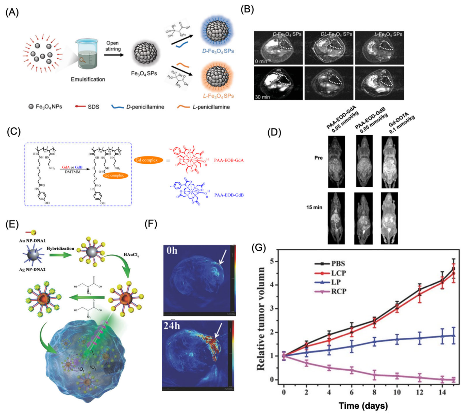
5.3.4. Photoacoustic imaging application of chiral nanoparticles
Photoacoustic imaging (PAI) has emerged as a powerful non-invasive and non-ionizing imaging modality that combines the high optical contrast of optical imaging with the deep penetration and spatial resolution of ultrasound, making it highly attractive for biomedical applications [170,171]. The rapid development of chiral nanomaterials has further expanded the capabilities of PAI by providing contrast agents with enhanced optical absorption, chirality-dependent interactions, and excellent tumor-targeting potential. Owing to their unique chiroptical and plasmonic properties, these nanomaterials have also been integrated with photothermal and photodynamic therapies to establish multifunctional theranostic platforms for imaging-guided cancer treatment. A notable example was reported by Xu and co-workers, who developed the first DNA-directed gold-core/silver-shell satellite-like chiral nanoassemblies for photoacoustic imaging and photodynamic therapy [169] (Figure 12E). The assemblies were functionalized with either L- or D-cysteine, producing strong plasmonic chiroptical responses in the visible region. Among the different formulations, the D-cysteine-modified nanoassemblies containing 15 nm Au cores and Ag shells exhibited the highest tumor-associated photoacoustic signal 24 h after intravenous administration (Figure 12F). Upon circularly polarized light (CPL) irradiation, these chiral nanostructures efficiently generated reactive oxygen species, resulting in significant tumor cell destruction through photodynamic effects (Figure 12G). This work demonstrated that DNA-programmed chiral plasmonic nanoassemblies can simultaneously provide enhanced photoacoustic contrast, real-time tumor visualization, and CPL-triggered therapeutic activity, highlighting their promise as multifunctional agents for PAI-guided cancer theranostics.
5.3.5. NIR fluorescence imaging
The integration of chirality with fluorescence imaging has led to the development of multifunctional nanoprobes capable of simultaneously providing optical visualization and chiroptical information [162]. By combining CD with fluorescence or upconversion luminescence (UCL), these dual-modal platforms offer enhanced sensitivity and improved analytical reliability compared with conventional fluorescence probes [172]. Such systems typically exploit stimulus-responsive changes in chiral assembly, allowing biological events to be monitored through concurrent variations in fluorescence intensity and chiroptical signals. One representative strategy employs metal–organic framework (MOF)-based chiral nanostructures for intracellular biomarker detection. For example, CuₓOS@ZIF-8 nanoparticles incorporating the fluorescent dye Cy3 were designed as dual-responsive probes for hydrogen sulfide (H₂S) (Figure 13A) [173]. In the absence of H₂S, the fluorescence of Cy3 remained quenched within the nanostructure. Upon exposure to H₂S, decomposition of the probe released the dye, producing a concentration-dependent fluorescence recovery accompanied by a gradual decrease in the CD signal (Figure 13B-C). This complementary optical response enabled both quantitative determination of intracellular H₂S and fluorescence imaging in living systems (Figure 13D).
Upconversion nanoparticles (UCNPs) have also emerged as attractive building blocks for chiral bioimaging because of their excellent photostability, deep tissue penetration, and low background autofluorescence [174]. DNA-programmed self-assembly has been utilized to fabricate chiral nanopyramids capable of real-time intracellular microRNA sensing. The intact nanostructure generated a pronounced CD response while suppressing UCL emission. Hybridization with the target microRNA triggered disassembly of the nanopyramid, leading to disappearance of the chiroptical signal together with restoration of UCL intensity [175]. This switchable dual-readout mechanism enabled sensitive intracellular microRNA detection by combining quantitative CD analysis with fluorescence imaging (Figure 13E-F). Similar design principles have subsequently been extended for monitoring reactive oxygen species (ROS) in cultured cells and tumor-bearing animal models. Another approach combines UCNPs with MOF-based hybrid architectures to construct ratiometric chiral imaging probes. Encapsulation of UCNP cores within NiSₓ-containing ZIF-8 shells produced nanocomposites exhibiting distinct CD activity together with wavelength-dependent modulation of upconversion emission [176]. Oxidative stimulation selectively altered the green emission while maintaining the red emission, allowing ROS concentrations to be quantified through fluorescence intensity ratios in parallel with CD signal changes. This dual-modal strategy provided highly sensitive imaging of intracellular hydrogen peroxide and enabled dynamic visualization of oxidative stress in vivo. Beyond molecular imaging, chiral fluorescence probes have also demonstrated considerable potential for pathogen detection. Chiral heterodimers assembled from antibody-functionalized gold nanoparticles and polymyxin-B-modified UCNPs generated amplified CD signals accompanied by quenched fluorescence [177]. Differences in the interaction of polymyxin B with antibiotic-sensitive and resistant Escherichia coli strains resulted in distinct optical responses, enabling discrimination between bacterial phenotypes and non-invasive visualization of infection in animal models.
Figure 13.
Representative chiral nanoprobes for fluorescence- and CD-based molecular imaging. (A) Schematic illustration of the CuxOS@ZIF-8 nanosensor designed for endogenous H₂S detection. (B,C) Chiroptical and fluorescence characterization of the CuxOS-Cy3@ZIF-8 nanoprobe before and after reaction with Na₂S, showing changes in the (B) circular dichroism (CD) and (C) fluorescence spectra. (D) In vivo fluorescence imaging of endogenous H₂S in a tumor-bearing mouse model following subcutaneous injection of the nanoprobe. Images were acquired at different time points for (a) probe-treated tumors and (b) tumors pretreated with 50 mg L⁻¹ PAG prior to probe administration. (E) CD spectra and (F) confocal fluorescence images of HeLa cells containing different levels of miR-21 after incubation with the chiral probe: (a,b) miR-21-transfected cells, (c) non-transfected cells, and (d,e) cells transfected with antisense miR-21 sequences. Scale bar = 20 μm. The inset in (E) presents a three-dimensional cryo-TEM tomographic reconstruction of the chiral pyramidal nanostructures. Panel A-D reproduced with permission from [173], Copyright 2020, Wiley Publishers. Panel E-F reproduced with permission from [175], Copyright 2016, American Chemical Society.
Figure 13.
Representative chiral nanoprobes for fluorescence- and CD-based molecular imaging. (A) Schematic illustration of the CuxOS@ZIF-8 nanosensor designed for endogenous H₂S detection. (B,C) Chiroptical and fluorescence characterization of the CuxOS-Cy3@ZIF-8 nanoprobe before and after reaction with Na₂S, showing changes in the (B) circular dichroism (CD) and (C) fluorescence spectra. (D) In vivo fluorescence imaging of endogenous H₂S in a tumor-bearing mouse model following subcutaneous injection of the nanoprobe. Images were acquired at different time points for (a) probe-treated tumors and (b) tumors pretreated with 50 mg L⁻¹ PAG prior to probe administration. (E) CD spectra and (F) confocal fluorescence images of HeLa cells containing different levels of miR-21 after incubation with the chiral probe: (a,b) miR-21-transfected cells, (c) non-transfected cells, and (d,e) cells transfected with antisense miR-21 sequences. Scale bar = 20 μm. The inset in (E) presents a three-dimensional cryo-TEM tomographic reconstruction of the chiral pyramidal nanostructures. Panel A-D reproduced with permission from [173], Copyright 2020, Wiley Publishers. Panel E-F reproduced with permission from [175], Copyright 2016, American Chemical Society.
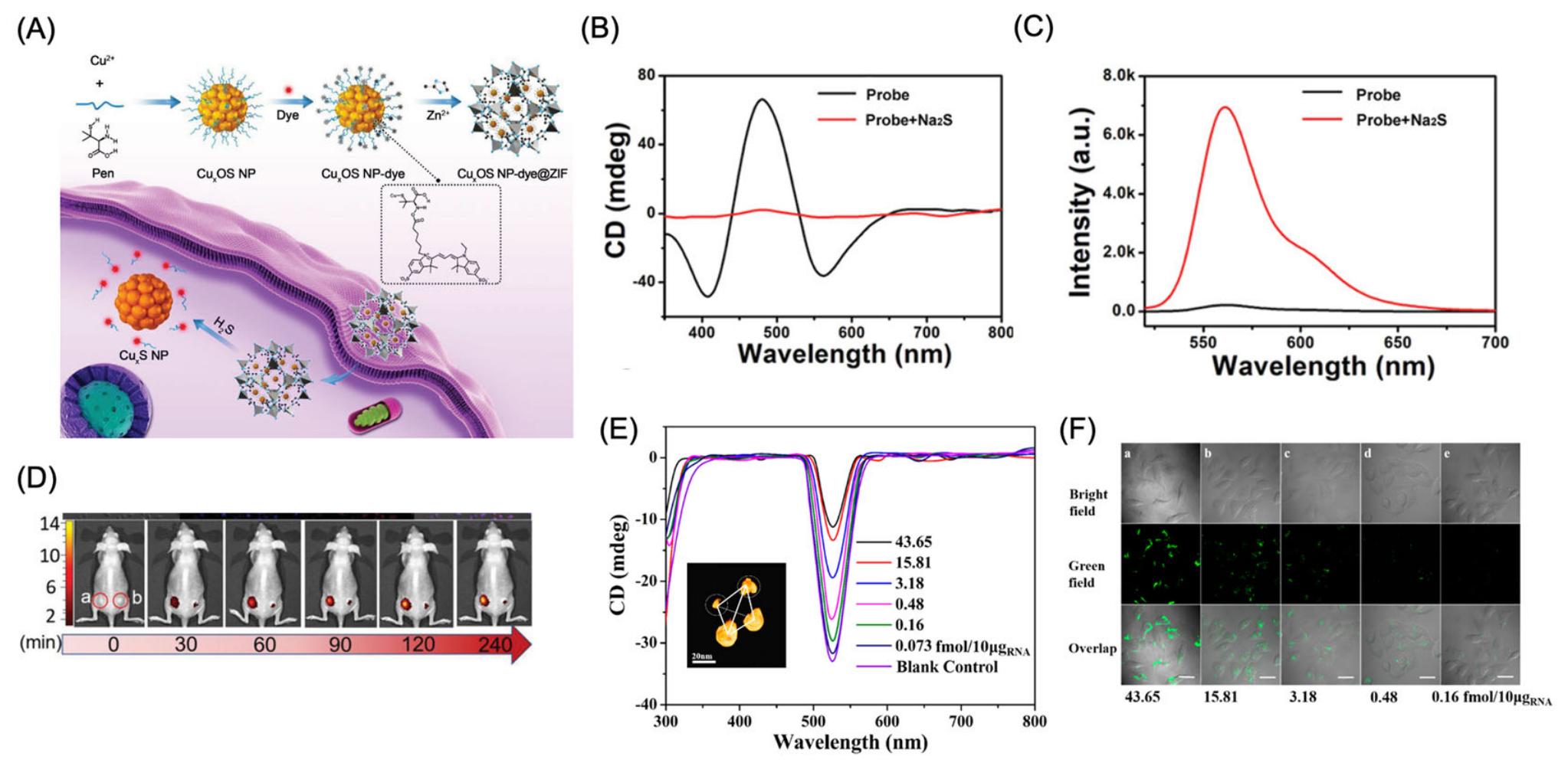
Near-infrared II (NIR-II, 1000–1700 nm) fluorescence imaging has emerged as one of the most promising optical imaging modalities because of its superior tissue penetration, reduced light scattering, and minimal background autofluorescence compared with conventional visible and NIR-I imaging. The incorporation of chirality into NIR-II nanoprobes further enhances imaging performance by coupling stereoselective biological recognition with optimized optical emission. Consequently, chiral nanomaterials have attracted considerable attention as tumor-targeted imaging agents capable of improving both probe accumulation and imaging sensitivity. One of the earliest demonstrations of this concept was reported using chiral Ag₂S quantum dots, where the influence of nanoparticle chirality on in vivo biodistribution was systematically investigated [178]. In an orthotopic renal tumor model, D-enantiomeric Ag₂S quantum dots accumulated substantially more efficiently within tumors than their L-counterparts, reaching approximately 12.3% injected dose per gram of tissue after 24 h, which corresponded to nearly 2.1-fold greater tumor retention. These findings established that nanoparticle chirality plays a decisive role in regulating tumor homing and in vivo pharmacokinetics, providing direct evidence that stereochemical design can significantly improve the targeting efficiency of NIR-II imaging probes. Further improvements in imaging performance have been achieved by integrating chirality with rare-earth engineering. Xu and co-workers developed ultrasmall chiral Ag₂Se nanoparticles co-doped with Nd³⁺, Yb³⁺, and Er³⁺ ions, producing a highly efficient NIR-II fluorescent probe. Rare-earth incorporation dramatically increased fluorescence brightness, yielding an emission intensity approximately 146-fold greater than that of undoped Ag₂Se nanoparticles while shifting the emission maximum to 1550 nm, a wavelength well suited for deep-tissue imaging [179]. In addition to optical enhancement, nanoparticle chirality strongly influenced biological recognition. The R-enantiomer displayed exceptionally strong binding toward the CD44 receptor, exhibiting a dissociation constant of only 0.9 nM and nearly 927-fold higher affinity than the S-enantiomer. This stereoselective interaction translated into markedly improved tumor accumulation in vivo, enabling high-contrast NIR-II fluorescence imaging with a signal-to-noise ratio approaching 18 and the visualization of tumor margins with a spatial resolution of approximately 0.3 mm, thereby facilitating the detection of small metastatic lesions.
The application of chiral plasmonic materials for bioimaging applications are represented in Table 3.
5.4. Chiral nanoparticles for therapy
The unique physicochemical and biological characteristics of chiral nanomaterials have established them as promising platforms for a wide range of therapeutic applications [180]. Beyond their distinctive chiroptical properties, these nanomaterials exhibit stereochemistry-dependent interactions with biological systems, enabling selective cellular uptake, differential protein adsorption, receptor-specific recognition, and immune modulation. Such enantioselective behavior significantly influences their biodistribution, pharmacological activity, and therapeutic efficacy, providing opportunities to improve treatment precision while minimizing off-target effects. Recent advances have demonstrated that the biological activity of chiral nanomaterials extends far beyond imaging and biosensing. Their ability to regulate cellular signaling pathways, oxidative stress, inflammatory responses, and immune activation has enabled the development of innovative therapeutic strategies for diverse diseases. Chirality-dependent nanoplatforms have shown considerable promise in enhancing cancer therapy through improved tumor targeting and therapeutic selectivity, while also offering new opportunities for the treatment of neurodegenerative disorders and age-associated diseases [25]. Increasing evidence further indicates that stereochemical engineering can modulate biological responses at the molecular level, providing mechanistic insights into how chirality governs interactions with proteins, membranes, and intracellular pathways.

Table 3.
List of chiral nanomaterials for bioimaging applications.
| Biomedical Modality | Role of Chirality | Representative Chiral Nanomaterials | Representative Applications | Ref |
|---|---|---|---|---|
| MRI | Chirality enhances molecular recognition and magnetic signal modulation | L-/D-aspartic acid-modified cobalt hydroxide nanoparticles; chiral magnetic nanostructures | ROS-responsive imaging, tumor microenvironment analysis, enantioselective cellular uptake | [181,182,183] |
| FL/Chiroptical | Chiral interactions regulate fluorescence resonance energy transfer (FRET), circularly polarized luminescence (CPL), and circular dichroism (CD) | UCNP-based hybrid nanoprobes; Au–UCNP heterostructures; chiral nanopyramids | Intracellular imaging, ROS sensing, microRNA detection, bacterial identification | [175,177,184,185,186] |
| PAI | Chiral plasmonic assemblies improve optical absorption and photothermal conversion | DNA-guided Au–Ag nanoassemblies; L-/D-cysteine-functionalized plasmonic nanoparticles | Deep-tissue tumor imaging, photoacoustic-guided phototherapy, real-time treatment monitoring | [169] |
| Biosensing/Imaging | Chirality improves molecular selectivity through CD, plasmonic coupling, and FRET | Cysteine-capped quantum dots; chiral Co(OH)₂ nanostructures; chiral triangular nanorings | Enantiomer recognition, biomarker detection, amyloid-β and α-synuclein sensing | [167,187,188] |
| SERS | Handedness-dependent plasmonic near fields produce distinct Raman enhancement for enantiomers, enabling sensitive chiral discrimination. | Amino acid capped nanoparticles, template assisted synthesize, Chiral nanotubes, helicoids, nanoring, nanofilms | Enantiomer recognition, biomarker detection, amyloid sensing | [54,146,189] |
| Chiral membranes/nanochannels | Chiral interfaces regulate selective molecular transport and separation | Cu₂−xSe/AAO membranes; Fe₃O₄ nanofilms; β-cyclodextrin-modified COF membranes | Enantioselective transport, drug separation, membrane-based biosensing | [190,191,192,193] |
5.4.1. Chiral Nanomaterials for Phototherapy and Radiotherapy
Phototherapy has emerged as an important non-invasive strategy for cancer treatment, with nanomaterials playing a central role in improving therapeutic efficiency through enhanced light absorption and selective tumor accumulation. Among the available approaches, photothermal therapy (PTT) eradicates tumor cells by converting absorbed light into localized heat, whereas photodynamic therapy (PDT) relies on the light-triggered generation of reactive oxygen species (ROS) to induce oxidative damage and apoptosis [34]. The introduction of chirality into nanomaterials has further expanded the potential of these therapies by enabling polarization-dependent light–matter interactions that enhance phototherapeutic performance [194,195].
Unlike achiral nanostructures, chiral nanomaterials interact preferentially with circularly polarized light (CPL) resulting in more efficient optical energy harvesting than under linearly polarized light. This unique characteristic can significantly improve ROS production and phototherapeutic efficacy [162]. A representative example is provided by chiral supramolecular nanostructures constructed from cysteine-derived building blocks, which exhibited markedly enhanced singlet oxygen generation when irradiated with CPL of matching handedness [169]. Under these conditions, ROS production was approximately two-fold higher than that obtained under linearly polarized illumination. The enhanced photodynamic activity translated into pronounced chirality-dependent cytotoxicity, where D- and L-configured nanostructures achieved maximum therapeutic efficacy under right- and left-handed CPL, respectively. Importantly, efficient tumor cell destruction was achieved at relatively low photosensitizer concentrations, and complete tumor regression was observed in animal models following CPL-guided treatment.
A representative example was reported by Liu and co-workers, who developed D- and L-penicillamine-modified Cu₂−xSe nanoparticles for combined chemodynamic and photothermal therapy under second near-infrared (NIR-II) laser irradiation (Figure 14 A) [196]. Compared with the L-enantiomer, the D-configured nanoparticles produced stronger therapeutic responses, primarily due to their higher photothermal conversion efficiency and enhanced ROS generation (Figure 14B-C). In addition, localized photothermal heating accelerated the release of therapeutic species, promoting hydroxyl radical production and resulting in more effective tumor cell destruction. Beyond direct tumor ablation, chiral nanomaterials have also been employed to facilitate postoperative tissue repair. Wang et al. developed a supramolecular hydrogel by incorporating polydopamine nanoparticles into an L-phenylalanine-derived gel matrix [197]. Under near-infrared irradiation, the nanoparticles generated sufficient heat to eliminate residual cancer cells, whereas the chiral hydrogel provided an extracellular matrix-like environment that supported fibroblast migration and tissue regeneration. This multifunctional system simultaneously suppressed tumor recurrence and accelerated wound healing following surgery. Li et al. introduced cysteine-functionalized MoO₃−x nanoparticles possessing intrinsic chiral architectures generated through metal-to-ligand charge transfer interactions (Figure 14D). These nanoparticles exhibited broad visible-light absorption and efficiently converted absorbed light into heat, leading to rapid temperature elevation during laser irradiation. Combined with their low cytotoxicity and efficient cellular internalization, the chiral MoO₃−x nanostructures represent promising and economical photothermal agents (Figure 14E) [198]. Despite these promising results, further investigations are required to evaluate the long-term stability, pharmacokinetics, and tumor accumulation efficiency of these supramolecular assemblies under physiologically relevant conditions before clinical translation can be considered.
Beyond light-mediated therapy, chirality has also been explored to improve the efficacy of radiotherapy [199,200]. Chiral gold nanoclusters have recently attracted attention as radiosensitizers capable of amplifying radiation-induced oxidative stress. In one study, D-configured Au nanoclusters demonstrated superior radiosensitizing activity compared with their L-enantiomeric counterparts [201]. The enhanced therapeutic response was attributed to improved intracellular distribution, which promoted ROS generation, DNA double-strand damage, cell-cycle arrest, and apoptotic cell death following X-ray irradiation. Although these ultrasmall nanoclusters produced encouraging antitumor outcomes, their extremely small dimensions (~2 nm) may result in rapid renal clearance and limited circulation time, potentially restricting tumor retention. Consequently, optimizing nanoparticle size, surface chemistry, administration protocols, and dosing strategies will be essential for maximizing their therapeutic benefits.
5.4.2. Chiral Nanomaterials for Cancer Immunotherapy
Cancer immunotherapy has transformed modern oncology by stimulating the host immune system to recognize and eradicate malignant cells while establishing long-term immune memory [202]. However, its clinical efficacy is often compromised by poor antigen presentation, inadequate activation of immune effector cells, limited infiltration of cytotoxic lymphocytes, and the immunosuppressive tumor microenvironment. Chiral nanomaterials have recently emerged as a new class of immunomodulatory platforms because stereochemical engineering not only improves nanoparticle delivery but also regulates immune-cell recognition, activation, and cytokine signaling, thereby enhancing antitumor immunity [203].
A pioneering study by Xu and co-workers demonstrated that chiral gold nanoadjuvants can simultaneously enhance innate and adaptive immune responses in a chirality-dependent manner (Figure 15A-B) [204]. These plasmonic nanostructures, synthesized under circularly polarized light, possessed exceptionally strong chiroptical activity, and their immunostimulatory efficacy increased with the optical anisotropy (g) factor. Mechanistic studies revealed that left-handed nanoadjuvants exhibited stronger interactions with G-protein-coupled receptors, including CD97 and EMR1, leading to enhanced cellular uptake, accelerated dendritic-cell maturation, and increased secretion of pro-inflammatory cytokines. Activation of the NLRP3 inflammasome further promoted antigen presentation and immune-cell activation. Consequently, left-handed nanoadjuvants generated substantially stronger antigen-specific antibody responses and, in tumor models, significantly enhanced infiltration of CD8⁺ cytotoxic T lymphocytes while activating natural killer (NK) cells through increased expression of NKG2D, granzyme B, and perforin, ultimately producing superior antitumor efficacy (Figure 15C-D).
Beyond regulating immune activation, chirality has also been exploited to improve nanoparticle pharmacokinetics and tumor delivery. D-configured MoS₂/CoS₂ nanozymes displayed prolonged circulation and enhanced tumor accumulation through stereoselective interactions with biological components [205]. Following tumor localization, these nanozymes efficiently generated reactive oxygen species, reprogramming immunosuppressive M2 tumor-associated macrophages into pro-inflammatory M1 macrophages and thereby amplifying local antitumor immune responses. Similarly, left-handed helical artificial antigen-presenting cells (aAPCs-L) minimized protein corona formation through stereochemical mismatch with endogenous serum proteins, substantially reducing phagocytic clearance, extending blood circulation beyond 24 h, and improving tumor accumulation, which ultimately resulted in complete melanoma regression.
Chiral engineering has also demonstrated considerable promise in therapeutic vaccination and oncolytic immunotherapy. Chen and co-workers reported that L-configured peptide hydrogels elicited significantly stronger antitumor immunity than the corresponding D-enantiomers, effectively suppressing immunosuppressive microenvironment formation and reducing both primary tumor growth and postoperative recurrence [206]. Likewise, Li and co-workers developed virus-inspired chiral oncolytic particles (VOPs), in which D-enantiomeric particles exhibited superior tumor-selective cytotoxicity and approximately five-fold greater oncolytic activity than the clinically used peptide LTX-315 [207]. Treatment with D-VOPs markedly increased intratumoral CD8⁺ cytotoxic T-cell infiltration and induced robust systemic antitumor immunity through chirality-dependent regulation of tumor recognition, cellular interactions, biodegradation resistance, and immune activation. Collectively, these studies demonstrate that chirality serves as a powerful molecular regulator of cancer immunotherapy by simultaneously enhancing nanoparticle delivery, controlling immune-cell signaling, remodeling the tumor microenvironment, and strengthening adaptive immune responses. These advances establish stereochemical engineering as a promising strategy for developing next generation nanoadjuvants, therapeutic cancer vaccines, and immunotherapeutic nanomedicines with improved precision and therapeutic efficacy.
5.4.3. Chiral Nanomaterials for Neurodegenerative Disease Therapy
Neurodegenerative disorders, including Alzheimer’s disease (AD) and Parkinson’s disease (PD), are characterized by progressive neuronal loss resulting from protein misfolding, oxidative stress, mitochondrial dysfunction, chronic neuroinflammation, and impaired neuronal regeneration. Despite considerable advances in pharmacological interventions, effective disease-modifying therapies remain elusive. Chiral nanomaterials have recently emerged as promising therapeutic platforms because their stereochemistry-dependent interactions with biological macromolecules enable selective recognition of pathological proteins, modulation of oxidative stress, and regulation of neural cell behavior.
One of the most extensively investigated applications of chiral nanomaterials is the inhibition of pathological protein aggregation. Chiral gold nanoparticles functionalized with glutathione demonstrated enantiomer-dependent binding toward amyloid-β (Aβ₄₂), with D-enantiomeric nanoparticles exhibiting significantly stronger affinity than their L-counterparts [208]. The enhanced interaction effectively suppressed Aβ fibrillation, improved blood-brain barrier penetration, and produced superior cognitive recovery in Alzheimer’s disease models. Similar stereoselective effects have been reported for D-penicillamine-modified FeₓCuᵧSe nanoparticles, which not only inhibited Aβ aggregation but also promoted the disassembly of preformed amyloid fibrils under near-infrared irradiation through enhanced ROS generation. Likewise, chiral CuₓCoᵧS superparticles effectively inhibited α-synuclein aggregation and facilitated fibril dissociation in Parkinson’s disease models, highlighting the broad applicability of chirality-guided protein recognition for treating protein-misfolding disorders [209].
Beyond regulating protein aggregation, chiral nanomaterials have also shown considerable promise in alleviating oxidative stress, a central pathological feature shared by most neurodegenerative diseases. Nanozymes possessing superoxide dismutase-, catalase-, glutathione peroxidase-, and peroxidase-like activities efficiently eliminated intracellular reactive oxygen species, thereby reducing neuronal oxidative damage and improving cognitive function in Parkinson’s disease models [210]. Morphology-dependent catalytic activity has also been demonstrated in Mn₃O₄ nanostructures, where nanorod-shaped particles exhibited superior ROS-scavenging capability compared with other morphologies [211]. In addition, antibody-functionalized chiral gold nanoparticles selectively eliminated senescent microglia through apoptosis, reducing α-synuclein accumulation and mitigating neuroinflammation [212]. These studies illustrate the potential of chiral nanozymes to simultaneously regulate oxidative stress, cellular senescence, and inflammatory signaling within the diseased brain.
Chiral nanomaterials have further emerged as powerful tools for neural regeneration by directing stem-cell fate through stereoselective mechanobiological signaling. Chiral plasmonic gold films modified with enantiomeric penicillamine exhibited opposite effects on neuronal cell adhesion and differentiation under circularly polarized light, demonstrating that surface chirality can regulate neuronal development [213]. More recently, DNA-programmed chiral nanoassemblies have been employed to precisely control neural stem-cell differentiation [214]. Under circularly polarized illumination, these nanostructures generated chiroplasmonic mechanical forces through interactions with cytoskeletal proteins, activating neuron-specific genetic programs and promoting efficient neuronal differentiation. Following transplantation into Alzheimer’s disease models, the differentiated neurons integrated into damaged brain tissue and significantly improved cognitive performance, suggesting that light-responsive chiral nanomaterials may provide an innovative strategy for neuronal regeneration.
Despite these encouraging advances, several translational challenges remain. Most reported chiral nanotherapeutics require direct intracranial administration because their relatively large dimensions limit efficient penetration across the blood-brain barrier. Future research should therefore prioritize the development of ultrasmall, biodegradable, and actively targeted chiral nanoplatforms capable of crossing the blood-brain barrier following systemic administration while maintaining high stereoselectivity and long-term biosafety. Addressing these challenges will be critical for translating chiral nanomedicine from proof-of-concept studies toward clinical treatment of neurodegenerative diseases.
5.4.4. Chiral Nanomaterials for Age-Related Diseases
The progressive accumulation of senescent cells is recognized as a major contributor to aging and numerous age-associated disorders through persistent secretion of pro-inflammatory factors, mitochondrial dysfunction, and impaired tissue regeneration. Consequently, the selective elimination of senescent cells (senolysis) has emerged as a promising therapeutic strategy to delay age-related functional decline while preserving healthy tissues [215]. In this context, chiral nanomaterials offer unique advantages because their stereochemistry-dependent cellular interactions enable selective recognition, targeted intracellular delivery, and controlled therapeutic activation. One of the earliest demonstrations of chiral nanomaterial-mediated senolysis was reported using mitochondria-targeted core–shell spiky nanorods (CSNRs) functionalized with triphenylphosphonium and β₂-microglobulin antibodies [216]. These nanoplatforms selectively accumulated within senescent cells, where near-infrared (NIR) irradiation induced mitochondrial membrane disruption and reactive oxygen species (ROS) generation, triggering apoptosis. In addition to direct phototherapeutic effects, activation of immune responses further promoted senescent cell clearance, resulting in improved hair regeneration and enhanced physical performance in doxorubicin-induced aging models. Despite these encouraging outcomes, optimization of in vivo stability, targeting specificity, and long-term biosafety remains essential for future clinical translation. Chirality has also been exploited to improve the efficiency of senescent-cell targeting through stereoselective cellular uptake. Photo- and magnetically responsive chiral CuₓCoᵧS nanoparticles demonstrated pronounced enantiomer-dependent internalization, with D-configured nanoparticles exhibiting approximately 2.5-fold greater uptake than their L-counterparts because of stronger interactions with cell membrane components [217]. Combined NIR irradiation and alternating magnetic field stimulation generated synergistic oxidative stress and cytoskeletal disruption, leading to activation of caspase-3-mediated apoptosis and efficient elimination of senescent cells in vivo. These findings highlight how stereochemical engineering can enhance both nanoparticle delivery and therapeutic efficacy through multimodal stimulation.
Beyond phototherapeutic approaches, programmable chiral nanostructures have also enabled stimulus-responsive senolytic drug delivery. DNA-bridged tetrahedral nanoplatforms consisting of upconversion nanoparticles surrounded by gold nanoparticles were engineered for the controlled release of granzyme B [218]. Following selective recognition of senescent cells via β₂-microglobulin antibodies, NIR irradiation disrupted the DNA framework, releasing granzyme B to initiate apoptosis. Simultaneously, fluorescence signal conversion provided real-time monitoring of therapeutic activity, enabling image-guided senolytic treatment. Application of this platform effectively reduced senescent-cell burden and reversed aging-associated phenotypes in both accelerated-aging and doxorubicin-induced senescence models.
Collectively, these studies demonstrate that chirality provides a versatile strategy for engineering next generation senolytic nanomedicines by enhancing cellular selectivity, therapeutic precision, and multimodal treatment efficacy. Future efforts should focus on developing biodegradable chiral nanoplatforms with improved targeting specificity, long-term biosafety, and clinically applicable delivery routes to facilitate translation for the treatment of aging and age-associated disorders. The application of chiral plasmonic materials for age and neurogenerative diseases are represented in Table 4.
6. Conclusions and Future Perspectives
Chiral nanomaterials have emerged as one of the most rapidly advancing areas of nanoscience, bridging chemistry, materials science, catalysis, and biomedicine. This review has highlighted recent progress in the rational synthesis of chiral nanostructures through molecular induction, chiral templating, circularly polarized light (CPL), magnetic-field-assisted assembly, and self-assembly strategies, demonstrating how precise control over nanoscale chirality enables unique optical, catalytic, and biological functions. These advances have significantly expanded the application landscape of chiral nanomaterials, ranging from asymmetric catalysis and enantioselective sensing to bioimaging, disease diagnosis, cancer therapy, immunotherapy, and regenerative medicine.
A key advantage of chiral nanomaterials lies in their ability to exploit stereospecific interactions that are inaccessible to conventional achiral nanostructures. In catalytic systems, chirality provides new opportunities for asymmetric photocatalysis, enantioselective electrocatalysis, and biomimetic nanozymes with enhanced selectivity and catalytic efficiency. In the biomedical field, stereochemical engineering has enabled selective recognition of proteins, nucleic acids, and cell-surface receptors, resulting in improved biosensing, targeted imaging, precision drug delivery, immune modulation, and disease therapy. Increasing evidence indicates that nanoparticle chirality not only governs molecular recognition but also regulates intracellular signaling, protein adsorption, cellular uptake, immune activation, and therapeutic outcomes, establishing chirality as a powerful design principle for next-generation nanomedicine.
Despite these remarkable achievements, several challenges continue to limit the broader application and clinical translation of chiral nanomaterials. The scalable synthesis of nanostructures with well-defined chirality, high optical anisotropy, and excellent batch-to-batch reproducibility remains difficult. Furthermore, the fundamental mechanisms governing chirality-dependent interactions with complex biological systems are still incompletely understood. Current studies often focus on phenomenological observations, whereas detailed molecular-level investigations combining advanced spectroscopy, cryogenic electron microscopy, molecular dynamics simulations, artificial intelligence-assisted structural prediction, and multi-omics analyses are required to establish robust structure–activity relationships. Standardized evaluation protocols are also essential to enable meaningful comparisons between different chiral nanoplatforms.
From a biomedical perspective, future efforts should prioritize the development of biodegradable, biocompatible, and clinically translatable chiral nanomaterials with predictable pharmacokinetics, long-term biosafety, and efficient clearance. Improving blood circulation, active tissue targeting, blood–brain barrier penetration, and tumor-specific accumulation while minimizing off-target toxicity will be critical for successful clinical implementation. In parallel, integrating chirality with multifunctional nanoplatforms capable of combining imaging, biosensing, targeted drug delivery, immunotherapy, phototherapy, gene regulation, and catalytic therapy is expected to accelerate the emergence of personalized theranostic systems. The incorporation of responsive elements activated by circularly polarized light, magnetic fields, ultrasound, or endogenous biochemical stimuli may further enable precise spatiotemporal control over therapeutic activity.
Looking ahead, several exciting research directions are anticipated to shape the next generation of chiral nanotechnology. The development of intrinsically chiral inorganic nanocrystals, programmable supramolecular assemblies, chiral metal–organic frameworks, and hybrid organic–inorganic architectures will provide unprecedented opportunities for tailoring optical and catalytic properties. Expanding the role of chirality in photoelectrochemical catalysis, artificial photosynthesis, enzyme-inspired catalysis, quantum nanomaterials, and intelligent nanosystems represents another promising frontier. Moreover, combining machine learning with high-throughput materials discovery and automated synthesis could substantially accelerate the rational design of chiral nanostructures with optimized physicochemical and biological performance.
Future advances in chiral nanomaterials are expected to focus on multifunctional platforms that integrate molecular sensing, bioimaging, targeted drug delivery, and therapeutic functions within a single nanostructure. Stimuli-responsive chiral systems capable of adapting to disease-specific microenvironments (e.g., pH, ROS, or enzymatic activity) will further improve targeting precision and therapeutic efficacy. In addition, chirality-mediated immune modulation represents a promising strategy for enhancing immunotherapy. The integration of artificial intelligence, molecular simulations, and data-driven material design is also anticipated to accelerate the rational development and optimization of next-generation chiral nanomedicines, facilitating their translation toward precision diagnostics and personalized therapy.
Overall, chiral nanomaterials have evolved from a fundamental scientific curiosity into a versatile platform with broad implications for catalysis, sensing, imaging, and precision medicine. Continued interdisciplinary collaboration among chemists, materials scientists, engineers, computational researchers, and clinicians will be essential to address the remaining scientific and translational challenges. With continued advances in stereochemical engineering, mechanistic understanding, and scalable manufacturing, chiral nanomaterials are expected to play an increasingly important role in sustainable catalysis, intelligent diagnostics, and next-generation precision therapeutics.
Funding
This research received no external funding.
Institutional Review Board Statement
Not applicable.
Informed Consent Statement
Not applicable.
Data Availability Statement
No new data were created or analyzed in this study.
Acknowledgments
P.P.P.K. shows sincere gratitude to the Department of Biomedical Engineering, Michigan State University, for the facilities and use of resources for the literature collection. “During the preparation of this manuscript the author(s) used [ChatGPT 5.2] for the purposes of language polishing and used for graphical abstract making. The authors have reviewed and edited the output and take full responsibility for the content of this publication.”.
Conflicts of Interest
The author declares no conflicts of interest.
References
- Ribó, J.M. Chirality: The Backbone of Chemistry as a Natural Science. Symmetry 2020, 12, 1982. [CrossRef]
- Ahn, H.-Y.; Yoo, S.; Cho, N.H.; Kim, R.M.; Kim, H.; Huh, J.-H.; Lee, S.; Nam, K.T. Bioinspired Toolkit Based on Intermolecular Encoder toward Evolutionary 4D Chiral Plasmonic Materials. Acc. Chem. Res. 2019, 52, 2768–2783. [CrossRef]
- Cho, N.H.; Guerrero-Martínez, A.; Ma, J.; Bals, S.; Kotov, N.A.; Liz-Marzán, L.M.; Nam, K.T. Bioinspired Chiral Inorganic Nanomaterials. Nat. Rev. Bioeng. 2023, 1, 88–106. [CrossRef]
- Ma, W.; Xu, L.; De Moura, A.F.; Wu, X.; Kuang, H.; Xu, C.; Kotov, N.A. Chiral Inorganic Nanostructures. Chem. Rev. 2017, 117, 8041–8093. [CrossRef]
- Blackmond, D.G. The Origin of Biological Homochirality. Cold Spring Harb. Perspect. Biol. 2010, 2, a002147–a002147. [CrossRef]
- Im, S.W.; Ahn, H.; Kim, R.M.; Cho, N.H.; Kim, H.; Lim, Y.; Lee, H.; Nam, K.T. Chiral Surface and Geometry of Metal Nanocrystals. Adv. Mater. 2020, 32, 1905758. [CrossRef]
- Hong, T.; Guan, J.; Zhou, W.; Tan, S.; Cai, Z. Sustainable Methods for the Synthesis of Chiral Nanomaterials. Cell Rep. Phys. Sci. 2025, 6, 102785. [CrossRef]
- Urban, M.J.; Shen, C.; Kong, X.-T.; Zhu, C.; Govorov, A.O.; Wang, Q.; Hentschel, M.; Liu, N. Chiral Plasmonic Nanostructures Enabled by Bottom-Up Approaches. Annu. Rev. Phys. Chem. 2019, 70, 275–299. [CrossRef]
- Yang, G.; Sun, L.; Zhang, Q. Multicomponent Chiral Plasmonic Hybrid Nanomaterials: Recent Advances in Synthesis and Applications. Nanoscale Adv. 2024, 6, 318–336. [CrossRef]
- Hentschel, M.; Schäferling, M.; Duan, X.; Giessen, H.; Liu, N. Chiral Plasmonics. Sci. Adv. 2017, 3, e1602735. [CrossRef]
- Zhou, B.; Zhang, Y.; Lei, Q.; Zhang, Z.; Wang, R.; Liu, P.; Xu, X.; Jia, T.; Cheng, J. Chiral Noble-Metal Nanomaterials: Chiral Origins and Biomedical Applications. Smart Mater. Med. 2026, 7, 59–76. [CrossRef]
- Karimova, N.V.; Aikens, C.M. Chiral Noble Metal Nanoparticles and Nanostructures. Part & Part Syst Charact 2019, 36, 1900043. [CrossRef]
- Ugras, T.J.; Carson, R.B.; Lynch, R.P.; Li, H.; Yao, Y.; Cupellini, L.; Page, K.A.; Wang, D.; Arbe, A.; Bals, S.; et al. Transforming Achiral Semiconductors into Chiral Domains with Exceptional Circular Dichroism. Science 2025, 387, eado7201. [CrossRef]
- Kumar, J.; Thomas, K.G.; Liz-Marzán, L.M. Nanoscale Chirality in Metal and Semiconductor Nanoparticles. Chem. Commun. 2016, 52, 12555–12569. [CrossRef]
- Xiao, S.; Liang, J.; Li, J.; Cheng, J.; Zhu, X.; He, T. Tunable Optical Activities in Chiral Transition Metal Oxide Nanoparticles. Nanoscale 2022, 14, 15414–15421. [CrossRef]
- Jung, W.; Lee, D.; Kim, H.; Son, B.; Oh, S.; Gong, J.E.; Kim, D.; Yoon, J.; Yeom, J. Universal Chiral Nanopaint for Metal Oxide Biomaterials. ACS Nano 2025, 19, 8632–8645. [CrossRef]
- Zhou, J.; Gao, Y.; Zhang, D.; Ren, K.; Dai, M.; Wang, H.; Qi, L. Concavity-Enhanced Chiral Self-Assembly of Anisotropic Nanoparticles toward Strong Chiroptical Activity. Nat. Commun. 2025, 16, 6897. [CrossRef]
- Zhang, F.; Bi, Y.; Wei, J.; Yang, Z. Recent Advances in Chiral Nanoparticle Superstructures with Long-Range Order. Chem. Commun. 2025, 61, 14087–14096. [CrossRef]
- Bedair, A.; Algethami, F.K.; Hamed, M.; Mansour, F.R. Emerging Materials for Chiral Recognition by Nanoscale Optical Sensors. Microchemical Journal 2026, 119234. [CrossRef]
- Gogoi, A.; Mazumder, N.; Konwer, S.; Ranawat, H.; Chen, N.-T.; Zhuo, G.-Y. Enantiomeric Recognition and Separation by Chiral Nanoparticles. Molecules 2019, 24, 1007. [CrossRef]
- Li, S.; Xu, X.; Xu, L.; Lin, H.; Kuang, H.; Xu, C. Emerging Trends in Chiral Inorganic Nanomaterials for Enantioselective Catalysis. Nat.Commun. 2024, 15, 3506. [CrossRef]
- Cao, H.; Yang, E.; Kim, Y.; Zhao, Y.; Ma, W. Biomimetic Chiral Nanomaterials with Selective Catalysis Activity. Adv. Sci. 2024, 11, 2306979. [CrossRef]
- Qu, A.; Xu, L.; Xu, C.; Kuang, H. Chiral Nanomaterials for Biosensing, Bioimaging, and Disease Therapies. Chem. Commun. 2022, 58, 12782–12802. [CrossRef]
- Mejía-Salazar, J.R. Nanophotonic Strategies for Chiral Biosensing: Nanoparticles, Metasurfaces, Magneto-Optical, and Quantum Approaches. Adv. Photon. Res. 2026, 7, e202500242. [CrossRef]
- Huang, Z.; Zhou, H.; Zhang, S.; Song, S.; Feng, C.; Ke, Z.; Tang, J.; Dai, Y. Chirality-Engineered Nanomaterials for Biomedical Applications. Journal of Controlled Release 2026, 395, 115015. [CrossRef]
- Im, S.; Mousavi, S.; Chen, Y.-S.; Zhao, Y. Perspectives of Chiral Nanophotonics: From Mechanisms to Biomedical Applications. npj Nanophoton. 2024, 1, 46. [CrossRef]
- Yang, G.; Wang, X.; Sun, L.; Zhang, Q. Intrinsically Chiral Plasmonic Hybrid Nanomaterials with Tailored Chiroptical Activities. ACS Appl. Nano Mater. 2026, 9, 10434–10447. [CrossRef]
- Zhang, R.; Gu, Y.; Zhao, L. Photothermal Effect and the Corresponding Applications of Chiral Nanomaterials. CrystEng. Comm. 2026, 28, 2000–2017. [CrossRef]
- Zheng, J.; Fu, Y.; Wang, J.; Zhang, W.; Lu, X.; Lin, H.-Q.; Shao, L.; Wang, J. Circularly Polarized OLEDs from Chiral Plasmonic Nanoparticle-Molecule Hybrids. Nat.Commun. 2025, 16, 1658. [CrossRef]
- Burrow, J.A.; Alam, M.S.; Yahiaoui, R.; Smith, E.M.; Laing, R.; Gardener, J.; Searles, T.A.; Shah, P.J.; Vangala, S.; Hendrickson, J.R.; et al. Chiral Phase Change Nanomaterials. Adv. Funct. Mater. 2026, e76202. [CrossRef]
- Kelly, K.L.; Coronado, E.; Zhao, L.L.; Schatz, G.C. The Optical Properties of Metal Nanoparticles: The Influence of Size, Shape, and Dielectric Environment. J. Phys. Chem. B 2003, 107, 668–677. [CrossRef]
- Jain, P.K.; Huang, X.; El-Sayed, I.H.; El-Sayed, M.A. Noble Metals on the Nanoscale: Optical and Photothermal Properties and Some Applications in Imaging, Sensing, Biology, and Medicine. Acc. Chem. Res. 2008, 41, 1578–1586. [CrossRef]
- Huang, X.; El-Sayed, M.A. Gold Nanoparticles: Optical Properties and Implementations in Cancer Diagnosis and Photothermal Therapy. Journal of Advanced Research 2010, 1, 13–28. [CrossRef]
- Kumar, P.P.P.; Lim, D.-K. Photothermal Effect of Gold Nanoparticles as a Nanomedicine for Diagnosis and Therapeutics. Pharmaceutics 2023, 15, 2349. [CrossRef]
- Paiva-Marques, W.; Reyes Gómez, F.; N. Oliveira, O.; Mejía-Salazar, J.R. Chiral Plasmonics and Their Potential for Point-of-Care Biosensing Applications. Sensors 2020, 20, 944. [CrossRef]
- Chen, H.; Kou, X.; Yang, Z.; Ni, W.; Wang, J. Shape- and Size-Dependent Refractive Index Sensitivity of Gold Nanoparticles. Langmuir 2008, 24, 5233–5237. [CrossRef]
- Ruan, Q.; Shao, L.; Shu, Y.; Wang, J.; Wu, H. Growth of Monodisperse Gold Nanospheres with Diameters from 20 Nm to 220 Nm and Their Core/Satellite Nanostructures. Adv. Opt. Mater. 2014, 2, 65–73. [CrossRef]
- Zhu, Z.; Liu, W.; Li, Z.; Han, B.; Zhou, Y.; Gao, Y.; Tang, Z. Manipulation of Collective Optical Activity in One-Dimensional Plasmonic Assembly. ACS Nano 2012, 6, 2326–2332. [CrossRef]
- Probst, P.T.; Dong, Y.; Zhou, Z.; Aftenieva, O.; Fery, A. Bottom-Up Assembly of Inorganic Particle-Based Chiroptical Materials. Adv. Opt. Mater. 2024, 12, 2301834. [CrossRef]
- Zhao, X.; Zang, S.-Q.; Chen, X. Stereospecific Interactions between Chiral Inorganic Nanomaterials and Biological Systems. Chem. Soc. Rev. 2020, 49, 2481–2503. [CrossRef]
- Zhang, L.; Wang, T.; Shen, Z.; Liu, M. Chiral Nanoarchitectonics: Towards the Design, Self-Assembly, and Function of Nanoscale Chiral Twists and Helices. Adv. Mater. 2016, 28, 1044–1059. [CrossRef]
- Tan, L.; Fu, W.; Gao, Q.; Wang, P. Chiral Plasmonic Hybrid Nanostructures: A Gateway to Advanced Chiroptical Materials. Adv. Mater. 2024, 36, 2309033. [CrossRef]
- Zhao, Y.; Xie, J.; Tian, Y.; Mourdikoudis, S.; Fiuza-Maneiro, N.; Du, Y.; Polavarapu, L.; Zheng, G. Colloidal Chiral Carbon Dots: An Emerging System for Chiroptical Applications. Adv. Sci. 2024, 11, 2305797. [CrossRef]
- Yang, Y.; Liu, L.; Wei, Z. Chiral Conjugated Molecular Assemblies Interact with Substances and Light. Acc. Mater. Res. 2024, 5, 329–346. [CrossRef]
- Govorov, A.O.; Fan, Z.; Hernandez, P.; Slocik, J.M.; Naik, R.R. Theory of Circular Dichroism of Nanomaterials Comprising Chiral Molecules and Nanocrystals: Plasmon Enhancement, Dipole Interactions, and Dielectric Effects. Nano Lett. 2010, 10, 1374–1382. [CrossRef]
- Govorov, A.O. Plasmon-Induced Circular Dichroism of a Chiral Molecule in the Vicinity of Metal Nanocrystals. Application to Various Geometries. J. Phys. Chem. C 2011, 115, 7914–7923. [CrossRef]
- Fan, Z.; Govorov, A.O. Chiral Nanocrystals: Plasmonic Spectra and Circular Dichroism. Nano Lett. 2012, 12, 3283–3289. [CrossRef]
- Zheng, G.; He, J.; Kumar, V.; Wang, S.; Pastoriza-Santos, I.; Pérez-Juste, J.; Liz-Marzán, L.M.; Wong, K.-Y. Discrete Metal Nanoparticles with Plasmonic Chirality. Chem. Soc. Rev. 2021, 50, 3738–3754. [CrossRef]
- Sherman, Z.M.; Kang, J.; Milliron, D.J.; Truskett, T.M. Illuminating Disorder: Optical Properties of Complex Plasmonic Assemblies. J. Phys. Chem. Lett. 2024, 15, 6424–6434. [CrossRef]
- Amirjani, A.; Sadrnezhaad, S.K. Computational Electromagnetics in Plasmonic Nanostructures. J. Mater. Chem. C 2021, 9, 9791–9819. [CrossRef]
- Sørensen, L.K.; Gerasimov, V.S.; Karpov, S.V.; Ågren, H. Development of Discrete Interaction Models for Ultra-Fine Nanoparticle Plasmonics. Phys. Chem. Chem. Phys. 2024, 26, 24209–24245. [CrossRef]
- Cho, N.H.; Kim, H.; Kim, J.W.; Lim, Y.-C.; Kim, R.M.; Lee, Y.H.; Nam, K.T. Chiral Inorganic Nanomaterials for Biomedical Applications. Chem 2024, 10, 1052–1070. [CrossRef]
- Lee, H.-E.; Ahn, H.-Y.; Mun, J.; Lee, Y.Y.; Kim, M.; Cho, N.H.; Chang, K.; Kim, W.S.; Rho, J.; Nam, K.T. Amino-Acid- and Peptide-Directed Synthesis of Chiral Plasmonic Gold Nanoparticles. Nature 2018, 556, 360–365. [CrossRef]
- Kumar, P.P.P.; Kim, M.; Lim, D. Amino Acid-Modulated Chirality Evolution and Highly Enantioselective Chiral Nanogap-Enhanced Raman Scattering. Adv. Opt. Mater. 2023, 11, 2301503. [CrossRef]
- Kim, H.; Im, S.W.; Cho, N.H.; Seo, D.H.; Kim, R.M.; Lim, Y.; Lee, H.; Ahn, H.; Nam, K.T. γ-Glutamylcysteine- and Cysteinylglycine-Directed Growth of Chiral Gold Nanoparticles and Their Crystallographic Analysis. Angew Chem. Int. Ed. 2020, 59, 12976–12983. [CrossRef]
- Cho, N.H.; Kim, Y.B.; Lee, Y.Y.; Im, S.W.; Kim, R.M.; Kim, J.W.; Namgung, S.D.; Lee, H.-E.; Kim, H.; Han, J.H.; et al. Adenine Oligomer Directed Synthesis of Chiral Gold Nanoparticles. Nat. Commun. 2022, 13, 3831. [CrossRef]
- Sun, X.; Sun, L.; Lin, L.; Guo, S.; Yang, Y.; Zhang, B.; Liu, C.; Tao, Y.; Zhang, Q. Tuning the Geometry and Optical Chirality of Pentatwinned Au Nanoparticles with 5-Fold Rotational Symmetry. ACS Nano 2024, 18, 9543–9556. [CrossRef]
- González-Rubio, G.; Mosquera, J.; Kumar, V.; Pedrazo-Tardajos, A.; Llombart, P.; Solís, D.M.; Lobato, I.; Noya, E.G.; Guerrero-Martínez, A.; Taboada, J.M.; et al. Micelle-Directed Chiral Seeded Growth on Anisotropic Gold Nanocrystals. Science 2020, 368, 1472–1477. [CrossRef]
- Chen, J.; Gao, X.; Zheng, Q.; Liu, J.; Meng, D.; Li, H.; Cai, R.; Fan, H.; Ji, Y.; Wu, X. Bottom-Up Synthesis of Helical Plasmonic Nanorods and Their Application in Generating Circularly Polarized Luminescence. ACS Nano 2021, 15, 15114–15122. [CrossRef]
- Wang, W.; Zhao, J.; Hao, C.; Hu, S.; Chen, C.; Cao, Y.; Xu, Z.; Guo, J.; Xu, L.; Sun, M.; et al. The Development of Chiral Nanoparticles to Target NK Cells and CD8+ T Cells for Cancer Immunotherapy. Advanced Materials 2022, 34, 2109354. [CrossRef]
- Tao, Y.; Sun, L.; Liu, C.; Yang, G.; Sun, X.; Zhang, Q. Site-Selective Chiral Growth of Anisotropic Au Triangular Nanoplates for Tuning the Optical Chirality. Small 2023, 19, 2301218. [CrossRef]
- Zheng, J.; Boukouvala, C.; Lewis, G.R.; Ma, Y.; Chen, Y.; Ringe, E.; Shao, L.; Huang, Z.; Wang, J. Halide-Assisted Differential Growth of Chiral Nanoparticles with Threefold Rotational Symmetry. Nat. Commun. 2023, 14, 3783. [CrossRef]
- Sun, X.; Yang, J.; Sun, L.; Yang, G.; Liu, C.; Tao, Y.; Cheng, Q.; Wang, C.; Xu, H.; Zhang, Q. Tunable Reversal of Circular Dichroism in the Seed-Mediated Growth of Bichiral Plasmonic Nanoparticles. ACS Nano 2022, 16, 19174–19186. [CrossRef]
- Wu, F.; Tian, Y.; Luan, X.; Lv, X.; Li, F.; Xu, G.; Niu, W. Synthesis of Chiral Au Nanocrystals with Precise Homochiral Facets for Enantioselective Surface Chemistry. Nano Lett. 2022, 22, 2915–2922. [CrossRef]
- Hong, T.; Guan, J.; Zhou, W.; Tan, S.; Cai, Z. Sustainable Methods for the Synthesis of Chiral Nanomaterials. Cell Rep. Phys. Sci. 2025, 6, 102785. [CrossRef]
- Kim, D.-S.; Kim, M.; Seo, S.; Kim, J.-H. Nature-Inspired Chiral Structures: Fabrication Methods and Multifaceted Applications. Biomimetics 2023, 8, 527. [CrossRef]
- Wu, T.; Luo, Z.; Wang, H.; Zhang, Z.; Wang, G.; Bai, F. Chiral Plasmonic Structures: From Intrinsically Optical Properties to Nanophotonic Applications. ACS Materials Lett. 2025, 7, 2617–2632. [CrossRef]
- Hao, C.; Wang, G.; Chen, C.; Xu, J.; Xu, C.; Kuang, H.; Xu, L. Circularly Polarized Light-Enabled Chiral Nanomaterials: From Fabrication to Application. Nano-Micro Lett. 2023, 15, 39. [CrossRef]
- Schaaff, T.G.; Knight, G.; Shafigullin, M.N.; Borkman, R.F.; Whetten, R.L. Isolation and Selected Properties of a 10.4 kDa Gold:Glutathione Cluster Compound. J. Phys. Chem. B 1998, 102, 10643–10646. [CrossRef]
- Cui, J.; Qi, X. A First-Principles Study of l -Glutathione Adsorption on Chiral Gold Nanoparticles. ACS Mater. Au 2026, 6, 767–778. [CrossRef]
- Zheng, Y.; Wang, Q.; Sun, Y.; Huang, J.; Ji, J.; Wang, Z.; Wang, Y.; Chen, H. Chiral Active Surface Growth via Glutathione Control. Adv. Opt. Mater. 2023, 11, 2202858. [CrossRef]
- Yao, H.; Fukui, T.; Kimura, K. Chiroptical Responses of d -/ l -Penicillamine-Capped Gold Clusters under Perturbations of Temperature Change and Phase Transfer. J. Phys. Chem. C 2007, 111, 14968–14976. [CrossRef]
- Boussouar, I.; Chen, Q.; Chen, X.; Zhang, Y.; Zhang, F.; Tian, D.; White, H.S.; Li, H. Single Nanochannel Platform for Detecting Chiral Drugs. Anal. Chem. 2017, 89, 1110–1116. [CrossRef]
- Gautier, C.; Bürgi, T. Chiral N -Isobutyryl-Cysteine Protected Gold Nanoparticles: Preparation, Size Selection, and Optical Activity in the UV−vis and Infrared. J. Am. Chem. Soc. 2006, 128, 11079–11087. [CrossRef]
- Delgado-Pérez, T.; Bouchet, L.M.; de la Guardia, M.; Galian, R.E.; Pérez-Prieto, J. Sensing Chiral Drugs by Using CdSe/ZnS Nanoparticles Capped with N -Acetyl- L -Cysteine Methyl Ester. Chemistry A European J 2013, 19, 11068–11076. [CrossRef]
- Knoppe, S.; Dharmaratne, A.C.; Schreiner, E.; Dass, A.; Bürgi, T. Ligand Exchange Reactions on Au38 and Au40 Clusters: A Combined Circular Dichroism and Mass Spectrometry Study. J. Am. Chem. Soc. 2010, 132, 16783–16789. [CrossRef]
- Si, S.; Gautier, C.; Boudon, J.; Taras, R.; Gladiali, S.; Bürgi, T. Ligand Exchange on Au25 Cluster with Chiral Thiols. J. Phys. Chem. C 2009, 113, 12966–12969. [CrossRef]
- Zhu, M.; Qian, H.; Meng, X.; Jin, S.; Wu, Z.; Jin, R. Chiral Au25 Nanospheres and Nanorods: Synthesis and Insight into the Origin of Chirality. Nano Lett. 2011, 11, 3963–3969. [CrossRef]
- Cathcart, N.; Kitaev, V. Silver Nanoclusters: Single-Stage Scaleable Synthesis of Monodisperse Species and Their Chirooptical Properties. J. Phys. Chem. C 2010, 114, 16010–16017. [CrossRef]
- Tamura, M.; Fujihara, H. Chiral Bisphosphine BINAP-Stabilized Gold and Palladium Nanoparticles with Small Size and Their Palladium Nanoparticle-Catalyzed Asymmetric Reaction. J. Am. Chem. Soc. 2003, 125, 15742–15743. [CrossRef]
- Zhang, Y.; Li, Y.; Wang, G.; Ullah, A. One-Pot Synthesis of Chiral Cysteine-Capped Gold Nanoparticles for Use as Vaccine Adjuvants in Mice. J. Drug Deliv. Sci. Technol. 2024, 102, 106333. [CrossRef]
- Maniappan, S.; Dutta, C.; Cheran, A.; Solís, D.M.; Kumar, J. Engineering Copper Plasmonic Chirality via Ligand-Induced Dissolution for Enantioselective Recognition of Amino Acids. Chem. Sci. 2024, 15, 7121–7129. [CrossRef]
- Gao, X.; Chen, X.; Li, Z.H.; He, H. Direct Synthesis of In-Situ Chirally Modified Palladium Nanocrystals without Capping Agents and Their Application in Heterogeneous Enantioselective Hydrogenations. ACS Catal. 2019, 9, 6100–6110. [CrossRef]
- Ince, S.; Öner, Ö.; Yılmaz, M.K.; Keleş, M.; Güzel, B. Highly Enantioselective Binaphthyl-Based Chiral Phosphoramidite Stabilized-Palladium Nanoparticles for Asymmetric Suzuki C–C Coupling Reactions. Inorg. Chem. 2023, 62, 4637–4647. [CrossRef]
- Ni, B.; González-Rubio, G.; Van Gordon, K.; Bals, S.; Kotov, N.A.; Liz-Marzán, L.M. Seed-Mediated Growth and Advanced Characterization of Chiral Gold Nanorods. Adv. Mater. 2024, 36, 2412473. [CrossRef]
- Zhang, Y.; Wang, X.; Zheng, X.; Ni, B. Synthesis of Chiral Ag Nanostructures: Wet-Chemical Methods and Physicalfielddriven Methods. ChemNanoMat 2026, 12, e202600017. [CrossRef]
- Pang, Y.; Tao, X.; Qin, Z.; Jiang, M.; Song, E.; Song, Y. Chiral Silver Nanoparticles with Surface-Anchored L(D)-Cys Exhibit Dissimilar Biological Characteristics in Vitro but Not in Vivo. Toxicol. Lett. 2024, 398, 28–37. [CrossRef]
- Obaid, A.; Mohd Jamil, A.K.; Saharin, S.M.; Mohamad, S. L-cysteine Capped Silver Nanoparticles as Chiral Recognition Sensor for Ketoprofen Enantiomers. Chirality 2021, 33, 810–823. [CrossRef]
- Ni, B.; Zhou, J.; Stolz, L.; Cölfen, H. A Facile and Rational Method to Tailor the Symmetry of Au@Ag Nanoparticles. Adv. Mater. 2023, 2209810. [CrossRef]
- González-Rubio, G.; Mosquera, J.; Kumar, V.; Pedrazo-Tardajos, A.; Llombart, P.; Solís, D.M.; Lobato, I.; Noya, E.G.; Guerrero-Martínez, A.; Taboada, J.M.; et al. Micelle-Directed Chiral Seeded Growth on Anisotropic Gold Nanocrystals. Science 2020, 368, 1472–1477. [CrossRef]
- Golla, M.; Albert, S.K.; Atchimnaidu, S.; Perumal, D.; Krishnan, N.; Varghese, R. DNA-Decorated, Helically Twisted Nanoribbons: A Scaffold for the Fabrication of One-Dimensional, Chiral, Plasmonic Nanostructures. Angew Chem. Int. Ed. 2019, 58, 3865–3869. [CrossRef]
- Scanga, R.A.; Reuther, J.F. Helical Polymer Self-Assembly and Chiral Nanostructure Formation. Polym. Chem. 2021, 12, 1857–1897. [CrossRef]
- Sun, X.; Yang, J.; Sun, L.; Yang, G.; Liu, C.; Tao, Y.; Cheng, Q.; Wang, C.; Xu, H.; Zhang, Q. Tunable Reversal of Circular Dichroism in the Seed-Mediated Growth of Bichiral Plasmonic Nanoparticles. ACS Nano 2022, 16, 19174–19186. [CrossRef]
- Martens, K.; Binkowski, F.; Nguyen, L.; Hu, L.; Govorov, A.O.; Burger, S.; Liedl, T. Long- and Short-Ranged Chiral Interactions in DNA-Assembled Plasmonic Chains. Nat. Commun. 2021, 12, 2025. [CrossRef]
- Shemer, G.; Krichevski, O.; Markovich, G.; Molotsky, T.; Lubitz, I.; Kotlyar, A.B. Chirality of Silver Nanoparticles Synthesized on DNA. J. Am. Chem. Soc. 2006, 128, 11006–11007. [CrossRef]
- Yan, W.; Xu, L.; Xu, C.; Ma, W.; Kuang, H.; Wang, L.; Kotov, N.A. Self-Assembly of Chiral Nanoparticle Pyramids with Strong R / S Optical Activity. J. Am. Chem. Soc. 2012, 134, 15114–15121. [CrossRef]
- Liu, J.; Yin, F.; Hu, J.; Ju, Y. Isomerism and Silver Ion-Regulated Triterpenoid-Based Nanostructures for In Situ Synthesis of Ag Nanoparticles with Plasmonic Chirality. ACS Appl. Nano Mater. 2022, 5, 10735–10743. [CrossRef]
- Lan, X.; Su, Z.; Zhou, Y.; Meyer, T.; Ke, Y.; Wang, Q.; Chiu, W.; Liu, N.; Zou, S.; Yan, H.; et al. Programmable Supra-Assembly of a DNA Surface Adapter for Tunable Chiral Directional Self-Assembly of Gold Nanorods. Angew Chem. Int. Ed. 2017, 56, 14632–14636. [CrossRef]
- Lu, J.; Chang, Y.-X.; Zhang, N.-N.; Wei, Y.; Li, A.-J.; Tai, J.; Xue, Y.; Wang, Z.-Y.; Yang, Y.; Zhao, L.; et al. Chiral Plasmonic Nanochains via the Self-Assembly of Gold Nanorods and Helical Glutathione Oligomers Facilitated by Cetyltrimethylammonium Bromide Micelles. ACS Nano 2017, 11, 3463–3475. [CrossRef]
- Auguié, B.; Alonso-Gómez, J.L.; Guerrero-Martínez, A.; Liz-Marzán, L.M. Fingers Crossed: Optical Activity of a Chiral Dimer of Plasmonic Nanorods. J. Phys. Chem. Lett. 2011, 2, 846–851. [CrossRef]
- Mastroianni, A.J.; Claridge, S.A.; Alivisatos, A.P. Pyramidal and Chiral Groupings of Gold Nanocrystals Assembled Using DNA Scaffolds. J. Am. Chem. Soc. 2009, 131, 8455–8459. [CrossRef]
- Wu, X.; Xu, L.; Liu, L.; Ma, W.; Yin, H.; Kuang, H.; Wang, L.; Xu, C.; Kotov, N.A. Unexpected Chirality of Nanoparticle Dimers and Ultrasensitive Chiroplasmonic Bioanalysis. J. Am. Chem. Soc. 2013, 135, 18629–18636. [CrossRef]
- Rothemund, P.W.K. Folding DNA to Create Nanoscale Shapes and Patterns. Nature 2006, 440, 297–302. [CrossRef]
- Sikeler, C.; Kempter, S.; Sekulic, I.; Burger, S.; Liedl, T. Chiral Plasmonic Crystals Self-Assembled by DNA Origami. J. Phys. Chem. C 2025, 129, 5116–5121. [CrossRef]
- Kuzyk, A.; Schreiber, R.; Fan, Z.; Pardatscher, G.; Roller, E.-M.; Högele, A.; Simmel, F.C.; Govorov, A.O.; Liedl, T. DNA-Based Self-Assembly of Chiral Plasmonic Nanostructures with Tailored Optical Response. Nature 2012, 483, 311–314. [CrossRef]
- Lan, X.; Lu, X.; Shen, C.; Ke, Y.; Ni, W.; Wang, Q. Au Nanorod Helical Superstructures with Designed Chirality. J. Am. Chem. Soc. 2015, 137, 457–462. [CrossRef]
- Shen, X.; Asenjo-Garcia, A.; Liu, Q.; Jiang, Q.; García De Abajo, F.J.; Liu, N.; Ding, B. Three-Dimensional Plasmonic Chiral Tetramers Assembled by DNA Origami. Nano Lett. 2013, 13, 2128–2133. [CrossRef]
- Tian, Y.; Wang, T.; Liu, W.; Xin, H.L.; Li, H.; Ke, Y.; Shih, W.M.; Gang, O. Prescribed Nanoparticle Cluster Architectures and Low-Dimensional Arrays Built Using Octahedral DNA Origami Frames. Nature Nanotech. 2015, 10, 637–644. [CrossRef]
- Shen, X.; Zhan, P.; Kuzyk, A.; Liu, Q.; Asenjo-Garcia, A.; Zhang, H.; García De Abajo, F.J.; Govorov, A.; Ding, B.; Liu, N. 3D Plasmonic Chiral Colloids. Nanoscale 2014, 6, 2077. [CrossRef]
- Negrín-Montecelo, Y.; Movsesyan, A.; Gao, J.; Burger, S.; Wang, Z.M.; Nlate, S.; Pouget, E.; Oda, R.; Comesaña-Hermo, M.; Govorov, A.O.; et al. Chiral Generation of Hot Carriers for Polarization-Sensitive Plasmonic Photocatalysis. J. Am. Chem. Soc. 2022, 144, 1663–1671. [CrossRef]
- Song, C.; Blaber, M.G.; Zhao, G.; Zhang, P.; Fry, H.C.; Schatz, G.C.; Rosi, N.L. Tailorable Plasmonic Circular Dichroism Properties of Helical Nanoparticle Superstructures. Nano Lett. 2013, 13, 3256–3261. [CrossRef]
- Xie, J.; Duan, Y.; Che, S. Chirality of Metal Nanoparticles in Chiral Mesoporous Silica. Adv. Funct. Mater. 2012, 22, 3784–3792. [CrossRef]
- Cai, J.; Zhang, W.; Xu, L.; Hao, C.; Ma, W.; Sun, M.; Wu, X.; Qin, X.; Colombari, F.M.; De Moura, A.F.; et al. Polarization-Sensitive Optoionic Membranes from Chiral Plasmonic Nanoparticles. Nat. Nanotechnol. 2022, 17, 408–416. [CrossRef]
- Kim, R.M.; Huh, J.-H.; Yoo, S.; Kim, T.G.; Kim, C.; Kim, H.; Han, J.H.; Cho, N.H.; Lim, Y.-C.; Im, S.W.; et al. Enantioselective Sensing by Collective Circular Dichroism. Nature 2022, 612, 470–476. [CrossRef]
- Zhang, W.; Ai, J.; Ouyang, T.; Yu, L.; Liu, A.; Han, L.; Duan, Y.; Tian, C.; Chu, C.; Ma, Y.; et al. Chiral Nanostructured Ag Films for Multicarbon Products from CO2 Electroreduction. J. Am. Chem. Soc. 2024, jacs.4c08445. [CrossRef]
- Gilroy, C.; Koyroytsaltis-McQuire, D.J.P.; Gadegaard, N.; Karimullah, A.S.; Kadodwala, M. Superchiral Hot-Spots in “Real” Chiral Plasmonic Structures. Mater. Adv. 2022, 3, 346–354. [CrossRef]
- Lv, J.; Hou, K.; Ding, D.; Wang, D.; Han, B.; Gao, X.; Zhao, M.; Shi, L.; Guo, J.; Zheng, Y.; et al. Gold Nanowire Chiral Ultrathin Films with Ultrastrong and Broadband Optical Activity. Angew Chem. Int. Ed. 2017, 56, 5055–5060. [CrossRef]
- Liu, Z.; Ai, J.; Kumar, P.; You, E.; Zhou, X.; Liu, X.; Tian, Z.; Bouř, P.; Duan, Y.; Han, L.; et al. Enantiomeric Discrimination by Surface-Enhanced Raman Scattering–Chiral Anisotropy of Chiral Nanostructured Gold Films. Angew Chem. Int. Ed. 2020, 59, 15226–15231. [CrossRef]
- Kim, J.-Y.; Yeom, J.; Zhao, G.; Calcaterra, H.; Munn, J.; Zhang, P.; Kotov, N. Assembly of Gold Nanoparticles into Chiral Superstructures Driven by Circularly Polarized Light. J. Am. Chem. Soc. 2019, 141, 11739–11744. [CrossRef]
- Xu, L.; Wang, X.; Wang, W.; Sun, M.; Choi, W.J.; Kim, J.-Y.; Hao, C.; Li, S.; Qu, A.; Lu, M.; et al. Enantiomer-Dependent Immunological Response to Chiral Nanoparticles. Nature 2022, 601, 366–373. [CrossRef]
- Wang, H.; Liu, Y.; Yu, J.; Luo, Y.; Wang, L.; Yang, T.; Raktani, B.; Lee, H. Selectively Regulating the Chiral Morphology of Amino Acid-Assisted Chiral Gold Nanoparticles with Circularly Polarized Light. ACS Appl. Mater. Interfaces 2022, 14, 3559–3567. [CrossRef]
- Saito, K.; Tatsuma, T. Chiral Plasmonic Nanostructures Fabricated by Circularly Polarized Light. Nano Lett. 2018, 18, 3209–3212. [CrossRef]
- Morisawa, K.; Ishida, T.; Tatsuma, T. Photoinduced Chirality Switching of Metal-Inorganic Plasmonic Nanostructures. ACS Nano 2020, 14, 3603–3609. [CrossRef]
- Saito, K.; Tatsuma, T. Chiral Plasmonic Nanostructures Fabricated by Circularly Polarized Light. Nano Lett. 2018, 18, 3209–3212. [CrossRef]
- Xu, L.; Wang, X.; Wang, W.; Sun, M.; Choi, W.J.; Kim, J.-Y.; Hao, C.; Li, S.; Qu, A.; Lu, M.; et al. Enantiomer-Dependent Immunological Response to Chiral Nanoparticles. Nature 2022, 601, 366–373. [CrossRef]
- Singh, G.; Chan, H.; Baskin, A.; Gelman, E.; Repnin, N.; Král, P.; Klajn, R. Self-Assembly of Magnetite Nanocubes into Helical Superstructures. Science 2014, 345, 1149–1153. [CrossRef]
- Zhuang, T.-T.; Li, Y.; Gao, X.; Wei, M.; García De Arquer, F.P.; Todorović, P.; Tian, J.; Li, G.; Zhang, C.; Li, X.; et al. Regioselective Magnetization in Semiconducting Nanorods. Nat. Nanotechnol. 2020, 15, 192–197. [CrossRef]
- Lee, J.; Yang, J.; Kwon, S.G.; Hyeon, T. Nonclassical Nucleation and Growth of Inorganic Nanoparticles. Nat Rev Mater 2016, 1, 16034. [CrossRef]
- Ji, C.-Y.; Chen, S.; Han, Y.; Liu, X.; Liu, J.; Li, J.; Yao, Y. Artificial Propeller Chirality and Counterintuitive Reversal of Circular Dichroism in Twisted Meta-Molecules. Nano Lett. 2021, 21, 6828–6834. [CrossRef]
- Zhang, J.; Winget, S.A.; Wu, Y.; Su, D.; Sun, X.; Xie, Z.-X.; Qin, D. Ag@Au Concave Cuboctahedra: A Unique Probe for Monitoring Au-Catalyzed Reduction and Oxidation Reactions by Surface-Enhanced Raman Spectroscopy. ACS Nano 2016, 10, 2607–2616. [CrossRef]
- Liu, J.; Ni, Z.; Nandi, P.; Mirsaidov, U.; Huang, Z. Chirality Transfer in Galvanic Replacement Reactions. Nano Lett. 2019, 19, 7427–7433. [CrossRef]
- Ni, Z.; Zhu, Y.; Liu, J.; Yang, L.; Sun, P.; Gu, M.; Huang, Z. Extension of Compositional Space to the Ternary in Alloy Chiral Nanoparticles through Galvanic Replacement Reactions. Adv. Sci. 2020, 7, 2001321. [CrossRef]
- Wolf, A.; Kodanek, T.; Dorfs, D. Tuning the LSPR in Copper Chalcogenide Nanoparticles by Cation Intercalation, Cation Exchange and Metal Growth. Nanoscale 2015, 7, 19519–19527. [CrossRef]
- Nan, J.; Yan, X.-P. Facile Fabrication of Chiral Hybrid Organic–Inorganic Nanomaterial with Large Optical Activity for Selective and Sensitive Detection of Trace Hg2+. Chem. Commun. 2010, 46, 4396. [CrossRef]
- Yan, W.; Xu, L.; Xu, C.; Ma, W.; Kuang, H.; Wang, L.; Kotov, N.A. Self-Assembly of Chiral Nanoparticle Pyramids with Strong R / S Optical Activity. J. Am. Chem. Soc. 2012, 134, 15114–15121. [CrossRef]
- Li, S.; Xu, L.; Ma, W.; Wu, X.; Sun, M.; Kuang, H.; Wang, L.; Kotov, N.A.; Xu, C. Dual-Mode Ultrasensitive Quantification of MicroRNA in Living Cells by Chiroplasmonic Nanopyramids Self-Assembled from Gold and Upconversion Nanoparticles. J. Am. Chem. Soc. 2016, 138, 306–312. [CrossRef]
- Li, S.; Xu, L.; Ma, W.; Wu, X.; Sun, M.; Kuang, H.; Wang, L.; Kotov, N.A.; Xu, C. Dual-Mode Ultrasensitive Quantification of MicroRNA in Living Cells by Chiroplasmonic Nanopyramids Self-Assembled from Gold and Upconversion Nanoparticles. J. Am. Chem. Soc. 2016, 138, 306–312. [CrossRef]
- Kim, R.M.; Huh, J.-H.; Yoo, S.; Kim, T.G.; Kim, C.; Kim, H.; Han, J.H.; Cho, N.H.; Lim, Y.-C.; Im, S.W.; et al. Enantioselective Sensing by Collective Circular Dichroism. Nature 2022, 612, 470–476. [CrossRef]
- Zhao, Y.; Xu, L.; Ma, W.; Wang, L.; Kuang, H.; Xu, C.; Kotov, N.A. Shell-Engineered Chiroplasmonic Assemblies of Nanoparticles for Zeptomolar DNA Detection. Nano Lett. 2014, 14, 3908–3913. [CrossRef]
- Song, Y.; Wei, W.; Qu, X. Colorimetric Biosensing Using Smart Materials. Advanced Materials 2011, 23, 4215–4236. [CrossRef]
- Zor, E.; Bekar, N. Lab-in-a-Syringe Using Gold Nanoparticles for Rapid Colorimetric Chiral Discrimination of Enantiomers. Biosensors and Bioelectronics 2017, 91, 211–216. [CrossRef]
- Lu, X.; Wang, X.; Wang, S.; Ding, T. Polarization-Directed Growth of Spiral Nanostructures by Laser Direct Writing with Vector Beams. Nat. Commun. 2023, 14, 1422. [CrossRef]
- Liu, J.; Zou, G.; Hou, S. Chiral Gold Nanoparticles/Graphene Oxide Membranes with Ultrahigh and Stable Permeance for Sieving and Enantioseparation. Chem. Eng. J. 2023, 467, 143366. [CrossRef]
- Kumar, P.P.P.; Kaushal, S.; Lim, D.-K. Recent Advances in Nano/Microfabricated Substrate Platforms and Artificial Intelligence for Practical Surface-Enhanced Raman Scattering-Based Bioanalysis. Trends Anal. Chem. 2023, 168, 117341. [CrossRef]
- Kumar, P.P.P.; Saxena, S.; Joshi, R. Advancements in SERS: Revolutionizing Biomedical Analysis and Applications. Nanotheranostics 2025, 9, 216–261. [CrossRef]
- Liu, Z.; Ai, J.; Kumar, P.; You, E.; Zhou, X.; Liu, X.; Tian, Z.; Bouř, P.; Duan, Y.; Han, L.; et al. Enantiomeric Discrimination by Surface-Enhanced Raman Scattering–Chiral Anisotropy of Chiral Nanostructured Gold Films. Angew Chem. Int. Ed. 2020, 59, 15226–15231. [CrossRef]
- Wu, F.; Li, F.; Tian, Y.; Lv, X.; Luan, X.; Xu, G.; Niu, W. Surface Topographical Engineering of Chiral Au Nanocrystals with Chiral Hot Spots for Plasmon-Enhanced Chiral Discrimination. Nano Lett. 2023, 23, 8233–8240. [CrossRef]
- Leong, S.X.; Koh, C.S.L.; Sim, H.Y.F.; Lee, Y.H.; Han, X.; Phan-Quang, G.C.; Ling, X.Y. Enantiospecific Molecular Fingerprinting Using Potential-Modulated Surface-Enhanced Raman Scattering to Achieve Label-Free Chiral Differentiation. ACS Nano 2021, 15, 1817–1825. [CrossRef]
- Tian, Y.; Wu, F.; Lv, X.; Luan, X.; Li, F.; Xu, G.; Niu, W. Enantioselective Surface-Enhanced Raman Scattering by Chiral Au Nanocrystals with Finely Modulated Chiral Fields and Internal Standards. Adv. Mater. 2024, 36, 2403373. [CrossRef]
- Luo, H.; Waqas, M.; Wen, X.; Hameed, H.; Yu, X.; Yang, H.; Yang, C.; Zhang, X.; Yang, H. Engineering Intrinsic Chirality in Pd@AuPd Alloy Nanoparticles: Decoupling Surface Catalytic Activity from Enantioselectivity for Robust Multiplexed Biosensing. Adv Funct Materials 2026, 36, e74607. [CrossRef]
- Kang, Y.-J.; Oh, J.-W.; Kim, Y.-R.; Kim, J.S.; Kim, H. Chiral Gold Nanoparticle-Based Electrochemical Sensor for Enantioselective Recognition of 3,4-Dihydroxyphenylalanine. Chem. Commun. 2010, 46, 5665. [CrossRef]
- Liu, C.; Tao, Y.; Wan, J.; Sun, X.; Wang, Z.; Yang, G.; Sun, L.; Zhang, Q. Correlating Descriptors of Chiral Au Nanoparticles with Their Capability toward Electrochemical Sensing of Enantiomers. ACS Appl. Mater. Interfaces 2026, 18, 25417–25427. [CrossRef]
- Luan, X.; Wu, F.; Tian, Y.; Li, F.; Gao, W.; Xu, G.; Zheng, G.; Wong, W.-L.; Wong, K.-Y.; Niu, W. Structural Origin of Hierarchical Chirality in Pt@Au Nanostructures with Step-Rich Surfaces. Nat Commun 2026, 17, 5914. [CrossRef]
- Wu, F.; Li, F.; Tian, Y.; Lv, X.; Luan, X.; Xu, G.; Niu, W. Surface Topographical Engineering of Chiral Au Nanocrystals with Chiral Hot Spots for Plasmon-Enhanced Chiral Discrimination. Nano Lett. 2023, 23, 8233–8240. [CrossRef]
- Luo, H.; Waqas, M.; Wen, X.; Hameed, H.; Yu, X.; Yang, H.; Yang, C.; Zhang, X.; Yang, H. Engineering Intrinsic Chirality in Pd@AuPd Alloy Nanoparticles: Decoupling Surface Catalytic Activity from Enantioselectivity for Robust Multiplexed Biosensing. Adv Funct Materials 2026, 36, e74607. [CrossRef]
- Li, S.; Xu, X.; Xu, L.; Lin, H.; Kuang, H.; Xu, C. Emerging Trends in Chiral Inorganic Nanomaterials for Enantioselective Catalysis. Nat. Commun. 2024, 15, 3506. [CrossRef]
- Wang, F.; Yang, W.; Ding, Q.; Xing, X.; Xu, L.; Lin, H.; Xu, C.; Li, S. Chiral Au@CeO2 Helical Nanorods with Spatially Separated Structures for Polarization-Dependent N2 Photofixation. Angew Chem Int Ed 2025, 64, e202415031. [CrossRef]
- Negrín-Montecelo, Y.; Movsesyan, A.; Gao, J.; Burger, S.; Wang, Z.M.; Nlate, S.; Pouget, E.; Oda, R.; Comesaña-Hermo, M.; Govorov, A.O.; et al. Chiral Generation of Hot Carriers for Polarization-Sensitive Plasmonic Photocatalysis. J. Am. Chem. Soc. 2022, 144, 1663–1671. [CrossRef]
- Zhou, Y.; Sun, H.; Xu, H.; Matysiak, S.; Ren, J.; Qu, X. Mesoporous Encapsulated Chiral Nanogold for Use in Enantioselective Reactions. Angew Chem. Int. Ed. 2018, 57, 16791–16795. [CrossRef]
- Li, S.; Liu, J.; Ramesar, N.S.; Heinz, H.; Xu, L.; Xu, C.; Kotov, N.A. Single- and Multi-Component Chiral Supraparticles as Modular Enantioselective Catalysts. Nat. Commun. 2019, 10, 4826. [CrossRef]
- Gao, X.; Chen, X.; Li, Z.H.; He, H. Direct Synthesis of In-Situ Chirally Modified Palladium Nanocrystals without Capping Agents and Their Application in Heterogeneous Enantioselective Hydrogenations. ACS Catal. 2019, 9, 6100–6110. [CrossRef]
- Hao, C. Recent Advances in Chiral Nanomaterials for Tumor Theranostics. Nanoscale 2026, 18, 11591–11608. [CrossRef]
- Wang, G.; Qu, A.; Sun, M.; Xu, J.; Kuang, H. Chemical Mechanisms and Biological Effects of Chiral Nanomaterials. Acc. Mater. Res. 2024, 5, 1221–1236. [CrossRef]
- Li, C.; Zhao, J.; Gao, X.; Hao, C.; Hu, S.; Qu, A.; Sun, M.; Kuang, H.; Xu, C.; Xu, L. Chiral Iron Oxide Supraparticles Enable Enantiomer-Dependent Tumor-Targeted Magnetic Resonance Imaging. Adv. Mater. 2023, 35, 2308198. [CrossRef]
- Ding, Q.; Zhao, J.; Zhang, H.; Li, C.; Sun, M.; Chen, C.; Lin, H.; Xu, C.; Kuang, H.; Xu, L. Enantiomeric NIR-II Emitting Rare-Earth-Doped Ag2 Se Nanoparticles with Differentiated In Vivo Imaging Efficiencies. Angew Chem. Int. Ed. 2022, 61, e202210370. [CrossRef]
- Zhou, X.; Ruan, X.; Li, X.; Xu, W.; Xiao, X.; Chen, A.; Ding, Y.; Zhang, J.; Mo, G.; Jian, Y.; et al. Chiral Strategy for Developing High-Performance Polymeric Gadolinium-Based MRI Contrast Agents for Vascular and Tumor Imaging. Chemical & Biomedical Imaging 2025, 3, 387–397. [CrossRef]
- Li, C.; Li, S.; Zhao, J.; Sun, M.; Wang, W.; Lu, M.; Qu, A.; Hao, C.; Chen, C.; Xu, C.; et al. Ultrasmall Magneto-Chiral Cobalt Hydroxide Nanoparticles Enable Dynamic Detection of Reactive Oxygen Species in Vivo. J. Am. Chem. Soc. 2022, 144, 1580–1588. [CrossRef]
- Li, C.; Li, S.; Zhao, J.; Sun, M.; Wang, W.; Lu, M.; Qu, A.; Hao, C.; Chen, C.; Xu, C.; et al. Ultrasmall Magneto-Chiral Cobalt Hydroxide Nanoparticles Enable Dynamic Detection of Reactive Oxygen Species in Vivo. J. Am. Chem. Soc. 2022, 144, 1580–1588. [CrossRef]
- Gao, F.; Sun, M.; Ma, W.; Wu, X.; Liu, L.; Kuang, H.; Xu, C. A Singlet Oxygen Generating Agent by Chirality-dependent Plasmonic Shell-Satellite Nanoassembly. Adv. Mater. 2017, 29, 1606864. [CrossRef]
- Kumar, P.P.P.; Lim, D.-K.; Kim, T. Photoacoustic Imaging for Women’s Gynecological Health: Advances and Clinical Prospects. Bioengineering 2026, 13, 476. [CrossRef]
- Choi, S.; Kim, J.; Jeon, H.; Kim, C.; Park, E.-Y. Advancements in Photoacoustic Detection Techniques for Biomedical Imaging. npj Acoust. 2025, 1, 1. [CrossRef]
- Lu, Y.; Liu, X.; Yan, D.; Yang, Y.; Li, B.; Yan, X.; Liu, F.; Sun, P.; Lu, G. Fluorescent/Circular Dichroic Dual-Mode Chiral Upconversion Nanocomposite for Diagnosis and Treatment of Rheumatoid Arthritis. Nano Lett. 2024, 24, 15331–15339. [CrossRef]
- Yuan, A.; Hao, C.; Wu, X.; Sun, M.; Qu, A.; Xu, L.; Kuang, H.; Xu, C. Chiral Cu xOS@ZIF-8 Nanostructures for Ultrasensitive Quantification of Hydrogen Sulfide In Vivo. Adv. Mater. 2020, 32, 1906580. [CrossRef]
- Ghorbanizamani, F.; Moulahoum, H.; Dmonte, D.J.; Deshmukh, K. Revisiting the Potential of Upconversion Nanoparticles in Biomedical Applications: Advances and Emerging Perspectives. Materials Today Bio 2026, 39, 103352. [CrossRef]
- Li, S.; Xu, L.; Ma, W.; Wu, X.; Sun, M.; Kuang, H.; Wang, L.; Kotov, N.A.; Xu, C. Dual-Mode Ultrasensitive Quantification of MicroRNA in Living Cells by Chiroplasmonic Nanopyramids Self-Assembled from Gold and Upconversion Nanoparticles. J. Am. Chem. Soc. 2016, 138, 306–312. [CrossRef]
- Hao, C.; Wu, X.; Sun, M.; Zhang, H.; Yuan, A.; Xu, L.; Xu, C.; Kuang, H. Chiral Core–Shell Upconversion Nanoparticle@MOF Nanoassemblies for Quantification and Bioimaging of Reactive Oxygen Species in Vivo. J. Am. Chem. Soc. 2019, 141, 19373–19378. [CrossRef]
- Sun, M.; Qu, A.; Hao, C.; Wu, X.; Xu, L.; Xu, C.; Kuang, H. Chiral Upconversion Heterodimers for Quantitative Analysis and Bioimaging of Antibiotic-Resistant Bacteria In Vivo. Advanced Materials 2018, 30, 1804241. [CrossRef]
- Qu, S.; Jia, Q.; Li, Z.; Wang, Z.; Shang, L. Chiral NIR-II Fluorescent Ag2S Quantum Dots with Stereospecific Biological Interactions and Tumor Accumulation Behaviors. Sci. Bull. 2022, 67, 1274–1283. [CrossRef]
- Ding, Q.; Zhao, J.; Zhang, H.; Li, C.; Sun, M.; Chen, C.; Lin, H.; Xu, C.; Kuang, H.; Xu, L. Enantiomeric NIR-II Emitting Rare-Earth-Doped Ag2 Se Nanoparticles with Differentiated In Vivo Imaging Efficiencies. Angew Chem. Int. Ed. 2022, 61, e202210370. [CrossRef]
- Hao, C. Recent Advances in Chiral Nanomaterials for Tumor Theranostics. Nanoscale 2026, 18, 11591–11608. [CrossRef]
- Van Gordon, K.; Baúlde, S.; Mychinko, M.; Heyvaert, W.; Obelleiro-Liz, M.; Criado, A.; Bals, S.; Liz-Marzán, L.M.; Mosquera, J. Tuning the Growth of Chiral Gold Nanoparticles Through Rational Design of a Chiral Molecular Inducer. Nano Lett. 2023, 23, 9880–9886. [CrossRef]
- Zhuo, H.; Cho, Y.I.; Gao, K.; Wang, Z.; Li, Z.; Chu, X.; Cui, T.; Choi, W.; Chang, G.; Ahn, J.; et al. Enhancing Optoelectronic Performance of Organic Phototransistors through Surface Doping of Tetra-Bromo Perylene Diimide Single Crystals. J. Mater. Chem. C 2025, 13, 8077–8083. [CrossRef]
- Li, C.; Li, S.; Zhao, J.; Sun, M.; Wang, W.; Lu, M.; Qu, A.; Hao, C.; Chen, C.; Xu, C.; et al. Ultrasmall Magneto-Chiral Cobalt Hydroxide Nanoparticles Enable Dynamic Detection of Reactive Oxygen Species in Vivo. J. Am. Chem. Soc. 2022, 144, 1580–1588. [CrossRef]
- Hao, C.; Wu, X.; Sun, M.; Zhang, H.; Yuan, A.; Xu, L.; Xu, C.; Kuang, H. Chiral Core–Shell Upconversion Nanoparticle@MOF Nanoassemblies for Quantification and Bioimaging of Reactive Oxygen Species in Vivo. J. Am. Chem. Soc. 2019, 141, 19373–19378. [CrossRef]
- Li, H.; Cui, Y.; Tao, M.; Sun, S.; Yan, X.; Xiao, Y. Discriminatory Fluorescence and FRET in the Chiral-Perovskite/RhB System. Phys. Chem. Chem. Phys. 2024, 26, 10515–10519. [CrossRef]
- Dong, H.; Lei, J.; Ju, H.; Zhi, F.; Wang, H.; Guo, W.; Zhu, Z.; Yan, F. Target-Cell-Specific Delivery, Imaging, and Detection of Intracellular MicroRNA with a Multifunctional SnO2 Nanoprobe. Angew Chem. Int. Ed. 2012, 51, 4607–4612. [CrossRef]
- Carrillo-Carrión, C.; Cárdenas, S.; Simonet, B.M.; Valcárcel, M. Selective Quantification of Carnitine Enantiomers Using Chiral Cysteine-Capped CdSe(ZnS) Quantum Dots. Anal. Chem. 2009, 81, 4730–4733. [CrossRef]
- Zhao, Q.; Yang, J.; Zhang, J.; Wu, D.; Tao, Y.; Kong, Y. Single-Template Molecularly Imprinted Chiral Sensor for Simultaneous Recognition of Alanine and Tyrosine Enantiomers. Anal. Chem. 2019, 91, 12546–12552. [CrossRef]
- Wang, G.; Hao, C.; Ma, W.; Qu, A.; Chen, C.; Xu, J.; Xu, C.; Kuang, H.; Xu, L. Chiral Plasmonic Triangular Nanorings with SERS Activity for Ultrasensitive Detection of Amyloid Proteins in Alzheimer’s Disease. Adv. Mater. 2021, 33, 2102337. [CrossRef]
- Cai, J.; Zhao, J.; Gao, X.; Ma, W.; Meng, D.; Zhang, H.; Hao, C.; Sun, M.; Kuang, H.; Xu, C.; et al. Magnetic Field Tuning Ionic Current Generated by Chiromagnetic Nanofilms. ACS Nano 2022, 16, 11066–11075. [CrossRef]
- Meng, D.; Hao, C.; Cai, J.; Ma, W.; Chen, C.; Xu, C.; Xu, L.; Kuang, H. Tailored Chiral Copper Selenide Nanochannels for Ultrasensitive Enantioselective Recognition and Detection. Angew Chem Int Ed 2021, 60, 24997–25004. [CrossRef]
- Zhang, S.; Zhou, J.; Li, H. Chiral Covalent Organic Framework Packed Nanochannel Membrane for Enantioseparation. Angew Chem. Int. Ed. 2022, 61, e202204012. [CrossRef]
- Yuan, C.; Wu, X.; Gao, R.; Han, X.; Liu, Y.; Long, Y.; Cui, Y. Nanochannels of Covalent Organic Frameworks for Chiral Selective Transmembrane Transport of Amino Acids. J. Am. Chem. Soc. 2019, 141, 20187–20197. [CrossRef]
- Guan, T.; Liu, Y.; Li, J.; Chen, M.; Shang, X.; Hu, P.; Li, R.; Gao, H.; Tu, D.; Zheng, W.; et al. Near-Infrared-Triggered Chirality-Dependent Photodynamic Therapy Based on Hybrid Upconversion Nanoparticle Hydrogels. Chemical Engineering Journal 2023, 474, 145429. [CrossRef]
- Hao, C.; Wang, G.; Chen, C.; Xu, J.; Xu, C.; Kuang, H.; Xu, L. Circularly Polarized Light-Enabled Chiral Nanomaterials: From Fabrication to Application. Nanomicro Lett. 2023, 15, 39. [CrossRef]
- Liu, Y.; Li, H.; Li, S.; Zhang, X.; Xiong, J.; Jiang, F.; Liu, Y.; Jiang, P. Chiral Cu 2– x Se Nanoparticles for Enhanced Synergistic Cancer Chemodynamic/Photothermal Therapy in the Second Near-Infrared Biowindow. ACS Appl. Mater. Interfaces 2021, 13, 60933–60944. [CrossRef]
- Wang, X.; Wu, B.; Zhang, Y.; Dou, X.; Zhao, C.; Feng, C. Polydopamine-Doped Supramolecular Chiral Hydrogels for Postoperative Tumor Recurrence Inhibition and Simultaneously Enhanced Wound Repair. Acta Biomaterialia 2022, 153, 204–215. [CrossRef]
- Li, Y.; Miao, Z.; Shang, Z.; Cai, Y.; Cheng, J.; Xu, X. A Visible- and NIR-Light Responsive Photothermal Therapy Agent by Chirality-Dependent MoO3− xNanoparticles. Adv Funct Materials 2020, 30, 1906311. [CrossRef]
- Huang, Z.; Gu, R.; Huang, S.; Chen, Q.; Yan, J.; Cui, X.; Jiang, H.; Yao, D.; Shen, C.; Su, J.; et al. Chiral Coordination Polymer Nanowires Boost Radiation-Induced in Situ Tumor Vaccination. Nat. Commun. 2024, 15, 3902. [CrossRef]
- Feng, J.; Zhai, Y.-N.; Dong, Z.-L.; Geng, Y.; Dong, Y.-B. Chiral Nanomaterials for Oncology and beyond: Advancing Disease Treatment. Coordination Chemistry Reviews 2026, 555, 217644. [CrossRef]
- Zeinizade, E.; Yousefalizideh, G.; Aminfar, P.; Horn, M.; Ding, L.; Pires, L.; Jaglanian, A.; Malbeteau, L.; Harrington, K.; Calçada, C.; et al. Atomically-Precise Au22(Lys-Cys-Lys)16 Nanoclusters for Radiation Sensitization. J. Nanobiotechnol. 2025, 23, 185. [CrossRef]
- Tan, S.; Li, D.; Zhu, X. Cancer Immunotherapy: Pros, Cons and Beyond. Biomedicine & Pharmacotherapy 2020, 124, 109821. [CrossRef]
- Wang, W.; Zhao, J.; Hao, C.; Hu, S.; Chen, C.; Cao, Y.; Xu, Z.; Guo, J.; Xu, L.; Sun, M.; et al. The Development of Chiral Nanoparticles to Target NK Cells and CD8+ T Cells for Cancer Immunotherapy. Adv. Mater. 2022, 34, 2109354. [CrossRef]
- Xu, L.; Wang, X.; Wang, W.; Sun, M.; Choi, W.J.; Kim, J.-Y.; Hao, C.; Li, S.; Qu, A.; Lu, M.; et al. Enantiomer-Dependent Immunological Response to Chiral Nanoparticles. Nature 2022, 601, 366–373. [CrossRef]
- Zhang, Y.; Cui, T.; Yang, J.; Huang, Y.; Ren, J.; Qu, X. Chirality-Dependent Reprogramming of Macrophages by Chiral Nanozymes. Angew Chem Int Ed 2023, 62, e202307076. [CrossRef]
- Ding, J.; Wang, T.; Lin, Z.; Li, Z.; Yang, J.; Li, F.; Rong, Y.; Chen, X.; He, C. Chiral Polypeptide Hydrogels Regulating Local Immune Microenvironment and Anti-Tumor Immune Response. Nat. Commun. 2025, 16, 1222. [CrossRef]
- Li, Y.; Deng, K.; Shen, C.; Liang, X.; Zeng, Z.; Liu, L.; Xu, X. Enantiomeric Virus-Inspired Oncolytic Particles for Efficient Antitumor Immunotherapy. ACS Nano 2023, 17, 17320–17331. [CrossRef]
- Hou, K.; Zhao, J.; Wang, H.; Li, B.; Li, K.; Shi, X.; Wan, K.; Ai, J.; Lv, J.; Wang, D.; et al. Chiral Gold Nanoparticles Enantioselectively Rescue Memory Deficits in a Mouse Model of Alzheimer’s Disease. Nat. Commun. 2020, 11, 4790. [CrossRef]
- Shi, B.; Qu, A.; Wang, W.; Lu, M.; Xu, Z.; Chen, C.; Hao, C.; Sun, M.; Xu, L.; Xu, C.; et al. Chiral Cux Coy S Supraparticles Ameliorate Parkinson’s Disease. CCS Chem 2022, 4, 2440–2451. [CrossRef]
- Hao, C.; Qu, A.; Xu, L.; Sun, M.; Zhang, H.; Xu, C.; Kuang, H. Chiral Molecule-Mediated Porous CuxO Nanoparticle Clusters with Antioxidation Activity for Ameliorating Parkinson’s Disease. J. Am. Chem. Soc. 2019, 141, 1091–1099. [CrossRef]
- Xu, Z.; Qu, A.; Wang, W.; Lu, M.; Shi, B.; Chen, C.; Hao, C.; Xu, L.; Sun, M.; Xu, C.; et al. Facet-Dependent Biodegradable Mn3 O4 Nanoparticles for Ameliorating Parkinson’s Disease. Adv. Healthcare Mater. 2021, 10, 2101316. [CrossRef]
- Xu, Z.; Qu, A.; Zhang, H.; Wang, W.; Hao, C.; Lu, M.; Shi, B.; Xu, L.; Sun, M.; Xu, C.; et al. Photoinduced Elimination of Senescent Microglia Cells in Vivo by Chiral Gold Nanoparticles. Chem. Sci. 2022, 13, 6642–6654. [CrossRef]
- Zhao, X.; Xu, L.; Sun, M.; Ma, W.; Wu, X.; Xu, C.; Kuang, H. Tuning the Interactions between Chiral Plasmonic Films and Living Cells. Nat. Commun. 2017, 8, 2007. [CrossRef]
- Qu, A.; Sun, M.; Kim, J.-Y.; Xu, L.; Hao, C.; Ma, W.; Wu, X.; Liu, X.; Kuang, H.; Kotov, N.A.; et al. Stimulation of Neural Stem Cell Differentiation by Circularly Polarized Light Transduced by Chiral Nanoassemblies. Nat. Biomed. Eng 2020, 5, 103–113. [CrossRef]
- Saliev, T.; Singh, P.B. Targeting Senescence: A Review of Senolytics and Senomorphics in Anti-Aging Interventions. Biomolecules 2025, 15, 860. [CrossRef]
- Lu, M.; Qu, A.; Li, S.; Sun, M.; Xu, L.; Kuang, H.; Xu, C. Mitochondria-Targeting Plasmonic Spiky Nanorods Increase the Elimination of Aging Cells in Vivo. Angew Chem. Int. Ed. 2020, 59, 8698–8705. [CrossRef]
- Li, S.; Sun, M.; Hao, C.; Qu, A.; Wu, X.; Xu, L.; Xu, C.; Kuang, H. Chiral CuxCoyS Nanoparticles under Magnetic Field and NIR Light to Eliminate Senescent Cells. Angew Chem. Int. Ed. 2020, 59, 13915–13922. [CrossRef]
- Qu, A.; Wu, X.; Li, S.; Sun, M.; Xu, L.; Kuang, H.; Xu, C. An NIR-Responsive DNA-Mediated Nanotetrahedron Enhances the Clearance of Senescent Cells. Adv. Mater. 2020, 32, 2000184. [CrossRef]
- Wang, Z.; Shang, Y.; Xi, L.; Wang, Y.; Liu, M.; Feng, Y.; Zhong, Y.; Zhang, Y.; Wu, Q.; Wang, J.; et al. Three-Dimensional Windmill-Shaped CAuNP Based on Chirality Transfer and Size Engineering for Highly Sensitive Immunochromatography Assay. Chemical Engineering Journal 2025, 506, 160079. [CrossRef]
- Hao, J.; Tian, Y.; Tang, J.; Zhu, N.; Li, Z.; Li, L.; Wang, Y.; Jiang, G. Enantiomer-Dependent Uptake of Chiral Nanoparticles in Macrophages Modulates the Inflammatory Response through the NF-κB Pathway. Environ. Sci. Technol. 2025, 59, 6428–6439. [CrossRef]
- Zhang, Y.; Wang, H.-Y.; He, X.-W.; Li, W.-Y.; Zhang, Y.-K. Homochiral Fluorescence Responsive Molecularly Imprinted Polymer: Highly Chiral Enantiomer Resolution and Quantitative Detection of L-Penicillamine. Journal of Hazardous Materials 2021, 412, 125249. [CrossRef]
- Xu, Z.; Qu, A.; Wang, W.; Lu, M.; Shi, B.; Chen, C.; Hao, C.; Xu, L.; Sun, M.; Xu, C.; et al. Facet-Dependent Biodegradable Mn3 O4 Nanoparticles for Ameliorating Parkinson’s Disease. Adv Healthcare Materials 2021, 10, 2101316. [CrossRef]
Figure 1.
Structural diversity and optical characteristics of plasmonic gold nanostructures. (A) Representative transmission electron microscopy (TEM) images of gold nanoparticles with different morphologies, including (a) nanospheres, (b) nanocubes, (c) nanobranches, (d–f) gold nanorods with progressively increasing aspect ratios, and (g–j) gold nanobipyramids exhibiting different aspect ratios. (B) Normalized extinction spectra illustrating the morphology-dependent optical responses of representative gold nanospheres, nanocubes, and nanorods. (C) Experimental and Mie theory-simulated extinction spectra of gold nanospheres with increasing particle diameters, demonstrating the influence of nanoparticle size on localized surface plasmon resonance (LSPR). (D) UV–Vis–NIR absorption spectra of individual gold nanorods and one-dimensional assemblies induced by different cysteine (Cys) enantiomers, highlighting plasmonic coupling following self-assembly. (E) Circular dichroism (CD) spectra of pristine and cysteine-assembled gold nanorods, together with the CD spectra of the corresponding cysteine enantiomers (inset), illustrating the transfer and amplification of molecular chirality through plasmonic self-assembly. Pane A-B reproduced with permission from [36]; Copyright 2008, American Chemical Society. Panel C reproduced with permission from [37]; Copyright 2014, Wiley Publishers. Panel D-E reproduced with permission from [38]; Copyright 2012, American Chemical Society.
Figure 1.
Structural diversity and optical characteristics of plasmonic gold nanostructures. (A) Representative transmission electron microscopy (TEM) images of gold nanoparticles with different morphologies, including (a) nanospheres, (b) nanocubes, (c) nanobranches, (d–f) gold nanorods with progressively increasing aspect ratios, and (g–j) gold nanobipyramids exhibiting different aspect ratios. (B) Normalized extinction spectra illustrating the morphology-dependent optical responses of representative gold nanospheres, nanocubes, and nanorods. (C) Experimental and Mie theory-simulated extinction spectra of gold nanospheres with increasing particle diameters, demonstrating the influence of nanoparticle size on localized surface plasmon resonance (LSPR). (D) UV–Vis–NIR absorption spectra of individual gold nanorods and one-dimensional assemblies induced by different cysteine (Cys) enantiomers, highlighting plasmonic coupling following self-assembly. (E) Circular dichroism (CD) spectra of pristine and cysteine-assembled gold nanorods, together with the CD spectra of the corresponding cysteine enantiomers (inset), illustrating the transfer and amplification of molecular chirality through plasmonic self-assembly. Pane A-B reproduced with permission from [36]; Copyright 2008, American Chemical Society. Panel C reproduced with permission from [37]; Copyright 2014, Wiley Publishers. Panel D-E reproduced with permission from [38]; Copyright 2012, American Chemical Society.
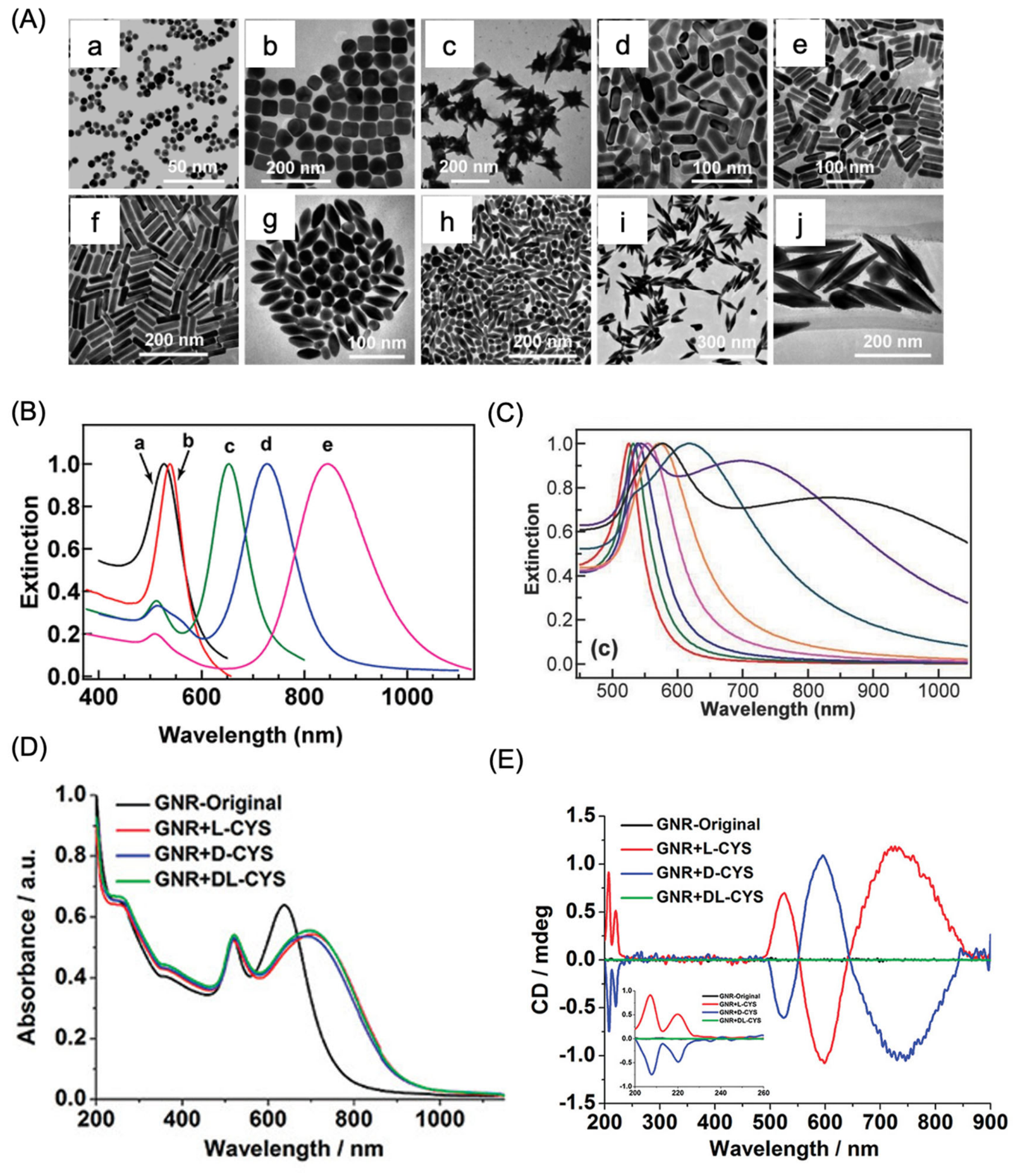
Figure 2.
Representative mechanisms underlying plasmon-enhanced chirality. (A) Schematic illustration of a plasmonic nanoparticle–molecule complex showing the relative nanoparticle–molecule geometry (i) and the associated light-induced (ii) and (iii) Coulomb-mediated electronic transitions responsible for plasmon-enhanced chiroptical interactions. (B) Simulated extinction (iv) and (v) circular dichroism (CD) spectra of a silver nanoparticle–molecule complex, demonstrating the influence of nanoparticle–molecule separation on the plasmon-enhanced CD response. (C) Schematic of a nanoparticle dimer coupled with a chiral molecule positioned within the interparticle gap (vi), (vii) together with calculated CD spectra illustrating the enhanced chiroptical response generated by plasmonic hotspots compared with a single nanoparticle. (D) Representative intrinsically chiral gold nanoparticles with opposite handedness and their corresponding calculated CD spectra, highlighting the effect of nanoparticle geometry on optical activity (viii). Panel A reproduced with permission from [45]; Copyright 2010 American Chemical Society. Panels B and C reproduced with permission from [46]; Copyright 2011 American Chemical Society. Panel D reproduced with permission from [47]; Copyright 2012 American Chemical Society.
Figure 2.
Representative mechanisms underlying plasmon-enhanced chirality. (A) Schematic illustration of a plasmonic nanoparticle–molecule complex showing the relative nanoparticle–molecule geometry (i) and the associated light-induced (ii) and (iii) Coulomb-mediated electronic transitions responsible for plasmon-enhanced chiroptical interactions. (B) Simulated extinction (iv) and (v) circular dichroism (CD) spectra of a silver nanoparticle–molecule complex, demonstrating the influence of nanoparticle–molecule separation on the plasmon-enhanced CD response. (C) Schematic of a nanoparticle dimer coupled with a chiral molecule positioned within the interparticle gap (vi), (vii) together with calculated CD spectra illustrating the enhanced chiroptical response generated by plasmonic hotspots compared with a single nanoparticle. (D) Representative intrinsically chiral gold nanoparticles with opposite handedness and their corresponding calculated CD spectra, highlighting the effect of nanoparticle geometry on optical activity (viii). Panel A reproduced with permission from [45]; Copyright 2010 American Chemical Society. Panels B and C reproduced with permission from [46]; Copyright 2011 American Chemical Society. Panel D reproduced with permission from [47]; Copyright 2012 American Chemical Society.
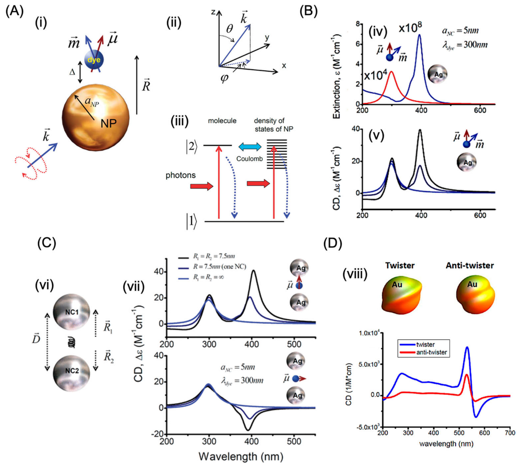
Figure 3.
Representative examples of ligand-induced chiral noble metal nanoclusters. (A) Molecular structure of atomically precise Au₂₅(PET)₁₈* nanoclusters, where PET* represents SCH₂C*H(Me)Ph. (B) Circular dichroism (CD) spectra of the corresponding R- and S-enantiomeric Au₂₅ nanoclusters, demonstrating mirror-image chiroptical responses arising from chiral ligand coordination. (C) CD (left) and UV–Vis absorption (right) spectra of silver nanoclusters stabilized with different ligand compositions, including captopril, glutathione, and a mixed captopril/glutathione system (molar ratio 62:38). The inset illustrates the molecular structure of captopril. (D) Chiroptical properties of BINAP-functionalized gold nanoparticles. The molecular structure of 2,2′-bis(diphenylphosphino)-1,1′-binaphthyl (BINAP) is shown together with (i) transmission electron microscopy (TEM) images of BINAP-modified Au nanoparticles and the CD spectra of (ii) (R)-BINAP-Au and (iii) (S)-BINAP-Au dispersed in chloroform, highlighting their opposite optical activities. Panel A-B reproduced with permission from [78]; Copyright 2011, American Chemical Society. Panel C reproduced with permission from [79]; Copyright 2010, American Chemical society. Panel D reproduced with permission from [80]; Copy right 2003 , American Chemical Society.
Figure 3.
Representative examples of ligand-induced chiral noble metal nanoclusters. (A) Molecular structure of atomically precise Au₂₅(PET)₁₈* nanoclusters, where PET* represents SCH₂C*H(Me)Ph. (B) Circular dichroism (CD) spectra of the corresponding R- and S-enantiomeric Au₂₅ nanoclusters, demonstrating mirror-image chiroptical responses arising from chiral ligand coordination. (C) CD (left) and UV–Vis absorption (right) spectra of silver nanoclusters stabilized with different ligand compositions, including captopril, glutathione, and a mixed captopril/glutathione system (molar ratio 62:38). The inset illustrates the molecular structure of captopril. (D) Chiroptical properties of BINAP-functionalized gold nanoparticles. The molecular structure of 2,2′-bis(diphenylphosphino)-1,1′-binaphthyl (BINAP) is shown together with (i) transmission electron microscopy (TEM) images of BINAP-modified Au nanoparticles and the CD spectra of (ii) (R)-BINAP-Au and (iii) (S)-BINAP-Au dispersed in chloroform, highlighting their opposite optical activities. Panel A-B reproduced with permission from [78]; Copyright 2011, American Chemical Society. Panel C reproduced with permission from [79]; Copyright 2010, American Chemical society. Panel D reproduced with permission from [80]; Copy right 2003 , American Chemical Society.
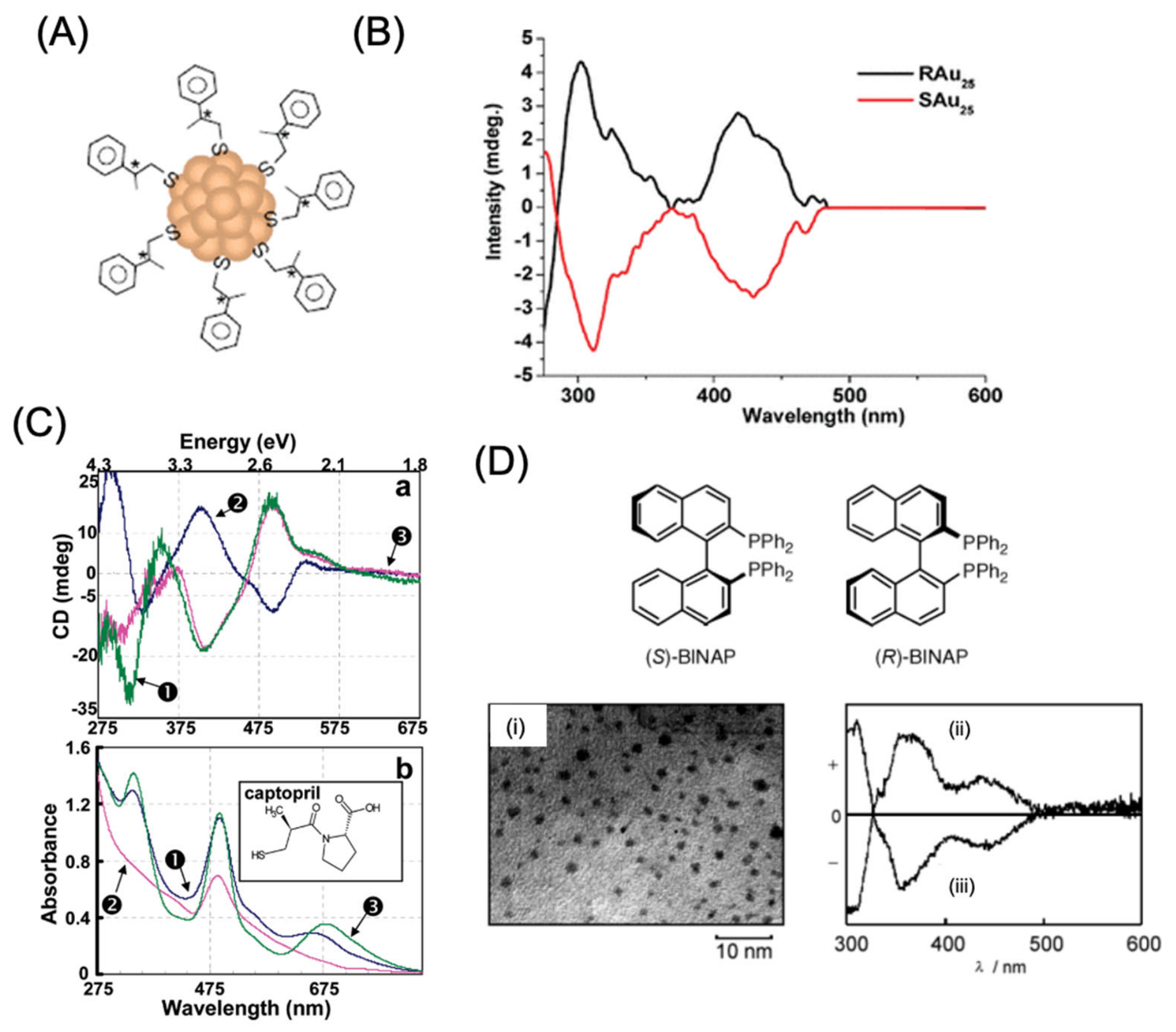
Figure 4.
Chiral nanostructures using seed mediated method. (A,B) Schematic illustration showing the structural evolution of stellated octahedral (A) and rhombic dodecahedral (B) gold nanocrystals containing right-handed (R) and left-handed (S) high-index surface domains. The presence of chiral ligands, including cysteine and glutathione, directs asymmetric crystal growth, resulting in distinct helicoidal plasmonic nanostructures. (C–F) Representative scanning electron microscopy (SEM) images together with the corresponding UV–Vis absorption and circular dichroism (CD) spectra of the synthesized chiral gold nanoparticles: 432 Helicoid I (C), 432 Helicoid II (D), 432 Helicoid III (E), and 432 Helicoid IV (F). Panel A-F reproduced with permission from [53]; Copy right, 2018, Nature publishing group.
Figure 4.
Chiral nanostructures using seed mediated method. (A,B) Schematic illustration showing the structural evolution of stellated octahedral (A) and rhombic dodecahedral (B) gold nanocrystals containing right-handed (R) and left-handed (S) high-index surface domains. The presence of chiral ligands, including cysteine and glutathione, directs asymmetric crystal growth, resulting in distinct helicoidal plasmonic nanostructures. (C–F) Representative scanning electron microscopy (SEM) images together with the corresponding UV–Vis absorption and circular dichroism (CD) spectra of the synthesized chiral gold nanoparticles: 432 Helicoid I (C), 432 Helicoid II (D), 432 Helicoid III (E), and 432 Helicoid IV (F). Panel A-F reproduced with permission from [53]; Copy right, 2018, Nature publishing group.
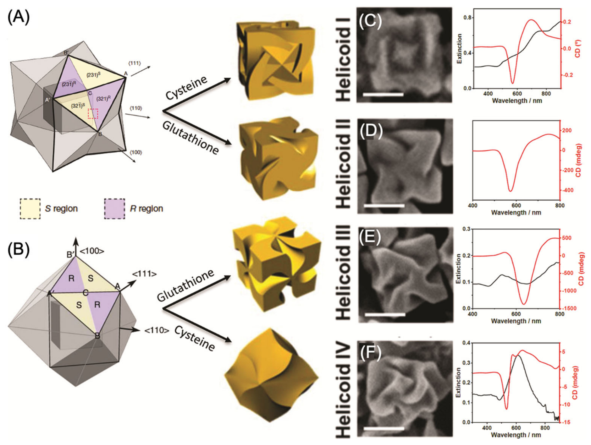
Figure 5.
Growth mechanism and chiroptical characteristics of seed-mediated Au@Ag chiral nanorods. (A) Schematic illustration depicting the proposed evolution of Au@Ag nanostructures into truncated octahedral morphologies during silver shell growth. (B) Proposed growth pathway highlighting successive stages of Ag deposition. Newly formed Ag atomic layers are represented by black lines, while the {111} crystallographic facets are indicated by red dashed lines. Silver nucleates on the Au nanorod surface, generating localized surface irregularities (green dashed lines) that progressively spread across the growing shell as growth fronts (green arrows) while Ag precursors are continuously consumed. (C) Representative STEM image of chiral rod 5 (CR5) showing the final particle morphology (scale bar: 100 nm). (D) Correlation between the maximum chiroptical g-factor and the geometric dimensions of the Au nanorod seeds, demonstrating the influence of seed morphology on the optical dissymmetry of the resulting Au@Ag nanostructures. Reproduced with permission from [89]; Copyright 2023, Wiley Publishers.
Figure 5.
Growth mechanism and chiroptical characteristics of seed-mediated Au@Ag chiral nanorods. (A) Schematic illustration depicting the proposed evolution of Au@Ag nanostructures into truncated octahedral morphologies during silver shell growth. (B) Proposed growth pathway highlighting successive stages of Ag deposition. Newly formed Ag atomic layers are represented by black lines, while the {111} crystallographic facets are indicated by red dashed lines. Silver nucleates on the Au nanorod surface, generating localized surface irregularities (green dashed lines) that progressively spread across the growing shell as growth fronts (green arrows) while Ag precursors are continuously consumed. (C) Representative STEM image of chiral rod 5 (CR5) showing the final particle morphology (scale bar: 100 nm). (D) Correlation between the maximum chiroptical g-factor and the geometric dimensions of the Au nanorod seeds, demonstrating the influence of seed morphology on the optical dissymmetry of the resulting Au@Ag nanostructures. Reproduced with permission from [89]; Copyright 2023, Wiley Publishers.
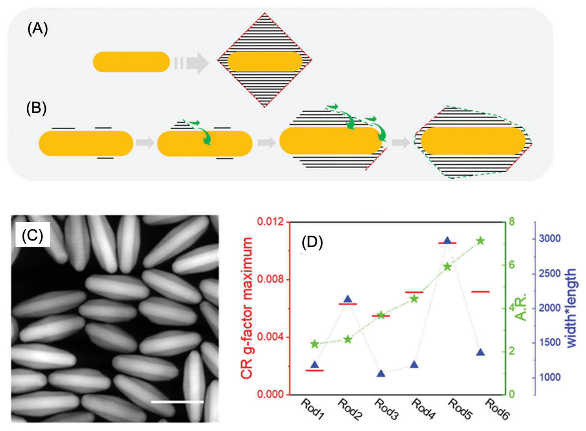
Figure 8.
Bottom-up fabrication of chiral plasmonic nanofilms. (A) Photograph of a macroscopic chiral nanofilm. (B) SEM image of the left-handed (L-type) nanofilm. (C) Circular dichroism (CD) spectra demonstrating the enhanced chiroptical response of the assembled nanofilm. (D) Schematic illustration of a two-dimensional (2D) helicoid crystal assembled from chiral Au nanoparticles. (E) SEM image showing the hexagonal arrangement of helicoids into a periodic 2D crystal. (F) CD spectra illustrating the enhancement of collective plasmonic resonances in the 2D helicoid assembly. Panel A-C reproduced with permission from [118]; Copyright 2022, Nature Publishers. Panel D-F reproduced with permission from [114]; Copyright 2022, Nature Publishers.
Figure 8.
Bottom-up fabrication of chiral plasmonic nanofilms. (A) Photograph of a macroscopic chiral nanofilm. (B) SEM image of the left-handed (L-type) nanofilm. (C) Circular dichroism (CD) spectra demonstrating the enhanced chiroptical response of the assembled nanofilm. (D) Schematic illustration of a two-dimensional (2D) helicoid crystal assembled from chiral Au nanoparticles. (E) SEM image showing the hexagonal arrangement of helicoids into a periodic 2D crystal. (F) CD spectra illustrating the enhancement of collective plasmonic resonances in the 2D helicoid assembly. Panel A-C reproduced with permission from [118]; Copyright 2022, Nature Publishers. Panel D-F reproduced with permission from [114]; Copyright 2022, Nature Publishers.
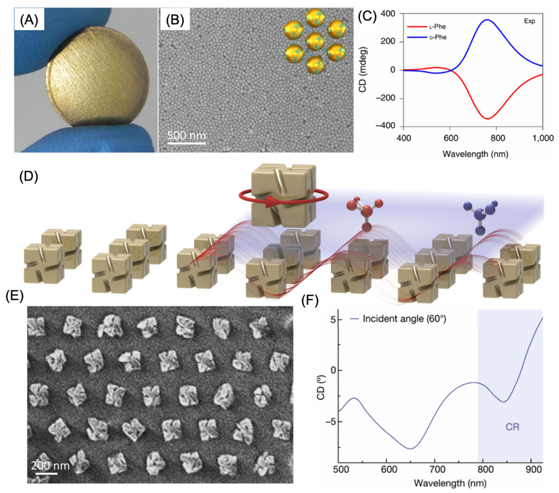
Figure 9.
Chiral plasmonic nanostructures for enantioselective sensing and intracellular miRNA detection. (A) Schematic representation of the Au–UCNP pyramidal nanoassembly designed for intracellular miRNA sensing. (B) DNA framework employed to direct the assembly of the pyramidal nanostructure in the presence of the target miRNA. (C) Circular dichroism (CD) spectra and (D) upconversion luminescence spectra of individual Au nanoparticles, UCNPs, DNA-functionalized Au nanoparticles and UCNPs, Au nanoparticle dimers, UCNP dimers, Au NP/UCNP mixtures, and assembled pyramidal nanostructures in 10 mM Tris buffer and cell culture medium under 980 nm near-infrared (NIR) laser irradiation (0.8 W). (E) Calibration curve showing the relationship between the CD intensity at 521 nm and intracellular miR-21 concentration. (F) Illustration of the experimental setup used for chiral sensing with a two-dimensional helicoid crystal mounted in a cuvette (optical path length ≈ 8.9 mm) containing the analyte solution. (G) CD spectra of the helicoid crystal recorded in deionized water and in solutions containing 0.1–1.0 M L-proline or D-proline. Unlike the localized surface plasmon resonance (LSPR) band (~650 nm), which remains nearly unchanged, the collective CD bands (~825 and 910 nm) exhibit pronounced spectral variations upon exposure to the two enantiomers. (H) Differential collective CD response (ΔCD = CDMolecule − CDDW) induced by 1.0 M L-proline and D-proline. The magnitude of the chiral response was determined from the difference between the maximum and minimum ΔCD values (ΔCDmax − ΔCDmin) revealing a significantly stronger modulation of the collective chiroptical response by D-proline than by L-proline. Panel A-E reproduced from [137]; Copyright, 2015, American Chemical Society. Panel F-H reproduced from [114]; Copyright, 2022 Nature publishing group.
Figure 9.
Chiral plasmonic nanostructures for enantioselective sensing and intracellular miRNA detection. (A) Schematic representation of the Au–UCNP pyramidal nanoassembly designed for intracellular miRNA sensing. (B) DNA framework employed to direct the assembly of the pyramidal nanostructure in the presence of the target miRNA. (C) Circular dichroism (CD) spectra and (D) upconversion luminescence spectra of individual Au nanoparticles, UCNPs, DNA-functionalized Au nanoparticles and UCNPs, Au nanoparticle dimers, UCNP dimers, Au NP/UCNP mixtures, and assembled pyramidal nanostructures in 10 mM Tris buffer and cell culture medium under 980 nm near-infrared (NIR) laser irradiation (0.8 W). (E) Calibration curve showing the relationship between the CD intensity at 521 nm and intracellular miR-21 concentration. (F) Illustration of the experimental setup used for chiral sensing with a two-dimensional helicoid crystal mounted in a cuvette (optical path length ≈ 8.9 mm) containing the analyte solution. (G) CD spectra of the helicoid crystal recorded in deionized water and in solutions containing 0.1–1.0 M L-proline or D-proline. Unlike the localized surface plasmon resonance (LSPR) band (~650 nm), which remains nearly unchanged, the collective CD bands (~825 and 910 nm) exhibit pronounced spectral variations upon exposure to the two enantiomers. (H) Differential collective CD response (ΔCD = CDMolecule − CDDW) induced by 1.0 M L-proline and D-proline. The magnitude of the chiral response was determined from the difference between the maximum and minimum ΔCD values (ΔCDmax − ΔCDmin) revealing a significantly stronger modulation of the collective chiroptical response by D-proline than by L-proline. Panel A-E reproduced from [137]; Copyright, 2015, American Chemical Society. Panel F-H reproduced from [114]; Copyright, 2022 Nature publishing group.
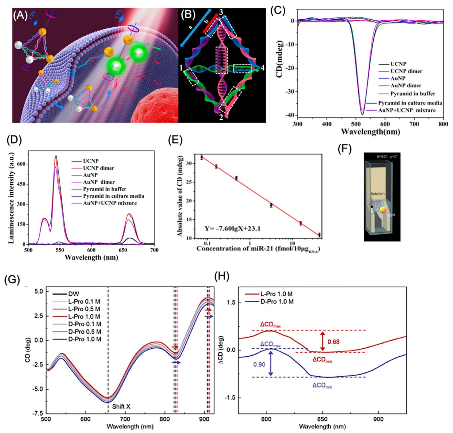
Figure 10.
Chiral plasmonic SERS platforms for enantiomer discrimination and chiral sensing. (A) Schematic of a conventional Raman system integrated with a chiral plasmonic substrate. (B) SERS-based chiral anisotropy (SERS-ChA) measurement and determination of the dissymmetry (g) factor for enantiomeric analytes. (C) Calibration strategy for identifying absolute configuration and enantiomeric excess (ee) of unknown samples. (D,E) Representative Raman spectra and correlation between SERS peak intensity and ee values (−100% to +100%) for 2-butanol. (F) FE-SEM images of chiral Au nanostructures synthesized in the presence of six different L- or D-amino acids (scale bar: 50 nm). (G) Colorimetric response of L-Trp Helicoid I after exposure to increasing concentrations of L-cysteine (0–10 µM). (H) Raman spectra of L-Trp Helicoid I in the presence of pure L-Cys, pure D-Cys, and L/D-Cys mixtures with different enantiomeric compositions. Panel A-E reproduced with permission from [146]; Copyright , 2020, Wiley Publishing group. Panel F-H reproduced with permission from [54]; Copyright, 2023, Wiley Publishing group.
Figure 10.
Chiral plasmonic SERS platforms for enantiomer discrimination and chiral sensing. (A) Schematic of a conventional Raman system integrated with a chiral plasmonic substrate. (B) SERS-based chiral anisotropy (SERS-ChA) measurement and determination of the dissymmetry (g) factor for enantiomeric analytes. (C) Calibration strategy for identifying absolute configuration and enantiomeric excess (ee) of unknown samples. (D,E) Representative Raman spectra and correlation between SERS peak intensity and ee values (−100% to +100%) for 2-butanol. (F) FE-SEM images of chiral Au nanostructures synthesized in the presence of six different L- or D-amino acids (scale bar: 50 nm). (G) Colorimetric response of L-Trp Helicoid I after exposure to increasing concentrations of L-cysteine (0–10 µM). (H) Raman spectra of L-Trp Helicoid I in the presence of pure L-Cys, pure D-Cys, and L/D-Cys mixtures with different enantiomeric compositions. Panel A-E reproduced with permission from [146]; Copyright , 2020, Wiley Publishing group. Panel F-H reproduced with permission from [54]; Copyright, 2023, Wiley Publishing group.
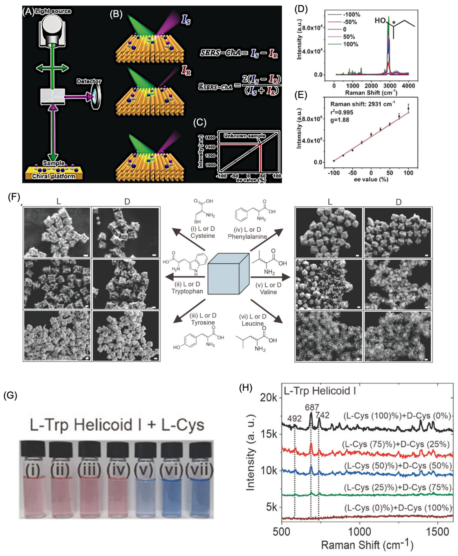
Figure 11.
(A) Schematic illustration of circularly polarized light (CPL)-driven nitrogen photofixation using left- and right-handed Au@CeO₂ helical nanorods (L/D-Au@CeO₂ HNRs). (B) Comparison of photocatalytic N₂ fixation rates for L/D-Au@CeO₂ HNRs under linearly polarized light (LPL), left circularly polarized light (LCPL), and right circularly polarized light (RCPL). (C) Ammonia generation by L- and D-Au@CeO₂ HNRs under LCPL and RCPL irradiation, demonstrating chirality-dependent photocatalytic performance (mean ± SD, n = 3). (D) Fabrication strategy for chiral plasmonic photocatalysts through the assembly of Au and TiO₂ nanoparticles onto helical SiO₂ nanoribbon templates. (E) Mechanism illustrating polarization-selective plasmonic excitation, where matching the handedness of the nanostructure with the incident CPL enhances hot-electron transfer from Au to the TiO₂ conduction band. (F, G) Photocatalytic degradation of rhodamine B (RhB) under LCPL and RCPL using chiral SiO₂@Au@TiO₂ hybrid nanoribbons. Photoreactions were conducted under a 150 W xenon lamp equipped with CPL filters (400–800 nm) at 20 °C for 8 h. Data represent three independent experiments for L-SiO₂@Au@TiO₂ and two independent experiments for R-SiO₂@Au@TiO₂. Panel A-C Reproduced with permission from [157]; Copyright 2025, Wiley Publishing group. Panel D-G reproduced with permission from [158]; Copyright, 2022, American Chemical Society. .
Figure 11.
(A) Schematic illustration of circularly polarized light (CPL)-driven nitrogen photofixation using left- and right-handed Au@CeO₂ helical nanorods (L/D-Au@CeO₂ HNRs). (B) Comparison of photocatalytic N₂ fixation rates for L/D-Au@CeO₂ HNRs under linearly polarized light (LPL), left circularly polarized light (LCPL), and right circularly polarized light (RCPL). (C) Ammonia generation by L- and D-Au@CeO₂ HNRs under LCPL and RCPL irradiation, demonstrating chirality-dependent photocatalytic performance (mean ± SD, n = 3). (D) Fabrication strategy for chiral plasmonic photocatalysts through the assembly of Au and TiO₂ nanoparticles onto helical SiO₂ nanoribbon templates. (E) Mechanism illustrating polarization-selective plasmonic excitation, where matching the handedness of the nanostructure with the incident CPL enhances hot-electron transfer from Au to the TiO₂ conduction band. (F, G) Photocatalytic degradation of rhodamine B (RhB) under LCPL and RCPL using chiral SiO₂@Au@TiO₂ hybrid nanoribbons. Photoreactions were conducted under a 150 W xenon lamp equipped with CPL filters (400–800 nm) at 20 °C for 8 h. Data represent three independent experiments for L-SiO₂@Au@TiO₂ and two independent experiments for R-SiO₂@Au@TiO₂. Panel A-C Reproduced with permission from [157]; Copyright 2025, Wiley Publishing group. Panel D-G reproduced with permission from [158]; Copyright, 2022, American Chemical Society. .
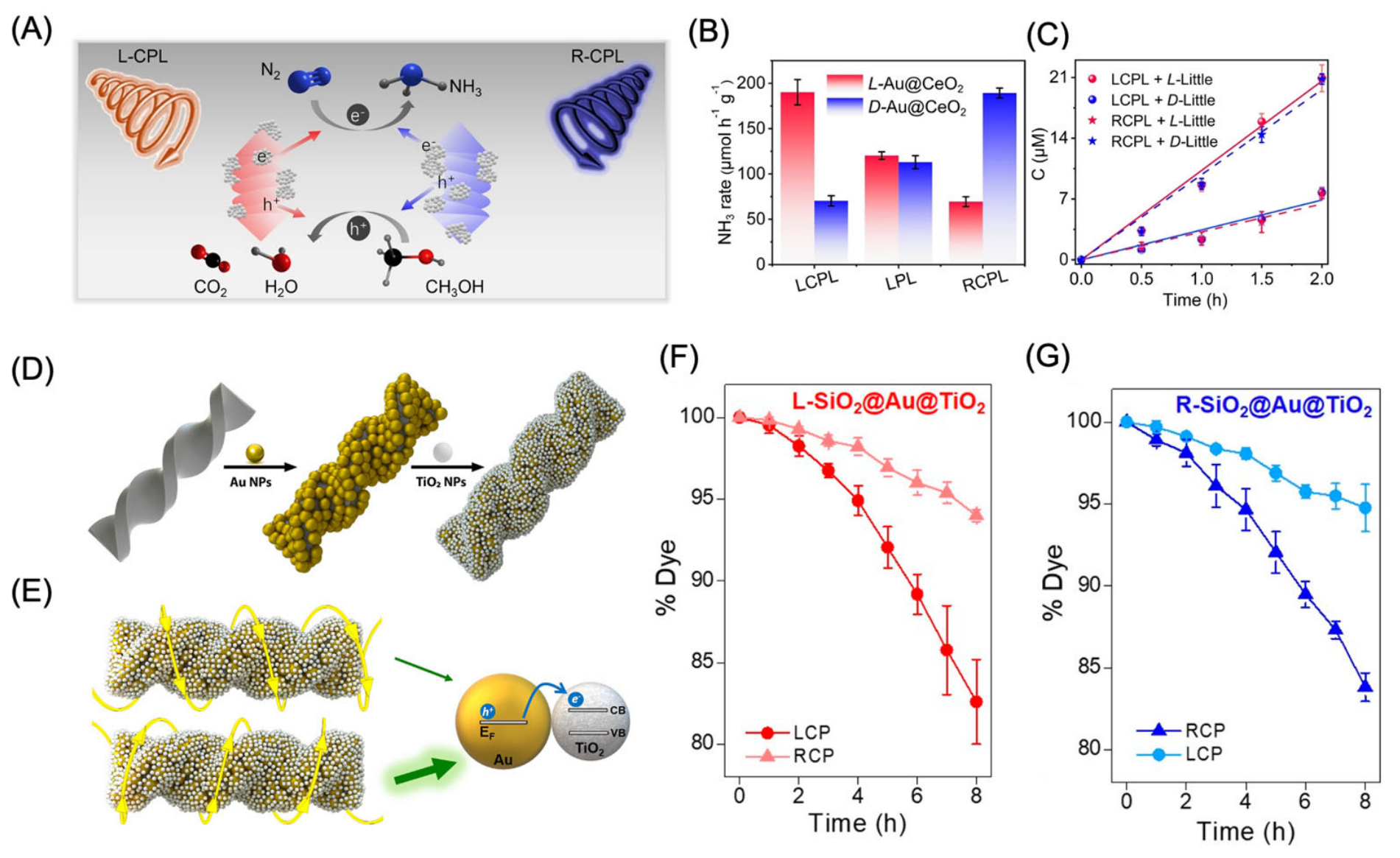
Figure 14.
Chiral nanomaterials for photothermal cancer therapy. (A) Schematic representation of the synthesis of D-/L-penicillamine-functionalized Cu₂−xSe nanoparticles. (B) Representative infrared thermal images of tumors following different treatment groups. (C) Tumor growth curves demonstrating the therapeutic efficacy of the various treatments (p < 0.05, p < 0.01, p < 0.001). (D) Preparation of chiral Cys-MoO₃−x nanoparticles and their application in circularly polarized light (CPL)-mediated photothermal therapy. Pristine MoS₂ was first oxidized to MoO₃ nanodots, followed by chiral cysteine-mediated reduction to generate Cys-MoO₃−x nanoparticles, which were subsequently employed for visible- and near-infrared (NIR)-driven photothermal ablation of tumor cells. (E) Representative LIVE/DEAD fluorescence images of HeLa cells treated with D-Cys-MoO₂.₈ or L-Cys-MoO₂.₈ nanoparticles (150 μg mL⁻¹) after irradiation with right circularly polarized (RCP), linearly polarized (LP), or left circularly polarized (LCP) 808 nm laser light (1 W cm⁻², 15 min). Scale bar = 100 μm. Panel A-C reproduced with permission from [196], Copyright 2021, American Chemical Society. Panel D-E reproduced with permission from [198]; Copyright 2020, Wiley Publishing group.
Figure 14.
Chiral nanomaterials for photothermal cancer therapy. (A) Schematic representation of the synthesis of D-/L-penicillamine-functionalized Cu₂−xSe nanoparticles. (B) Representative infrared thermal images of tumors following different treatment groups. (C) Tumor growth curves demonstrating the therapeutic efficacy of the various treatments (p < 0.05, p < 0.01, p < 0.001). (D) Preparation of chiral Cys-MoO₃−x nanoparticles and their application in circularly polarized light (CPL)-mediated photothermal therapy. Pristine MoS₂ was first oxidized to MoO₃ nanodots, followed by chiral cysteine-mediated reduction to generate Cys-MoO₃−x nanoparticles, which were subsequently employed for visible- and near-infrared (NIR)-driven photothermal ablation of tumor cells. (E) Representative LIVE/DEAD fluorescence images of HeLa cells treated with D-Cys-MoO₂.₈ or L-Cys-MoO₂.₈ nanoparticles (150 μg mL⁻¹) after irradiation with right circularly polarized (RCP), linearly polarized (LP), or left circularly polarized (LCP) 808 nm laser light (1 W cm⁻², 15 min). Scale bar = 100 μm. Panel A-C reproduced with permission from [196], Copyright 2021, American Chemical Society. Panel D-E reproduced with permission from [198]; Copyright 2020, Wiley Publishing group.
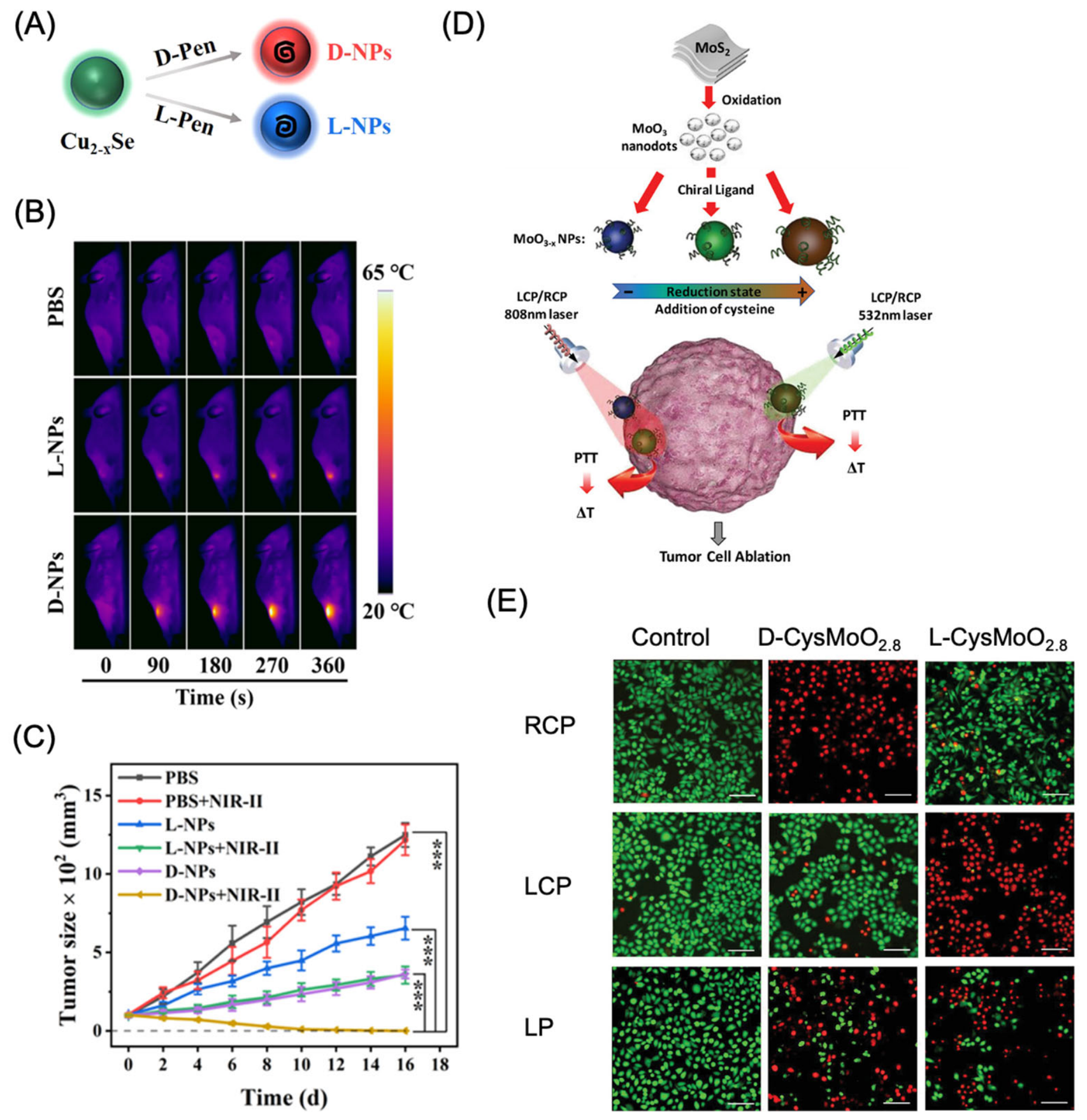
Figure 15.
Chiral nanoparticles for immunotherapy applications. (A) Schematic illustration of the interaction between chiral nanoparticles and the extracellular epidermal growth factor (EGF)-like domains of adhesion G protein-coupled receptors (aGPCRs). (B) Proposed pathway illustrating chiral nanoparticle-mediated activation of the immune response. (C) Relative mean fluorescence intensity (MFI) of mouse BMDCs after treatment with receptor and endocytosis inhibitors, revealing that chiral nanoparticle internalization primarily occurs through CD97/EMR1-mediated, clathrin- and dynamin-dependent endocytosis. (D,E) Time-dependent confocal fluorescence images of mouse bone marrow-derived dendritic cells (BMDCs) incubated with 2 μg mL⁻¹ MPL, 20 μg mL⁻¹ OVA, and 2 nM L-P⁺ nanoparticles for up to 4 h, showing the colocalization of nanoparticles with (F) CD97 and (G) EMR1 receptors. Nuclei were stained with DAPI (blue), CD97 or EMR1 with Cy5 (red), and L-P⁺ nanoparticles with Cy3 (green). Scale bar = 10 μm. (F) Two-photon laser (TPL) fluorescence images of mouse BMDCs incubated with 2 nM L-P⁺ nanoparticles for different time intervals (0–4 h), illustrating intracellular trafficking and colocalization with dynamin (orange) and clathrin (pink). Nanoparticles are shown in white, and nuclei were counterstained with DAPI (blue). Scale bar = 10 μm. Reproduced with permission from [120]. Copyright 2022, Nature Publishing group.
Figure 15.
Chiral nanoparticles for immunotherapy applications. (A) Schematic illustration of the interaction between chiral nanoparticles and the extracellular epidermal growth factor (EGF)-like domains of adhesion G protein-coupled receptors (aGPCRs). (B) Proposed pathway illustrating chiral nanoparticle-mediated activation of the immune response. (C) Relative mean fluorescence intensity (MFI) of mouse BMDCs after treatment with receptor and endocytosis inhibitors, revealing that chiral nanoparticle internalization primarily occurs through CD97/EMR1-mediated, clathrin- and dynamin-dependent endocytosis. (D,E) Time-dependent confocal fluorescence images of mouse bone marrow-derived dendritic cells (BMDCs) incubated with 2 μg mL⁻¹ MPL, 20 μg mL⁻¹ OVA, and 2 nM L-P⁺ nanoparticles for up to 4 h, showing the colocalization of nanoparticles with (F) CD97 and (G) EMR1 receptors. Nuclei were stained with DAPI (blue), CD97 or EMR1 with Cy5 (red), and L-P⁺ nanoparticles with Cy3 (green). Scale bar = 10 μm. (F) Two-photon laser (TPL) fluorescence images of mouse BMDCs incubated with 2 nM L-P⁺ nanoparticles for different time intervals (0–4 h), illustrating intracellular trafficking and colocalization with dynamin (orange) and clathrin (pink). Nanoparticles are shown in white, and nuclei were counterstained with DAPI (blue). Scale bar = 10 μm. Reproduced with permission from [120]. Copyright 2022, Nature Publishing group.
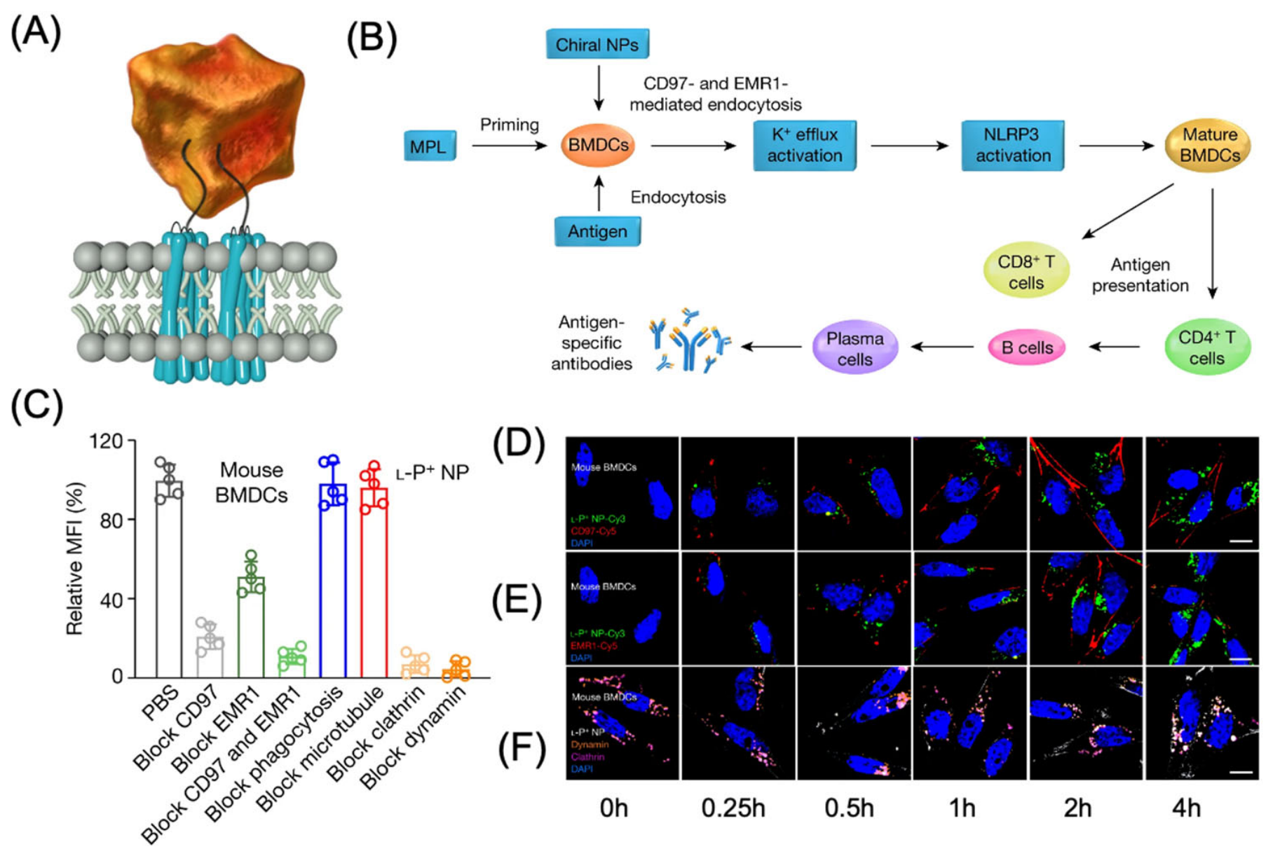
Table 1.
Representative chiral plasmonic nanostructures, chiral ligands, optical activity, and representative applications.
Table 1.
Representative chiral plasmonic nanostructures, chiral ligands, optical activity, and representative applications.
| Nanostructure | Chiral Induction Strategy | Typical g-factor | Representative Application | Ref. |
|---|---|---|---|---|
| Au Octahedra | L/D-Cys, GSH, | 0.20 | Polarization control, chiral optics | [53] |
| Au Nanocubes | L/D-Cys, Trp, Tyr | 0.03, 0.018 and 0.011 | SERS, plasmonics | [54] |
| Au Octahedra | peptide ligands (g-Glu-cys/Cys-Gly) | 0.02 | Chiral optics | [55] |
| Au Octahedra | ssDNA | 0.04 | Chiral optics | [56] |
| Au Decahedra | CYP/GSH | — | Chiral plasmonics | [57] |
| Au Nanorods | L/D-Cys, 4-ATP | 0.03-0.1 |
CPL generation, optical modulation | [58,59] |
| Au Nanotriangles | CYP, Cys | 0.08–0.44 | Cancer immunotherapy | [60,61] |
| Au Nanodiscs | GSH | 0.10 | Chiral optics | [62] |
| Rhombicuboctahedra | CYP | 0.01 | Plasmonic chirality | [63] |
| Trisoctahedra | Cys | 0.02 | Enantiomer discrimination | [64] |
Where: Cys: Cysteine, GSH: Glutathione, CPT: Trp: Tryptophan, gly: Glycine, CYP: Cysteine-Phenylalanine, ssDNA: Single stranded DNA.
Table 2.
Representative synthetic strategies for chiral plasmonic nanomaterials: principles, advantages, limitations, and representative examples.
Table 2.
Representative synthetic strategies for chiral plasmonic nanomaterials: principles, advantages, limitations, and representative examples.
| Synthesis strategy | Principle | Representative plasmonic nanomaterials | Advantages | Limitations | Typical applications |
|---|---|---|---|---|---|
| Chiral ligand-directed growth | Chiral molecules selectively adsorb on specific crystal facets, directing asymmetric nanoparticle growth. | Au, Ag, Au–Ag nanostructures | Simple synthesis; scalable; tunable chirality; strong chiroptical activity | Ligand dependence; moderate stability; mechanism not fully understood | Enantioselective sensing, catalysis, bioimaging |
| DNA-programmed self-assembly | DNA hybridization precisely organizes plasmonic nanoparticles into chiral architectures. | Au nanoparticle assemblies, Au@Ag satellites | Precise structural control; programmable geometry; large CD response | High cost; limited stability in biological media; complex synthesis | Biosensing, PAI, photodynamic therapy |
| Peptide/protein-guided assembly | Biomolecules serve as chiral templates for nanoparticle nucleation and organization. | Au, Ag nanoparticles | Excellent biocompatibility; biomimetic synthesis; aqueous conditions | Lower structural reproducibility; sensitive to environmental conditions | Biosensing, bioimaging, drug delivery |
| Template-assisted synthesis | Hard or soft templates (silica helices, liquid crystals, polymers) guide formation of chiral plasmonic structures. | Helical Au, Ag nanostructures | High structural uniformity; controllable morphology | Multi-step fabrication; template removal may be required | Chiroptical devices, catalysis, optical materials |
| Seed-mediated asymmetric growth | Preformed seeds undergo anisotropic growth under chiral induction. | Au nanorods, nanocubes, bipyramids | High crystallinity; excellent shape control; scalable | Sensitive to reaction conditions; reproducibility challenges | SERS, sensing, photothermal therapy |
| Galvanic replacement reaction | Sacrificial templates undergo controlled replacement to generate hollow or hybrid chiral nanostructures. | Au–Ag nanocages, hollow nanostructures | Hollow structures; tunable plasmon resonance; multifunctionality | Difficult to precisely control composition and chirality | Catalysis, imaging, phototherapy |
| Magnetic-field-induced assembly | External magnetic fields orient nanoparticles into chiral superstructures. | Au–Fe₃O₄ hybrids | Reversible assembly; dynamic chirality; external control | Requires magnetic components; limited structural complexity | Magneto-optics, MRI, responsive materials |
| Circularly polarized light (CPL)-assisted synthesis | CPL induces enantioselective growth or assembly of plasmonic nanostructures. | Au nanostructures | No molecular chiral inducer required; direct optical control | Low yield; specialized optical setup; limited scalability | Chiroptics, optical information processing |
Table 4.
Application of chiral plasmonic materials for neuro and age-related diseases.
| Neurological Disorder / Biological Target | Representative Chiral Nanoplatform | Chirality-Associated Advantages | Primary Therapeutic Mechanism | Key Therapeutic Benefit | Ref |
|---|---|---|---|---|---|
| Alzheimer’s disease (Aβ pathology) | Glutathione-capped Au nanoparticles; D-/L-penicillamine-functionalized Fe–Cu–Se nanoparticles | Enantiomer-dependent interaction with Aβ aggregates, improved BBB penetration, enhanced ROS regulation | Disassembly of amyloid fibrils, ROS-mediated plaque clearance, neuroprotection | Lower amyloid burden and improved cognitive performance in AD models | [219,220,221] |
| Parkinson’s disease (α-synuclein pathology) | Chiral CuₓCoᵧ superparticles; Mn₃O₄ nanorods; porous Cu₂O nanoclusters | Chirality influences ROS scavenging efficiency and protein recognition | Suppression of α-synuclein aggregation, oxidative stress reduction, NIR-assisted fibril disruption | Restoration of motor function and neuronal protection in PD models | [217,222] |
| Neuroinflammation (Senescent microglia) | Antibody-conjugated chiral gold nanoparticles | Selective recognition and elimination of senescent microglial cells | Activation of Fas-mediated apoptosis and targeted clearance of dysfunctional microglia | Reduced neuroinflammation and improved cognitive function | [212] |
| Neural regeneration | Chiral Au nanofilms; CPL-responsive DNA nanoassemblies | Chirality and circularly polarized light promote neuronal adhesion and differentiation | Mechanical stimulation and activation of neuronal signaling pathways | Accelerated neural stem cell differentiation and enhanced tissue regeneration | [213,214] |
| Cellular senescence and aging | Chiral core–shell nanorods; CuₓCoᵧ nanoparticles | D-enantiomer exhibits superior cellular uptake and senolytic performance | Mitochondrial dysfunction induction, ROS generation, caspase activation | Efficient removal of senescent cells and improvement of age-related tissue function | [216] |
| Photoresponsive senolytic theranostics | Chiral UCNP–AuNP–DNA tetrahedral nanostructures | Chirality enhances targeting accuracy and phototherapeutic responsiveness | NIR-triggered DNA cleavage, controlled granzyme B release, FRET-guided imaging | Simultaneous imaging and selective elimination of senescent cells | [218] |
Disclaimer/Publisher’s Note: The statements, opinions and data contained in all publications are solely those of the individual author(s) and contributor(s) and not of MDPI and/or the editor(s). MDPI and/or the editor(s) disclaim responsibility for any injury to people or property resulting from any ideas, methods, instructions or products referred to in the content. |
© 2026 by the authors. Licensee MDPI, Basel, Switzerland. This article is an open access article distributed under the terms and conditions of the Creative Commons Attribution (CC BY) license (http://creativecommons.org/licenses/by/4.0/).
Copyright: This open access article is published under a Creative Commons CC BY 4.0 license, which permit the free download, distribution, and reuse, provided that the author and preprint are cited in any reuse.