

Submitted:
05 August 2026
Posted:
07 August 2026
You are already at the latest version
Abstract
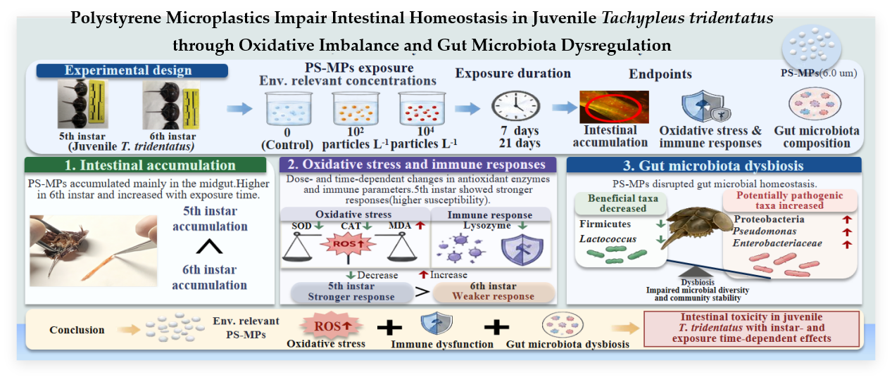
Microplastics (MPs) are ubiquitous marine pollutants that pose increasing ecological risks, yet their effects on intestinal physiology and gut microbial homeostasis during the juvenile developmental stages of Tachypleus tridentatus remain unclear. In this study, fifth- and sixth-instar juveniles of T. tridentatus were exposed to environmentally relevant concentrations of 6.0 μm polystyrene microplastics (PS-MPs; 0, 10², and 10⁴ particles/L) for 7 and 21 days to investigate intestinal toxicity and its underlying mechanisms. Intestinal accumulation, oxidative stress, innate immune responses, and gut microbiota were comprehensively evaluated. PS-MPs exposure induced dose- and time-dependent alterations in superoxide dismutase, catalase, malondialdehyde, and lysozyme, indicating excessive oxidative stress, impaired antioxidant defense, and compromised innate immunity. Fifth-instar juveniles exhibited stronger oxidative stress and immune responses than sixth-instar individuals, suggesting higher susceptibility during earlier developmental stages. Gut microbiota analysis revealed pronounced dysbiosis, characterized by a reduced relative abundance of Firmicutes, enrichment of Proteobacteria, depletion of beneficial taxa (e.g., Lactococcus), and increased abundance of opportunistic bacteria, including Pseudomonas and members of Enterobacteriaceae. These physiological and microbial alterations collectively suggest that environmentally relevant PS-MPs impair intestinal homeostasis in juvenile T. tridentatus by inducing oxidative imbalance, modifying innate immune responses, and reshaping gut microbial communities, with clear instar- and exposure time-dependent effects. These findings highlighting the importance of developmental stages in ecological risk assessment for benthic arthropods.
Keywords:
Tachypleus tridentatus
; polystyrene microplastics
; intestinal toxicity
; oxidative stress
; gut microbiota
; ecological risk
1. Introduction
Tachypleus tridentatus (Chinese horseshoe crab) is an ancient marine chelicerate and one of only four extant horseshoe crab species worldwide. Owing to its highly conserved morphology over more than 400 million years of evolution, it is widely regarded as a “living fossil” and has considerable value in evolutionary biology, comparative physiology, and biomedicine, particularly as the natural source of limulus amebocyte lysate (LAL) reagents [1]. Despite its ecological and biomedical importance, wild populations of T. tridentatus have experienced severe declines in recent decades because of habitat degradation, coastal development, and overexploitation, resulting in its classification as an endangered species [2]. Juvenile horseshoe crabs primarily inhabit estuarine, intertidal, and shallow coastal environments, where they remain closely associated with sediments during burrowing and feeding activities. These habitats are recognized as major sinks for microplastics, exposing juvenile horseshoe crabs to persistent sediment-associated contaminants throughout their early development [1,3]. Recent studies have indicated that microplastic exposure adversely affects growth, behaviour, and physiological performance in juvenile T. tridentatus, while early developmental stages appear particularly susceptible to contaminant-induced oxidative stress [4,5]. Collectively, these characteristics highlight T. tridentatus as an ecologically relevant model for evaluating the biological effects of microplastic pollution in coastal benthic ecosystems.
Among the diverse environmental pressures affecting coastal ecosystems, microplastic pollution has emerged as a pervasive threat to aquatic organisms. Microplastics (MPs, <5 mm) are ubiquitous contaminants that have been detected in marine waters, estuaries, and sediments worldwide, with concentrations in many coastal environments reaching environmentally relevant levels of 10²–10⁴ particles /L [6,7,8,9,10]. Polystyrene microplastics (PS-MPs), one of the most frequently detected polymer types in aquatic environments, exhibit high environmental persistence and physicochemical stability, enabling long-distance transport through hydrological processes and trophic transfer within aquatic food webs [11,12,13,14]. Consequently, PS-MPs have become an important focus of ecotoxicological research because of their widespread occurrence and increasing ecological risks to marine organisms. Although previous studies have documented adverse effects of MPs on growth, behaviour, and reproduction, their impacts on intestinal physiology and host–microbiota interactions remain incompletely understood, particularly in marine benthic arthropods.
Accumulating evidence indicates that the intestine is one of the primary target organs of microplastic toxicity in aquatic organisms. In addition to causing physical abrasion and obstruction within the digestive tract, MPs can induce excessive production of reactive oxygen species (ROS), leading to oxidative stress, impairment of antioxidant defense systems, and impairment of innate immune function [15]. As the largest interface between the host and the external environment, the intestine harbors complex microbial communities that are essential for nutrient metabolism, immune regulation, and maintenance of intestinal barrier integrity [16,17]. Disturbance of gut microbial homeostasis is frequently characterized by the depletion of beneficial microorganisms and enrichment of opportunistic pathogens, thereby exacerbating intestinal inflammation and oxidative damage and ultimately compromising intestinal homeostasis [18]. Consistent with this mechanism, microplastic exposure has been shown to induce intestinal inflammation accompanied by gut microbial dysbiosis in zebrafish [19]. However, most previous studies have focused on teleost fishes and adult organisms, whereas the intestinal responses of marine benthic arthropods, particularly during juvenile development, remain largely unexplored [20,21]. Consequently, the mechanisms through which environmentally relevant MPs affect intestinal physiology and gut microbial homeostasis in juvenile horseshoe crabs remain poorly understood.
Juvenile development represents a particularly vulnerable life-history stage in horseshoe crabs because of their Type III survivorship strategy, characterized by high fecundity but extremely low juvenile survival. The fifth- and sixth-instar stages encompass rapid somatic growth and physiological differentiation, during which the intestine plays a pivotal role in nutrient assimilation, immune maturation, and maintenance of host health. Although the gut microbiota of wild juvenile T. tridentatus has been preliminarily characterized [22], and several studies have evaluated antioxidant responses to nanoplastics and heavy metals in horseshoe crabs [23,24], important knowledge gaps remain. In particular, few studies have systematically investigated the accumulation of environmentally relevant PS-MPs within the gastrointestinal tract together with concurrent alterations in intestinal antioxidant capacity, innate immune responses, and gut microbial communities. Furthermore, whether susceptibility to microplastic exposure differs between juvenile developmental stages remains unknown. Addressing these knowledge gaps is essential for improving our understanding of the physiological vulnerability of endangered horseshoe crabs to emerging contaminants and for strengthening ecological risk assessments in coastal benthic ecosystems.
Unlike teleost fishes, horseshoe crabs represent an ancient lineage of marine chelicerates with distinct physiological characteristics, including hemolymph-based innate immunity and sediment-associated benthic lifestyles. Their close interaction with coastal sediments, where microplastics are increasingly accumulated, may result in unique exposure pathways and toxicological responses compared with pelagic organisms. Therefore, investigating microplastic-induced intestinal alterations in juvenile horseshoe crabs provides valuable insights into contaminant sensitivity in understudied marine invertebrate taxa.
Therefore, the present study exposed fifth- and sixth-instar juvenile T. tridentatus to environmentally relevant concentrations of polystyrene microplastics (0, 10², and 10⁴ particles/L) for 7 and 21 days. The objectives were to (i) characterize the accumulation of PS-MPs in the intestinal, (ii) evaluate intestinal accumulation together with changes in antioxidant and innate immune responses, and (iii) determine shifts in gut microbial community composition using 16S rRNA gene sequencing. We hypothesized that environmentally relevant PS-MPs disrupt intestinal homeostasis in juvenile T. tridentatus by inducing oxidative stress, impairing innate immune function, and altering gut microbial communities, and that these responses vary with developmental stage and exposure duration. By integrating physiological, histological, and microbial endpoints, this study provides a comprehensive evaluation of intestinal toxicity induced by PS-MPs in an endangered marine arthropod. The findings contribute to a better mechanistic understanding of microplastic toxicity in benthic organisms and provide scientific evidence for ecological risk assessment and conservation of the endangered horseshoe crab T. tridentatus.
2. Materials and Methods
2.1. Polystyrene Microplastics
Fluorescently labeled and non-labeled polystyrene microspheres with a diameter of 6 m were purchased from Polysciences Inc. (Warrington, PA, USA). The stock suspension of PS microspheres had a concentration of 2.10 × 10⁸ particles/mL.
The stock solution was thoroughly homogenized using a vortex mixer and subsequently diluted with distilled water to prepare working stock solutions at concentrations of 2.10 × 10⁴ particles/mL and 2.10 × 10⁶ particles/mL, respectively. All stock solutions were stored at 4 °C until further use. Prior to exposure experiments, the stock suspensions were vortexed again to ensure uniform dispersion, and then added to filtered seawater to prepare the desired exposure concentrations.
2.2. Experimental Animals
A total of 300 healthy 5th- and 6th-instar juvenile T. tridentatus were provided by Guangxi Marine Research Co., Ltd. (Breeding permit No.: (Guangxi) Aquatic Wild Breeding No. 2023-0503007). The molting interval between the 5th and 6th instars is approximately six months.Prior to the experiment, all juveniles were acclimated for 10 days in artificial seawater filtered through a 0.22 μm membrane under laboratory conditions. All individuals were in good physiological condition during acclimation (Figure S1).
Morphometric characteristics of juveniles were as follows: 5th-instar individuals had a body weight of 0.47 ± 0.07 g, body length of 3.38 ± 0.41 cm, caudal spine length of 1.64 ± 0.18 cm, prosomal width of 1.96 ± 0.10 cm, and opisthosomal width of 1.36 ± 0.12 cm. For 6th-instar individuals, body weight was 1.28 ± 0.22 g, body length 5.02 ± 0.35 cm, caudal spine length 2.45 ± 0.28 cm, prosomal width 2.69 ± 0.15 cm, and opisthosomal width 2.02 ± 0.15 cm. The photoperiod was set at 14 h light : 10 h dark. Water temperature was maintained at 25 ± 1 °C, salinity at 28–30, pH at 8.3–8.6, and dissolved oxygen ≥ 6 mg/L. Juveniles were fed daily at 17:00 with newly hatched brine shrimp (Artemia), at a ration equivalent to approximately 1–2% of their body weight.
2.3. Experimental Design
The concentrations of polystyrene microplastics used in this study were selected based on previously reported levels in coastal marine environments [6,8,9,10]. Stock suspensions were diluted to achieve the final exposure concentrations of 0, 1 × 10² particles/L, and 1 × 10⁴ particles/L, respectively, and added into each experimental tank to prepare PS-MPs exposure media. The artificial seawater used for preparing PS-MPs suspensions was pre-filtered through GF/A nitrocellulose membrane filters (0.22 μm pore size, Millipore, USA) to eliminate background particulate contamination.
Prior to exposure, all juvenile T. tridentatus were starved for 24 h to minimize the influence of residual gut contents. Individuals with similar body size, healthy external appearance, and relatively uniform initial body mass were selected and randomly assigned to experimental groups.Both 5th- and 6th-instar juveniles were exposed to three concentrations of PS-MPs (0, 1 × 10² particles/L, and 1 × 10⁴ particles/L) for two exposure durations (7 days and 21 days), respectively. During the light period, the exposure suspensions were gently stirred every 4 h using a sterile glass rod to maintain homogeneous distribution of PS-MPs in the water column. The exposure media were completely renewed every two days, with continuous aeration maintained throughout the experiment. Husbandry conditions during exposure were consistent with those used during acclimation. Each tank contained 15 L of exposure medium containing PS-MPs. The two instars were exposed separately: each 5th-instar and 6th-instar treatment group consisted of 25 individuals per tank (Figure S1).
All juveniles were randomly assigned into six treatment groups according to exposure duration and concentration. For the 7-day exposure groups (R series, R1–R6): R1–R3 represented 6th-instar juveniles, including the control group (no PS-MPs), the low concentration group (10² particles/L), and the high concentration group (10⁴ particles/L), respectively. R4R6 represented 5th-instar juveniles under the same exposure conditions. Each R group contained five biological replicates, with sample identifiers as follows: R1 (SC1–SC5), R2 (SL1–SL5), R3 (SH1–SH5), R4 (FC1–FC5), R5 (FL1–FL5), and R6 (FH1–FH5). For the 21-day exposure groups (G series, G1–G6): G1–G3 represented 6th-instar juveniles, including control (no PS-MPs), low concentration (10² particles/L), and high concentration (10⁴ particles/L), respectively. G4–G6 represented 5th-instar juveniles under the same exposure conditions. Each G group also contained five biological replicates, with sample identifiers as follows: G1 (SN1–SN5), G2 (SP1–SP5), G3 (SS1–SS5), G4 (FN1–FN5), G5 (FP1–FP5), and G6 (FS1–FS5).
2.4. Fluorescence Microscopic Observation of PS-MPs Accumulation in the Gastrointestinal Tract of Juvenile T. tridentatus
After 7 and 21 days of exposure in fluorescent microplastic suspensions, three individuals of similar body size were randomly selected from each treatment group. The juveniles were anesthetized using 200 mg/L ethyl 3-aminobenzoate methanesulfonate (MS-222).
The gastrointestinal tracts were carefully dissected on ice and immediately subjected to observation. The morphology, density, and accumulation patterns of PS-MPs within the digestive tract were examined using an inverted fluorescence microscope (Axio Observer Z1, Carl Zeiss, Oberkochen, Germany)(10×eyepiece and 10×objective lenses, total magnification: 100×)..
2.5. Determination of Gastrointestinal Physiological Parameters
After 7 and 21 days of PS-MPs exposure, nine juvenile T. tridentatus were randomly selected from each treatment group. Every three individuals were pooled as one composite sample, resulting in three biological replicates per group (n = 3).
The juveniles were anesthetized using 200 mg/L ethyl 3-aminobenzoate methanesulfonate (MS-222), and the gastrointestinal tracts were dissected on ice. The collected tissues were rapidly flash-frozen in liquid nitrogen and stored at −80 °C. All samples were homogenized and processed for crude enzyme extraction within 24 h.A measured amount of intestinal tissue was accurately weighed and homogenized in 600 μL of ice-cold 0.9% physiological saline using a glass homogenizer under ice bath conditions. The homogenates were centrifuged at 2,500 rpm for 15 min at 4 °C. The resulting supernatants were aliquoted into 1.5 mL centrifuge tubes and used for subsequent biochemical analyses, including total protein (TP) content, catalase (CAT) activity, superoxide dismutase (SOD) activity, malondialdehyde (MDA) content, and lysozyme (LZM) activity.
All assay kits were purchased from Nanjing Jiancheng Bioengineering Institute (Nanjing, China). The measurements of all physiological parameters were conducted strictly according to the manufacturer’s instructions.
2.6. Gastrointestinal Tract Microbiota Composition Analysis
Five juvenile T. tridentatus were randomly selected from each treatment group, and their gastrointestinal tracts were collected to generate five biological replicates.
Total genomic DNA was extracted from intestinal contents using the TIANamp Stool DNA Kit (Tiangen Biotech, Beijing, China) according to the manufacturer’s instructions. DNA quality and concentration were evaluated by agarose gel electrophoresis, and DNA samples were diluted with sterile nuclease-free water to a final concentration of 1 ng/μL. The V4 hypervariable region of the bacterial 16S rRNA gene was amplified using the universal primer pair 515F (5′-GTGYCAGCMGCCGCGGTAA-3′) and 806R (5′-GGACTACNVGGGTWTCTAAT-3′).
Sequencing libraries were prepared and sequenced on the Illumina NovaSeq 6000 platform. Raw paired-end reads were subjected to quality filtering, adapter trimming, paired-end merging, and chimera removal to obtain high-quality sequences. Sequence denoising was subsequently performed using the DADA2 pipeline to infer amplicon sequence variants (ASVs), generating a high-resolution ASV feature table and representative sequences [25]. Representative ASV sequences were taxonomically assigned against the SILVA reference database (v138) to obtain taxonomic classifications and relative abundance profiles [26].
Microbial α-diversity was evaluated using the Chao1, Shannon, Simpson, and Good’s coverage indices based on the ASV abundance table. Differences in microbial community composition (β-diversity) were assessed using weighted UniFrac distance matrices and visualized by principal coordinates analysis (PCoA). Statistical significance of community dissimilarities among treatment groups was evaluated using permutational multivariate analysis of variance (PERMANOVA, 999 permutations). Differentially abundant microbial taxa were identified using linear discriminant analysis effect size (LEfSe), whereas differences in the relative abundance of individual taxa between treatment groups were evaluated using Student’s t-test where appropriate [27].
2.7. Statistical Analysis
All experimental data are presented as the mean ± standard deviation (SD). Data processing and statistical analyses were performed using WPS Office (v12.1.0.21541), Microsoft Excel (v2512, 64-bit), and SPSS Statistics (v26.0.0.0). Prior to analysis, all datasets were tested for normality and homogeneity of variance.
For normally distributed data, one-way analysis of variance (one-way ANOVA) followed by Tukey’s honestly significant difference (HSD) post hoc test was used to determine significant differences among treatment groups. For non-normally distributed data, the Kruskal–Wallis non-parametric ANOVA was applied.
Statistical significance was defined as p < 0.05. All statistical analyses were conducted using SPSS Statistics (v26.0.0.0).
3. Results
3.1. Accumulation and Distribution of PS-MPs in the Gastrointestinal Tract of 5th- and 6th-Instar T. tridentatus
Fluorescence microscopy suggested the presence and distribution of fluorescent PS-MPs in the gastrointestinal tract of 5th- and 6th-instar juvenile T. tridentatus after both 7 and 21 days of exposure (Figure 1). Fluorescent particles were predominantly localized in the midgut, indicating that this region is the principal site of particle retention within the digestive tract (Figure S2).
Qualitatively, fluorescence signals were more intense in intestines containing abundant digesta than in those with little or no intestinal contents, suggesting that gut contents may facilitate the temporary retention of PS-MPs during digestion. In addition, stronger fluorescence signals were generally observed in the high-concentration exposure groups than in the low-concentration groups at the same exposure duration, indicating that particle occurrence in the gastrointestinal tract increased with exposure concentration.
Developmental-stage differences were also evident. Under comparable exposure conditions, sixth-instar juveniles generally exhibited stronger intestinal fluorescence signals than fifth-instar juveniles. Because fluorescence microscopy provides qualitative rather than quantitative evidence, these observations should be interpreted as differences in the apparent distribution and retention of PS-MPs rather than direct measurements of particle burden. Nevertheless, the results suggest that both exposure concentration and developmental stage influence the gastrointestinal occurrence of PS-MPs in juvenile horseshoe crabs.
3.2. Effects of PS-MPs on Antioxidant Responses and Innate Immune Parameters in the Gastrointestinal Tract of Juvenile T. tridentatus
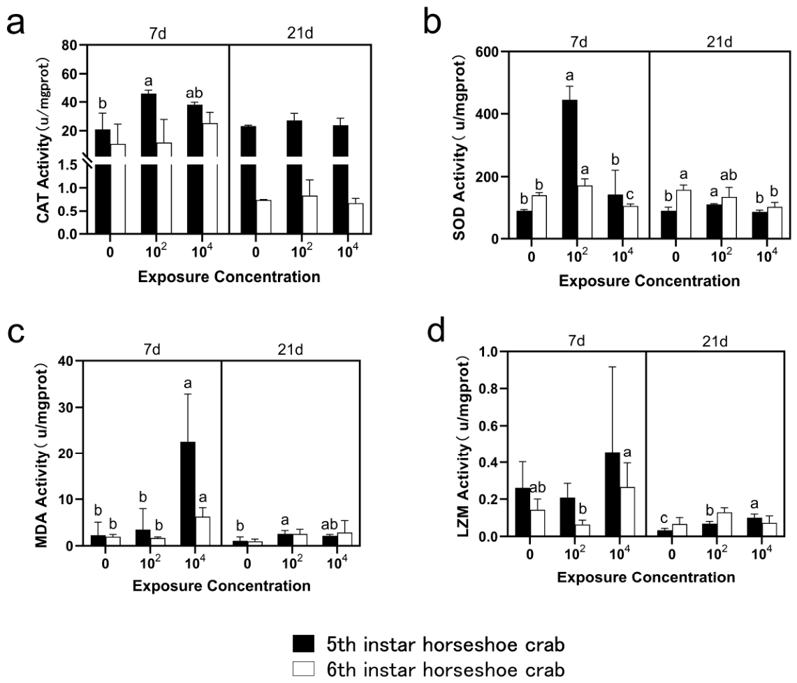
Exposure to PS-MPs significantly affected antioxidant defense, lipid peroxidation, and innate immune responses in the gastrointestinal tract of juvenile T. tridentatus, with responses varying according to exposure concentration, duration, and developmental stage (Figure 2). Both CAT and SOD exhibited rapid antioxidant responses during early exposure. After 7 days, CAT activity increased with PS-MP exposure in fifth-instar juveniles, reaching a significant 2.20-fold increase over the control in the low-concentration group, whereas no significant changes were observed in sixth-instar juveniles. SOD activity showed greater sensitivity than CAT. In fifth-instar juveniles, SOD activity increased significantly to 4.86-fold and 1.57-fold of the control under low- and high-concentration exposure, respectively. In contrast, sixth-instar juveniles displayed only a slight increase under low-concentration exposure (1.23-fold of the control), whereas SOD activity declined under high-concentration exposure (0.76-fold of the control). After 21 days, CAT activity no longer differed significantly among treatments, whereas SOD activity remained elevated only in the low-concentration group of fifth-instar juveniles but was significantly suppressed in sixth-instar juveniles exposed to the high concentration (0.66-fold of the control). Notably, basal SOD activity was consistently higher in sixth-instar than in fifth-instar control animals.
Changes in MDA content indicated that oxidative damage was most pronounced during the early exposure period. After 7 days, MDA levels increased markedly in both instars, reaching 9.84-fold of the control in fifth-instar juveniles and 3.23-fold in sixth-instar juveniles under high-concentration exposure. Following 21 days of exposure, MDA levels generally declined, with a significant increase persisting only in the low-concentration group of fifth-instar juveniles (2.42-fold of the control), suggesting a partial physiological acclimation to prolonged PS-MP exposure.
Innate immune responses, as reflected by lysozyme (LZM) activity, were comparatively moderate and exhibited distinct instar-specific patterns. In fifth-instar juveniles, LZM activity showed no significant changes after 7 days but increased significantly after 21 days in both exposure groups, reaching 2.07-fold and 3.02-fold of the control in the low- and high-concentration treatments, respectively. In contrast, sixth-instar juveniles exhibited a transient increase in LZM activity after 7 days (1.85-fold of the control under high-concentration exposure), whereas no significant differences were detected after 21 days. Overall, PS-MP exposure elicited concentration-, exposure duration-, and instar-dependent physiological responses. Compared with sixth-instar juveniles, fifth-instar individuals displayed stronger antioxidant activation, greater lipid peroxidation, and more pronounced immune alterations, indicating higher physiological sensitivity to PS-MP exposure.
Figure 2.
Effects of PS-MPs exposure on oxidative stress and immune-related enzyme activities in the gastrointestinal tract of juvenile Tachypleus tridentatus.(a) Catalase (CAT) activity; (b) Superoxide dismutase (SOD) activity; (c) Malondialdehyde (MDA) content; (d) Lysozyme (LZM) activity. Values are presented as mean ± SD (n = 3 biological replicates per group, each pooled from three individuals). Different lowercase letters (a, b) above the plots indicate significant differences among PS-MPs concentrations within the same instar and exposure duration according to one-way ANOVA followed by Tukey’s HSD test (p < 0.05). Groups sharing the same letter are not significantly different.
Figure 2.
Effects of PS-MPs exposure on oxidative stress and immune-related enzyme activities in the gastrointestinal tract of juvenile Tachypleus tridentatus.(a) Catalase (CAT) activity; (b) Superoxide dismutase (SOD) activity; (c) Malondialdehyde (MDA) content; (d) Lysozyme (LZM) activity. Values are presented as mean ± SD (n = 3 biological replicates per group, each pooled from three individuals). Different lowercase letters (a, b) above the plots indicate significant differences among PS-MPs concentrations within the same instar and exposure duration according to one-way ANOVA followed by Tukey’s HSD test (p < 0.05). Groups sharing the same letter are not significantly different.
3.3. PS-MPs Reduced Gut Microbial Diversity
Rarefaction analysis indicated that sequencing depth was sufficient to capture the gut microbial diversity of all samples, as richness and diversity curves approached saturation and Good’s coverage values were close to 1.0 (Figure S3). PS-MP exposure substantially reduced gut microbial richness and diversity in juvenile T. tridentatus (Table 1, Figure 3). Compared with the corresponding controls, the Chao1, Shannon, and Simpson indices declined progressively with increasing exposure duration, with the strongest reductions observed after 21 days in the high-concentration groups. Fifth-instar juveniles exhibited a clear concentration-dependent decrease in all diversity indices after 21 days, indicating greater sensitivity of the gut microbiota to prolonged PS-MP exposure. Consistent with the diversity indices, ASV analysis revealed a marked decline in both shared and unique ASVs following prolonged exposure (Figure 4, Figure S4). The number of core ASVs shared among all treatment groups decreased from 137 after 7 days to 54 after 21 days, demonstrating progressive destabilization of the core gut microbiota. Collectively, these results indicate that chronic PS-MP exposure substantially reduced gut microbial diversity and altered community stability in juvenile T. tridentatus.
3.4. Differential Bacterial Taxa Identified by LEfSe
Gut microbial composition was dominated by Proteobacteria, Firmicutes, and Bacteroidota across all treatments (Figure 5a, b; Figure S5). PS-MP exposure induced marked shifts in the relative abundance of these dominant phyla, characterized by enrichment of Proteobacteria and depletion of Firmicutes, particularly after 21 days of high-concentration exposure. These changes were more pronounced in fifth-instar juveniles, in which Proteobacteria increased to 81.13% while Firmicutes decreased to 12.85%. At the genus level, Pseudomonas, Morganella, and Vagococcus predominated the intestinal microbiota (Figure S5). Prolonged PS-MP exposure resulted in a pronounced enrichment of Pseudomonas accompanied by a progressive decline in Vagococcus, with the strongest responses observed in fifth-instar juveniles. Heatmap analysis further indicated widespread taxonomic restructuring following PS-MP exposure, with Firmicutes-associated genera showing the greatest reductions, whereas multiple taxa belonging to Proteobacteria became enriched across treatment groups (Figure 5c, d; Figure S6).
3.5. LEfSe Analysis of Key Discriminatory Taxa
LEfSe analysis identified distinct microbial biomarkers associated with PS-MP exposure across developmental stages and exposure durations (Figure 5e; Figure S7). Firmicutes, Proteobacteria, and Actinobacteriota were the principal discriminatory phyla, whereas Bacilli, Alphaproteobacteria, and Gammaproteobacteria were the dominant discriminatory classes. At lower taxonomic levels, control groups were characterized by several taxa associated with normal gut microbiota, including Lactobacillales, Vagococcus, and Lactococcus, whereas prolonged high-concentration exposure enriched taxa such as Pseudomonas, Pseudomonadaceae, Enterobacteriaceae, Blautia, and Bifidobacterium. T-test analysis suggested significant alterations in the abundance of key bacterial genera, particularly the concentration-dependent enrichment of Pseudomonas after 21 days of exposure in fifth-instar juveniles (p = 0.034), together with significant changes in Blautia, Escherichia–Shigella, Bacteroides, and Cohaesibacter (Figure S8).
3.6. Overall Shifts in Microbial Community Structure
Principal coordinate analysis based on weighted UniFrac distances indicated clear separation of gut microbial communities according to PS-MP concentration, exposure duration, and developmental stage (Figure S9). PC1 and PC2 explained 66.28% and 15.07% of the total variation, respectively, accounting for 81.35% of the cumulative variance. Samples from the 7- and 21-day exposure groups formed distinct clusters, with the greatest separation observed among fifth-instar juveniles after 21 days. These results indicate that both prolonged exposure and developmental stage contributed substantially to gut microbial community divergence following PS-MP exposure.
4. Discussion
The present study demonstrates the accumulation characteristics of the emerging marine contaminant PS-MPs in the gastrointestinal tract of juvenile T. tridentatus during critical developmental stages (5th and 6th instars) and reveals their combined toxic effects, including oxidative injury, immune suppression, and gut microbial dysbiosis. These findings highlight the complex impacts of PS-MPs on intestinal microecological homeostasis in juvenile T. tridentatus and further confirm that exposure concentration, exposure duration, and host developmental stage are key determinants of toxicological responses.
4.1. Ingestion, Retention, and Instar-Dependent Accumulation Characteristics of PS-MPs in the Midgut
In the present study, juvenile T. tridentatus primarily ingested PS-MPs via feeding and water intake, with pronounced accumulation observed in the midgut. This uptake pathway is consistent with trophic transfer patterns reported in other aquatic organisms, such as zebrafish and tuna [14,28].
Sultan et al.[14] indicated that nanoplastics can be efficiently transferred through a trophic chain (Artemia → zebrafish), resulting in significant gastrointestinal accumulation and a clear bioaccumulation gradient in zebrafish. In the present study, fluorescence microscopy revealed that intestinal tracts containing food residues exhibited higher fluorescence intensity, whereas intestines with little or no food content showed markedly weaker signals. Moreover, strong fluorescent signals were also detected in fecal material, indicating that MPs associated with food particles can be rapidly excreted, although a fraction remains retained in the gut, leading to persistent accumulation.Wang et al.[28] further confirmed that yellowfin tuna (Thunnus albacares) from coastal waters of southern Kaohsiung, Taiwan, can directly ingest microplastics through both respiration (filtration and retention via gills) and drinking (uptake of MPs suspended in water), as evidenced by the abundance and morphology of MPs detected in gill and stomach tissues.
Our observations indicate that PS-MPs accumulation exhibits a clear dose–response relationship, with exposure concentration being a key determinant of intestinal burden. Higher environmental concentrations led to greater ingestion and retention of PS-MPs in the gut. This finding is consistent with Danting Wang et al.[29], who reported that fluorescence intensity of red fluorescent polystyrene nanoplastics in zebrafish larvae increased with rising exposure concentrations.Furthermore, under identical exposure conditions, 6th-instar juveniles exhibited higher accumulation levels than 5th-instar individuals. This may be attributed to higher feeding rates and greater intestinal content volume in 6th-instar juveniles, which increases the likelihood of particle retention and adhesion within the digestive tract.
The presence of MPs in intestinal contents may further prolong retention time through mucus encapsulation and particle aggregation, forming so-called “intestinal mucus–microplastic aggregates,” thereby exacerbating accumulation. A similar mechanism was proposed by Hoang and Felix-Kim [30], who observed in juvenile soft-mouth minnows that ingested MPs could be re-ingested during excretion, and that expelled MPs coated with intestinal mucus tended to aggregate. This cyclical ingestion–excretion process, combined with the strong adhesive properties of MPs, ultimately contributes to prolonged retention and accumulation in the gut. Therefore, the strong fluorescence signals observed in this study are likely attributable to mucus-mediated aggregation and retention of PS-MPs within the intestinal environment.
4.2. Instar-Specific Oxidative Imbalance and Intestinal Immune Impairment
Oxidative stress is considered one of the primary mechanisms underlying microplastic toxicity in aquatic organisms. In the present study, PS-MP exposure induced pronounced alterations in the antioxidant defense system of juvenile T. tridentatus, indicating that intestinal redox homeostasis was impaired under environmentally relevant exposure conditions. The transient elevation of CAT and SOD activities during early exposure is consistent with a compensatory activation of antioxidant defenses in response to excessive reactive oxygen species (ROS), whereas the diminished responses after prolonged exposure suggest that sustained oxidative stress may gradually overwhelm cellular antioxidant capacity [31,32].
The concurrent increase in MDA further demonstrates that antioxidant activation was insufficient to completely prevent membrane lipid peroxidation, particularly in fifth-instar juveniles exposed to higher PS-MP concentrations. As MDA is a reliable indicator of oxidative injury [33], these findings suggest that earlier developmental stages experienced greater oxidative injury, whereas sixth-instar juveniles maintained relatively stronger redox homeostasis. Similar developmental-stage differences were also reflected in innate immune responses. Lysozyme activity showed comparatively moderate changes, yet older juveniles exhibited greater capacity to maintain immune stability under prolonged exposure, indicating a more effective physiological compensation [34].
Together, these findings demonstrate that the susceptibility of juvenile T. tridentatus to PS-MP-induced oxidative stress is strongly influenced by developmental stage. The greater physiological sensitivity of fifth-instar juveniles suggests that early post-hatching development may represent a particularly vulnerable period during which oxidative and immune disturbances could have disproportionate consequences for subsequent growth and survival.
An important observation in the present study is that PS-MP accumulation did not correspond directly to toxicity severity. Although sixth-instar juveniles consistently exhibited higher intestinal accumulation of PS-MPs than fifth-instar individuals, the latter showed stronger oxidative stress responses, greater lipid peroxidation, and more pronounced microbial dysbiosis. This discrepancy indicates that body burden alone is insufficient to predict toxicological outcomes in developing T. tridentatus.
Several factors may explain this pattern. First, sixth-instar juveniles appeared to possess a more mature antioxidant defense system, as evidenced by their higher basal SOD activity and relatively stable physiological responses, enabling more efficient maintenance of redox homeostasis under chronic PS-MP exposure. Similar developmental-stage differences in antioxidant capacity have been reported in juvenile T. tridentatus exposed to plastic-related pollutants [24]. Second, older juveniles likely have a more stable and resilient gut microbial ecosystem, which may buffer the disruptive effects of microplastics on intestinal homeostasis. Third, the stronger fluorescence observed in sixth-instar individuals may partly reflect larger food intake and greater gut content volume rather than a proportionally higher degree of particle internalization into intestinal tissues. Consequently, a substantial fraction of ingested particles may remain within the gut lumen and be eliminated through normal digestive processes, reducing direct contact with epithelial cells.
Collectively, these findings indicate that physiological maturity, rather than microplastic accumulation per se, is the primary determinant of toxic susceptibility during juvenile development of T. tridentatus. This interpretation is consistent with recent evidence showing that developmental stage strongly influences the biological responses of juvenile T. tridentatus to polystyrene microplastics [4] and supports the broader view that host physiological condition is a key modifier of microplastic toxicity in marine invertebrates [3].
4.3. PS-MPs Impair the Gut Microbiota Through Oxidative Stress-Mediated Alterations of the Intestinal Microenvironment
PS-MP exposure markedly altered the diversity and composition of the intestinal microbiota in juvenile T. tridentatus, with fifth-instar juveniles exhibiting greater reductions in microbial richness and diversity than sixth-instar individuals. The decline in Chao1 and Shannon indices together with the reduction in shared ASVs indicates that prolonged PS-MP exposure progressively destabilized the intestinal microbial community. Rather than representing isolated microbial changes, these alterations likely reflect disturbances in the intestinal microenvironment initiated by oxidative stress.
Increasing evidence suggests that excessive reactive oxygen species (ROS) generated during microplastic exposure act not only as cytotoxic molecules but also as ecological drivers shaping gut microbial communities. Elevated ROS levels can alter intestinal oxygen availability, mucus composition, epithelial permeability, and nutrient gradients, thereby creating selective conditions that favor facultative aerobic or opportunistic bacteria while suppressing beneficial anaerobic commensals. In the present study, the marked enrichment of Proteobacteria, accompanied by the decline of Firmicutes, is consistent with microbial signatures of intestinal dysbiosis reported in aquatic organisms exposed to environmental contaminants [35]. Similar community shifts have frequently been associated with impaired intestinal barrier function and increased susceptibility to inflammation.
At the genus level, the expansion of Pseudomonas and the simultaneous reduction of beneficial genera such as Lactococcus further support this ecological transition. Beneficial lactic acid bacteria contribute to pathogen exclusion, maintenance of epithelial integrity, and immune homeostasis, whereas opportunistic genera including Pseudomonas readily proliferate under oxidative and inflammatory conditions. Therefore, the microbial alterations observed here are unlikely to represent direct toxic effects of PS-MPs on bacteria alone but instead appear to arise from ROS-mediated remodeling of the intestinal microenvironment, linking oxidative stress with microbial dysbiosis.
Collectively, these findings suggest that oxidative stress represents an upstream driver connecting microplastic exposure with gut microbial instability in juvenile T. tridentatus.
4.4. Gut Dysbiosis Amplifies Oxidative Imbalance Through Impairment of the Gut–Microbiota–Immune Axis
The present study further suggests that intestinal dysbiosis is not merely a consequence of PS-MP exposure but may actively amplify host toxicity through disruption of the gut-microbiota-immune axis. Following oxidative stress-induced alterations of the intestinal environment, the loss of beneficial bacteria together with the enrichment of opportunistic pathogens is expected to impair mucosal immune function and intestinal barrier integrity, thereby establishing a self-reinforcing cycle of intestinal injury.
This hypothesis is supported by the LEfSe analysis, which revealed the emergence of distinct biomarkers associated with potentially pathogenic taxa after prolonged high-concentration exposure. For example, Enterobacteriaceae, detected as a characteristic biomarker in sixth-instar juveniles after 21 days of high-concentration exposure, is widely recognized as an indicator of intestinal dysbiosis and inflammatory disorders. Excessive proliferation of Enterobacteriaceae can activate host inflammatory pathways, aggravate epithelial damage, and further disturb microbial homeostasis [36]. Likewise, the increased abundance of Campilobacterota, which contains numerous gastrointestinal pathogens [37], suggests that chronic PS-MP exposure may increase susceptibility to intestinal infection.
Conversely, beneficial taxa including Lactococcus, which produce antimicrobial metabolites and contribute to colonization resistance [38], declined following PS-MP exposure. The reduction of these protective microorganisms may weaken pathogen exclusion and compromise innate immune defenses, consistent with the altered lysozyme responses observed in the present study.
Taken together, our findings support a positive feedback mechanism linking oxidative stress, microbial dysbiosis, and immune dysfunction. Initial PS-MP accumulation triggers excessive ROS production, which impairs intestinal epithelial homeostasis and alters the gut microenvironment. These changes selectively promote opportunistic bacteria while suppressing beneficial commensals. The resulting dysbiosis may further impair immune function and intestinal barrier integrity, thereby enhancing oxidative injury and sustaining chronic intestinal inflammation. This oxidative stress–microbiota–immune feedback loop provides a mechanistic framework for understanding the chronic gastrointestinal toxicity of PS-MPs in juvenile T. tridentatus.
4.5. Proposed Mechanism Underlying PS-MP-Induced Intestinal Alterations in Juvenile T. tridentatus
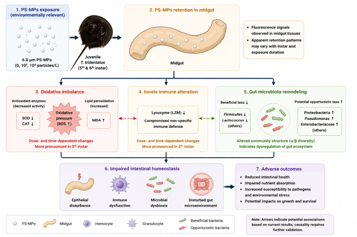
Based on the present physiological and microbial findings, a conceptual mechanism model was proposed to illustrate the potential responses of juvenile T. tridentatus to PS-MP exposure (Figure 6). Following ingestion, PS-MPs may be retained in the midgut and interact with intestinal tissues, leading to oxidative imbalance characterized by reduced antioxidant enzyme activities and increased lipid peroxidation. Meanwhile, alterations in lysozyme activity suggest that PS-MPs may influence innate immune responses. In parallel, PS-MP exposure reshaped gut microbial communities, including decreased relative abundances of beneficial taxa and enrichment of potential opportunistic bacteria. These interconnected changes may collectively contribute to impaired intestinal homeostasis.
However, this model represents a hypothesis based on observed associations rather than confirmed causal pathways. Further investigations involving direct measurements of ROS generation, intestinal barrier-related genes, inflammatory signaling pathways, and microbial functional analyses are required to validate the proposed mechanisms.
Figure 6.
Conceptual model depicting the potential mechanisms underlying PS-MP-induced intestinal alterations in juvenile Tachypleus tridentatus.
Figure 6.
Conceptual model depicting the potential mechanisms underlying PS-MP-induced intestinal alterations in juvenile Tachypleus tridentatus.
5. Conclusions
This study demonstrates that environmentally relevant concentrations of PS-MPs predominantly accumulate in the midgut of endangered juvenile Tachypleus tridentatus and impair intestinal homeostasis via three interconnected pathways: oxidative stress, innate immune dysfunction, and gut microbial dysbiosis. These toxic responses exhibit clear concentration-, exposure duration-, and instar-dependent patterns, with fifth-instar juveniles showing consistently higher susceptibility than sixth-instar individuals, confirming that early ontogeny represents a critical vulnerability window for this ancient marine arthropod. By integrating histological, physiological, and microbial endpoints, this work establishes a mechanistic framework for instar-specific microplastic toxicity in benthic arthropods; our findings provide empirical support for refining ecological risk assessments of coastal microplastic pollution and inform targeted conservation strategies for T. tridentatus in microplastic-impacted intertidal ecosystems, with broader implications for coastal benthic biodiversity protection under anthropogenic disturbance. Future research incorporating multi-omics approaches, long-term exposure regimes, and field-relevant co-exposure scenarios will help translate laboratory toxicological findings into population- and community-level ecological outcomes, further strengthening the scientific basis for marine invertebrate conservation.
Supporting Information:
The following supporting information can be downloaded at the website of this paper posted on Preprints.org, The original contributions presented in the study are included in the article/Supplementary Materials. Further inquiries can be directed to the corresponding author.
Author Contributions
Yuhong Li: Conceptualization, Writing—review & editing, Project administration, Funding acquisition. Yiran Tan: Methodology, Investigation, and Laboratory Experiments, Visualization, Writing—original draft. Liyun Han: Methodology, Investigation, and Laboratory Experiments, Visualization, Writing—original draft. Yue Liu: Methodology, Investigation, and Laboratory Experiments, Visualization, Writing—original draft. Mingxiao Liu: Methodology, Investigation, and Laboratory Experiments. Caoqun Zheng: Visualization. Wenxin Jin: Writing—original draft. Tianshuai Zhang: Writing—original draft. Bosen Weng: Visualization and Writing—original draft. Zhaohong Weng: Conceptualization and Funding acquisition. Jun Bo: Conceptualization, Writing—original draft.
Institutional Review Board Statement
The animal procedures were approved by the ethics committee of the Third Institute of Oceanography, Ministry of Natural Resources, China (No. TIO-IACUC-01-2024-03-08, date: 2024.3.8).
Data Availability Statement
Data will be made available on request.
Conflicts of Interest
The authors declare that they have no known competing financial interests or personal relationships that could have appeared to influence the work reported in this paper.
Acknowledgments
This work was financially supported by the Natural Science Foundation of Fujian Province (Grant No. 2023J01134) and the Open Project of Fujian Key Laboratory of Marine Fishery Resources and Ecological Environment.
References
- Zhu, J.; Kwan, K.Y.; Zeng, Y.; Yang, X.; Zou, Y.; Zhong, X.; Zhou, H.; Chen, Z.; Tan, K.A.; Xu, P.; Zhang, C. Tetrodotoxin in Asian horseshoe crabs Carcinoscorpius rotundicauda and Tachypleus tridentatus across different life stages from northern Beibu Gulf, China. Mar. Pollut. Bull. 2022, 184, 114195. [Google Scholar] [CrossRef] [PubMed]
- Xie, M.; Chen, X.; Xie, X.; Deng, C. Environmental DNA effectively reveals spatial patterns of benthos in a nursery habitat of the highly endangered Tachypleus tridentatus. J. Environ. Manag. 2025, 391, 126376. [Google Scholar] [CrossRef] [PubMed]
- Hara, J.; Vercauteren, M.; Janssen, C.R.; Blust, R.; Asselman, J.; Town, R.M. Systematic assessment of the mechanisms and risks of micro- and nanoplastic particle exposure in marine invertebrates. Ecotoxicol. Environ. Saf. 2026, 311, 119877. [Google Scholar] [CrossRef] [PubMed]
- Jiang, Y.; Yang, Z.Y.; Jiang, L.F.; Chong, C.M.; Gul, S.; Wang, Y.J.; Waiho, K.; Ma, X.W.; Chen, Z.; Hu, M.H. Polystyrene microplastics impede growth and induce behavioural toxicity in endangered juvenile horseshoe crabs (Tachypleus tridentatus). Mar. Environ. Res. 2025, 214, 107758. [Google Scholar] [CrossRef] [PubMed]
- Chen, B.; Wu, R.; Zhu, P.; Liu, J.; Liu, Y.; Yan, L.; Ma, Z.; Gao, F.; Liu, Z.; Zhang, Z.; Li, S.; Jia, J.; Zhang, Y.; Liao, Y. Azadirachtin-induced oxidative stress in early life stages of the endangered horseshoe crab Tachypleus tridentatus: Implications for coastal conservation. Mar. Pollut. Bull. 2026, 226, 119339. [Google Scholar] [CrossRef] [PubMed]
- Andrady, A.L. Microplastics in the marine environment. Mar. Pollut. Bull. 2011, 62, 1596–1605. [Google Scholar] [CrossRef] [PubMed]
- Chen, B.; Zhang, Z.; Wang, T.; Hu, H.; Qin, G.; Lu, T.; Hong, W.; Hu, J.; Penuelas, J.; Qian, H. Global distribution of marine microplastics and potential for biodegradation. J. Hazard. Mater. 2023, 451, 131198. [Google Scholar] [CrossRef] [PubMed]
- Chen, J.; Fang, C.; Zheng, R.; Chen, M.; Kim, D.; Lee, Y.; Bailey, C.; Wang, K.; Lee, J.; Bo, J. Environmentally relevant concentrations of microplastics modulated the immune response and swimming activity, and impaired the development of marine medaka Oryzias melastigma larvae. Ecotoxicol. Environ. Saf. 2022, 241, 113843. [Google Scholar] [CrossRef] [PubMed]
- Andersen, R.; Harsaae, A.L.; Kellner, A.; Smyth, A.; Westermann, T.A.R.; Green, M.; Vollertsen, J.; Syberg, K.; Lorenz, C. Abundance, distribution and characteristics of microplastics in the North and South Atlantic Ocean. Mar. Pollut. Bull. 2024, 209, 117217. [Google Scholar] [CrossRef] [PubMed]
- Ho, C.M.; Feng, W.; Li, X.; Ngien, S.K.; Yu, X.; Song, F.; Yang, F.; Liao, H. Microplastic distribution and its implications for human health through marine environments. J. Environ. Manag. 2025, 382, 125427. [Google Scholar] [CrossRef] [PubMed]
- Akash, S.; Rameshwar, S.S.; Rajamohan, N.; Sivaprakash, B.; Paramasivam, S.; Cappellini, G.; Gatto, G. Advances in polystyrene nanoplastic remediation: A review of detection methods, toxicity, removal strategies, and economic insights. J. Hazard. Mater. Adv. 2025, 20, 100889. [Google Scholar] [CrossRef]
- Das, S.; Chatterjee, N.H.; Choudhury, A.; Ray, A.; Rana, N.; Banerjee, A.; Ray, M.; Ray, S. Characterization and ecological risk assessment of microplastics accumulated in sea water, sand, sediment, shell water and selected tissues of hermit crab of Sundarban Biosphere Reserve. Environ. Pollut. 2024, 357, 124484. [Google Scholar] [CrossRef] [PubMed]
- Luo, X.; Zhang, Y.; Kang, S.; Chen, R.; Gao, T.; Allen, S. Atmospheric emissions of microplastics entrained with dust from potential source regions. J. Hazard. Mater. 2025, 488, 137509. [Google Scholar] [CrossRef] [PubMed]
- Sultan, M.; Cai, Z.; Bao, L.; Duan, J.; Liu, Y.; Yang, G.; Pei, D. Trophic transfer induced gut inflammation, dysbiosis, and inflammatory pathways in zebrafish via Artemia franciscana: A differential analysis of nanoplastic toxicity. J. Hazard. Mater. 2024, 480, 136030. [Google Scholar] [CrossRef] [PubMed]
- Zhang, W.; Tian, D.; Yu, Y.; Tong, D.; Zhou, W.; Yu, Y.; Lu, L.; Li, W.; Liu, G.; Shi, W. Micro/nanoplastics impair the feeding of goldfish by disrupting the complicated peripheral and central regulation of appetite. Sci. Total Environ. 2024, 946, 174112. [Google Scholar] [CrossRef] [PubMed]
- Zhang, L.; Liu, X.; Zhang, C. Effect of PET microplastics on the growth, digestive enzymes, and intestinal flora of the sea cucumber Apostichopus japonicus. Mar. Environ. Res. 2023, 190, 106125. [Google Scholar] [CrossRef] [PubMed]
- He, Y.; Shaoyong, W.; Chen, Y.; Li, M.; Gan, Y.; Sun, L.; Liu, Y.; Wang, Y.; Jin, M. The functions of gut microbiota-mediated bile acid metabolism in intestinal immunity. J. Adv. Res. 2025, 80, 351–370. [Google Scholar] [CrossRef] [PubMed]
- Shi, W.; Xi, M.; Zhang, K.; Yang, J.; Cheng, X.; Zang, H.; Fan, W. Gut microbiota as a central mediator in hydrogen gas-induced alleviation of colitis via TLR4/NF-κB and Nrf2 pathway regulation. Int. Immunopharmacol. 2025, 167, 115671. [Google Scholar] [CrossRef] [PubMed]
- Jin, Y.; Xia, J.; Pan, Z.; Yang, J.; Wang, W.; Fu, Z. Polystyrene microplastics induce microbiota dysbiosis and inflammation in the gut of adult zebrafish. Environ. Pollut. 2018, 235, 322–329. [Google Scholar] [CrossRef] [PubMed]
- Mut, N.N.N.; Cao, Z.; Na, J.; Yuzir, A.; Tollefsen, K.E.; Jung, J. Adverse outcome pathway networks of microplastic ecotoxicity to aquatic organisms: A critical review. Aquat. Toxicol. 2025, 289, 107594. [Google Scholar] [CrossRef] [PubMed]
- You, X.; Zhang, Z.; Tian, G.; Zhang, Y.; Pei, Y.; Wu, Y.; Li, G.; Wang, Q.; Yang, Y. The impact of polyethylene microplastics exposure on the growth performance, reproductive performance, antioxidant capacity, and intestinal microbiota of quails. Ecotoxicol. Environ. Saf. 2025, 296, 118218. [Google Scholar] [CrossRef] [PubMed]
- Qu, Z.; Leung, T.C.N.; Nong, W.; Yip, H.Y.; Lee, I.H.T.; Cheung, S.G.; Ming, N.S.; So, W.L.; Bendena, W.G.; Tobe, S.S.; Hui, J.H.L. Hemolymph proteomics and gut microbiota of horseshoe crabs Tachypleus tridentatus and Carcinoscorpius rotundicauda. Front. Mar. Sci. 2020, 7, 579706. [Google Scholar] [CrossRef]
- Pan, Y.; Qian, J.; Ma, X.; Huang, W.; Fang, J.K.; Arif, I.; Wang, Y.; Shang, Y.; Hu, M. Response of moulting genes and gut microbiome to nano-plastics and copper in juvenile horseshoe crab Tachypleus tridentatus. Mar. Environ. Res. 2023, 191, 106128. [Google Scholar] [CrossRef] [PubMed]
- Huang, M.L.; Ma, Y.X.; Qian, J.; Sokolova, I.M.; Zhang, C.Q.; Waiho, K.; Fang, J.K.H.; Ma, X.W.; Wang, Y.J.; Hu, M.H. Combined effects of norfloxacin and polystyrene nanoparticles on the oxidative stress and gut health of the juvenile horseshoe crab Tachypleus tridentatus. J. Hazard. Mater. 2024, 468, 133801. [Google Scholar] [CrossRef] [PubMed]
- Callahan, B.J.; McMurdie, P.J.; Rosen, M.J.; Han, A.W.; Johnson, A.J.A.; Holmes, S.P. DADA2: High-resolution sample inference from Illumina amplicon data. Nat. Methods 2016, 13, 581–583. [Google Scholar] [CrossRef] [PubMed]
- Quast, C.; Pruesse, E.; Yilmaz, P.; Gerken, J.; Schweer, T.; Yarza, P.; Peplies, J.; Glöckner, F.O. The SILVA ribosomal RNA gene database project: improved data processing and web-based tools. Nucleic Acids Res. 2013, 41, D590–D596. [Google Scholar] [CrossRef] [PubMed]
- Gu, H.; Wang, S.; Wang, X.; Yu, X.; Hu, M.; Huang, W.; Wang, Y. Nanoplastics impair the intestinal health of the juvenile large yellow croaker Larimichthys crocea. J. Hazard. Mater. 2020, 397, 122773. [Google Scholar] [CrossRef] [PubMed]
- Wang, M.; Gonzalez, J.B.; Chen, C.; Hsu, C.; Chen, C.; Dong, C. Microplastic ingestion and phthalate esters bioaccumulation in yellowfin tuna (Thunnus albacares): tissue-specific distribution across gills, stomach, and muscles. Mar. Pollut. Bull. 2025, 219, 118257. [Google Scholar] [CrossRef] [PubMed]
- Wang, D.; Lan, J.; Meng, W.; Liu, J.; Zhou, R.; Chen, C.; Qin, L.; Tan, B.; Huang, C. Co-exposure to triclosan and polystyrene nanoplastics on neurodevelopmental toxicity and gut microbiota dysbiosis in zebrafish (Danio rerio). Environ. Pollut. 2025, 381, 126643. [Google Scholar] [CrossRef] [PubMed]
- Hoang, T.C.; Felix-Kim, M. Microplastic consumption and excretion by fathead minnows (Pimephales promelas): Influence of particles size and body shape of fish. Sci. Total Environ. 2020, 704, 135433. [Google Scholar] [CrossRef] [PubMed]
- Zhuang, Z.; Wang, Y.; Zhang, Z.; Zhang, W.; Ding, J.; Weng, Z.; Chen, X.; Hu, F. Trade-off strategy under stress: Growth, reproduction, and antioxidant defense in Daphnia magna exposed to 6PPD-Q. J. Hazard. Mater. 2025, 496, 139337. [Google Scholar] [CrossRef] [PubMed]
- Liu, J.; Chen, Y.; Song, Y.; Xu, D.; Gu, Y.; Wang, J.; Song, W.; Sun, B.; Jiang, Z.; Xia, B. Evidence of size-dependent toxicity of polystyrene nano- and microplastics in sea cucumber Apostichopus japonicus (Selenka, 1867) during the intestinal regeneration. Environ. Pollut. 2024, 357, 124394. [Google Scholar] [CrossRef] [PubMed]
- Liu, X.; Zhang, X.; Zhu, J.; Zou, W.; Liang, L.; Zhang, J.; Wen, C.; Li, Y.; Liu, G.; Xu, X. BbF-induced liver injury in Balb/c mice: AhR activation as the conductor of metabolism, oxidative stress, lipid metabolism disorder, and inflammatory response. Free Radic. Biol. Med. 2025, 241, 617630. [Google Scholar] [CrossRef]
- Watts, C. Lysosomes and lysosome-related organelles in immune responses. FEBS Open Bio 2022, 12, 678–693. [Google Scholar] [CrossRef] [PubMed]
- Rehman, A.; Habumugisha, T.; Huang, F.; Zhang, Z.; Kiki, C.; Al, M.A.; Yan, C.; Shaheen, U.; Zhang, X. Impacts of polystyrene nanoplastics on zebrafish gut microbiota and mechanistic insights. Ecotoxicol. Environ. Saf. 2025, 299, 118332. [Google Scholar] [CrossRef] [PubMed]
- Feng, G.; Xu, M.; Liang, H.; Peng, J.; Sun, T.; Du, Y.; Huang, Y.; Fang, J. Enterobacteriaceae-targeting tungsten nanoclusters for precision IBD therapy: A quadruple-action strategy integrating targeted delivery, ROS scavenging, microbiome reprogramming and anti-inflammation. Chem. Eng. J. 2026, 529, 173286. [Google Scholar] [CrossRef]
- Burcham, Z.M.; Tweedie, J.L.; Farfán-García, A.E.; Nolan, V.G.; Donohoe, D.; Gómez-Duarte, O.G.; Johnson, J.G. Campylobacter infection of young children in Colombia and its impact on the gastrointestinal environment. mSphere 2024, 9, e324–e342. [Google Scholar] [CrossRef] [PubMed]
- Huang, F.; Ju, Z.; Hou, Y.; Zhao, G.; Yang, Y.; Yue, B.; Zhang, X. Exploring advanced antimicrobial effects of Pediococcus pentosaceus and Lactococcus lactis derived from Bufo gargarizans: In vitro analysis and in vivo evaluation in mice. LWT 2024, 210, 116851. [Google Scholar] [CrossRef]
Figure 1.
Accumulation of PS-MPs in the midgut of juvenile Tachypleus tridentatus under high-concentration exposure.(a) and (b) Midgut accumulation of PS-MPs in 5th- and 6th-instar juveniles after 7 days of exposure, respectively; (c) and (d) Midgut accumulation of PS-MPs in 5th- and 6th-instar juveniles after 21 days of exposure, respectively. The positions indicated by blue triangles represent fluorescent signals. Images were obtained using an inverted fluorescence microscope at 200 μm scale.
Figure 1.
Accumulation of PS-MPs in the midgut of juvenile Tachypleus tridentatus under high-concentration exposure.(a) and (b) Midgut accumulation of PS-MPs in 5th- and 6th-instar juveniles after 7 days of exposure, respectively; (c) and (d) Midgut accumulation of PS-MPs in 5th- and 6th-instar juveniles after 21 days of exposure, respectively. The positions indicated by blue triangles represent fluorescent signals. Images were obtained using an inverted fluorescence microscope at 200 μm scale.
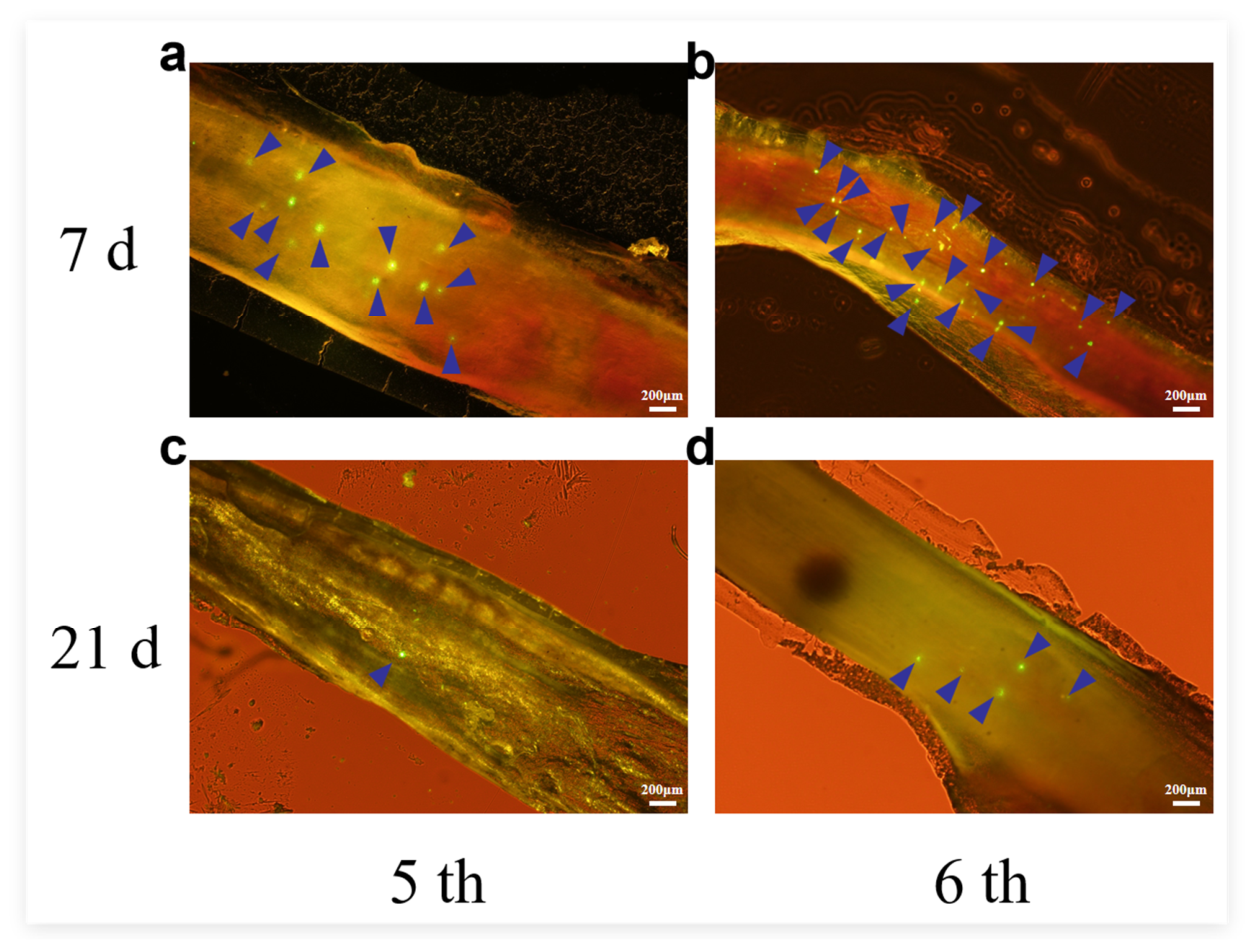
Figure 3.
Effects of polystyrene microplastics (PS-MPs; 0, 10², and 104 particles·L-1) on the Shannon diversity index of the gut microbiota in juvenile Tachypleus tridentatus at two developmental stages (5th and 6th instars) and two exposure durations (7 and 21 days). Black and white boxplots represent the 7-day and 21-day exposure groups, respectively. Values are presented as mean ± SD (n = 5 individual biological replicates per group). Statistical significance between the 7-day and 21-day groups at the same concentration and instar was determined by two-tailed t-test. ns indicates not significant (p > 0.05); *p < 0.05; **p < 0.01. Exact p values are shown above the brackets.
Figure 3.
Effects of polystyrene microplastics (PS-MPs; 0, 10², and 104 particles·L-1) on the Shannon diversity index of the gut microbiota in juvenile Tachypleus tridentatus at two developmental stages (5th and 6th instars) and two exposure durations (7 and 21 days). Black and white boxplots represent the 7-day and 21-day exposure groups, respectively. Values are presented as mean ± SD (n = 5 individual biological replicates per group). Statistical significance between the 7-day and 21-day groups at the same concentration and instar was determined by two-tailed t-test. ns indicates not significant (p > 0.05); *p < 0.05; **p < 0.01. Exact p values are shown above the brackets.
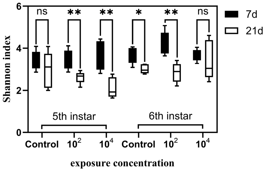
Figure 4.
Venn diagrams showing shared and unique ASVs among gastrointestinal microbial communities of juvenile Tachypleus tridentatus under different PS-MP exposure durations. (a) Shared and unique ASVs among 5th- and 6th-instar juveniles under control, low-concentration, and high-concentration PS-MPs exposure after 7 days; (b) Shared and unique ASVs among 5th- and 6th-instar juveniles under control, low-concentration, and high-concentration PS-MPs exposure after 21 days. Numbers in overlapping regions indicate shared ASVs among groups, whereas numbers in non-overlapping regions represent unique ASVs within each group. Sample group definitions: R-series (7-day exposure groups): R1–R3 represent 6th-instar juveniles (Tachypleus tridentatus) (R1: control, R2: low-concentration PS-MPs, R3: high-concentration PS-MPs); R4–R6 represent 5th-instar juveniles (R4: control, R5: low-concentration PS-MPs, R6: high-concentration PS-MPs).
Figure 4.
Venn diagrams showing shared and unique ASVs among gastrointestinal microbial communities of juvenile Tachypleus tridentatus under different PS-MP exposure durations. (a) Shared and unique ASVs among 5th- and 6th-instar juveniles under control, low-concentration, and high-concentration PS-MPs exposure after 7 days; (b) Shared and unique ASVs among 5th- and 6th-instar juveniles under control, low-concentration, and high-concentration PS-MPs exposure after 21 days. Numbers in overlapping regions indicate shared ASVs among groups, whereas numbers in non-overlapping regions represent unique ASVs within each group. Sample group definitions: R-series (7-day exposure groups): R1–R3 represent 6th-instar juveniles (Tachypleus tridentatus) (R1: control, R2: low-concentration PS-MPs, R3: high-concentration PS-MPs); R4–R6 represent 5th-instar juveniles (R4: control, R5: low-concentration PS-MPs, R6: high-concentration PS-MPs).
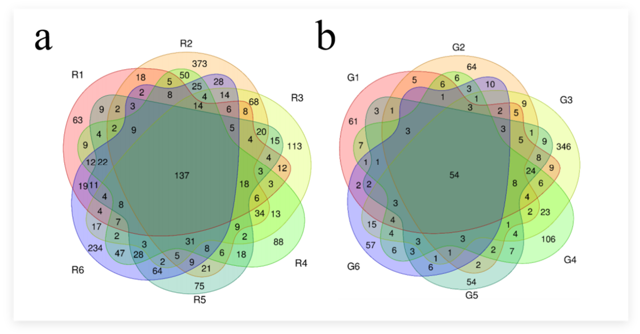
Figure 5.
Relative abundance, clustering heatmaps, and LEfSe biomarker analysis of the gut microbiota in juvenile Tachypleus tridentatus.(a) Stacked bar plot of relative abundance at the phylum level; (b) Stacked bar plot of relative abundance at the genus level; (c) Hierarchical clustering heatmap at the phylum level (top 35 most abundant bacterial phyla); (d) Hierarchical clustering heatmap at the genus level (top 35 most abundant bacterial genera); (e) LEfSe cladogram showing taxonomic biomarkers of gut microbial communities. In the heatmaps, red indicates higher relative abundance, and blue indicates lower relative abundance. In the cladogram, taxa are shown from the inner to outer rings, representing the phylum, class, order, family, and genus levels. Yellow nodes indicate taxa with no significant differences among groups, while nodes in other colors represent significantly different taxa among treatment groups. Sample group definitions: R-series (7-day exposure groups): R1–R3 represent 6th-instar juveniles (Tachypleus tridentatus) (R1: control, R2: low-concentration PS-MPs, R3: high-concentration PS-MPs); R4–R6 represent 5th-instar juveniles (R4: control, R5: low-concentration PS-MPs, R6: high-concentration PS-MPs).
Figure 5.
Relative abundance, clustering heatmaps, and LEfSe biomarker analysis of the gut microbiota in juvenile Tachypleus tridentatus.(a) Stacked bar plot of relative abundance at the phylum level; (b) Stacked bar plot of relative abundance at the genus level; (c) Hierarchical clustering heatmap at the phylum level (top 35 most abundant bacterial phyla); (d) Hierarchical clustering heatmap at the genus level (top 35 most abundant bacterial genera); (e) LEfSe cladogram showing taxonomic biomarkers of gut microbial communities. In the heatmaps, red indicates higher relative abundance, and blue indicates lower relative abundance. In the cladogram, taxa are shown from the inner to outer rings, representing the phylum, class, order, family, and genus levels. Yellow nodes indicate taxa with no significant differences among groups, while nodes in other colors represent significantly different taxa among treatment groups. Sample group definitions: R-series (7-day exposure groups): R1–R3 represent 6th-instar juveniles (Tachypleus tridentatus) (R1: control, R2: low-concentration PS-MPs, R3: high-concentration PS-MPs); R4–R6 represent 5th-instar juveniles (R4: control, R5: low-concentration PS-MPs, R6: high-concentration PS-MPs).
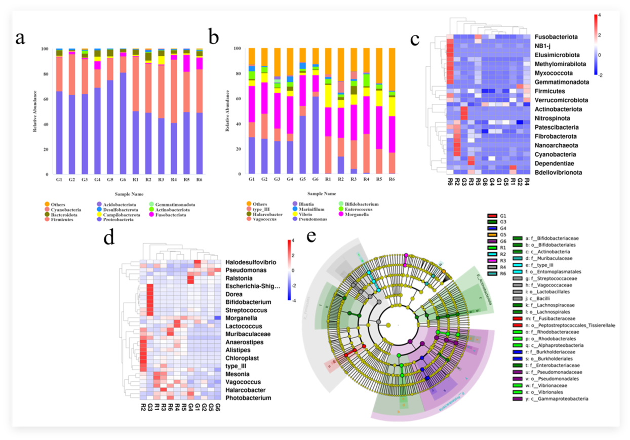

Table 1.
Effects of PS-MPs on the diversity indices of gut bacterial communities in Tachypleus tridentatus. Definition of sample group codes used in this study. R-series (7-day exposure groups): R1–R3 represent 6th-instar juveniles of Tachypleus tridentatus (R1: control; R2: low-concentration PS-MPs exposure; R3: high-concentration PS-MPs exposure); R4–R6 represent 5th-instar juveniles (R4: control; R5: low-concentration PS-MPs exposure; R6: high-concentration PS-MPs exposure).G-series (21-day exposure groups): G1–G3 represent 6th-instar juveniles (G1: control; G2: low-concentration PS-MPs exposure; G3: high-concentration PS-MPs exposure); G4–G6 represent 5th-instar juveniles (G4: control; G5: low-concentration PS-MPs exposure; G6: high-concentration PS-MPs exposure).
Table 1.
Effects of PS-MPs on the diversity indices of gut bacterial communities in Tachypleus tridentatus. Definition of sample group codes used in this study. R-series (7-day exposure groups): R1–R3 represent 6th-instar juveniles of Tachypleus tridentatus (R1: control; R2: low-concentration PS-MPs exposure; R3: high-concentration PS-MPs exposure); R4–R6 represent 5th-instar juveniles (R4: control; R5: low-concentration PS-MPs exposure; R6: high-concentration PS-MPs exposure).G-series (21-day exposure groups): G1–G3 represent 6th-instar juveniles (G1: control; G2: low-concentration PS-MPs exposure; G3: high-concentration PS-MPs exposure); G4–G6 represent 5th-instar juveniles (G4: control; G5: low-concentration PS-MPs exposure; G6: high-concentration PS-MPs exposure).
| Chao1 | Shannon | Simpson | |
|---|---|---|---|
| R1 | 185.02±27.09 | 3.66±0.34 | 0.84±0.04 |
| R2 | 354.62±102.87 | 4.25±0.50 | 0.85±0.04 |
| R3 | 226.62±39.51 | 3.68±0.25 | 0.83±0.03 |
| R4 | 235.20±30.39 | 3.41±0.41 | 0.78±0.07 |
| R5 | 229.01±41.17 | 3.45±0.42 | 0.80±0.08 |
| R6 | 272.37±112.87 | 3.76±0.66 | 0.82±0.08 |
| G1 | 88.89±11.99 | 3.00±0.22 | 0.76±0.05 |
| G2 | 87.64±10.85 | 2.83±0.46 | 0.73±0.09 |
| G3 | 166.31±103.65 | 3.41±0.93 | 0.75±0.12 |
| G4 | 120.23±29.01 | 2.96±0.85 | 0.72±0.17 |
| G5 | 95.35±17.09 | 2.61±0.28 | 0.70±0.05 |
| G6 | 75.87±9.92 | 2.11±0.48 | 0.57±0.14 |
Disclaimer/Publisher’s Note: The statements, opinions and data contained in all publications are solely those of the individual author(s) and contributor(s) and not of MDPI and/or the editor(s). MDPI and/or the editor(s) disclaim responsibility for any injury to people or property resulting from any ideas, methods, instructions or products referred to in the content. |
© 2026 by the authors. Licensee MDPI, Basel, Switzerland. This article is an open access article distributed under the terms and conditions of the Creative Commons Attribution (CC BY) license (http://creativecommons.org/licenses/by/4.0/).
Copyright: This open access article is published under a Creative Commons CC BY 4.0 license, which permit the free download, distribution, and reuse, provided that the author and preprint are cited in any reuse.


