

Submitted:
06 August 2026
Posted:
07 August 2026
You are already at the latest version
Abstract
Rabies is a fatal zoonotic disease of tyhe central nervous system of mammals caused by Lyssavirus rabies (RABV), a member of the Mononegavirales. Its single-stranded negative-sense RNA genome encodes five structural proteins: nucleoprotein (N), phosphoprotein (P), matrix protein (M), glycoprotein (G), and large protein (L). N plays a pivotal role in nucleocapsid assembly, regulation of viral transcription and replication, and the formation of Negri bodies, cytoplasmic viral factories essential for viral replication. Consequently, overexpression of N may disrupt RABV replicative dynamics, offering insight into the mechanisms governing its replication cycle while identifying potential targets for antiviral intervention. This study evaluated the effects of transient expression of RABV N mRNA on viral replication using a plasmid-based expression system in BHK-21 cells. BHK-21 cells were transfected with the pcDNA3.1(+)/C-DYK plasmid containing the N gene from the Challenge Virus Standard (CVS-11) strain of RABV. Viral mRNA transcription and total genomic viral load were quantified using absolute RT-qPCR. Overexpression of N mRNA led to a downward trend in viral mRNA transcription levels, although total viral genomic load remained largely unaffected. These findings provide further insight into the regulatory function of N during RABV replication and suggest that modulating N expression influences viral transcriptional dynamics.
Keywords:
rabies virus
; nucleoprotein
; RT-qPCR
; transfection
; viral replication
1. Introduction
Although rabies is an immunopreventable disease through proper wound care, post-exposure prophylaxis, and administration of rabies immunoglobulin, it remains almost universally fatal upon symptom onset. The disease causes approximately 59,000 human deaths annually worldwide, with the vast majority of cases transmitted through the bites of domestic dogs [1,2].
Lyssavirus rabies (Rabies virus, RABV) is a neurotropic virus belonging to the family Rhabdoviridae, order Mononegavirales. Its negative-strand RNA genome of approximately 12 kb encodes five structural proteins in sequential order: nucleoprotein (N), phosphoprotein (P), matrix protein (M), glycoprotein (G), and large protein (L) [3,4,5,6,7].
Morphologically, RABV exhibits a characteristic bullet-shaped structure, measuring approximately 200 nm in length and 80 nm in diameter. The virion consists of a host-derived lipid envelope surrounding an internal ribonucleoprotein (RNP) complex, which is composed of genomic RNA tightly encapsidated by the N protein in close association with the P and L proteins [5,8]. The RNP functions as the functional template for viral transcription and replication [5,8]. The transmembrane glycoprotein (G) forms spikes on the viral envelope and mediates host receptor binding, membrane fusion, and viral entry, serving as the primary target for neutralizing antibodies [5,6,8,9]. The matrix protein (M) condensation drives virion assembly and budding while mediating nucleocapsid condensation [10]. The L protein functions as the RNA-dependent RNA polymerase (RdRp), executing viral transcript synthesis and genomic RNA replication [8,9]. The phosphoprotein (P) acts as an essential non-catalytic cofactor for L and a molecular chaperone for N, while concurrently modulating host immune signaling to evade antiviral responses [8].
The accumulation of free N protein acts as a critical regulatory switch during the viral life cycle. At low intracellular concentrations of unencapsidated N, the viral RdRp functions primarily as a transcriptase, producing capped and polyadenylated subgenomic viral mRNAs for the five viral proteins [5,11,12]. As N protein accumulates, it binds to nascent leader RNAs and encapsidates full-length genomic and antigenomic RNA strands by binding to the leader sequences, preventing ribosome accessibility to subgenomic transcripts, now encapsidated by N, and shifting viral polymerase activity from transcription to full-length genomic RNA replication required for virion assembly [5,11,12]. Thus, the intracellular concentration of available N protein serves as a rate-limiting regulator balancing viral transcription and replication dynamics [8,11,12].
Furthermore, assembly of functional RABV RNP complexes requires strict stoichiometric balance among its components. Nucleoprotein is the most abundant structural constituent of the nucleocapsid, maintaining an approximate 2:1 ratio relative to P, which is vital for maintaining the structural integrity and enzymatic activity of the replicative machinery [11,12]. Together, N and P are also fundamental components of Negri bodies—phase-separated liquid viral microenvironments where RABV transcription and replication occur [13]. Disrupting the stoichiometric balance between N and P compromises the formation and functional dynamics of these replication factories, rendering the N–P interaction an attractive target for novel antiviral strategies [13,14].
The objective of this investigation was to evaluate the impact of overexpressing RABV N mRNA via plasmid transfection on RABV replication and transcriptional activity in vitro.
2. Materials and Methods
2.1. Cells and Virus
Baby hamster kidney cells (BHK-21) were maintained in Minimal Essential Medium (MEM) supplemented with 10% fetal bovine serum (FBS) at 37 °C in a humidified atmosphere containing 5% CO2. The Challenge Virus Standard strain of RABV (CVS-11), propagated in BHK-21 cells with a viral titer of 106.685 TCID50/mL, was kindly provided by the Pasteur Institute, São Paulo, Brazil.
2.2. Plasmid Construction
The complete nucleotide sequence of the N gene of the CVS-11 strain was determined by Sanger sequencing using the BigDye™ Terminator v3.1 Cycle Sequencing Kit (Applied Biosystems, Foster City, CA, USA) with primers 21G, 304, and 784, as described previously by Orciari et al. [15].
The mammalian expression vector pcDNA3.1(+)/C-DYK (GenScript Biotech, Piscataway, NJ, USA), driven by the human cytomegalovirus (CMV) promoter, was used to express recombinant RABV N mRNA. The CVS-11 N coding sequence was inserted into the vector via BamHI restriction sites. A consensus Kozak sequence was added immediately upstream of the initiation codon to optimize translational efficiency in mammalian cells. The C-terminal region was cloned using the CloneEZ strategy to fuse the FLAG tag in-frame to the N protein without introducing unnecessary restriction site sequences. Sequence accuracy of the recombinant plasmid (constructed by FastBio Brasil) was confirmed by Sanger sequencing using T7 promoter (5'-TAATACGACTCACTATAGGG-3') and bGH reverse (5'-ATAGAGCAGATTTTCGAGACAGC-3') primers, alongside RABV N-specific primers 510 and 784 [15].
2.3. Viral Infection and Transfection Assays
BHK-21 cells were seeded in 96-well culture plates at 100 µL per well in MEM containing 10% FBS. For viral infection groups, CVS-11 virus stock was diluted 1:10 in MEM with 10% FBS. Inoculation was performed at the time of cell plating by adding 50 µL of viral suspension per well (final volume of 150 µL per well). Uninfected control wells received 50 µL of MEM with 10% FBS. Plates were incubated at 37 °C under 5% CO2 for 24, 48, or 72 h.
Transfection was performed at 24, 48, or 72 h post-infection. Supernatants were aspirated, and cells were incubated with 100 µL of transfection complex containing 200 ng of plasmid DNA and Lipofectamine™ 3000 (Thermo Fisher Scientific, Waltham, MA, USA) in serum-free MEM according to manufacturer instructions. Non-transfected control wells received 100 µL of serum-free MEM. Monolayers were harvested 24 h post-transfection. Prior to collection, culture supernatants were removed, and cells were washed ten times with serum-free MEM to eliminate extracellular residual plasmid DNA. Total RNA was extracted from the cell monolayers using the QIAamp Viral RNA Mini Kit (Qiagen, Hilden, Germany). All experimental conditions were conducted in triplicate across three independent experiments.
2.4. Quantitative RT-qPCR Analysis
Real-time RT-qPCR assays were carried out to quantify total CVS-11 viral RNA and recombinant plasmid-derived N mRNA. Primers JW12 (5'-ATGTAACACCYCTACAATG-3') and N165-146 (5'-GCAGGGTACTTGTACTCATA-3') with probe RABVprobeptnN (5'-56-FAM/ACAAGATTGTATTCAAAGTCAATAATCAG/3IABkFQ-3') were used for RABV N gene detection. Recombinant mRNA was amplified using the T7 promoter forward primer (5'-TAATACGACTCACTATAGGG-3') paired with N165-146 reverse primer and RABVprobeptnN probe.
Absolute quantification standard curves were generated using serial 10-fold dilutions of pGEM-T Easy vector containing the RABV N gene (PV strain) [16]. For recombinant plasmid mRNA quantification, standard curves were prepared using serial 10-fold dilutions of pcDNA3.1(+)/C-DYK containing CVS-11 N insert. Host cell β-actin gene expression served as an endogenous normalizer using primers actβ_for (5'-ATTGGCAACGAGCGGTT-3') and actβ_rev (5'-ACGTCACACTTCATGATGGA-3') with probe actβ_probe (5'-5FAM/ATTCCATAC/ZEN/CCAGGAAGGAACGCTGG/IBFQ-3'). Assays were conducted using the SuperScript III Platinum One-Step qRT-PCR Kit (Invitrogen, Carlsbad, CA, USA). To control for residual plasmid DNA contamination, parallel reactions were executed in the presence (+RT) and absence (-RT) of reverse transcriptase; net RNA copy numbers were calculated as [# copies with RT] - [# copies without RT].
2.5. Statistical Analysis
Data analysis was performed using GraphPad Prism software (version 11.0.0; GraphPad Software, San Diego, CA, USA). Differences across experimental groups and time points were analyzed using two-way analysis of variance (ANOVA) followed by Bonferroni's post-hoc test for multiple comparisons. Differences were considered statistically significant at p < 0.05. Outliers were screened using the ROUT test (Q = 1%), with no extreme values identified. Graphical representation was executed in R using the ggplot2 package.
3. Results
3.1. Plasmid-Derived N mRNA Expression
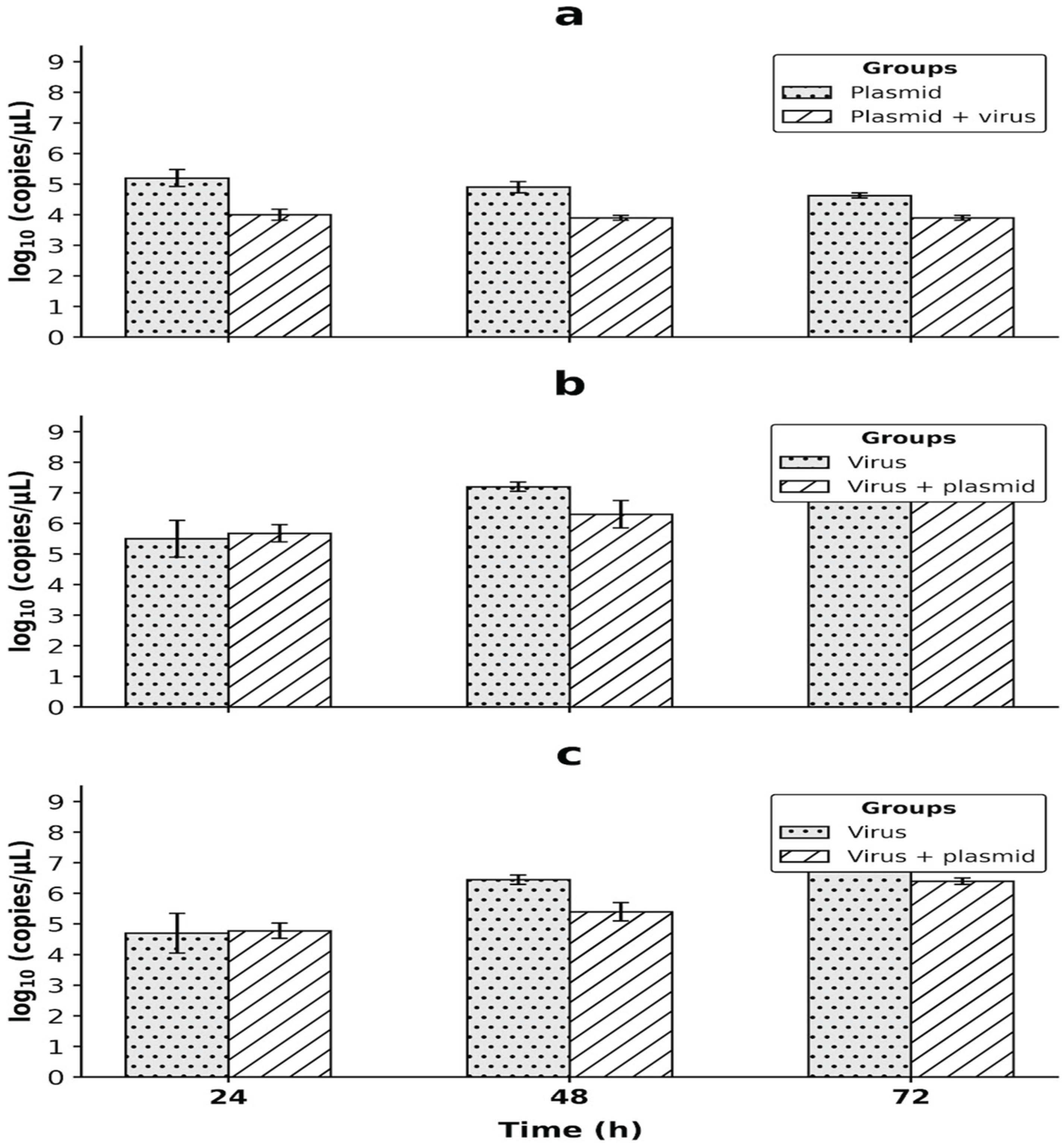
Plasmid-derived N mRNA expression was quantified in BHK-21 cells transfected with the plasmid alone or co-incubated with RABV CVS-11 at 24, 48, and 72 h post-infection. Two-way ANOVA revealed no significant main effect of time (p = 0.1425) or time × group interaction (p = 0.3348). However, a significant group effect was observed (p < 0.0001), demonstrating that RABV co-infection significantly diminished plasmid-derived transcript accumulation (Figure 1). Bonferroni's multiple comparisons test confirmed significantly reduced plasmid mRNA levels in the plasmid + virus group relative to plasmid-only cells at all evaluated time points (24 h: p = 0.0006; 48 h: p = 0.0024; 72 h: p = 0.0274).
3.2. Effect of N mRNA Overexpression on Total Viral RNA
Total CVS-11 viral RNA accumulation was evaluated in cells infected with virus alone versus cells infected and transfected with plasmid at 24, 48, and 72 h post-infection. Two-way ANOVA indicated a significant main effect of time (p = 0.0004), consistent with ongoing active viral replication. No statistically significant main effect was observed between groups (p = 0.1253) or for the interaction term (p = 0.3269) (Figure 1). Post-hoc Bonferroni testing showed no significant differences between groups at any individual time point (24 h: p > 0.9999; 48 h: p = 0.2840; 72 h: p = 0.6164), although a downward trend in total viral load was noted in the virus + plasmid group at 48 and 72 h.
3.3. Effect of Plasmid Transfection on Viral N mRNA Synthesis
Viral N mRNA transcription levels were analyzed across 24, 48, and 72 h post-infection. Two-way ANOVA showed significant main effects for both time (p = 0.0002) and group treatment (p = 0.0390), with no interaction effect (p = 0.2306) (Figure 1). While viral mRNA increased progressively over time in both groups, mean viral mRNA levels were consistently lower in the virus + plasmid group at 48 h (p = 0.1381) and 72 h (p = 0.2334) compared to cells infected with virus alone, demonstrating that overexpression of plasmid-encoded N mRNA exerts a downregulatory effect on viral transcription.
4. Discussion
This study investigated the impact of exogenous RABV nucleoprotein mRNA overexpression on RABV CVS-11 replication dynamics in BHK-21 cells using a plasmid transfection approach.
Plasmid-derived N transcript levels were readily detectable at early time points, following typical transient expression profiles wherein plasmid DNA remains unintegrated and is progressively lost or degraded over time [17]. Intriguingly, co-infection with RABV resulted in a significant decrease in plasmid-derived N mRNA levels compared to uninfected cells (Figure 1). Rabies virus infection is well recognized to shut down host cellular transcription and alter overall translational efficiency [18]. Viral disruption of host metabolic and transcriptional machinery likely reduced the cell's capacity to express recombinant mRNA from the exogenous plasmid promoter.
Regarding viral dynamics, total viral RNA increased progressively over the 72-h period, confirming active replication (Figure 1). While a downward trend in total viral genomic load was observed at 48 and 72 h in plasmid-transfected cells, these reductions were not statistically significant. However, direct evaluation of viral N mRNA synthesis revealed a significant overall treatment effect, with viral mRNA levels remaining lower in plasmid-transfected cells at 48 and 72 h (Figure 1). These findings indicate that superexpression of nucleoprotein mRNA downregulates Rabies virus transcription.
In unsegmented negative-sense RNA viruses, the precise balance between viral transcription and replication depends heavily on the availability and stoichiometry of RNP components, particularly N and P [8,11,12,19]. Free N protein acts as a molecular trigger: when unencapsidated N accumulates, it encapsidates nascent genomic RNA, signaling the viral polymerase to switch from transcribing subgenomic mRNAs to replicating full-length anti-genomes and genomes [11,12].
An artificial excess of N transcript—and presumably translated N protein—may accelerate premature encapsidation or alter the required 2:1 N:P ratio, sequestering functional P chaperones and suppressing viral transcriptase activity [11,13,14]. Furthermore, because Negri body formation and functionality require strict N–P stoichiometry, altered N expression could disrupt the liquid-liquid phase separation dynamics of these viral factories [13,20].
Several limitations should be noted. First, an empty vector or non-viral protein control plasmid was not included to rule out non-specific cellular responses to nucleic acid transfection. Second, this study utilized the laboratory-adapted CVS-11 strain; wild-type field isolates may display distinct replication kinetics [16,21]. Finally, while target mRNA dynamics were thoroughly quantified, western blotting or immunofluorescence assays will be essential in future studies to confirm protein-level expression and stoichiometric balance.In conclusion, overexpression of plasmid-derived N mRNA negatively modulates RABV mRNA transcription in vitro. Further investigation into stoichiometric regulation of the RNP complex may inform novel therapeutic interventions against rabies.
Author Contributions
LB and JA performed the in vitro experiments and RT-qPCR assays. CLM performed the statistical analyses. SAT designed the recombinant plasmid construct and supervised RT-qPCR analyses. PEB conceptualized and supervised the study. All authors read and approved the final manuscript.
Funding
This study was supported by the São Paulo Research Foundation (FAPESP, grant no. 2022/07115-7) and the National Council for Scientific and Technological Development (CNPq, grant no. 302503/2021-8). Publication fees were supported by the Coordenação de Aperfeiçoamento de Pessoal de Nível Superior - Brasil (CAPES).
Data Availability Statement
The data supporting the findings of this study are available from the corresponding author upon reasonable request.
Acknowledgments
The authors thank the Pasteur Institute of São Paulo for providing the RABV CVS-11 strain.
Conflicts of Interest
The authors declare that they have no conflicting interest.
References
- Cleaveland, S.; Sharp, J.; Abela-Ridder, B.; Allan, K.J.; Bányai, K.; Brierley, L.; Claes, F.; Daniel-Ribeiro, C.T.; de Balogh, K.; Dufour, B.; et al. Proof of concept of mass dog vaccination for the control and elimination of canine rabies. Rev. Sci. Tech. 2018, 37, 559–568. [Google Scholar] [CrossRef] [PubMed]
- World Health Organization. Zero by 30: The Global Strategic Plan to End Human Deaths from Dog- Mediated Rabies by 2030; WHO: Geneva, Switzerland, 2022; Available online: https://www.who.int/news/item/28-09-2022-who-ntd-reaffirms-ts-commitment-to-zero-by-30- (accessed on 16 May 2023).
- Fooks, A.R.; Banyard, A.C.; Horton, D.L.; Johnson, N.; McElhinney, L.M.; Jackson, A.C. Current status of rabies and prospects for elimination. Lancet 2014, 384, 1389–1399. [Google Scholar] [CrossRef] [PubMed]
- Rupprecht, C.E.; Freuling, C.M.; Mani, R.S.; Jackson, A.C. A history of rabies. In Rabies: Scientific Basis of the Disease and Its Management, 4th ed.; Fooks, A.R., Jackson, A.C., Eds.; Academic Press: Boston, MA, USA, 2020; pp. 1–42. [Google Scholar]
- Schnell, M.J.; McGettigan, J.P.; Wirblich, C.; Papaneri, A. The cell biology of rabies virus. Nat. Rev. Microbiol. 2010, 8, 51–61. [Google Scholar] [CrossRef] [PubMed]
- Davis, B.M.; Rall, G.F.; Schnell, M.J. Everything you always wanted to know about rabies virus (but were afraid to ask). Annu. Rev. Virol. 2015, 2, 451–471. [Google Scholar] [CrossRef]
- Guo, Y.; Duan, M.; Wang, X.; Gao, J.; Guan, Z.; Zhang, M. Early events in rabies virus infection: Attachment, entry, and intracellular trafficking. Virus Res. 2019, 263, 217–225. [Google Scholar] [CrossRef] [PubMed]
- Fisher, C.R.; Streicker, D.G.; Schnell, M.J. The spread and evolution of rabies virus: Conquering new frontiers. Nat. Rev. Microbiol. 2018, 16, 241–255. [Google Scholar] [CrossRef] [PubMed]
- Zandi, F.; Eslami, N.; Sohrabi, A.; Howeizi, N.; Fayaz, A. Rabies infection: An overview of lyssavirus- host protein interactions. Iran. Biomed. J. 2021, 25, 226–242. [Google Scholar] [CrossRef] [PubMed]
- Wunner, W.H.; Conzelmann, K.K. Rabies virus. In Rabies: Scientific Basis of the Disease and Its Management, 4th ed.; Fooks, A.R., Jackson, A.C., Eds.; Academic Press: Boston, MA, USA, 2020; pp. 43–81. [Google Scholar] [CrossRef]
- Wunner, W.H. Rabies virus. In Rabies, 2nd ed.; Jackson, A.C., Wunner, W.H., Eds.; Academic Press: London, UK, 2007; pp. 23–68. [Google Scholar] [CrossRef]
- Albertini, A.A.V.; Ruigrok, R.W.H.; Blondel, D. Rabies virus transcription and replication. Adv. Virus Res. 2011, 79, 1–22. [Google Scholar] [CrossRef] [PubMed]
- Nikolic, J.; Le Bars, R.; Lama, Z.; Scrima, N.; Lagaudrière-Gesbert, C.; Gaudin, Y.; Blondel, D. Structure and function of Negri bodies. Adv. Exp. Med. Biol. 2019, 1215, 111–127. [Google Scholar] [CrossRef] [PubMed]
- Zhan, J.; Huang, Y.; Lu, S.; Wu, X.; Tan, Y. Analysis of mechanisms of the rabies virus P protein- nucleocapsid interaction using engineered N-protein peptides and potential applications in antivirals design. Antivir. Res. 2025, 234, 106075. [Google Scholar] [CrossRef] [PubMed]
- Orciari, L.A.; Niezgoda, M.; Hanlon, C.A.; Shaddock, J.H.; Sanderlin, D.W.; Yager, P.A.; Rupprecht, C.E. Rapid clearance of SAG-2 rabies virus from dogs after oral vaccination. Vaccine 2001, 19, 4511–4518. [Google Scholar] [CrossRef] [PubMed]
- Conselheiro, J.A.; Silva, O.C.R.; Carnieli, P.; Oliveira, R.N.; Brandão, P.E. Evolution of rabies virus isolates: Virulence signatures and effects of modulation by neutralizing antibodies. Pathogens 2022, 11, 1556. [Google Scholar] [CrossRef] [PubMed]
- Chong, Z.X.; Yeap, S.K.; Ho, W.Y. Transfection types, methods and strategies: A technical review. PeerJ 2021, 9, e11165. [Google Scholar] [CrossRef] [PubMed]
- Finke, S.; Mueller-Waldeck, R.; Conzelmann, K.K. Rabies virus matrix protein regulates the balance of virus transcription and replication. J. Gen. Virol. 2003, 84, 1613–1621. [Google Scholar] [CrossRef] [PubMed]
- Finke, S.; Conzelmann, K.K. Replication strategies of rabies virus. Virus Res. 2005, 111, 120–131. [Google Scholar] [CrossRef] [PubMed]
- Liu, P.; Yang, J.; Wu, X.; Fu, Z.F. Interactions amongst rabies virus nucleoprotein, phosphoprotein and genomic RNA. J. Gen. Virol. 2004, 85, 3725–3734. [Google Scholar] [CrossRef] [PubMed]
- Fahl, W.O.; Garcia, A.I.; Asano, K.M.; de Lucca, T.; Iamamoto, K.; Mori, E.; Peixoto, Z.M.; Brandão, P.E.; Scheffer, K.C. Desmodus rotundus and Artibeus spp. bats m ight present distinct rabies virus lineages. Braz. J. Infect. Dis. 2012, 16, 545–551. [Google Scholar] [CrossRef] [PubMed]
Figure 1.
Real-time RT-qPCR absolute quantification (log10 RNA copies/µL) in BHK-21 cells at 24, 48, and 72 h post-infection. a Expression of plasmid-derived N mRNA in plasmid alone vs. plasmid + RABV CVS-11. b Total RABV CVS-11 genomic RNA quantification RABV CVS-11 alone vs. RABV CVS-11 + plasmid. c RABV CVS-11 viral N mRNA levels in RABV CVS-11 alone vs. RABV CVS-11 + plasmid.
Figure 1.
Real-time RT-qPCR absolute quantification (log10 RNA copies/µL) in BHK-21 cells at 24, 48, and 72 h post-infection. a Expression of plasmid-derived N mRNA in plasmid alone vs. plasmid + RABV CVS-11. b Total RABV CVS-11 genomic RNA quantification RABV CVS-11 alone vs. RABV CVS-11 + plasmid. c RABV CVS-11 viral N mRNA levels in RABV CVS-11 alone vs. RABV CVS-11 + plasmid.
Disclaimer/Publisher’s Note: The statements, opinions and data contained in all publications are solely those of the individual author(s) and contributor(s) and not of MDPI and/or the editor(s). MDPI and/or the editor(s) disclaim responsibility for any injury to people or property resulting from any ideas, methods, instructions or products referred to in the content. |
© 2026 by the authors. Licensee MDPI, Basel, Switzerland. This article is an open access article distributed under the terms and conditions of the Creative Commons Attribution (CC BY) license (http://creativecommons.org/licenses/by/4.0/).
Copyright: This open access article is published under a Creative Commons CC BY 4.0 license, which permit the free download, distribution, and reuse, provided that the author and preprint are cited in any reuse.