

Submitted:
05 August 2026
Posted:
06 August 2026
You are already at the latest version
Abstract
The present paper aimed to study the effects of 40% ethanolic extracts (5 mg GAE/mL) from Ajuga reptans (Ajre), Geranium phaeum (Geph) and Helianthemum nummularium (Henu) on mouse normal fibroblasts and human blood mononuclear cell lines L929 and SC, as well as in silico ADMET and molecular docking assessments of the major and key phenolics found in the three test medicinal species, against the widespread protein disease cyclooxygenase 2 (COX-2), in comparison with the native ligand diclofenac. Briefly, in silico studies revealed flavonoid derivatives as being more likely to induce metabolic effects in humans, by interfering with the activity of cytochrome P450 (CYP450) isoenzymes, of P-glycoprotein transporters (P-gp), and of potassium channels (hERG) respectively, at the same time more effective inhibitors of COX-2 in comparison with phenolic acids. In vitro studies on Ajre indicated augmented inhibitory effects on the viability of L929 (IC50 = 184.70 µg GAE/mL) and SC (IC50 = 91.30 µg GAE/mL); Geph did not affect the viability of L929 (IC50 = 414.70 µg GAE/mL) and showed putative effects on SC (152.00 µg GAE/mL); Henu revealed weak inhibitory effects on L929 (IC50 = 350.00 µg GAE/mL) versus high stimulatory effects on the viability of SC line (IC50 = 183.50 μg GAE/mL), up to 21% more intense than those induced by LPS positive control.
Keywords:
blue bugle
; dusky cranesbill and rock-rose ethanolic extracts
; key effects in humans
; in silico ADMET
; molecular docking on COX-2
; studies on L929 and SC lines
1. Introduction
As it is well established, the body’s response to a combination of pharmacologically active compounds, such as complex plant extracts, is in accordance with the overall receptor occupancy and the intrinsic efficacy of the compounds on a specific molecular target [1,2,3]. Hence, only those compounds that simultaneously have bioavailability and a high affinity for an active molecule can be theoretically successful in vivo.
Practically, there are many other physical, chemical and metabolic constraints that make the plant extracts with high pharmacological activity in vitro [4,5,6,7], to not accomplish a similar efficacy in vivo. The main constraints in obtaining a concordant in vitro-in vivo pharmacological activity are the following: 1)the secondary metabolites from the green plants are mostly in the form of glycosides and other conjugated forms, which are pharmacologically inactive; 2)their active forms, aglycones, are mostly released through the activity of the microbial enzymes, therefore their amount is depending on the individual microbiota; 3)the released aglycones are in fact xenobiotics, thus being the subject of phase I and II metabolism in humans, resulting a dramatic reduction of the compounds available for tissues. Finally, 4)the antioxidant activity is not a single process but a system of oxidation and reduction reactions preventing the oxidation of a third molecule [8], therefore, the processes need a perfectly matching.
In terms of biological activity, it is also well known that the compounds with high efficacy in the plant defence [9] also are the compounds with high pharmacological activity in humans; thus, the polyphenols in the series of phenolic acids and flavonoids, alongside bitter compounds, terpenes, alkaloids, volatile oils, and the genus markers are the major active compounds, both in plants and humans. Due to this prevalence of phenolics in green plants, there is a series of common primary pharmacological effects expected from all ingested plant derived products and phytomedicines. These are: 1) antioxidant, anti-inflammatory, chemopreventive effects [10,11,12,13,14,15,16], 2) effects upon the immune cells [17,18], 3) effects upon the gut microbiota [19,20,21], and 4) effects upon the general metabolism [22,23,24]. Yet, the study of the key physiological effects in vivo is very difficult, while through complementary in vitro-in silico studies there can be obtained important pharmacokinetic and pharmacological data. For example, in silico tools today can offer information about the most probable absorption, distribution, metabolism, excretion and toxicity features (namely ADMED) of the small plant compounds, while the molecular docking tools can estimate their probable efficacy on a specific protein disease and molecular target in cells. Similarly, in vitro studies on cell lines with general predictive value can direction the future in vitro and in vivo researches on those direction resulted as useful and properly for the test products. In silico-in vitro studies are all the more useful as they can offer information regarding both, potential benefits versus potential limitations in humans.
In this context, the present paper aimed to study the polar extracts from three alpine medicinal species by complementary in silico-in vitro approaches, in the purpose of estimating their compositional and pharmacokinetic features, alongside general cytotoxicity and immune-related effects in humans; the plant species under studies were Ajuga reptans (commonly blue bugle), Geranium phaeum (commonly dusky cranesbill), and Helianthemum nummularium (commonly rock-rose). Studies first of all established the major and key phenolics present in the polar alcoholic extracts from the three test plant species; the chemical compositional aspects were accomplished by HPTLC analysis [25,26] of the 70% ethanolic extracts. After that, the in silico studies aimed to analyse the ADMET (Absorption, Distribution, Metabolism, Excretion, and Toxicity) parameters of the major and key phenolics achievable in the alcoholic extracts from each one test species. Next, the compounds assigned with the ability to pass the humans’ gastrointestinal (GI) and blood brain barriers (BBB) were tested through molecular docking (CLC) tool in the purpose to estimate their ability to inhibit the function of a major protein disease, involved in all chronic pathologies in humans, the cyclooxygenase 2 (COX-2) respectively; studies were done by comparison with the native ligand, diclofenac. The 40% ethanolic standardized extracts (made as 5 mg gallic acid equivalents/GAE per 1 mL test extract) from each one test plant species were further tested in relation with their ability to interfere with the viability of normal mouse fibroblasts and human blood mononuclear cells (L929 and SC lines) in vitro, in the purpose of estimating their potential limits alongside the potential benefits in humans.
2. Materials and Methods
2.1. Plant Materials Description
Studies were done on the flower part of Ajuga reptans (Ajre15-1.17) and the aerial part (herba et flores) of Helianthemum nummularium (Henu16-2.17) and Geranium phaeum (Geph24-3.17), respectively. The medicinal plants were collected from the Romanian Carpathians and the adjacent regions, Prahova region respectively, at 600 - 1.400 m altitude. Taxonomic identification was done by the botanist’s team of National Institute of Chemical-Pharmaceutical Research and Development (ICCF Bucharest); the voucher specimens are deposited in the ICCF plant material storing room.
2.2. Plant Extracts Preparation
The three plant species were dried at 40-45 °C, then ground in a cross-beater mill equipped with a 2 mm sieve. The resulting plant powders (samples of 20 g of each test species) were separately extracted with 200 mL of 70% ethanol (v/v); the process was conducted at the reflux temperature, for 40 minutes, in continuously agitation status. The resulting whole ethanolic extracts were submitted to vacuum filtration using a medium pore filter paper. The filtered ethanolic extracts (on average 1400 mL) were used as follows: 1000 mL of ethanolic extract of each vegetal species was subjected to acidic hydrolysis to release the aglycone compounds from their glycosides; the other 400 mL was stored at 4℃and,and used for current analytical qualitative/HPTLC and quantitative investigations. The acidic hydrolysis was conducted as follows: 1000 mL of each genuine 70% ethanolic extract was concentrated to a spiss residue, after that the spiss residue was solved into 100 mL 1N HCl and hydrolysed for 30 minutes at the boiling temperature. The hydrolysed extract was extracted with 100 mL ethyl acetate, three times serial, after that the three ethyl acetate fractions were mixed and desicated on a sodium carbonate paper filter; the resulted (three) desicated extracts, corresponding to the three test species, were vacuum concentrated to the residue, then the residues were solved in 70% ethanol to achieve the final volumes of 20 mL test sample. The hydrolysed series and the genuine 70% ethanolic extract series were subjected to analytical qualitative (HPTLC) and quantitative investigations.
Similarly, other three series of 20 g plant powders were separately extracted with 200 mL of 70% ethanol (v/v), at the reflux temperature, for 40 minutes, in continuously agitation status. The resulting whole ethanolic extracts were vacuum filtration through a medium pore filter paper, after that the filtered extracts (on average 1400 mL) were analysed as concerning the total phenolic content in samples; quantitative analyses were done by Folin–Ciocalteau method in comparison with gallic acid reference substance calibration curve (R2= 0.991, n = 3) [27], using the UV/Vis Hélios γ, Thermo Electron Corporation spectrophotometer (Waltham, MA, US); the results were computed as mg gallic acid equivalents [GAE] per 1 mL sample. The quantified whole ethanolic extracts were concentrated to the residues (spiss), after that each one spiss residue was solved in 40% ethanol solution to achieve the precise concentration of 5 mg [GAE] per 1 mL plant extract (g/v), respectively. The three standardized extracts, codified Ajre, Geph and Henu, were subjected to in vitro pharmacological studied.
2.3. Chemical Analysis of Test Plant Extracts
The chemical qualitative composition of polyphenols in the two series of test plant extracts (polyphenols fingerprint) was obtained through the high-performance thin- layer chromatography (HPTLC) method, following Wagner and Bladt [25] and Reich and Schibli [26] recommendations. Polyphenols fingerprint was analysed by using Linomat 5 instrument (CAMAG, Muttenz, Switzerland), following two standard methods and solvent systems: the standard method for polyphenols glycosides’ assessment using the mobile phase ethyl acetate–formic acid–glacial acetic acid–water (100:11:11:26)/System I, and NP-PEG (Natural Product Reagent) and VS (Vanillin-Sulphuric Acid Reagent) visualisation reagents, to reveal the polyphenolic species found in genuine 70% ethanolic extracts from each one test plant species (chromatograms A); the standard method for saponin aglycones’ assessment using the mobile phase chloroform-glacial acetic acid-methanol-water (64:32:12:8)/System II and NP-PEG treatment, to assess the polyphenol aglycones in the hydrolized extracts (chromatograms B). The polyphenols assignment in each test species/extract, meaning the analysis of Rf and colour’ spot similarity, was done by comparison with the reference substance (REF) used, pure phenolic acids and flavonoids, respectively.
The pure compounds, reference substances (REF) in the series of plant polyphenols were purchased from Sigma Aldrich (Merck Darmstadt, Germany) in Romania. The REF used are as follows: quercetin (95%), quercetin-3-O-galactoside/hyperoside (>97%), quercetin-3-O-glucoside/isoquercitrin (>90%), quercetin-3-O-rhamnoside/quercitrin (>90%), quercetin-3-O-rutinoside/rutin (min. 95%), kaempferol (95%), kaempferol-3- O-glucoside/astragalin (99%), kaempferol-3-O-rutinoside/nictoflorin (min. 95%), kaempferol-3-O-rhamnoside/afzelin (98%), kaempferol-3-O-arabinoside/avicularin (98%), kaempferol-3-O-galactopyranoside/trifolin (>98%), apigenin (>97%), apigenin-7- O-glucoside/cosmosiin (97%), apigenin-8-C-glucoside/vitexin (>96%), apigenin-6- C-glucoside/isovitexin (>98%), vitexin-2ˈˈO-rhamnoside (>98%), apigenin 7-O-apiosyl glucoside/apiin (>99%), luteolin (>98%), luteolin-7-O-glucoside/cynaroside (analytical standard), luteolin-8-C-glucoside/orientin (>97%), myricetin (>98%), myricitrin (analytical standard), caffeic acid (99%), chlorogenic acid (>95%), cynarin (analytical standard), rosmarinic acid (97%), gallic acid (95%), ferulic acid (99%), ellagic acid (>96%), protocatechuic acid (>97%), gentisic acid (>99%) umbelliferone (>99%), and scopoletin (>99%). REF was prepared as solutions 10-3M in 70% ethanol.
2.4. In Silico ADMET Studies
In silico ADMET studies were done by Biosig.Lab [28] and SwissADME [29] free web tools. There were analysed bioavailability, metabolic and toxicity features of 55 common plant phenolics, including the three series of major and key phenolics achievable in the three test medicinal plant species under studies; particularly, there were analysed the ability of test plant compounds to impact the activity of cytochrome P450 oxidase system in humans (by interfering with the activity of CYP1, CYP2, CYP3 isoenzymes), of glycoprotein P transporters (acting as substrate/P-gps and/or inhibitor/P-gpi), of hERG I/II potassium channels, of OCT2, as well as the potentiality to induce hepatotoxicity in humans. Tables S1, S2, S3 in the supplementary files present SwissADME computational data on each one series of major and key phenolics achievable in the three plant species under studies.
2.5. In Silico Molecular Docking Studies
In silico molecular docking studies were done by CLC Drug Discovery Workbench Software (QIAGEN, Aarhus, Denmark) [30], and they intended the computation of the docking scores. The docking score used in the Drug Discovery Workbench, namely the PLANTSPLP score, is calculated according to Korb et al [31]. This score is associated with the potential energy change when the ligand interacts with the target protein to form a complex together. Punctually, the PLANTSPLP score (1) is given by all contributions from heavy atoms contacts between the ligand and the molecules included in the binding site setup, comprising rewarded contacts (hydrogen bond interactions, Lone-pair - metal ion interactions, non-polar interactions) and punished contacts (non-polar - polar contacts or repulsive contacts (hydrogen bond donor-donor contacts, hydrogen bond donor-metal contacts, hydrogen bond acceptor-acceptor contacts).
According to Korb et al [31], a very negative score is associated to a strong binding, while a less negative, or a positive score corresponds to a weak or non-existing binding.
The score is attended by a root Root Mean Square Deviation (RMSD) value, measuring “the differences between values predicted by a model and the actual observed values”; punctually, the RMSD value in docking indicates the accuracy with which the test ligand position in the binding site was reproduced compared to that validated co-crystallized (the native ligand); specifically, a RMSD value lower than 2 indicates that the docking protocol has been validated. The current docking studies aimed to test the interaction between the cyclooxigenase 2 (COX-2) and the compounds resulted with bioavailability in humans (noticed as GI and BBB in Tables S1, S2 and S3) alongside the marker genus ajugalactone, ajugasterone and geraniin. The assessments were done by comparison with the native ligand, diclofenac, the Protein Data Bank PDB ID: 1PXX [32], respectively. This way, the COX-2 was first of all prepared by setting-up the binding site and the binding pocket at the specific bond legs of the protein/chain A (Å3). The hydrogen bonds formed between the native ligand, diclofenac, with the amino acids from group interaction atoms were further used to predict the binding modes, the binding affinities and the orientation of the docked compounds in the active site of the molecular target. Secondly, the molecular docking validation is done by re-docking the native ligand in comparison with the tested ligands. Finally, the punctual hydrogen bonds and the docking scores are compared, the native ligand vs the tested ligands, to obtain their scale of efficacy. The optimized molecular structure of the test compounds and the numbering of the atoms in the binding pocket is according to the Spartan`24 software [33].
2.6. In Vitro Pharmacological Studies
In vitro studies were done using the CellTiter 96AQueous One Solution Cell Proliferation Assay Promega Corporation (Madison, WI, USA) protocol [34]; the MTS assay is based on the selective ability of the viable cells in culture to reduce the 3-(4,5-dimethylthiazol-2-yl)-5-(3-carboxymethoxyphenyl)-2-(4-sulfophenyl)-2H-tetrazolium (MTS) in medium into a purple coloured formazan crystals which can be further measured at 492 nm. The three series of test extracts were prepared as seven dilution point series, using each one cell line (L929, SC) medium in the study; there were also prepared two negative control series, one consisting in the growth medium alone and another one consisting in 40% ethanol solvent in dilution series applied for the test samples; in the specific case of SC line, a positive control series consisting in lipopolysaccharide (LPS) solution made in phosphate buffered saline for cell culture (PBS) counting 5 µg LPS/1 mL has also been prepared. The test series (triplicates) as well as the negative and positive (LPS) control series (also as triplicates) were applied on the two cell lines, SC and L929, by following the cytotoxicity protocol. Briefly, after reaching the cell confluence necessary in cytotoxicity type assay, the cells are detached from the flask with Trypsin-EDTA, after that the cell suspension is centrifuged at 2000 rpm for 5 min, and resulted sediment is re-suspended in the respective cell growth medium: Iscove’s modified Dulbecco’s medium (IMDM; ATCC 30-2005) with 4 mM L-glutamine adjusted to contain 1.5 g/L sodium bicarbonate and supplemented with 0.05 mM 2-mercaptoethanol, 0.1 mM hypoxanthine and 0.016 mM thymidine, 90% and fetal bovine serum (FBS; ATCC 30-2020), 10% (purchased from ATCC, LGC Standards, Poland Office) for SC line and Eagle’s Minimum Essential Medium (ATCC 30-2003) and horse serum (Gibco™ Horse Serum ABP-040, purchased from ATCC, LGC Standards, Poland Office and ANTISEL the distributor of Gibco products in Romania) to a final concentration of 10% for L929 line. The cells were after-that seeded in 96-well plates at a density of 6.000 cells per well respectively, in 200 μL culture medium. After 20 hours of the cell exposure to the test/control samples, the culture medium was removed. The cells were after that incubated with the MTS solution for 2 hours and the viability of the adherent cells was measured by reading the absorbance of the test/control solutions at 492 nm (BMR-100 Microplate Reader, Boeco, Germany). The Optical Densities (O.D.) at 492 nm (mean value, n=3) were then used to compute the viability percentages (%) along the dilution series. The punctual results were computed for their statistical significance by using the Student “t” test. The notation (*) means results without statistical significance and p > 0.05; the notation (**) means results with statistical significance and 0.05 < p < 0.01; the notation (***) means results with statistical significance and p < 0.01.
3. Results
3.1. Chemical Compositional Data on the Three Test Medicinal Plant Species
Analytical assessments were done on the 70% genuine ethanolic extracts from the three alpine medicinal plant species, as described in the Section 2.2. The 70% ethanol has the advantage of being a properly solubilization solvent for most of the plant phenolics, at the same time acting as a technological sterilization medium for the vegetal material. The HPTLC method also has the advantage of effectively visualising the major phenolics from a plant extract, at the same time offering a plethora of qualitative and quantitative data on the flavonoids’ subclasses (meaning the presence of apigenin, kaempferol, luteolin, quercetin or miricetin derivatives), their degree of glycosylation, or esterification in the case of phenylcarboxylic acid derivatives, as well as the position of the glycosyl chains on the flavonoid core, and the presence of free aglycones in the genuine and hydrolysed extracts. Given these advantages, the HPTLC is factually used in the purpose of finding adulterants in the herbal derived products for human use.
3.1.1. Chemical Compositional Analysis of Ajuga reptans Flores Extract
According to the literature data available, there are over 300 wild species of Ajuga reptans L. (Lamiaceae) spread all over in Europe, Asia, North America, North Africa, and Australia [35]. In terms of medicinal activity, the traditional and folk medicine, as well as the scientific studies today, all paid close attention to this plant genus and particularly to Ajuga reptans species, commonly known as the blue bugle [36,37].
As compositional aspects, analytical studies on different aqueous and alcoholic extracts isolated from different plant parts of Ajuga reptans [38,39,40,41] indicated the prevalence of phenylpropanoids (e.g., verbascoside, isoverbascoside), of phenolic acids (e.g., 3,4-dihydroxyphenylacetic acid, rosmarinic acid), of flavonoids (e.g., mainly isoquercitrin derivatives) and of iridoids (e.g., 8-O-acetylharpagide), alongside two steroidal compounds and genus markers, namely ajugalactone and ajugasterone.
The current HPTLC studies were done on the flower part of Ajuga reptans collected from the Romanian Carpathians; Figure 1, chromatogram A1 shows the fingerprint of the major polyphenols compounds found in the 70% extract from the flower part of blue bugle, while the chromatogram A2 reveals the status of the bitter compounds.
The test extract, the tracks T2 in chromatogram A1, reveals quercetin derivatives (yellow-fluorescent/fl. spots at Rf~0.26/0.32/0.60/0.66/0.70) and ferulic acid derivatives (blue-indigo fl. spots at Rf~0.20/0.42/0.55/0.80/0.95) as being the major phenolic compounds found in Ajuga reptans flower extracts. By comparison with the reference substances used (tracks T1, T3, T4) and specialized data on the Rf and colour of the leading plant polyphenols [26,27], the two major quercetin derivatives were attributed to hyperoside (Rf~0.60) and isoquercitrin (Rf~0.65). The chromatogram A2 obtained through vanillin sulphuric acid reagent (VS treatment) also revealed the presence of two major bitter compounds (the brown zones at Rf~0.15 and Rf~0.22) in Ajuga reptans flower extracts. Studies on the hydrolysed extract, Figure S3 in the supplementary files, punctually the chromatogram B1, track T2H in comparison with the reference substances used (tracks T4-T18) confirmed the occurrence of ferulic acid (blue fl. spot at Rf~0.96), of caffeic acid (blue fl. spot at Rf~0.78) and of rosmarinic acid (blue fl. spot at Rf~0.51) in the Ajuga reptans flower extract.
By corroborating the current HPTLC results with specialized HPLC data [38,39,40,41], it resulted the following series of major and key phenolics expectable in Ajuga reptans polar hydroalcoholic extracts: quercetin, quercetin-3-O-glucoside/isoquercetin, quercetin-3-O-galactoside/hyperoside, isorhamnetin, isorhamnetin-3-O-glucoside, isorhamnetin-3-O-rutinoside/narcisin, rosmarinic acid, ferulic acid, caffeic and chlorogenic acids, 3,4-dihydroxyphenylacetic acid alongside 8-O-acetylharpagide, verbascoside, and ajugalactone and ajugasterone genus markers and key compounds.
3.1.2. Chemical Compositional Analysis of Geranium phaeum Herba Extract
Geranium phaeum L. (Geraniaceae), commonly called dusky cranesbill, is one of the 400 Geranium species found throughout the entire planet. The genus is used from ancient times for medicinal purposes, while the scientific studies revealed the major contribution of geraniin and geraniol, the genus markers, alongside the flavonoids, ellagic acid, and sesquiterpenes from Geranium extracts [42,43,44,45,46,47,48]. Geraniin is a hydrolyzable tannin principally present in the leaf part; it contains a glucose molecule linked with two ellagic acid residue esters at 2,4 and 3,6 carbons and a galloyl ester in C1 [45,46]; it must be noted that ellagic acid is synthesized in the root (rhizome) part only, while gallic acid is synthesized in roots and leaf parts, as well [45,46].
Analytical studies on the Geranium genus revealed a high phytochemical diversity, depending on the geographical location of the species; particularly, studies on 78 species collected from different geographic regions revealed that quercetin, kaempferol, and myricetin-3/7-O-glucoside/galactoside/rhamnoside derivatives are the common chemical denominator of leaves of all tested species [42,43,44]. The extracts from Geranium phaeum indicated the presence of luteolin and apigenin derivatives, punctually of orientin, vitexin, isovitexin, and vicenin flavonoids [47], while the floral part indicated high amounts of malvidin-3,5-O-diglucoside [48].
The HPTLC analysis in the current study, Figure 2, shows the fingerprints of the major polyphenols (chromatogram A1) and bitter compounds (chromatogram A2) found in 70% genuine ethanolic extract from the aerial part of Geranium phaeum collected from the Romanian Carpathians. The test extract, tracks T3-T4 in chromatogram A1, in comparison with the reference substance used (tracks T1, T2, T5), revealed the prevalence of caffeoylquinic acid derivatives (the light blue fl. spots s3,s4,s5,s6,s10), mainly chlorogenic acid (s5) and isochlorogenic acid (s6) isomers. The green fl. spots s1, s2 and s7, s9 were attributed to cosmosiin (Rf~0.69) and vitexin (Rf~0.74); the yellow-green fl. spot s8 likely apart to the luteolin C-glycoside namely orientin [47].
The chromatogram A2 (by VS treatment), particularly the dark green-brown zones at Rf values 0.1 - 0.3 in track T3, also reveals the attendance of bitter compounds in Geranium phaeum polar extracts. Studies on the hydrolysed extract, Figure S4 in the supplementary files, chromatogram B1, track T3H in comparison with the reference substances used (tracks T12 - T21) confirmed the prevalence of caffeoylquinic acid derivatives (the blue fl. spots along the entire chromatogram B1), particularly the preponderance of chlorogenic and caffeic acid aglycones’, as well as the presence of luteolin-7-O-glucoside namely cynaroside (the yellow-green fl spot at Rf~0.35).
Summing the current HPTLC data with HPLC data on similar extracts [43,44,47] it resulted in the following list of major and key phenolics achievable in the polar extracts from Geranium phaeum: geraniin, kaempferol-3-O-glucoside/astragalin, kaempferol-3,7- O-dirhamnoside/lespedin, quercetin-3-O-glucoside/isoquercitrin, quercetin-3,7-O- diglucoside, luteolin-8-C-glucoside/orientin, luteolin-7-O-glucoside/cynaroside, apigenin-8-C-glucoside/vitexin, apigenin-6,8-C-diglucopyranoside/vicenin, apigenin-6- C-glucoside/ isovitexin, alongside gallic, ellagic, caffeic, and chlorogenic phenolic acids.
3.1.3. Chemical Compositional Analysis of Helianthemum nummularium Herba Extract
Helianthemum nummularium L. (Fam. Cistaceae), commonly known as rock-rose, is a bush growing at high altitude in the Alps and Carpathians. The plants growing in the Mediterranean regions (e.g., Tunisia, Morocco, Libia and Algeria) are known as Cistus species. The genus includes more that 110 different species, most of them assigned with medicinal, cosmetic and fragrance value, as well. Being plants growing in harsh conditions, the Cistus species are assigned with very active secondary metabolites [49,50,51]. For example, the Cistus species growing in Portugal are notorious for their content in 2,2,6-trimethylcyclohexanone and ethyl dihydrocinnamate, both used as flavourand and fragrance agents, and to secure the wines from the Douro region [52]; Cistus ladanifer and Cistus monspeliensis from Morocco are noticed with high contents of antioxidant, antimicrobial compounds in the series of 1,8-cineole (19.27%), viridiflorol (16.38%), bornyl acetate (9.14%) and a-pinene (5.84%) [53]; Cistus monspeliensis, Cistus libanotis and Cistus villosus are notorious for diterpene type compounds (namely resins), also known as labdanum, used as a primary ingredient in the perfumery industry [54]. Yet, the medicinal value of the genus appears to mainly be done through the flavonoid content. For example, the species Helianthemum kahiricum, Cistus ladanifer and Cistus monspeliensis were assigned with high contents of kaempferol-3-O-glycosides [52,55]. Two Helianthemum species growing in Greece regions indicated the presence of quercetin-3- O-rutinoside/rutin, quercetin-3-O-galactoside/hyperoside, quercetin-3-O-glucoside/ isoquercetin, quercetin-3-O-glucuronide/querciturone, kaempferol-3-O-glucoside/ astragalin, kaempferol-3-O-β-D-(6″-O-€-p-coumaroyl)glucopyranoside/tiliroside, kaempferol-3-O-rutinoside/nictoflorin alongside caffeic acid, gallic acid, chlorogenic acid and 5-O-feruloylquinic acid, plus hydroxycinnamic and (4)-hydroxybenzoic acids, and methyl gallate and a key compound namely syringoyl hexoside [56].
The current HPTLC study, Figure 3, shows the major polyphenols (chromatogram A1) and bitter compounds (chromatogram A2) found in 70% genuine ethanolic extract from the aerial part H. nummularium collected from the Romanian Carpathians.
The test extract, the tracks T3 in chromatogram A1, in comparison with the reference substances used (tracks T1, T2 and T4) reveals the occurrence of seven yellow-orange fl.spots (s1,s5,s6,s8,s9,s10,s11) attributed to quercetin derivatives, rutin/s5, hyperoside/s8, isoquercitrin/s9, quercitrin/s10 and avicularin/s11, respectively. The tracks T3 in chromatogram A1 also reveal the presence of numerous kaempferol glycosides (green fl. spots s2,s3,s4), of chlorogenic acid (blue fl. spot s7) and of gallic acid (blue-indigo fl. spot s12) active phenolics. The chromatogram A2 (by VS treatment), specifically the green-brown spots at the Rf values from 0.1 - 0.5, also confirmed the presence of bitter compounds in rock-rose polar extracts. Studies on the hydrolysed extract, Figure S5 in the supplementary files, particularly the chromatogram B1, track T3H in comparison with the reference substances used (tracks T7 - T14) confirmed the attendance of gallic acid (indigo fl. spot at Rf~0.42), of quercetin (yellow fl. spot at Rf~0.64), of caffeic acid (blue fl. spot at Rf~0.70) and of kaempferol (green fl. spot at Rf~0.80) aglycones in the hydrolysed extracts from the aerial part of H. nummularium.
The current HPTLC data corroborated with the literature data on other species growing in the European regions [49], resulted in the following list of major phenolics achievable in the polar extracts from H. nummularium: quercetin-3-O-glucoside/ isoquercetin, quercetin-3-O-galactoside/hyperoside, quercetin-3-O-glucuronide/ querciturone, kaempferol-3-O-glucoside/astragalin, kaempferol-3-O- rutinoside/ nictoflorin, kaempferol-3-(6″-O-coumaroylglucopyranoside/tiliroside), kaempferol-3-O- arabinoside/avicularin, alongside 4-hydroxybenzoic and 5-O-feruloylquinic caffeic acids, gallic, chlorogenic and syringic acids, and methyl gallate and syringoyl hexoside.
3.2. In Silico ADMET Analysis of Common and Key Phenolics Found in Test Plant Species
In silico ADMET analysis is chiefly used for studying molecular, pharmacokinetic, and many other toxicity and medicinal chemistry aspects of the newly developed synthetic drugs (namely drug design), but also for investigating the existing small active compounds of natural origin. In the context of banning in vivo studies, the active compounds of plant origin are especially feasible for ADMET analysis, in the purpose of estimating the most properly formulation technology to obtain a high bioavailability and pharmacological efficacy in humans.
Table 1 shows a series of suggestive ADMET parameters for 50 common phenolics alongside 5 key compounds found in green plants and in the three medicinal species under studies, studied by pkCSM computational free web tool [28]; studies were done in the purpose to obtain a general view of the test compounds bioavailability, potential metabolic interferences and potential toxicity in humans. The parameters selected are as follows: the molecular mass (MW), the solubility in water and their water/oil rate (namely LogS and LopP), the ability to passively cross the gastrointestinal and blood brain barriers (Caco-2 and BBB), the impact upon the activity of cytochrome P450 (CYP450i) and P-glycoprotein transporters in humans (the status of substrate/P-gps and/or inhibitor/P-gpi), the impact upon potassium channels (hERGi) and the ability to interfere with the renal organic cation transporters (OCT2) in humans.
As it is also well established, the ability to pass the human body barriers is related with the compound’ ability to dissolve in the gastrointestinal fluids; this way, the molecular weight (MW) and the solubility in water and oil (logS and logP) are the major parameters that indicate its potentiality to achieve the oral bioavailability [57,58,59,60]. Specifically, the compounds with MW < 500, logS between -4 and 0.5 and logP < 5 (ideally between 1.35 and 1.8) are considered with a good oral and intestinal absorption, therefore with GI bioavailability; the drugs targeting the central nervous system, therefore compounds ascribed as BBB, should ideally have a logP value between 2 and 3.
In relation to the common and key phenolic compounds under studies, both the pkCSM computational tool [28] as well as the SwissADME computational tool [29], see Table 1 and Tables S1, S2, S3 in the supplementary files respectively, indicate that flavonoid aglycones and non-esterified carboxylic acids have the potentiality to pass the human body barriers. Particularly, the pkCSM computational tool through the Caco-2 and BBB parameters (Table 1) quantitatively measures the ability of the small compounds to cross the human intestinal epithelial cells and blood brain barrier; exactly, the compounds with Caco-2 values higher than −5.15 log cm/s are in the series of those with high probability of oral and intestinal absorption in humans, while the compounds with BBB values >2 and logP between 2 and 3 likely can across the blood brain barrier [61,62]. The SwissADME computational tool offer instead two qualitative parameters, namely GI and BBB, also depicting the ability of compounds to cross or not the gastrointestinal (GI) and blood brain barrier (BBB) in humans. In terms of metabolic interactions, the SwissADME tool offers data on the status of P-gps status and the ability of small compounds to inhibit five major CYP450 isoenzymes in humans; the pkCSM computational tool offers cumulative CYP450, P-gps and P-gpi data, OCT2, hERGi, hepatotoxicity and many other citotoxicity data.
This way, the pkCSM data in Table 1 reveals that the compounds isorhamnetin-3-O-rutinoside, kaempferol 3-(6″O-coumaroyl) glucopyranoside/tiliroside, 4-caffeoylquinic acid, ajugalactone, verbascoside and geraniin are able to act as both, P-gp substrates and P-gp inhibitors in humans, while all flavonoid aglycones alongside isorhamnetin, ellagic acid, cynarin and 4-caffeoylquinic acid have the ability to interfere with the activity of CYP450 isoenzymes; quercetin and myricetin derivatives also are attributed with the ability to inhibit the activity of hERG II channels.
From the side of cytochrome P450 interactions, it is well known that CYP enzymes are involved in the absorption and metabolism of all xenobiotics, as well as of many important endogenous compounds such as neurotransmitters, vitamin D and hormones [63,64]; this way, the plant derived products abounding in compounds capable to interfere with the activity of major CYP enzymes can lead to important intestinal and liver metabolic ineffectiveness. Similarly, the plant compounds capable to inhibit the activity of the P-gp transporters could also disturb the absorption and metabolism of the drugs in the series of calcium channel blockers, digoxin, opioids, and anticancer agents, therefore also lead to important impairments in humans. Yet, being efflux proteins over-expressed on the membrane of cancer cells, the plant compounds with inhibitory activity upon the P-gp transporters are theoretically able to decrease the cell resistance to drugs [65,66], therefore these compounds could be usefully in cancer approach.
The potential effects of the plant compounds upon the activity of potassium channels should be seen in the context in which the inhibition of the hERG I and hERG II results in the ventricular arrhythmia in humans (the illness is called acquired long QT syndrome), which can be fatal [67,68,69]. Furthermore, due to the fact that the organic cation transporters (OCT1, OCT2, and OCT3) are especially expressed in the organs involved in drug absorption and excretion (such as small intestine, liver, and kidneys), at the same time the OCTs were shown to have partially overlapping binding sites both for substrates and inhibitors, the plant compounds able to act as OCT substrates/inhibitors can lead to the increased risk of drug-drug interactions, and therefore more intense and widely adverse reactions in humans [70,71].
The ADMET analysis on the punctual series of major and key phenolics achievable in the alcoholic extracts from the three test medicinal species revealed the following aspects: the alcoholic products from the flowers of Ajuga reptans, especially the so-called polyphenolic concentrates could interfere with the metabolic function in humans through ajugalactone, ajugasterone and isorhamnetin-3-O-rutinoside/narcisin ability to inhibit the function of the P-gp I and P-gp II, through isoquercetin, hyperoside, isorhamnetin, isorhamnetin-3-O-glucoside and narcisin ability to inhibit the hERG II channels, and through the ability of isorhamnetin to inhibit the function of CYP1A2; Geranium phaeum alcoholic extracts through ellagic acid could interfere with the activity of CYP1A2, through geraniin could interfere with OCT2, with P-gp I/II and hERG II, and through major flavonoids kaempferol-3,7-O-dirhamnoside/lespedin, quercetin-3- O-glucoside/isoquercitrin, quercetin-3,7-O-diglucoside, and apigenin-6/8-diglucoside/ vicenin also could interfere with the activity of hERG II channels; Helianthemum nummularium alcoholic extracts through kaempferol derivative namely tiliroside could inhibit the activity of P-gp I/II transporters and hERG II channels, while isoquercitrin, hyperoside and nictoflorin could inhibit the activity of hERG II channels.
3.3. In Silico Molecular Docking on the COX-2 Target (1PXX)
In silico molecular docking studies were done by CLC Drug Discovery Workbench Software (QIAGEN, Aarhus, Denmark) [30] and Spartan 24 software [33]. The CLC studies aimed to test the interaction between the cyclooxygenase 2 (COX-2) and the compounds with bioavailability in humans (GI and BBB in Figure 2, Figure 4 and Figure 6) alongside the marker genus ajugalactone, ajugasterone, and geraniin, in comparison with the native ligand, diclofenac, assigned with the Protein Data Bank (PDB) ID: 1PXX [31].
Considering the universal antioxidant anti-inflammatory chemopreventive value of the medicinal plants, the cyclooxygenase-2 (COX-2) is a protein-disease and molecular target feasible for studying all plant derived products today [72,73]. The COX-2 variant is an inducible enzyme normally absent in the human cells, but synthesized in large quantities in diseases involving destructive, infectious, degenerative, and inflammatory processes in humans, at the same time accompanying the tumoringenensis and malignancy processes in tissues [74,75]. The possibility of finding selective, natural or synthetic inhibitors is based on a phylogenetic feature rendering in small differences found at the level of aminoacids in the binding pocket of the COX-2 isoform only.
Table 2 presents the punctual hydrogen bonds and the docking scores for the native ligand versus the tested ligands tested on the molecular target COX-2 chain A (the binding pocket at 78.34 ų), in the purpose to compare their probable scale of efficacy. Table S4 (in the supplementary) presents the illustration of the molecular interactions hydrogen bonding (a) and interacting amino acids groups (b) of the investigated ligands against 2, COX-2 (PDB ID: 1PXX, chain A, binding pocket at 78.34 Å3) [76].
According to the results in Table 2, the native ligand, diclofenac, establishes two hydrogen bonds with the amino acid serine in position 530 (SER530) and one hydrogen bond with the amino acid tyrosine in position 385 (TYR385), resulting in stronger stability of the complex COX-2-diclofenac; these three bonds ensure the docking score of − 68.53. By comparing with the docking score of the native ligand (the positive control), there can be drawn the following scale of efficacy alongside the test series: diclofenac > isorhamnetin > ellagic acid > ferulic acid > caffeic acid > 3,4-dihydroxyphenylacetic acid > syringic scid > methyl gallate > 4-hydroxybenzoic acid > gallic acid > ajugasterone. Ajugalactone and geraniin both revealed positive scores, suggesting that the two ligands are not a good match; they may have unfavorable steric conformations, or the interaction may not be spontaneous or stable, or it may be energetically unstable with respect to the protein receptor.
3.4. In Vitro Pharmacological Results
Pharmacological studies were done on the 40% ethanolic extracts (each one standardized as 5 mg GAE per 1 mL test extract) from the three medicinal alpine plant species (Ajre, Geph, and Henu) following the MTS Promega cytotoxicity protocol [34]. Tests were done on two suggestive cell lines: the mouse normal fibroblasts cell line L929 (ATCC-CCL-1) and the human blood mononuclear cell line SC (ATTC CRL-9855). The mouse normal fibroblasts L929 cells are widely employed as a standard model for assessing the general cytotoxicity and biocompatibility, offering a robust baseline for the evaluation of cellular tolerance. The human monocytes SC cells allow the investigations of potential effects of (plant) compounds on the immune-related cellular responses, which is particularly important for the discernment of their potential anti-inflammatory use. The combined evaluation on these two cell lines enhances the predictive value of the study by integrating data from both, a general cytotoxicity model, and a human immune-relevant system, therefore allowing a comprehensive assessment of safety and pharmacological activity of plant derived products for human use purposes.
The current in vitro studies were done in the range from 1 to 100 µL test plant extract (as shown in Figure 4a and Figure 5a), therefore covering the concentration range from 5 to 500 µg total phenolics expressed as gallic acid equivalents [GAE/mL test sample] (as shown in Figure 4b and Figure 5b); where the notation (*) means results without statistical significance and p > 0.05; the notation (**) means results with statistical significance and 0.01 < p < 0.05; the notation (***) means results with statistical significance and p < 0.01.
3.4.1. Cytotoxicity Results on the Ajuga reptans, Geranium phaeum and Helianthemum nummularium 40% Ethanolic Extract Tested on Mouse Normal Fibroblasts (L929 Line)
Figure 4(a,b) presents in vitro cytotoxicity results on 40% ethanolic extracts from the three alpine species under studies tested on L929 cell line, after 24 h of cell exposure.
Punctually the comparison with the negative control series in Figure 4 (the green line for medium only and the purple line for 40% ethanol solvent), meaning the viability of the mouse normal fibroblasts after the exposure to Ajre, Geph, and Henu dilution series indicated very low variations (<5%) or no effects in the interval from 1 to 10 µL test extract per 1 mL test sample. Starting with the concentration of 25 µL/mL test sample, Ajre revealed the ability to decrease the cell viability, achieving 8%, 13%, and 25% inhibitory potency at 25, 50, and 100 µL/mL, respectively. Henu indicated the lack of effects up to 25 µL/mL test sample, followed by 6 to 12% cell viability inhibitory potential from 50 to 100 µL/mL, respectively. Geph indicated the lack of effects up to 50 µL/mL test sample and a potential 9% inhibitory magnitude at the maximum concentration level in the study. The IC50 values for the three test samples were computed at: 184.70 µg/mL Ajre, 414,70 µg/mL Geph and 350.00 µg/mL Henu.
3.4.2. Cytotoxicity Results on the Ajuga reptans, Geranium phaeum and Helianthemum nummularium 40% Ethanolic Extract Tested on Human Blood Mononuclear Cells (SC Line)
Figure 5(a,b) presents in vitro cytotoxicity results on 40% ethanolic extracts from the three alpine species under studies tested on SC line, after 24 h of cell exposure.
Particularly, the comparison with the negative control series in Figure 5 (the green line and the purple line), indicated that each one plant extract could impact the viability of the blood mononuclear cells in vitro. Particularly, Ajuga reptans flowers’ 40% ethanolic extract (Ajre) indicated very low or no effects in the interval from 1 to 5 µL/mL test sample; at higher concentrations, Ajre induced augmented inhibitory effects, achieving 20%, 56%, 68%, and 70% inhibitory activity at 10, 25, 50, and 100 µL/mL test sample, respectively. Geph indicated the ability to increase the viability of human blood mononuclear cells (up to 16%) in the interval from 1 to 5 µL/mL test sample; at higher concentrations, Geph revealed the potentiality to induce inhibitory effects, estimated at a magnitude of 11%, 17%, and 27% at 25, 50, and 100 µL/mL, respectively. Henu indicated a high capability to stimulate the growth of the human blood mononuclear cells in the interval from 2.5 to 25 µL/mL test sample; the maximum stimulatory potency was revealed in the interval from 2.5 and 5 µL/mL, and computed at 34% to 45% stimulatory activity; at the maximum concentration in the study, 100 µL/mL, Henu indicated the ability to decrease the cell viability up to 13%. The IC50 values for the three test samples were computed at: 91.30 µg/mL Ajre, 152.00 µg/mL Geph and 183.50 µg/mL Henu.
The comparison with the positive control series, the red line in Figure 5, first of all reveals that LPS at 5 µg/sample generate a 20% stimulatory activity upon the normal viability of the SC line. Henu at 5 µL/sample also indicated the ability to stimulate the viability of SC, up to 45% by comparison with the negative control samples, and up to 21% by comparison with the positive control series (the LPS sample). Geph at 5 µL/mL also indicated 16% stimulatory potency by comparison with the negative control series, and similar effects by comparison with positive control sample (20%). Ajre at 2.5 µL/mL indicated 9% stimulatory potency by comparison with the negative control series, therefore inferior to the positive control (LPS).
4. Discussion
Both traditional and folk medicine recommend Ajuga species for alleviating arthritic, respiratory inflammations, and digestive disorders, particularly stomach and gallstone deficiencies, as well as for fighting against viral, bacterial, and protozoa (malaria) infections; the modern studies on various Ajuga reptans whole or separate plant parts revealed analgesic, anti-inflammatory, antimicrobial, wound healing, anti-oedematous, anti-hemorrhagic, digestive stimulatory, and diaphoretic effects, especially in the case of of methanolic and aqueous-methanolic extracts [77,78]. Studies on the aerial part of Ajuga reptans prepared as aqueous and (50% and 70%) aqueous-ethanolic extracts abounding in tannins, hydroxycinnamic acids, and only traces of flavonoid derivatives indicated hepatoprotective, anti-inflammatory, and wound healing effects; the major beneficial activity was noticed in the case of 50% ethanolic extracts [79].
In relation to the chemopreventive and anti-tumor potency, in vitro studies [78] on the aqueous and methanolic extracts from different plant parts of Ajuga reptans tested on human lung cancer cell line A549, breast cancer cell line MCF7 and prostate adenocarcinoma PC line comparative to the normal embryonic kidney cell line HEK293 indicated a high anti-proliferative against the prostate tumor (PC3 line), especially in the case of methanolic extract from the flower part; the selectivity index on the PC3 line was four times better than that of the reference substance used, colchicine [78]. Other in vitro studies on the murine colon carcinoma C26 and murine melanoma B16.F10 cell lines also proved the anti-tumor efficacy of the alcoholic extracts from A. laxmannii, A. chamaepitys, and A. genevensis; particularly, the cytostatic effects were attributed to the ability of Ajuga extracts to inhibit the activity of nuclear factor NF-κB-p65 proinflammatory cytokine, known to be responsible for the high oxidative stress in all cancer cells [80]. The current cytotoxicity studies on the 40% ethanolic extract (5 mg GAE/mL) from the flower part of Ajuga reptans (Ajre) in the interval from 1 to 10 mg GAE/sample induced very low or no effects upon the viability of the mouse normal fibroblasts in vitro; at higher concentrations, Ajre started to decrease the viability of the normal fibroblasts, achieving 8%, 13%, and 25% inhibitory potency at 25, 50, and 100 mg GAE/sample, respectively. Similarly, Ajre in doses of 1 to 100 mg GAE/sample tested on human blood mononuclear cell line SC by comparison with both, the negative control series (cells treated with the growth medium only) and the positive control series (cells treated with LPS solution at a fixed concentration) indicated that Ajre does not affect the viability of the cells in the interval from 1 to 5 mg GAE/sample; at upper concentrations, Ajre induce augmented inhibitory effects, achieving 20%, 56%, 68% and 70% inhibitory potency at 10, 25, 50 and 100 mg GAE/sample, respectively. On the other side, the in silico ADMET studies on the major and key phenolics achievable in Ajuga reptans polar extracts revealed the following aspects: the compounds ajugastrerone, isorhamnetin, 3,4-dihydroxyphenylacetic acid, ferulic acid, and caffeic acid can cross the GI barriers in humans, therefore these are the top active compounds from Ajuga reptans polar type extracts. Except for three small phenolics (3,4-dihydroxyphenylaetic acid, ferulic acid and caffeic acid), all tested phenolics revealed the ability to act as P-gp substrate; also, all major flavonoid glycosides revealed the ability to act as hERG (II) inhibitors, while ajugalactone, ajugasterone and narcisin also shown the ability to inhibit the activity of P-gp transporters; none of the tested compounds indicated the value of renal OCT-2 substrate, or hepatotoxicity; isorhamnetin, a 3-methylquercetin derivative, indicated the ability to pass the GI barrier, but also the ability to inhibit the activity of three major CYP isoenzymes in humans, CYP1A2, CYP2D6 and CYP3A4, respectively. Cumulative, from an in silico approach, it can be estimated that the products enriched in polyphenolic fraction from flowers of Ajuga reptans could impact the efficacy of xenobiotics, could affect the function of the liver, and could cause ventricular arrhythmia in humans. From an in vitro approach, it can also be predicted that in long-term use, the products based on extracts from the floral part of Ajuga reptans could impact the healing processes in the body and could decrease the activity of the immune cells and the systemic innate response in humans; therefore, they can affect basic functions of the human body. These results are all the most important as the key compound ajugasterone, similar to turkesterone from Ajuga turkestanica, are ecdysteroids able to mimic the activity of the steroids but without hormonal suppression, therefore Ajuga derived products are of high interest in sport nutrition products [81].
As previously noticed, the Geranium genus comprises a huge medicinal history throughout time. The pharmacological utility of Geranium species comes from geraniin-geraniol and geraniin-flavonoid-sesquiterpene combinations, proved with augmented antibacterial, anti-inflammatory, and anti-tumor efficacy in vitro [82,83,84,85,86]. Very recent studies conducted on 99 publications covering the period from 1974 to 2025 concluded that geraniin is effective in controlling glycolipid metabolism, blood pressure, and bone metabolism, also indicating protective activity on brain, cardiovascular, pulmonary, gastrointestinal, and renal systems; at the same time, geraniin indicated augmented anti-tumor potency on lung, breast, colorectal, and cervical tumors, and with a broad spectrum of antiviral activity. As the most probable molecular mechanism, geraniin “facilitated cross-disease modulation through involvement in the oxidative-inflammatory network via NF-κB–Nrf2 and MAPK pathways and the cell survival network via PI3K/Akt and Wnt/β-catenin pathways” [87], allowing cumulative organ-protective, anti-tumor, and metabolic regulatory functions in humans. Furthermore, the in vitro studies on lipopolysaccharide (LPS)-primed macrophages exposed to adenosine triphosphate (ATP), nigericin (an antibiotic isolated from Streptomyces hygroscopicus), and monosodium urate (MSU) crystals in the purpose to activate the NLRP3 inflammasome pathway indicated the ability of geraniin to decrease the release of lactate dehydrogenase (LDH) and interleukin (IL)-1β in macrophages, to block the oligomerization of apoptosis-associated speck-like protein containing a caspase recruitment domain (ASC), to prevent the interaction of ASC with the NLRP3 inflammasome protein leading to macrophages’ activation resulting in lytic cell death damages, to reduce the ROS production in cells, to maintain the mitochondrial membrane potential, to diminish the monosodium urate (MSU) crystal formation and IL-1β expression in swelling joints, and to block the recruitment of neutrophils and macrophages to the mice joints’ synovium [88]. Finally, studies proved the ability of geraniin to protect the spinal cord from degenerative injury (SCI) in vivo [89]; by comparison with untreated rats, dosed 2.5, 5, and 10 mg/kg weight-drop SCI rat groups indicated the acceleration of the rats’ recovery, while the recovery process was linked with a decreased expression of Bcl2, Bax, COX-2, caspase-3, -8, and -9 [89]. Ellagic acid, the second key active compound found in the rhizomes part only also was proved with cumulative anti-inflammatory and tissue-healing properties; the tests on the human dermal fibroblasts stimulated with IL-1β, IL-6, TNF, and LPS revealed that both ellagic acid and its derivative namely punicalagin, in the range from 10⁻⁶ M to 10⁻⁷ M, modulated the inflammation process and the viability of the fibroblastss in vitro [90]. Overall, the anti-atherogenic, anti-inflammatory, and neuroprotective effects of ellagic acid appear to be done through its cumulative ability to reduce the level of the pro-inflammatory mediators (TNF-α, IL-1β, and IL-6) and the activity of the NF-κB transcription factor involved in the signal transduction of the inflammation processes, at the same time inducing the increase of expression of the nuclear factor erythroid 2-related factor 2 [91]. Ellagic acid also was proved with inhibitory effects upon the cell apoptosis, cell proliferation, and cell metastasis, and stimulatory activity upon the autophagy and tumor cell function reprogramming activity, being effective on gastric, liver, pancreatic, breast, colorectal, and lung tumour cell lines [92,93]. The current cytotoxicity studies on the 40% ethanolic standardized extract (5 mg GAE/mL) from the aerial part of Geranium phaeum (Geph) tested on the L929 cell line indicated that Geph in doses from 1 to 50 mg GAE/test sample induced very low or no effects upon the viability of the mouse normal fibroblasts in vitro; at the highest concentration in the study, 100 mg GAE/sample respectively, Geph revealed 9% inhibitory magnitude upon the viability of the fibroblasts. In the specific case of the human blood mononuclear cell line SC, Geph by comparison with the negative control series (cells treated with the growth medium only) indicated the ability to increase the viability of the human blood mononuclear cells in the interval from 1 to 5 mg GAE/sample (up to 16%); at higher concentrations, Geph induced up to 11%, 17%, and 27% inhibitory effects, at 25, 50, and 100 mg GAE/test sample, respectively. By comparison with the positive control series (cells treated with the LPS solution), Geph at 5 mg GAE/sample revealed stimulatory effects similar to those induced by LPS sample. The complementary in silico ADMET studies on the major and key phenolics achievable in Geranium phaeum revealed the following aspects: being with high bioavailability in humans, ellagic, gallic and caffeic phenolic acids appear as the top active compounds in Geranium phaeum polar extracts; among these, ellagic and gallic acids also appear with the ability to inhibit the activity of CYP1A2 and CYP3A4 isoenzymes; except for two small phenolics, gallic and caffeic acids, all major phenolics revealed the ability to act as P-gp substrates; astragalin, lespedin, quercetin-3,7-diglucoside, and vicenin revealed the ability to act as hERG (II) inhibitors; geraniin, the genus marker, indicated the ability to act as P-gp and OCT-2 substrate, as well as hERG (II) inhibitor; given the high efficacy in cancer approach, the ability of geraniin to interfere with P-gp should be mainly seen in the interest of opposing to the drug resistance [94]. Cumulative, from an in silico approach, Geranium derived products seems to be all the more active as they combine extracts from roots and aerial parts together, formulated in a manner to overcome the lack of bioavailability of geraniin key compound; from the side of in vitro studies on L929 and SC cell lines, it resulted that 40% ethanolic extracts from the aerial part of Geranium phaeum in doses lower than 100 mg GAE/sample should not affect the healing capacity of the body; yet, doses from 1 to 5 mg GAE/sample indicated the ability to increase the viability of the SC line, while higher doses indicated inhibitory effects, therefore the use of polar extracts from Geranium phaeum should be carefully analysed.
Cistus and Helianthemum species were both proved with high antioxidant activity through DPPH [2,2-di(4-tert-octylphenyl)-1-picrylhydrazyl], FRAP (ferric reducing antioxidant power), ATBS (2,2′-azinobis-(3-ethylbenzothiazoline-6-sulfonic acid)), β-carotene bleaching [95,96], and chemiluminescence [97] methods. Traditionally, the rock rose is recommended internally for infectious diseases (e.g., diarrhea and colds), for various skin conditions [98], and for emotional balancing and different brain risks [99]. Scientific studies indeed proved the ability of extractive products from five Cistus species to inhibit the activity of acetylcholinesterase (AChE) and butyrylcholinesterase (BChE) [100], and to prevent some neurodegenerative transformations [101]. Similarly, there were proved the antimicrobial, antiviral, antiprotozoal, and anti-inflammatory properties of variouas Cistus species [95,96,102]. The current cytotoxicity studies on the 40% ethanolic extract (5 mg GAE/mL) from the aerial part of Helianthemum nummularium (Henu) by comparison with the negative control series indicated that Henu in doses from 1 to 25 mg GAE/sample induced low or no effects upon the viability of the mouse normal fibroblasts (L929 line) in vitro; at bigger concentrations, from 50 to 100 mg GAE/test sample respectively, Henu revealed the ability to induce 6 to 12% cell viability inhibitory effects on the viability of mouse fibroblasts. In the specific case of SC line, Henu by comparison with the negative control series (cells treated with the growth medium only) indicated a high potentiality to stimulate the growth of the human blood mononuclear cells in vitro; the maximum stimulatory potency (between 34% and 45% intensity) was noted in the interval from 2.5 to 5 mg GAE/sample; it is to notice that Henu from 2.5 to 5 mg GAE/sample exceeded the stimulatory potency of the LPS treatment, therefore the ethanolic extract from H. nummularium appears with a huge potentiality to stimulate the immune cells and innate immune response in humans. The complementary in silico ADMET studies on the major and key phenolics achievable in Helianthemum nummularium revealed the following aspects: 4-hydroxybenzoic acid, gallic acid, methyl gallate, syringic acid, and caffeic acid can cross the human body barriers, therefore, these compounds are the top active compounds from the polar extracts and polyphenolic concentrates from H. nummularium. Similar to other two medicinal plants under studies, except for 4-hydroxybenzoic acid, gallic acid, and caffeic acid small phenolics, all other major and key phenolics indicated the ability to act as P-gp substrates, while tiliroside appeared to also inhibit the activity of P-gp I and P-gp II transporters, as well. The compounds isoquercetin, hyperoside, tiliroside, and nicotiflorin revealed the potentiality to act as hERG (II) inhibitors, while gallic acid indicated the capacity to inhibit the activity of CYP3A4 isoenzyme. From an in vitro approach, the 40% ethanolic extract from the aerial part of H. nummularium indicated a huge stimulatory potentiality upon the immune cells and innate immune system in humans, therefore the use of H. nummularium derived products for anti-inflammatory purposes should be carefully analysed.
Figure 6(a,b,c) display in a figurative manner the main pharmacokinetic data for the major and key active phenolics achievable in the three medicinal plant species.
Figure 6.
a). Pharmacokinetic aspects of major and key phenolics from Ajuga reptans.
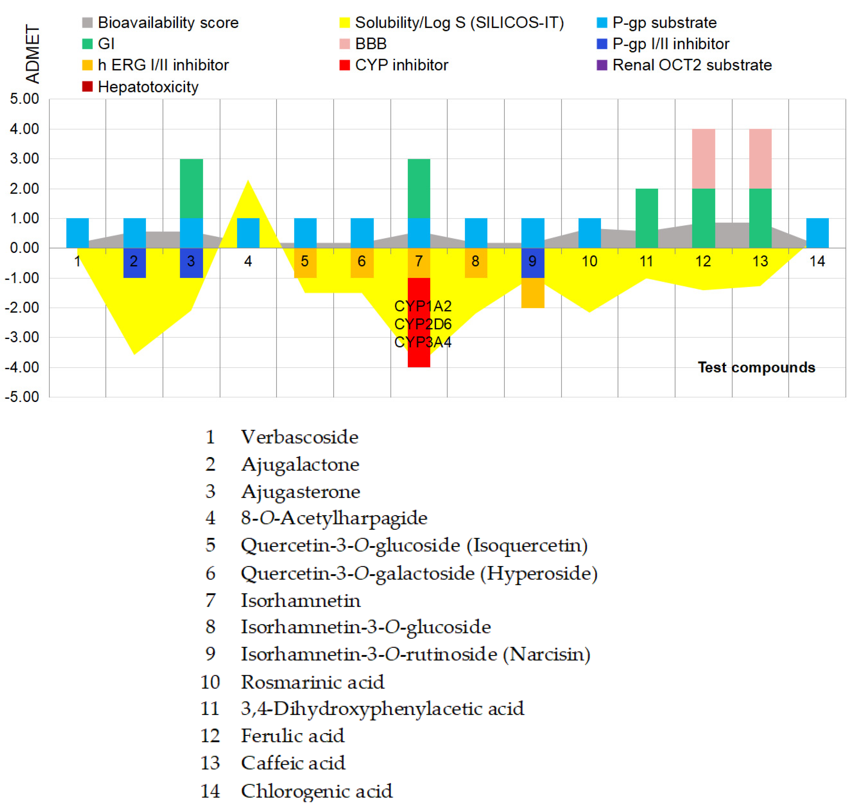
Figure 6.
b). Pharmacokinetic aspects of major and key phenolics from Geranium phaeum.
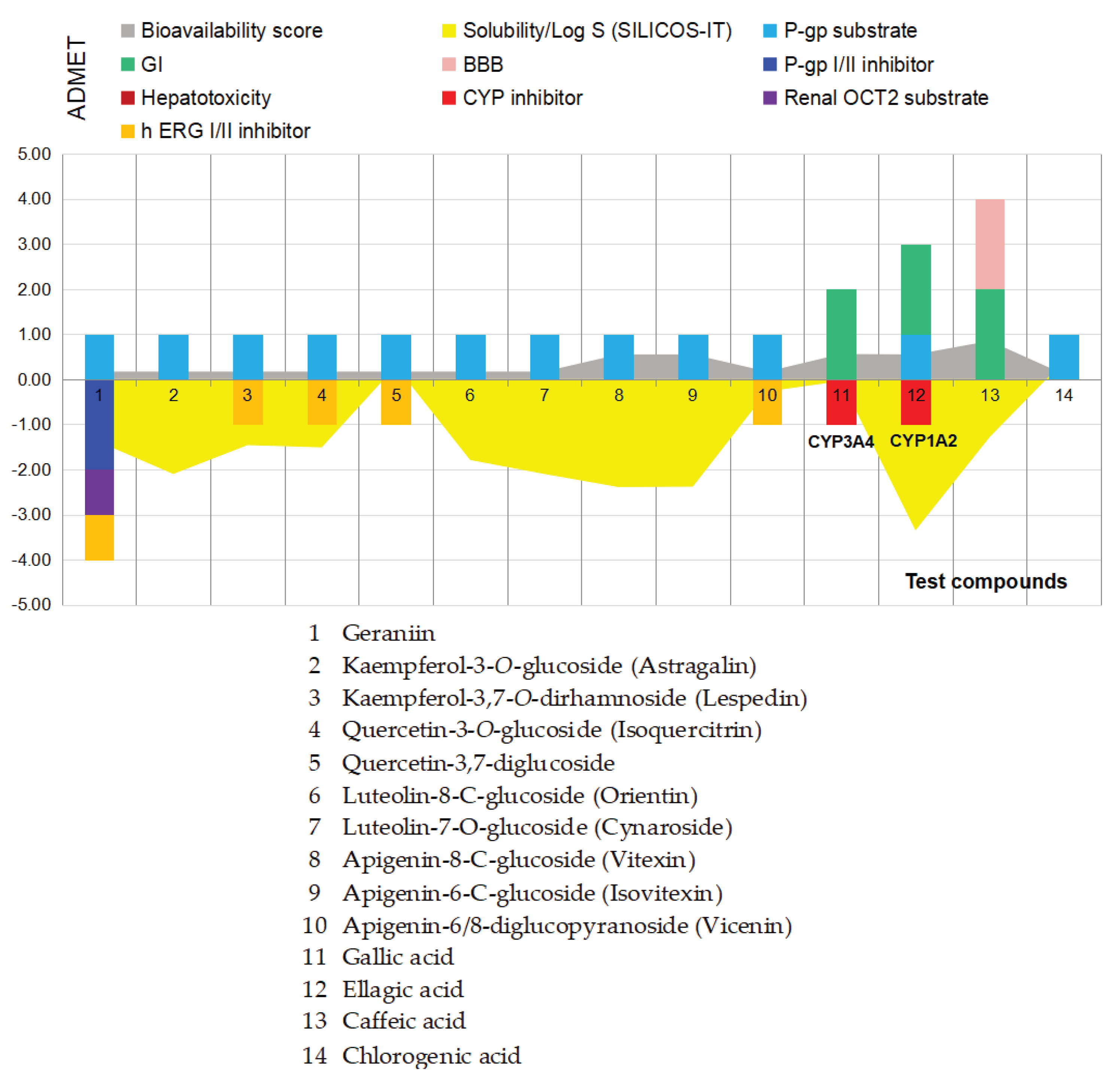
Figure 6.
c). Pharmacokinetic aspects of major and key phenolics from H. nummularium.
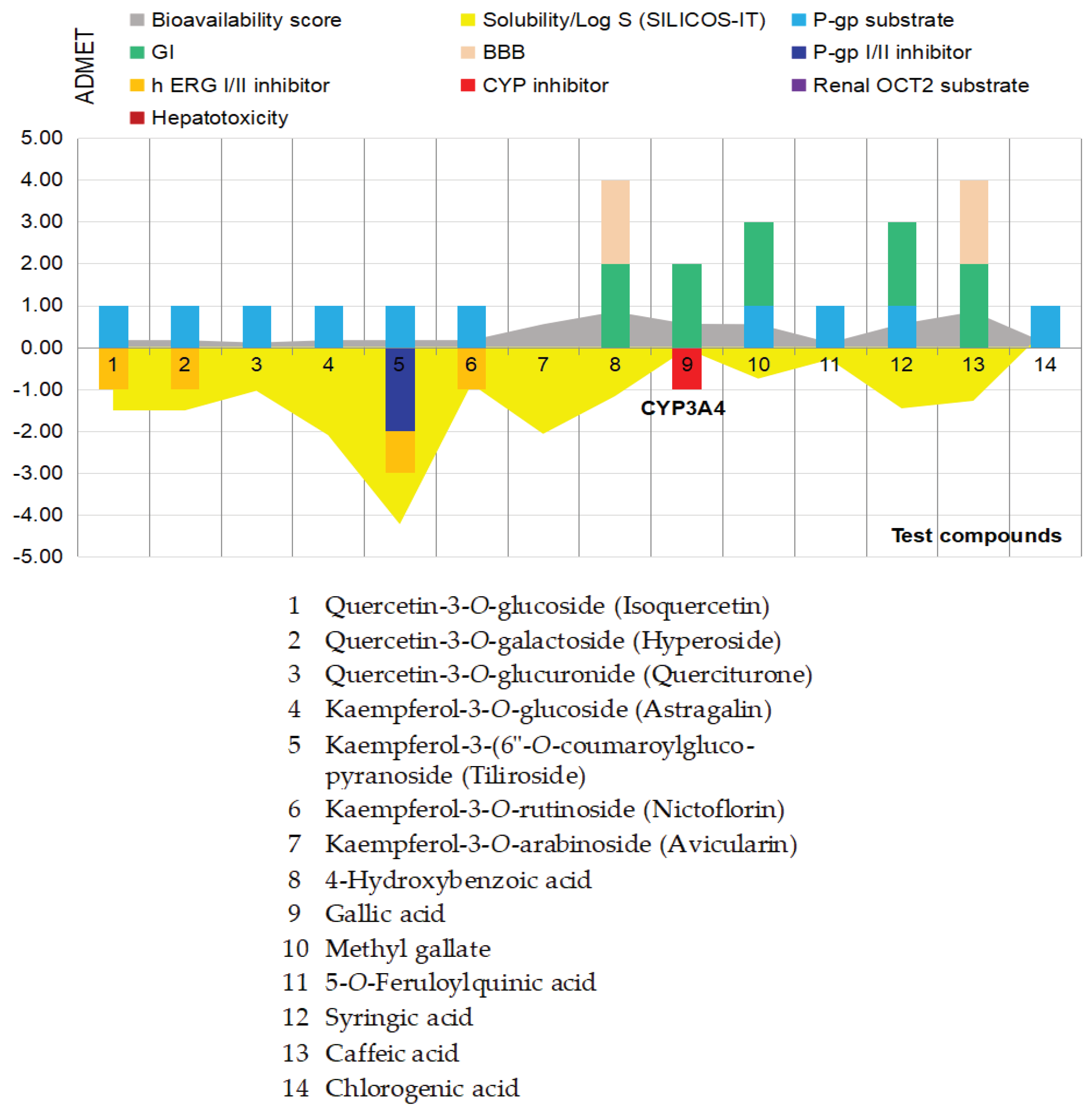
The three diagrams cumulate BiosigLab [28] and SwissADME [29] data as follows: the quantitative data, solubility/Log S (SILICOS-IT) and bioavailability scores correspond to the exactly values computed with the SwissADME tool (see Tables S1, S2, S3 in the supplementary files); the qualitative data, GI and BBB status, the status of P-gp, CYP450, hERG and OCT inhibitors and the potentiality as acting as hepatotoxic agents, are the parameters assigned with Yes and Not in both, Table 1 and Tables S1, S2, S3.
This way, the qualitative parameters were assigned with positive (+) and negative (-) values, function on their potential positive or negative potentiality on humans. Specifically, being the most important pharmacokinetic features, GI and BBB status [29] received the value +2 ; being revealed as a usual condition of plant phenolics, the status of P-gp substrate received the value +1 [28]; the status of P-gp inhibitor received the value -1 [28] ; the status of CYP450, hERG I/II, and OCT2 inhibitors as well as the potential hepatotoxic effects, each one received the value -1 [28].
Altogether, in silico assessments on the three series of major and key phenolics from the three medicinal species confirmed the ability of small phenolic acids to pass the human body barriers; caffeic acid, ferulic acid, and 4-hydroxybenzoic acid revealed the ability to cross the blood-brain barrier, too. In the specific case of flavonoids, except for methyl derivative namely isorhamnetin, none of the compounds investigated indicate the ability to pass the human body barriers. As it is well known, there are only few flavonoid glycosides able to actively pass the GI (e.g., quercetin-4’-O- glucoside/spiraeoside, quercetin-3-O-glucoside/isoquercitrin, quercetin-3-O-galactoside/ hyperoside, luteolin-8-C-glucoside/orientin), while the most part of the flavonoid derivatives have a low bioavailability in humans (estimated as less than 5%); the aglycone forms and the methyl derivatives are instead recognized with high bioavailability in humans [103,104,105,106]. Yet, the use of pure flavonoids should take into consideration complementary studies on 16 flavonoid aglycones analysed in relation with their ability to inhibit the activity of trypsin digestive enzyme [107]; briefly, their IC50 values, especially in the case of quercetin and myricetin, were situated well bellow other pharmacological effects in vitro, therefore flavonoid aglycones have the potentiality to interfere with the process of digestion of proteins in humans before to induce particular pharmacological effects.
Furthermore, the flavonoid aglycones also revealed the potentially to induce mutations in humans, oxidative stress and also inhibitory activity upon the enzymes involved in the hormonal metabolism [108].
Related to the relevancy of the current in vitro studies, as it is well known, the tissue repair occurs through the activation of the dormant fibroblasts and their transformation into a proliferative and contractile mass called “myofibroblasts” [109]; thus, the inhibition of viability of the normal fibroblasts in humans leads to impairments in maintaining the structural framework of tissues and organs, in tissue elasticity, and in tissue repair and wound healing by affecting the production of extracellular matrix proteins in tissues [110]; opposite, the over-activation of the fibroblast lead to the fibrotic disease and a large amount of extracellular matrix accumulated in the interstitial space of respective organ. This way, the ability of Ajuga reptans flower extract to decrease the normal mouse fibroblast viability in vitro (up to 25% inhibitory potency at 100 mg GAE/sample) represents an argument for further cross-linked studies on macrophage and fibroblast dynamics [111], representative for the fibrosis diseases.
Similarly, finding plant extracts able to inhibit or to stimulate the immune response in humans, both are of high interest for practical purposes, while the comparison with the LPS effects [112,113,114,115] is highly relevant. This way, the plant derived products with high stimulatory effects upon the immune cells are not practicable for chronic and immune-mediated inflammatory diseases (IMIDs) [116], but useful to increase the immune response of the patients with depressed immunity as well as for boosting the immunity of the healthy people during the seasonal infections. In this context, both Helianthemum nummularium and Geranium phaeum ethanolic extracts indicated moderate to high immunostimulatory effects, therefore paying attention to their use.
5. Conclusions
In silico - in vitro approach of Ajuga reptans revealed that the ethanolic extracts from the flowers in long-term use could impact the healing processes of the body, could decrease the immune cells’ viability and the systemic innate immune response in humans, and could impact the function of P-gp transporters, hERG II receptors, and CYP1A2, CYP2D6, and CYP3A4 oxidative enzymes. The inhibitory activity of Ajre upon the viability of L929 line was estimated at IC50 = 184.70 µg GAE/mL and upon the SC line at IC50 = 91.30 µg GAE/mL, respectively. These data are particularly useful in the case of using Ajuga-derived products for the purpose of sports nutrition products.
The ethanolic extracts from the aerial part of Geranium phaeum also revealed the potentiality to impact the function of P-gp transporters, of hERG II, and of CYP1A2, CYP2D6, and CYP3A4 oxidative enzymes; the key compound, geraniin, indicated the ability to interfere with the absorption and metabolism of xenobiotics in humans. In vitro, Geph at doses from 1 to 10 µL/mL indicated the ability to stimulate the viability of the immune cells at a magnitude similar to that induced by LPS positive control (16% vs. 20%), while at higher concentrations indicated inhibitory activities upon the viability of both L929 and SC; the inhibitory effects were estimated at IC50 = 414.70 and 152.00 µg GAE/mL, respectively. These results are particularly useful in the cancer approach.
The ethanolic extract from the aerial part of H. nummularium (Henu) indicated the lowest metabolic interferences. Henu in vitro revealed weak inhibitory effects upon the viability of L929 line (IC50 = 350.00 µg GAE/mL) versus high stimulatory effects upon the viability of SC line (IC50 = 183.50 μg GAE/mL); it is to note that the stimulatory activity was 21% higher than that induced by positive control, LPS at 5 µg/mL. Due to this tremendous stimulatory potency upon the viability of the immune cells, it can be estimated that the ethanolic extracts from the aerial part of H. nummularium should be avoided in an inflammatory context, since these could boost the existing inflammation process. At the opposite, the polar extracts from the aerial part of H. nummularium appear to be useful for boosting the immunity of the healthy people.
Supplementary Materials
The following supporting information can be downloaded at the website of this paper posted on Preprints.org, Figure S1 presenting the HPTLC (high-performance thin-layer chromatography) aspects of the reference substances using System I and NP-PEG visualisation reagent. Figure S2 presenting the HPTLC (high-performance thin-layer chromatography) aspects of the reference substances using System II and NP-PEG visualisation reagent. Figures S3-S5 presenting the (HP)TLC aspects of 70% ethanolic extract from the three alpine medicinal species after acidic hydrolysis (1N HCl) in comparison with the reference substances used; Tables S1-S3 presenting the in silico SwissADME pharmacokinetic data on major and key phenolics ascribed to the three alpine medicinal species; Tables S4 presenting the in silico CLC docking studies on COX-2 against the key compounds and compounds ascribed with bioavailability in humans by comparison with native ligand, diclofenac.
Author Contributions
Conceptualization, L.C.P.; methodology, L.C.P. and G.N.; software, L.CP. and A.S.; validation, L.C.P., G.N. and A.S.; formal analysis, L.C.P. and G.N.; investigation, L.C.P. and G.N.; resources, L.C.P.; data curation, L.C.P. and G.N.; writing—original draft preparation, L.C.P.; writing—review and editing, L.C.P. and A.S.; visualization, L.C.P.; supervision, L.C.P. and G.N.; project administration, L.C.P.; funding acquisition, L.C.P. All authors have read and agreed to the published version of the manuscript.
Funding
This research received no external funding.
Institutional Review Board Statement
Not applicable.
Informed Consent Statement
Not applicable.
Conflicts of Interest
The authors declare no conflicts of interest.
Acknowledgments
This work was done with the support of Romanian National Authority for Scientific Research, Competitiveness Operational Programme POC-A1-A1.2.3-G-2015, Project title “Innovative technologies for new, natural health products”, ID P_40_406, SMIS 105542.
References
- Manuals, Merck. SD Manuals. Drug-Receptor Interactions by Abimbola Farinde. Available online: https://www.merckmanuals.com (accessed on 6 April 2026).
- Niu, J.; Straubinger, R.M.; Mager, D.E. Pharmacodynamic Drug–Drug Interactions. Clin. Pharmacol. 2019, 105, 1395–1406. [Google Scholar] [CrossRef] [PubMed]
- Jonker, D.M.; Visser, S.A.G.; van der Graaf, P.H.; Voskuyl, R.A.; Danhof, M. Towards a mechanism-based analysis of pharmacodynamic drug–drug interactions in vivo. Pharmacology&Therapeutics 2005, 106, 1–18. [Google Scholar] [CrossRef] [PubMed]
- Chen, Z.; Zheng, S.; Li, L.; Jiang, H. Metabolism of Flavonoids in Human: A Comprehensive Review. Curr. Drug Metab. 2014, 15, 48–61. [Google Scholar] [CrossRef] [PubMed]
- Nunes, C.D.R.; Barreto Arantes, M.; Menezes de Faria Pereira, S.; Leandro da Cruz, L.; de Souza Passos, M.; Pereira de Moraes, L.; Vieira, I.J.C.; Barros de Oliveira, D. Plants as Sources of Anti-Inflammatory Agents. Molecules 2020, 25, 3726. [Google Scholar] [CrossRef] [PubMed]
- Aware, C.B.; Patil, D.N.; Suryawanshi, S.S.; Mali, P.R.; Rane, M.R.; Gurav, R.G.; Jadhav, Y.P. Natural bioactive products as promising therapeutics: A review of natural product-based drug development. South Afr. J. Bot. 2022, 151, 512–528. [Google Scholar] [CrossRef]
- Latif, R.; Nawaz, T. Medicinal plants and human health: a comprehensive review of bioactive compounds, therapeutic effects, and applications. Phytochem Rev. 2026, 25, 2299–2342. [Google Scholar] [CrossRef]
- Forman, H.J.; Davies, K.J.; Ursini, F. How do nutritional antioxidants really work: nucleophilic tone and para-hormesis versus free radical scavenging in vivo. Free Radic. Biol. Med. 2014, 66, 24–35. [Google Scholar] [CrossRef]
- Diaz, I. Plant Defense Genes against Biotic Stresses. Int. J. Mol. Sci. 2018, 19, 446. [Google Scholar] [CrossRef] [PubMed]
- Farkas, O.; Jakus, J.; Héberger, K. Quantitative structure-antioxidant activity relationships of flavonoid compounds. Molecules 2004, 9, 1079–1088. [Google Scholar] [CrossRef] [PubMed]
- Chen, J.; Yang, J.; Ma, L.; et al. Structure-antioxidant activity relationship of methoxy, phenolic hydroxyl, and carboxylic acid groups of phenolic acids. Sci. Rep. 2020, 10, 5666. [Google Scholar] [CrossRef] [PubMed]
- Moazzen, A.; Öztinen, N.; Ak-Sakalli, E.; Koşar, M. Structure-antiradical activity relationships of 25 natural antioxidant phenolic compounds from different classes. Heliyon 2022, 8, e10467. [Google Scholar] [CrossRef] [PubMed]
- Hussain, T.; Tan, B.; Yin, Y.; Blachier, F.; Tossou, M.C.; Rahu, N. Oxidative Stress and Inflammation: What Polyphenols Can Do for Us? Oxid. Med. Cell Longev. 2016, 7432797. [Google Scholar] [CrossRef] [PubMed]
- Vuolo, M.M.; Lima, V.S.; Maróstica-Junior, M.R. Phenolic compounds: Structure, classification, and antioxidant power. In Bioactive Compounds: Health Benefits and Potential Applications; Segura-Campos, M.R., Ed.; Elsevier Inc.: Amsterdam, The Netherlands, 2018; pp. 33–50. [Google Scholar]
- Kim, Y.S.; Young, M.R.; Bobe, G.; Colburn, N.H.; Milner, J.A. Bioactive food components, inflammatory targets, and cancer prevention. Cancer Prev. Res. 2009, 2, 200–208. [Google Scholar] [CrossRef] [PubMed]
- Arroo, R.R.J.; Wang, M.F.; Bhambra, A.S. Chemopreventive Potential of Flavones, Flavonols, and their Glycosides. In Handbook of Dietary Phytochemicals; Xiao, J., Sarker, S., Asakawa, Y., Eds.; Springer: Singapore, 2020. [Google Scholar] [CrossRef]
- Saad, A.M.; Mohammed, D.M.; Alkafaas, S.S.; Ghosh, S.; Negm, S.H.; Salem, H.M.; Fahmy, M.A.; Semary, H.; Ibrahim, E.H.; AbuQamar, S.F.; El-Tarabily, K.A.; El-Saadony, M.T. Dietary polyphenols and human health: sources, biological activities, nutritional and immunological aspects, and bioavailability– a comprehensive review. Front. Immunol. 2025, 16, 1653378. [Google Scholar] [CrossRef] [PubMed]
- Gelabert-Mora, A.; Chiang, B.; Lee, A.; Sinha, M.; Abuabara, K. Global trends in immune mediated inflammatory diseases: Psoriasis, atopic dermatitis, multiple sclerosis, inflammatory bowel disease, and rheumatoid arthritis. JAAD Int. 2024, 18, 177–179. [Google Scholar] [CrossRef] [PubMed]
- Catalkaya, G.; Venema, K.; Lucini, L.; Rocchetti, G. Interaction of Dietary Polyphenols and Gut Microbiota: Microbial Metabolism of Polyphenols, Influence on the Gut Microbiota and Implications on Host Health. Food Front. 2020, 1, 109–33. [Google Scholar] [CrossRef]
- Zhang, W.; Qi, S.; Xue, X.; Al Naggar, Y.; Wu, L.; Wang, K. Understanding the Gastrointestinal Protective Effects of Polyphenols using Foodomics-Based Approaches. Front Immunol. 2021, 12, 671150. [Google Scholar] [CrossRef] [PubMed]
- Scalbert, A.; Morand, C.; Manach, C.; Rémésy, C. Absorption and metabolism of polyphenols in the gut and impact on health. Biomed. Pharmacother. 2002, 56, 276–282. [Google Scholar] [CrossRef] [PubMed]
- Kimura, Y.; Ito, H.; Ohnishi, R.; Hatano, T. Inhibitory effects of polyphenols on human cytochrome P450 3A4 and 2C9 activity. Food Chem. Toxicol. 2010, 1, 429–435. [Google Scholar] [CrossRef] [PubMed]
- Cosme, P.; Rodríguez, A.B.; Espino, J.; Garrido, M. Plant Phenolics: Bioavailability as a Key Determinant of Their Potential Health-Promoting Applications. Antioxidants 2020, 9, 1263. [Google Scholar] [CrossRef] [PubMed]
- Vicidomini, C.; Palumbo, R.; Moccia, M.; Roviello, G.N. Oxidative Processes and Xenobiotic Metabolism in Plants: Mechanisms of Defense and Potential Therapeutic Implications. J. Xenobiot. 2024, 14, 1541–1569. [Google Scholar] [CrossRef] [PubMed]
- Wagner, H.; Bladt, S. Plant Drug Analysis. A Thin Layer Chromatography Atlas, 2nd ed.; 2nd Edition; Springer-Verlag: Berlin/Heidelberg, Germany, 1996. [Google Scholar]
- Reikh, E.; Schibli, A. HPTLC for the Analysis of Medicinal Plants; Thieme, 2008; Available online: N.Y.-Stuttgart. [Google Scholar]
- Collective Work. Romanian Pharmacopoeia, Xth ed.; Medicala: Bucharest, Romania, 1993.
- pkCSM: predicting small-molecule pharmacokinetic properties using graph-based signatures. J. Med. Chem. 2015, 58, 4066–4072. Available online: https://biosig.lab.uq.edu.au/pkcsm/admet_prediction (accessed on 21 May 2026). [CrossRef] [PubMed]
- SwissADME: a free web tool to evaluate pharmacokinetics, drug-likeness and medicinal chemistry friendliness of small molecules. Sci. Rep. 2017, 7, 42717. Available online: http://www.swissadme.ch. (accessed on 18 October 2025). [CrossRef] [PubMed]
- CLC Drug Discovery Workbench 2.4, 2015. QIAGEN Aarhus A/S Silkeborgvej 2 Prismet DK-8000 Aarhus C Denmark.
- Korb, O.; Stützle, T.; Exner, T.E. Empirical scoring functions for advanced protein-ligand docking with plants. J. Chem. Inf. Model 2009, 49, 84–96. [Google Scholar] [CrossRef] [PubMed]
- Rowlinson, S.W.; Kiefer, J.R.; Prusakiewicz, J.J.; Pawlitz, J.L.; Kozak, K.R.; Kalgutkar, A.S.; Stallings, W.C.; Kurumbail, R.G.; Marnett, L.J. A Novel Mechanism of Cyclooxygenase-2 Inhibition Involving Interactions with Ser-530 and Tyr-385. J. Biol. Chem. 2003, 278, 45763–45769. [Google Scholar] [CrossRef] [PubMed]
- SPARTAN’24 Software, Wavefunction, Inc. 18401 Von Karman Avenue, Suite 435 Irvine, CA 92612, USA.
- Protocols & Applications Guide. Available online: www.promega.com (accessed on 23 March 2026).
- Israili, Z.; Lyoussi, B. Ethnopharmacology of the plants of genus Ajuga. Pak. J. Pharm. Sci. 2009, 22, 425–462. [Google Scholar] [PubMed]
- Goetz, P. Ajuga reptans. Phytothérapie 2022, 20, 273–275. [Google Scholar] [CrossRef]
- Rahiminiya, A.; Herizchi Ghadim, H.; Sadati Lamardi, S.N.; Haghir Ebrahimabadi, M.; Fazljou, S.M.; Ayati, M.H. Medicinal Importance of Ajuga Species in Iran: Ethnobotanical and Traditional Applications, Phytochemical, and Pharmacological Studies. Jundishapur. J. Nat. Pharm. Prod. 2022, 17, 109209. [Google Scholar] [CrossRef]
- Luan, F.; Han, K.; Li, M.; Zhang, T.; Liu, D.; Yu, L.; Lv, H. Ethnomedicinal Uses, Phytochemistry, Pharmacology, and Toxicology of Species from the Genus Ajuga L.: A Systematic Review. Am. J. Chin. Med. 2019, 47, 959–1003. [Google Scholar] [CrossRef] [PubMed]
- Toiu, A.; Mocan, A.; Vlase, L.; Pârvu, A.E.; Vodnar, D.C.; Gheldiu, A.-M.; Moldovan, C.; Oniga, I. Comparative Phytochemical Profile, Antioxidant, Antimicrobial and In Vivo Anti-Inflammatory Activity of Different Extracts of Traditionally Used Romanian Ajuga genevensis L. and A. reptans L. (Lamiaceae). Molecules 2019, 24, 1597. [Google Scholar] [CrossRef] [PubMed]
- Maliuvanchuk, S.; Grytsyk, A.; Popadynets, O.; Kotyk, T.; Raal, A.; Koshovyi, O. Ajuga reptans L. Herb Extracts: Phytochemical Composition and Pharmacological Activity Screening. Plants 2025, 14, 219. [Google Scholar] [CrossRef] [PubMed]
- Dziki, A.; Malinowska, M.A.; Szopa, A.; Sikora, E. Comparative Study of the Phytochemical Profile and Biological Activity of Ajuga reptans L. Leaf and Root Extracts. Appl. Sci. 2024, 14, 5105. [Google Scholar] [CrossRef]
- Alshehri, B. The geranium genus: A comprehensive study on ethnomedicinal uses, phytochemical compounds, and pharmacological importance. Saudi J. Biol. Sci. 2024, 31, 103940. [Google Scholar] [CrossRef] [PubMed]
- Graça, V.C.; Ferreira, I.C.F.R.; Santos, P.F. Bioactivity of the Geranium genus: a comprehensive review. Curr. Pharm. Des. 2020, 26, 1838–1865. [Google Scholar] [CrossRef] [PubMed]
- Harborne, J.B.; Williams, C.A. Phytochemistry of the Genus Geranium. In CRC Press; 2002; pp. 32–41. [Google Scholar]
- Okuda, T.; Mori, K.; Hatano, T. The distribution of geraniin and mallotusinic acid in the order geraniales. Phytochemistry 1980, 19, 547–551. [Google Scholar] [CrossRef]
- Okuda, T.; Yoshida, T.; Hatano, T. Pharmacologically active tannins isolated from medicinal plants. Plant Polyphen. 1992, 539–569. [Google Scholar] [CrossRef] [PubMed]
- Alshehri, B. The geranium genus: A comprehensive study on ethnomedicinal uses, phytochemical compounds, and pharmacological importance. Saudi J. Biol. Sci. 2024, 31(4), 103940. [Google Scholar] [CrossRef] [PubMed]
- Markham, K.R.; Mitchell, K.A.; Boase, M.R. Malvidin-3-O-glucoside-5-O-(6-acetylglucoside) and its colour manifestation in ‘Johnson’s Blue’and other ‘Blue’geraniums. Phytochemistry 1997, 45, 417–423. [Google Scholar] [CrossRef]
- Mouffouk, S.; Mouffouk, C.; Mouffouk, S.; Haba, H. Medicinal, Pharmacological and Biochemical Progress on the Study of Genus Helianthemum: A Review. Curr. Chem. Biol. 2023, 17, 147–159. [Google Scholar] [CrossRef]
- Bernacka, K.; Bednarska, K.; Starzec, A.; Mazurek, S.; Fecka, I. Antioxidant and Antiglycation Effects of Cistus × incanus Water Infusion, Its Phenolic Components, and Respective Metabolites. Molecules 2022, 27, 2432. [Google Scholar] [CrossRef] [PubMed]
- Agostini, M.; Hininger-Favier, I.; Marcourt, L.; Boucherle, B.; Gao, B.; Hybertson, B.M.; Bose, S.K.; McCord, J.M.; Millery, A.; Rome, M.; Ferreira-Queiroz, E.; Wolfender, J.L.; Gallet, C.; Boumendjel, A. Phytochemical and Biological Investigation of Helianthemum nummularium, a High-Altitude Growing Alpine Plant Overrepresented in Ungulates Diets. Planta Med. 2020, 86, 1185–1190. [Google Scholar] [CrossRef] [PubMed]
- De Freitas, V.A.; Ramalho, P. S.; Azevedo, Z.; Macedo, A. Identification of Some Volatile Descriptors of the Rock-Rose-Like Aroma of Fortified Red Wines from Douro Demarcated Region. J. Agric. Food Chem. 1999, 47, 4327–31. [Google Scholar] [CrossRef] [PubMed]
- Viuda-Martos, M.; Sendra, E.; Perez-Alvarez, J.A.; Abrini, J. Identification of Flavonoid Content and Chemical Composition of the Essential Oils of Moroccan Herbs: Myrtle (Myrtus communis L.), Rockrose (Cistus ladanifer L.) and Montpellier Cistus (Cistus monspeliensis L.). J. Essent. Oil Res. 2011, 23, 1–9. [Google Scholar] [CrossRef]
- Nicoletti, M.; Toniolo, C.; Venditti, A.; Bruno, M.; Jemia, M.B. Antioxidant Activity and Chemical Composition of Three Tunisian Cistus: Cistus monspeliensis, Cistus villosus and Cistus libanotis. Nat. Prod. Res. 2013, 29, 223–30. [Google Scholar] [CrossRef] [PubMed]
- Bouzergoune, F.; Bitam, F.; Aberkane, M.C.; Aberkane, A. Preliminary Phytochemical and Antimicrobial Activity Investigations on the Aerial Parts of Helianthemum kahiricum. Chem. Nat. Compd. 2013, 49, 751–2. [Google Scholar] [CrossRef]
- Panou, E.; Graikou, K.; Tsafantakis, N.; Sakellarakis, F.N.; Chinou, I. Phytochemical Profiling and Biological Activities of Two Helianthemum Species Growing in Greece. Sci. Pharm. 2024, 92. [Google Scholar] [CrossRef]
- Ghose, A.K.; Crippen, G.M. Atomic physicochemical parameters for three-dimensional-structure-directed quantitative structure-activity relationships. 2. Modeling dispersive and hydrophobic interactions. J. Chem. Inf. Comput. Sci. 1987, 27, 21–35. [Google Scholar] [CrossRef] [PubMed]
- Leo, A.; Hansch, C.; Elkins, D. Partition Coefficients and their uses. Chem. Rev. 1971, 6, 525–616. [Google Scholar] [CrossRef]
- Daina, A.; Michielin, O.; Zoete, V. SwissADME: A free web tool to evaluate pharmacokinetics, drug-likeness and medicinal chemistry friendliness of small molecules. Sci. Rep. 2017, 7, 42717. [Google Scholar] [CrossRef] [PubMed]
- Lipinski, C.A.; Lombardo, F.; Dominy, B.W.; Feeney, P.J. Experimental and computational approaches to estimate solubility and permeability in drug discovery and development settings. Adv. Drug Deliv. Rev. 2001, 46, 3–26. [Google Scholar] [CrossRef] [PubMed]
- Pham-The, H.; González-Álvarez, I.; Bermejo, M.; Garrigues, T.; Le-Thi-Thu, H.; Cabrera-Pérez, M.Á. The Use of Rule-Based and QSPR Approaches in ADME Profiling: A Case Study on Caco-2 Permeability. Mol. Inf. 2013, 32, 459–479. [Google Scholar] [CrossRef] [PubMed]
- Wang, N.-N.; Dong, J.; Deng, Y.-H.; Zhu, M.-F.; Wen, M.; Yao, Z.-J.; Lu, A.-P.; Wang, J.-B.; Cao, D.-S. ADME Properties Evaluation in Drug Discovery: Prediction of Caco-2 Cell Permeability Using a Combination of NSGA-II and Boosting. J. Chem. Inf. Model. 2016, 56, 763–773. [Google Scholar] [CrossRef] [PubMed]
- Croom, E. Chapter Three—Metabolism of Xenobiotics of Human Environments. In Progress in Molecular Biology and Translational Science; Hodgson, E., Ed.; Academic Press: Cambridge, MA, USA, 2012; Volume 112, pp. 31–38. [Google Scholar] [CrossRef] [PubMed]
- Sayyed, Y.; Rehman, K.; Akash, M.S.H.; Kamran, S.H.; Badran, M. Chapter 13—Xenobiotics and drug-metabolizing enzymes: Challenges and strategies. In Biochemistry of Drug Metabolizing Enzymes; Academic Press: Cambridge, MA, USA, 2022; pp. 305–321. [Google Scholar] [CrossRef]
- Lee, C.A.; Cook, J.A.; Reyner, E.L.; Smith, D.A. P-glycoprotein related drug interactions: Clinical importance and a consideration of disease states. Expert Opin. Drug Metab. Toxicol. 2010, 6, 603–619. [Google Scholar] [CrossRef] [PubMed]
- P-Glycoprotein Substrate ADMET Feature Prediction. Available online: https://dev.drugbank.com/guides/terms/p-glycoprotein-substrate (accessed on 5 January 2026).
- Garrido, A.; Lepailleur, A.; Mignani, S.M.; Dallemagne, P.; Rochais, C. hERG toxicity assessment: Useful guidelines for drug design. Eur. J. Med. Chem. 2020, 195, 112290. [Google Scholar] [CrossRef] [PubMed]
- Yuan, Y.C.; Liu, C.; Xie, R.R.; Zhang, L.; Wang, H.; Yang, J.K. The role of the HERG channel in the secretion of glucagon-like peptide-1 (GLP-1) from murine intestinal L-cells. Curr. Med. 2025, 2, 5. [Google Scholar] [CrossRef]
- Yang, J.K.; Liu, C.; Wang, H.; Yuan, Y.C. 1656-P: Glucagon-Like peptide-1 (GLP-1) Secretion from Intestinal L-Cells Is Mediated by hERG Potassium Channels. Diabetes 2023, 72 (Supplement_1), 1656–P. [Google Scholar] [CrossRef]
- Jonker, J.; Schinkel, A. Pharmacological and Physiological Functions of the Polyspecific Organic Cation Transporters: OCT1, 2, and 3 (SLC22A1-3). J. Pharmacol. Exp. Ther. 2004, 308, 2–9. [Google Scholar] [CrossRef] [PubMed]
- Ailabouni, A.; Prasad, B. Organic cation transporters 2: Structure, regulation, functions, and clinical implications. Drug Metab. Dispos. 2025, 53, 100044. [Google Scholar] [CrossRef] [PubMed]
- Kurumbail, R.G.; Stevens, A.M.; Gierse, J.K.; McDonald, J.J.; Stegeman, R.A.; Pak, J.Y.; Gildehaus, D.; Miyashiro, J.M.; Penning, T.D.; Seibert, K.; Isakson, P.C.; Stallings, W.C. Structural basis for selective inhibition of cyclo-oxygenase-2 by anti-inflammatory agents. Nature 1996, 384, 644–648. [Google Scholar] [CrossRef] [PubMed]
- Steinbach, G.; Lynch, P.M.; Phillips, R.K.; Walace, M.H.; Hawk, E.; Gordon, G.B.; Wakayashi, N.; Saunders, B.; Shen, Y.; Fujimura, T.; Su, L.K.; Levin, B. The effect of Celecoxib, a cyclo-oxygenase inhibitor, in Familial Adenomatous Polyposis. N. Engl. J. Med. 2000, 342, 1946–1952. [Google Scholar] [CrossRef] [PubMed]
- Roelofs, H.M.; te Morsche, R.H.; van Heumen, B.W.; Nagengast; Peters, F.M. Over-expression of COX-2 mRNA in colorectal cancer. BMC Gastroenterol. 2014, 14, 9005. [Google Scholar] [CrossRef] [PubMed]
- Li, S.; Jiang, M.; Wang, L.; Yu, S. Combined chemotherapy with cyclooxygenase-2 (COX-2) inhibitors in treating human cancers: Recent advancement. Biomed. Pharmacother. 2020, 129, 110389. [Google Scholar] [CrossRef] [PubMed]
- Pirvu, L.; Stefaniu, A.; Neagu, G.; Pintilie, L. Studies on Anemone nemorosa L. extracts; polyphenols profile, antioxidant activity, and effects on Caco-2 cells by in vitro and in silico studies. Open Chem. 2022, 20, 299–312. [Google Scholar] [CrossRef]
- Atay, I.; Kirmizibekmez, H.; Kaiser, M.; Akaydin, G.; Yesilada, E.; Tasdemir, D. Evaluation of in Vitro Antiprotozoal Activity of Ajuga laxmannii and Its Secondary Metabolites. Pharm. Biol. 2016, 54, 1808–1814. [Google Scholar] [CrossRef] [PubMed]
- Caskurlu, A.; Karavus, S.N.; Biltekin, S.N.; Helvaci, E.Z.; Karadag, A.E. In Vitro Cytotoxicity Evaluation and Phytochemical Analysis of Ajuga reptans L. Extracts. J. Res. Pharm. 2023, 27, 2115–2123. [Google Scholar] [CrossRef]
- Maliuvanchuk, S.; Grytsyk, A.; Popadynets, O.; Kotyk, T.; Raal, A.; Koshovyi, O. Ajuga reptans L. Herb Extracts: Phytochemical Composition and Pharmacological Activity Screening. Plants 2025, 14, 219. [Google Scholar] [CrossRef] [PubMed]
- Rauca, V.F.; Vlase, L.; Casian, T.; Sesarman, A.; Gheldiu, A.M.; Mocan, A.; Banciu, M.; Toiu, A. Biologically Active Ajuga Species Extracts Modulate Supportive Processes for Cancer Cell Development. Front. Pharmacol. 2019, 10, 334. [Google Scholar] [CrossRef] [PubMed]
- Todorova, V.; Ivanova, S.; Chakarov, D.; Kraev, K.; Ivanov, K. Ecdysterone and Turkesterone-Compounds with Prominent Potential in Sport and Healthy Nutrition. Nutrients 2024, 16, 1382. [Google Scholar] [CrossRef] [PubMed]
- Kosuge, T.; Yokota, M.; Sugiyama, K.; Yamamoto, T.; Ni, M.Y.; Yan, S.C. Studies on antitumor activities and antitumor principles of Chinese herbs. I. Antitumor activities of Chinese herbs. Yakugaku Zasshi J. Pharm. Soc. Jpn. 1985, 105, 791–795. [Google Scholar] [CrossRef] [PubMed]
- Kashiwada, Y.; Nonaka, G.I.; Nishioka, I.; Chang, J.J.; Lee, K.H. Antitumor agents, 129. Tannins and related compounds as selective cytotoxic agents. J. Nat. Prod. 1992, 55, 1033–1043. [Google Scholar] [CrossRef] [PubMed]
- Chalchat, J.C.; Petrovic, S.D.; Maksimovic, Z.A.; Gorunovic, M.S. A comparative study on essential oils of Geranium macrorrhizum L. and Geranium phaeum L., Geraniaceae from Serbia. J. Essent. Oil Res. 2002, 14, 333–335. [Google Scholar] [CrossRef]
- Acharya, J.; Hildreth, M.B.; Reese, R.N. In vitro screening of forty medicinal plant extracts from the United States Northern Great Plains for anthelmintic activity against Haemonchus contortus. Vet. Parasitol. 2014, 201, 75–81. [Google Scholar] [CrossRef] [PubMed]
- Nam, H.H.; Choo, B.K. Geranium koreanum, a medicinal plant Geranii Herba, ameliorate the gastric mucosal injury in gastritis-induced mice. J. Ethnopharmacol. 2021, 265, 113041. [Google Scholar] [CrossRef] [PubMed]
- Cai, J.; Wang, L.; Xu, M.; Zhu, S.; Chen, Y.; Zhong, J.; Li, J.; Zhang, P.; Zhang, T.; Ye, Q.; Ao, H. Multi-target therapeutics of geraniin from Geranium wilfordii Maxim. and related species: Mechanisms and applications. J. Ethnopharmacol. 2026, 120765. [Google Scholar] [CrossRef] [PubMed]
- Zhou, X.; Qin, M.; He, L.; Zhang, Y.; Liu, A.; Chen, D.; Pan, H. Geraniin restricts inflammasome activation and macrophage pyroptosis by preventing the interaction between ASC and NLRP3 to exert anti-inflammatory effects. Int. Immunopharmacol. 2024, 129, 111656. [Google Scholar] [CrossRef] [PubMed]
- Wang, Y.; Chu, X.; Hu, G.; Chang, J. Anti-oxidant, anti-apoptotic and anti-inflammatory effects of geraniin in spinal cord injury in rat: role of COX-2. Folia Morphol (Warsz) 2024. [Google Scholar] [CrossRef] [PubMed]
- Ramos-Torrecillas, J.; González-Acedo, A.; Melguizo-Rodríguez; Ruiz, C.; De Luna-Bertos, E.; Illescas-Montes, R.; García-Martínez, O. Anti-Inflammatory and Antimicrobial Effect of Ellagic Acid and Punicalagin in Dermal Fibroblasts. Int. J. Mol. Sci. 2025, 26, 8681. [Google Scholar] [CrossRef] [PubMed]
- Ríos, J.L.; Giner, R.M.; Marín, M.; Recio, M.C. A Pharmacological Update of Ellagic Acid. Planta Med. 2018, 84, 1068–1093. [Google Scholar] [CrossRef] [PubMed]
- Ceci, C.; Lacal, P.M.; Tentori, L.; De Martino, M.; Miano, R.; Graziani, G. Experimental Evidence of the Antitumor, Antimetastatic and Antiangiogenic Activity of Ellagic Acid. Nutrients 2018, 10, 1756. [Google Scholar] [CrossRef] [PubMed]
- Lu, G.; Wang, X.; Cheng, M.; Wang, S.; Ma, K. The multifaceted mechanisms of ellagic acid in the treatment of tumors: State-of-the-art. Biomed. Pharmacother. 2023, 165, 115132. [Google Scholar] [CrossRef] [PubMed]
- Guo, Y.; Ashrafizadeh, M.; Tambuwala, M.M.; Ren, J.; Orive, G.; Yu, G. P-glycoprotein (P-gp)-driven cancer drug resistance: biological profile, non-coding RNAs, drugs and nanomodulators. Drug Discov. Today 2024, 29, 104161. [Google Scholar] [CrossRef] [PubMed]
- Rubio-Moraga, Á.; Argandoña, J.; Mota, B.; Pérez, J.; Verde, A.; Fajardo, J.; Gómez-Navarro, J.; Castillo-López, R.; Ahrazem, O.; Gómez-Gómez, L. Screening for polyphenols, antioxidant and antimicrobial activities of extracts from eleven Helianthemum taxa (Cistaceae) used in folk medicine in south-eastern Spain. J. Ethnopharmacol. 2013, 148, 287–296. [Google Scholar] [CrossRef] [PubMed]
- Wafa, N.; Sofiane, G. In-vitro Antioxidant and anti-inflammatory activities valorisation of tannin crude extract of Helianthemum helianthemoïdes (Desf.) Grosser. J. Drug Deliv. Ther. 2020, 10, 135–139. [Google Scholar] [CrossRef]
- Pirvu, L.; Nicu, I. Polyphenols content, antioxidant and antimicrobial activity of ethanol extracts from the aerial part of rock rose (Helianthemum nummularium) species. J. Agric. Sci. Technol. A 2017, 7, 61–67. [Google Scholar] [CrossRef]
- Natural Medicines Comprehensive Database. Rock Rose. Therapeutic Research Faculty. 2017. Available online: http://naturaldatabase.therapeuticresearch.com (accessed on 18 May 2025).
- Bach Flower Co. Bach Flower—Rock Rose (Helianthemum nummularium). 2015. Available online: http://www.bachflower.org/rock_rose.htm (accessed on 18 May 2025).
- Thaler, K.; Kaminski, A.; Chapman, A.; Langley, T.; Gartlehner, G. Bach flower remedies for psychological problems and pain: A systematic review. BMC Complement. Altern. Med. 2009, 9, 16. [Google Scholar] [CrossRef] [PubMed]
- Loizzo, M.R.; Jemia, M.B.; Senatore, F.; Bruno, M.; Menichini, F.; Tundis, R. Chemistry and Functional Properties in Prevention of Neurodegenerative Disorders of Five Cistus Species Essential Oils. Food Chem. Toxicol. 2013, 59, 586–94. [Google Scholar] [CrossRef] [PubMed]
- Ay, L.; Karaman, Ik.; Lu, S.; Ilyurt, E.; Yar, T.M. Investigation of in vitro Anti-trichomoniasis Effect of Helianthemum ledifolium L. (Mill.) Varieties against Trichomonas vaginalis. Ann. Med. Res. 2023, 30, 1557–1562. [Google Scholar] [CrossRef]
- Manach, C.; Scalbert, A.; Morand, C.; Rémésy, C.; Jimenez, L. Polyphenols: food sources and bioavailability. Am. J. Clin. Nutr. 2004, 79, 727–747. [Google Scholar] [CrossRef] [PubMed]
- D’Archivio, M.; Filesi, C.; Varì, R.; Scazzocchio, B.; Masella, R. Bioavailability of the Polyphenols: Status and Controversies. Int. J. Mol. Sci. 2010, 11, 1321–1342. [Google Scholar] [CrossRef] [PubMed]
- Canivenc-Lavier, M. C.; Vernevaut, M.F.; Totis, M.; Siess, M. H.; Magdalou, J.; Suschetet, M. Comparative effects of flavonoids and model inducers on drug-metabolizing enzymes in rat liver. Toxicology 1996, 114, 19–27. [Google Scholar] [CrossRef] [PubMed]
- Xie, L.; Deng, Z.; Zhang, J.; Dong, H.; Wang, W.; Xing, B.; Liu, X. Comparison of flavonoid O-glycoside, C-glycoside and their aglycones on antioxidant capacity and metabolism during in vitro digestion and in vivo. Foods 2022, 11, 882. [Google Scholar] [CrossRef] [PubMed]
- Maliar, T.; Jedinák, A.; Kadrabová, J.; Šturdík, E. Structural aspects of flavonoids as trypsin inhibitors. Eur. J. Med. Chem. 2004, 39, 241–248. [Google Scholar] [CrossRef] [PubMed]
- Skibola, C.F.; Smith, M.T. Potential health impacts of excessive flavonoid intake. Free Radic. Biol. Med. 2000, 29, 375–383. [Google Scholar] [CrossRef] [PubMed]
- Wang, S.; Liang, Y.; Dai, C. Metabolic Regulation of Fibroblast Activation and Proliferation during Organ Fibrosis. Kidney Dis. 2022, 8, 115–125. [Google Scholar] [CrossRef] [PubMed]
- Zhang, X.; Zhang, Y.; Liu, Y. Fibroblast activation and heterogeneity in fibrotic disease. Nat. Rev. Nephrol. 2025, 21, 613–632. [Google Scholar] [CrossRef] [PubMed]
- Froom, S.C.S.Z.; Callaghan, N.I.; Huyer, L.D. Cellular crosstalk in fibrosis: Insights into macrophage and fibroblast dynamics. J. Biol. Chem. 2025, 110203. [Google Scholar] [CrossRef] [PubMed]
- Park, B.; Lee, J.O. Recognition of lipopolysaccharide pattern by TLR4 complexes. Exp. Mol. Med. 2013, 45, e66. [Google Scholar] [CrossRef] [PubMed]
- Pugin, J.; Schurer-Maly, C.C.; Leturcq, D.; Moriarty, A.; Ulevitch, R.J.; Tobias, P.S. Lipopolysaccharide activation of human endothelial and epithelial cells is mediated by lipopolysaccharide-binding protein and soluble CD14. Proc. Natl. Acad. Sci. USA 1993, 90, 2744–2748. [Google Scholar] [CrossRef] [PubMed]
- Chow, J.C.; Young, D.W.; Golenbock, D.T.; Christ, W.J.; Gusovsky, F. Toll-like receptor-4 mediates lipopolysaccharide-induced signal transduction. J. Biol. Chem. 1999, 274, 10689–10692. [Google Scholar] [CrossRef] [PubMed]
- Zhang, X.; Tian, X.; Wang, Y.; Yan, Y.; Wang, Y.; Su, M.; Lv, H.; Li, K.; Hao, X.; Xing, X.; Song, S. Application of lipopolysaccharide in establishing inflammatory models. Int. J. Biol. Macromol. 2024, 279, 135371. [Google Scholar] [CrossRef]
- Gelabert-Mora, A.; Chiang, B.; Lee, A.; Sinha, M.; Abuabara, K. Global trends in immune mediated inflammatory diseases: Psoriasis, atopic dermatitis, multiple sclerosis, inflammatory bowel disease, and rheumatoid arthritis. JAAD Int. 2024, 18, 177–179. [Google Scholar] [CrossRef] [PubMed]
Figure 1.
(HP)TLC aspects of genuine 70% ethanolic extract from Ajuga reptans in comparison with pure polyphenols, reference substances (REF) solved in 70% ethanol; a general view on polyphenols (A1) and bitter compounds (A2) content. Chromatogram A1: T1/REF: rutin (Rf~0.40), chlorogenic acid (Rf~0.50), gallic acid (Rf~0.94) and caffeic acid (Rf~0.95); T2 - 70% ethanolic extract from A. reptans by NP-PEG treatment (duplicate sample); T3/REF: isoquercitrin (Rf~0.66); T4/REF: hyperoside (Rf~0.60) and quercitrin (Rf~0.80); Chromatogram A2: T2 - 70% ethanolic extract from A. reptans after VS treatment.
Figure 1.
(HP)TLC aspects of genuine 70% ethanolic extract from Ajuga reptans in comparison with pure polyphenols, reference substances (REF) solved in 70% ethanol; a general view on polyphenols (A1) and bitter compounds (A2) content. Chromatogram A1: T1/REF: rutin (Rf~0.40), chlorogenic acid (Rf~0.50), gallic acid (Rf~0.94) and caffeic acid (Rf~0.95); T2 - 70% ethanolic extract from A. reptans by NP-PEG treatment (duplicate sample); T3/REF: isoquercitrin (Rf~0.66); T4/REF: hyperoside (Rf~0.60) and quercitrin (Rf~0.80); Chromatogram A2: T2 - 70% ethanolic extract from A. reptans after VS treatment.
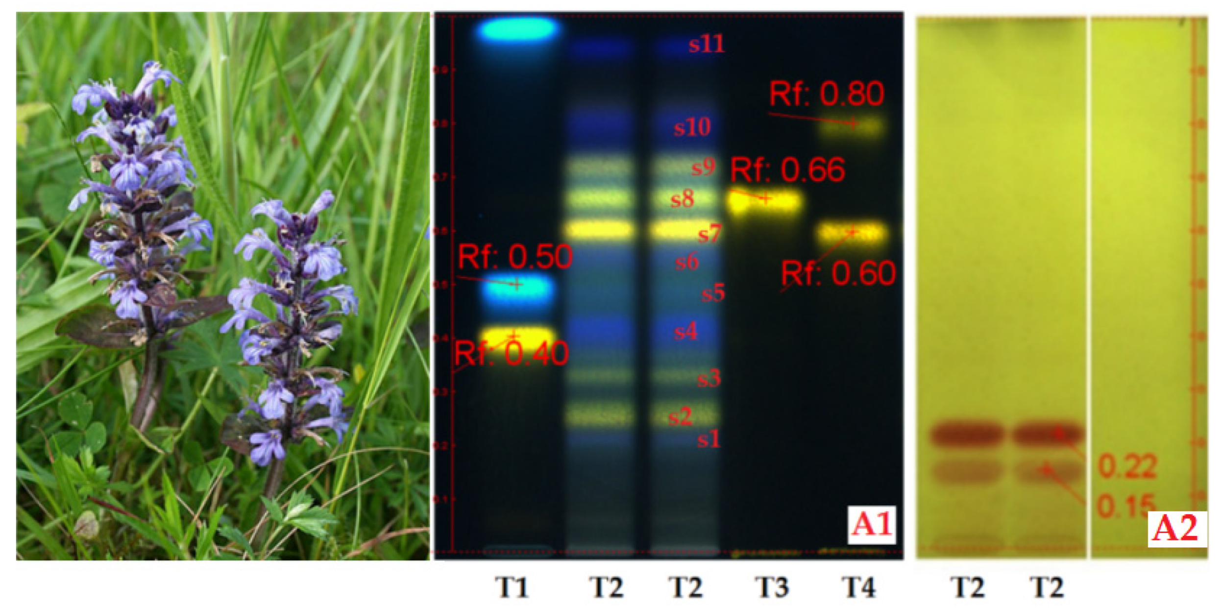
Figure 2.
(HP)TLC aspects of genuine 70% ethanolic extract from Geranium phaeum in comparison with pure polyphenols, reference substances (REF) solved in 70% ethanol; a view on polyphenols (A1) and bitter compounds (A2) content. Chromatogram A1: T1/REF: rutin (Rf~0.43), vitexin (Rf~0.74), protocatechuic acid (Rf~0.98) and apigenin (Rf~0.99); T2/REF: hyperoside (Rf~0.64), cosmosiin (Rf~0.69), rosmarinic acid (Rf~0.96), kaempferol (Rf~0.98); T3 and T4 - 70% ethanolic extract from Geranium phaeum by NP-PEG treatment (duplicate sample); T5/REF: rutin (Rf~0.43), chlorogenic acid (Rf~0.52), gallic acid (Rf~0.95) and caffeic acid (Rf~0.97); Chromatogram A2: T3 - 70% ethanolic extract from Geranium phaeum after VS treatment.
Figure 2.
(HP)TLC aspects of genuine 70% ethanolic extract from Geranium phaeum in comparison with pure polyphenols, reference substances (REF) solved in 70% ethanol; a view on polyphenols (A1) and bitter compounds (A2) content. Chromatogram A1: T1/REF: rutin (Rf~0.43), vitexin (Rf~0.74), protocatechuic acid (Rf~0.98) and apigenin (Rf~0.99); T2/REF: hyperoside (Rf~0.64), cosmosiin (Rf~0.69), rosmarinic acid (Rf~0.96), kaempferol (Rf~0.98); T3 and T4 - 70% ethanolic extract from Geranium phaeum by NP-PEG treatment (duplicate sample); T5/REF: rutin (Rf~0.43), chlorogenic acid (Rf~0.52), gallic acid (Rf~0.95) and caffeic acid (Rf~0.97); Chromatogram A2: T3 - 70% ethanolic extract from Geranium phaeum after VS treatment.
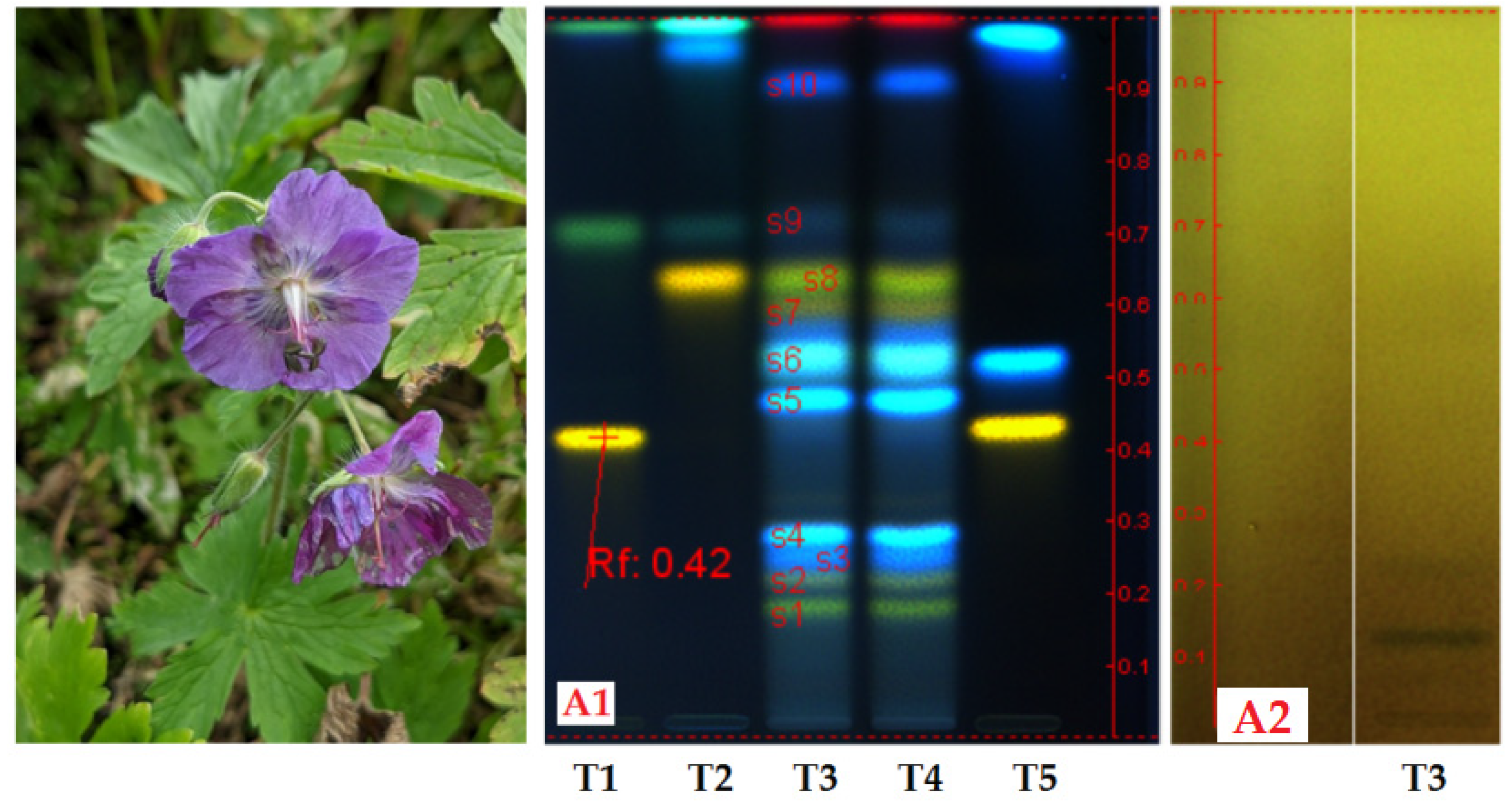
Figure 3.
(HP)TLC aspects of 70% ethanolic extract from H. nummularium in comparison with pure polyphenols, reference substances (REF) solved in 70% ethanol; a view on polyphenols (A1) and bitter compounds (A2) content. Chromatogram A1: T1/REF: rutin (Rf~0.43), vitexin (Rf~0.74), protocatechuic acid (Rf~0.98) and apigenin (Rf~0.99); T2/REF: hyperoside (Rf~0.64), cosmosiin (Rf~0.69), rosmarinic acid (Rf~0.96), kaempferol (Rf~0.98); T3 - 70% ethanolic extract from H. nummularium by NP-PEG treatment (duplicate sample); T4/REF: rutin (Rf~0.43), chlorogenic acid (Rf~0.55), gallic acid (Rf~0.94) and caffeic acid (Rf~0.96); Chromatogram A2: T3 - 70% ethanolic extract from H. nummularium after VS treatment.
Figure 3.
(HP)TLC aspects of 70% ethanolic extract from H. nummularium in comparison with pure polyphenols, reference substances (REF) solved in 70% ethanol; a view on polyphenols (A1) and bitter compounds (A2) content. Chromatogram A1: T1/REF: rutin (Rf~0.43), vitexin (Rf~0.74), protocatechuic acid (Rf~0.98) and apigenin (Rf~0.99); T2/REF: hyperoside (Rf~0.64), cosmosiin (Rf~0.69), rosmarinic acid (Rf~0.96), kaempferol (Rf~0.98); T3 - 70% ethanolic extract from H. nummularium by NP-PEG treatment (duplicate sample); T4/REF: rutin (Rf~0.43), chlorogenic acid (Rf~0.55), gallic acid (Rf~0.94) and caffeic acid (Rf~0.96); Chromatogram A2: T3 - 70% ethanolic extract from H. nummularium after VS treatment.
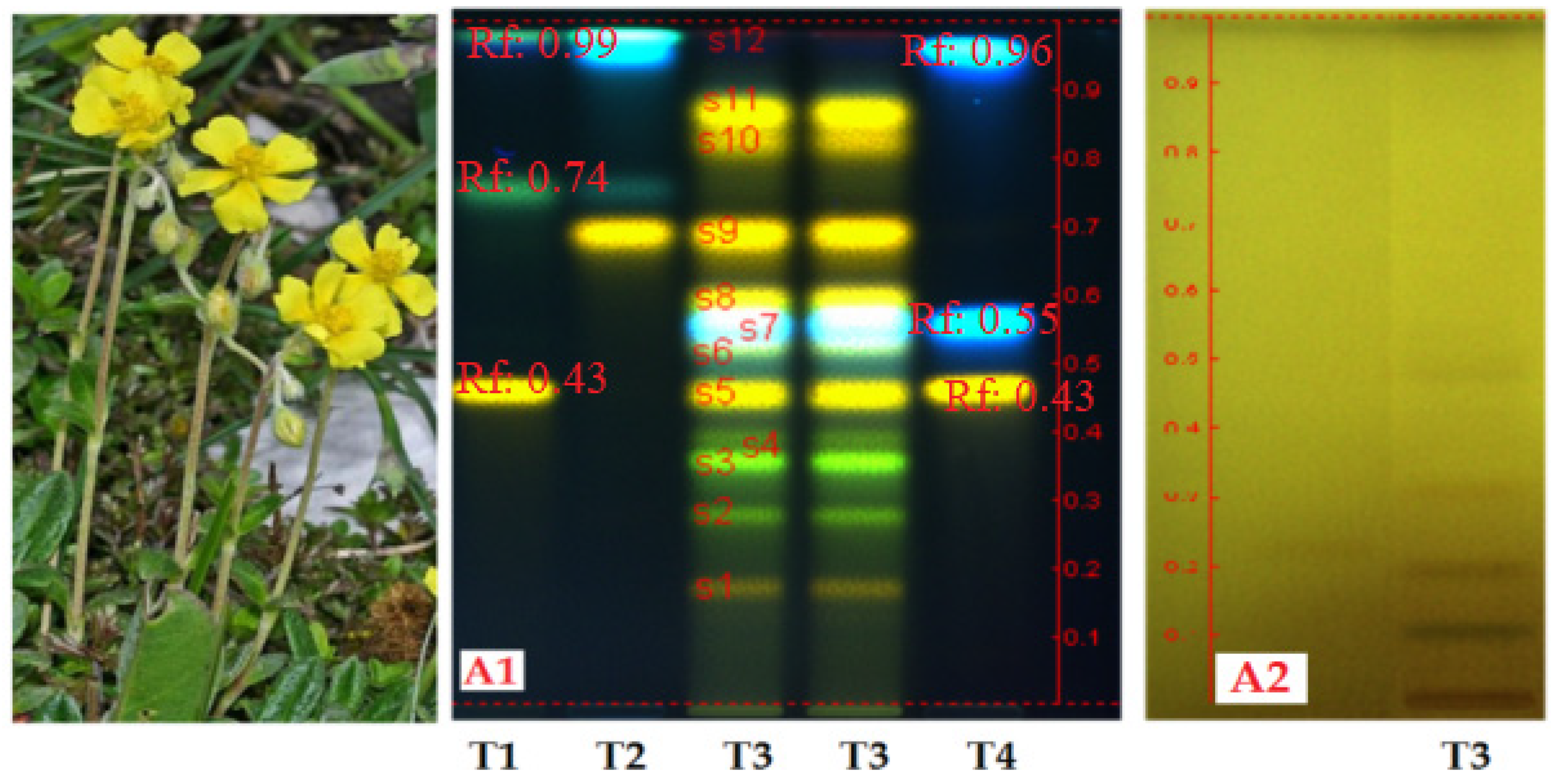
Figure 4.
a,b). The effects of the 40% ethanolic extracts from Ajuga reptans (Ajre), Geranium phaeum (Geph) and Helianthemum nummularium (Henu) upon the viability of mouse normal fibroblasts (L929 cell line), cytotoxicity MTS assay at 24 h of cell exposure; the cell viability (%) along the dilution series in variants µL test extract/mL sample and µg GAE/mL sample, by comparison with the negative control series (the green line), respectively. Where: Notation (*) means results without statistical significance (p > 0.05); Notation (**) means results with statistical significance and 0.01 < p < 0.05; Notation (***) means results with statistical significance and p < 0.01; mean values, n=3.
Figure 4.
a,b). The effects of the 40% ethanolic extracts from Ajuga reptans (Ajre), Geranium phaeum (Geph) and Helianthemum nummularium (Henu) upon the viability of mouse normal fibroblasts (L929 cell line), cytotoxicity MTS assay at 24 h of cell exposure; the cell viability (%) along the dilution series in variants µL test extract/mL sample and µg GAE/mL sample, by comparison with the negative control series (the green line), respectively. Where: Notation (*) means results without statistical significance (p > 0.05); Notation (**) means results with statistical significance and 0.01 < p < 0.05; Notation (***) means results with statistical significance and p < 0.01; mean values, n=3.
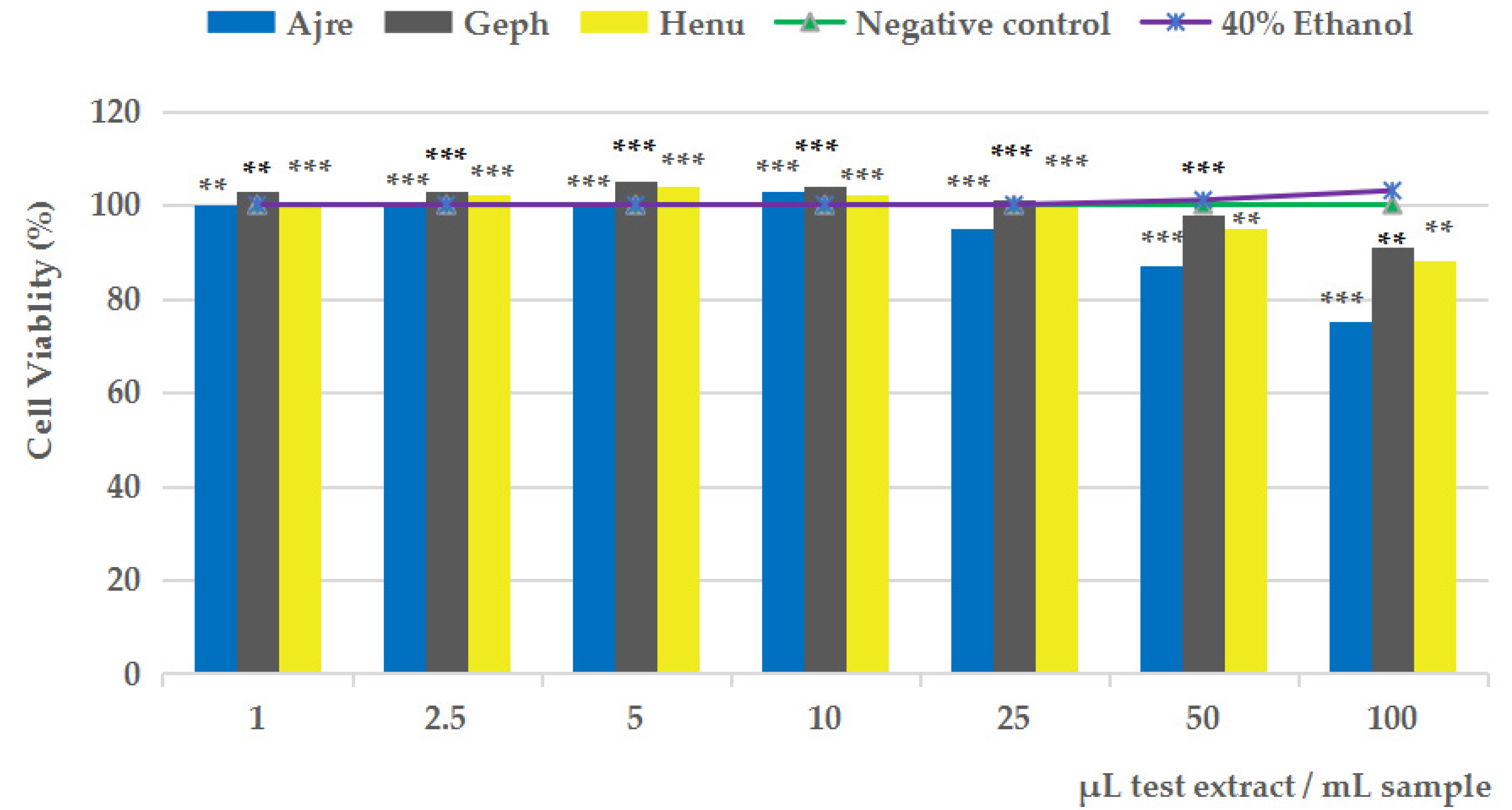
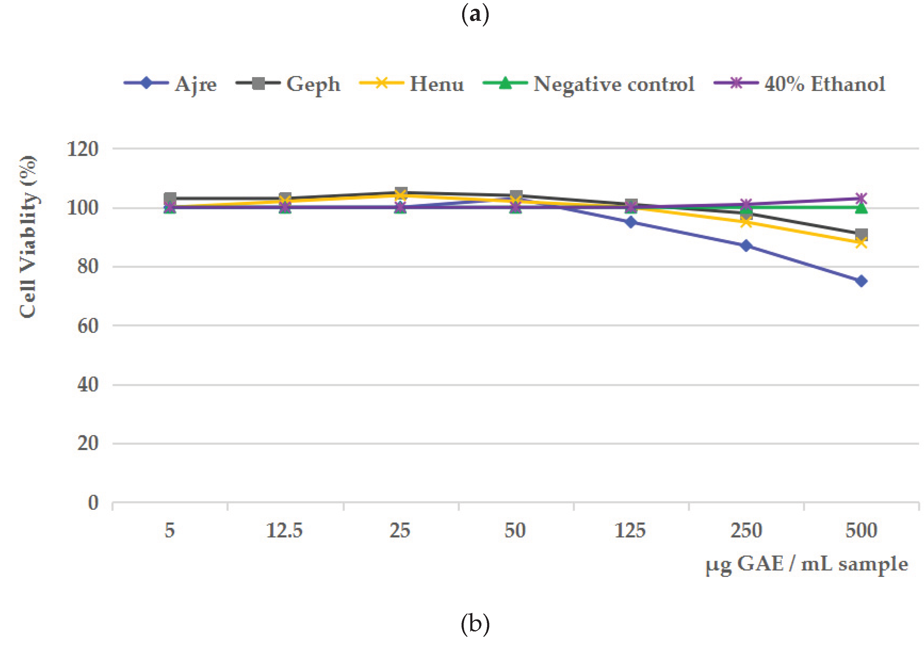
Figure 5.
a,b). The effects of the 40% ethanolic extracts from Ajuga reptans (Ajre), Geranium phaeum (Geph) and Helianthemum nummularium (Henu) upon the viability of human blood mononuclear cells (SC line), cytotoxicity MTS assay at 24 h of cell exposure; the cell viability (%) along the dilution series in variants µL test extract/mL sample and µg GAE/mL sample, by comparison with the negative control series (the green line) and positive control series (the red line), respectively. Where: Notation (*) means results without statistical significance (p > 0.05); Notation (**) means results with statistical significance and 0.01 < p < 0.05; Notation (***) means results with statistical significance and p < 0.01; mean values, n=3.
Figure 5.
a,b). The effects of the 40% ethanolic extracts from Ajuga reptans (Ajre), Geranium phaeum (Geph) and Helianthemum nummularium (Henu) upon the viability of human blood mononuclear cells (SC line), cytotoxicity MTS assay at 24 h of cell exposure; the cell viability (%) along the dilution series in variants µL test extract/mL sample and µg GAE/mL sample, by comparison with the negative control series (the green line) and positive control series (the red line), respectively. Where: Notation (*) means results without statistical significance (p > 0.05); Notation (**) means results with statistical significance and 0.01 < p < 0.05; Notation (***) means results with statistical significance and p < 0.01; mean values, n=3.
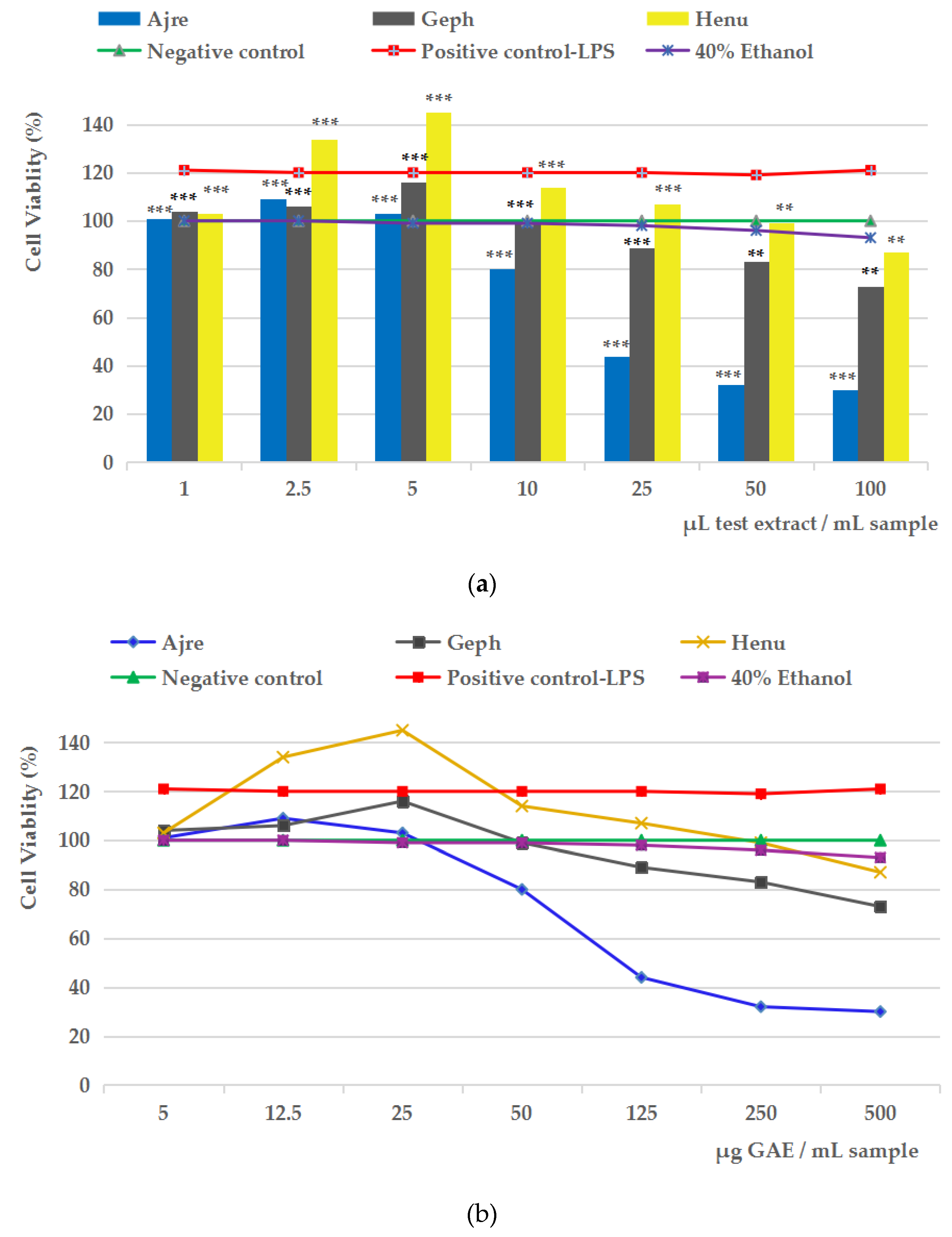

Table 1.
The ADMET parameters for phenolics frequently found in medicinal plant species.
| No. | Test compounds | 1MW | 2LogS | 3LogP | 4Caco-2 | 5BBB | 6P-gps | 7P-gpi | 8CYP450i | 9hERGi | 10OCT2 | ||
| Myricetin derivatives | |||||||||||||
| 1 | Myricetin | 318.237 | -2.915 | 1.6936 | 0.095 | -1.493 | Yes | No | CYP1A2 | No | No | ||
| 2 | Myricetin-3-O-glucoside | 480.378 | -2.904 | -0.833 | -1.34 | -2.078 | Yes | No | No | hERG II | No | ||
| 3 | Myricetin-3-O-galactoside | 480.378 | -2.904 | -0.833 | -1.34 | -2.078 | Yes | No | No | hERG II | No | ||
| 4 | Myricetin-3-O-rhamnoside | 464.379 | -2.892 | 0.1943 | -0.982 | -1.811 | Yes | No | No | hERG II | No | ||
| 5 | Myricetin-3-O-rutinoside | 626.52 | -2.892 | -1.9815 | -1.138 | -2.215 | Yes | No | No | hERG II | No | ||
| Quercetin derivatives | |||||||||||||
| 6 | Quercetin | 302.238 | -2.925 | 1.988 | -0.229 | -1.098 | Yes | No | CYP1A2 | No | No | ||
| 7 | Quercetin-3-O-glucoside | 464.379 | -2.925 | -0.5389 | 0.242 | -1.688 | Yes | No | No | hERG II | No | ||
| 8 | Quercetin-7-O-glucoside | 464.379 | -2.881 | -0.5389 | 0.271 | -1.623 | Yes | No | No | hERG II | No | ||
| 9 | Quercetin-3-O-galactoside | 464.379 | -2.925 | -0.5389 | 0.242 | -1.688 | Yes | No | No | hERG II | No | ||
| 10 | Quercetin-3-O-rhamnoside | 448.38 | -2.903 | 0.4887 | 0.048 | -1.495 | Yes | No | No | No | No | ||
| 11 | Quercetin-3-O-glucuronide | 478.362 | -2.894 | -0.4466 | 0.999 | -1.656 | Yes | No | No | No | No | ||
| 12 | Quercetin-3,7-O-diglucoside | 626.52 | -2.704 | -3.0658 | -0.977 | -2.358 | Yes | No | No | hERG II | No | ||
| 13 | Quercetin-3-O-rutinoside | 772.662 | -2.891 | -4.214 | -1.076 | -2.591 | Yes | No | No | hERG II | No | ||
| 14 | Isorhamnetin | 316.265 | -3 | 2.291 | -0.003 | -1.135 | Yes | No | CYP1A2 | No | No | ||
| 15 | Isorhamnetin-3-O-glucoside | 478.406 | -2.909 | -0.2359 | 0.333 | -1.723 | Yes | No | No | hERG II | No | ||
| 16 | Isorhamnetin-3-O-rutinoside | 624.548 | -2.899 | -1.3841 | 0.201 | -1.754 | Yes | P-gp II | No | hERG II | No | ||
| Luteolin derivatives | |||||||||||||
| 17 | Luteolin | 286.239 |
-3.094 |
2.2824 | 0.096 | -0.907 | Yes |
No |
CYP1A2 CYP2C9 |
No |
No |
||
| 18 | Luteolin-5-O-glucoside | 448.38 | -2.678 | -0.2445 | 0.325 | -1.502 | Yes | No | No | No | No | ||
| 19 | Luteolin-7-O-glucoside | 448.38 | -2.716 | -0.2445 | 0.248 | -1.564 | Yes | No | No | No | No | ||
| 20 | Luteolin-6-C-glucoside | 448.38 | -2.9 | -0.2027 | -0.912 | -1.564 | Yes | No | No | No | No | ||
| 21 | Luteolin-8-C-glucoside | 448.38 | -2.905 | -0.2027 | -1.25 | -1.639 | Yes | No | No | No | No | ||
| 22 | Luteolin-7-O-rutinoside | 594.522 | -2.904 | -1.3927 | -1.523 | -1.979 | Yes | No | No | hERG II | No | ||
| Kaempferol derivatives | |||||||||||||
| 23 | Kaempferol | 286.239 | -3.04 | 2.2824 | 0.032 | -0.939 | Yes | No | CYP1A2 | No | No | ||
| 24 | Kaempferol-3-O-glucoside | 448.38 | -2.863 | -0.2445 | 0.306 | -1.514 | Yes | No | No | No | No | ||
| 25 | Kaempferol-7-O-glucoside | 448.38 | -2.746 | -0.2445 | 0.353 | -1.465 | Yes | No | No | No | No | ||
| 26 | Kaempferol-3-O-galactoside | 448.38 | -2.863 | -0.2445 | 0.306 | -1.514 | Yes | No | No | No | No | ||
| 27 | Kaempferol-3-O-rhamnoside | 432.381 | -2.969 | 0.7831 | 0.123 | -1.265 | Yes | No | No | hERG II | No | ||
| 28 | Kaempferol 3-(6″O-couma royl) glucoside | 594.525 |
-2.923 |
1.7254 | -0.113 | -1.585 | Yes |
P-gp I P-gp II |
No |
hERG II |
No |
||
| 29 | Kaempferol-3-O-rutinoside | 594.522 | -2.9 | -1.3927 | 0.189 | -1.669 | Yes | No | No | hERG II | No | ||
| Apigenin and derivatives | |||||||||||||
| 30 | Apigenin | 270.24 |
-3.329 |
2.5768 | 1.007 | -0.734 | Yes |
No |
CYP1A2 CYP2C9 |
No |
No |
||
| 31 | Apigenin-6-C-glucoside | 432.381 | -2.812 | 0.0917 | -0.618 | -1.375 | Yes | No | No | No | No | ||
| 32 | Apigenin-7-O-glucoside | 432.381 | -2.559 | 0.0499 | 0.33 | -1.391 | Yes | No | No | No | No | ||
| 33 | Apigenin-8-C-glucoside | 432.381 | -2.845 | 0.0917 | -0.956 | -1.449 | Yes | No | No | No | No | ||
| 34 | Apigenin-6,8-di-C-diglucoside | 594.522 | -2.844 | -2.3934 | -1.127 | -1.927 | Yes | No | No | hERG II | No | ||
| 35 | Apigenin-7-O-apioglucoside | 564.496 | -2.851 | -1.4852 | -0.966 | -1.793 | Yes | No | No | hERG II | No | ||
| Phenolic acids | |||||||||||||
| 36 | 4-Hydroxybenzoic acid | 138.124 | 1.877 | 1.0904 | 1.151 | -0.334 | No | No | No | No | No | ||
| 33 | 3,4-Dihydroxyphenylacetic acid/ Protocatechuic acid | 168.148 |
-2.267 |
0.7249 | 0.637 | 0.639 | No |
No |
No |
No |
No |
||
| 38 | Ferulic acid | 194.186 | -2.817 | 1.4986 | 0.176 | -0.239 | No | No | No | No | No | ||
| 39 | Syringic acid | 198.174 | -2.223 | 1.1076 | 0.495 | -0.191 | Yes | No | No | No | No | ||
| 40 | Gallic acid | 170.12 | -2.56 | 0.5016 | -0.081 | -1.102 | No | No | No | No | No | ||
| 41 | Methyl gallate | 184.147 | -2 | 0.59 | -0.056 | -1.046 | Yes | No | No | No | No | ||
| 42 | Ellagic acid | 302.194 | -3.181 | 1.3128 | 0.335 | -1.272 | Yes | No | CYP1A2 | No | No | ||
| 43 | Caffeic acid | 164.16 | -2.378 | 1.49 | 1.21 | -0.225 | No | No | No | No | No | ||
| 44 | Chlorogenic acid | 354.311 | -2.449 | -0.6459 | -0.84 | -1.407 | Yes | No | No | No | No | ||
| 45 | Isochlorogenic acid | 354.311 | -2.449 | -0.6459 | -0.84 | -1.407 | Yes | No | No | No | No | ||
| 46 | Neochlorogenic acid | 354.311 | -2.449 | -0.6459 | -0.84 | -1.407 | Yes | No | No | No | No | ||
| 47 | Rosmarinic acid | 360.318 | -3.059 | 1.7613 | -0.937 | -1.378 | Yes | No | No | No | No | ||
| 48 | 4,5-Dicaffeoylquinic acid | 516.455 | -2.955 | 1.0296 | -1.203 | -2.08 | Yes | No | No | No | No | ||
| 49 | Cynarin | 516.455 | -2.962 | 1.0296 | -1.092 | -1.983 | Yes | No | CYP3A4 | No | No | ||
| 50 | 4-Caffeoylquinic acid | 761.371 | -5.778 | 8.8418 | 0.959 | -2.412 | No | P-gp I | CYP3A4 | No | N0 | ||
| Genus markers and other key compounds achievable in test plant species | |||||||||||||
| 51 | Ajugalactone | 516.631 | -4.712 | 2.163 | 0.425 | -0.898 | Yes | P-gp I | No | No | No | ||
| 52 | Ajugasterone C | 480.642 | -4.097 | 1.7099 | 0.394 | -0.856 | Yes | No | No | No | No | ||
| 53 | Verbascoside | 624.592 | -2.906 | -1.0159 | 0.096 | -1.86 | Yes | P-gp I | No | hERG II | No | ||
| 54 | 8-O-Acetylharpagide | 406.384 | -2.297 | -2.8934 | 0.42 | -1.275 | Yes | No | No | No | No | ||
| 55 | Geraniin | 952.648 |
-2.892 |
-1.1015 |
-1.384 |
-3.549 |
Yes |
P-gp I P-gp II |
No |
hERG II |
OCT2 |
||
Where: 1MW represents the molecular weight of the test compounds (the optimal values are lower than 500 u); 2LogS represents the water solubility, ensuring the test compound actually dissolves in the bodily fluids (the optimal values are between −4 and 0.5); 3LogP represents the solubility in fat/oil versus the solubility in water, estimating how well the test compound permeates cell membranes in the purpose of absorption and distribution in human body (the optimal values are between 1.35 and 1.8); 4Caco-2 value indicates the passive intestinal permeability (the optimal values are those higher than −5.15 log cm/s); 5BBB indicates the ability of a compound to cross the blood-brain barrier (these compounds are usually ascribed with values >2 and logP between 2 and 3); 6P-gps indicates the potentially to act as P-glycoprotein substrate; 7P-gpi indicates the potentially to act as P-glycoprotein I and/or P-glycoprotein II inhibitors; 8CYP450i indicates the compounds potentially acting as inhibitors of different oxidative enzymes composing the cytochrome P450 in humans, for example CYP1A2, CYP2C9, CYP2C19, CYP2D6 and CYP3A4 known as the major isoenzymes assisting the phase I metabolic processes in humans; 9hERGi indicates the compounds potentially acting as potassium channels (hERG I and hERG II) inhibitors; 10OCT2 indicates the compounds potentially acting as renal organic cation transporter substrate.
Table 2.
The results of molecular docking study on cyclooxygenase 2, COX-2 (PDB ID: 1PXX, chain A, binding pocket at 78.34 Å3) [76].
Table 2.
The results of molecular docking study on cyclooxygenase 2, COX-2 (PDB ID: 1PXX, chain A, binding pocket at 78.34 Å3) [76].
| Test Ligand | Interacting group /1PXX Chain A | Hydrogen bonds: Å | Score / RMSD |
| Diclofenac | TYR355, SER353, LEU352, VAL349, TYR348, TRP387, SER530, TYR385, LEU384, PHE381, LEU525, PRO528, GLY526, MET522, VAL 523, ARG120, GLU524, ALA527, VAL523, PHE518, PHE518 | Osp2 – Osp3 SER530: 2.653 Osp2 – Osp3 SER530: 2.905 Osp2 – Osp3 TYR385: 2.729 |
-68.76 / 0.15 |
| Isorhamnetin | ARG120, SER530,ALA527, VAL344, VAL349, TYR355, HIS90, GLY354, SER353, TYR348, VAL523, LEU352, GLY526, PHE205, PHE381, LEU384, TYR385, TRP387, MET522, PHE518, ILE517, ARG513, GLN192 | Osp3 – Osp3 SER530: 2.981 – Osp3 TYR385: 2.803 Osp3 – Osp3 TYR355: 2.988 Osp3 – Osp3 TYR355: 3.271 Osp3 – Osp2 SER353: 3.154 Osp3 – Osp2 LEU352: 3.222 Osp3 – Osp2 GLN192: 3.068 |
-59.77 / 0.02 |
| Ellagic acid | MET113, VAL116, LEU359, LEU117, ARG120, LEU532, LEU534, SER530, ILE345, ALA527, GLY526, VAL523, MET522, SER353, LEU352, PHE518, TRP387, TYR385, PHE381, TYR355, VAL349 | Osp3 – Osp3 SER530: 2.706 Osp2 – Osp3 SER530: 3.140 Osp3 – Osp3 TYR355: 2.992 |
-44.33 / 0.00353 |
| Ferulic acid | LEU384, TRP387, TYR385, GLY526, MET522, SER530, PRO528, PHE518, ALA527, LEU531, TYR348, VAL349, LEU352, SER353, TYR355 | Osp3 – Osp3 SER530: 3.080 Osp3 – Osp3 SER530: 2.897 – Osp3 TYR385: 2.974 |
-40.94 / 0.45 |
| Caffeic acid | VAL523, MET522, ALA527, GLY526, PHE381, LEU 384, SER530, PHE518, TYR355, SER353, LEU352, VAL349, TYR348, TRP387, TYR385 | Osp3 – Osp2 LEU352: 3.192 Osp2 – Osp3 SER530: 3.243 Osp2 – Osp3 SER530: 2.968 Osp2 – Osp3 TYR385: 2.942 |
-39.47 / 0.04 |
| 3,4-Dihydroxy phenylacetic acid | PHE381, TYR385, LEU384, TRP387, SER530, GLY526, ALA527, MET522, VAL523, PHE518, LEU352, SER353, GLY354, TYR348, VAL349 | Osp3 – Osp3 SER530: 3.012 Osp3 – Osp3 SER530: 3.099 – Osp3 TYR385: 3.105 Osp2 – Nsp2 SER353: 3.182 Osp2 – Osp2 LEU352: 3.130 |
-39.43 / 2.63 |
| Syringic acid | PHE518, VAL523,ARG120, VAL116, GLY256, ALA527, TYR355, SER353, TRP387, LEU352, VAL349, TYR348, ILE345, LEU534, LEU531, SER530 | Osp3 –Osp2 SER530: 3.118 Osp2–Osp3 TYR355: 2.598 – Nsp2ARG120: 3.112 Osp2 – Nsp2ARG120: 2.988 |
-38.75 / 0.03 |
| Methyl gallate | LEU534, SER530, VAL344, ALA527, VAL349, TYR348, LEU352, VAL523, GLY526, PHE381, TYR385, TRP387, PHE518, MET522 | Osp2 – Osp3 SER530: 2.924 Osp2 – Osp3 SER530: 2.924 Osp2 – Osp3 SER530: 2.976 – Osp3 TYR385: 2.814 |
-35.94 / 0.0052 |
| 4-Hydroxybenzoic acid | LEU352, VAL349, TYR348, VAL344, LEU534, SER530, PHE381, LEU384, TYR385, GLY526, TRP387, ALA527, MET522, VAL523, PHE518 | Osp2 – Osp3 SER530: 3.008 Osp2 – Osp3 SER530: 2.949 Osp2 – Osp3 TYR355: 2.760 Osp3 – Osp2 MET522: 3.014 |
-35.93 / 0.02 |
| Gallic acid | VAL344, TYR348, LEU534, VAL349, LEU352, SER353, SER530, GLY526, ALA527, PHE518, VAL523, MET522, LEU384, PHE381, TYR385, TRP387 | Osp2 – Osp3 SER530: 3.042 Osp2 – Osp3 SER530: 2.989 – Osp3 TYR385: 2.933 Osp3 – Osp2 MET522: 3.142 Osp3 – Osp2 MET522: 3.054 – Nsp2 VAL523: 3.274 |
-35.70 / 0.16 |
| Ajugasterone | PHE357, ILE112, LEU93, TYR355, TYR115, VAL89, SER119, ALA116, LEU117, SER353, GLN350, LEU359, ILE345, TYR348, VAL349, LEU352, LEU531, ARG120, SER119, VAL523, ALA527, PHE518, PRO528, VAL523, GLY526, MET522, LEU525, LEU384, TRP387, TYR385, LEU384, SER530 | Osp3 – Osp2 ALA527: 2.733 Osp2 – Osp3 TYR385: 2.638 |
-12.24 / 0.13 |
| Ajugalactone | - | - | + 6.14 / 0.079 |
| Geranin | - | - | +231.30 / 0.01 |
* Where: 1a very negative score is associated to a strong binding, while a less negative, or a positive score corresponds to a weak or non-existing binding [31]; 2a RMSD value lower than 2 indicates that the docking protocol has been validated.
Disclaimer/Publisher’s Note: The statements, opinions and data contained in all publications are solely those of the individual author(s) and contributor(s) and not of MDPI and/or the editor(s). MDPI and/or the editor(s) disclaim responsibility for any injury to people or property resulting from any ideas, methods, instructions or products referred to in the content. |
© 2026 by the authors. Licensee MDPI, Basel, Switzerland. This article is an open access article distributed under the terms and conditions of the Creative Commons Attribution (CC BY) license (http://creativecommons.org/licenses/by/4.0/).
Copyright: This open access article is published under a Creative Commons CC BY 4.0 license, which permit the free download, distribution, and reuse, provided that the author and preprint are cited in any reuse.