

Submitted:
04 August 2026
Posted:
06 August 2026
You are already at the latest version
Abstract
A lime-juice-assisted route was used to synthesize CoFe₂O₄ nanoparticles, which were subsequently immobilized in a chitosan matrix to obtain a magnetically recoverable CoFe₂O₄/chitosan (CoFe₂O₄/CS) nanocomposite for methylene blue (MB) removal under visible light. X-ray diffraction confirmed the cubic spinel phase and its retention after composite formation, while FTIR and XPS supported interfacial interactions between CoFe₂O₄ and the amino/hydroxyl functionalities of chitosan. Electron mi-croscopy showed predominantly spherical-to-quasi-spherical ferrite nanoparticles (approximately 10–20 nm) distributed on the polymer matrix with reduced agglom-eration. Chitosan incorporation decreased the BET surface area from 81.9 to 51.3 m² g⁻¹ and the saturation magnetization from 51.5 to 31.4 emu g⁻¹, but the nanocomposite remained readily separable with an external magnet. The optical band gap increased from 2.17 to 2.61 eV, whereas photoluminescence quenching was consistent with lower radiative charge-carrier recombination. At pH 9, an initial MB concentration of 20 mg L⁻¹, and a catalyst dosage of 0.7 g L⁻¹, the composite achieved 98.5% MB removal within 120 min under a 30 W visible-light LED, with a pseudo-first-order rate constant of 0.035 min⁻¹. Removal remained 90.3% after five cycles. Scavenger tests indicated that h⁺ and •OH were the dominant reactive species. These findings suggest the potential of bio-assisted CoFe₂O₄/CS nanocomposites as magnetically recoverable photocatalysts for wastewater treatment.
Keywords:
lime-juice-assisted synthesis
; cobalt ferrite
; chitosan
; magnetic recovery
; visible-light photocatalysis
; methylene blue
1. Introduction
Synthetic dyes released by textile, leather, pharmaceutical, and related industries remain a persistent challenge for aquatic environments because many dye molecules are chemically stable, intensely colored, and poorly biodegradable [1,2,3]. Methylene blue (MB), a cationic phenothiazine dye, is frequently used as a model contaminant in photocatalytic studies owing to its strong visible absorption and resistance to conventional biological treatment [1,3]. Even at relatively low concentrations, dye-containing effluents can impede light penetration and oxygen transfer, disrupt aquatic photosynthesis, and create potential human-health concerns[1,2,3,4,5]. Accordingly, treatment technologies must combine high removal efficiency with low chemical demand, operational simplicity, and catalyst reusability.
Semiconductor photocatalysis is an attractive advanced oxidation process because absorbed photons generate electron–hole pairs that can initiate the formation of reactive oxygen species, including hydroxyl radicals (•OH) and superoxide species (•O₂⁻), under comparatively mild conditions [2,5,6,7,8,9]. Practical performance, however, is often limited by rapid charge recombination, insufficient visible-light utilization, nanoparticle aggregation, and difficult post-treatment separation. Material design must therefore balance optical activity, interfacial charge transfer, accessible adsorption sites, and recoverability rather than optimize any single property in isolation.
Spinel ferrites (MFe₂O₄; M = Co, Zn, Ni, Mn, Mg, or Cu) are promising photocatalytic platforms because their electronic structures support visible-light response and their magnetic properties enable separation from treated water [10,11]. CoFe₂O₄ is particularly attractive because of its chemical stability, high magnetocrystalline anisotropy, coercivity, and saturation magnetization [12,13,14,15]. Nevertheless, bare CoFe₂O₄ nanoparticles tend to aggregate through magnetic and high-surface-energy interactions, reducing the number of accessible sites and complicating reproducible photocatalytic operation.
CoFe₂O₄ has been prepared by co-precipitation, sol–gel, hydrothermal, vapor-phase, laser-ablation, and mechanochemical methods [16,17,18,19,20,21]. Several of these routes require synthetic complexing agents, hazardous solvents, strong reducing agents, or energy-intensive processing [22]. Bio-assisted synthesis partially addresses these limitations by using plant-derived metabolites as complexing, capping, and stabilizing species [14,22,23,24,25,26,27,28]. Citrus juices contain citric and ascorbic acids, reducing sugars, flavonoids, polyphenols, and other oxygen-containing compounds that can coordinate metal ions, regulate nucleation, and suppress uncontrolled particle growth [29,30,31,32]. Their successful application to ZnO, CuO, MgO, TiO₂, and related nanomaterials supports the use of lime juice as a low-cost synthesis medium [29,30,33,34].
Metal oxide–polymer composites have been widely explored to improve semiconductor photocatalysis through enhanced light harvesting, reduced charge recombination, and better catalytic performance [35]. Chitosan (CS), a renewable polysaccharide obtained by deacetylation of chitin, offers complementary functionality through abundant amino and hydroxyl groups [36,37,38]. These groups can bind cationic and molecular pollutants through electrostatic interactions, hydrogen bonding, complexation, and physical adsorption [37,39,40]. As a polymeric support, chitosan can also limit ferrite aggregation and improve nanoparticle dispersion [41,42]. The resulting composite may therefore couple adsorption-assisted concentration of MB near the catalyst surface with more effective utilization of photogenerated charge carriers. Importantly, such synergy can improve apparent removal even when polymer coverage decreases the measured BET surface area.
Despite growing interest in ferrite–biopolymer composites, the relationships among citrus-assisted synthesis, CoFe₂O₄/CS interfacial chemistry, optical response, magnetic recovery, and adsorption-coupled photocatalysis remain insufficiently resolved. A rigorous assessment should therefore integrate structural, surface, optical, textural, and magnetic characterization with kinetic, operational-parameter, recycling, and reactive-species analyses.
In this work, CoFe₂O₄ nanoparticles were synthesized using lime juice as a natural complexing and stabilizing medium and subsequently incorporated into chitosan. The effects of calcination temperature on ferrite formation were investigated, and the resulting CoFe₂O₄/CS nanocomposite was characterized using structural, morphological, optical, surface, textural, and magnetic techniques. Its photocatalytic MB removal performance was evaluated under different irradiation times, pH values, dye concentrations, and catalyst dosages, together with reusability and scavenger tests
2. Materials and Methods
2.1. Materials
All reagents were of analytical grade and were used without further purification. Cobalt(II) nitrate hexahydrate [Co(NO₃)₂·6H₂O, 99%], iron(III) nitrate nonahydrate [Fe(NO₃)₃·9H₂O, 99%], medium-molecular-weight chitosan (degree of deacetylation, 75–85%), and methylene blue (97%) were purchased from Sigma-Aldrich (St. Louis, MO, USA). Sodium hydroxide (NaOH, 98%), acetic acid (CH₃COOH, 99%), and hydrochloric acid (HCl, 37%; density, 1.19 g mL⁻¹) were obtained from Aladdin Reagent Co., Ltd. (Shanghai, China). Deionized water was used throughout.
2.2. Preparation of Lime-Juice Extract
Fresh limes obtained in Nghe An Province, Vietnam, were washed thoroughly with deionized water and mechanically pressed. The juice was centrifuged at 13,000 rpm for 10 min and filtered, and the clear supernatant was stored at 4 °C until use.
2.3. Synthesis of CoFe₂O₄ Nanoparticles and CoFe₂O₄/CS Nanocomposite
CoFe₂O₄ nanoparticles were synthesized by dissolving 0.005 mol Co(NO₃)₂·6H₂O and 0.010 mol Fe(NO₃)₃·9H₂O in 50 mL of deionized water under stirring, thereby maintaining the stoichiometric Co:Fe ratio of 1:2. Lime-juice extract (20 mL) was added at room temperature, and the mixture was stirred at 80 °C until a gel formed. The gel was dried at 80 °C for 4 h, heated to the selected calcination temperature at 5 °C min⁻¹, held for 2 h, and cooled naturally. Samples calcined at 300, 400, 500, and 700 °C were used to evaluate phase development; the material calcined at 500 °C was used for composite preparation and subsequent photocatalytic tests.
For nanocomposite fabrication, 1.0 g of chitosan was dissolved in 100 mL of 1% (v/v) aqueous acetic acid. CoFe₂O₄ nanoparticles (0.30 g) were dispersed in the chitosan solution by magnetic stirring followed by sonication for 30 min. The pH was adjusted gradually to approximately 11 with 2 M NaOH, and the dispersion was sonicated for another 30 min and heated at 80 °C for 3 h. The solid was collected by filtration, washed repeatedly with deionized water to neutral pH, and dried at 60 °C for 5 h.
2.4. Characterization
Crystalline phases were analyzed by X-ray diffraction (XRD; D8 Advance, Bruker, Germany) using Cu Kα radiation (λ = 0.15406 nm) over 2θ = 10–70°. Crystallite size was estimated from the (311) reflection using the Scherrer equation, and lattice parameters, unit-cell volume, and X-ray density were calculated from the diffraction data.
Surface functional groups and ferrite–polymer interactions were examined by Fourier-transform infrared spectroscopy (FTIR; Thermo-Nicolet Nexus 670). Morphology and particle distribution were observed by field-emission scanning electron microscopy (FESEM; Hitachi S-4800) and transmission electron microscopy (TEM; JEOL JEM-1400). Elemental composition was assessed by energy-dispersive X-ray spectroscopy (EDX; HORIBA 7593-H) coupled to the SEM.
Surface elemental composition and oxidation states were investigated by X-ray photoelectron spectroscopy (XPS; ESCALab 250, Thermo VG, UK). Nitrogen adsorption–desorption measurements were performed on a Tristar-3000 analyzer, and the specific surface area and pore-size distribution were determined using the BET and BJH methods, respectively.
Diffuse-reflectance spectra were recorded with a Cary 5000 UV–Vis–NIR spectrometer. Reflectance data were transformed using the Kubelka–Munk function, and optical band gaps were estimated from Tauc plots assuming a direct allowed transition. Photoluminescence spectra were measured with a Fluorolog-3 spectrofluorometer (FL3C-22, HORIBA Scientific) to compare the relative radiative recombination behavior of CoFe₂O₄ and CoFe₂O₄/CS.
Magnetic hysteresis loops were recorded at room temperature using a Lake Shore 7404 vibrating sample magnetometer (VSM). Saturation magnetization (Mₛ), remanent magnetization (Mᵣ), and coercivity (Hc) were extracted from the hysteresis curves. Magnetic separation from water was assessed qualitatively using an external permanent magnet.
2.5. Photocatalytic Experiments
Photocatalytic tests were performed in a glass reactor containing 50 mL of MB solution and the required amount of catalyst. A 30 W visible-light LED was positioned 20 cm above the liquid surface. Before irradiation, each suspension was stirred in the dark for 60 min to establish adsorption–desorption equilibrium. Aliquots were withdrawn at 30 min intervals, the catalyst was separated, and the residual MB concentration was determined by UV–Vis spectrophotometry. Control experiments included direct photolysis and dark adsorption by CS, CoFe₂O₄, and CoFe₂O₄/CS. The MB removal efficiency (RE) was calculated as follows:
where C₀ and Cₜ (mg L⁻¹) are the MB concentrations at the beginning of the test and at irradiation time t, respectively.
The apparent photocatalytic kinetics were evaluated using the pseudo-first-order expression:
where k is the pseudo-first-order rate constant (min⁻¹), and t is the irradiation time (min). The value of k was obtained from the slope of the linear plot of ln(C₀/Cₜ) versus t.
Operational parameters were examined over the following ranges: initial pH, 3–11; initial MB concentration, 10–40 mg L⁻¹; and CoFe₂O₄/CS dosage, 0.2–1.0 g L⁻¹. Unless a parameter was being varied, the remaining conditions were held constant. The point of zero charge (pHPZC) of the nanocomposite was determined using the pH-drift method.
Reactive-species tests were performed by adding p-benzoquinone (BQ), isopropanol (IPA), ethylenediaminetetraacetic acid disodium salt dihydrate (Na₂EDTA·2H₂O, abbreviated as EDTA), or AgNO₃ as scavengers for •O₂⁻, •OH, h⁺, and e⁻, respectively. The resulting removal efficiencies were compared with a scavenger-free control under otherwise identical irradiation conditions.
Reusability was evaluated for five consecutive cycles under the optimized conditions (pH 9, 20 mg L⁻¹ MB, 0.7 g L⁻¹ catalyst, and 120 min irradiation). After each cycle, the nanocomposite was recovered with an external magnet, washed with ethanol and deionized water, vacuum-dried at 80 °C for 5 h, and reused in a freshly prepared MB solution. Phase stability was additionally assessed by comparing the XRD patterns of fresh material and a separately recovered specimen after five irradiation–recovery cycles.
All photocatalytic experiments were conducted in triplicate. Results are reported as mean ± standard deviation (SD), and error bars represent three independent measurements (n = 3).
3. Results and Discussion
3.1. Calcination-Dependent Formation of CoFe₂O₄
Figure 1a shows the XRD patterns of the ferrite precursors calcined at 300–700 °C. The sample treated at 300 °C exhibited only weak, broad features, indicating limited crystallization. At 400 °C, reflections appeared at 2θ = 30.3°, 35.6°, 43.3°, 53.7°, 57.3°, and 62.9°, corresponding to the (220), (311), (400), (422), (511), and (440) planes of cubic spinel CoFe₂O₄ (JCPDS 22-1086). Increasing the calcination temperature sharpened and intensified these reflections, consistent with progressive crystallite growth and improved long-range order.
The mean crystallite size D was estimated from the (311) reflection using the Scherrer equation [43]:
where K = 0.9 is the shape factor, λ = 1.5406 Å is the Cu Kα wavelength, β is the full width at half maximum of the (311) peak (in radians), and θ is the corresponding Bragg angle.
where dₕₖₗ is the interplanar spacing, h, k, and l are the Miller indices, M is the molar mass of CoFe₂O₄, Nₐ is Avogadro’s constant (6.022 × 10²³ mol⁻¹), and the factor 8 is the number of formula units per spinel unit cell.
The calculated crystallographic parameters are summarized in Table 1.
The crystallite size increased from 8.1 nm at 400 °C to 10.0 nm at 500 °C and 18.9 nm at 700 °C, reflecting thermally activated domain growth and coalescence [14,15]. Concurrently, a increased from 8.3304 to 8.3530 Å and V from 578.09 to 582.81 ų, while ρₓ decreased slightly from 5.39 to 5.35 g cm⁻³. The shift of the (220) and (311) reflections toward lower 2θ values (Figure 1b) is consistent with this small lattice expansion [44,45,46].
A calcination temperature of 500 °C was selected for subsequent work because it produced well-defined spinel CoFe₂O₄ while retaining a comparatively small crystallite size (10.0 nm). The 700 °C treatment increased crystallinity but nearly doubled the crystallite size, a change expected to reduce external surface area and potentially diminish photocatalytic accessibility. The selected temperature therefore represents a compromise between phase purity and nanoscale dimensions.
3.2. Crystal Structure of the CoFe₂O₄/CS Nanocomposite
The XRD patterns of CS, CoFe₂O₄, and CoFe₂O₄/CS are compared in Figure 2. Chitosan displayed a broad maximum near 2θ = 20°, characteristic of its semi-crystalline polysaccharide structure [47,48].
The nanocomposite retained the characteristic spinel reflections of CoFe₂O₄ together with the broad chitosan contribution. The reduced ferrite peak intensities primarily reflect dilution by the polymer matrix and partial attenuation of the diffracted signal; interfacial disorder may provide an additional contribution. No new crystalline impurity phase or substantial peak displacement was observed, demonstrating that composite formation preserved the CoFe₂O₄ spinel framework [49,50].
3.3. FTIR Analysis
Figure 3 compares the FTIR spectra of CS, CoFe₂O₄, and CoFe₂O₄/CS.
Chitosan exhibited a broad band at 3462.6 cm⁻¹ assigned to overlapping O–H and N–H stretching vibrations. The bands at 2933.5 and 2877.6 cm⁻¹ arose from asymmetric and symmetric C–H stretching, respectively [51,52,53,54].
The band at 1660.5 cm⁻¹ was associated with residual amide C=O stretching, whereas the features at 1592.1 and 1394.1 cm⁻¹ were assigned to amino-group bending (amide II region) and C–H deformation, respectively [55,56,57,58,59]. Bands at 1171.9 and 1073.4 cm⁻¹ corresponded to C–O–C stretching and coupled C–O/C–N vibrations of the glucosamine framework [55,56,57].
CoFe₂O₄ displayed broad O–H stretching and adsorbed-water bending bands at 3451.2 and 1635.3 cm⁻¹, respectively [15,41,60]. The feature at 1383.7 cm⁻¹ was attributed to residual surface-bound organic species from the bio-assisted synthesis [61]. The bands at 586.2 and 416.7 cm⁻¹ are characteristic of metal–oxygen vibrations at tetrahedral and octahedral sites of the spinel lattice, respectively [62].
Both polymer and ferrite vibrations were present in CoFe₂O₄/CS. The metal–oxygen bands shifted from 586.2 and 416.7 cm⁻¹ to 575.6 and 408.8 cm⁻¹, and the broad O–H/N–H band shifted from 3462.6 to 3441.7 cm⁻¹. These coordinated shifts, together with intensity changes, support hydrogen-bonding and/or coordination interactions between surface metal/oxygen sites and the –NH₂/–OH groups of chitosan [63,64].
3.4. Morphology and Elemental Composition
Pristine CoFe₂O₄ consisted of predominantly spherical-to-quasi-spherical particles with an approximate size range of 10–20 nm (Figure 4a and inset). The particles formed aggregates, as expected from their high surface energy and magnetic interactions [65]. After incorporation into chitosan, ferrite domains were distributed across the polymer matrix and appeared more spatially separated (Figure 4c). TEM likewise indicated a lower degree of compact agglomeration in the composite. These observations support the role of chitosan as a steric and interfacial stabilizer that restricts direct ferrite–ferrite contact [63,66].
EDX detected Co, Fe, and O in the ferrite sample and additionally C and N in CoFe₂O₄/CS, confirming the presence of the chitosan phase (Figure 4b,d). The lower relative Co and Fe signals in the composite are consistent with polymer dilution and surface coverage of the ferrite particles.
3.5. Optical Absorption and Photoluminescence
Figure 5a shows the diffuse-reflectance response of CS, CoFe₂O₄, and CoFe₂O₄/CS. Chitosan absorbed mainly in the ultraviolet region, whereas CoFe₂O₄ exhibited broad visible-light absorption. Tauc analysis (Figure 5b) yielded optical band gaps of 2.17 eV for CoFe₂O₄ and 2.61 eV for CoFe₂O₄/CS. The blue shift after polymer incorporation may arise from interfacial electronic perturbation, altered surface states, and the lower optical contribution of ferrite in the composite. Thus, the enhanced photocatalytic removal of CoFe₂O₄/CS cannot be attributed to band-gap narrowing; it must instead involve interfacial, adsorption, and charge-utilization effects.
PL emission provides a comparative indication of radiative electron–hole recombination [6,67,68,69]. CoFe₂O₄ showed a broad emission centered near 430 nm, whereas CoFe₂O₄/CS exhibited substantially lower intensity without a pronounced shift in the emission maximum (Figure 6). The quenching is consistent with reduced radiative recombination and/or more efficient trapping and interfacial transfer of photoexcited carriers. Because the composite also contains a lower fraction of emissive ferrite, dilution may contribute to the absolute intensity decrease; nevertheless, the PL trend, together with the higher rate constant, supports improved charge utilization in CoFe₂O₄/CS.
3.6. XPS Analysis
The XPS survey spectrum of CoFe₂O₄/CS confirmed the presence of C, N, O, Co, and Fe (Figure 7a). The comparatively weak Co and Fe intensities are consistent with partial coverage of the ferrite domains by the chitosan-rich surface layer rather than an absence of the inorganic phase.
The high-resolution Fe 2p spectrum contained satellite features at 718.1 and 732.4 eV and components assigned to Fe³⁺ in octahedral and tetrahedral coordination (Figure 7b) [70,71,72]. The Co 2p spectrum (Figure 7c) showed Co²⁺ spin–orbit components at 779.1/794.5 eV and 781.3/795.8 eV, together with satellites at 785.1 and 801.1 eV [70,71,73]. No distinct Co³⁺ contribution was resolved [74]. These assignments are consistent with the expected mixed-site cation distribution of spinel CoFe₂O₄.
The O 1s envelope was resolved into components at 530.0, 531.4, and 532.9 eV, attributable to lattice oxygen, carbonyl/defect-associated oxygen, and hydroxyl/ether oxygen from chitosan, respectively (Figure 7d) [40,71]. The C 1s spectrum (Figure 7e) contained contributions from C–C/C–H (284.5 eV), C–N/C–O (286.1 eV), and O–C–O/C=O (287.6 eV), whereas the N 1s spectrum (Figure 7f) showed amino and protonated-amino environments at 399.5 and 400.3 eV [75,76]. The coexistence of ferrite lattice oxygen and chitosan-derived C/N/O environments corroborates the composite structure indicated by XRD and FTIR.
3.7. BET Analysis and Magnetic Properties
Both materials exhibited type-IV nitrogen adsorption–desorption isotherms with H3 hysteresis, indicating mesoporous structures (Figure 8). CoFe₂O₄ had a BET surface area of 81.9 m² g⁻¹ and pore volume of 0.149 cm³ g⁻¹, whereas CoFe₂O₄/CS showed corresponding values of 51.3 m² g⁻¹ and 0.122 cm³ g⁻¹ (Table 2). The decrease is attributable to polymer coverage of ferrite surfaces and partial occupation or shielding of pore space. The mean pore diameter changed only slightly, from 6.8 to 7.4 nm, indicating that mesoporosity was broadly retained [41].
Room-temperature magnetic hysteresis loops are shown in Figure 9. CoFe₂O₄ exhibited Mₛ = 51.5 emu g⁻¹, Mᵣ = 3.8 emu g⁻¹, and Hc = 70.3 Oe. For CoFe₂O₄/CS, Mₛ decreased to 31.4 emu g⁻¹ because of dilution by non-magnetic chitosan, while Mᵣ and Hc increased to 7.46 emu g⁻¹ and 679.8 Oe, respectively. The change in coercivity may reflect altered interparticle interactions, anisotropy, and magnetic-domain reversal after immobilization in the polymer matrix.
Despite the lower saturation magnetization, CoFe₂O₄/CS was rapidly collected from water with an external magnet (Figure 9 inset). This recoverability is operationally important because it enables catalyst removal without filtration and supports repeated use. The textural and magnetic results therefore reveal a deliberate trade-off: chitosan decreases accessible ferrite surface area and magnetization but provides a dispersing, adsorptive, and recoverable composite architecture.
3.8. Photocatalytic Performance and Mechanistic Analysis
3.8.1. Time-Dependent MB Removal and Kinetics
The rate constant increased from 0.016 min⁻¹ for CoFe₂O₄ to 0.035 min⁻¹ for CoFe₂O₄/CS (Figure 10b), corresponding to an approximately 2.2-fold enhancement. This improvement occurred despite the lower BET surface area and wider apparent optical band gap of the composite. This improvement may be associated with a cooperative mechanism in which chitosan promotes MB enrichment near the interface, helps reduce ferrite aggregation, and creates an interfacial environment consistent with lower radiative recombination and more effective generation or utilization of oxidizing specie [76].
3.8.2. Effect of Initial pH
MB removal increased from 72.6% at pH 3 to 84.7%, 90.9%, and 98.5% at pH 5, 7, and 9, respectively, before decreasing slightly to 96.7% at pH 11 (Figure 11a). The corresponding rate constants were 0.011, 0.016, 0.020, 0.035, and 0.028 min⁻¹ (Figure 11b). Thus, pH 9 provided the highest efficiency and kinetic rate under the investigated conditions.
The pH dependence is consistent with the measured pHPZC of 6.7 (Figure S1) and with protonation/deprotonation of chitosan amino groups [77,78,79]. Below pHPZC, the composite surface and protonated –NH₃⁺ groups are predominantly positive, producing electrostatic repulsion toward cationic MB and limiting adsorption near photocatalytic sites. Above pHPZC, a more negative surface favors MB accumulation and increases the local probability of oxidation. The modest decline at pH 11 may arise from excessive alkalinity, increased suspension screening/competition at surface sites, and altered radical chemistry [80].
The close correspondence between removal efficiency and k confirms that the pH effect was kinetic rather than solely an endpoint adsorption phenomenon. The optimum near pH 9 reflects a balance between favorable electrostatic uptake of MB and efficient photochemical oxidation at the composite–solution interface.
3.8.3. Effect of MB Concentration
At initial MB concentrations of 10, 20, 30, and 40 mg L⁻¹, removal efficiencies after 120 min were 99.3%, 98.5%, 90.5%, and 84.1%, respectively (Figure 12a). The decline at higher concentration reflects saturation of a finite number of adsorption/photocatalytic sites and stronger attenuation of incident light by the intensely colored solution [1,2,4]. These effects reduce the ratio of reactive species to dye molecules and slow removal.
The rate constants decreased from 0.044 to 0.035, 0.019, and 0.015 min⁻¹ as the MB concentration increased from 10 to 40 mg L⁻¹ (Figure 12b). An initial concentration of 20 mg L⁻¹ was selected for subsequent optimization because it retained near-complete removal and a relatively high rate constant while representing a more demanding dye loading than 10 mg L⁻¹.
3.8.4. Effect of Catalyst Dosage
Catalyst dosage controls the number of accessible reaction sites and the fraction of incident light that reaches the suspended particles [81]. CoFe₂O₄/CS loadings from 0.2 to 1.0 g L⁻¹ were therefore examined (Figure 13).
Increasing the dosage from 0.2 to 0.7 g L⁻¹ raised MB removal from 65.3% to 84.5% and then to 98.5%. The rate constant increased in parallel from 0.009 to 0.015 and 0.035 min⁻¹. The improvement is attributed to the larger number of adsorption and photocatalytic sites and the resulting increase in productive photon absorption [1,4].
At 1.0 g L⁻¹, removal decreased slightly to 96.7% and k to 0.027 min⁻¹. Excess catalyst can increase turbidity, light scattering, and particle shielding and may promote aggregation, thereby lowering the effectively illuminated surface area [2,80]. A dosage of 0.7 g L⁻¹ was therefore selected as the optimum.
3.8.5. Reusability, Reactive Species, and Proposed Mechanism
Under the optimized conditions, MB removal decreased from 98.5% in the first cycle to 96.2%, 95.7%, 92.6%, and 90.3% in cycles 2–5, respectively (Figure 14a). The loss after five cycles was 8.2 percentage points, indicating that most of the initial performance was retained. The gradual decline may result from incomplete desorption of MB/intermediates, partial blockage of pores and active sites, small handling losses during recovery, or progressive surface modification [1,5,81]. Magnetic collection simplified catalyst recovery and is a practical advantage of the composite architecture.
Scavenger tests provided qualitative evidence for the dominant reactive pathways (Figure 14b). In the absence of scavengers, removal was 98.5%. BQ and AgNO₃ reduced removal to 83.6% and 87.4%, respectively, suggesting secondary contributions from •O₂⁻- and electron-mediated reactions. EDTA and IPA caused much larger decreases, to 54.8% and 60.7%, demonstrating that photogenerated holes and •OH radicals were the principal oxidizing species [81].
The XRD pattern of a separately recovered sample after five irradiation–recovery cycles retained the characteristic spinel reflections with no evident new crystalline phase (Figure 15). This result indicates good phase-level stability, although it does not exclude gradual surface fouling, amorphous changes, or metal-ion leaching.
To evaluate the thermodynamic feasibility of the proposed reactive pathways, the conduction-band (ECB) and valence-band (EVB) edge potentials were estimated using Mulliken electronegativity theory [82]:
Here, χ is the absolute electronegativity of the semiconductor (5.8 eV for CoFe₂O₄), Eₑ is the free-electron energy on the hydrogen scale (4.5 eV vs. NHE), and Eg is the measured optical band gap. Using Eg = 2.61 eV for CoFe₂O₄/CS gave ECB ≈ −0.005 eV and EVB ≈ +2.605 eV versus NHE. The strongly positive valence-band potential is sufficient for direct hole oxidation and for oxidation of H₂O/OH⁻ to •OH, consistent with the strong inhibition caused by EDTA and IPA [83]. By contrast, ECB is less negative than the standard O₂/•O₂⁻ potential, indicating that direct superoxide generation is not the dominant thermodynamic pathway. The moderate BQ effect may therefore arise from secondary oxygen-reduction routes associated with defects/interfacial states or from non-ideal scavenger selectivity.
The proposed mechanism is summarized in Figure 16. Visible-light excitation generates conduction-band electrons and valence-band holes. Interfacial interactions with chitosan and reduced radiative recombination increase the probability that holes reach the surface, where they can oxidize adsorbed MB directly or react with H₂O/OH⁻ to form •OH. Chitosan simultaneously concentrates cationic MB near the reactive interface, creating an adsorption-coupled oxidation pathway. Electrons and oxygen-derived species make a smaller contribution, in agreement with the scavenger results.
The dominant reactions can be represented schematically as follows:
CoFe₂O₄/CS + hν → CoFe₂O₄/CS (e⁻(CB) + h⁺(VB))
H₂O + h⁺(VB) → •OH + H⁺
OH⁻ + h⁺(VB) → •OH
MB + •OH/h⁺(VB) → CO₂ + H₂O + other products
These reactions describe a proposed degradation pathway rather than demonstrated complete mineralization. Because total organic carbon/chemical oxygen demand, degradation intermediates, cobalt/iron leaching, and real-wastewater matrix effects were not measured, the present results should be interpreted primarily as evidence of efficient MB removal and decolorization under controlled laboratory conditions. These measurements are important priorities for future scale-up and environmental-safety assessment.
4. Conclusions
A magnetically recoverable CoFe₂O₄/CS nanocomposite was prepared by combining lime-juice-assisted CoFe₂O₄ synthesis with chitosan immobilization. XRD confirmed preservation of the cubic spinel phase, whereas FTIR and XPS supported interfacial interactions between ferrite surface sites and chitosan amino/hydroxyl groups. Chitosan reduced ferrite aggregation and improved dark MB uptake, although it decreased the BET surface area from 81.9 to 51.3 m² g⁻¹, increased the apparent optical band gap from 2.17 to 2.61 eV, and lowered Mₛ from 51.5 to 31.4 emu g⁻¹. The composite nevertheless remained magnetically separable and exhibited a lower PL intensity and superior light-assisted removal. Under the optimized conditions (pH 9, 20 mg L⁻¹ MB, 0.7 g L⁻¹ catalyst, 30 W visible-light LED), 98.5% removal was achieved within 120 min with k = 0.035 min⁻¹, approximately 2.2 times the value for pristine CoFe₂O₄. Removal remained 90.3% after five cycles, and h⁺, •OH were identified as the dominant reactive species. This improvement may be partly associated with reduced radiative recombination of photogenerated charge carriers. Overall, these findings support the potential practical application of Lime-Juice-Assisted CoFe₂O₄/CS nanocomposites as magnetically recoverable photocatalysts for MB-containing wastewater.
Supplementary Materials
The following supporting information can be downloaded at the website of this paper posted on Preprints.org, Figure S1: pH-drift curve used to determine the point of zero charge (pHPzc) of CoFe₂O₄/CS.
Author Contributions
Conceptualization, N.X.D.; methodology, L.T.H., P.T.M.H., and N.X.D.; investigation and data curation, L.T.H. and P.T.M.H.; validation and supervision, N.X.D.; writing—original draft preparation, L.T.H. and P.T.M.H.; writing—review and editing, all authors. All authors have read and agreed to the published version of the manuscript.
Funding
This research was funded by the Vietnam Ministry of Education and Training under grant number B2024-TDV-10.
Institutional Review Board Statement
Not applicable.
Informed Consent Statement
Not applicable.
Data Availability Statement
The data supporting the findings of this study are available from the corresponding author upon reasonable request.
Conflicts of Interest
The authors declare no conflict of interest.
References
- Tran, D.-T.; Nguyen, K.-H.; Nghiem, L.D.; Van, H. High-Performance and Easily Recoverable Bismuth Vanadate Based Photocatalyst for Boosting Degradation of Organic Dyes by Visible Light. Ceram. Int. 2026, 52, 24010–24022. [Google Scholar] [CrossRef]
- Geldasa, F.T.; Kebede, M.A.; Shura, M.W.; Hone, F.G. Experimental and Computational Study of Metal Oxide Nanoparticles for the Photocatalytic Degradation of Organic Pollutants: A Review. RSC Adv. 2023, 13, 18404–18442. [Google Scholar] [CrossRef] [PubMed]
- Ahmadi, B.; Fallah, A.; Ghamarpoor, R.; Jamshidi, M. Methylene Blue beyond the Dye: A Critical Review on Its Role as a Benchmark Pollutant in Photocatalyst Design. Results Chem. 2025, 18, 102910. [Google Scholar] [CrossRef]
- Kouser, H.A.; Kumar, E.V.; Kamat, V.; Naik, H.S.B. Photocatalytic Degradation Phenomena of Methylene Blue Dye by ZnFe2O4 Decorated with RGO Nanocomposites under Visible Light Irradiation. Next Nanotechnol. 2026, 9, 100342. [Google Scholar] [CrossRef]
- Kumar, H.; Nike, T.; Kaushal, D.; Sheel, V.; Chauhan, V.; Shandilya, P.; Kumar, M. Fabrication of Synergistic CuBi2O4/CaTiO3 Heterojunction towards Methylene Blue Dye Degradation under Sunlight. Nano Trends 2026, 14, 100207. [Google Scholar] [CrossRef]
- Chandel, N.; Sharma, K.; Sudhaik, A.; Raizada, P.; Hosseini-Bandegharaei, A.; Thakur, V.K.; Singh, P. Magnetically Separable ZnO/ZnFe2O4 and ZnO/CoFe2O4 Photocatalysts Supported onto Nitrogen Doped Graphene for Photocatalytic Degradation of Toxic Dyes. Arab. J. Chem. 2019, 13, 4324–4340. [Google Scholar] [CrossRef]
- Stiadi, Y.; Wendari, T.P.; Zilfa; Zulhadjri; Rahmayeni. Tuning the Structural, Magnetic, and Optical Properties of ZnO/NiFe2O4Heterojunction Photocatalyst for Simultaneous Photodegradation of Rhodamine B and Methylene Blue under Natural Sunlight. Environ. Eng. Res. 2022, 28, 220074–0. [Google Scholar] [CrossRef]
- Suharyadi, E.; Muzakki, A.; Nofrianti, A.; Istiqomah, N.I.; Kato, T.; Iwata, S. Photocatalytic Activity of Magnetic Core-Shell CoFe2O4@ZnO Nanoparticles for Purification of Methylene Blue. Mater. Res. Express 2020, 7, 085013. [Google Scholar] [CrossRef]
- Malakootian, M.; Asadzadeh, S.N.; Mehdipoor, M.; Kalantar-Neyestanaki, D. A New Approach in Photocatalytic Degradation of Tetracycline Using Biogenic Zinc Oxide Nanoparticles and Peroxymonosulfate under UVC Irradiation. Desalin. Water Treat. 2021, 222, 302–312. [Google Scholar] [CrossRef]
- Patnaik, S.; Das, K.K.; Mohanty, A.; Parida, K. Enhanced Photo Catalytic Reduction of Cr (VI) over Polymer-Sensitized g-C3N4/ZnFe2O4 and Its Synergism with Phenol Oxidation under Visible Light Irradiation. Catal. Today 2018, 315, 52–66. [Google Scholar] [CrossRef]
- Kumar Das, K.; Sahoo, D.P.; Mansingh, S.; Parida, K. ZnFe2O4@WO3–X /Polypyrrole: An Efficient Ternary Photocatalytic System for Energy and Environmental Application. ACS Omega 2021, 6, 30401–30418. [Google Scholar] [CrossRef] [PubMed]
- Ramadan, R.; Uskoković, V.; El-Masry, M.M. Triphasic CoFe2O4/ZnFe2O4/CuFe2O4 Nanocomposite for Water Treatment Applications. J. Alloys Compd. 2023, 954, 170040. [Google Scholar] [CrossRef]
- El-Masry, M.M.; El-Shahat, M.; Ramadan, R.; Abdelhameed, R.M. Selective Photocatalytic Reduction of Nitroarenes into Amines Based on Cobalt/Copper Ferrite and Cobalt-Doped Copper Ferrite Nano-Photocatalyst. J. Mater. Sci. 2021, 32, 18408–18424. [Google Scholar] [CrossRef]
- Banifatemi, S.S.; Davar, F.; Aghabarari, B.; Segura, J.A.; Alonso, F.J.; Ghoreishi, S.M. Green Synthesis of CoFe2O4 Nanoparticles Using Olive Leaf Extract and Characterization of Their Magnetic Properties. Ceram. Int. 2021, 47, 19198–19204. [Google Scholar] [CrossRef]
- Purnama, B.; Suwandi, A.D.; Hartono, R.; Alim, S.; Bawono, T.; Utari, U.; Aldila, H.; Rahwanto, A.; Kusumandari, K. Annealing Temperature Dependence on Magnetic Properties, Crystalline Structure and Photocatalyst Activity of Coprecipitated Cobalt Ferrite (CoFe2O4). J. Phys. Sci. 2023, 34, 75–89. [Google Scholar] [CrossRef]
- Yu, W.; Li, Y.; Shu, M.; Liu, C.; Liang, Y.; Mao, Y.; Tan, J.; Liu, Y.; Ai, T. CS/CoFe2O4 Nanocomposite as a High-Effective and Steady Chainmail Catalyst for Tetracycline Degradation with Peroxymonosulfate Activation: Performance and Mechanism. Environ. Geochem. Health 2024, 46, 40. [Google Scholar] [CrossRef] [PubMed]
- Zhang, J.; Zhang, S.; Bian, X.; Yin, Y.; Huang, W.; Liu, C.; Liang, X.; Li, F. High Efficiency Removal Performance of Tetracycline by Magnetic CoFe2O4/NaBiO3 Photocatalytic Synergistic Persulfate Technology. Molecules 2024, 29, 4055. [Google Scholar] [CrossRef] [PubMed]
- Ibrahim, I.; Belessiotis, G. V.; Elseman, A.M.; Mohamed, M.M.; Ren, Y.; Salama, T.M.; Mohamed, M.B.I. Magnetic TiO2/CoFe2O4 Photocatalysts for Degradation of Organic Dyes and Pharmaceuticals without Oxidants. Nanomaterials 2022, 12, 3290. [Google Scholar] [CrossRef] [PubMed]
- Dong, S.; Dai, J.; Yang, Y.; Zada, A.; Qi, K. Extended Interfacial Charge Transference in CoFe2O4/WO3 Nanocomposites for the Photocatalytic Degradation of Tetracycline Antibiotics. Molecules 2024, 29, 4561. [Google Scholar] [CrossRef] [PubMed]
- Aspe, B.; Malyeyev, A.; Vakilinejad, A.; Menguelti, K.; Michels, A.; Bahlawane, N. Chemical Vapor Deposition of CoFe2O4 Micropillar Arrays with Enhanced Magnetic Properties. J. Alloys Compd. 2022, 890, 161758. [Google Scholar] [CrossRef]
- Cedeño-Mattei, Y.; Perales-Pérez, O.; Uwakweh, O.N.C. Effect of High-Energy Ball Milling Time on Structural and Magnetic Properties of Nanocrystalline Cobalt Ferrite Powders. J. Magn. Magn. Mater. 2013, 341, 17–24. [Google Scholar] [CrossRef]
- Ahmed, R.; Manik, K.H.; Islam, Md.S.; Rhine, A.; Mim, J.J.; Hossain, N. Green Synthesis Methods for Nanoparticles: Principles, Biological Routes, and Physicochemical Approaches toward Sustainable Nanotechnology. Next Mater. 2026, 11, 101929. [Google Scholar] [CrossRef]
- Saxena, R.; Kotnala, S.; Bhatt, S.C.; Uniyal, M.; Rawat, B.S.; Negi, P.; Riyal, M.K. A Review on Green Synthesis of Nanoparticles toward Sustainable Environment. Sustain. Chem. Clim. Action 2025, 6, 100071. [Google Scholar] [CrossRef]
- Refat, N.M.; Nassar, M.Y.; Sadeek, S.A. A Controllable One-Pot Hydrothermal Synthesis of Spherical Cobalt Ferrite Nanoparticles: Synthesis, Characterization, and Optical Properties. RSC Adv. 2022, 12, 25081–25095. [Google Scholar] [CrossRef] [PubMed]
- Alotaibi, A.M.; Alansi, A.M.; Alade, I.; Qahtan, T.F. Green Nanochemistry Approaches for the Sustainable Synthesis of Biomedical Nanomaterials and Their Emerging Applications. Adv. Powder Technol. 2026, 37, 105226. [Google Scholar] [CrossRef]
- Kumar, R.; Yadav, V.; Gagnish; Rishu; Bhardwaj, V.K. Green Synthesis of Fe2O3 Nanoparticles Using Sunflower Seed Extract for Enhanced Photodegradation of Rhodamine B. Mater. Chem. Phys. 2026, 360, 132662. [Google Scholar] [CrossRef]
- Ger, T.-Y.; Arokia Vijaya Anand, M.; Yang, C.-J.; Yao, C.-H.; Lai, J.-Y. Recent Progress in Green Synthesis of Bioinspired Nanoceria for Therapeutic Applications. Mater. Des. 2026, 267, 116334. [Google Scholar] [CrossRef]
- El Ouardy, K.; Akhrouf, A.; Faik, A.; Mir, Y. Green Synthesis of Metal and Metal Oxide Nanoparticles: A Pathway to Sustainable Energy and Sensing Applications. Nanotechnol. Rev. 2026, 15, 1–33. [Google Scholar] [CrossRef]
- Alvarado, J.A.; Conzalez, G.S.A.; Arce-Plaza, A.; Reyes-Carmona, S. New Approach in Effective and Reproducible Green Synthesis of Pure ZnO Nanoparticles Using Lemon Juice with Less Solvent and without Strong Base Chemical. Ceram. Int. 2025, 51, 18348–18355. [Google Scholar] [CrossRef]
- Jebali, M.; Gómez-Merino, A.I.; Colangelo, G. Influence of the Lemon (Citrus Limon L.) Juice Amount on the Green Synthesis of CuO Nanoparticles: Characterization, Stability and Thermal Conductivity. Ceram. Int. 2025, 51, 72–84. [Google Scholar] [CrossRef]
- Mahiuddin, M.; Ochiai, B. Comprehensive Study on Lemon Juice-Based Green Synthesis and Catalytic Activity of Bismuth Nanoparticles. ACS Omega 2022, 7, 35626–35634. [Google Scholar] [CrossRef]
- Borowska, M.; Pszczoła, J.; Pawlak, K.; Ruzik, L.; Ombugadu, J.N.; Trzaskowski, M.; Jankowski, K. Microwave-Assisted Green Synthesis of Selenium Nanoparticles Using Citrus Extracts: Insights into Size-Controlled Formation and Surface Characteristics. Colloids Surf. A Physicochem. Eng. Asp. 2025, 725, 137516. [Google Scholar] [CrossRef]
- Alam, Md.K.; Sahadat Hossain, Md.; Tabassum, S.; Bahadur, N.M.; Ahmed, S. Green Synthesis of Nano-MgO Using Lemon Juice for Amplified Photocatalytic Degradation of Organic Pollutants. Open Ceram. 2024, 19, 100625. [Google Scholar] [CrossRef]
- Rahman, A.; Akter, S.; Prapti, B.B.R.; Rafsan, A.; Hossain, Md.I.; Chouhan, C.S.; Hossain, K.A.; Rahman, Md.B.; Siddique, M.P. Green Synthesis of Antibacterial TiO₂ Nanoparticles Using Citrus Lemon Extract to Combat MDR Clostridium Perfringens. Environ. Technol. Innov. 2025, 39, 104347. [Google Scholar] [CrossRef]
- Akhtar, M.; Shahzadi, S.; Arshad, M.; Akhtar, T.; Janju, M.R.S.A. Metal Oxide-Polymer Hybrid Composites: A Comprehensive Review on Synthesis and Multifunctional Applications. RSC Adv. 2025, 15, 18173–18208. [Google Scholar] [CrossRef] [PubMed]
- Ahmad, S.; Shah, S.A.; Ahmed, S. A New Chitosan Schiff Base-Modified Cobalt Ferrite: Mechanistic Insights and Performance Enhancement in Photocatalysis of Rhodamine B Dye and Antibacterial Applications. Int. J. Biol. Macromol. 2024, 283, 136841. [Google Scholar] [CrossRef] [PubMed]
- Dawy Badry, M.; Ahmed, W.M.; Khaled, R.K.; Ali, M.M. Hydrothermally Assisted Synthesis of Magnetic Iron Oxide-Chitosan Nanocomposites: Electrical and Biological Evaluation. Biointerface Res. Appl. Chem. 2022, 12, 2229–2241. [Google Scholar] [CrossRef]
- Elemike, E.E.; Onwudiwe, D.C.; Mbonu, J.I. Green Synthesis, Structural Characterization and Photocatalytic Activities of Chitosan-ZnO Nano-composite. J. Inorg. Organomet. Polym. Mater. 2021, 31, 3356–3367. [Google Scholar] [CrossRef]
- Al-Kadhi, N.S.; Abdelrahman, E.A.; Alamro, F.S.; Shah, R.K.; Saad, F.A.; ur Rehman, K. Synthesis of Novel Magnesium Ferrite Schiff Base Chitosan Nanocomposite for Efficient Removal of Pb(II) Ions from Aqueous Media. Sci. Rep. 2025, 15, 4153. [Google Scholar] [CrossRef] [PubMed]
- Aadnan, I.; Zegaoui, O.; Daou, I.; Esteves da Silva, J.C.G. Synthesis and Physicochemical Characterization of a ZnO-Chitosan Hybrid-Biocomposite Used as an Environmentally Friendly Photocatalyst under UV-A and Visible Light Irradiations. J. Environ. Chem. Eng. 2020, 8, 104260. [Google Scholar] [CrossRef]
- dos Santos, J.M.N.; Pereira, C.R.; Pinto, L.A.A.; Frantz, T.; Lima, É.C.; Foletto, E.L.; Dotto, G.L. Synthesis of a Novel CoFe2O4/Chitosan Magnetic Composite for Fast Adsorption of Indigotine Blue Dye. Carbohydr. Polym. 2019, 217, 6–14. [Google Scholar] [CrossRef] [PubMed]
- Simonescu, C.M.; Tătăruş, A.; Culiţă, D.C.; Stănică, N.; Ionescu, I.A.; Butoi, B.; Banici, A.-M. Comparative Study of CoFe2O4 Nanoparticles and CoFe2O4-Chitosan Composite for Congo Red and Methyl Orange Removal by Adsorption. Nanomaterials 2021, 11, 711. [Google Scholar] [CrossRef] [PubMed]
- Patterson, A.L. The Scherrer Formula for X-Ray Particle Size Determination. Phys. Rev. 1939, 56, 978–982. [Google Scholar] [CrossRef]
- Vichery, C.; Poggi, M.; Bonville, P.; Gacoin, T.; Maurin, I. Post-Synthesis Annealing of Coprecipitated CoFe2O4 Nanoparticles in Silica Matrix. J. Magn. Magn. Mater. 2018, 465, 186–192. [Google Scholar] [CrossRef]
- Kumar, P.; Pathak, S.; Singh, A.; Khanduri, H.; Basheed, G.A.; Wang, L.; Pant, R.P. Microwave Spin Resonance Investigation on the Effect of the Post-Processing Annealing of CoFe2O4 Nanoparticles. Nanoscale Adv. 2020, 2, 1939. [Google Scholar] [CrossRef] [PubMed]
- Monalisa; Sharma, S.; Satyapal, H.K.; Singh, R.K. Correlation between Lattice Strain and Magnetic Properties Enhancement of Nanocrystalline Cobalt Ferrite with Controlled Annealing. J. Mater. Sci. Mater. Electron. 2021, 32, 23843–23853. [Google Scholar] [CrossRef]
- Ali, M.E.A.; Aboelfadl, M.M.S.; Selim, A.M.; Khalil, H.F.; Elkady, G.M. Chitosan Nanoparticles Extracted from Shrimp Shells, Application for Removal of Fe(II) and Mn(II) from Aqueous Phases. Sep. Sci. Technol. 2018, 53, 2870–2881. [Google Scholar] [CrossRef]
- Hu, X.; Jia, X.; Zhi, C.; Jin, Z.; Miao, M. Improving the Properties of Starch-Based Antimicrobial Composite Films Using ZnO-Chitosan Nanoparticles. Carbohydr. Polym. 2019, 210, 204–209. [Google Scholar] [CrossRef] [PubMed]
- Dananjaya, S.H.S.; Kumar, R.S.; Yang, M.; Nikapitiya, C.; Lee, J.; De Zoysa, M. Synthesis, Characterization of ZnO-Chitosan Nanocomposites and Evaluation of Its Antifungal Activity against Pathogenic. Int. J. Biol. Macromol. 2018, 108, 1281–1288. [Google Scholar] [CrossRef] [PubMed]
- Saad, A.Halim.A.; Azzam, A.M.; El-Wakeel, S.T.; Mostafa, B.B.; Abd El-latif, M.B. Removal of Toxic Metal Ions from Wastewater Using ZnO@Chitosan Core-Shell Nanocomposite. Environ. Nanotechnol. Monit. Manag. 2018, 9, 67–75. [Google Scholar] [CrossRef]
- Lazăr, A.I.; Șelaru, A.; Croitoru, A.M.; Motelica, L.; Oprea, O.C.; Trușcă, R.D.; Ficai, D.; Văireanu, D.I.; Ficai, A.; Dinescu, S. Conductive Chitosan–Graphene Oxide Scaffold with Applications in Peripheral Nerve Tissue Engineering. Polymers 2025, 17, 2398. [Google Scholar] [CrossRef] [PubMed]
- Fahmy, T.; Sarhan, A. Characterization and Molecular Dynamic Studies of Chitosan–Iron Complexes. Bull. Mater. Sci. 2021, 44, 142. [Google Scholar] [CrossRef]
- Tiama, T.M.; Ismail, A.M.; Elhaes, H.; Ibrahim, M.A. Structural and Spectroscopic Studies for Chitosan/Fe3O4 Nanocomposites as Glycine Biosensors. Biointerface Res. Appl. Chem. 2023, 13, 547. [Google Scholar] [CrossRef]
- Habtoor, S.S.; Basri, H.B.; Zaini, M.; Rahmawati, A.; Shah, T. Ex Situ Synthesis and Characterization of Chitosan-ZnO Nanocomposites Using ZnO Nanoparticles Prepared by the Precipitation Method. AIMS Mater. Sci. 2025, 12, 686–702. [Google Scholar] [CrossRef]
- Milosavljević, N.B.; Ristić, M.Đ.; Perić-Grujić, A.A.; Filipović, J.M.; Štrbac, S.B.; Rakočević, Z.Lj.; Krušić, M.T.K. Removal of Cu2+ Ions Using Hydrogels of Chitosan, Itaconic and Methacrylic Acid: FTIR, SEM/EDX, AFM, Kinetic and Equilibrium Study. Colloids Surf. A Physicochem. Eng. Asp. 2011, 388, 59–69. [Google Scholar] [CrossRef]
- Nordin, N.; Zaini Ambia, N.F.A.; Majid, S.R.; Abu Bakar, N. Efficient Encapsulation of a Model Drug in Chitosan Cathodic Electrodeposition: Preliminary Analysis Using FTIR, UV–Vis, and NMR Spectroscopy. Carbohydr. Polym. 2025, 348, 122830. [Google Scholar] [CrossRef] [PubMed]
- Butwong, N.; Thatujirangkul, T.; Kunthadong, P.; Mukdasai, S.; Luong, J.H.T. Molecularly Imprinted Film Based on Chitosan-GQDs Using Citric Acid as a Crosslinker for Creatinine Adsorption and Detection by FTIR-ATR. Results Chem. 2026, 20, 103045. [Google Scholar] [CrossRef]
- Kumar, N.S.; Krishnakumar, B.B.; Sobral, A. Bio-Based (Chitosan/PVA/ZnO) Nanocomposites Film: Thermally Stable and Photoluminescence Material for Removal of Organic Dye. Carbohydr. Polym. 2019, 205, 559–564. [Google Scholar] [CrossRef] [PubMed]
- Saral Sarojini, K.; Indumathi, M.P.; Rajarajeswari, G.R. Mahua Oil-Based Polyurethane/Chitosan/Nano ZnO Composite Films for Biodegradable Food Packaging Applications. Int. J. Biol. Macromol. 2019, 124, 163–174. [Google Scholar] [CrossRef] [PubMed]
- Sharifi, E.; Reisi, F.; Yousefiasl, S.; Elahian, F.; Barjui, S.P.; Sartorius, R.; Fattahi, N.; Zare, E.N.; Rabiee, N.; Gazi, E.P.; et al. Chitosan Decorated Cobalt Zinc Ferrite Nanoferrofluid Composites for Potential Cancer Hyperthermia Therapy: Anti-Cancer Activity, Genotoxicity, and Immunotoxicity. Adv. Compos. Hybrid. Mater. 2023, 6, 191. [Google Scholar] [CrossRef]
- Suharyadi, E.; Muzakki, A.; Istiqomah, N.I.; Puspitarum, Deska.L.; Purnama, B.; Djuhana, D. Reusability of Photocatalytic CoFe2O4@ZnO Core-Shell Nanoparticles for Dye Degradation. ECS J. Solid State Sci. Technol. 2022, 11, 023004. [Google Scholar] [CrossRef]
- Zhang, J.; Zhao, Y.; Zhang, K.; Zada, A.; Qi, K. Sonocatalytic Degradation of Tetracycline Hydrochloride with CoFe2O4/g-C3N4 Composite. Ultrason. Sonochem. 2023, 94, 106325. [Google Scholar] [CrossRef] [PubMed]
- Alhokbany, N.; Alshehri, S.M. Highly Porous Chitosan Based Magnetic Polymeric Nanocomposite (PNC) for the Removal of Radioactive, Cs(I) and Sr(II) Ions from Aqueous Solution. J. King Saud. Univ. Sci. 2022, 34, 102036. [Google Scholar] [CrossRef]
- Ansari, H.; Miralinaghi, M.; Azizinezhad, F. CoFe2O4/Chitosan Magnetic Nanocomposite: Synthesis, Characterization and Application for Adsorption of Acidic Yellow Dye from Aqueous Solutions. Cellul. Chem. Technol. 2019, 53, 191–204. [Google Scholar] [CrossRef]
- Ashraf, M.A.; Peng, W.; Zare, Y.; Rhee, K.Y. Effects of Size and Aggregation/Agglomeration of Nanoparticles on the Interfacial/Interphase Properties and Tensile Strength of Polymer Nanocomposites. Nanoscale Res. Lett. 2018, 13, 214. [Google Scholar] [CrossRef] [PubMed]
- Preethi, S.; Abarna, K.; Nithyasri, M.; Kishore, P.; Deepika, K.; Ranjithkumar, R.; Bhuvaneshwari, V.; Bharathi, D. Synthesis and Characterization of Chitosan/Zinc Oxide Nanocomposite for Antibacterial Activity onto Cotton Fabrics and Dye Degradation Applications. Int. J. Biol. Macromol. 2020, 164, 2779–2787. [Google Scholar] [CrossRef] [PubMed]
- Pawariya, V.; De, S.; Dutta, J. Synthesis and Characterization of a New Developed Modified-Chitosan Schiff Base with Improved Antibacterial Properties for the Removal of Bismarck Brown R. Carbohydr. Polym. Technol. Appl. 2023, 6, 100352. [Google Scholar] [CrossRef]
- Sahoo, D.P.; Das, K.K.; Patnaik, S.; Parida, K. Double Charge Carrier Mechanism through 2D/2D Interface-Assisted Ultrafast Water Reduction and Antibiotic Degradation over Architectural S,P Co-Doped g-C3N4/ZnCr LDH Photocatalyst. Inorg. Chem. Front. 2020, 7, 3695–3717. [Google Scholar] [CrossRef]
- Das, K.K.; Mohanty, U.A.; Mohanty, R.; Sarangi, P.P.; Sahoo, D.P.; Parida, K. Improving Charge Carrier Separation through S-Scheme-Based 2D–2D WS 2/Sulfur-Doped g-C 3N4 Heterojunctions for a Superior Photocatalytic O2 Reduction Reaction. ACS Appl. Energy Mater. 2024, 7, 6360–6375. [Google Scholar] [CrossRef]
- Arman, M.M.; El-Dek, S.I. Structural, Surface, Magnetic Study and Application of Nanoparticles CoFe2O4, ZnO and Its Nanocomposite. J. Supercond. Nov. Magn. 2023, 36, 1913–1925. [Google Scholar] [CrossRef]
- Reyes-Vallejo, O.; Cano, F.J.; Sánchez-Albores, R.; Luévano-Hipólito, E.; Escorcia-García, J.; Torres-Martínez, L.M.; Joseph Sebastian, P. Orange Peel − Derived CoFe₂O₄ Spinel: A Sustainable Nanocatalyst for Dye Removal, Water Splitting, and CO₂ Reduction. Sustain. Mater. Technol. 2026, 47, e01869. [Google Scholar] [CrossRef]
- Chen, X.; Wu, C.; Guo, Z. Synthesis of Efficient Cu/CoFe2O4 Catalysts for Low Temperature CO Oxidation. Catal. Lett. 2019, 149, 399–409. [Google Scholar] [CrossRef]
- Li, R.; Sun, C.; Liu, J.; Zhen, Q. Sulfur-Doped CoFe2O4 Nanopowders for Enhanced Visible-Light Photocatalytic Activity and Magnetic Properties. RSC Adv. 2017, 7, 50546–50554. [Google Scholar] [CrossRef]
- Magno De Lima Alves, T.; Amorim, B.F.; Morales Torres, M.A.; Bezerra, C.G.; Nóbrega De Medeiros, S.; Gastelois, P.L.; Fernandez Outon, L.E.; Augusto De Almeida Macedo, W. Wasp-Waisted Behavior in Magnetic Hysteresis Curves of CoFe2O4 Nanopowder at a Low Temperature: Experimental Evidence and Theoretical Approach. RSC Adv. 2017, 7, 22187–22196. [Google Scholar] [CrossRef]
- Zhang, W.; Li, X.; Zou, R.; Wu, H.; Shi, H.; Yu, S.; Liu, Y. Multifunctional Glucose Biosensors from Fe3O4 Chitosan/Graphene Nanocomposites. Sci. Rep. 2015, 5. [Google Scholar] [CrossRef] [PubMed]
- Bashal, A.H.; Riyadh, S.M.; Alharbi, W.; Alharbi, K.H.; Farghaly, T.A.; Khalil, K.D. Bio-Based (Chitosan-ZnO) Nanocomposite: Synthesis, Characterization, and Its Use as Recyclable, Ecofriendly Biocatalyst for Synthesis of Thiazoles Tethered Azo. Polymers 2022, 14, 386. [Google Scholar] [CrossRef] [PubMed]
- Chaudhari, R.; Adhale, M.; Bhapkar, A.; Desai, M.; Bhame, S.; Sartale, S.; Kasabe, S.; Bhongale, C. ZnO Nanoparticles Synthesis by Sacrificial Composite Monolith Method and Enhanced Photocatalytic Degradation of Methylene Blue Dye. Sustain. Chem. Environ. 2025, 12, 100295. [Google Scholar] [CrossRef]
- Huyen, P.T.M.; Hung, L.T.; Tuyet, P.T.H.; Dan, N.H.; Ha, L.T.V.; Quynh, T.T.N.; Dung, N.X. Synthesis and Characterization of ZnO/Chitosan Nanocomposites for Photocatalytic Degradation of Tetracycline in Water Media. Polymers 2026, 18, 1114. [Google Scholar] [CrossRef] [PubMed]
- Gao, X.; Yin, H.; Guo, C.; Yan, B.; Li, M.; Xin, L.; Wu, Z. Comprehensive Removal of Various Dyes by Thiourea Modified Chitosan/Nano ZnS Composite via Enhanced Photocatalysis: Performance and Mechanism. Int. J. Biol. Macromol. 2023, 247, 125677. [Google Scholar] [CrossRef] [PubMed]
- Saint, U.K.; Chandra Baral, S.; Sasmal, D.; Maneesha, P.; Datta, S.; Naushin, F.; Sen, S. Effect of PH on Photocatalytic Degradation of Methylene Blue in Water by Facile Hydrothermally Grown TiO2 Nanoparticles under Natural Sunlight. JCIS Open 2025, 19, 100150. [Google Scholar] [CrossRef]
- Ashouri, F.; Khoobi, M.; Ganjali, M.R.; Karimi, M.S. Construction, Characterization, and Photocatalytic Study of La2Ti2O7/C3N4+xHy and La2Ti2O7/GO Nanocomposites as Efficient Catalysts toward Photodegradation of Harmful Organic Dyes. J. Photochem. Photobiol. A Chem. 2023, 435, 114279. [Google Scholar] [CrossRef]
- Varghese, D.; S. R, N.; P, J.S.J.; S, M.; J, M.; M, V.A.R. Synergistic Design of CuO/CoFe2O4/MWCNTs Ternary Nanocomposite for Enhanced Photocatalytic Degradation of Tetracycline under Visible Light. Sci. Rep. 2025, 1 2025(15), 320. [Google Scholar] [CrossRef] [PubMed]
- Varghese, D.; Joe, M.; Ruban, R.; Joselene, P.; Jennifer, S.; Anniecanisius, D.; Chakko, S.; Muthupandi, S.; Madhavan, J.; Victor, M.; et al. Comprehensive Analysis of NiFe2O4/MWCNTs Nanocomposite to Degrade a Healthcare Waste–Tetracycline. RSC Adv. 2023, 13, 28339–28361. [Google Scholar] [CrossRef] [PubMed]
Figure 1.
(a) XRD patterns of CoFe₂O₄ nanoparticles calcined at 300, 400, 500, and 700 °C. (b) Enlarged (220)/(311) region showing the shift toward lower 2θ values with increasing calcination temperature.The cubic lattice parameter (a), unit-cell volume (V), and theoretical X-ray density (ρ) were calculated using the following relationships:.
Figure 1.
(a) XRD patterns of CoFe₂O₄ nanoparticles calcined at 300, 400, 500, and 700 °C. (b) Enlarged (220)/(311) region showing the shift toward lower 2θ values with increasing calcination temperature.The cubic lattice parameter (a), unit-cell volume (V), and theoretical X-ray density (ρ) were calculated using the following relationships:.
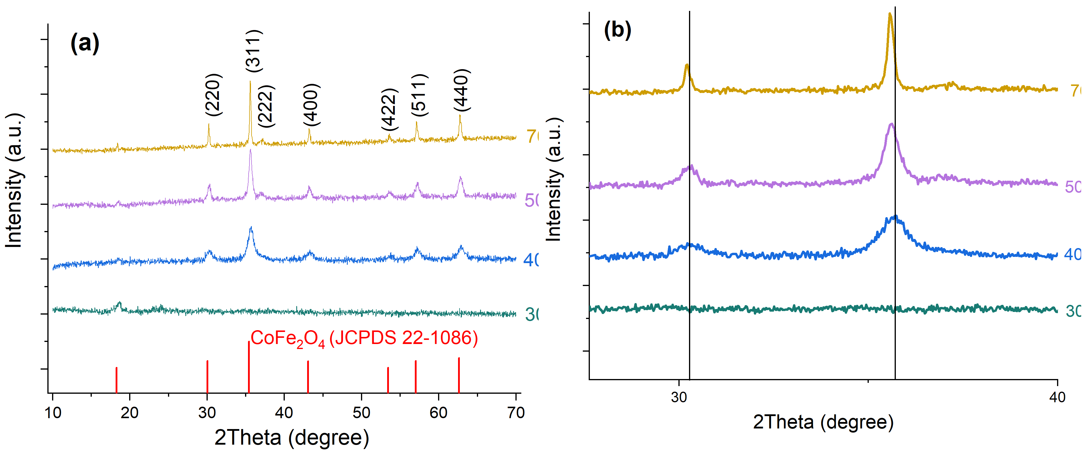
Figure 2.
XRD patterns of CS, CoFe₂O₄, and the CoFe₂O₄/CS nanocomposite.
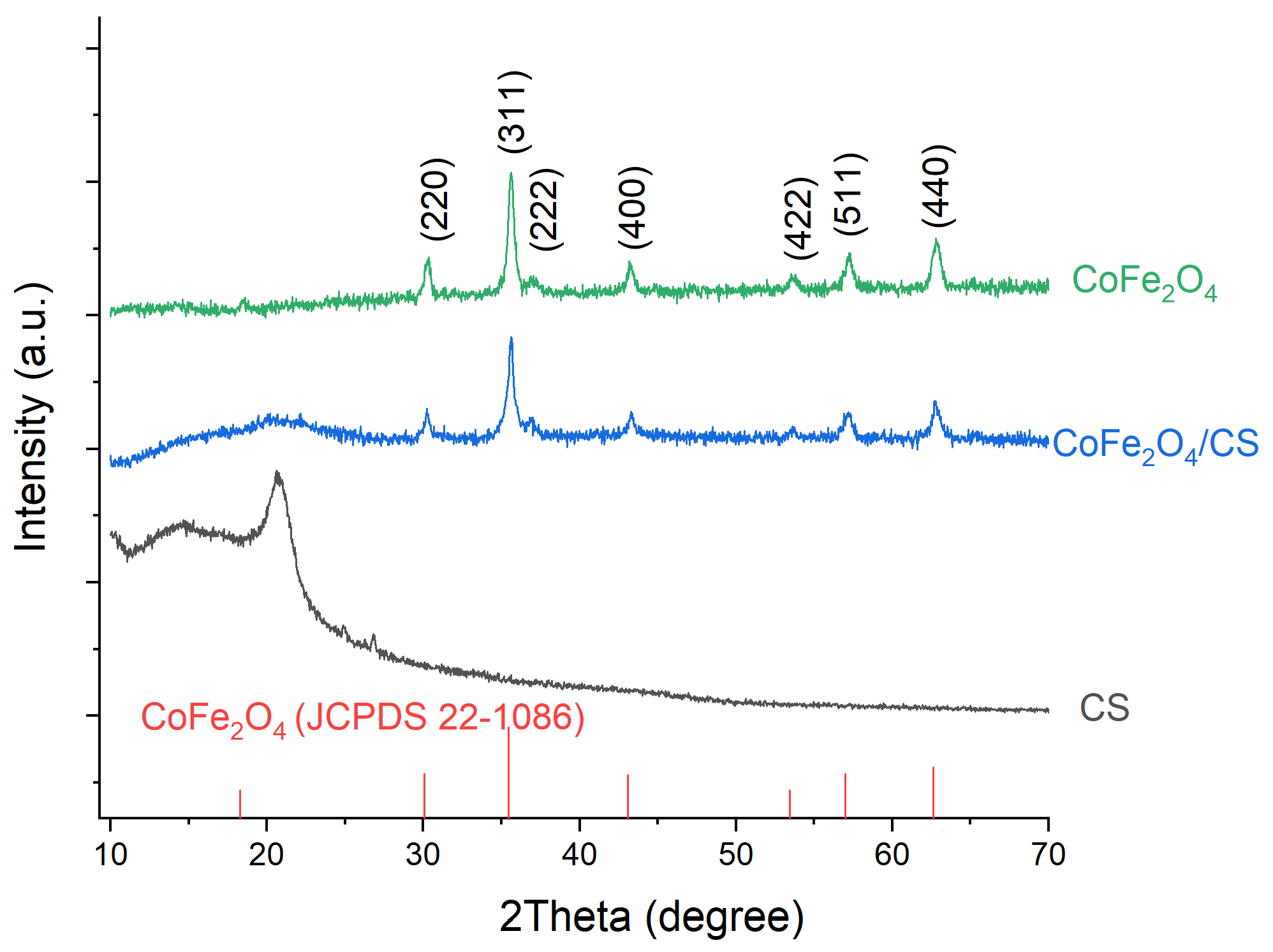
Figure 3.
FTIR spectra of CS, CoFe₂O₄, and the CoFe₂O₄/CS nanocomposite.
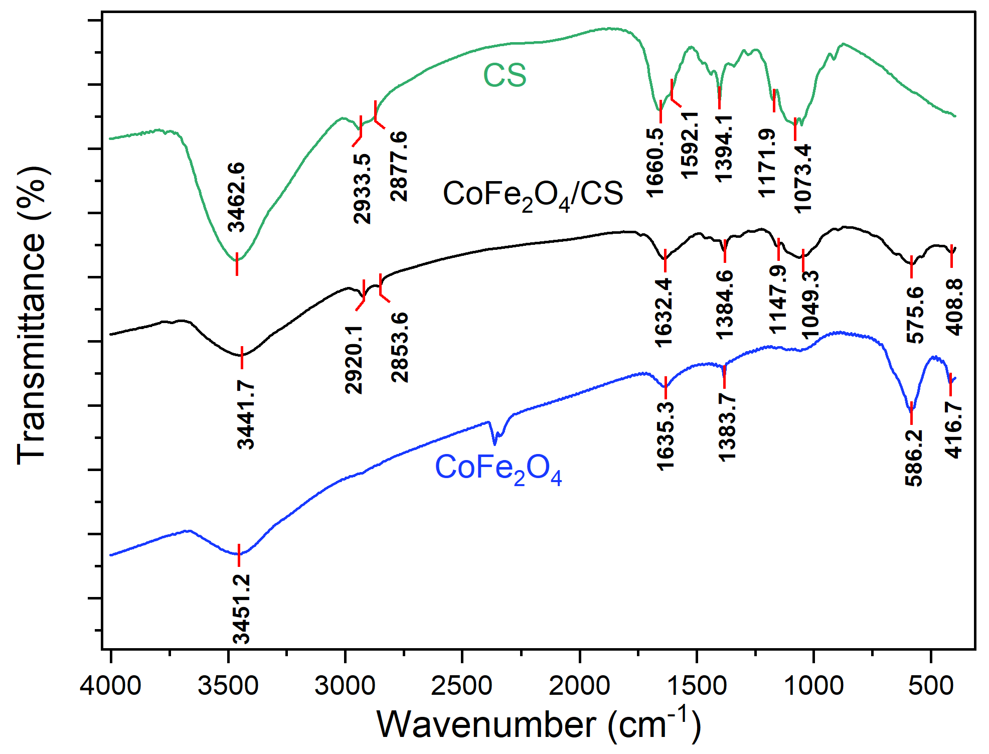
Figure 4.
(a) FESEM image and (b) EDX spectrum of CoFe₂O₄ nanoparticles; (c) FESEM image and (d) EDX spectrum of the CoFe₂O₄/CS nanocomposite. Insets show the corresponding TEM images.
Figure 4.
(a) FESEM image and (b) EDX spectrum of CoFe₂O₄ nanoparticles; (c) FESEM image and (d) EDX spectrum of the CoFe₂O₄/CS nanocomposite. Insets show the corresponding TEM images.
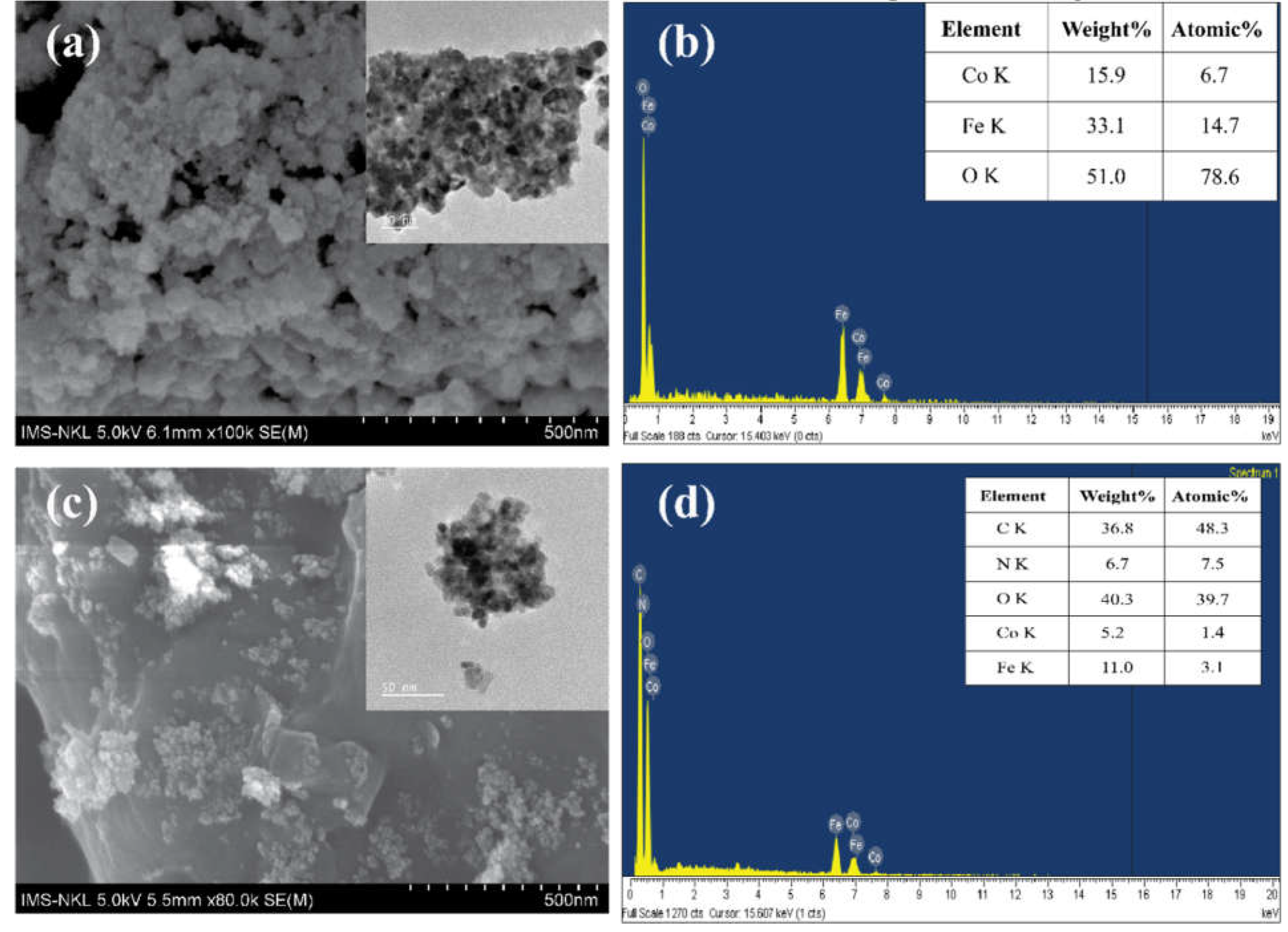
Figure 5.
(a) UV–Vis diffuse-reflectance spectra of CS, CoFe₂O₄, and CoFe₂O₄/CS; (b) Tauc plots used to estimate the optical band gaps of CoFe₂O₄ and CoFe₂O₄/CS.
Figure 5.
(a) UV–Vis diffuse-reflectance spectra of CS, CoFe₂O₄, and CoFe₂O₄/CS; (b) Tauc plots used to estimate the optical band gaps of CoFe₂O₄ and CoFe₂O₄/CS.
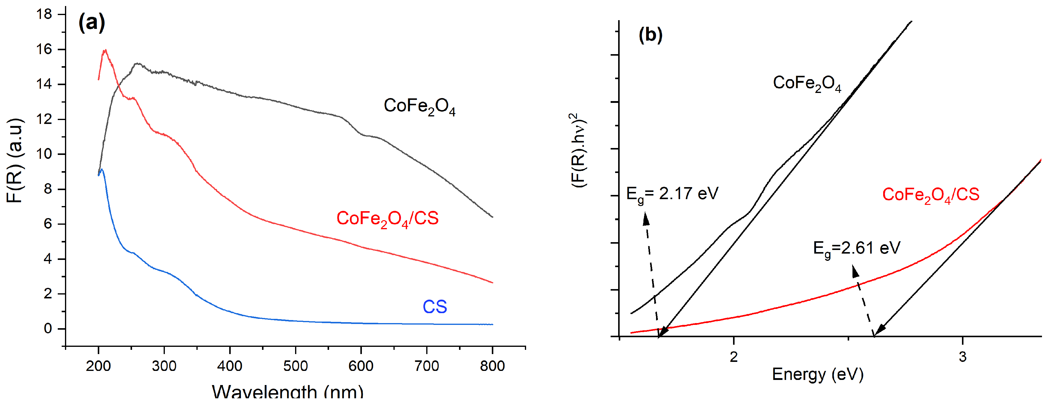
Figure 6.
Photoluminescence spectra of CoFe₂O₄ and CoFe₂O₄/CS.
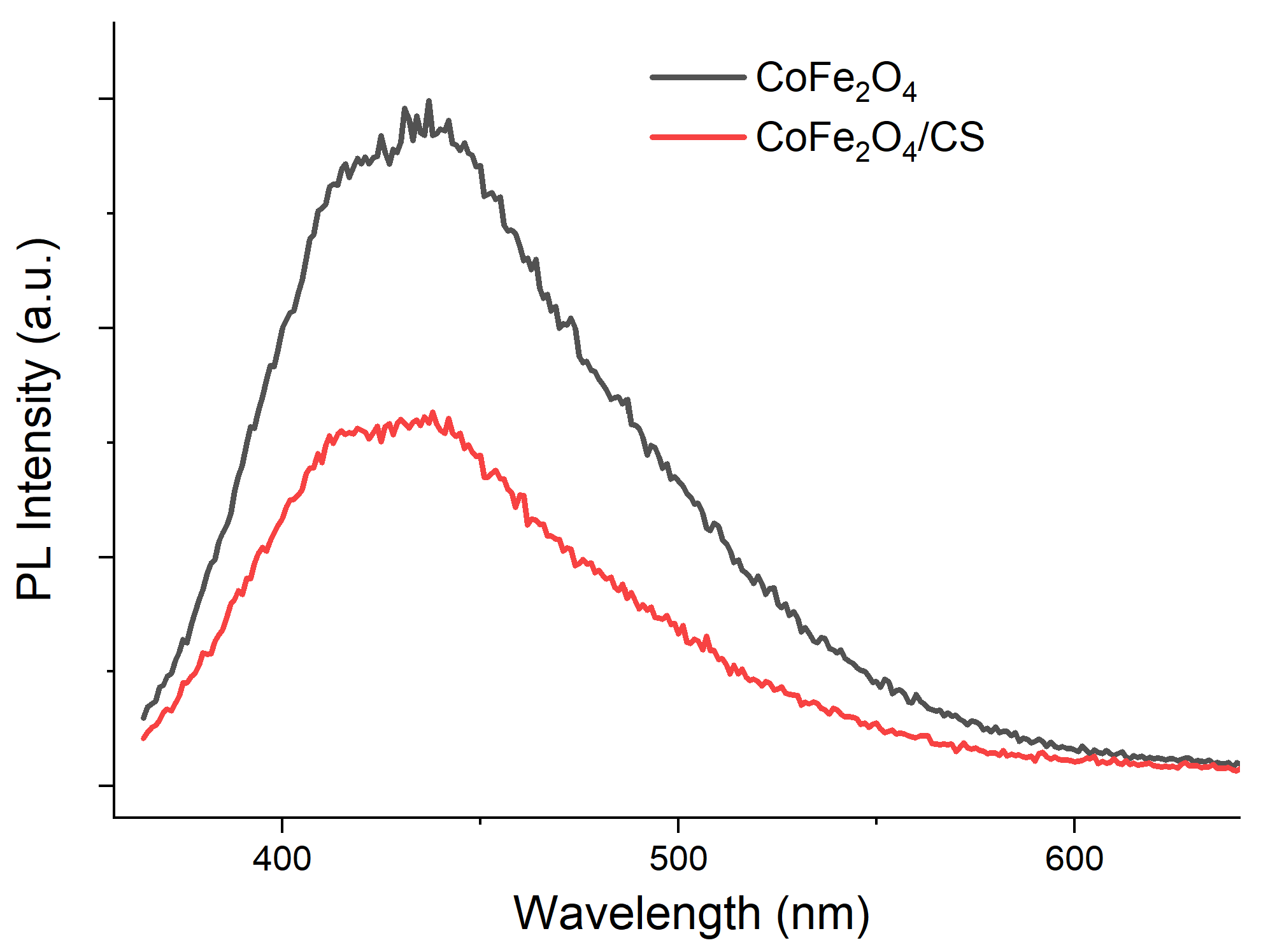
Figure 7.
XPS spectra of CoFe₂O₄/CS: (a) survey spectrum; high-resolution (b) Fe 2p, (c) Co 2p, (d) O 1s, (e) C 1s, and (f) N 1s spectra.
Figure 7.
XPS spectra of CoFe₂O₄/CS: (a) survey spectrum; high-resolution (b) Fe 2p, (c) Co 2p, (d) O 1s, (e) C 1s, and (f) N 1s spectra.
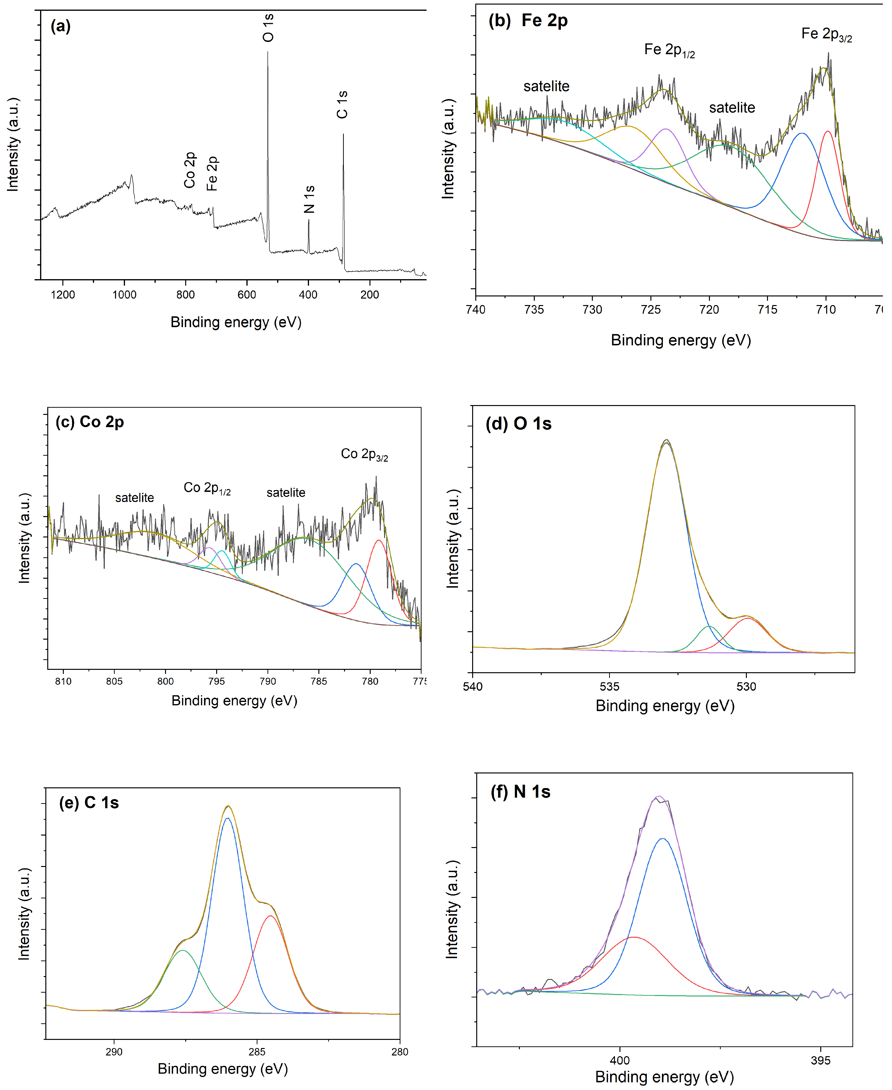
Figure 8.
Nitrogen adsorption–desorption isotherms and BJH pore-size distributions of (a) CoFe₂O₄ and (b) CoFe₂O₄/CS.
Figure 8.
Nitrogen adsorption–desorption isotherms and BJH pore-size distributions of (a) CoFe₂O₄ and (b) CoFe₂O₄/CS.
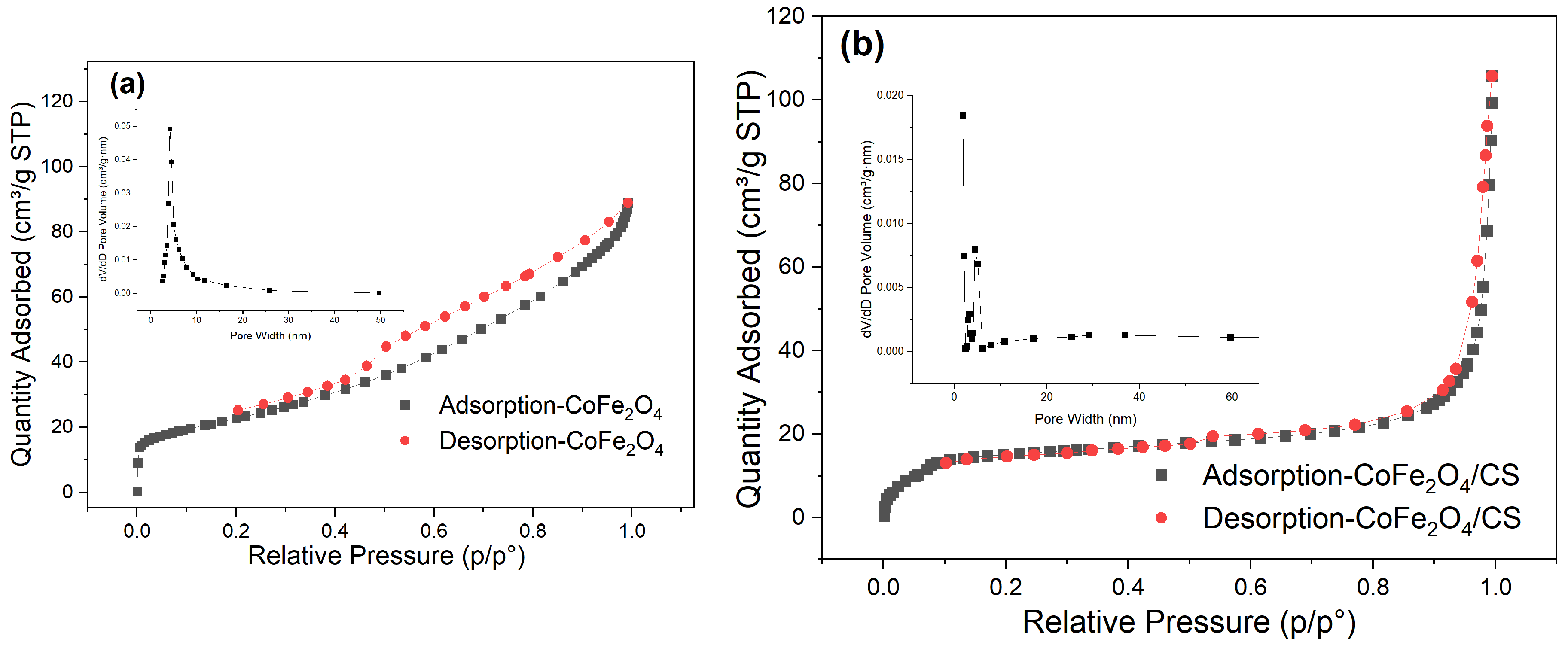
Figure 9.
Room-temperature magnetic hysteresis loops of CoFe₂O₄ and CoFe₂O₄/CS. The inset shows the response of CoFe₂O₄/CS in water to an external magnet.
Figure 9.
Room-temperature magnetic hysteresis loops of CoFe₂O₄ and CoFe₂O₄/CS. The inset shows the response of CoFe₂O₄/CS in water to an external magnet.
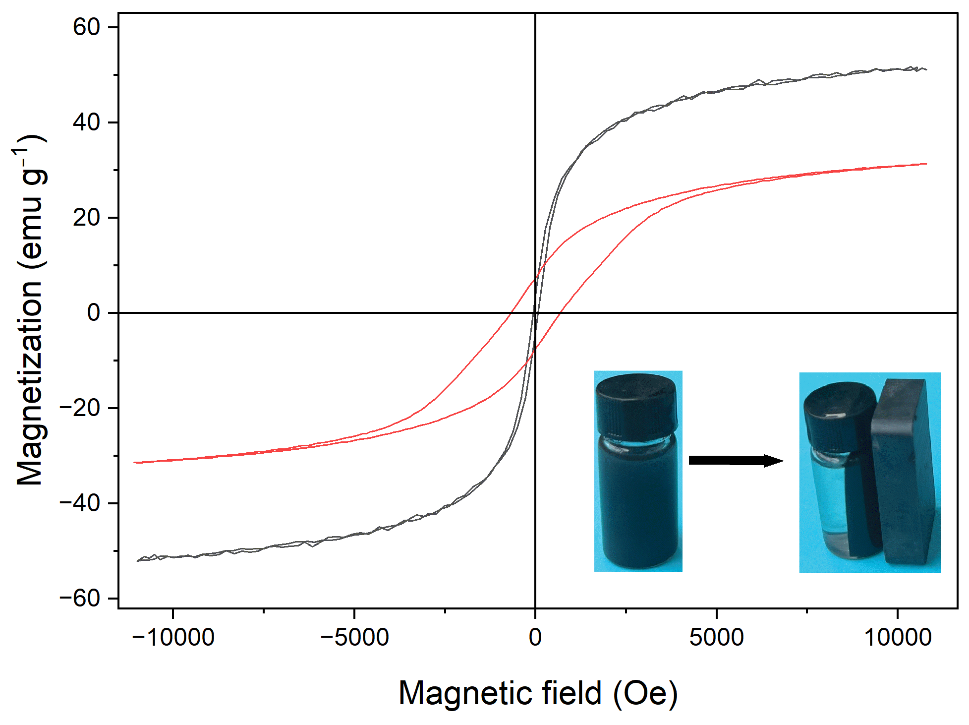
Figure 10.
(a) Time-dependent MB removal under dark and visible-light conditions and (b) pseudo-first-order kinetic plots for CoFe₂O₄ and CoFe₂O₄/CS.
Figure 10.
(a) Time-dependent MB removal under dark and visible-light conditions and (b) pseudo-first-order kinetic plots for CoFe₂O₄ and CoFe₂O₄/CS.
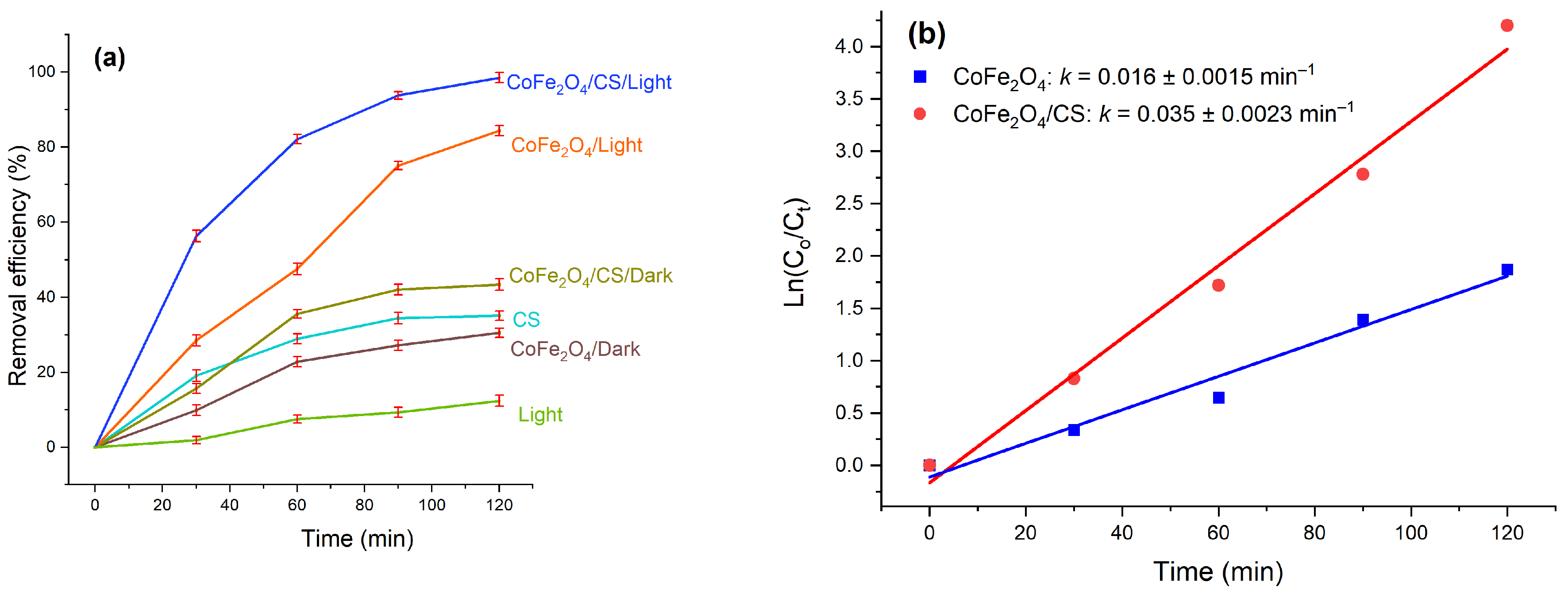
Figure 11.
(a) Effect of initial pH on MB removal and (b) Pseudo-first-order kinetic plots at different pH values.
Figure 11.
(a) Effect of initial pH on MB removal and (b) Pseudo-first-order kinetic plots at different pH values.
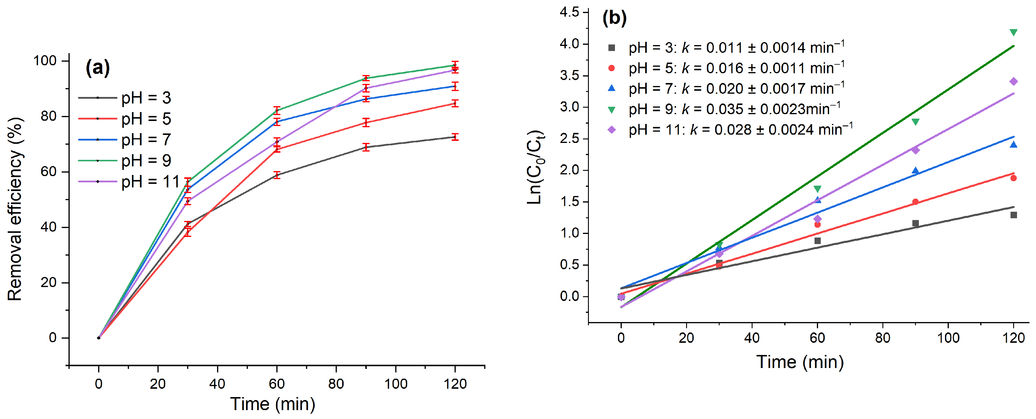
Figure 12.
(a) Effect of initial MB concentration on removal efficiency and (b) pseudo-first-order kinetic plots at different initial concentrations.
Figure 12.
(a) Effect of initial MB concentration on removal efficiency and (b) pseudo-first-order kinetic plots at different initial concentrations.
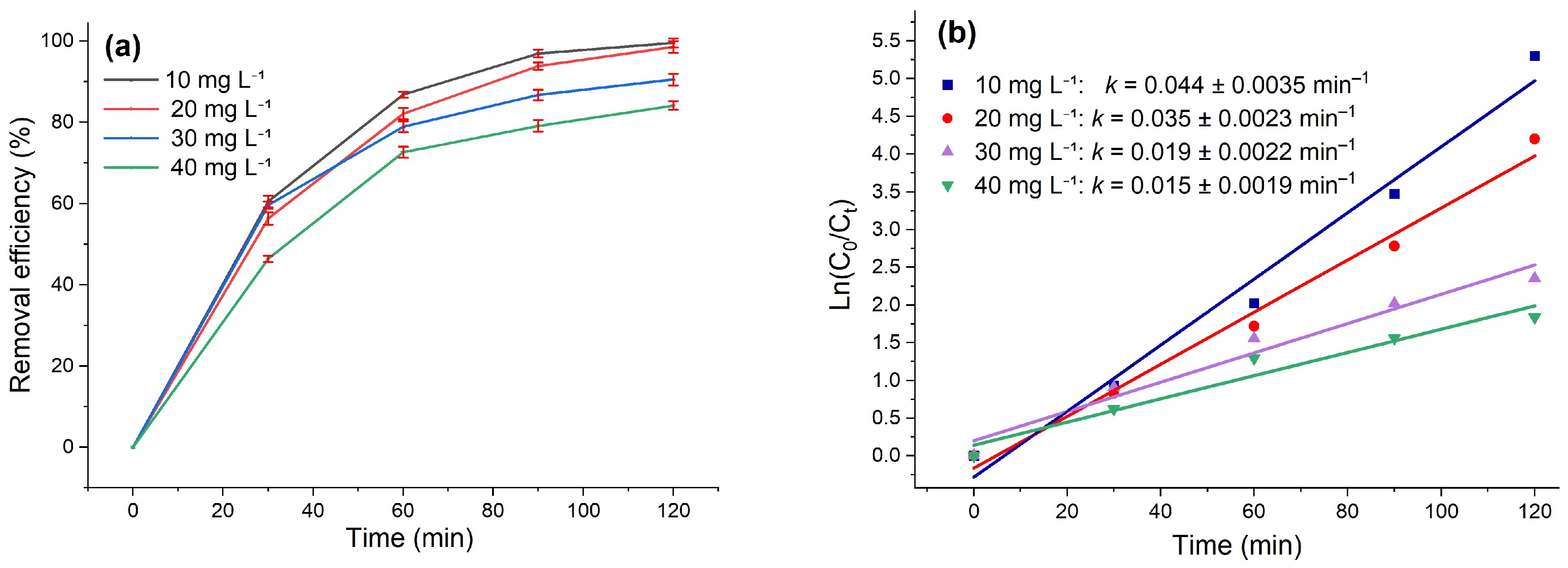
Figure 13.
(a) Effect of CoFe₂O₄/CS dosage on MB removal and (b) pseudo-first-order kinetic plots at different catalyst dosages.
Figure 13.
(a) Effect of CoFe₂O₄/CS dosage on MB removal and (b) pseudo-first-order kinetic plots at different catalyst dosages.
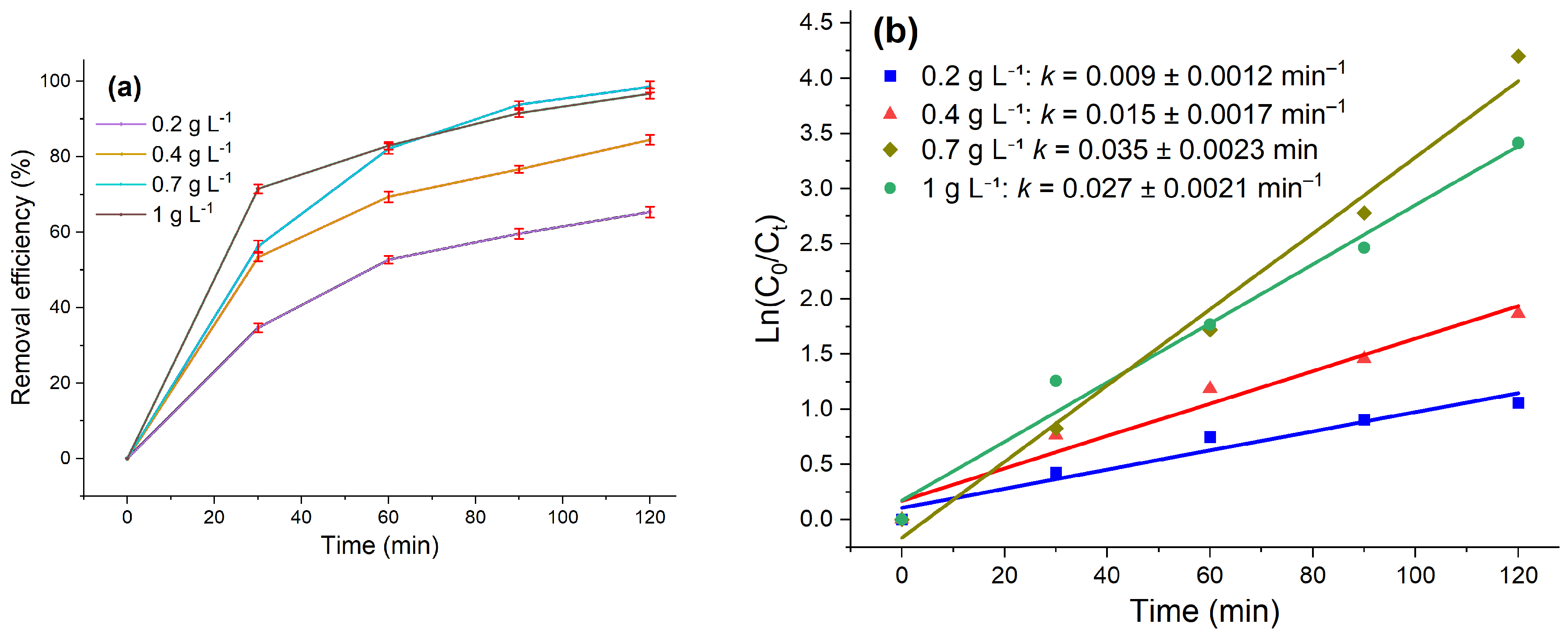
Figure 14.
(a) Reusability of CoFe₂O₄/CS for MB removal over five cycles and (b) effect of EDTA, IPA, BQ, and AgNO₃ scavengers on MB removal.
Figure 14.
(a) Reusability of CoFe₂O₄/CS for MB removal over five cycles and (b) effect of EDTA, IPA, BQ, and AgNO₃ scavengers on MB removal.
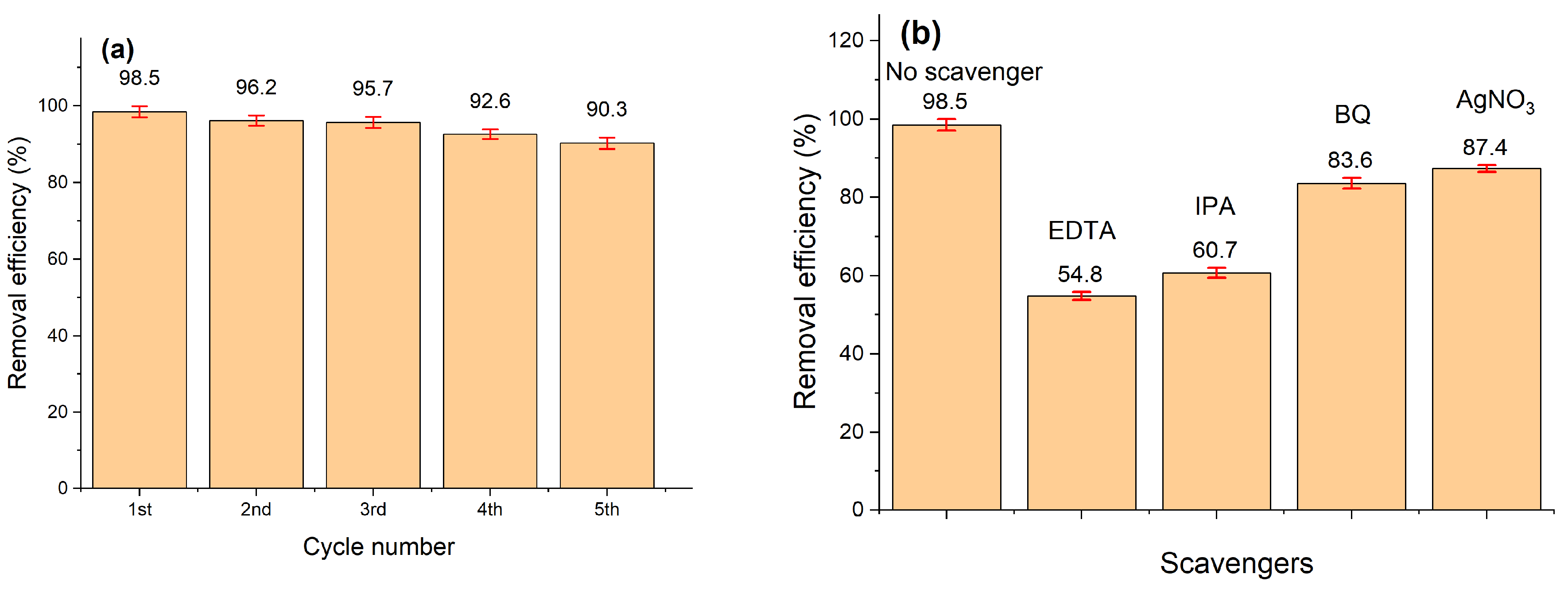
Figure 15.
XRD patterns of the CoFe₂O₄/CS catalyst before and after eight photocatalytic cycles.
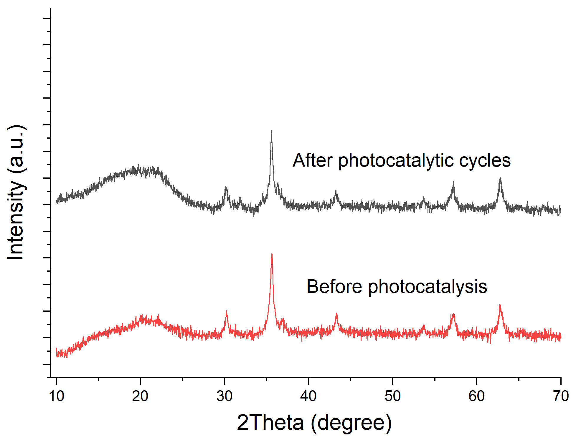
Figure 16.
Proposed adsorption-coupled photocatalytic mechanism for MB removal over CoFe₂O₄/CS.
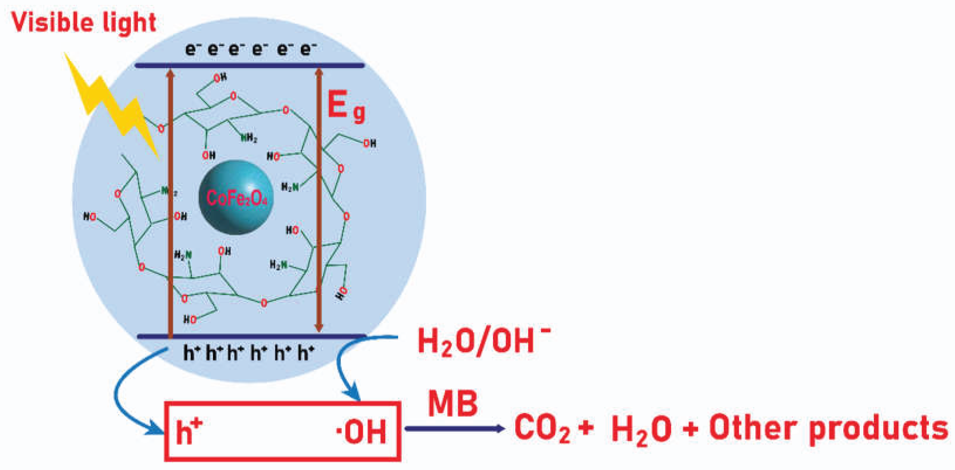

Table 1.
Crystallite size, lattice parameter, unit-cell volume, and X-ray density of CoFe₂O₄ nanoparticles calcined at different temperatures.
Table 1.
Crystallite size, lattice parameter, unit-cell volume, and X-ray density of CoFe₂O₄ nanoparticles calcined at different temperatures.
| Annealing Temperature (°C) | D (nm) | a (Å) | V (ų) | ρ (g cm⁻³) |
| 300 | - | - | - | - |
| 400 | 8.1 | 8.3304 | 578.09 | 5.39 |
| 500 | 10.0 | 8.3349 | 579.03 | 5.38 |
| 700 | 18.9 | 8.3530 | 582.81 | 5.35 |
| Reference CoFe₂O₄ (JCPDS 22-1086) | 8.3920 | 591.01 | 5.27 |
Table 2.
The BET surface area, pore volumes, and pore sizes of CoFe₂O₄, CoFe₂O₄/CS.
| Material |
BET Surface Area (m2 g−1) |
Pore Volume (cm3 g−1) |
Mean Pore Size (nm) |
| CoFe₂O₄ | 81.9 | 0.149 | 6.8 |
| CoFe₂O₄/CS | 51.3 | 0.122 | 7.4 |
Disclaimer/Publisher’s Note: The statements, opinions and data contained in all publications are solely those of the individual author(s) and contributor(s) and not of MDPI and/or the editor(s). MDPI and/or the editor(s) disclaim responsibility for any injury to people or property resulting from any ideas, methods, instructions or products referred to in the content. |
© 2026 by the authors. Licensee MDPI, Basel, Switzerland. This article is an open access article distributed under the terms and conditions of the Creative Commons Attribution (CC BY) license (http://creativecommons.org/licenses/by/4.0/).
Copyright: This open access article is published under a Creative Commons CC BY 4.0 license, which permit the free download, distribution, and reuse, provided that the author and preprint are cited in any reuse.