

Submitted:
04 August 2026
Posted:
05 August 2026
You are already at the latest version
Abstract
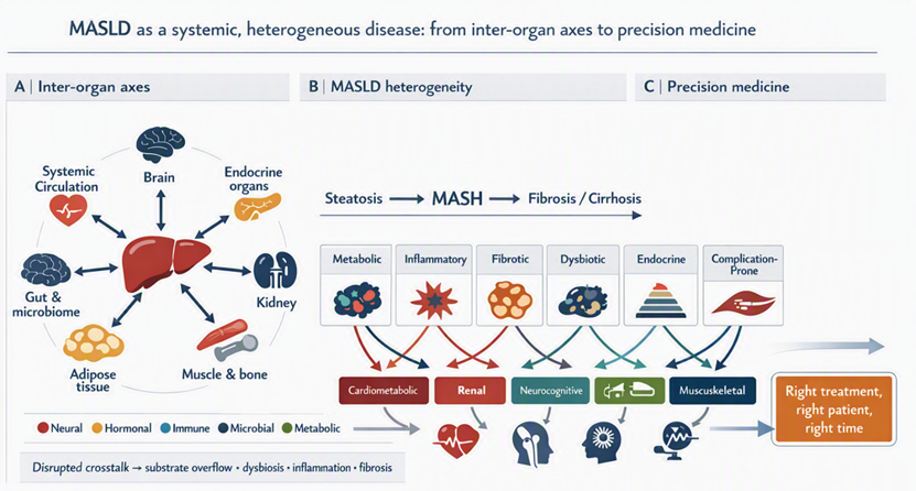
Metabolic dysfunction-associated steatotic liver disease (MASLD) is a systemic disorder shaped by inter-organ crosstalk: dynamic, bidirectional communication through which the liver and endocrine organs, gut, adipose tissue, brain, kidney, skeletal muscle, and bone exchange signals to coordinate metabolism, immunity, and tissue homeostasis. Across these axes, neural circuits, hormones, cytokines, adipokines, hepatokines, myokines, osteokines, bile acids, microbial metabolites, lipids, extracellular vesicles, and microRNAs integrate nutrient handling, insulin action, immunity, mitochondrial function, and tissue remodeling. Perturbation of these networks converts physiological homeostasis into self-reinforcing loops of substrate overflow, endocrine dysregulation, dysbiosis, inflammation, and fibrogenesis, while hepatic dysfunction propagates renal, neurocognitive, cardiometabolic, and musculoskeletal complications. This framework helps explain why individuals with comparable steatosis show divergent trajectories of metabolic dysfunction-associated steatohepatitis (MASH), fibrosis, extrahepatic disease, and treatment response. It also highlights tractable points of intervention, including restoration of adipose buffering, modulation of gut microbial and bile-acid signaling, correction of endocrine drivers, preservation of muscle and bone, and integrated cardio–kidney–liver risk reduction across different disease stages and clinical phenotypes. We argue that precision hepatology should move beyond isolated assessment of liver fat and fibrosis towards multidimensional phenotyping of dominant crosstalk mechanisms. Longitudinal multi-omic studies and trials incorporating outcomes across organs are now required to distinguish causal signals from disease correlates, define clinically actionable endotypes, and test whether targeting one node can restore durable metabolic and functional resilience throughout the interconnected MASLD network, while improving patient-centered outcomes across the disease course.
Keywords:
gut microbiome
; insulin resistance
; bile acids and salts
; extracellular vesicles
; intercellular signaling peptides and proteins
; MASLD
1. Introduction
Metabolic dysfunction-associated steatotic liver disease (MASLD), formerly non-alcoholic fatty liver disease, is a major multisystem disorder defined by hepatic steatosis with cardiometabolic risk. Its spectrum extends from metabolically associated fat accumulation to metabolic dysfunction-associated steatohepatitis (MASH), progressive fibrosis, cirrhosis and hepatocellular carcinoma [10]. Prevalence is increasing worldwide, with substantial geographical variation and particularly high estimates in parts of the Middle East [11]. Outcomes nevertheless extend beyond liver-related events to cardiovascular disease, chronic kidney disease, endocrine dysfunction, neurocognitive impairment and loss of musculoskeletal resilience.
This clinical breadth is incompatible with a model of the liver as an isolated lipid repository. Homeostasis depends on dynamic, bidirectional axes linking the liver with the brain, gut, endocrine organs, adipose tissue, kidney, muscle and bone [1,2,3,4,5,6,7]. Neural circuits, hormones, cytokines, microbial metabolites, bile acids, lipids, microRNAs and extracellular vesicles coordinate energy balance, insulin action, immunity and stress adaptation. When these networks fail, physiological signaling becomes a self-reinforcing driver of substrate overflow, insulin resistance, inflammation, mitochondrial dysfunction and tissue injury [8].
Adipose dysfunction increases fatty-acid delivery and inflammatory signaling; endocrine disturbances alter lipid handling and mitochondrial function; intestinal dysbiosis and barrier failure expose the portal circulation to microbial products and reshape bile-acid signaling; and impaired muscle glucose disposal magnifies substrate excess. Conversely, the steatotic and fibrotic liver releases metabolites, lipoproteins, hepatokines, cytokines and extracellular vesicles that affect distant tissues. These reciprocal loops help explain why similar degrees of steatosis can yield divergent inflammatory activity, fibrosis progression, extrahepatic complications and treatment responses.
A systems perspective has direct clinical implications. It shifts assessment beyond liver fat towards the dominant biological circuits sustaining disease in each patient. Treatment may therefore require restoration of adipose buffering, correction of endocrine drivers, modulation of gut microbial and bile-acid signaling, preservation of muscle and bone, and integrated cardio–kidney–liver risk reduction, alongside liver-directed therapy. In this Review, we synthesize evidence across the endocrine–liver, gut–liver, adipose tissue–liver, liver–brain, liver–kidney and muscle–bone axes. We examine how neural, hormonal, immune, microbial, metabolic and vesicular signals shape disease initiation, progression and therapeutic responsiveness, and identify tractable opportunities for mechanism-based stratification and intervention.
2. Methods
This narrative review was designed to synthesize current concepts of inter-organ communication in metabolic dysfunction-associated steatotic liver disease (MASLD), with an emphasis on the endocrine–liver, gut–liver, adipose tissue–liver, liver–brain, liver–kidney, and MASLD–muscle–bone axes. MASLD was considered according to the current nomenclature as hepatic steatosis occurring in the presence of at least one cardiometabolic risk factor and in the absence of harmful alcohol consumption or other dominant causes of steatosis. The review was conceived as a mechanistic and translational synthesis rather than as a systematic review or meta-analysis, because the available literature spans heterogeneous experimental, epidemiological, clinical, multi-omic, and therapeutic studies across several organ systems. Relevant articles were identified through targeted searches of PubMed/MEDLINE, Web of Science, Scopus, and Google Scholar, supplemented by manual screening of reference lists from key reviews, guidelines, mechanistic studies, and landmark clinical trials.
Searches combined terms related to MASLD and its previous nomenclature with terms describing organ crosstalk and individual axes, including “metabolic dysfunction-associated steatotic liver disease,” “MASLD,” “NAFLD,” “MASH,” “organ crosstalk,” “inter-organ communication,” “endocrine–liver axis,” “gut–liver axis,” “adipose tissue–liver axis,” “liver–brain axis,” “liver–kidney axis,” “sarcopenia,” “myosteatosis,” “osteoporosis,” “extracellular vesicles,” “exosomes,” “microRNA,” “bile acids,” “hepatokines,” “myokines,” “adipokines,” and “gut microbiota.” Priority was given to articles published in English, with an emphasis on recent publications, international guidelines, high-impact reviews, meta-analyses, prospective cohort studies, translational studies, and mechanistic experimental work relevant to MASLD pathogenesis, progression, heterogeneity, and treatment responsiveness. Older seminal studies were included when they provided foundational concepts for functional organ axes, neuroendocrine regulation, gut-derived signaling, adipose inflammation, insulin resistance, hepatic encephalopathy, chronic kidney disease (CKD), sarcopenia, bone metabolism, or extracellular-vesicle biology. Studies were considered eligible when they addressed bidirectional or functionally relevant communication between the liver and at least one extrahepatic organ system, or when they clarified molecular mediators such as hormones, cytokines, adipokines, hepatokines, myokines, osteokines, bile acids, microbial metabolites, extracellular vesicles, microRNAs, lipid intermediates, or neural pathways. Articles focused exclusively on isolated hepatic lipid metabolism without relevance to systemic signaling, extrahepatic disease, or therapeutic implications were considered of lower priority.
The evidence was organized into thematic sections reflecting major biological axes rather than according to study design, because MASLD is increasingly understood as a multisystem disorder embedded within dynamic endocrine, metabolic, immune, neural, microbial, renal, musculoskeletal, and adipose networks. For each axis, we extracted and synthesized information on anatomical and physiological foundations, principal molecular mediators, mechanisms contributing to steatosis, inflammation, fibrogenesis, extrahepatic complications, clinical heterogeneity, and therapeutic opportunities. Particular attention was paid to reciprocal signaling loops, because several axes operate bidirectionally, with the liver acting both as a recipient of pathogenic extrahepatic signals and as an active endocrine, metabolic, inflammatory, and hemodynamic organ influencing distant tissues. Where available, clinical evidence was integrated with mechanistic data to distinguish associations from plausible causal pathways and to identify areas in which experimental evidence remains stronger than human validation. Because terminology has evolved from NAFLD and NASH to MASLD and MASH, older studies using previous nomenclature were retained when their populations and metabolic phenotypes were conceptually applicable to MASLD, while acknowledging that definitions are not always fully interchangeable. The final synthesis was developed through iterative discussion among the authors, with the aim of producing an integrative framework that connects organ-specific mechanisms with precision risk stratification and mechanism-based therapeutic development in MASLD.
3. The Endocrine-Liver Axis in MASLD
The liver is both a major target of endocrine signals and a central organ for hormone synthesis, biotransformation and clearance [12]. Accordingly, disruption of neuroendocrine and peripheral glandular circuits can establish bidirectional feedback loops that intensify lipid accumulation, lipotoxicity and inflammation [13].
MASLD is therefore not simply a consequence of caloric excess and insulin resistance [14], but a multisystem disease embedded within endocrine and metabolic networks [15,16,17]. This section considers four principal axes: hypothalamus–pituitary, pancreas–liver, thyroid–liver and gonad–liver.
3.1. The Hypothalamus-Pituitary Axis
The hypothalamus integrates neural and hormonal cues to coordinate appetite, energy expenditure and peripheral metabolism [18]. Disruption of hypothalamic–pituitary signaling is an established experimental cause of liver injury, and progressive MASLD/MASH is observed after neurosurgery for craniopharyngioma [19].
3.1.1. Growth Hormone Deficiency and Steatotic Liver Disease
Growth hormone (GH) induces hepatic insulin-like growth factor-1 (IGF-1), mainly through Janus kinase 2–STAT5 signaling [20]. Together, GH and IGF-1 promote mitochondrial β-oxidation, restrain DNL, improve insulin sensitivity and induce hepatic stellate-cell senescence, thereby limiting fibrogenesis [20]. The relationship between this axis and MASLD is bidirectional.
A meta-analysis of 18 studies comprising 20,520 middle-aged participants found inverse associations between circulating GH or IGF-1 and both the presence and severity of MASLD [21].
In adults with GH deficiency or panhypopituitarism, loss of this hepatoprotective axis is associated with rapid progression from steatosis to advanced MASH. Low IGF-1 might also facilitate fibrosis through direct effects on stellate cells [22]. Although evidence remains limited, recombinant GH or GH-releasing hormone analogues can reduce liver fat and might slow fibrosis [22].
3.1.2. Prolactin and Adrenocorticotropic Alterations
Prolactin (PRL), secreted by the anterior pituitary, is emerging as a regulator of metabolic homeostasis and liver health [23].
Very high (>100 µg/L) and very low (<7 µg/L) PRL concentrations are associated with adverse outcomes, whereas moderately elevated concentrations within or just above the physiological range—termed HomeoFIT-PRL—seem metabolically favourable. In experimental models and humans, HomeoFIT-PRL improves insulin resistance, glucose tolerance, adipose hypertrophy and SLD through coordinated actions in the pancreas, liver, adipose tissue and hypothalamus [24].
PRL also activates hypothalamic proopiomelanocortin neurons, increasing hepatic sympathetic outflow and suppressing fatty acid synthase (FAS)-dependent lipogenesis [25]. This PRL-dependent brain–liver circuit is a potential therapeutic target in MASLD.
By contrast, hypothalamic–pituitary–adrenal dysregulation increases adrenocorticotropic hormone and cortisol exposure. Chronic subclinical hypercortisolism promotes visceral adiposity and free-fatty-acid delivery to the liver, thereby increasing triglyceride synthesis [26].
In transgenic mice, corticosterone-induced expression of hepatic lipid-metabolism proteins contributes to high-fat-diet-induced MASLD [27].
In humans, MASLD is more prevalent in Cushing’s syndrome than in controls, with risk determined mainly by metabolic burden, disease duration and aetiology [28,29]. SLD may regress only partly after remission, although fibrosis risk declines most clearly in patients with high baseline fibrosis. These data support systematic liver assessment in Cushing’s syndrome, particularly after prolonged disease exposure or in patients with substantial metabolic risk [28].
3.2. The Pancreas-Liver Axis
Pancreas–liver crosstalk is central to MASLD: pancreatic insulin regulates systemic glucose and lipid metabolism, whereas the liver determines insulin sensitivity and clearance.
3.2.1. Hyperinsulinemia and De Novo Lipogenesis
Postprandial insulin normally suppresses gluconeogenesis and adipose lipolysis. In MASLD, diacylglycerols and ceramides impair insulin receptor substrate-1 signaling, producing hepatic insulin resistance [30]. However, lipogenic insulin signaling is relatively preserved; hyperinsulinaemia activates sterol regulatory element-binding protein 1c (SREBP-1c) and carbohydrate response element-binding protein (ChREBP) [31], increasing DNL and hepatic triglyceride accumulation [32].
3.2.2. Defective Insulin Clearance via Carcinoembryonic Antigen-Related Cell Adhesion Molecule 1
Hepatic insulin clearance depends partly on carcinoembryonic antigen-related cell adhesion molecule 1 (CEACAM1), which mediates internalization and degradation of the insulin–receptor complex. Nutrient excess suppresses CEACAM1, sustaining hyperinsulinaemia, systemic insulin resistance, lipid flux and inflammatory signaling [33].
3.2.3. The Mutual and Bidirectional Relationship Between Type 2 Diabetes and MASLD
T2D is a major determinant of adverse liver outcomes: more than 70% of affected individuals have MASLD and up to 30% develop MASH, with greater inflammation and fibrosis. Conversely, MASLD impairs glycaemic control and accelerates pancreatic β-cell failure [34].
T2D independently increases the risk of liver-stiffness progression and liver-related events in MASLD. Adequate glycaemic control attenuates stiffness progression, although clear effects on regression or liver-related events remain unproven [35].
3.3. The Thyroid-Liver Axis
The thyroid–liver axis integrates thyroid hormone signaling with hepatic lipid handling, mitochondrial function and cholesterol metabolism [36].
The liver is both a target and a component of the hypothalamic–pituitary–thyroid axis: it produces thyroid hormone-binding proteins and expresses deiodinases that convert thyroxine (T4) to triiodothyronine (T3), thereby shaping local and systemic hormone availability [37].
3.3.1. Thyroid Hormone Receptor Beta Signaling
Most hepatic effects of T3 are mediated by thyroid hormone receptor-β (THR-β), the predominant hepatocyte isoform. THR-β promotes mitochondrial biogenesis and fatty-acid oxidation, partly through carnitine palmitoyltransferase 1A, and supports autophagic disposal of lipotoxic substrates [38]. It also increases low-density lipoprotein (LDL) receptor expression, linking thyroid signaling to cholesterol clearance [39].
3.3.2. Hypothyroidism and Direct TSH Pathogenicity
Both overt and subclinical hypothyroidism are enriched in MASLD, supporting a clinically relevant association between impaired thyroid homeostasis and metabolic liver disease [40,41].
Reduced thyroid hormone availability is accompanied by lower hepatic THR-β expression, particularly in advanced steatosis. Impaired receptor signaling reduces mitochondrial fatty-acid oxidation and favours accumulation of unesterified fatty acids, reinforcing hepatocellular lipotoxicity [42].
Hepatocytes also express functional thyroid-stimulating hormone receptors (TSH-R) [43]. Elevated TSH can therefore act directly on the liver, independently of thyroid hormone availability [44]. TSH-R engagement activates cAMP signaling, increases SREBP-1c activity and suppresses AMP-activated protein kinase (AMPK), shifting metabolism towards lipogenesis [45].
These mechanisms motivated the development of liver-directed selective THR-β agonists, designed to reduce hepatic lipid accumulation and improve MASH histology while limiting extrahepatic T3-related effects [46].
Because MASH can progress to cirrhosis and hepatocellular carcinoma (HCC), preclinical evidence that resmetirom suppresses midkine–LDL receptor-related protein 1-mediated immunosuppressive crosstalk provides a rationale for investigating whether this approved MASH therapy can also limit hepatocarcinogenesis [36].
3.4. The Gonad-Liver Axis
Gonadal signaling contributes to MASLD pathobiology and its marked sexual dimorphism [47]. Premenopausal women are relatively protected from severe steatosis and fibrosis compared with age-matched men, but this advantage diminishes after menopause, consistent with loss of estrogen-dependent hepatometabolic protection [48].
The high prevalence and rapid progression of liver disease in women with polyendocrine metabolic ovary syndrome (PMOS), previously termed polycystic ovary syndrome (PCOS), further implicate sex hormones in liver health [49].
3.4.1. The Ovary–Liver Axis in MASLD
In women, ovarian endocrine function modifies MASLD risk through effects on adipose distribution, glucose–lipid homeostasis and hepatic inflammation. This ovary–liver axis is driven mainly by estradiol acting through nuclear estrogen receptors and membrane-initiated pathways [50].
Endogenous estrogens, particularly 17β-estradiol, exert antisteatotic, anti-inflammatory and antifibrotic actions, principally through hepatic ER-α. ER-α suppresses lipogenic genes including SREBP1c, FASN and acetyl-CoA carboxylase (ACC), whereas ER-α/ER-β programs promote lipid oxidation. G protein-coupled estrogen receptor signaling also regulates glucose uptake, mitochondrial function and inflammation through rapid phosphoinositide 3-kinase (PI3K)–MAPK pathways [51].
Estrogen signaling preserves mitochondrial integrity, limits reactive oxygen species and restrains Kupffer-cell activation. Menopause or ovarian dysfunction therefore removes an important hepatometabolic brake, favouring triglyceride accumulation, cytokine activation, steatohepatitis and collagen deposition [50,51].
3.4.2. Androgenic Dichotomy: Testosterone and PMOS
Testosterone exerts sex- and context-dependent effects. In men, physiological androgen receptor signaling is generally hepatoprotective, whereas hypogonadism promotes insulin resistance, visceral adiposity and MASLD. Testosterone suppresses SREBP-1c-, FAS- and ACC-dependent lipogenesis; promotes peroxisome proliferator-activated receptor-α (PPARα), CPT1 and AMPK-dependent oxidation; and supports VLDL export through microsomal triglyceride transfer protein (MTTP) and apolipoprotein B. It may also activate nuclear factor erythroid 2-related factor 2 (Nrf2), limit nuclear factor kappa-light-chain-enhancer of activated B cells (NF-κB)/TLR4 signaling and Kupffer-cell recruitment, and inhibit transforming growth factor-β (TGF-β)–SMAD-dependent stellate-cell activation [51].
Thus, physiological testosterone can restrain steatosis, inflammation and fibrosis, whereas androgen deficiency and supraphysiological anabolic-androgenic steroid exposure may promote liver injury [51].
In women, the association is reversed: functional hyperandrogenism in PMOS promotes MASLD independently of body mass index by activating hepatic androgen receptor signaling, increasing fatty-acid synthesis, impairing insulin clearance and accelerating steatosis [49].
3.5. Conclusion
MASLD progression reflects the convergence of hepatic lipid stress with systemic endocrine dysfunction (Table 1).
Disruption of hypothalamic–pituitary, pancreas–liver, thyroid–liver and gonad–liver signaling amplifies steatosis, inflammation and fibrosis, reinforcing the view of MASLD as a multisystem disorder. This endocrine framework supports mechanism-based interventions, including GH replacement in selected deficiency states, selective THR-β agonism and context-specific modulation of sex-hormone signaling. Integrating neuroendocrine phenotypes with liver staging may improve risk stratification and treatment selection.
4. Gut-Liver Axis
The gut–liver axis constitutes one of the most anatomically direct and biologically consequential inter-organ communication systems in MASLD. Through portal circulation, the liver is continuously exposed to nutrients, microbial products, bile acids, and immunomodulatory metabolites generated within the intestinal lumen [54]. Under physiological conditions, this arrangement supports metabolic flexibility, immune tolerance, and enterohepatic bile acid recycling, whereas disruption of intestinal barrier integrity and microbial community structure can promote steatohepatitis, fibrosis, and cirrhosis [55]. In metabolic dysfunction, diet-induced dysbiosis, impaired epithelial barrier integrity, and altered microbial metabolism convert this homeostatic circuit into a pathogenic amplifier of steatosis, inflammation, and fibrogenesis [54,56].
4.1. Disruption of the Intestinal Barrier: Microbial Danger Signals in MASLD
A central feature of this transition is disruption of the intestinal barrier: high-fat, high-fructose, and ultra-processed dietary patterns reduce microbial diversity, deplete barrier-supporting commensals, and weaken tight-junction architecture, thereby increasing intestinal permeability [54,55]. This state, often termed metabolic endotoxemia, facilitates portal translocation of pathogen-associated molecular patterns (PAMPs), including lipopolysaccharide (LPS), peptidoglycan, and bacterial DNA, linking barrier failure to hepatic inflammatory activation [54,57]. In the liver, these signals are sensed by Kupffer cells, hepatic stellate cells, sinusoidal endothelial cells, and hepatocytes through pattern-recognition receptors such as Toll-like receptor 4 (TLR4), Toll-like receptor 9 (TLR9), and nucleotide-binding oligomerization domain-like receptors [58]. The resulting activation of Myeloid differentiation primary response 88 (MyD88)–NF-κB, interferon-regulatory, and inflammasome pathways promotes production of tumor necrosis factor (TNF), interleukin-1β (IL-1β), interleukin-6 (IL-6), and chemokines that recruit inflammatory monocytes and neutrophils, thereby linking intestinal barrier failure to steatohepatitis and fibrotic progression [54,55].
4.2. From Dysbiosis to Hepatotoxic Signaling
Dysbiosis does not act solely by increasing microbial translocation; it also reshapes the metabolic output of the intestinal ecosystem [54,55]. MASLD and MASH have been associated with expansion of Proteobacteria and ethanol-producing taxa, relative depletion of butyrate-producing bacteria, and altered Bacteroidetes–Firmicutes relationships, although taxonomic signatures vary across geography, diet, medication exposure, and disease stage [55,59]. A recurring functional pattern is nevertheless emerging: loss of microbial functions that preserve mucosal integrity and expansion of pathways that generate hepatotoxic or pro-inflammatory metabolites [54]. Endogenous ethanol and acetaldehyde may aggravate oxidative stress and hepatocellular injury [60], whereas trimethylamine-derived trimethylamine-N-oxide (TMAO), branched-chain amino acid (BCAA) derivatives, and imidazole propionate can impair insulin signaling and reinforce hepatic lipid accumulation [55,61]. Conversely, short-chain fatty acids (SCFAs), particularly butyrate and propionate, can support epithelial energy metabolism, regulatory T-cell differentiation, glucagon-like peptide-1 secretion, and AMPK activity, although their effects remain dose-, compartment- and context-dependent [54,55].
4.3. Bile Acids Link Dysbiosis with Hepatic Injury
Bile acids provide a second major communication layer between gut microbes and hepatic metabolism. Primary bile acids synthesized in hepatocytes are modified by intestinal bacteria into secondary bile acids, which signal through nuclear and membrane receptors including farnesoid X receptor (FXR) and Takeda G protein-coupled receptor 5 (TGR5), thereby linking the liver, gut microbiota, and immune system [54,62]. In the healthy state, intestinal FXR–fibroblast growth factor 19 signaling restrains hepatic bile acid synthesis, modulates gluconeogenesis and lipogenesis, and supports intestinal barrier integrity; recent experimental evidence further indicates that microbiota-dependent bile salt hydrolase activity can activate the intestinal FXR–fibroblast growth factor (FGF)19 axis and attenuate hepatic lipid accumulation [63]. In MASLD, microbial remodeling of the bile acid pool may blunt FXR activity, alter TGR5-dependent energy expenditure and immune signaling, and increase exposure to hydrophobic secondary bile acids such as deoxycholic acid [54,55]. These shifts can promote hepatocyte stress, stellate-cell activation, and a pro-fibrogenic niche, particularly when combined with lipotoxicity and innate immune activation. Conversely, ileal secondary bile acid accumulation can activate TGR5–mTOR–oxidative phosphorylation signaling in CD8+ T cells, promote ileitis, impair enterohepatic circulation, and suppress hepatic FXR activity, thereby exacerbating MASLD progression [64].
4.4. Role of the Gut–Liver Axis Also in the Transition from Metabolic Stress to Fibrosis
Repeated exposure to LPS and other microbial ligands primes Kupffer cells and recruited macrophages, increasing transforming growth factor-β, platelet-derived growth factor and reactive oxygen species; this portal delivery of PAMPs and microbial metabolites is increasingly viewed as a fibrogenic arm of the gut–liver axis [54,55]. These mediators activate hepatic stellate cells and portal fibroblasts, enhancing extracellular matrix deposition and tissue stiffness, consistent with evidence that gut-derived LPS and altered microbial metabolites can amplify hepatic inflammatory and fibrogenic cascades [54,65]. In parallel, dysregulated bile acid signaling, microbial ethanol production, and reduced indole-mediated aryl hydrocarbon receptor activation may weaken epithelial repair mechanisms and perpetuate inflammatory exposure [60,64,66]. Thus, fibrosis in MASLD can be interpreted not only as the end product of hepatocellular lipotoxicity but also as the consequence of sustained portal delivery of microbial and metabolic danger signals [54,56].
4.5. Clinical Relevance
4.5.1. MASLD Heterogeneity
Gut-derived signals may help explain heterogeneity in MASLD progression, because microbial composition, barrier integrity, and metabolite output differ across patients and can influence inflammatory activity, fibrosis trajectory, and therapeutic responsiveness [54,55]. Individuals with similar hepatic fat content can therefore differ markedly in necroinflammatory activity and fibrotic progression, consistent with multi-omic evidence that gut barrier dysfunction and host–microbiome interactions stratify MASLD severity in metabolically high-risk populations [57]. Microbiome composition, intestinal permeability, bile acid profiles, and circulating markers of microbial exposure, such as LPS-binding protein and soluble CD14, may therefore complement liver-centered biomarkers. Multi-omics approaches integrating metagenomics, metabolomics, transcriptomics and non-invasive fibrosis assessment are beginning to define microbial-host endotypes and MASLD subtypes, although causality remains difficult to establish in humans owing to confounding by diet, drugs, obesity, diabetes, and genetic background [56,67,68].
4.5.2. Therapeutics
Dietary patterns rich in fiber and minimally processed plant-derived foods can enrich short-chain fatty acid-producing bacteria, reduce endotoxemia, and improve insulin sensitivity [54,55]. Probiotics, synbiotics, and postbiotics have shown encouraging effects on aminotransferases, steatosis indices, and inflammatory markers, but strain specificity, treatment duration, and patient selection remain unresolved [69,70]. Fecal microbiota transplantation provides proof of concept that microbial ecosystems can be remodeled, yet its role in MASLD remains constrained by donor variability, durability, safety, and mixed trial results [71,72]. Pharmacological modulation of bile acid signaling, intestinal FXR activity, incretin pathways, and microbial metabolite production may provide more controllable approaches, particularly when aligned with biomarker-defined endotypes [54,63,73].
4.5.3. Conclusions
Conceptually, the gut–liver axis shifts MASLD pathogenesis from a hepatocentric model to an ecological model in which host metabolism, diet, microbes, and mucosal immunity operate as an integrated disease network. The intestine supplies both injurious signals and protective metabolites, whereas the liver shapes the intestinal environment through bile acid secretion, immune mediators, and systemic metabolic regulation. Dissecting this reciprocal circuitry will be essential for moving beyond broad microbiome associations toward causal, stage-specific, and therapeutically tractable mechanisms. In this respect, gut-directed strategies are unlikely to replace liver-targeted or cardiometabolic therapies, but they may become important adjuncts for reducing inflammatory tone, limiting fibrogenic progression, and personalizing treatment in MASLD. Figure 1 illustrates the pathophysiology of the gut–liver axis.
5. Adipose Tissue-Liver Axis
5.1. Adipose Failure and Hepatic Lipid Overflow
The adipose tissue–liver axis is a dominant metabolic circuit in MASLD because white adipose tissue determines the magnitude, timing, and inflammatory context of lipid delivery to the liver [73,74]. In healthy energy surplus, subcutaneous adipose tissue buffers excess nutrients through adipocyte hyperplasia, triglyceride storage, and insulin-sensitive suppression of lipolysis. When this storage compartment becomes hypertrophic, hypoxic, and inflamed, its buffering capacity fails, and non-esterified fatty acids are released chronically into the circulation. Adipose-derived fatty acids are a major source of hepatic triglyceride stores in MASLD [75]. The liver is then exposed to a sustained substrate flux that exceeds mitochondrial oxidative capacity and very-low-density lipoprotein (VLDL) export, favoring hepatocellular triglyceride accumulation, toxic lipid intermediates, oxidative stress, and endoplasmic reticulum stress [17,73]. This substrate-driven view has been reinforced by recent high-impact syntheses that position white adipose tissue insulin resistance, non-esterified fatty acid flux, gut-derived metabolites, and hepatic mitochondrial dysfunction as integrated determinants of steatosis, MASH progression, and treatment heterogeneity [73,76].
5.2. Adipose Insulin Resistance Drives Steatogenesis
In insulin-sensitive adipocytes, postprandial insulin restrains hormone-sensitive lipase and adipose triglyceride lipase, thereby limiting release of fatty acids during feeding; impaired suppression of adipose lipolysis is a hallmark of MASLD and correlates with steatosis, necroinflammation, and fibrosis [75]. In obesity-associated adipose dysfunction, this antilipolytic action is blunted, creating a paradoxical state in which hyperinsulinemia coexists with persistent lipolysis and increased non-esterified fatty acid flux to the liver [73,74]. The liver therefore receives both an excess of fatty acids and an insulin signal that remains sufficiently active to induce sterol regulatory element-binding protein 1c and ChREBP response element-binding protein, amplifying DNL [73,77]. This dual input—increased adipose-derived lipid delivery and preserved hepatic lipogenic signaling—helps explain why MASLD can progress despite systemic insulin resistance. Visceral adipose tissue is particularly relevant because its venous drainage and inflammatory phenotype promote direct metabolic coupling with the liver, whereas limited expandability of subcutaneous adipose tissue favors ectopic lipid deposition across liver, muscle, and pancreas [17,76].
5.3. Adipose Tissue as an Immune–Endocrine Organ
Expansion of hypertrophic adipocytes is accompanied by mechanical stress, impaired vascularization, local hypoxia, and cell death, which recruit monocyte-derived macrophages and promote crown-like structures around dying adipocytes [78]. These macrophage-rich niches release TNF, IL-1β, IL-6, and chemokines that propagate systemic low-grade inflammation and impair insulin receptor signaling in adipose tissue and liver [79]. In parallel, adipocytes and immune cells alter the adipokine milieu. Adiponectin, which normally activates AMPK and peroxisome proliferator-activated receptor-α pathways, enhances fatty acid oxidation, and restrains stellate-cell activation, is reduced in obesity and advanced MASLD [80,81]. Conversely, leptin, resistin, chemerin, and other pro-inflammatory adipokines may promote hepatic inflammation, endothelial dysfunction, and fibrogenic signaling, although their effects depend on disease stage, receptor expression, and metabolic context [80]. The clinical importance of this inflammatory adipose–liver interface is underscored by recent syntheses of MASLD as a multisystem metabolic disease [17].
5.4. Mediators of the Adipose Tissue-Liver Axis
Beyond soluble cytokines and adipokines, adipose tissue communicates with the liver through extracellular vesicles (EVs), bioactive lipids, and microRNAs. Adipocyte-derived EVs have emerged as endocrine nanovectors capable of transferring regulatory proteins, lipids, and RNAs to distant metabolic tissues, thereby shaping insulin sensitivity and systemic metabolic homeostasis [82]. Insulin further regulates the sorting and secretion of adipocyte small-extracellular-vesicle microRNAs, including miRNA species linked to obesity and insulin resistance, providing a mechanism through which hyperinsulinemia may remodel adipose–liver communication [83]. In MASLD, adipocyte small-extracellular-vesicle-derived microRNA-30a-3p can exacerbate hepatocyte lipotoxicity and hepatic steatosis, whereas adipose tissue macrophage-derived small EVs enriched in miR-155 and miR-34a activate hepatic stellate cells and promote liver fibrosis [84,85]. Thus, the adipose tissue–liver axis is not merely a flux of fuels but also an information network in which vesicle cargo reprograms hepatocyte metabolism, macrophage polarization, and extracellular matrix remodeling. This concept aligns with broader models of MASLD as a systemic inflammatory and metabolic disease, in which the steatotic liver receives pathogenic adipose signals and feeds back to extrahepatic organs through hepatokines, dyslipidemia, glucose overproduction, procoagulant factors, and circulating microRNAs [76,86].
5.5. Role in Hepatic Fibrogenesis
Saturated fatty acids, ceramides, and diacylglycerols induce mitochondrial overload, reactive oxygen species production, and hepatocyte ballooning; dying hepatocytes release damage-associated molecular patterns that activate Kupffer cells and recruit inflammatory monocytes [17,73]. In this primed hepatic environment, adipose-derived cytokines and leptin can enhance transforming growth factor-β and platelet-derived growth factor signaling, promoting hepatic stellate-cell activation and matrix deposition [87]. In contrast, adiponectin deficiency removes an antifibrotic brake by reducing AMPK-dependent lipid oxidation and weakening inhibition of stellate-cell proliferation [88]. Thus, the adipose tissue–liver axis links the earliest metabolic lesion of excess lipid storage to the later architectural lesion of fibrosis. Importantly, this relationship is bidirectional: as hepatic inflammation progresses, the liver contributes to systemic insulin resistance through altered hepatokine secretion, impaired lipid handling, and increased inflammatory mediators, thereby worsening adipose dysfunction and closing a self-reinforcing loop [73,89].
5.6. Clinical Relevance
5.6.1. MASLD Heterogeneity
This axis also helps explain the marked heterogeneity of MASLD, including differences in disease progression, cardiometabolic associations, and treatment responsiveness [76,90]. Individuals with similar body mass index can differ substantially in adipose tissue distribution, adipocyte expandability, insulin sensitivity, inflammatory cell composition, and adipokine profile, supporting the concept that adipose tissue function may be more informative than body size alone [74,76]. Visceral adiposity, rather than total adiposity, is more closely aligned with cardiometabolic risk, hepatic fat accumulation, and fibrotic progression, whereas metabolically healthier subcutaneous adipose tissue expansion may limit ectopic lipid deposition [10,90]. These observations support a shift from weight-centered classification toward adipose-function-centered phenotyping. Biomarkers such as adiponectin, leptin-to-adiponectin ratio, circulating inflammatory mediators, lipidomic signatures, and imaging-based measures of visceral and subcutaneous adiposity may refine risk stratification when integrated with liver stiffness, genetic risk variants, and cardiometabolic comorbidities [10,76].
5.6.2. Therapeutics
Sustained weight loss remains the cornerstone of adipose tissue–directed MASLD therapy because it improves adipose insulin sensitivity, reduces free fatty acid flux, restores a more favorable adipokine profile, and decreases hepatic steatosis, with greater weight reduction generally required for steatohepatitis resolution and fibrosis regression [10,90]. Exercise can improve adipose tissue inflammation, insulin responsiveness, and cardiometabolic risk even when weight loss is modest, supporting lifestyle intervention as a mechanism-based treatment rather than a purely weight-reduction strategy [10,76]. Incretin-based therapies act on the adipose–liver axis by reducing adipose mass, improving glycemic control, and lowering hepatic fat; in patients with MASH and stage F2–F3 fibrosis, semaglutide and tirzepatide have shown histological benefits, consistent with systemic metabolic remodeling rather than isolated hepatic targeting [90,91,92]. Bariatric surgery provides the strongest clinical proof that durable correction of adipose dysfunction can improve MASLD outcomes in patients with obesity, although patient selection, perioperative risk, and long-term nutritional surveillance remain essential [10,90]. Future approaches may therefore move beyond global weight reduction toward targeted restoration of adipose tissue health, enhancing expandability, reducing macrophage activation, modulating adipokine signaling, and interrupting vesicle-mediated inflammatory communication [76].
5.6.3. Conclusion
Conceptually, adipose tissue should therefore be viewed not as a passive fat reservoir but as an active determinant of hepatic destiny. Its capacity to safely store energy, restrain lipolysis, maintain an anti-inflammatory adipokine profile, and communicate appropriately with immune and metabolic networks determines whether nutrient excess remains metabolically contained or is exported to the liver as lipotoxic and inflammatory stress. Dissecting the molecular grammar of adipose–liver communication will be essential for precision hepatology, because patients with adipose-driven MASLD may require therapies that restore adipose tissue function as much as agents that directly target hepatic steatosis, inflammation, or fibrosis.
6. Liver-Brain Axis
6.1. Definitions, Mechanisms and Outcomes
Communication across the liver–brain axis is mediated by convergent neural, endocrine, metabolic, immune, and microbial pathways. Afferent and efferent vagal fibers provide a rapid anatomical route linking hepatic sensory information to central autonomic nuclei and returning brain-derived signals that influence hepatic glucose production, lipid handling, and inflammatory tone [94,95]. In parallel, circulating nutrients, lipids, and hepatokines convey information on hepatic metabolic state to the brain [96,97]. When hepatic detoxification and immunometabolic control are impaired, ammonia, microbial products, and systemic inflammatory mediators may escape hepatic clearance, enter the circulation, and perturb blood–brain barrier integrity, microglial activation, and neuronal function [98]. This circuitry is embedded within the broader gut–liver–brain axis, in which intestinal dysbiosis, bile acids, and microbial metabolites modify hepatic inflammation and, secondarily, brain health [99].
Disruption of this axis produces a spectrum of neurological consequences. At one extreme, decompensated cirrhosis and portosystemic shunting give rise to the canonical syndrome of hepatic encephalopathy, driven by impaired ammonia handling, systemic inflammation, blood–brain barrier dysfunction, and glial activation [98,100]. In MASLD, however, brain involvement is likely to emerge earlier and more insidiously, through chronic low-grade inflammation, insulin resistance, dyslipidemia, altered bile acid and amino acid metabolism, endothelial dysfunction, impaired ketogenesis, and disrupted neurotrophic signaling. Together, these pathways provide a biological rationale for fatigue, sleep disturbance, impaired attention, mood symptoms, cognitive decline, and brain atrophy before advanced liver failure becomes clinically evident [93,101].
MASLD therefore extends the liver–brain paradigm beyond the setting of advanced liver failure. It highlights how hepatic metabolic stress and inflammatory activation can influence neural homeostasis during earlier disease stages. Hepatic inflammation may propagate neuroinflammatory responses associated with cognitive dysfunction, behavioral alterations, and brain atrophy, whereas reduced brain-derived neurotrophic factor signaling may weaken adaptive liver–brain communication and compromise neural resilience [101,102,103,104].
6.2. Relevance for MASLD Heterogeneity
Viewed through a liver–brain framework, MASLD heterogeneity extends beyond differences in individual histological liver features—steatosis, inflammation, and fibrosis—to encompass variation in neurovascular, neuroimmune, and neuroendocrine susceptibility. For a given burden of intrahepatic fat, patients may differ markedly in fatigue, sleep disruption, cognitive performance, and dementia vulnerability because liver-derived signals are shaped by inflammatory activity, fibrosis stage, diabetes control, vascular disease, sarcopenia, endothelial dysfunction, and systemic metabolic load [103]. Conversely, brain-derived autonomic and endocrine outputs regulate appetite, energy expenditure, sympathetic tone, adipose lipolysis, cortisol exposure, and insulin dynamics, thereby feeding back onto hepatic lipid handling and inflammatory tone. This reciprocal circuitry is further modified by sex, reproductive status, and endocrine context, including menopause, androgen deficiency, PMOS, hypothalamic–pituitary disruption, and long-standing T2D [93].
In this model, neurocognitive manifestations of MASLD do not simply mirror hepatic fat content, but arise from interacting metabolic, vascular, and inflammatory networks. High-impact syntheses have emphasized that compromised brain health in MASLD is probably driven by convergent effects of systemic inflammation, vascular disease, brain aging, and neurodegeneration [103]. Mechanistic reviews of chronic liver disease similarly position the liver as an active communicator with the central nervous system through gut–liver–brain pathways, immune mediators, metabolites, and neural circuits [93].
Meta-analytic and population-based evidence is consistent with this model. In a systematic review and meta-analysis including seven studies and 891,562 individuals, a history of NAFLD was associated with cognitive impairment, although pooled associations with all-cause dementia and Alzheimer’s disease were not significant; this pattern suggests that liver-related brain effects may first become detectable as subtle cognitive dysfunction rather than as clinically diagnosed dementia [105]. A complementary meta-analysis of cohort studies involving 2,345,929 participants found a modest but statistically significant association between NAFLD and incident dementia, while also highlighting heterogeneity across studies and the influence of large population cohorts [106]. More recent prospective analyses using contemporary MAFLD and MASLD definitions refine this interpretation by showing that MASLD is associated particularly with vascular dementia, whereas associations with Alzheimer’s disease are weaker or absent; importantly, diabetes-defined and lean metabolic subtypes appear to carry greater vascular dementia risk than obesity-defined subtypes [107].
Importantly, dementia risk seems to scale with the severity of chronic liver disease rather than with steatosis alone. In a meta-analysis of eight longitudinal cohorts including 1,115,759 middle-aged individuals, of whom 31,129 had liver fibrosis at baseline, liver fibrosis was associated with a 32% higher long-term risk of dementia over a median follow-up of 14 years, independently of demographic, social, anthropometric, and cardiometabolic confounders. A dose–response relationship was also evident, with more advanced fibrosis conferring progressively greater risk [108].
Together, these findings imply that MASLD-related brain outcomes are not determined by steatosis alone, but by interactions among vascular, inflammatory, glycemic, and neuroendocrine pathways that exhibit inter-individual variability among patients. The liver–brain axis, therefore, provides a mechanistic basis for why some individuals with MASLD develop fatigue, cognitive slowing, or dementia-prone vascular injury, whereas others remain neurologically resilient despite exhibiting comparable intrahepatic fat content.
6.3. Impact on Disease Stratification and Therapeutics
Because dementia is multifactorial and disease-modifying therapies remain limited, prevention depends on identifying actionable risk markers. In this context, liver fibrosis can be viewed as a systemic biomarker of biological vulnerability [109], signaling a population at moderately increased risk of cognitive decline and potentially suitable for closer surveillance, risk-factor modification, and earlier intervention. Extending this concept to MASLD care, the liver–brain axis broadens disease stratification beyond steatosis, inflammation, and fibrosis to include neurocognitive vulnerability, autonomic tone, sleep disruption, mood symptoms, and vascular brain risk. This expanded phenotype is biologically plausible because cognitive dysfunction in MASLD probably reflects the convergence of systemic inflammation, insulin resistance, endothelial dysfunction, altered bile acid and amino acid metabolism, impaired ketogenesis, gut–liver–brain dysbiosis, and neurodegenerative susceptibility, rather than hepatic fat content alone [93,103]. Clinically, this framework supports layered assessment of fibrosis stage, diabetes status, vascular risk, sarcopenia, sleep quality, and cognitive baseline alongside conventional liver biomarkers, particularly in patients with fibrosis or metabolically high-risk MASLD whose fatigue, sleep disturbance, or cognitive symptoms are disproportionate to liver biochemistry. Therapeutically, it argues for multidomain intervention: liver-directed disease modification should be integrated with cardiometabolic, vascular, behavioral, endocrine, and gut-directed strategies, including weight loss, physical activity, optimized diabetes control, incretin-based therapies, selective thyroid hormone receptor-β agonism, correction of endocrine drivers, microbiome modulation, and vascular-risk prevention. Future trials should therefore test whether therapies that improve MASLD histology also normalize liver–brain signaling and improve cognition, fatigue, sleep, quality of life, and long-term neurocognitive risk.
7. Liver-Kidney Axis in MASLD
7.1. Definitions and Burden
Metabolic dysfunction-associated steatotic liver disease (MASLD), the current nomenclature for hepatic steatosis occurring in the presence of at least one cardiometabolic risk factor and in the absence of harmful alcohol intake or other dominant causes of steatosis, encompasses a continuum from simple steatosis to metabolic dysfunction-associated steatohepatitis (MASH), progressive fibrosis, cirrhosis, and HCC [10]. CKD, conversely, is defined by persistent structural or functional kidney abnormalities, most commonly reduced estimated glomerular filtration rate (eGFR) and/or albuminuria, and is staged functionally rather than histologically [110]. The liver–kidney axis in MASLD therefore refers not merely to the late hepatorenal syndrome of decompensated cirrhosis, but to a broad, bidirectional, and often subclinical network linking steatotic, inflamed, or fibrotic liver tissue with glomerular, tubular, vascular, and interstitial renal injury. Lonardo and colleagues emphasized that this physiological axis often remains clinically invisible until portal hypertension becomes manifest as ascites, refractory ascites, or hepatorenal syndrome, but that non-cirrhotic NAFLD/MASLD has been associated since 2008 with higher CKD risk independent of obesity, T2D, and other conventional renal risk factors [110]. Its epidemiological relevance is considerable: MASLD affects roughly one third of adults worldwide, while CKD affects approximately 10% of the global population and accounts for substantial mortality and years of life lost; in high-income settings, CKD is most often linked to metabolic disorders such as T2D, obesity, and hypertension, whereas infectious and environmental causes contribute more prominently in lower-income settings [110].
7.2. Epidemiological Evidence
Epidemiological evidence consistently links MASLD with a higher burden of CKD, particularly in the presence of MASH or advanced fibrosis [111,112]. Although this association partly reflects shared cardiometabolic risk factors—including obesity, T2D, hypertension, dyslipidemia, and aging—longitudinal studies and meta-analyses indicate that MASLD is associated with incident CKD independently of these conventional determinants [112]. In an updated meta-analysis of 13 observational studies, comprising 1,222,032 participants and 33,840 incident CKD cases, NAFLD was associated with an approximately 45% higher long-term risk of incident CKD stage ≥3 after adjustment for age, sex, obesity, hypertension, diabetes, and other established renal risk factors [113]. Thus, CKD should be viewed not simply as a comorbidity of MASLD, but as part of its systemic cardiometabolic phenotype. This concept is reinforced by cardiovascular outcome data: patients with coexistent MASLD and CKD carry an especially adverse risk profile, characterized by greater obesity, hypertension, dyslipidemia, and diabetes burden, and by increased risk of coronary heart disease and heart failure [114]. Liver fat severity, renal function, sex, and menopausal status may also interact to shape subclinical atherosclerosis, suggesting that the liver–kidney–metabolic axis is modified by biological sex and reproductive aging [115]. A meta-analysis by Kueh and colleagues further quantifies this overlap: across 36 observational studies, CKD affected 15.22% of adults with MASLD, with an incidence of 22.17 cases per 1,000 person-years; diabetes, hypertension, and dyslipidemia substantially increased CKD odds, and coexistence of MASLD and CKD more than doubled all-cause mortality compared with MASLD alone [116]. Collectively, these data position CKD as a frequent, prognostically meaningful component of the liver–kidney–cardiometabolic axis, rather than an incidental extrahepatic finding.
7.3. Mechanisms
Mechanistically, the liver–kidney axis in MASLD is best understood as the convergence of shared cardiometabolic injury and liver-derived systemic signaling. Insulin resistance, adipose dysfunction, chronic low-grade inflammation, oxidative and endoplasmic reticulum stress, mitochondrial dysfunction, and activation of the renin–angiotensin–aldosterone system create a permissive milieu for both steatohepatitis and renal microvascular, glomerular, and tubulointerstitial injury [111,117]. Beyond this common metabolic soil, the steatotic and fibrotic liver may actively amplify kidney damage through hepatokines, pro-inflammatory cytokines, procoagulant mediators, EVs, dyslipidemia, and altered hemodynamics, whereas progressive fibrosis and subclinical portal hypertension can promote neurohumoral activation, renal vasoconstriction, sodium retention, and impaired renal functional reserve [110,118]. Gut-derived signals add a further layer of crosstalk: intestinal dysbiosis, increased permeability, LPS translocation, and microbial metabolites such as indoxyl sulfate, p-cresyl sulfate, and TMAO can reinforce systemic inflammation, endothelial dysfunction, bile acid dysregulation, and renal fibrogenesis [119]. This framework explains why liver fibrosis, rather than steatosis alone, is increasingly viewed as the hepatic phenotype most closely aligned with CKD risk, cardiovascular amplification, and adverse systemic outcomes [113,117].
The axis is mechanistically plausible because the steatotic liver is an active endocrine, inflammatory, metabolic, and hemodynamic organ: hepatic insulin resistance increases gluconeogenesis, VLDL production, and atherogenic dyslipidemia; lipotoxic hepatocytes and activated Kupffer and stellate cells release pro-inflammatory cytokines, procoagulant factors, hepatokines, EVs, and microRNAs; and progressive fibrosis promotes systemic vascular dysfunction and may alter renal perfusion through neurohumoral activation. The kidney phenotypes potentially involved include diabetic nephropathy, with glomerular basement membrane thickening, podocyte loss, and mesangial expansion, and obesity-related glomerulopathy, with glomerular hypertrophy, perihilar sclerosis, interstitial fibrosis, and podocyte injury; however, systematic renal histological studies in MASLD remain lacking, so clinical research still depends mainly on functional CKD definitions based on eGFR and proteinuria [110]. Renal injury, in turn, feeds back on the liver by intensifying oxidative stress, low-grade inflammation, endothelial dysfunction, uremic toxin accumulation, altered lipid handling, and changes in gut microbiota-derived metabolites. The gut–liver–kidney axis provides a particularly attractive explanatory framework: dysbiosis, increased intestinal permeability, and bacterial products such as LPS amplify systemic inflammation, while microbial metabolism of aromatic amino acids, choline, and carnitine generates indoxyl sulfate, p-cresyl sulfate, and TMAO, which have been linked to kidney fibrosis, endothelial inflammation, hepatic steatosis, and disturbed bile acid/FXR signaling [119]. SCFAs and bile acids may be protective or harmful depending on concentration, context, and host phenotype, highlighting that the axis is not a single pathway but a dynamic metabolic ecosystem. Portal hypertension may also matter before overt cirrhosis: subclinical portal hypertension has been demonstrated in non-cirrhotic NAFLD/MASLD and may adversely affect renal vasoregulation through a hepatorenal reflex, sympathetic activation, renal vasoconstriction, sodium retention, and impaired renal functional reserve, providing a specific liver-driven pathway to gradual kidney dysfunction that remains underinvestigated [110]. These mechanisms converge with activation of the renin–angiotensin–aldosterone system, oxidative stress, mitochondrial dysfunction, endoplasmic reticulum stress, inflammasome signaling, adipose tissue dysfunction, and profibrotic transforming growth factor-β pathways, which are common to MASH, diabetic kidney disease, and obesity-related glomerulopathy.
7.4. Relevance for MASLD Heterogeneity
This framework helps explain the clinical heterogeneity of MASLD. Patients with comparable hepatic fat burden may follow divergent trajectories depending on renal reserve, albuminuria, fibrosis stage, adipose distribution, duration of diabetes, genetic background, diet, microbiome configuration, sex-hormone status, menopausal state, sarcopenia, alcohol exposure, and socioeconomic context [76]. Conversely, among individuals with CKD, the presence of occult advanced MASLD or MASH-related fibrosis may identify a subgroup with disproportionately high cardiovascular risk. This interpretation is consistent with the notion that MASLD is heterogeneous in pathogenesis, cardiometabolic outcomes, and treatment response, and that many patients die from cardiovascular rather than liver-related causes [76]. The liver–kidney axis should therefore be viewed not only as a disease-amplifying circuit, but also as a clinically useful stratification framework that captures the clustering of hepatic, renal, and cardiometabolic risk and may reveal patients in whom conventional cardiovascular risk scores underestimate vulnerability.
7.5. Implications for Treatment
The therapeutic relevance of this axis is equally important. Lifestyle interventions remain foundational, because weight loss, Mediterranean-style dietary patterns, reduced ultra-processed food intake, physical activity, and treatment of sleep apnea can improve hepatic fat, insulin resistance, blood pressure, and albuminuria. However, the axis supports integrated rather than organ-siloed treatment. Statins should not be withheld in MASLD when indicated for cardiovascular prevention; renin–angiotensin–aldosterone system inhibitors are central for albuminuric CKD and hypertension; sodium–glucose cotransporter 2 inhibitors reduce kidney and heart failure outcomes in CKD, as shown in large outcome trials of dapagliflozin and empagliflozin, and may improve hepatic steatosis indirectly through weight, glycemic, urinemic, and hemodynamic effects [120,121]. Glucagon-like peptide-1 receptor agonists and dual incretin agonists promote weight loss, improve glycemia, and show benefits on steatohepatitis and cardiometabolic risk, with semaglutide producing significantly higher NASH resolution than placebo in a phase 2 trial, although without significant fibrosis improvement [122]; emerging MASH-directed agents, including thyroid hormone receptor-β agonism with resmetirom in non-cirrhotic MASH with significant fibrosis, require renal and cardiovascular outcome integration, especially because the phase 3 MAESTRO-NASH trial showed superiority of resmetirom over placebo for both NASH resolution and fibrosis improvement by at least one stage [10,123]. Microbiome- and bile acid-targeted approaches, fiber enrichment, non-lethal choline analogs, FXR agonists, and precision nutrition are promising but need trials that enroll patients across liver, kidney, and cardiovascular phenotypes [119,124]. In practice, recognition of the liver–kidney axis argues for routine eGFR and albuminuria assessment in MASLD, noninvasive liver fibrosis assessment in CKD and diabetes clinics, and multidisciplinary cardio–kidney–liver–metabolic care. Future trials should avoid treating MASLD, CKD, and cardiovascular disease as separate endpoints; instead, they should test whether modifying one node of the axis improves the others.
7.6. Conclusion
In summary, the liver–kidney axis reframes MASLD as a systemic cardio–renal–metabolic disorder rather than a liver-confined disease. Hepatic steatosis, inflammation, and fibrosis interact with adipose dysfunction, insulin resistance, gut-derived metabolites, endothelial injury, and neurohumoral activation to promote renal vulnerability, whereas CKD feeds back on the liver through uremic toxins, oxidative stress, vascular inflammation, and cardiometabolic instability. This bidirectional circuitry helps explain heterogeneity in MASLD progression and supports integrated risk stratification using fibrosis stage, albuminuria, eGFR rate, diabetes control, and vascular risk. Clinically, the axis argues for combined cardio–kidney–liver–metabolic care and for trials that evaluate whether targeting one node of this network can improve outcomes across organs. Figure 2 schematically illustrates the pathophysiological interconnections of the liver–kidney axis.
8. MASLD-Muscle-Bone Axis
8.1. Definitions
The MASLD–muscle–bone axis denotes bidirectional communication between the steatotic, inflamed liver and skeletal muscle quantity and quality, bone turnover and fracture vulnerability. Sarcopenia comprises reduced muscle strength with low muscle quantity or quality and, when severe, impaired physical performance [125]. Myosteatosis is ectopic lipid deposition within and between muscle fibres, and can be present despite preserved muscle mass [126]. Osteopenia and osteoporosis denote reduced bone mineral density, whereas osteosarcopenia captures the coexistence of low bone mass and sarcopenia [127]. In MASLD, these phenotypes are systemic manifestations of insulin resistance, inflammation, endocrine disruption and ectopic lipid deposition that interact with liver activity, fibrosis and cardiometabolic risk [126,128,129].
Muscle mass and muscle quality should be distinguished. Sarcopenia is assessed using appendicular skeletal muscle mass, grip strength, chair-stand performance or gait speed; myosteatosis is evaluated by computed tomography attenuation, Magnetic resonance imaging proton density fat fraction, ultrasound echogenicity or, less often, biopsy and lipid extraction [130].
Bone assessment similarly extends beyond density. Dual-energy X-ray absorptiometry, fracture history and bone turnover markers remain standard, but higher body weight can preserve areal bone mineral density while bone quality, turnover and fracture resistance deteriorate [131]. Precision phenotyping should therefore integrate muscle strength, lipid infiltration, bone quantity and quality, and liver fibrosis rather than relying on body mass index or liver fat alone [126,131,132].
8.2. Epidemiology
Sarcopenia commonly accompanies MASLD, especially in older adults and people with T2D, obesity, metabolic syndrome or advanced fibrosis. The association is reciprocal: metabolic syndrome promotes both conditions, whereas loss of metabolically active muscle worsens insulin resistance, reduces glucose disposal and increases hepatic lipid availability [128]. Muscle depletion is also prognostic. Among 19,867 adults with MASLD without baseline CKD, sarcopenia predicted incident CKD, and progressive muscle loss conferred additional dose-dependent risk, particularly with more severe fibrosis [133].
Myosteatosis can be more informative than muscle mass. It is detectable in non-cirrhotic MASLD and correlates with disease activity when muscle density is measured by computed tomography [126]. Thus, obesity can coexist with preserved muscle mass but poor contractile quality and impaired insulin-stimulated glucose uptake, whereas lean individuals can have an adverse fat-to-muscle phenotype that body mass index misses. Fat-to-muscle ratio might therefore refine risk prediction by capturing ectopic adiposity relative to muscle reserve [127].
Bone involvement is heterogeneous. Meta-analyses suggest increased osteoporosis and fracture risk, although bone mineral density varies by sex, site, ethnicity and adiposity. One synthesis found lower density and greater osteoporosis and fracture risk, particularly at the femoral neck and hip in men, with ethnic differences [132]. Another reported higher femoral density in some subgroups, especially women and people with overweight, but greater osteoporosis or fracture risk and lower turnover markers [131]. These findings are consistent with impaired bone quality and low-turnover disease despite apparently preserved density.
Sex modifies this axis. Before midlife, men generally have higher MASLD prevalence and may be more vulnerable to adverse muscle–liver coupling as muscle mass declines; Japanese data linked low skeletal muscle mass index and elevated phase angle with MASLD in men but not women [134]. After menopause, loss of estrogenic protection promotes visceral adiposity, insulin resistance and bone loss. Regional composition also matters: leg fat-to-muscle ratio might be particularly informative for MASLD risk in women, whereas other patterns could better predict liver outcomes in men [135].
8.3. Mechanisms
The mechanistic core is a self-reinforcing loop of ectopic lipid deposition, insulin resistance and inflammation. Skeletal muscle is the principal site of insulin-stimulated glucose disposal; muscle loss or myocellular lipid accumulation reduces glucose uptake and mitochondrial oxidative capacity, increasing circulating glucose and insulin. Hyperinsulinaemia and substrate excess promote hepatic DNL, while steatosis and MASH amplify systemic inflammation, dyslipidaemia and hepatokine release. Myosteatosis is therefore both a marker and potential driver of hepatic lipid overload, impaired contractility and physical inactivity [126,136].
Crosstalk is conveyed by hepatokines, myokines, adipokines, osteokines, cytokines, bile acids, gut metabolites and EVs. Altered secretion of myostatin, irisin, IL-6, IL-15, apelin, brain-derived neurotrophic factor and FGF21 modifies insulin sensitivity, lipid oxidation, mitochondrial biogenesis and inflammation [137,138]. Exercise-induced myokines generally favour hepatic oxidation and metabolic flexibility, whereas myostatin, TNF-α and sustained inflammatory IL-6 signaling impair anabolism, inhibit Mechanistic target of rapamycin (mTOR) signaling and promote sarcopenia and hepatic fibrogenesis [139].
Hepatic FGF21 is often increased in MASLD, indicating metabolic stress and possible FGF21 resistance; high concentrations can therefore mark severity without restoring efficient lipid oxidation or muscle metabolism [140].
Bone is an active endocrine participant. Osteocalcin, osteopontin, sclerostin and FGF23 connect remodeling with glucose metabolism, inflammation, phosphate homeostasis and liver injury. Low osteocalcin can impair insulin secretion and sensitivity, whereas osteopontin and FGF23 associate with inflammatory and fibrogenic phenotypes. Inflammation, oxidative stress, vitamin D deficiency, altered sex steroids, GH–IGF-1 disruption, reduced loading and dysbiosis converge to suppress formation and promote osteosarcopenia [141,142]. Muscle loss compounds this process by reducing anabolic mechanical loading, increasing falls and removing supportive myokine signals [129].
Sex hormones influence every component. Estrogens restrain visceral adiposity, hepatic inflammation, muscle insulin resistance and bone resorption; menopause therefore favours a combined MASLD–osteopenia–sarcopenia phenotype [129,143]. In men, physiological testosterone supports protein synthesis, strength, bone formation and favourable composition, whereas hypogonadism promotes visceral adiposity, insulin resistance, sarcopenia and low bone mass [144,145]. Androgen effects are context-dependent: hyperandrogenism in women with polyendocrine metabolic ovary syndrome favours MASLD, whereas deficiency worsens liver and muscle phenotypes in men. Diagnostic thresholds and priorities should therefore reflect sex and reproductive stage [51].
The gut–liver–muscle–bone axis adds further integration. Dysbiosis and permeability increase exposure to LPS and microbial products, sustaining inflammation and impairing insulin signaling in liver and muscle [146,147]. Microbial metabolites, bile acids and SCFAs also regulate muscle mitochondria, hepatic lipid metabolism and bone remodeling [147,148].
Inactivity and poor diet amplify this loop; fibre-rich diets and exercise can improve microbial diversity, muscle metabolism and liver fat. Protein-energy insufficiency and deficiencies of calcium or vitamin D can coexist with obesity and contribute to sarcopenia and osteopenia [141,142,143,144,145,146,147,148,149,150,151]. In a small MASLD cohort, probable sarcopenia was common and associated with lower calcium intake, emphasizing diet quality as well as caloric excess [152].
8.4. Clinical Implications for Precision Medicine Approaches
Recognition of this axis broadens clinical phenotyping beyond liver enzymes, steatosis and fibrosis to muscle strength and quality, physical performance, bone density, fracture risk, nutrition, sex-hormone status and menopause [128,129]. This is particularly important in older adults, lean MASLD, postmenopausal women, men with hypogonadism, T2D, rapid pharmacological or surgical weight loss and advanced fibrosis, in whom weight reduction without lean-mass preservation might improve liver fat while worsening frailty. A precision workflow should combine non-invasive fibrosis assessment with body composition and function. Computed tomography or MRI can opportunistically quantify muscle area and attenuation; Dual-energy X-ray absorptiometry assesses appendicular lean mass and bone mineral density; bioelectrical impedance estimates muscle mass and phase angle; and grip strength, chair-stand testing and fracture-risk algorithms provide functional context [153,154,155]. Fat-to-muscle ratio and regional adiposity are promising but require standardized, sex-specific and ethnically validated thresholds.
Myosteatosis and sarcopenia identify patients in whom MASLD reflects systemic metabolic failure rather than isolated steatosis [126,128]. Osteopenia or osteosarcopenia identifies vulnerability to falls, fractures, disability and intolerance of intensive weight-loss treatment [127,156]. Because the meaning of a given muscle mass, bone density or liver-fat burden differs by age, sex and reproductive stage [157,158], precision hepatology should integrate fibrosis, cardiometabolic burden, muscle reserve and skeletal fragility.
8.5. Principles of Treatment
Treatment should target the axis. Structured exercise simultaneously reduces liver fat, improves insulin sensitivity, preserves strength, reduces myosteatosis and loads bone [128,159]. Aerobic training improves oxidation and cardiorespiratory fitness; resistance training supports protein synthesis, strength and skeletal health; combined, individualized programmes are likely to be optimal [159,160].
Exercise activates AMPK, peroxisome proliferator-activated receptor gamma coactivator 1-α (PGC-1α), Nrf2 and Akt, enhances mitochondrial biogenesis, reduces oxidative stress and shifts myokines towards an insulin-sensitizing, anti-inflammatory profile [137].
Nutrition should couple liver-directed weight loss to musculoskeletal preservation. Energy restriction should be accompanied by adequate protein, resistance exercise and correction of vitamin D and calcium deficiency [156,161]. Mediterranean-style diets, minimally processed foods, fibre and high-quality protein are preferable to calorie-centred prescriptions [150,151].
In older adults and postmenopausal women, aggressive weight loss must be balanced against sarcopenia and bone loss; endocrine assessment can be relevant in men with hypogonadism or low muscle reserve [145,162]. Bariatric surgery and incretin-based therapies improve MASLD but require prospective monitoring of strength, muscle mass, nutrition and bone health [163,164].
Pharmacological strategies remain largely indirect. Incretin-based therapies, THR-β agonists, insulin sensitizers and lipid-lowering agents can improve hepatic disease, whereas osteoporosis treatment should follow fracture-risk indications [129,165,166]. Agents targeting myostatin, mitochondria, FGF21, adiponectin or gut-derived inflammation could affect both MASLD and musculoskeletal decline, but trials should measure muscle quality, physical function and skeletal fragility alongside liver histology [73,128,167,168]. Remission should ultimately denote restored resilience across liver, muscle and bone.
8.6. Conclusion
The MASLD–muscle–bone axis explains why similar liver-fat burdens can produce divergent trajectories. Myosteatosis captures muscle quality, sarcopenia loss of reserve, and osteopenia or osteosarcopenia skeletal fragility within one systemic network. Sex, ageing and reproductive endocrine status modify each node. Integrating these dimensions into assessment and trials is essential for precision approaches that protect liver status, mobility, fracture resistance, renal and cardiovascular health, and long-term independence.
9. Mechanisms of Organ Crosstalk in MASLD
Inter-organ crosstalk in MASLD is organized as a multilayered network in which neural pathways, hormones, cytokines, adipokines, hepatokines, myokines, osteokines, bile acids, microbial metabolites, EVs, microRNAs, lipid intermediates and immune signals connect the liver to distant tissues [73,169]. This framework recasts MASLD from a hepatic lipid-storage disorder to a systemic disease arising from reciprocal signaling among the endocrine system, gut, adipose tissue, brain, kidney, muscle and bone [170,171]. Organ axes are defined by bidirectional information flow, integration of tissue-specific responses and disease emergence when these signaling architectures become maladaptive [170].
These pathways converge on a limited set of recurrent processes: insulin resistance, adipose failure, dysbiosis, bile acid remodeling, endocrine disruption, inflammation and mitochondrial stress [76]. The principal molecular conduits are EVs and exosomes, microRNAs, lipid droplets and metabolites, which together transmit both injury and adaptive responses across the MASLD network. Network topology also changes with disease stage. Early compensatory signaling can maintain substrate partitioning and tissue repair, whereas chronic nutrient excess converts the same channels into positive-feedback loops. Consequently, mediator concentration, cellular source, recipient competence and temporal sequence can be as important as molecular identity. This context dependence helps explain why isolated biomarkers often perform poorly and why similar hepatic fat burdens produce divergent systemic phenotypes.
9.1. Extracellular Vesicles as Inter-Organ Transport Systems
EVs are membrane-bound particles released into blood, urine and cerebrospinal fluid that deliver biologically active cargo to local and distant cells [172]. In MASLD, stressed hepatocytes, dysfunctional adipocytes, activated macrophages, endothelial and immune cells, and potentially muscle and bone cells release EVs containing proteins, lipids, metabolites, mRNAs, microRNAs and other non-coding RNAs [169]. Recipient cells can therefore be reprogrammed at the levels of metabolism, inflammation, fibrosis and gene expression.
This mode of communication is well suited to a disease characterized by concurrent lipotoxicity, oxidative stress, endothelial dysfunction and extracellular matrix remodeling [169]. In the adipose tissue–liver axis, EVs complement free fatty acids, cytokines and adipokines by transferring complex regulatory cargo [172]. Adipocyte-derived EVs can thus function as endocrine nanovectors, linking adipose stress to systemic insulin sensitivity and hepatic metabolism.
Insulin regulates the sorting and secretion of microRNAs into adipocyte-derived small EVs, implying that hyperinsulinemia can reshape adipose–liver communication. In experimental MASLD, adipocyte small-EV microRNA-30a-3p aggravates hepatocyte lipotoxicity and steatosis [84].
Similarly, adipose tissue macrophage-derived small EVs enriched in miR-155 and miR-34a activate hepatic stellate cells and promote fibrosis, linking adipose inflammation directly to matrix remodeling [85].
Communication is reciprocal. The steatotic and inflamed liver releases EV-associated cargo alongside hepatokines, inflammatory and procoagulant mediators and circulating microRNAs, thereby influencing adipose tissue, kidney, brain, vasculature and immune compartments. Within the liver–kidney axis, EVs are candidate mediators of renal injury together with cytokines, dyslipidemia and altered hemodynamics [169].
Potential renal effects include endothelial dysfunction, glomerular injury, tubulointerstitial inflammation and fibrosis, although systematic renal histology in MASLD remains limited [169]. More broadly, EVs extend classical endocrine signaling by transferring coordinated molecular packages rather than single soluble factors—an important property in a disease driven by simultaneous changes in lipid flux, immunity, mitochondria, endocrine function and microbial exposure. The biological effect of an EV depends not only on cargo but also on release rate, circulation time, surface targeting and the metabolic state of the recipient cell. Thus, identical vesicles may elicit distinct responses in insulin-sensitive and insulin-resistant tissues. Resolving these variables will require lineage tracing, single-vesicle profiling and paired sampling across organs rather than conventional bulk measurements.
9.2. Exosomes
Exosomes are widely studied small EVs, commonly defined by a diameter of approximately 50–150 nm, that carry proteins, lipids and nucleic acids capable of reprogramming recipient cells [172]. Surface integrins and receptor systems contribute to tissue targeting; after binding, exosomes can activate membrane signaling or release cargo into the cytoplasm. They therefore support long-range communication across adipose–liver, gut–liver, liver–kidney, liver–brain and muscle–liver axes [169]. Lipotoxicity, oxidative stress, hypoxia, inflammation and insulin resistance can each alter exosome abundance and cargo. In adipose tissue, hypertrophy and macrophage accumulation generate vesicular signals that modify hepatic metabolism and fibrogenesis. Within the liver, exchange among hepatocytes, Kupffer cells, hepatic stellate cells, sinusoidal endothelial cells and recruited immune cells can amplify injury and matrix deposition [85].
Although intrahepatic, this vesicle exchange converts systemic inputs into inflammatory and fibrogenic outputs. Circulating exosomes can then disseminate a localized perturbation to tissues controlling glucose disposal, lipid handling, immunity and vascular homeostasis [169]. Their cargo also records aspects of the producing tissue, creating biomarker potential [173].
Vesicle-associated microRNAs, proteins and lipids could help identify adipose-driven MASLD, inflammatory MASH, fibrotic progression, renal vulnerability or systemic metabolic stress. Translation requires standardized isolation, cargo quantification, tissue-of-origin assignment and validation against histology, imaging and clinical outcomes [174]. Therapeutic strategies might inhibit pathogenic release, modify cargo or use engineered vesicles to deliver protective molecules to hepatocytes, stellate cells, macrophages or extrahepatic targets [175]. These approaches remain investigational but exemplify mechanism-based modulation of inter-organ networks. A further challenge is nomenclature: exosomes are defined by endosomal biogenesis, which cannot usually be established from size alone. Many clinical studies therefore measure small EVs rather than proven exosomes. Greater methodological precision is essential if vesicle signatures are to become reproducible biomarkers or therapeutic targets.
9.3. MicroRNAs as Endocrine-Like Regulators
MicroRNAs are short non-coding RNAs that bind mRNAs to suppress translation or promote degradation, thereby coordinating cellular phenotype [176]. Many are packaged into EVs or protein complexes and circulate as endocrine-like regulators. This mechanism is particularly relevant to MASLD because microRNAs can simultaneously influence hepatic lipid metabolism, insulin signaling, inflammatory activation, stellate-cell behavior and systemic energy balance [176,177].
Adipose-derived microRNAs provide a clear example. Vesicles from adipocytes and adipose tissue macrophages reach the liver and modify hepatocyte or stellate-cell function [82]. Exosomal miR-34a has been linked to insulin resistance and obesity [178]; adipocyte-derived microRNA-30a-3p aggravates steatosis; and macrophage-derived miR-155 and miR-34a promote stellate-cell activation and fibrosis [85]. MicroRNAs therefore encode tissue stress and transmit the consequences of adipose hypertrophy, hypoxia, macrophage infiltration and insulin-resistant lipolysis across organ boundaries.
Conversely, hepatocyte- and stellate-cell-derived microRNAs might feed back on adipose tissue, kidney, vascular endothelium, immune compartments and brain [176,179]. These signals are not merely markers of injury: they remodel target-cell gene networks. Pathogenic profiles can enhance DNL, suppress mitochondrial oxidation, promote inflammatory signaling or activate stellate cells, whereas protective profiles might favour oxidation, insulin sensitivity, anti-inflammatory macrophage states and matrix resolution [176,179].
MicroRNAs are relatively stable in circulation when enclosed within vesicles or protein complexes [180], making them attractive adjuncts to liver stiffness, imaging, adipokines, inflammatory markers, bile acid profiles and microbiome-derived biomarkers [181]. However, measured concentrations integrate contributions from multiple tissues and vary with disease stage, comorbidities, medication and sample processing. Future studies should therefore map microRNA cargo to defined organ axes, histological phenotypes and outcomes rather than treat it as a nonspecific signature of liver injury. Causal inference will also require repeated measurements, because circulating microRNA profiles can change rapidly with feeding, exercise, weight loss and pharmacological treatment. Dynamic responses may prove more informative than single baseline values and could reveal whether an intervention restores adaptive inter-organ signaling before conventional liver endpoints improve.
9.4. Lipid Droplets and Lipid-Based Intercellular Communication
Lipid droplets are dynamic organelles that buffer energy excess and protect cells from lipotoxicity [182]. This distinction is central to MASLD: triglyceride storage can initially be adaptive, whereas failure to store or mobilize lipid safely generates toxic intermediates, oxidative stress and inflammation. Lipid droplets interact with mitochondria and the endoplasmic reticulum to coordinate fatty-acid storage and oxidation, membrane synthesis and stress responses [182]. In hepatocytes, disrupted droplet homeostasis increases exposure to saturated fatty acids, ceramides and diacylglycerols, promoting mitochondrial overload, reactive oxygen species, insulin resistance and ballooning.
Adipose storage failure is the dominant systemic source of this burden. Insulin-resistant subcutaneous and visceral depots release non-esterified fatty acids that contribute substantially to hepatic triglycerides [183]. The liver simultaneously receives excess lipid and preserved insulin-dependent activation of SREBP-1c and ChREBP, a dual input that explains steatosis despite systemic insulin resistance. When mitochondrial oxidation and VLDL export cannot match this flux, lipid droplets expand and bioactive lipid species accumulate [183].
The resulting oxidative and endoplasmic reticulum stress links adipose failure to hepatocyte injury and MASH. Lipid droplets, or selected lipids within them, can also move between cells [182]. Such transfer might be adaptive when it relieves an overloaded cell, but maladaptive when it propagates lipotoxic lipids, inflammatory cues or mitochondrial stress. In the liver, lipid exchange among hepatocytes, macrophages, stellate cells and endothelial cells can therefore shape inflammation, fibrosis and vascular dysfunction. The chemical composition of stored lipid is equally important. Relatively inert triglyceride can sequester fatty acids, whereas ceramides, diacylglycerols and oxidized lipids interfere with insulin signaling and activate stress pathways. Spatial lipidomics should help distinguish protective storage from pathogenic lipid partitioning within and between organs.
In skeletal muscle, myosteatosis impairs contractile quality, mitochondrial function and insulin-stimulated glucose uptake, thereby increasing hyperinsulinemia and hepatic DNL [136]. Lipid droplets are thus not passive histological features but metabolic hubs that determine whether substrate excess remains buffered or becomes inflammatory, fibrogenic and systemic.
9.5. Metabolites, Bile Acids, and Additional Mediators of Organ Communication
Beyond vesicles, microRNAs and lipid droplets, crosstalk is mediated by microbial metabolites, bile acids, SCFAs, amino acid derivatives, hormones, cytokines and neural signals [184,185]. The gut–liver axis is the clearest metabolite-driven circuit because portal blood continuously exposes the liver to nutrients, microbial products and immunomodulatory molecules generated in the intestine [186]. Physiologically, this arrangement supports metabolic flexibility, immune tolerance and enterohepatic recycling; with dysbiosis and barrier failure, it becomes a conduit for steatosis, inflammation and fibrosis.
Increased permeability permits translocation of LPS, peptidoglycan and bacterial DNA. Kupffer cells, hepatic stellate cells, sinusoidal endothelial cells and hepatocytes sense these signals through TLR4, TLR9 and nucleotide-binding oligomerization domain-like receptors [54]. MyD88–NF-κB, interferon-regulatory and inflammasome pathways then induce TNF, IL-1β, IL-6 and chemokines, connecting barrier disruption directly to steatohepatitis and fibrosis [54,187].
Dysbiosis also changes microbial metabolism, increasing hepatotoxic products while depleting barrier-supportive functions [55]. Endogenous ethanol and acetaldehyde intensify oxidative injury [60], whereas TMAO, BCAA derivatives and imidazole propionate impair insulin signaling and promote hepatic lipid accumulation [188].
Conversely, SCFAs such as butyrate and propionate support epithelial energy metabolism, regulatory T-cell differentiation, glucagon-like peptide-1 secretion and AMPK activity, although their effects are dose-, compartment- and context-dependent [55]. Bile acids provide another communication layer: hepatocyte-derived primary bile acids are converted by intestinal bacteria into secondary species that signal through FXR and TGR5 [188].
Intestinal FXR–FGF 19 signaling normally restrains hepatic bile acid synthesis, modulates gluconeogenesis and lipogenesis, and maintains barrier integrity. In MASLD, microbial remodeling can blunt FXR activity, alter TGR5-dependent energy expenditure and immunity, and increase hydrophobic secondary bile acids such as deoxycholic acid. Combined with lipotoxicity and innate immune activation, these changes promote hepatocyte stress, stellate-cell activation and a profibrogenic niche [189].
Metabolite signaling extends to kidney and brain. Within the gut–liver–kidney axis, LPS, indoxyl sulfate, p-cresyl sulfate and TMAO reinforce systemic inflammation, endothelial dysfunction, bile acid dysregulation and renal fibrosis [111,190]. In the liver–brain axis, ammonia, microbial products and inflammatory mediators escaping hepatic clearance disrupt the blood–brain barrier, activate microglia and impair neuronal function [93]. Endocrine signals—including GH, IGF-1, PRL, ACTH, cortisol, insulin, thyroid hormones, thyroid-stimulating hormone, estrogens and testosterone—further regulate hepatic lipid handling, mitochondrial oxidation, DNL, inflammation and fibrosis [51,191].
Neural communication, particularly vagal and autonomic circuitry, conveys hepatic sensory information to central nuclei and returns outputs that control glucose production, lipid handling and inflammatory tone [93,192]. At the subcellular level, membrane contact sites permit rapid ion and lipid exchange without vesicular transport [193]. Although local, these contacts determine how extracellular signals are integrated by mitochondria, endoplasmic reticulum and lipid droplets, thereby translating organ-level cues into cellular stress or adaptation.
Together, vesicular transport, RNA regulation, lipid trafficking, microbial and host metabolites, bile acid remodeling, endocrine disruption, neural signaling and immune activation form a distributed pathogenic system. Their effects are reciprocal and state-dependent: the same route can buffer stress early yet propagate injury once metabolic capacity is exceeded. Precision hepatology should therefore identify the dominant crosstalk module in each patient—adipose lipid overflow, gut-derived inflammation, endocrine dysfunction, renal vulnerability, neurocognitive susceptibility or muscle–bone failure—and align therapy accordingly. This perspective shifts treatment from a liver-confined strategy towards restoration of network resilience across the organs that determine steatosis, MASH, fibrosis, extrahepatic complications and therapeutic response. Figure 3 summarizes the mechanisms involved in organ crosstalk in MASLD.
10. Conclusion and Research Agenda
10.1. Conclusion
MASLD is best understood as a systemic disorder of inter-organ communication rather than a liver-confined consequence of lipid excess. The liver functions simultaneously as a sensor, integrator and source of endocrine, metabolic, microbial, immune and neural signals. Perturbation of these networks explains why similar degrees of steatosis can be accompanied by markedly different inflammatory activity, fibrosis trajectories, cardiometabolic burden, renal vulnerability, neurocognitive symptoms and musculoskeletal decline. Across the endocrine–liver, gut–liver, adipose tissue–liver, liver–brain, liver–kidney and MASLD–muscle–bone axes, recurrent drivers include insulin resistance, adipose storage failure, dysbiosis, altered bile acid signaling, endocrine disruption, mitochondrial stress and chronic inflammation. These processes are propagated by EVs, exosomes, microRNAs, hepatokines, adipokines, myokines, osteokines, cytokines, SCFAs, microbial metabolites, lipid intermediates and autonomic pathways. Their effects are contextual and bidirectional: adaptive signals can buffer metabolic stress early, but become self-reinforcing once tissue capacity is exceeded.
This network perspective has direct clinical implications. Adipose failure controls substrate delivery and inflammatory tone; the gut shapes microbial and bile acid exposure; endocrine organs regulate lipid oxidation, DNL and fibrosis; the kidney, brain, muscle and bone both receive liver-derived signals and feed back on systemic resilience. Accordingly, liver fat alone is an incomplete measure of disease. Precision phenotyping should combine steatosis and fibrosis assessment with cardiometabolic risk, adipose distribution, endocrine context, kidney function, cognitive and sleep symptoms, muscle strength and quality, and bone health. The aim is not to catalogue every abnormal axis, but to identify the dominant mechanisms that determine progression and treatment response in an individual patient.
10.2. Research Agenda
The research agenda should first establish longitudinal, deeply phenotyped cohorts spanning steatosis, MASH, advanced fibrosis, cirrhosis, HCC and extrahepatic complications. Repeated measures should integrate imaging, elastography and histology with metabolic and renal indices, cognitive and sleep assessment, body composition, bone density, diet, physical activity and medication exposure. Such designs are required to distinguish early causal signals from consequences of established tissue injury and to determine whether changes in one organ precede, accompany or follow changes in another.
Second, multi-omic studies should connect metagenomics, metabolomics, lipidomics, proteomics, transcriptomics, EV profiling, and single-cell and spatial biology to organ-specific physiology. The priority is to define reproducible crosstalk modules rather than generate additional descriptive signatures. Tissue of origin, target-cell response, disease stage and temporal dynamics must be resolved for candidate microbial metabolites, bile acid profiles, adipose-derived vesicles, hepatokines, myokines and microRNAs.
Third, experimental models should move beyond single-organ perturbations. Clinically relevant systems must incorporate combinations of obesity, T2D, ageing, sex-hormone change, hypothalamic–pituitary dysfunction, CKD, sarcopenia, dysbiosis and cardiovascular risk. Human organoids, linked microphysiological systems and spatially resolved tissue studies could complement animal models by testing directionality and context dependence across multiple organs.
Fourth, sex, reproductive stage and endocrine status should be embedded prospectively in study design. Men, premenopausal and postmenopausal women, and women with PMOS, formerly known as PCOS, differ in adipose distribution, estrogen and androgen signaling, muscle reserve, bone turnover and fibrosis risk. These variables should inform sampling, thresholds, endpoints and therapeutic priorities rather than be treated as post hoc modifiers.
Fifth, biomarkers should identify clinically actionable endotypes, including adipose-driven, gut-inflammatory, endocrine-associated, fibrosis-dominant, renal-risk, neurocognitive-risk and sarcopenic or osteosarcopenic MASLD. Validation must extend beyond association with steatosis to outcomes that matter: fibrosis progression, liver-related events, cardiovascular disease, CKD progression, cognitive decline, falls, fractures, frailty, quality of life and mortality.
Sixth, trials should test whether modifying one node restores resilience across the network. Incretin-based therapies, THR-β agonists, bariatric surgery, microbiome-directed interventions, endocrine correction, exercise and adipose-directed strategies should therefore include liver, glycaemic, renal, vascular, cognitive, functional and skeletal endpoints. Mechanistic substudies should determine whether extrahepatic improvement mediates histological response or emerges independently.
Lifestyle interventions should likewise be studied as mechanism-based treatments. Dietary quality, fibre, minimally processed plant-derived foods, adequate protein, calcium and vitamin D, aerobic and resistance exercise, and sleep optimization can simultaneously reshape microbiota, adipose inflammation, insulin sensitivity, muscle function, bone loading and hepatic lipid metabolism. Trials should define which combinations benefit specific endotypes and how lean mass and bone are preserved during substantial weight loss.
Ultimately, precision hepatology should match treatment intensity and combination to the dominant crosstalk architecture. Patients with adipose lipid overflow, dysbiosis-driven inflammation, endocrine-mediated steatosis, renal vulnerability, cognitive symptoms or osteosarcopenia are unlikely to require identical care. Future guidelines should therefore promote integrated cardio–renal–hepatic–metabolic and musculoskeletal pathways. Remission should denote more than reduced liver fat or improved fibrosis: it should encompass durable metabolic resilience, preserved organ function, prevention of extrahepatic complications and better long-term functional health.
Author Contributions
Conceptualization, A.L. and R.W.; methodology, A.L. and R.W.; validation, A.L. and R.W.; formal analysis, A.L. and R.W.; resources, A.L. and R.W.; data curation, A.L. and R.W.; writing—original draft preparation, A.L. and R.W.; writing—review and editing, A.L. and R.W.; visualization, A.L.; supervision, A.L. and R.W.; project administration, A.L. and R.W. All authors have read and agreed to the published version of the manuscript.
Funding
This research received no external funding.
Institutional Review Board Statement
Not applicable.
Informed Consent Statement
Not applicable.
Data Availability Statement
No new data were generated.
Acknowledgments
During the preparation of this manuscript, the authors used Microsoft Copilot for English-language editing and figure creation. The authors have reviewed and edited the output and take full responsibility for the content of this publication.
Conflicts of Interest
The authors declare no conflicts of interest.
Abbreviations
The following abbreviations are used in this manuscript:
| ACC | Acetyl-CoA carboxylase |
| ACTH | Adrenocorticotropic hormone |
| AMPK | AMP-activated protein kinase |
| BCAA | Branched-chain amino acid |
| CEACAM1 | Carcinoembryonic antigen-related cell adhesion molecule 1 |
| ChREBP | Carbohydrate response element-binding protein |
| CKD | Chronic kidney disease |
| CPT1A | Carnitine palmitoyltransferase 1A |
| DNL | De novo lipogenesis |
| eGFR | Estimated glomerular filtration rate |
| EVs | Extracellular vesicles |
| FAS | Fatty acid synthase |
| FGF | Fibroblast growth factor |
| FXR | Farnesoid X receptor |
| GH | Growth hormone |
| HCC | Hepatocellular carcinoma |
| IGF-1 | Insulin-like growth factor 1 |
| LDL | Low-density lipoprotein |
| LPS | Lipopolysaccharide |
| MAFLD | Metabolic dysfunction-associated fatty liver disease |
| MASH | Metabolic dysfunction-associated steatohepatitis |
| MASLD | Metabolic dysfunction-associated steatotic liver disease |
| MyD88 | Myeloid differentiation primary response 88 |
| NAFLD | Non-alcoholic fatty liver disease |
| NASH | Non-alcoholic steatohepatitis |
| NF-κB | Nuclear factor kappa-light-chain-enhancer of activated B cells |
| Nrf2 | Nuclear factor erythroid 2-related factor 2 |
| PAMPs | Pathogen-associated molecular patterns |
| PCOS | Polycystic ovary syndrome |
| PGC-1α | Peroxisome proliferator-activated receptor gamma coactivator 1-α |
| PI3K | Phosphoinositide 3-kinase |
| PMOS | Polyendocrine metabolic ovary syndrome |
| PPARα | Peroxisome proliferator-activated receptor-α |
| PRL | Prolactin |
| SCFAs | Short-chain fatty acids |
| SLD | Steatotic liver disease |
| SREBP-1c | Sterol regulatory element-binding protein 1c |
| T2D | Type 2 diabetes |
| T3 | Triiodothyronine |
| T4 | Thyroxine |
| TGF-β | Transforming growth factor-β |
| TGR5 | Takeda G protein-coupled receptor 5 |
| THR-β | Thyroid hormone receptor-β |
| TLR4 | Toll-like receptor 4 |
| TLR9 | Toll-like receptor 9 |
| TMAO | Trimethylamine-N-oxide |
| TNF | Tumor necrosis factor |
| TSH | Thyroid-stimulating hormone |
| TSH-R | Thyroid-stimulating hormone receptor |
| VLDL | Very-low-density lipoprotein |
References
- Sapolsky, R.M.; Romero, L.M.; Munck, A.U. How do glucocorticoids influence stress responses? Integrating permissive, suppressive, stimulatory, and preparative actions. Endocr. Rev. 2000, 21(1), 55–89. [Google Scholar] [CrossRef] [PubMed]
- Marsland, B.J.; Trompette, A.; Gollwitzer, E.S. The Gut-Lung Axis in Respiratory Disease. Ann. Am. Thorac. Soc. 2015, 12 Suppl 2, S150–S156. [Google Scholar] [CrossRef] [PubMed]
- Cryan, J.F.; O'Riordan, K.J.; Cowan, C.S.M.; Sandhu, K.V.; Bastiaanssen, T.F.S.; Boehme, M.; Codagnone, M.G.; Cussotto, S.; Fulling, C.; Golubeva, A.V.; Guzzetta, K.E.; Jaggar, M.; Long-Smith, C.M.; Lyte, J.M.; Martin, J.A.; Molinero-Perez, A.; Moloney, G.; Morelli, E.; Morillas, E.; O'Connor, R.; Cruz-Pereira, J.S.; Peterson, V.L.; Rea, K.; Ritz, N.L.; Sherwin, E.; Spichak, S.; Teichman, E.M.; van de Wouw, M.; Ventura-Silva, A.P.; Wallace-Fitzsimons, S.E.; Hyland, N.; Clarke, G.; Dinan, T.G. The Microbiota-Gut-Brain Axis. Physiol. Rev. 2019, 99(4), 1877–2013. [Google Scholar] [CrossRef] [PubMed]
- Li, X.; Yuan, F.; Zhou, L. Organ Crosstalk in Acute Kidney Injury: Evidence and Mechanisms. J. Clin. Med. 2022, 11(22), 6637. [Google Scholar] [CrossRef] [PubMed]
- Carabotti, M.; Scirocco, A.; Maselli, M.A.; Severi, C. The gut-brain axis: interactions between enteric microbiota, central and enteric nervous systems. Ann. Gastroenterol. Erratum in: Ann Gastroenterol. 2016;29(2):240. 2015, 28(2), 203–209. [Google Scholar] [PubMed]
- Richards, P.; Thornberry, N.A.; Pinto, S. The gut-brain axis: Identifying new therapeutic approaches for type 2 diabetes, obesity, and related disorders. Mol. Metab. 2021, 46, 101175. [Google Scholar] [CrossRef] [PubMed]
- Che, H.; Gao, Y.; Xu, Y.; Xu, H.; Eils, R.; Tian, M. Organ cross-talk: molecular mechanisms, biological functions, and therapeutic interventions for diseases. Signal Transduct. Target Ther. 2026, 11(1), 8. [Google Scholar] [CrossRef] [PubMed]
- Yang, R.; Fang, Z.; He, F.; Wang, L. Interorgan crosstalk in metabolic dysfunction-associated steatotic liver disease. Chin. Med. J. (Engl) 2026, 139(2), 166–181. [Google Scholar] [CrossRef] [PubMed]
- The Institute for Functional Medicine.
- European Association for the Study of the Liver (EASL); European Association for the Study of Diabetes (EASD); European Association for the Study of Obesity (EASO). EASL-EASD-EASO Clinical Practice Guidelines on the management of metabolic dysfunction-associated steatotic liver disease (MASLD). J. Hepatol. 2024, 81(3), 492–542. [Google Scholar] [CrossRef] [PubMed]
- Feng, G.; Targher, G.; Byrne, C.D.; Yilmaz, Y.; Wai-Sun Wong, V.; Adithya Lesmana, C.R.; Adams, L.A.; Boursier, J.; Papatheodoridis, G.; El-Kassas, M.; Méndez-Sánchez, N.; Sookoian, S.; Castera, L.; Chan, W.K.; Ye, F.; Treeprasertsuk, S.; Cortez-Pinto, H.; Yu, H.H.; Kim, W.; Romero-Gómez, M.; Nakajima, A.; Win, K.M.; Kim, S.U.; Holleboom, A.G.; Sebastiani, G.; Ocama, P.; Ryan, J.D.; Lupșor-Platon, M.; Ghazinyan, H.; Al-Mahtab, M.; Hamid, S.; Perera, N.; Alswat, K.A.; Pan, Q.; Long, M.T.; Isakov, V.; Mi, M.; Arrese, M.; Sanyal, A.J.; Sarin, S.K.; Leite, N.C.; Valenti, L.; Newsome, P.N.; Hagström, H.; Petta, S.; Yki-Järvinen, H.; Schattenberg, J.M.; Castellanos Fernández, M.I.; Leclercq, I.A.; Aghayeva, G.; Elzouki, A.N.; Tumi, A.; Sharara, A.I.; Labidi, A.; Sanai, F.M.; Matar, K.; Al-Mattooq, M.; Akroush, M.W.; Benazzouz, M.; Debzi, N.; Alkhatry, M.; Barakat, S.; Al-Busafi, S.A.; Rwegasha, J.; Yang, W.; Adwoa, A.; Opio, C.K.; Sotoudeheian, M.; Wong, Y.J.; George, J.; Zheng, M.H. Global burden of metabolic dysfunction-associated steatotic liver disease, 2010 to 2021. JHEP Rep. 2024, 7(3), 101271. [Google Scholar] [CrossRef] [PubMed]
- Vargas-Beltran, A.M.; Armendariz-Pineda, S.M.; Martínez-Sánchez, F.D.; Martinez-Perez, C.; Torre, A.; Cordova-Gallardo, J. Interplay between endocrine disorders and liver dysfunction: Mechanisms of damage and therapeutic approaches. World J. Gastroenterol. 2025, 31(32), 108827. [Google Scholar] [CrossRef] [PubMed]
- Lonardo, A.; Mantovani, A.; Lugari, S.; Targher, G. NAFLD in Some Common Endocrine Diseases: Prevalence, Pathophysiology, and Principles of Diagnosis and Management. Int. J. Mol. Sci. 2019, 20(11), 2841. [Google Scholar] [CrossRef] [PubMed]
- Sakamoto, K.; Butera, M.A.; Zhou, C.; Maurizi, G.; Chen, B.; Ling, L.; Shawkat, A.; Patlolla, L.; Thakker, K.; Calle, V.; Morgan, D.A.; Rahmouni, K.; Schwartz, G.J.; Tahiri, A.; Buettner, C. Overnutrition causes insulin resistance and metabolic disorder through increased sympathetic nervous system activity. Cell Metab. 2025, 37(1), 121–137.e6. [Google Scholar] [CrossRef] [PubMed]
- Hutchison, A.L.; Tavaglione, F.; Romeo, S.; Charlton, M. Endocrine aspects of metabolic dysfunction-associated steatotic liver disease (MASLD): Beyond insulin resistance. J. Hepatol. 2023, 79(6), 1524–1541. [Google Scholar] [CrossRef] [PubMed]
- Lonardo, A.; Jamalinia, M.; Zheng, M.-H. How to Identify MASLD Forms Secondary to Endocrine Derangements in Clinical Practice. EMJ Hepatol. 2025, 13, 86–93. [Google Scholar] [CrossRef]
- Targher, G.; Valenti, L.; Byrne, C.D. Metabolic Dysfunction-Associated Steatotic Liver Disease. N Engl. J. Med. 2025, 393(7), 683–698. [Google Scholar] [CrossRef] [PubMed]
- Tran, L.T.; Park, S.; Kim, S.K.; Lee, J.S.; Kim, K.W.; Kwon, O. Hypothalamic control of energy expenditure and thermogenesis. Exp. Mol. Med. 2022, 54(4), 358–369. [Google Scholar] [CrossRef] [PubMed]
- Lonardo, A.; Weiskirchen, R. From Hypothalamic Obesity to Metabolic Dysfunction-Associated Steatotic Liver Disease: Physiology Meets the Clinics via Metabolomics. Metabolites 2024, 14(8), 408. [Google Scholar] [CrossRef] [PubMed]
- Dichtel, L.E.; Cordoba-Chacon, J.; Kineman, R.D. Growth Hormone and Insulin-Like Growth Factor 1 Regulation of Nonalcoholic Fatty Liver Disease. J. Clin. Endocrinol. Metab. 2022, 107(7), 1812–1824. [Google Scholar] [CrossRef] [PubMed]
- Messetti, D.; Mantovani, A.; Morandin, R.; Rolli, N.; Molinaroli, E.; Giachetti, G.; Zoco, M.; Polyzos, S.A.; Targher, G. Dysregulated Growth Hormone–Insulin-Like Growth Factor-1 Axis in Adults with Metabolic Dysfunction–Associated Steatotic Liver Disease: A Meta-Analysis. Nutrition, Metabolism and Cardiovascular Diseases. Published online. 24 June 2026. [CrossRef]
- Polyzos, S.A.; Mantovani, A.; Targher, G. Adult Growth Hormone Deficiency and Metabolic Dysfunction-Associated Steatotic Liver Disease. Curr. Obes. Rep. 2026, 15(1), 10. [Google Scholar] [CrossRef] [PubMed]
- Zhang, P.; Ge, Z.; Wang, H.; Feng, W.; Sun, X.; Chu, X.; Jiang, C.; Wang, Y.; Zhu, D.; Bi, Y. Prolactin improves hepatic steatosis via CD36 pathway. J. Hepatol. 2018, 68(6), 1247–1255. [Google Scholar] [CrossRef] [PubMed]
- Macotela, Y.; Ruiz-Herrera, X.; Vázquez-Carrillo, D.I.; Ramírez-Hernandez, G.; Martínez de la Escalera, G.; Clapp, C. The beneficial metabolic actions of prolactin. Front Endocrinol. 2022, 13, 1001703. [Google Scholar] [CrossRef] [PubMed]
- Zhu, Y.; Wu, K.; Wu, T.; Fang, D.; Zhang, P.; Wang, J.; Bi, Y. Prolactin acts on proopiomelanocortin neurons to regulate hepatic lipid metabolism. Hepatol. Int. Epub ahead of print. 2026. [Google Scholar] [CrossRef] [PubMed]
- Woods, C.P.; Hazlehurst, J.M.; Tomlinson, J.W. Glucocorticoids and non-alcoholic fatty liver disease. J. Steroid Biochem Mol. Biol. 2015, 154, 94–103. [Google Scholar] [CrossRef] [PubMed]
- Tsai, S.F.; Hung, H.C.; Shih, M.M.; Chang, F.C.; Chung, B.C.; Wang, C.Y.; Lin, Y.L.; Kuo, Y.M. High-fat diet-induced increases in glucocorticoids contribute to the development of non-alcoholic fatty liver disease in mice. FASEB J. 2022, 36(1), e22130. [Google Scholar] [CrossRef] [PubMed]
- Ucgul, E.; Menekse, B.; Cankaya, R.; Gorgulu, M.B.; Ucan, B.; Cakal, E.; Kizilgul, M. Metabolic dysfunction-associated steatotic liver disease in Cushing's syndrome: prevalence, determinants, and changes after remission. Front Endocrinol. 2026, 17, 1790066. [Google Scholar] [CrossRef] [PubMed]
- Apaydin, T.; Keklikkiran, C.; Kani, H.T.; Yilmaz, Y.; Yavuz, D.G. Metabolic dysfunction-associated steatotic liver disease in cushing's syndrome: a case controlled FibroScan study. J. Endocrinol. Invest. Epub ahead of print. 2026. [Google Scholar] [CrossRef] [PubMed]
- Truong, X.T.; Lee, D.H. Hepatic Insulin Resistance and Steatosis in Metabolic Dysfunction-Associated Steatotic Liver Disease: New Insights into Mechanisms and Clinical Implications. Diabetes Metab. J. 2025, 49(5), 964–986. [Google Scholar] [CrossRef] [PubMed]
- Linden, A.G.; Li, S.; Choi, H.Y.; Fang, F.; Fukasawa, M.; Uyeda, K.; Hammer, R.E.; Horton, J.D.; Engelking, L.J.; Liang, G. Interplay between ChREBP and SREBP-1c coordinates postprandial glycolysis and lipogenesis in livers of mice. J. Lipid Res. 2018, 59(3), 475–487. [Google Scholar] [CrossRef] [PubMed]
- Xu, X.; So, J.S.; Park, J.G.; Lee, A.H. Transcriptional control of hepatic lipid metabolism by SREBP and ChREBP. Semin Liver Dis. 2013, 33(4), 301–11. [Google Scholar] [CrossRef] [PubMed]
- Najjar, S.M.; Caprio, S.; Gastaldelli, A. Insulin Clearance in Health and Disease. Annu Rev. Physiol. 2023, 85, 363–381. [Google Scholar] [CrossRef] [PubMed]
- Suresh, M.G.; Mohamed, S.; Geetha, H.S.; Prabhu, S.; Trivedi, N.; Ng, Z.C.; Mehta, P.D.; Brar, A.S.; Sohal, A.; Goyal, M.K.; Hatwal, J.; Batta, A. Metabolic dysfunction-associated steatotic liver disease and type 2 diabetes: Pathophysiology, diagnosis, and emerging therapeutic strategies. World J. Diabetes 2026, 17(2), 113149. [Google Scholar] [CrossRef] [PubMed]
- Zhou, X.D.; Chen, Q.F.; Kim, S.U.; Cheuk-Fung Yip, T.; Petta, S.; Nakajima, A.; Tsochatzis, E.; Boursier, J.; Bugianesi, E.; Hagström, H.; Chan, W.K.; Romero-Gomez, M.; Calleja, J.L.; de Lédinghen, V.; Castéra, L.; Sanyal, A.J.; Goh, G.B.; Newsome, P.N.; Fan, J.G.; Lai, M.; Fournier-Poizat, C.; Lee, H.W.; Lai-Hung Wong, G.; Armandi, A.; Shang, Y.; Pennisi, G.; Llop, E.; Yoneda, M.; Zoncapè, M.; de Saint-Loup, M.; Canivet, C.M.; Lara-Romero, C.; Gallego-Durán, R.; Carrillo-Fernández, P.; Asgharpour, A.; Kim-Jun Teh, K.; Sau-Wai Chan, M.; Lin, H.; Liu, W.Y.; Targher, G.; Byrne, C.D.; Wai-Sun Wong, V.; Zheng, M.H. Long-Term Glycemic Control and the Risk of Liver Stiffness Progression and Liver-Related Events in MASLD. Clin. Gastroenterol. Hepatol. 2026, 24(5), 1332–1343. [Google Scholar] [CrossRef] [PubMed]
- Lonardo, A.; Zheng, M.H.; Weiskirchen, R. Repurposing Resmetirom to Suppress MASLD/MASH-HCC in the Dysmetabolic Era. Oncol. Ther. Epub ahead of print. 2026. [Google Scholar] [CrossRef] [PubMed]
- Chouik, Y.; Caussy, C.; Tacke, F. The thyroid-liver axis in MASLD: From metabolic control to clinical translation. Med. 2026, 7(6), 101173. [Google Scholar] [CrossRef] [PubMed]
- Serviddio, G.; Giudetti, A.M.; Bellanti, F.; Priore, P.; Rollo, T.; Tamborra, R.; Siculella, L.; Vendemiale, G.; Altomare, E.; Gnoni, G.V. Oxidation of hepatic carnitine palmitoyl transferase-I (CPT-I) impairs fatty acid beta-oxidation in rats fed a methionine-choline deficient diet. PLoS ONE 2011, 6(9), e24084. [Google Scholar] [CrossRef] [PubMed]
- Soares De Oliveira, L.; Kaserman, J.E.; Van Der Spek, A.H.; Lee, N.J.; Undeutsch, H.J.; Werder, R.B.; Wilson, A.A.; Hollenberg, A.N. Thyroid hormone receptor beta (THRβ1) is the major regulator of T3 action in human iPSC-derived hepatocytes. Mol. Metab. 2024, 90, 102057. [Google Scholar] [CrossRef] [PubMed]
- Mantovani, A.; Csermely, A.; Bilson, J.; Borella, N.; Enrico, S.; Pecoraro, B.; Shtembari, E.; Morandin, R.; Polyzos, S.A.; Valenti, L.; Tilg, H.; Byrne, C.D.; Targher, G. Association between primary hypothyroidism and metabolic dysfunction-associated steatotic liver disease: an updated meta-analysis. Gut 2024, 73(9), 1554–1561. [Google Scholar] [CrossRef] [PubMed]
- Amdetsion, G.Y.; Pan, C.W.; Tebeje, H.; Sapkota, A.; Nandyal, S.; Kotwal, V. Association Between Subclinical Hypothyroidism and MASLD: A Systematic Review and Meta-Analysis. Int. J. Hepatol. 2025, 2025, 8133686. [Google Scholar] [CrossRef] [PubMed]
- Marschner, R.A.; Arenhardt, F.; Ribeiro, R.T.; Wajner, S.M. Influence of Altered Thyroid Hormone Mechanisms in the Progression of Metabolic Dysfunction Associated with Fatty Liver Disease (MAFLD): A Systematic Review. Metabolites 2022, 12(8), 675. [Google Scholar] [CrossRef] [PubMed]
- Zhang, W.; Tian, L.M.; Han, Y.; Ma, H.Y.; Wang, L.C.; Guo, J.; Gao, L.; Zhao, J.J. Presence of thyrotropin receptor in hepatocytes: not a case of illegitimate transcription. J. Cell Mol. Med. 2009, 13(11-12), 4636–42. [Google Scholar] [CrossRef] [PubMed]
- Bao, S.; Li, F.; Duan, L.; Jiang, X. Thyroid-Stimulating Hormone Regulates the Glucose Metabolism in Hepatocytes via Toll-Like Receptor 4/Tollip Pathway. Int. J. Endocrinol. 2025, 2025, 5528193. [Google Scholar] [CrossRef] [PubMed]
- Wang, Z.; Wang, H.; Sun, H. Thyroid-Stimulating Hormone: An Important Target for the Prevention of Nonalcoholic Fatty Liver Disease. Physiol. Res. 2025, 74(2), 175–187. [Google Scholar] [CrossRef] [PubMed]
- Hua, Y.; Liu, Y.; Xing, L.; Yu, X.; Zheng, P.; Yang, L.; Song, H. Targeting THR-β for MASLD: Mechanisms and Drug Development. Drug Des. Devel Ther. 2025, 19, 10473–10483. [Google Scholar] [CrossRef] [PubMed]
- Lonardo, A.; Jamalinia, M.; Weiskirchen, R. Biological determinants and outcomes of sex discrepancies in MASLD. Metab. Target Organ Damage 2026, 6, 4. [Google Scholar] [CrossRef]
- Jamalinia, M.; Saeian, S.; Nikkhoo, N.; Nazerian, A.; Lankarani, K.B. Sex and gender differences in MASLD: pathophysiological mechanisms, clinical implications, and future directions. Metab. Target Organ Damage 2025, 5, 60. [Google Scholar] [CrossRef]
- Stener-Victorin, E.; Carlsson, S.; Hagström, H.; Taebnia, N. MASLD, diabetes and PMOS across the female life stages. Diabetologia Epub ahead of print. 2026. [Google Scholar] [CrossRef] [PubMed]
- Lonardo, A.; Weiskirchen, R. Insulin Resistance at the Crossroads of Metabolic Inflammation, Cardiovascular Disease, Organ Failure and Cancer. Biomolecules 2025, 15(12), 1745. [Google Scholar] [CrossRef] [PubMed]
- Lonardo, A. Resmetirom: Finally, the Light at the End of the NASH Tunnel? Livers 2024, 4, 138–141. [Google Scholar] [CrossRef]
- Weiskirchen, R.; Lonardo, A. The Ovary-Liver Axis: Molecular Science and Epidemiology. Int. J. Mol. Sci. 2025, 26(13), 6382. [Google Scholar] [CrossRef] [PubMed]
- Weiskirchen, R.; Lonardo, A. Sex Hormones and Metabolic Dysfunction-Associated Steatotic Liver Disease. Int. J. Mol. Sci. 2025, 26(19), 9594. [Google Scholar] [CrossRef] [PubMed]
- Schnabl, B.; Damman, C.J.; Carr, R.M. Metabolic dysfunction-associated steatotic liver disease and the gut microbiome: pathogenic insights and therapeutic innovations. J. Clin. Invest. 2025, 135(7), e186423. [Google Scholar] [CrossRef] [PubMed]
- Lau, H.C.H.; Zhang, X.; Yu, J. Gut microbiome in metabolic dysfunction-associated steatotic liver disease and associated hepatocellular carcinoma. Nat. Rev. Gastroenterol. Hepatol. 2025, 22(9). [Google Scholar] [CrossRef] [PubMed]
- Zhou, J.; Zhu, B.; Bing, Z.; Wang, T.; Zhao, Y. The Gut–Liver Axis in MASLD: From Host–Microbiome Crosstalk to Precision Therapeutics. Microorganisms 2026, 14(2), 471. [Google Scholar] [CrossRef] [PubMed]
- Forlano, R.; Martinez-Gili, L.; Takis, P.; Miguens-Blanco, J.; Liu, T.; Triantafyllou, E.; Skinner, C.; Loomba, R.; Thursz, M.; Marchesi, J.R.; Mullish, B.H.; Manousou, P. Disruption of gut barrier integrity and host–microbiome interactions underlie MASLD severity in patients with type-2 diabetes mellitus. Gut Microbes 2024, 16(1), 2304157. [Google Scholar] [CrossRef] [PubMed]
- Mercado-Gómez, M.; Goikoetxea-Usandizaga, N.; Kerbert, A.J.C.; Uraga Gracianteparaluceta, L.; Serrano-Maciá, M.; Lachiondo-Ortega, S.; Rodriguez-Agudo, R.; Gil-Pitarch, C.; Simón, J.; González-Recio, I.; Fondevila, M.F.; Santamarina-Ojeda, P.; Fraga, M.F.; Nogueiras, R.; de Las Heras, J.; Jalan, R.; Martínez-Chantar, M.L.; Delgado, T.C. The lipopolysaccharide-TLR4 axis regulates hepatic glutaminase 1 expression promoting liver ammonia build-up as steatotic liver disease progresses to steatohepatitis. Metabolism 2024, 158, 155952. [Google Scholar] [CrossRef] [PubMed]
- Bahitham, W. “Trust your gut”: exploring the connection between gut microbiome dysbiosis and the advancement of Metabolic Associated Steatosis Liver Disease (MASLD)/Metabolic Associated Steatohepatitis (MASH): a systematic review of animal and human studies. Front Nutr. 2025, 12, 1637071. [Google Scholar] [CrossRef] [PubMed]
- Farràs Solé, N.; Wydh, S.; Alizadeh Bahmani, A.H.; Bui, T.P.N.; Nieuwdorp, M. Endogenous Ethanol Metabolism and Development of MASLD-MASH. Int. J. Mol. Sci. 2025, 26(17), 8609. [Google Scholar] [CrossRef] [PubMed]
- Molinaro, A.; Bel Lassen, P.; Henricsson, M.; Wu, H.; Adriouch, S.; Belda, E.; Chakaroun, R.; Nielsen, T.; Bergh, P.O.; Rouault, C.; André, S.; Marquet, F.; Andreelli, F.; Salem, J.E.; Assmann, K.; Bastard, J.P.; Forslund, S.; Le Chatelier, E.; Falony, G.; Pons, N.; Prifti, E.; Quinquis, B.; Roume, H.; Vieira-Silva, S.; Hansen, T.H.; Pedersen, H.K.; Lewinter, C.; Sønderskov, N.B. MetaCardis, C.o.n.s.o.r.t.i.u.m. Imidazole propionate is increased in diabetes and associated with dietary patterns and altered microbial ecology. Nat Commun. Erratum in: Nat Commun. 2020;11(1):6448. doi: 10.1038/s41467-020-20412-9. 2020, 11(1), 5881. [Google Scholar] [CrossRef] [PubMed]
- Fuchs, C.D.; Simbrunner, B.; Baumgartner, M.; Campbell, C.; Reiberger, T.; Trauner, M. Bile acid metabolism and signalling in liver disease. J. Hepatol. 2025, 82(1), 134–153. [Google Scholar] [CrossRef] [PubMed]
- Xia, Y.; Yang, J.; Lu, S.; Cheng, W.; Ren, M.; Liu, Z.; Yang, L.; Shen, Q.; Liang, Y.; Huang, H.; Chen, M.; Zhou, X.; Yu, M.; Ji, F.; Xu, C. Microbial changes resulting from VSG attenuate MASLD by modulating bile acid metabolism and the intestinal FXR-FGF19 axis. mSystems 2025, 10(11), e0063425. [Google Scholar] [CrossRef] [PubMed]
- Zheng, C.; Wang, L.; Zou, T.; Lian, S.; Luo, J.; Lu, Y.; Hao, H.; Xu, Y.; Xiang, Y.; Zhang, X.; Xu, G.; Zou, X.; Jiang, R. Ileitis promotes MASLD progression via bile acid modulation and enhanced TGR5 signaling in ileal CD8+ T cells. J. Hepatol. 2024, 80(5), 764–777. [Google Scholar] [CrossRef] [PubMed]
- Min, B.H.; Devi, S.; Kwon, G.H.; Gupta, H.; Jeong, J.J.; Sharma, S.P.; Won, S.M.; Oh, K.K.; Yoon, S.J.; Park, H.J.; Eom, J.A.; Jeong, M.K.; Hyun, J.Y.; Stalin, N.; Park, T.S.; Choi, J.; Lee, D.Y.; Han, S.H.; Kim, D.J.; Suk, K.T. Gut microbiota-derived indole compounds attenuate metabolic dysfunction-associated steatotic liver disease by improving fat metabolism and inflammation. Gut Microbes 2024, 16(1), 2307568. [Google Scholar] [CrossRef] [PubMed]
- Tan, E.Y.; Muthiah, M.D.; Sanyal, A.J. Metabolomics at the cutting edge of risk prediction of MASLD. Cell Rep. Med. 2024, 5(12), 101853. [Google Scholar] [CrossRef] [PubMed]
- Yuan, H.; Zhou, J.; Wu, X.; Wang, S.; Park, S. Enterotype-stratified gut microbial signatures in MASLD and cirrhosis based on integrated microbiome data. Front Microbiol. 2025, 16, 1568672. [Google Scholar] [CrossRef] [PubMed]
- Saeed, H.; Díaz, L.A.; Gil-Gómez, A.; Burton, J.; Bajaj, J.S.; Romero-Gomez, M.; Arrese, M.; Arab, J.P.; Khan, M.Q. Microbiome-centered therapies for the management of metabolic dysfunction-associated steatotic liver disease. Clin. Mol. Hepatol. 2025, 31 (Suppl), S94–S111. [Google Scholar] [CrossRef] [PubMed]
- Lu, J.; Dong, X.; Gao, Z.; Yan, H.; Shataer, D.; Wang, L.; Qin, Y.; Zhang, M.; Wang, J.; Cui, J.; Zhou, S. Probiotics as a therapeutic strategy for metabolic dysfunction-associated steatotic liver disease: A systematic review and meta-analysis. Curr. Res. Food Sci. 2025, 11, 101138. [Google Scholar] [CrossRef] [PubMed]
- Groenewegen, B.; Ruissen, M.M.; Crossette, E.; Menon, R.; Prince, A.L.; Norman, J.M.; Ballieux, B.E.P.B.; Lamb, H.J.; Terveer, E.M.; Keller, J.J.; Tushuizen, M.E. Consecutive fecal microbiota transplantation for metabolic dysfunction-associated steatotic liver disease: a randomized controlled trial. Gut Microbes 2025, 17(1), 2541035. [Google Scholar] [CrossRef] [PubMed]
- He, C.; Zhou, F.; Fang, X. Meta-analysis of the effectiveness of fecal microbiota transplantation in the treatment of metabolic-associated fatty liver disease: A systematic review based on liver inflammation indicators and fat content. Medicine 2026, 105(1), e46886. [Google Scholar] [CrossRef] [PubMed]
- Wang, J.; Gao, J.; Zhang, Q.; Lu, J.; Yang, Y.; Cai, X.; Dong, H.; Lu, L. Ileal FXR Knockdown Ameliorates MASLD Progression in Rats via Modulating Bile Acid Metabolism Mediated by Gut Microbiota. J. Gastroenterol. Hepatol. 2025, 40(8), 2091–2103. [Google Scholar] [CrossRef] [PubMed]
- Steinberg, G.R.; Valvano, C.M.; De Nardo, W.; Watt, M.J. Integrative metabolism in MASLD and MASH: Pathophysiology and emerging mechanisms. J. Hepatol. 2025, 83(2), 584–595. [Google Scholar] [CrossRef] [PubMed]
- Lee, W.H.; Kipp, Z.A.; Bates, E.A.; Pauss, S.N.; Martinez, G.J.; Hinds, T.D., Jr. The physiology of MASLD: molecular pathways between liver and adipose tissues. Clin. Sci. (Lond) 2025, 139(18), 1015–1046. [Google Scholar] [CrossRef] [PubMed]
- Luukkonen, P.K. Sympathetic activation of adipose tissue lipolysis underlies overnutrition-induced metabolic dysfunction. J. Hepatol. 2025, 83(3), 805–807. [Google Scholar] [CrossRef] [PubMed]
- Stefan, N.; Yki-Järvinen, H.; Neuschwander-Tetri, B.A. Metabolic dysfunction-associated steatotic liver disease: heterogeneous pathomechanisms and effectiveness of metabolism-based treatment. Lancet Diabetes Endocrinol. 2025, 13(2), 134–148. [Google Scholar] [CrossRef] [PubMed]
- Bo, T.; Gao, L.; Yao, Z.; Shao, S.; Wang, X.; Proud, C.G.; Zhao, J. Hepatic selective insulin resistance at the intersection of insulin signaling and metabolic dysfunction-associated steatotic liver disease. Cell Metab. 2024, 36(5), 947–968. [Google Scholar] [CrossRef] [PubMed]
- Colella, F.; Ramachandran, P. Adipose tissue macrophage dysfunction in human MASLD – Cause or consequence? J. Hepatol. 2024, 80(3), 390–393. [Google Scholar] [CrossRef] [PubMed]
- De Ponti, F.F.; Liu, Z.; Scott, C.L. Understanding the complex macrophage landscape in MASLD. JHEP Rep. 2024, 6(11), 101196. [Google Scholar] [CrossRef] [PubMed]
- Pezzino, S.; Puleo, S.; Luca, T.; Castorina, M.; Castorina, S. Adipokine and Hepatokines in Metabolic Dysfunction-Associated Steatotic Liver Disease (MASLD): Current and Developing Trends. Biomedicines 2025, 13(8), 1854. [Google Scholar] [CrossRef] [PubMed]
- Pezzino, S.; Luca, T.; Castorina, M.; Puleo, S.; Latteri, S.; Castorina, S. Role of Perturbated Hemostasis in MASLD and Its Correlation with Adipokines. Life 2024, 14(1), 93. [Google Scholar] [CrossRef] [PubMed]
- Le Lay, S.; Scherer, P.E. Exploring adipose tissue-derived extracellular vesicles in inter-organ crosstalk: implications for metabolic regulation and adipose tissue function. Cell Rep. 2025, 44(6), 115732. [Google Scholar] [CrossRef] [PubMed]
- Lino, M.; Garcia-Martin, R.; Muñoz, V.R.; Ruiz, G.P.; Nawaz, A.; Brandão, B.B.; Dreyfus, J.; Pan, H.; Kahn, C.R. Multi-step regulation of microRNA expression and secretion into small extracellular vesicles by insulin. Cell Rep. 2024, 43(7), 114491. [Google Scholar] [CrossRef] [PubMed]
- Zhang, T.; Hu, L.; Chen, D.; Chen, Y.; Zhou, F.; Lou, R.; Zhou, Y.; Wang, Y.; Shi, M.; Linghu, K.G.; Lin, L.; Guo, B. Adipocyte small extracellular vesicle-derived microRNA-30a-3p exacerbates hepatic steatosis in high fat diet-fed male mice. Nat. Commun. 2026, 17(1), 5060. [Google Scholar] [CrossRef] [PubMed]
- Rohm, T.V.; Gomes Dos Reis, F.C.; Cunha ERocha, K.; Isaac, R.; Strayer, S.; Murphy, C.; Bandyopadhyay, G.; Gao, H.; Ganguly, S.; Nguyen, T.; Wang, J.; Youhanna, J.E.; Pack, D.; Liu, X.; Kim, H.Y.; Jeelani, I.; Dhar, D.; Kisseleva, T.; Ying, W.; Olefsky, J.M. Adipose Tissue Macrophages in Metabolic Dysfunction-Associated Steatohepatitis Secrete Extracellular Vesicles That Activate Liver Fibrosis in Obese Male Mice. Gastroenterology 2025, 169(4), 691–704.e9. [Google Scholar] [CrossRef] [PubMed]
- Grossini, E.; Ola Pour, M.M.; Venkatesan, S. The Role of Extracellular Vesicles in the Pathogenesis of Metabolic Dysfunction-Associated Steatotic Liver Disease and Other Liver Diseases. Int. J. Mol. Sci. 2025, 26(11), 5033. [Google Scholar] [CrossRef] [PubMed]
- Kim, H.Y.; Rosenthal, S.B.; Liu, X.; Miciano, C.; Hou, X.; Miller, M.; Buchanan, J.; Poirion, O.B.; Chilin-Fuentes, D.; Han, C.; Housseini, M.; Carvalho-Gontijo Weber, R.; Sakane, S.; Lee, W.; Zhao, H.; Diggle, K.; Preissl, S.; Glass, C.K.; Ren, B.; Wang, A.; Brenner, D.A.; Kisseleva, T. Multi-modal analysis of human hepatic stellate cells identifies novel therapeutic targets for metabolic dysfunction-associated steatotic liver disease. J. Hepatol. 2025, 82(5), 882–897. [Google Scholar] [CrossRef] [PubMed]
- Zhao, S.; Zhu, Q.; Lee, W.H.; Funcke, J.B.; Zhang, Z.; Wang, M.Y.; Lin, Q.; Field, B.; Sun, X.N.; Li, G.; Ekane, M.; Onodera, T.; Li, N.; Zhu, Y.; Kusminski, C.M.; Hinds, TDJr; Scherer, P.E. The adiponectin-PPARγ axis in hepatic stellate cells regulates liver fibrosis. Cell Rep. 2025, 44(1), 115165. [Google Scholar] [CrossRef] [PubMed]
- Zhu, Y.; Cai, B. Mechanisms and therapeutic insights into MASH-associated fibrosis. Trends Endocrinol. Metab. 2025, S1043-2760(25)00196-1. [Google Scholar] [CrossRef] [PubMed]
- Tilg, H.; Petta, S.; Stefan, N.; Targher, G. Metabolic Dysfunction-Associated Steatotic Liver Disease in Adults: A Review. JAMA Erratum in: JAMA. 2026. doi: 10.1001/jama.2026.11797. 2026, 335(2), 163–174. [Google Scholar] [CrossRef] [PubMed]
- Sanyal, A.J.; Newsome, P.N.; Kliers, I.; Harms Østergaard, L.; Long, M.T.; Kjær, M.S.; Cali, A.M.G.; Bugianesi, E.; Rinella, M.E.; Roden, M.; Ratziu, V. ESSENCE Study Group. Phase 3 Trial of Semaglutide in Metabolic Dysfunction-Associated Steatohepatitis. N Engl. J. Med. 2025, 392(21), 2089–2099. [Google Scholar] [CrossRef] [PubMed]
- Loomba, R.; Hartman, M.L.; Lawitz, E.J.; Vuppalanchi, R.; Boursier, J.; Bugianesi, E.; Yoneda, M.; Behling, C.; Cummings, O.W.; Tang, Y.; Brouwers, B.; Robins, D.A.; Nikooie, A.; Bunck, M.C.; Haupt, A.; Sanyal, A.J.; SYNERGY-NASH Investigators. Tirzepatide for Metabolic Dysfunction-Associated Steatohepatitis with Liver Fibrosis. N Engl. J. Med. 2024, 391(4), 299–310. [Google Scholar] [CrossRef] [PubMed]
- Siddle, M.; Gallego Durán, R.; Goel, D.; Renquist, B.J.; Holt, M.K.; Hadjihambi, A. Mechanistic insights into the liver-brain axis during chronic liver disease. Nat. Rev. Gastroenterol. Hepatol. 2026, 23(2), 166–188. [Google Scholar] [CrossRef] [PubMed]
- Yang, X.; Qiu, K.; Jiang, Y.; Huang, Y.; Zhang, Y.; Liao, Y. Metabolic crosstalk between liver and brain: from diseases to mechanisms. Int. J. Mol. Sci. 2024, 25(14), 7621. [Google Scholar] [CrossRef] [PubMed]
- Breit, S.; Kupferberg, A.; Rogler, G.; Hasler, G. Vagus nerve as modulator of the brain–gut axis in psychiatric and inflammatory disorders. Front Psychiatry 2018, 9, 44. [Google Scholar] [CrossRef] [PubMed]
- Amir, M.; Yu, M.; He, P.; Srinivasan, S. Hepatic autonomic nervous system and neurotrophic factors regulate the pathogenesis and progression of non-alcoholic fatty liver disease. Front Med. 2020, 7, 62. [Google Scholar] [CrossRef] [PubMed]
- Chen, J.; Wang, L.; Zhou, Y.; Zhao, S.; Chen, Q.; Zheng, K. The liver as a metabolic and immune hub in Alzheimer’s disease: from mechanisms to therapeutic opportunities. J. Prev. Alzheimers Dis. 2026, 13(3), 100478. [Google Scholar] [CrossRef] [PubMed]
- Gallego-Durán, R.; Hadjihambi, A.; Ampuero, J.; Rose, C.F.; Jalan, R.; Romero-Gómez, M. Ammonia-induced stress response in liver disease progression and hepatic encephalopathy. Nat. Rev. Gastroenterol. Hepatol. 2024, 21(11), 774–791. [Google Scholar] [CrossRef] [PubMed]
- Yan, M.; Man, S.; Sun, B.; Ma, L.; Guo, L.; Huang, L.; Gao, W. Gut liver brain axis in diseases: the implications for therapeutic interventions. Signal Transduct. Target Ther. 2023, 8(1), 443. [Google Scholar] [CrossRef] [PubMed]
- Häussinger, D.; Dhiman, R.K.; Felipo, V.; Görg, B.; Jalan, R.; Kircheis, G.; Merli, M.; Montagnese, S.; Romero-Gomez, M.; Schnitzler, A.; Taylor-Robinson, S.D.; Vilstrup, H. Hepatic encephalopathy. Nat. Rev. Dis. Prim. 2022, 8(1), 43. [Google Scholar] [CrossRef] [PubMed]
- Mikkelsen, A.C.D.; Kjærgaard, K.; Schapira, A.H.V.; Mookerjee, R.P.M.; Thomsen, K.L. The liver–brain axis in metabolic dysfunction-associated steatotic liver disease. Lancet Gastroenterol. Hepatol. 2025, 10(3), 248–258. [Google Scholar] [CrossRef] [PubMed]
- He, X.; Hu, M.; Xu, Y.; et al. The gut–brain axis underlying hepatic encephalopathy in liver cirrhosis. Nat. Med. 2025, 31, 627–638. [Google Scholar] [CrossRef] [PubMed]
- Kelty, T.J.; Dashek, R.J.; Arnold, W.D.; Rector, R.S. Emerging links between nonalcoholic fatty liver disease and neurodegeneration. Semin Liver Dis. 2023, 43(1), 77–88. [Google Scholar] [CrossRef] [PubMed]
- Wang, L.; Sang, B.; Zheng, Z. Risk of dementia or cognitive impairment in non-alcoholic fatty liver disease: a systematic review and meta-analysis. Front Aging Neurosci. 2022, 14, 985109. [Google Scholar] [CrossRef] [PubMed]
- Lu, L.Y.; Wu, M.Y.; Kao, Y.S.; Hung, C.H. Non-alcoholic fatty liver disease and the risk of dementia: a meta-analysis of cohort studies. Clin. Mol. Hepatol. 2022, 28(4), 931–932. [Google Scholar] [CrossRef] [PubMed]
- Bao, X.; Kang, L.; Yin, S.; et al. Association of MAFLD and MASLD with all-cause and cause-specific dementia: a prospective cohort study. Alzheimers Res. Ther. 2024, 16, 136. [Google Scholar] [CrossRef] [PubMed]
- Jamalinia, M.; Zare, F.; Lonardo, A. Liver fibrosis and risk of incident dementia in the general population: systematic review with meta-analysis. Health Sci. Rep. 2025, 8(11), e71530. [Google Scholar] [CrossRef] [PubMed]
- Lonardo, A.; Ballestri, S.; Baffy, G.; Weiskirchen, R. Liver fibrosis as a barometer of systemic health by gauging the risk of extrahepatic disease. Metab. Target Organ Damage 2024, 4, 41. [Google Scholar] [CrossRef]
- European Association for the Study of the Liver (EASL); European Association for the Study of Diabetes (EASD); European Association for the Study of Obesity (EASO). EASL-EASD-EASO Clinical Practice Guidelines on the management of metabolic dysfunction-associated steatotic liver disease (MASLD). J. Hepatol. 2024, 81(3), 492–542. [Google Scholar] [CrossRef] [PubMed]
- Lonardo, A.; Mantovani, A.; Targher, G.; Baffy, G. Nonalcoholic Fatty Liver Disease and Chronic Kidney Disease: Epidemiology, Pathogenesis, and Clinical and Research Implications. Int. J. Mol. Sci. 2022, 23(21), 13320. [Google Scholar] [CrossRef] [PubMed]
- Sriperumbuduri, S.; Saravanan, P.B.; Gupta, G.; Sanyal, A.J. The Metabolic Dysfunction–Associated Steatotic Liver Disease–CKD Axis: Intersecting Pathways and Opportunities for Early Intervention. Kidney Int. Rep. 2026, 11(4), 103757. [Google Scholar] [CrossRef] [PubMed]
- Lonardo, A. Association of NAFLD/NASH, and MAFLD/MASLD with chronic kidney disease: an updated narrative review. Metab. Target Organ Damage 2024, 4, 16. [Google Scholar] [CrossRef]
- Mantovani, A.; Petracca, G.; Beatrice, G.; Csermely, A.; Lonardo, A.; Schattenberg, J.M.; Tilg, H.; Byrne, C.D.; Targher, G. Non-alcoholic fatty liver disease and risk of incident chronic kidney disease: an updated meta-analysis. Gut 2022, 71(1), 156–162. [Google Scholar] [CrossRef] [PubMed]
- Caussy, C.; Rieusset, J.; Koppe, L. The Gut Microbiome and the Gut-Liver-Kidney Axis in Metabolic-Associated Steatotic Liver Disease and Chronic Kidney Disease. Clin. J. Am. Soc. Nephrol. 2025, 20(11), 1626–1629. [Google Scholar] [CrossRef] [PubMed]
- Moncho, F.; Benlloch, S.; Górriz, J.L. The impact of metabolic dysfunction-associated steatotic liver disease on the high risk of cardiovascular disease in CKD: interconnections and management. Clin. Kidney J. 2025, 18(9), sfaf260. [Google Scholar] [CrossRef] [PubMed]
- Goh, R.; Koh, J.; Intaran, A.; Chin, Y.H.; Kong, G.; Chong, B.; Chia, J.; Chan, M.; Mehta, A.; Muthiah, M.; et al. Cardiovascular-kidney-liver-metabolic-health: a population-based study on the prognostic impact of the coexistence of metabolic dysfunction-associated steatotic liver disease and chronic kidney disease. Eur. Heart J. 2025, 46 (Suppl 1), ehaf784.3434. [Google Scholar] [CrossRef]
- Kueh, M.T.W.; Yeo, J.K.W.; Chen, Y.; Chua, N.Y.Z.; Koh, H.L.; Khaw, S.P.; Jabbar, H.; Wang, J.W.; Lee, K.; Chin, Y.H.; Sun, D.Q.; Mantovani, A.; Targher, G.; Chew, N.W.S.; Zhou, X.D.; Zheng, M.H. Global prevalence, incidence, and outcomes of coexisting MASLD and chronic kidney disease: a meta-analysis. Liver Int. 2026, 46(7), e70747. [Google Scholar] [CrossRef] [PubMed]
- Bagheri Lankarani, K.; Jamalinia, M.; Zare, F.; Heydari, S.T.; Ardekani, A.; Lonardo, A. Liver-kidney-metabolic health, sex, and menopause impact total scores and monovessel vs. multivessel coronary artery calcification. Adv. Ther. 2025, 42(4), 1729–1744. [Google Scholar] [CrossRef] [PubMed]
- Heerspink, H.J.L.; Stefánsson, B.V.; Correa-Rotter, R.; Chertow, G.M.; Greene, T.; Hou, F.F.; Mann, J.F.E.; McMurray, J.J.V.; Lindberg, M.; Rossing, P.; Sjöström, C.D.; Toto, R.D.; Langkilde, A.M.; Wheeler, D.C. DAPA-CKD Trial Committees and Investigators. Dapagliflozin in patients with chronic kidney disease. N Engl. J. Med. 2020, 383(15), 1436–1446. [Google Scholar] [CrossRef] [PubMed]
- EMPA-KIDNEYCollaborative; G.r.o.u.p.; Herrington, W.G.; Staplin, N.; Wanner, C.; Green, J.B.; Hauske, S.J.; Emberson, J.R.; Preiss, D.; Judge, P.; Mayne, K.J.; Ng, S.Y.A.; Sammons, E.; Zhu, D.; Hill, M.; Stevens, W.; Wallendszus, K.; Brenner, S.; Cheung, A.K.; Liu, Z.H.; Li, J.; Hooi, L.S.; Liu, W.; Kadowaki, T.; Nangaku, M.; Levin, A.; Cherney, D.; Maggioni, A.P.; Pontremoli, R.; Deo, R.; Goto, S.; Rossello, X.; Tuttle, K.R.; Steubl, D.; Petrini, M.; Massey, D.; Eilbracht, J.; Brueckmann, M.; Landray, M.J.; Baigent, C.; Haynes, R. Empagliflozin in patients with chronic kidney disease. N Engl. J. Med. 2023, 388(2), 117–127. [Google Scholar] [CrossRef] [PubMed]
- Newsome, P.N.; Buchholtz, K.; Cusi, K.; Linder, M.; Okanoue, T.; Ratziu, V.; Sanyal, A.J.; Sejling, A.S.; Harrison, S.A. NN9931-4296 Investigators. A placebo-controlled trial of subcutaneous semaglutide in nonalcoholic steatohepatitis. N Engl. J. Med. 2021, 384(12), 1113–1124. [Google Scholar] [CrossRef] [PubMed]
- Harrison, S.A.; Bedossa, P.; Guy, C.D.; Schattenberg, J.M.; Loomba, R.; Taub, R.; Labriola, D.; Moussa, S.E.; Neff, G.W.; Rinella, M.E.; Anstee, Q.M.; Abdelmalek, M.F.; Younossi, Z.M.; Baum, S.J.; Francque, S.; Charlton, M.R.; Newsome, P.N.; Lanthier, N.; Schiefke, I.; Mangia, A.; Pericàs, J.M.; Patil, R.; Sanyal, A.J.; Noureddin, M.; Bansal, M.B.; Alkhouri, N.; Castera, L.; Rudraraju, M.; Ratziu, V. MAESTRO-NASH Investigators. A phase 3, randomized, controlled trial of resmetirom in NASH with liver fibrosis. N Engl. J. Med. 2024, 390(6), 497–509. [Google Scholar] [CrossRef] [PubMed]
- Mojak, A.G.; Bronkowska, M. Dietary Fibre and Chronic Kidney Disease: A Systematic Review of Effects on Inflammation, Uraemic Toxins, Nutritional Status, Kidney Function, and Gut-Liver-Kidney Axis Mechanisms. Nutrients 2026, 18(9), 1341. [Google Scholar] [CrossRef] [PubMed]
- Liu, C.H.; Zeng, Q.M.; Kim, W.; Kim, S.U.; Younossi, Z.M.; Targher, G.; Byrne, C.D.; Mantzoros, C.S.; Charatcharoenwitthaya, P.; Leclercq, I.A.; Romero-Gómez, M.; Tang, H.; Zheng, M.H. Sarcopenia and MASLD: novel insights and the future. Nat. Rev. Endocrinol. 2026, 22(3), 139–152. [Google Scholar] [CrossRef] [PubMed]
- Henin, G.; Loumaye, A.; Leclercq, I.A.; Lanthier, N. Myosteatosis: diagnosis, pathophysiology and consequences in metabolic dysfunction-associated steatotic liver disease. JHEP Rep. 2024, 6(2), 100963. [Google Scholar] [CrossRef] [PubMed]
- Fan, A.; Zhang, J.; Ye, Q. Osteosarcopenia in metabolic dysfunction-associated steatotic liver disease: from mechanisms to management. Front Endocrinol. 2025, 16, 1706068. [Google Scholar] [CrossRef] [PubMed]
- Tao, J.; Li, H.; Wang, H.; Tan, J.; Yang, X. Metabolic dysfunction-associated fatty liver disease and osteoporosis: the mechanisms and roles of adiposity. Osteoporos. Int. 2024, 35(12), 2087–2098. [Google Scholar] [CrossRef] [PubMed]
- Li, X.; Luo, W.; Chen, K.; Peng, Y. Association of metabolic dysfunction-associated steatotic liver disease with bone health in adults: a systematic review and meta-analysis of observational studies. Front Endocrinol. 2025, 16, 1717852. [Google Scholar] [CrossRef] [PubMed]
- Kim et, a.l. Associations between skeletal muscle strength and chronic kidney disease in individuals with metabolic dysfunction-associated steatotic liver disease. Commun. Med. 2025. [Google Scholar] [CrossRef] [PubMed]
- Xi et, a.l. Association of total and regional fat-to-muscle ratio with incident metabolic dysfunction-associated steatotic liver disease. Hepatol. Int. 2026. [Google Scholar] [CrossRef] [PubMed]
- Tajirika, S.; Miwa, T.; Shimizu, M.; Yamamoto, M. Reduced skeletal muscle mass and elevated phase angle are linked to metabolic dysfunction-associated steatotic liver disease in Japanese males. Nutrition 2026, 146, 113118. [Google Scholar] [CrossRef] [PubMed]
- Marjot, T.; Armstrong, M.J.; Stine, J.G. Skeletal muscle and MASLD: mechanistic and clinical insights. Hepatol. Commun. 2025, 9(6), e0711. [Google Scholar] [CrossRef] [PubMed]
- Dobre, M.Z.; Virgolici, B.; Dunca-Stefan, D.C.A.; Doicin, I.C.; Stanescu-Spinu, I.I. Inflammation-Insulin Resistance Crosstalk and the Central Role of Myokines. Int. J. Mol. Sci. 2025, 27(1), 60. [Google Scholar] [CrossRef] [PubMed]
- Fuentes-Barría, H.; Aguilera-Eguía, R.; Maureira-Sánchez, J.; Alarcón-Rivera, M.; Garrido-Osorio, V.; López-Soto, O.P.; Aristizábal-Hoyos, J.A.; Angarita-Davila, L.; Rojas-Gómez, D.; Bermudez, V.; Flores-Fernández, C.; Roco-Videla, Á.; González-Casanova, J.E.; Urbano-Cerda, S.; Iulian Alexe, D. Effects of 12 Weeks of Interval Block Resistance Training Versus Circuit Resistance Training on Body Composition, Performance, and Autonomic Recovery in Adults: Randomized Controlled Trial. J. Funct. Morphol. Kinesiol. 2025, 10(2), 195. [Google Scholar] [CrossRef] [PubMed]
- Gamil, N.M.; Elsayed, H.A.; Salah, E.T.; Mostafa, H.A.; El-Shiekh, R.A.; Ghaiad, H.R.; Eitah, H.E. Decoding the mechanistic basis of liver-muscle communication in health and disease. Naunyn Schmiedebergs Arch. Pharmacol. 2026, 399(8), 11011–11033. [Google Scholar] [CrossRef] [PubMed]
- Almeida, N.S.; Rocha, R.; de Souza, C.A.; da Cruz, A.C.S.; Ribeiro, B.D.R.; Vieira, L.V.; Daltro, C.; Silva, R.; Sarno, M.; Cotrim, H.P. Prevalence of sarcopenia using different methods in patients with non-alcoholic fatty liver disease. World J. Hepatol. 2022, 14(8), 1643–1651. [Google Scholar] [CrossRef] [PubMed]
- Cruz-Jentoft, A.J.; Bahat, G.; Bauer, J.; Boirie, Y.; Bruyère, O.; Cederholm, T.; Cooper, C.; Landi, F.; Rolland, Y.; Sayer, A.A.; Schneider, S.M.; Sieber, C.C.; Topinkova, E.; Vandewoude, M.; Visser, M.; Zamboni, M. Writing Group for the European Working Group on Sarcopenia in Older People 2 (EWGSOP2), and the Extended Group for EWGSOP2. Sarcopenia: revised European consensus on definition and diagnosis. Age Ageing 2019, 48(1), 16–31. [Google Scholar] [CrossRef] [PubMed]
- Yi, Y.T.; Zhao, H.F.; Wang, W.Z.; Li, X. Osteosarcopenia: epidemiology, molecular mechanisms, and management. Front Endocrinol. 2025, 16, 1577758. [Google Scholar] [CrossRef] [PubMed]
- Garcia-Diez, A.I.; Porta-Vilaro, M.; Isern-Kebschull, J.; Naude, N.; Guggenberger, R.; Brugnara, L.; Milinkovic, A.; Bartolome-Solanas, A.; Soler-Perromat, J.C.; Del Amo, M.; Novials, A.; Tomas, X. Myosteatosis: diagnostic significance and assessment by imaging approaches. Quant. Imaging Med. Surg. 2024, 14(11), 7937–7957. [Google Scholar] [CrossRef] [PubMed]
- Gallego-Durán, R.; Ampuero, J.; Maya-Miles, D.; Pastor-Ramírez, H.; Montero-Vallejo, R.; Rivera-Esteban, J.; Álvarez-Amor, L.; et al. Fibroblast growth factor 21 is a hepatokine involved in MASLD progression. United Eur. Gastroenterol. J. 2024, 12(8), 1056–1068. [Google Scholar] [CrossRef] [PubMed]
- Wang, J.S.; Mazur, C.M.; Wein, M.N. Sclerostin and Osteocalcin: Candidate Bone-Produced Hormones. Front Endocrinol. 2021, 12, 584147. [Google Scholar] [CrossRef] [PubMed]
- Nowicki, J.K.; Jakubowska-Pietkiewicz, E. Osteocalcin: Beyond Bones. Endocrinol. Metab. 2024, 39(3), 399–406. [Google Scholar] [CrossRef] [PubMed]
- Dong, J.; Dennis, K.M.J.H.; Venkatakrishnan, R.; Hodson, L.; Tomlinson, J.W. The Impact of Estrogen Deficiency on Liver Metabolism: Implications for Hormone Replacement Therapy. Endocr. Rev. 2025, 46(6), 790–809. [Google Scholar] [CrossRef] [PubMed]
- Miller, C.; Madden-Doyle, L.; Jayasena, C.; McIlroy, M.; Sherlock, M.; O’Reilly, M.W. Mechanisms in endocrinology: hypogonadism and metabolic health in men—novel insights into pathophysiology. Eur. J. Endocrinol. 2024, 191(6), R1–R17. [Google Scholar] [CrossRef] [PubMed]
- Tenuta, M.; Hasenmajer, V.; Gianfrilli, D.; Isidori, A.M. Testosterone and Male Bone Health: A Puzzle of Interactions. J. Clin. Endocrinol. Metab. 2025, 110(7), e2121–e2135. [Google Scholar] [CrossRef] [PubMed]
- Mancin, L.; Wu, G.D.; Paoli, A. Gut microbiota–bile acid–skeletal muscle axis. Trends Microbiol. 2023, 31(3), 254–269. [Google Scholar] [CrossRef] [PubMed]
- Cebi, M.; Yilmaz, Y. Epithelial barrier hypothesis in the context of nutrition, microbial dysbiosis, and immune dysregulation in metabolic dysfunction-associated steatotic liver disease. Front Immunol. 2025, 16, 1575770. [Google Scholar] [CrossRef] [PubMed]
- Zhao, H.; Liu, Y.; Su, L.; Cui, P.; Sai, J.; Li, S.; Wang, N.; He, P. Gut–liver–muscle axis: linking gut microbiota dysbiosis to malnutrition and sarcopenia in liver disease. Front Med. 2025, 12, 1727270. [Google Scholar] [CrossRef] [PubMed]
- Xu, Y.; He, B. The gut-muscle axis: a comprehensive review of the interplay between physical activity and gut microbiota in the prevention and treatment of muscle wasting disorders. Front Microbiol. 2025, 16, 1695448. [Google Scholar] [CrossRef] [PubMed]
- Paredes-Marin, A.; He, Y.; Zhang, X. Dietary interventions in metabolic dysfunction-associated steatotic liver disease: a narrative review of evidence, mechanisms, and translational challenges. Nutrients 2025, 17(21), 3491. [Google Scholar] [CrossRef] [PubMed]
- Sheikh, M.Y.; Younus, M.F.; Shergill, A.; Hasan, M.N. Diet and lifestyle interventions in metabolic dysfunction-associated fatty liver disease: a comprehensive review. Int. J. Mol. Sci. 2025, 26(19), 9625. [Google Scholar] [CrossRef] [PubMed]
- Voulgaridou, G.; Tyrovolas, S.; Detopoulou, P.; Tsoumana, D.; Drakaki, M.; Apostolou, T.; Chatziprodromidou, I.P.; Papandreou, D.; Giaginis, C.; Papadopoulou, S.K. Diagnostic Criteria and Measurement Techniques of Sarcopenia: A Critical Evaluation of the Up-to-Date Evidence. Nutrients 2024, 16(3), 436. [Google Scholar] [CrossRef] [PubMed]
- Lavalle, S.; Scapaticci, R.; Masiello, E.; Messina, C.; Aliprandi, A.; Salerno, V.M.; Russo, A.; Pegreffi, F. Advancements in sarcopenia diagnosis: from imaging techniques to non-radiation assessments. Front Med. Technol. 2024, 6, 1467155. [Google Scholar] [CrossRef] [PubMed]
- Zhang, D.; Lam, S.K.; Zheng, Y. A Comprehensive Review of the Correlations of Measurement Parameters among Modern Technologies for Sarcopenia Assessment. Aging Dis. 2026, 17(3), 1423–1445. [Google Scholar] [CrossRef] [PubMed]
- Mocini, E.; Cardinali, L.; Di Vincenzo, O.; Moretti, A.; Baldari, C.; Iolascon, G.; Migliaccio, S. An Integrated Nutritional and Physical Activity Approach for Osteosarcopenia. Nutrients 2025, 17(17), 2842. [Google Scholar] [CrossRef] [PubMed]
- Gao, L.; Liu, Y.; Zhang, W.; Zheng, Y.L.; Zhang, Z.K.; Zhu, X.N.; Wang, Y.; Zhao, J. Age- and sex-specific relationships between bone mineral density, abdominal fat, and paravertebral muscle. Sci. Rep. 2025, 15, 22726. [Google Scholar] [CrossRef] [PubMed]
- Manjarrés, L.; Xavier, A.; González, L.; et al. Sex differences in the relationship between body composition and MASLD progression in a murine model of metabolic syndrome. iScience 2025, 28(2), 111863. [Google Scholar] [CrossRef] [PubMed]
- Lindstad, T.M.; Pourteymour, S.; Lee-Ødegård, S.; Drevon, C.A.; Norheim, F.A. Structured Exercise Interventions and Hepatic–Metabolic Outcomes in Adults with MASLD: A Narrative Review of Randomized Controlled Trials. Int. J. Mol. Sci. 2026, 27(7), 2941. [Google Scholar] [CrossRef] [PubMed]
- Mambrini, S.P.; Grillo, A.; Colosimo, S.; Zarpellon, F.; Pozzi, G.; Furlan, D.; Amodeo, G.; Bertoli, S. Diet and physical exercise as key players to tackle MASLD through improvement of insulin resistance and metabolic flexibility. Front Nutr. 2024, 11, 1426551. [Google Scholar] [CrossRef] [PubMed]
- Groenendijk, I.; de Groot, L.C.P.G.M.; Tetens, I.; Grootswagers, P. Discussion on protein recommendations for supporting muscle and bone health in older adults: a mini review. Front Nutr. 2024, 11, 1394916. [Google Scholar] [CrossRef] [PubMed]
- Chen, A.S.; Batsis, J.A. Treating Sarcopenic Obesity in the Era of Incretin Therapies: Perspectives and Challenges. Diabetes 2025, 74(12), 2179–2190. [Google Scholar] [CrossRef] [PubMed]
- Bloomgarden, Z.T. Monitoring Sarcopenia With Incretin Receptor Activator Treatment. J. Diabetes 2025, 17(6), e70117. [Google Scholar] [CrossRef] [PubMed]
- Heymsfield, S.B.; Coleman, L.A.; Miller, R.; et al. The impact of weight loss on fat-free mass, muscle, bone and fluid compartments: A systematic review. Metabolism 2024, 160, 155999. [Google Scholar] [CrossRef] [PubMed]
- Drygalski, K. Pharmacological Treatment of MASLD: Contemporary Treatment and Future Perspectives. Int. J. Mol. Sci. 2025, 26(13), 6518. [Google Scholar] [CrossRef] [PubMed]
- Ratziu, V.; Scanlan, T.S.; Bruinstroop, E. Thyroid hormone receptor-β analogues for the treatment of metabolic dysfunction-associated steatohepatitis (MASH). J. Hepatol. 2025, 82(2), 375–387. [Google Scholar] [CrossRef] [PubMed]
- Cui, X.; Sun, Q.; Wang, H. Targeting fibroblast growth factor (FGF)-21: a promising strategy for metabolic dysfunction-associated steatotic liver disease treatment. Front Pharmacol. 2025, 16, 1510322. [Google Scholar] [CrossRef] [PubMed]
- Mu, C.; Wang, S.; Wang, Z.; Tan, J.; Yin, H.; Wang, Y.; Dai, Z.; Ding, D.; Yang, F. Mechanisms and therapeutic targets of mitochondria in the progression of metabolic dysfunction-associated steatotic liver disease. Ann. Hepatol. 2025, 30(1), 101774. [Google Scholar] [CrossRef] [PubMed]
Figure 1.
Bidirectional gut–liver signaling in MASLD. Intestinal dysbiosis and impaired barrier integrity increase portal delivery of lipopolysaccharide and other microbial metabolites to the liver, promoting steatosis, inflammation, and fibrosis. Conversely, liver-derived bile acids influence the intestinal microbiota and activate protective enterohepatic signaling through the FXR–FGF19 pathway. Green symbols indicate protective signals, whereas red symbols indicate harmful signals. Abbreviations: FGF19, fibroblast growth factor 19; FXR, farnesoid X receptor; LPS, lipopolysaccharide; MASLD, metabolic dysfunction-associated steatotic liver disease; SCFAs, short-chain fatty acids.
Figure 1.
Bidirectional gut–liver signaling in MASLD. Intestinal dysbiosis and impaired barrier integrity increase portal delivery of lipopolysaccharide and other microbial metabolites to the liver, promoting steatosis, inflammation, and fibrosis. Conversely, liver-derived bile acids influence the intestinal microbiota and activate protective enterohepatic signaling through the FXR–FGF19 pathway. Green symbols indicate protective signals, whereas red symbols indicate harmful signals. Abbreviations: FGF19, fibroblast growth factor 19; FXR, farnesoid X receptor; LPS, lipopolysaccharide; MASLD, metabolic dysfunction-associated steatotic liver disease; SCFAs, short-chain fatty acids.
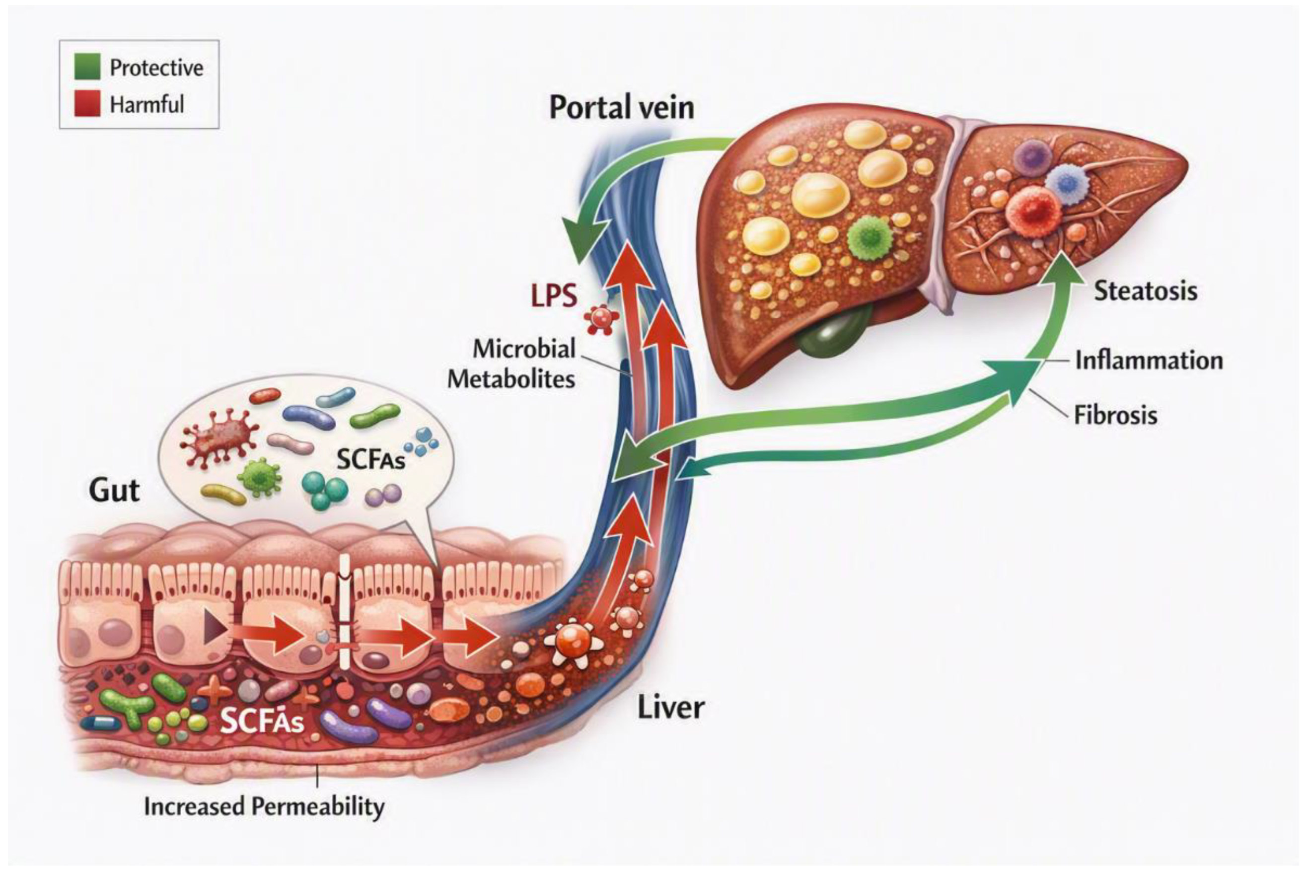
Figure 2.
Liver–kidney axis in MASLD. Shared metabolic and inflammatory drivers promote bidirectional crosstalk between the liver and kidneys. Hepatic inflammation, dyslipidemia, and hemodynamic changes contribute to renal injury, whereas uremic toxins and endothelial dysfunction may aggravate liver damage. This interaction promotes fibrosis, CKD, and CV risk, supporting integrated cardio–kidney–liver care based on weight management, healthy diet, physical activity, metabolic risk-factor control, kidney protection, and fibrosis assessment. Abbreviations: CKD, chronic kidney disease; CV, cardiovascular; MASLD, metabolic dysfunction-associated steatotic liver disease.
Figure 2.
Liver–kidney axis in MASLD. Shared metabolic and inflammatory drivers promote bidirectional crosstalk between the liver and kidneys. Hepatic inflammation, dyslipidemia, and hemodynamic changes contribute to renal injury, whereas uremic toxins and endothelial dysfunction may aggravate liver damage. This interaction promotes fibrosis, CKD, and CV risk, supporting integrated cardio–kidney–liver care based on weight management, healthy diet, physical activity, metabolic risk-factor control, kidney protection, and fibrosis assessment. Abbreviations: CKD, chronic kidney disease; CV, cardiovascular; MASLD, metabolic dysfunction-associated steatotic liver disease.
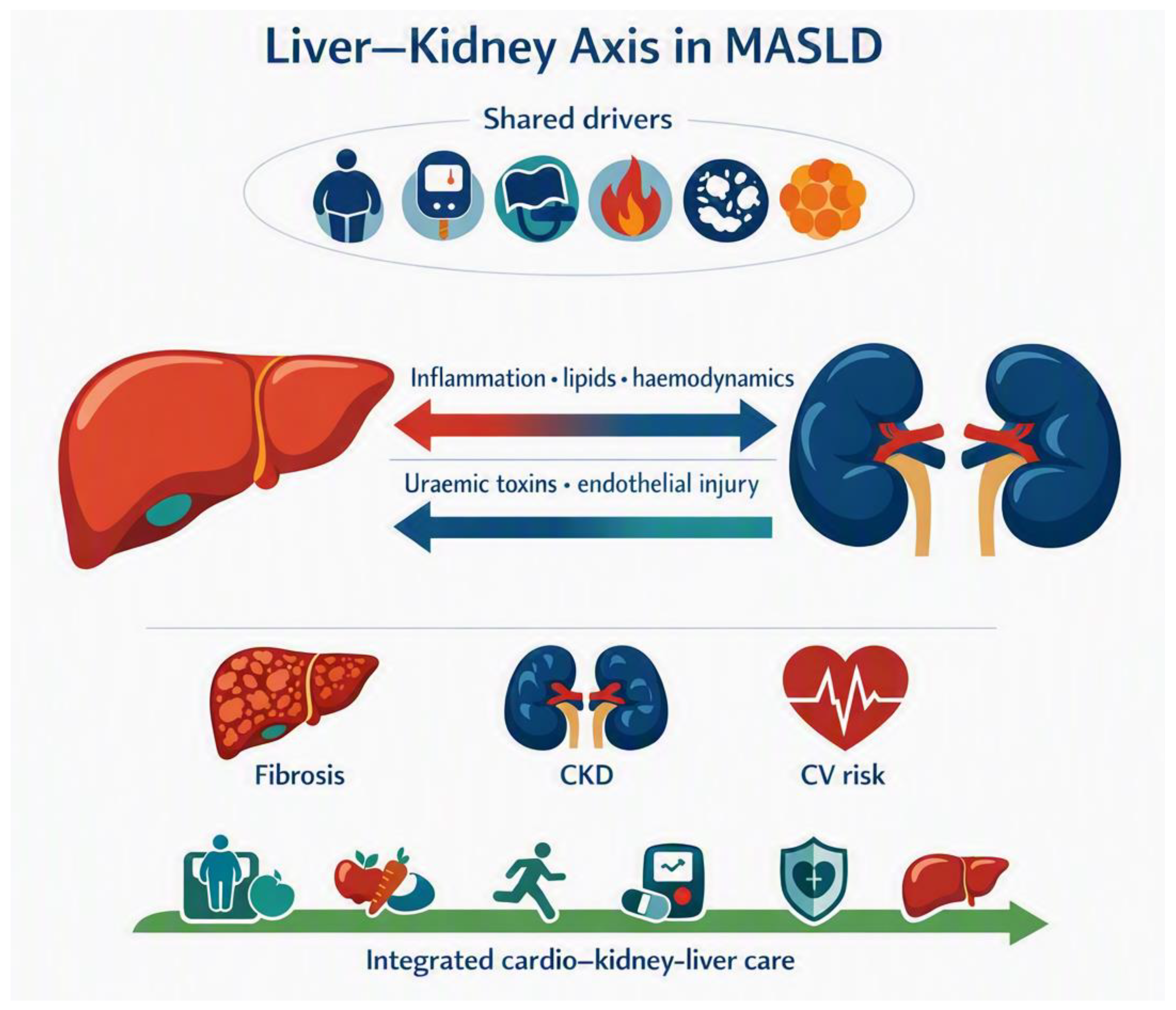
Figure 3.
Mechanisms of organ cross talks in MASLD. Schematic representation of the bidirectional inter-organ crosstalk involved in metabolic dysfunction-associated steatotic liver disease (MASLD). The MASLD liver interacts with adipose tissue, the gut, the brain, the kidney, skeletal muscle, bone, endocrine organs, and the immune system through extracellular vesicles (EVs), microRNAs (miRNAs), lipids, cytokines, bile acids, hormones, metabolites, microbial products, and neural signals. These interconnected pathways promote steatosis, insulin resistance, inflammation, mitochondrial stress, fibrosis, and systemic complications.
Figure 3.
Mechanisms of organ cross talks in MASLD. Schematic representation of the bidirectional inter-organ crosstalk involved in metabolic dysfunction-associated steatotic liver disease (MASLD). The MASLD liver interacts with adipose tissue, the gut, the brain, the kidney, skeletal muscle, bone, endocrine organs, and the immune system through extracellular vesicles (EVs), microRNAs (miRNAs), lipids, cytokines, bile acids, hormones, metabolites, microbial products, and neural signals. These interconnected pathways promote steatosis, insulin resistance, inflammation, mitochondrial stress, fibrosis, and systemic complications.
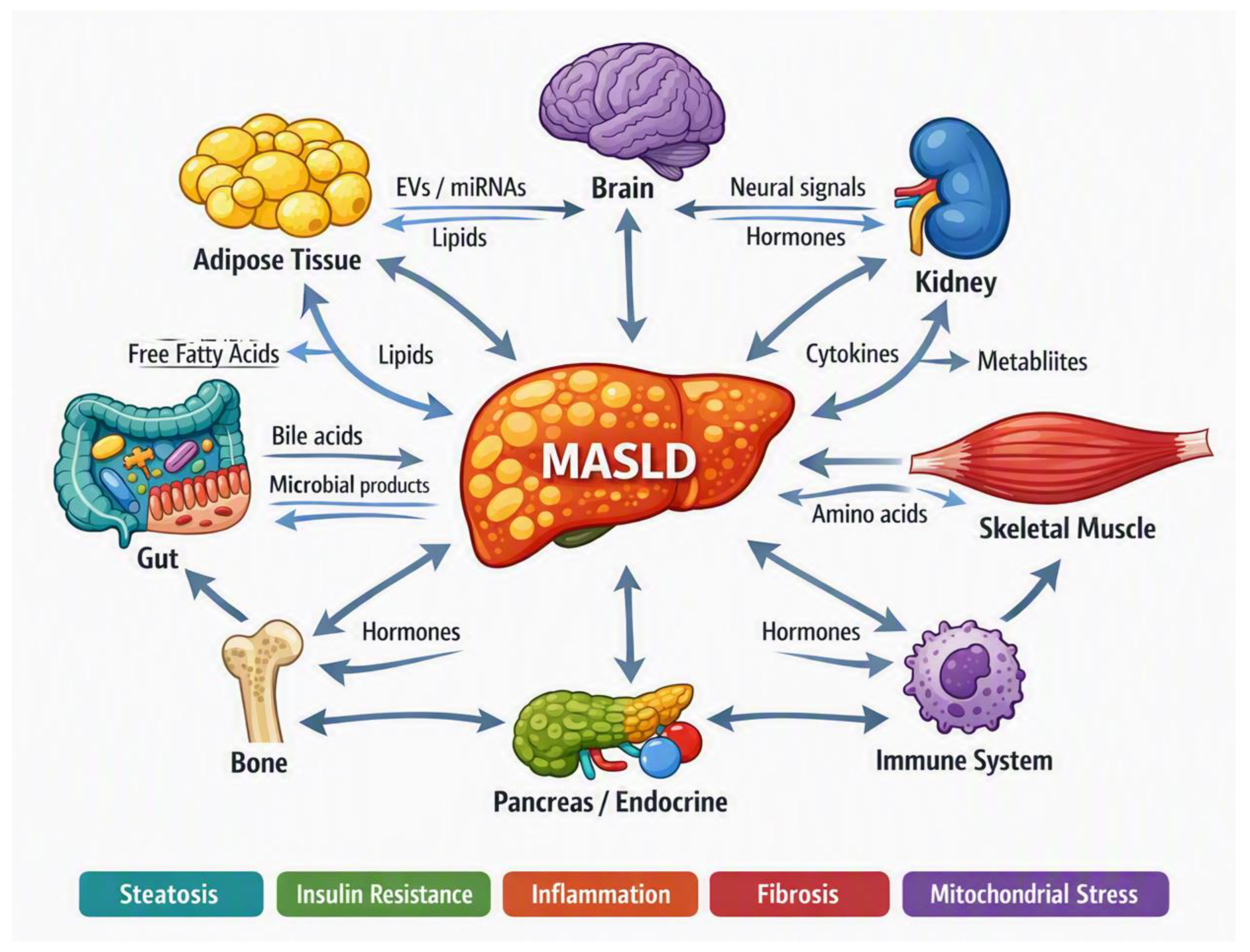

Table 1.
Overview of the endocrine axes involved in MASLD development and progression.
| Endocrine Axis | Primary Hormonal Mediators | Key Hepatic Mechanism in MASLD | Clinical Impact/Outcome | References |
|---|---|---|---|---|
| Hypothalamus-Pituitary-Liver | GH, IGF-1, PRL, ACTH | ↓ Mitochondrial oxidation, ↑ Cortisol-driven lipolysis |
Rapidly progressive MASH and accelerated fibrosis | [19] |
| Pancreas-Liver | Insulin | Hyperinsulinemia, ↓ CEACAM1 clearance, ↑ SREBP-1c/DNL |
Intracellular triglyceride buildup and severe lipotoxicity | [52] |
| Thyroid-Liver | T3, T4, TSH | Downregulated THR-β activity, TSH-R-mediated DNL induction | Reduced lipid clearance, chronic inflammation, advanced steatosis | [36,53] |
| Gonad-Liver | Estrogen (ER-α), Testosterone | ER-α blocks lipogenic enzymes, hyperandrogenism drives DNL | Postmenopausal/PMOS-associated progression risk, high sexual dimorphism | [50,51] |
Abbreviations: ACTH, adrenocorticotropic hormone; CEACAM1, carcinoembryonic antigen-related cell adhesion molecule 1, also known as CD66a; DNL, de novo lipogenesis; ER, estrogen receptor; GH, growth hormone; IGF, insulin-like growth factor; SREBP-1c, sterol regulatory element-binding protein 1c; MASH, metabolic dysfunction-associated steatohepatitis; PMOS, polyendocrine metabolic ovary syndrome (previously known as PCOS); PRL, prolactin; THR-β, thyroid hormone receptor-β; TSH, thyroid stimulating hormone.
Disclaimer/Publisher’s Note: The statements, opinions and data contained in all publications are solely those of the individual author(s) and contributor(s) and not of MDPI and/or the editor(s). MDPI and/or the editor(s) disclaim responsibility for any injury to people or property resulting from any ideas, methods, instructions or products referred to in the content. |
© 2026 by the authors. Licensee MDPI, Basel, Switzerland. This article is an open access article distributed under the terms and conditions of the Creative Commons Attribution (CC BY) license (http://creativecommons.org/licenses/by/4.0/).
Copyright: This open access article is published under a Creative Commons CC BY 4.0 license, which permit the free download, distribution, and reuse, provided that the author and preprint are cited in any reuse.