
Submitted:
04 August 2026
Posted:
05 August 2026
You are already at the latest version
Abstract
The global incidence of diseases caused by Mycobacterium abscessus (Mab), which is a nontuberculous mycobacterium (NTM), is increasing, driven by improved diagnostics and environmental changes that disrupt natural reservoirs in soil and water. These disruptions facilitate contamination of water systems. Immunocompromised individuals, particularly those with cystic fibrosis or chronic obstructive pulmonary disease, are at elevated risk, although Mab can also infect otherwise healthy individuals, causing pulmonary and cutaneous disease. Clinical management remains challenging due to intrinsic antibiotic resistance and the limited efficacy of standard anti-tuberculosis regimens. Despite this growing burden, the mechanisms governing Mab persistence and pathogenesis remain poorly defined. Progress has been constrained by the limitations of existing animal models. Mice typically clear infections rapidly, even when immunocompromised, limiting studies of chronic disease. Zebrafish support persistent infection but lack lungs and have a less developed adaptive immune system. These limitations highlight the need for alternative models. Here, we present a perspective highlighting the amphibian Xenopus laevis as a complementary model for investigating Mab chronic infection and disease. Xenopus tadpoles develop functional lungs early and possess conserved innate and adaptive immune components, including macrophages and antigen presentation pathways. Their external development allows precise control of environmental exposures and accessibility to experimentation, such as live imaging, to study Mab infection over 40 days. We discuss the application of Xenopus models to elucidate the potential role of a novel putative secondary metabolite gene cluster (SMGC) in encoding a virulence factor that promotes Mab colonization, interaction with macrophages, and persistence. Thus, X. laevis tadpoles provide a valuable experimental platform for investigating host–pathogen interactions and identifying novel therapeutic strategies.
Keywords:
infectious disease
; vertebrates
; comparative immunology
; xenopus
; animal model
1. Introduction
Nontuberculous Mycobacteria (NTMs) are a group of mycobacteria that are similar to, yet distinct from M. tuberculosis and M. leprae complexes, the causative agents of Tuberculosis (TB) and Leprosy, respectively [1]. NTMs are primarily found in soil and water; however, they are increasingly widespread outside their natural niches. The disruption of NTM natural niches through extreme weather events and natural disasters could increase exposure risks by aerosolizing the bacilli, making them easier to inhale [2]. There is both an increasing global incidence and prevalence of NTM infection [3]. Yet, it is important to note that there is variability in reporting due to either a lack of existing surveillance systems or the inability to put them in place [3,4]. Adding to the rise of NTM infections is the increase in plastic and elective surgeries, as NTMs can form biofilms on surgical equipment [5]. Currently, exposure and disease caused by NTMs, specifically Mycobacterium abscessus (Mab), are independent of the host's socio-economic standing and geography.
Mab is an opportunistic human pathogen that primarily infects individuals with compromised lung function, such as those with pulmonary conditions like Cystic Fibrosis (CF) and Chronic Obstructive Pulmonary Disease (COPD) [6]. Yet healthy people can also be infected, as Mab is known to cause skin lesions when it infects an open wound [7]. Clinically, a Mab infection presents similarly to tuberculosis. In the lungs, Mab infection results in nodes and opacities on chest X-rays, indicating solid granulomas [7], but Mab is resistant to anti-TB drugs, making treatment challenging [8,9]. The mechanisms of Mab persistence and pathogenesis are poorly defined; therefore, it is crucial to elucidate these mechanisms to better treat those infected.
Traditionally, most work on pathogenicity relevant to human health is conducted in mice. Mice are considered the “gold standard” of comparative medicine to investigate the host response to infection [10]. The mouse model has been used extensively to research TB [11,12,13]. However, multiple studies have shown that both healthy and immunocompromised mice can rapidly clear a Mab infection [14,15]. Therefore, it is difficult to study the mechanism of chronic disease when bacteria are unable to establish a lasting infection.
Another well-used and characterized comparative animal model of mycobacterial infection is Zebrafish [16,17,18,19]. In contrast to mice, Zebrafish are able to support a chronic Mab infection, but they do not have lungs - their gills function as the organ for gas exchange – and they have a relatively undeveloped adaptive immune system at the developmental stage used for research [9,19,20,21,22].
To bridge the gap between these two models introduced above, we have proposed an additional model: the amphibian, Xenopus laevis. X. laevis can maintain a chronic NTM infection; it has functioning lungs early in development, along with both functional innate and adaptive immune systems that are well characterized [14,23,24,25,26,27,28,29]. We present here a perspective detailing the use of X. laevis as an experimental model for investigating Mab chronic infection and elucidating host-Mab pathogen interactions, with the goal of aiding future identification of novel therapeutic strategies.
2. Mycobacterium abscessus Is an Emerging Pathogen
Mycobacterium abscessus (Mab) is naturally found in the soil and water [2,30]. It can readily form biofilms in aquatic environments and attach to surfaces, such as plumbing infrastructure and medical equipment [2,4,30,31,32]. This makes Mab very difficult to remove and kill. As an opportunistic pathogen, Mab can live outside its natural niche and cause disease when introduced into a host.
One of the many unique aspects of Mab is its ability to change its morphology during the course of infection by the decrease or loss of the expression of glycopeptidolipids (GPLs), located in the outer cell envelope [1,16,31,32,33]. The wild-type smooth morphotype produces GPLs, whereas the rough does not [1,8,16,33,34]. The loss of the GPLs is often associated with an increased bacterial burden and a more severe disease state; however, this is not always the case, as the smooth morphotype has been reported to persist in chronic Mab infections [35]. Infection with the rough and smooth morphotypes results in differential expression of host genes in murine macrophages, as determined through transcriptomics [36]. Downregulation of GPLs exposes bacterial antigens to the host immune system, such as the lipoproteins LpqH, LprA, and LprG [37]. Interestingly, these lipoproteins are recognized by the host via Toll-Like Receptor 2 (TLR-2) to initiate an immune response, which can potentially have therapeutic effects in terms of vaccine development [37].
Oftentimes, what allows an organism to transition into a pathogen is the production of a secondary metabolite. Primary metabolites are compounds necessary for survival, e.g., amino acids, nucleotides, and lipids [38]. Secondary metabolites are not required for survival, but can be used during periods of environmental stress, such as competition or hypoxia within a host macrophage, to promote survival [38]. Secondary metabolites can be toxins, antibiotics, have roles in antibiotic resistance, and modulate oxidative stress [38]. Within a host, these secondary metabolites can act as virulence factors or as mechanisms of antibiotic resistance and immune evasion [38].
Genetic material is often exchanged among different bacteria within an environmental niche via horizontal gene transfer (HGT). There are three main known mechanisms of HGT: transformation, transduction, and conjugation. Transformation refers to the uptake of extracellular DNA from the environment, primarily by competent bacteria scavenging for genetic material. Transduction is the process by which a bacteriophage functions as an intermediate and transfers genetic material from one bacterium to another. Conjugation is the mechanism by which a pilus extends from a donor bacterium to a recipient bacterium. These mechanisms are important factors in the evolution of bacterial pathogens and likely contributed to the development of pathogenesis in environmental NTMs [31].
3. Models Used for Mycobacterial Research
As noted in the Introduction, several animal and tissue models have been developed to investigate the pathogenesis of Mab infection. Here, we briefly summarize the principal features, strengths, and limitations of these models (Table 1), which have been reviewed in greater detail elsewhere [13,18,39,40]. This overview provides the context for discussing the attractive attributes of Xenopus laevis as a complementary model.
3.1. Mice:
For host-pathogen research, mice represent the prototypical model. Their immune system is well characterized and has been extensively compared to that of humans, making them a widely accepted model, to the point of being considered the “gold standard” of pathogenesis research [10]. Mice have a lung physiology similar to that of humans, including a branching bronchial tree and lungs subdivided into lobes [41]. This allows for exploration of a pulmonary infection over time, as the lungs can be dissected at different stages of infection. Mice have been widely used to study M. tuberculosis and M. leprae to varying degrees of success [42,43]. Often, a particular inbred strain or an immunocompromised line is used to develop susceptibility to infection with M. tuberculosis, M. leprae, or Mab [42,43,44,45]. Identifying the immunocompromised model that permits the pathogen to colonize and proliferate provides scientists with insight into the mechanisms of disease.
Unfortunately, investigating Mab chronic infection using the traditional immunocompromised mouse models is challenging because they typically clear the infection within a few weeks [14,15,46]. Attempts to use chemotherapy drugs (cyclophosphamide and dexamethasone) as immunosuppressants have been tried to allow colonization and proliferation of Mab; however, these studies note the limitations of this approach, as it does not fully mimic the chronic disease states of CF or COPD [13,47,48].
This is not to say that mice have no benefit when investigating the pathogenesis of Mab. For example, studies using primary mouse macrophages have revealed Mab infection with the rough morphotype leads to increased type 1 interferon production and inflammasome activation [49]. Additionally, mice with macrophages deficient in Dectin-1, a known pathogen recognition receptor (PRR) for mycobacteria, showed that Mab enters the macrophages independently of Dectin-1 [48]. In RAW264.7, a murine macrophage cell line, it was demonstrated that Dectin-1, in conjunction with TLR-2, was sufficient, but not necessary, for Mab to enter the macrophages, and resulted in the production of the pro-inflammatory cytokines TNFα, IL-6, and IL-12 [50]. Nevertheless, while mice and murine cell lines can provide insight into Mab pathogenesis, their intrinsic capacity to clear Mab infection is not ideal for investigating chronic infection.
3.2. Other Small Mammals:
The first animal model used by Robert Koch to study M. tuberculosis was a guinea pig [45]. Indeed, data show that guinea pigs are more susceptible to M. tuberculosis than humans, while they follow the same disease progression with primary and secondary necrotic lesions present in the lungs [45]. Additionally, guinea pigs have been used to investigate the cross-reactivity of NTMs with a TB skin test. However, there seems to be limited work with NTMs, notably Mab [51].
Rabbits are also used frequently to study M. tuberculosis, but they are more resistant to infection than guinea pigs. Rabbits are lagomorphs and the only species mentioned here that exhibit cavitation, a hallmark symptom of TB in humans [7,45]. Active work is underway to investigate the role of rabbits in NTM research, and current data suggest they are a promising model system [40,52]. As with guinea pigs, there is a lack of work investigating Mab in rabbits, primarily due to a lack of immunological reagents.
3.3. Zebrafish:
Another notable and well-established model in mycobacterial research is the Zebrafish (Danio rerio). Zebrafish are widely used as a vertebrate model for scientific research due to their short generation time, accessibility for experimentation, and genomic and genetic resources, including numerous transgenic lines. In terms of husbandry, Zebrafish are cost-effective owing to their large broods and low space requirements compared to rodents [53]. Another benefit of the zebrafish model is that the animals are translucent, making them particularly suited for live imaging [53]. Regarding mycobacteria, Zebrafish can maintain chronic NTM infections, including with Mab, which differs from the previously described rodents [19,22,54,55].
However, as with all bony fish, the Zebrafish do not have functional lungs that mimic those of mammals; instead, they have gills that filter oxygen from the water [20,22]. This is not ideal for studying host-pathogen interactions in the pulmonary environment, which are key to Mab infection and disease.
3.4. Myeloid Cell Lines:
In conjunction with animal models, it is important to recognize the benefits of in vitro cell culture systems. Immortalized cell lines have a homogeneous genetic background that reduces cell phenotype variability, enables long-term passaging, and eliminates confounding cell types [56]. Most work investigating mycobacterial infections focuses on the innate immune response, as mycobacteria typically interact with and infect macrophages [57]. And the complexity of B and T cells is difficult to replicate in vitro. Thus, myeloid-derived cell lines have been instrumental in understanding host-pathogen interactions at the cellular and molecular levels in a controlled environment.
Among cell lines, a large number of studies have been conducted with the human monocytic THP-1 cells that can be differentiated into macrophages by treatment with phorbol 12-myristate 13-acetate (PMA), lipopolysaccharide (LPS), or a combination of PMA and Vitamin D3 [26,33,56,58,59,60]. The THP-1 cell line was developed in the 1980s from the peripheral blood of a 1-year-old male patient diagnosed with monocytic leukemia [56]. As with any cell line derived from a cancer origin, there are considerations to keep in mind when comparing to in vivo or ex vivo cells, such as differences in metabolic requirements, baseline and stimulated inflammatory markers, and gene deregulation and mutation accumulation over time and passages [56]. However, THP-1 cells, both monocytes and differentiated macrophages, have served as a useful in vitro model system for mycobacterial research [36,56,58,59,61,62]. Macrophages infected with the rough or smooth morphotype of Mab exhibit distinct transcriptomic profiles, with the smooth morphotype inducing a more immunogenic response, as evidenced by increased IL-6 and IL-1β cytokine production [36]. For M. tuberculosis, THP-1 cells were used to show that hypoxic-grown, dormant M. tuberculosis, which resemble those present in a granuloma, are less virulent than actively growing bacilli, leading to the persistence phenotype [58].
Another common human cell line used to investigate host-pathogen interaction is the human U937 monocytic cell line derived from a patient with histiocytic lymphoma [56]. In contrast to THP-1 cells, which are from a blood cancer, U937 cells are from a solid tumor, making them more mature in terms of development [56]. When comparing the response of U937, THP-1, and peripheral blood mononuclear cells (PBMCs) to infection with M. tuberculosis, M. bovis, and the TB BCG vaccine, the THP-1 cells showed a more physiological relevant response (nitrate, IL-12, and TNFα), similar to the PBMCs, compared to U937 [57]. This suggests that not all myeloid cell lines perform the same under experimental conditions.
As mentioned earlier, murine cell lines have also been used to investigate mycobacteria host interaction at the cellular level. Notably, the macrophage line RAW264.7 has been used to elucidate the role of two component system MtrAB in Mab virulence and the function of the transmembrane protein RV3737 in the intracellular survival of M. tuberculosis [17,63]. An exhaustive description of cell lines used for mycobacteria research is beyond the scope of this review.
4. Xenopus laevis as a Model for the Human Immune System
To advance NTM research, we present the amphibian Xenopus laevis as an intersection that addresses the limitations of the animal models discussed above, while also encompassing their benefits. Like zebrafish, X. laevis has large broods and is transparent during its larval stages. Importantly, and unlike zebrafish, X. laevis have functioning lungs five days after birth [64]. Embryos and tadpoles develop independently of maternal influence, allowing for the interrogation of the immune response with few confounding factors [65].
The X. laevis immune system has been extensively characterized and is remarkably conserved to that of human as detailed in several comprehensive reviews [14,64,65]. This includes a thymus and a spleen; T and B cells with recombination-activating genes (RAGs)-mediated rearranging T-cell receptors (TCR) and immunoglobulin (Ig) receptors, respectively; activation-induced cytidine deaminase (AID)-mediated hypermutations and Ig isotype heterogeneity; key cytokines and major histocompatibility complex (MHC) class I and class II genes. T cell maturation and expression of key surface molecules are conserved between frogs and mammals [27]. MHC-restricted cytotoxic and helper T cell responses have been demonstrated. The fully sequenced and annotated genomes of both X. laevis and a close relative X. tropicalis, as well as large gene expression databases and advanced genomic resources (all accessible through Xenbase; https://www.xenbase.org/xenbase/) further empower the Xenopus model and provide additional compelling evidence of the high degree of conservation of gene repertoires in vertebrate immune systems [66,67].
The reliance on the innate immune system early in development is likely beneficial evolutionarily, as eggs hatch unprotected in the aquatic environment and are immediately exposed to an onslaught of pathogens [65]. Therefore, a functional immune system differentiates early during the larval stage. Blood circulation and prototypical innate immune cells, such as macrophages and neutrophils, are detected within a week post-fertilization [27,65]. Within a week of hatching (2 weeks post-fertilization), the larvae begin to display functional B cells and T cells[64]. However, the majority of these T cells are so-called innate-like (i)T cells that express a very limited T cell receptor repertoire and interact with non-polymorphic nonclassical MHC-Ib (mhc1-uba) rather than classical polymorphic MHC-Ia. One subset of iT-cells expressing the invariant TCR alpha Vα45-J1.14 rearrangement and interacting with mhc1-uba4 or XNC4 has been shown to be critical for tadpole resistance and immune response against the NTM M. marinum [24].
During metamorphosis, the tadpole begins to undergo a series of organ and structural remodeling. Namely, for the immune system, this results in a drastic decrease in the number of thymocytes and lymphocytes [64]. The thymus shrinks and migrates toward the tympanum; consequently, the immune system must generate new lymphocytes that undergo another round of negative selection to promote self-tolerance to adult antigens [64]. The expression of the classical MHC-I and MHC-II genes are also differentially regulated during metamorphosis [64]. Once adulthood is reached, there is a vast diversification of the B cell and T cell receptor repertoire similar to that of mammals, along with more efficient B cell and antibody responses [64].
As with other animal models, peritoneal leukocytes can be harvested from both the tadpole and adult stages, enabling ex vivo modeling [25,27]. Macrophages can be isolated through cell sorting techniques. Additionally, there are X. laevis cell lines, such as the kidney-derived A6 line, that can be cultured in the laboratory and used for in vitro experiments [27].
In summary, the amphibian X. laevis serves as a valuable comparative model for studying fundamental aspects of the immune system conserved across jawed vertebrates including human.. A collection of important reagents (e.g., monoclonal antibodies, recombinant proteins), as well as X. laevis transgenic lines and MHC-defined inbred strains important for immunological studies are available through our NIH-funded Xenopus Research Resource for Immunobiology (https://www.urmc.rochester.edu/microbiology-immunology/research/xenopus-laevis).
5. Xenopus laevis as an Established Model for Mycobacterial Research
Previous work from our lab has established X. laevis as a reliable mycobacterial experimental organism using M. marinum [23,24,26,28]. M. marinum is found in both fresh and saltwater sources, such as lakes, rivers, and estuaries, in addition to fish tanks, making its presence ubiquitous [68]. In aquatic animals, M. marinum exhibits a pathophysiology similar to that of M. tuberculosis in humans [68,69]. Because of this, M. marinum is often used as a model to study TB disease progression [69]. However, M. marinum is a pathogen in its own right, infecting aquatic animals and immunocompromised individuals, causing skin lesions and granulomas [69].
A clear benefit of using X. laevis as a model organism is its ease of transgenesis compared to mammalian models, enabling the generation of a variety of transgenic lines that can aid in answering immunological questions. Notably, our lab has contributed to the development of a transgenic line, mpeg:GFP, in which macrophages express the Green Fluorescent Protein for imaging by confocal microscopy or flow cytometry [25,26,28]. Thus, we can visually track macrophage recruitment to the site of infection and quantify the colocalization of macrophages and pathogens.
We have used these transgenic models, along with outbred lines, to investigate the pathogenesis of M. marinum in X. laevis. Our results show that tadpoles and adults exhibit distinct host-pathogen interactions during infection: tadpoles were tolerogenic, whereas adults cleared the infection [23]. More importantly, the results revealed that X. laevis is a valuable model organism, assays such as qPCR, RNA sequencing, colony-forming units, immunohistochemistry, flow cytometry, and confocal microscopy can all be used to quantify the host response to infection.
Additionally, we have applied CRISPR/Cas9 genome editing combined with transgenesis to disrupt different key immune genes, including nonclassical MHC-I; RAG2, which prevents V(D)J recombination and essentially halts BCR and TCR production; FOXN1 required for thymus development; and others that limit or severely compromise the immune system. Some of these knockout lines are generated and characterized with the National Xenopus Resource in Woods Hole MA, USA, directed by Marko Horb, PhD. These immune-deficient lines strengthen the X. laevis model to investigate the host response to Mab in vivo.
6. Perspective of Xenopus laevis and Mycobacterium Abscessus Virulence
Based upon our success with M. marinum in the Xenopus model, we propose using X. laevis as a useful comparative model system to study Mab pathogenesis and chronic disease. Published work from our lab demonstrates that X. laevis can elicit an immune response to a Mab infection, a phenomenon similarly to the clinical manifestations observed in humans [14,25].
Our work shows that X. laevis tadpoles can maintain the Mab infection for over 50 days [25], and in doing so, may delay metamorphosis – an anecdotal biological marker for environmental stress.
In 2009, Ripoll et al. sequenced the first Mab genome and identified multiple gene clusters that appear to result from HGT in the distant past. One such cluster, MAB_0248c-0305, lacked homology to any mycobacterial genes but is similar to gene clusters found in Streptomyces species that are responsible for the synthesis of various antibiotic secondary metabolites and the biosynthesis of pyocyanin, a virulence factor produced by Pseudomonas aeruginosa.
Analysis of the Mab gene cluster reveals similarities and differences between the P. aeruginosa cluster, notably for the production of phenazine-1-carboxylic acid intermediate from chorismic acid [70]. The Mab cluster encodes the early enzymes, but instead of producing phenazine-1-carboxylic acid, it appears to synthesize 3-hydroxyanthranilic acid, which is shared with the streptonigrin biosynthesis pathway of Streptomyces albus. The Mab pathway is less complex than the streptonigrin pathway, and we have proposed that it produces a simpler metabolite (Figure 1). To date, there is no proposed structure or established function; however, we present here a hypothesized biosynthetic pathway of the secondary metabolite based on the other genes present in the cluster (Figure 2).
In addition, published data from our lab support the hypothesis that this secondary metabolite pathway produces a compound that triggers a host response [25]. We have shown that when X. laevis tadpoles are infected with a mutant with a deletion in a gene (ΔphzC) for an early step in the pathway, there is a decrease in macrophage recruitment to the site of infection when compared to WT [25]. Our data further show that tadpoles infected with the ΔphzC mutant exhibit significantly improved survival compared with those infected with wild-type Mab, suggesting that the secondary metabolite produced by this biosynthetic gene cluster functions as a virulence factor. However, the mechanisms by which this pathway promotes host colonization and persistence remain to be elucidated. Figure 3 outlines the potential Mab/macrophage interactions. We think that investigating the role of this gene cluster is critical for understanding Mab pathogenesis, and the X. laevis model offers a powerful experimental platform to investigate these processes in vivo during chronic infection.
Future studies will be able to take advantage of our reliable tail wound inoculation system in mpeg::GFP transgenic tadpoles, where the GFP reporter is specifically expressed by macrophages[26]. The different Mab strains expressing DsRed reporter can be inoculated through a small wound in the middle region of the ventral fin with a 0.3 mm diameter surgical biopsy punch. The recruitment of macrophages and their infection by Mab can be monitored by live imaging confocal microscopy, as depicted in Figure 4. This is a powerful approach that will permit to visualize host-pathogen interaction in vivo in real time. Conveniently, live imaging by confocal microscopy can be recorded at different time points post-inoculation on the same tadpole under narcosis, and unlike mammalian model, the process does not require temperature control nor special sterile conditions. Finally, the dissemination of Mab infection from the tail wound to distant organs such as the lung can followed. Whole-mount tadpole lungs are relatively easy to prepare, and their small size and transparency are particularly convenient for live imaging. Each of these experimental techniques developed by our lab speaks to the versatility that X. laevis offers for future work investigating Mab and other NTMs.
7. Conclusion
The opportunist pathogen, M. abscessus (Mab), is increasingly causing disease worldwide with the rise in both environmental disasters and plastic/elective surgeries.
In conclusion, we propose X. laevis as a complementary model organism to mice and zebrafish for investigating the role of Mab/host interaction. We think that X. laevis has the potential to contribute significantly to elucidating the mechanisms of Mab pathogenesis, which can lead to better treatment approaches in the future.
Acknowledgements
The authors are funded by R24AI059830 from the National Institute of Allergy and Infectious Diseases (NIAID). Kexin Chen K, Francisco De Jesus Andino, Nicholas Miller.
References
- To, K.; Cao, R.; Yegiazaryan, A.; Owens, J.; Venketaraman, V. General Overview of Nontuberculous Mycobacteria Opportunistic Pathogens: Mycobacterium avium and Mycobacterium abscessus. J. Clin. Med. 2020, 9, 2541. [Google Scholar] [CrossRef] [PubMed]
- Mercaldo, R.A.; Marshall, J.E.; Cangelosi, G.A.; et al. Environmental risk of nontuberculous mycobacterial infection: Strategies for advancing methodology. Tuberculosis 2023, 139, 102305. [Google Scholar] [CrossRef] [PubMed]
- Dahl, V.N.; Mølhave, M.; Fløe, A.; et al. Global trends of pulmonary infections with nontuberculous mycobacteria: a systematic review. Int. J. Infect. Dis. 2022, 125, 120–131. [Google Scholar] [CrossRef] [PubMed]
- Dowdell, K.; Haig, S.J.; Caverly, L.J.; Shen, Y.; LiPuma, J.J.; Raskin, L. Nontuberculous mycobacteria in drinking water systems – the challenges of characterization and risk mitigation. Curr. Opin. Biotechnol. 2019, 57, 127–136. [Google Scholar] [CrossRef] [PubMed]
- Ghosh, R.; Das, S.; Kela, H.; De, A.; Haldar, J.; Maiti, K. Biofilm colonization of Mycobacterium abscessus: New threat in hospital-acquired surgical site infection. Indian J. Tuberc. 2017, 64, 178–182. [Google Scholar] [CrossRef] [PubMed]
- Marshall, J.E.; Mercaldo, R.A.; Lipner, E.M.; Prevots, D.R. Incidence of nontuberculous mycobacteria infections among persons with cystic fibrosis in the United States (2010–2019). BMC Infect. Dis. 2023, 23, 489. [Google Scholar] [CrossRef] [PubMed]
- Lee, M.R.; Sheng, W.H.; Hung, C.C.; Yu, C.J.; Lee, L.N.; Hsueh, P.R. Mycobacterium abscessus complex infections in humans. Emerg. Infect. Dis. 2015, 21, 1638–1646. [Google Scholar] [CrossRef]
- Nguyen, T.Q.; Heo, B.E.; Jeon, S.; et al. Exploring antibiotic resistance mechanisms in Mycobacterium abscessus for enhanced therapeutic approaches. Front Microbiol. 2024, 15. [Google Scholar] [CrossRef] [PubMed]
- Touré, H.; Galindo, L.A.; Lagune, M.; et al. Mycobacterium abscessus resists the innate cellular response by surviving cell lysis of infected phagocytes. PLoS Pathog. 2023, 19, e1011257. [Google Scholar] [CrossRef] [PubMed]
- Bryda, E.C. The Mighty Mouse: The Impact of Rodents on Advances in Biomedical Research; 2013; Available online: www.who.int/cardiovascular_diseases.
- Beamer, G.L.; Flaherty, D.K.; Assogba, B.D.; et al. Interleukin-10 Promotes Mycobacterium tuberculosis Disease Progression in CBA/J Mice 1. The Journal of Immunology. Published online October 15, 2008. [CrossRef] [PubMed]
- Silva Miranda, M.; Breiman, A.; Allain, S.; Deknuydt, F.; Altare, F. The Tuberculous Granuloma: An Unsuccessful Host Defence Mechanism Providing a Safety Shelter for the Bacteria? Clin. Dev. Immunol. 2012, 2012, 139127. [Google Scholar] [CrossRef] [PubMed]
- Soldevilla, P.; Vilaplana, C.; Cardona, P.J. Mouse Models for Mycobacterium tuberculosis Pathogenesis: Show and Do Not Tell. Pathogens 2022, 12, 49. [Google Scholar] [CrossRef] [PubMed]
- Lopez, A.; Shoen, C.; Cynamon, M.; et al. Developing tadpole xenopus laevis as a comparative animal model to study Mycobacterium abscessus pathogenicity. Int. J. Mol. Sci. 2021, 22, 1–12. [Google Scholar] [CrossRef] [PubMed]
- Obregón-Henao, A.; Arnett, K.A.; Henao-Tamayo, M.; et al. Susceptibility of Mycobacterium abscessus to Antimycobacterial Drugs in Preclinical Models. Antimicrob. Agents Chemother. 2015, 59, 6904. [Google Scholar] [CrossRef] [PubMed]
- Mendoza-Trujillo, I.; Diez-Echave, P.; Tontini, C.; Bulfone-Paus, S. The Crosstalk Between Mycobacterium abscessus and Immune Cells: Exploring Novel Interaction Modalities. Cells 2025, 14, 1829. [Google Scholar] [CrossRef] [PubMed]
- Zhang, J.; Ju, Y.; Li, L.; et al. MtrAB two-component system is crucial for the intrinsic resistance and virulence of Mycobacterium abscessus. Int. J. Antimicrob. Agents Published online April 5. 2024, 2024.04.05.588258. [CrossRef] [PubMed]
- van Leeuwen, L.M.; van der Sar, A.M.; Bitter, W. Animal Models of Tuberculosis: Zebrafish. Cold Spring Harb. Perspect. Med. 2015, 5, a018580. [Google Scholar] [CrossRef] [PubMed]
- Johansen, M.D.; Spaink, H.P.; Oehlers, S.H.; Kremer, L. Modeling nontuberculous mycobacterial infections in zebrafish. Trends Microbiol. 2024, 32, 663–677. [Google Scholar] [CrossRef] [PubMed]
- Messerli, M.; Aaldijk, D.; Haberthür, D.; et al. Adaptation mechanism of the adult zebrafish respiratory organ to endurance training. PLoS One Published online. 2020. [Google Scholar] [CrossRef] [PubMed]
- Bernut, A.; Nguyen-Chi, M.; Halloum, I.; Herrmann, J.L.; Lutfalla, G.; Kremer, L. Mycobacterium abscessus-Induced Granuloma Formation Is Strictly Dependent on TNF Signaling and Neutrophil Trafficking. PLoS Pathog Published online. 2016. [Google Scholar] [CrossRef] [PubMed]
- Bernut, A.; Herrmann, J.L.; Ordway, D.; Kremer, L. The diverse cellular and animal models to decipher the physiopathological traits of Mycobacterium abscessus infection. Front Cell Infect. Microbiol. 2017, 7(APR), 260117. [Google Scholar] [CrossRef]
- Rhoo, K.H.; Edholm, E.S.; Forzán, M.J.; et al. Distinct Host–Mycobacterial Pathogen Interactions between Resistant Adult and Tolerant Tadpole Life Stages of Xenopus laevis. J. Immunol. 2019, 203, 2679–2688. [Google Scholar] [CrossRef] [PubMed]
- Edholm, E.S.; Banach, M.; Rhoo, K.H.; Pavelka, M.S.; Robert, J. Distinct MHC class I-like interacting invariant T cell lineage at the forefront of mycobacterial immunity uncovered in Xenopus. Proc. Natl. Acad. Sci. U S A 2018, 115, E4023–E4031. [Google Scholar] [CrossRef] [PubMed]
- Miller, N.J.; Dimitrakopoulou, D.; Baglia, L.A.; Pavelka, M.S.; Robert, J. Exploring the Role of a Putative Secondary Metabolite Biosynthesis Pathway in Mycobacterium abscessus Pathogenesis Using a Xenopus laevis Tadpole Model. Microorganisms 2024, 12. [Google Scholar] [CrossRef] [PubMed]
- Hyoe, R.K.; Robert, J. A Xenopus tadpole alternative model to study innate-like T cell-mediated anti-mycobacterial immunity. Dev Comp Immunol 2019, 92, 253–259. [Google Scholar] [CrossRef] [PubMed]
- Robert, J. The Immune System of Amphibians. In Encyclopedia of Immunobiology; Elsevier Inc., 2016; Vol 1, pp. 486–492. [Google Scholar] [CrossRef]
- Paiola, M.; Dimitrakopoulou, D.; Pavelka, M.S.; Robert, J. Amphibians as a model to study the role of immune cell heterogeneity in host and mycobacterial interactions. Dev. Comp. Immunol. 2023, 139, 104594. [Google Scholar] [CrossRef] [PubMed]
- Robert, J.; Najafi-Majd, E. Evolutionary Perspective of Nonclassical MHC Class I and Innate-like T Cells Relevance in Immune Surveillance. Cells Published online. 2025. [Google Scholar] [CrossRef] [PubMed]
- Cristancho-Rojas, C.; Varley, C.D.; Lara, S.C.; Kherabi, Y.; Henkle, E.; Winthrop, K.L. Epidemiology of Mycobacterium abscessus. Clin. Microbiol. Infect. 2024, 30, 712–717. [Google Scholar] [CrossRef] [PubMed]
- Ripoll, F.; Pasek, S.; Schenowitz, C.; et al. Non mycobacterial virulence genes in the genome of the emerging pathogen Mycobacterium abscessus. PLoS ONE 2009, 4. [Google Scholar] [CrossRef] [PubMed]
- Chan, E.D.; Bai, X.; Kartalija, M.; Orme, I.M.; Ordway, D.J. Host immune response to rapidly growing mycobacteria, an emerging cause of chronic lung disease. Am. J. Respir. Cell Mol. Biol. 2010, 43, 387–393. [Google Scholar] [CrossRef] [PubMed]
- Kapoor, H.; Maves, A.M.; Bowder, M.A.; Danelishvili, L. Phage-mediated TLR2 signaling attenuates intracellular Mycobacterium abscessus survival in macrophages. Sci. Rep. 2025, 15, 28504. [Google Scholar] [CrossRef] [PubMed]
- Degiacomi, G.; Sammartino, J.C.; Chiarelli, L.R.; Riabova, O.; Makarov, V.; Pasca, M.R. Mycobacterium abscessus, an Emerging and Worrisome Pathogen among Cystic Fibrosis Patients. Int. J. Mol. Sci. 2019, Vol. 20, Page 5868 2019, 20, 5868. [Google Scholar] [CrossRef] [PubMed]
- Dedrick, R.M.; Smith, B.E.; Cristinziano, M.; et al. Phage Therapy of Mycobacterium Infections: Compassionate Use of Phages in 20 Patients With Drug-Resistant Mycobacterial Disease. Clin. Infect. Dis. 2023, 76, 103–112. [Google Scholar] [CrossRef] [PubMed]
- Nandanwar, N.; Gibson, J.E.; Neely, M.N. Transcriptome profiles of macrophages upon infection by morphotypic smooth and rough variants of Mycobacterium abscessus. Microbes Infect. 2024, 26, 5–6. [Google Scholar] [CrossRef] [PubMed]
- Le Moigne, V.; Roux, A.L.; Jobart-Malfait, A.; et al. A TLR2-Activating Fraction From Mycobacterium abscessus Rough Variant Demonstrates Vaccine and Diagnostic Potential. Front Cell Infect. Microbiol. 2020, 10, 432. [Google Scholar] [CrossRef]
- Perry, E.K.; Meirelles, L.A.; Newman, D.K. From the soil to the clinic: The impact of microbial secondary metabolites on antibiotic tolerance and resistance. Nat. Rev. Microbiol. 2021, 20, 129. [Google Scholar] [CrossRef] [PubMed]
- Clark, S.; Hall, Y.; Williams, A. Animal Models of Tuberculosis: Guinea Pigs. Cold Spring Harb. Perspect. Med. 2015, 5, a018572. [Google Scholar] [CrossRef] [PubMed]
- Arrazuria, R.; Juste, R.A.; Elguezabal, N. Mycobacterial Infections in Rabbits: From the Wild to the Laboratory. Transbound Emerg Dis. 2017, 64, 1045–1058. [Google Scholar] [CrossRef] [PubMed]
- Rydell-Törmänen, K.; Johnson, J.R. The applicability of mouse models to the study of human disease. Methods Mol. Biol. 2019, 1940, 3–22. [Google Scholar] [CrossRef]
- Adams, L.B. Susceptibility and resistance in leprosy: Studies in the mouse model. Immunol. Rev. 2021, 301, 157–174. [Google Scholar] [CrossRef] [PubMed]
- Lancaster, R.D.; Hilson, G.R.F.; McDougall, A.C.; Colston, M.J. Mycobacterium leprae Infection in Nude Mice: Bacteriological and Histological Responses to Primary Infection and Large Inocula. Infect. Immun. Published online. 1983, 865–872. [Google Scholar] [CrossRef] [PubMed]
- Chackerian, A.A.; Behar, S.M. Susceptibility to Mycobacterium tuberculosis: lessons from inbred strains of mice. Tuberculosis 2003, 83, 279–285. [Google Scholar] [CrossRef] [PubMed]
- McMurray, D.N. Disease model: pulmonary tuberculosis. Trends Mol. Med. 2001, 7, 135–137. [Google Scholar] [CrossRef] [PubMed]
- Dartois, V.; Bonfield, T.L.; Boyce, J.P.; et al. Preclinical murine models for the testing of antimicrobials against Mycobacterium abscessus pulmonary infections: current practices and recommendations. Tuberculosis 2024, 147, 102503. [Google Scholar] [CrossRef] [PubMed]
- Sun, Y.; Zhang, K.; Santos, M.D.; et al. An optimized cyclophosphamide-treated mouse model of Mycobacterium abscessus pulmonary infection. Antimicrob. Agents Chemother. 2024, 68, e01520-23. [Google Scholar] [CrossRef] [PubMed]
- Ignatius, E.H.; Rimal, B.; Panthi, C.M.; et al. Efficacies of omadacycline + amikacin + imipenem and an all-oral regimen omadacycline + clofazimine + linezolid in a mouse model of M. abscessus lung disease. mSphere 2024, 9, e00381-24. [Google Scholar] [CrossRef] [PubMed]
- Kim, B.R.; Kim, B.J.; Kook, Y.H.; Kim, B.J. Mycobacterium abscessus infection leads to enhanced production of type 1 interferon and NLRP3 inflammasome activation in murine macrophages via mitochondrial oxidative stress. PLoS Pathog. 2020, 16, e1008294. [Google Scholar] [CrossRef] [PubMed]
- Shin, D.M.; Yang, C.S.; Yuk, J.M.; et al. Mycobacterium abscessus activates the macrophage innate immune response via a physical and functional interaction between TLR2 and dectin-1. Cell Microbiol. 2008, 10, 1608–1621. [Google Scholar] [CrossRef] [PubMed]
- Fernández-Veiga, L.; Fuertes, M.; Geijo, M. V.; et al. Differences in skin test reactions to official and defined antigens in guinea pigs exposed to non-tuberculous and tuberculous bacteria. Sci. Rep. 2023, 13, 2936. [Google Scholar] [CrossRef] [PubMed]
- Kaya, F.; Ernest, J.P.; LoMauro, K.; et al. A Rabbit Model to Study Antibiotic Penetration at the Site of Infection for Nontuberculous Mycobacterial Lung Disease: Macrolide Case Study. Antimicrob. Agents Chemother. 2022, 66, e02212-21. [Google Scholar] [CrossRef] [PubMed]
- Teame, T.; Zhang, Z.; Ran, C.; et al. The use of zebrafish (Danio rerio) as biomedical models. Anim. Front. 2019, 9, 68. [Google Scholar] [CrossRef] [PubMed]
- Dirks, R.P.; Ordas, A.; Jong-Raadsen, S.; et al. The human pathogen Mycobacterium tuberculosis and the fish pathogen Mycobacterium marinum trigger the same core set of late innate immune response genes in zebrafish larvae. Biology Published online. 2024. [Google Scholar] [CrossRef] [PubMed]
- Kam, J.Y.; Hortle, E.; Krogman, E.; et al. Rough and smooth variants of Mycobacterium abscessus are differentially controlled by host immunity during chronic infection of adult zebrafish. Nat. Commun. 2022, 13, 952. [Google Scholar] [CrossRef] [PubMed]
- Chanput, W.; Mes, J.J.; Wichers, H.J. THP-1 cell line: An in vitro cell model for immune modulation approach. Int. Immunopharmacol. 2014, 23, 37–45. [Google Scholar] [CrossRef] [PubMed]
- Mendoza-Coronel, E.; Castañón-Arreola, M. Comparative evaluation of in vitro human macrophage models for mycobacterial infection study. Pathog. Dis. 2016, 74, 52. [Google Scholar] [CrossRef] [PubMed]
- Iona, E.; Pardini, M.; Gagliardi, M.C.; et al. Infection of human THP-1 cells with dormant Mycobacterium tuberculosis. Microbes Infect. 2012, 14, 959–967. [Google Scholar] [CrossRef] [PubMed]
- Liu, X.; Yang, M.; Xu, P.; et al. Kynurenine-AhR reduces T-cell infiltration and induces a delayed T-cell immune response by suppressing the STAT1-CXCL9/CXCL10 axis in tuberculosis. Cell Mol Immunol Published online. 2024. [Google Scholar] [CrossRef] [PubMed]
- Mol, B.A.; Wasinda, J.J.; Xu, Y.F.; Gentle, N.L.; Meyer, V. 1,25-dihydroxyvitamin D3 augments low-dose PMA-based monocyte-to-macrophage differentiation in THP-1 cells. J. Immunol. Methods 2024, 532, 113716. [Google Scholar] [CrossRef] [PubMed]
- Ruedas-López, A.; Tato, M.; Lerma, L.; et al. Infection model of THP-1 cells, growth dynamics, and antimicrobial susceptibility of clinical Mycobacterium abscessus isolates from cystic fibrosis patients: Results from a multicentre study. PLoS ONE 2025, 20, e0319710. [Google Scholar] [CrossRef] [PubMed]
- Maserumule, C.; Passemar, C.; H Oh, O.S.; et al. Phagosomal RNA sensing through TLR8 controls susceptibility to tuberculosis. Cell Rep Published online. 2025. [Google Scholar] [CrossRef]
- Peng, Z.; Xu, C.; You, T.; et al. Depletion of Mycobacterium tuberculosis transmembrane protein Rv3737 reduces pathogen survival and induces M1 macrophage polarization against tuberculosis. Front Cell Infect. Microbiol. 2025, 15, 1592296. [Google Scholar] [CrossRef]
- Robert, J.; Ohta, Y. Comparative and Developmental Study of the Immune System in Xenopus. Dev. Dyn. 2009, 238, 1249. [Google Scholar] [CrossRef] [PubMed]
- Robert, J.; Cohen, N. The genus Xenopus as a multispecies model for evolutionary and comparative immunobiology of the 21st century. Dev Comp Immunol Published online. 2011. [Google Scholar] [CrossRef] [PubMed]
- Session, A.M.; Uno, Y.; Kwon, T.; et al. Genome evolution in the allotetraploid frog Xenopus laevis. Nature 2016, 538, 336. [Google Scholar] [CrossRef] [PubMed]
- Hellsten, U.; Harland, R.M.; Gilchrist, M.J.; et al. The Genome of the Western Clawed Frog Xenopus tropicalis. Science (1979) 2010, 328, 633. [Google Scholar] [CrossRef] [PubMed]
- Aubry, A.; Mougari, F.; Reibel, F.; Cambau, E. Mycobacterium marinum. Microbiol. Spectr. 2017, 5. [Google Scholar] [CrossRef] [PubMed]
- Menon, A.R.; Prest, R.J.; Tobin, D.M.; Champion, P.A. Mycobacterium marinum as a model for understanding principles of mycobacterial pathogenesis. J. Bacteriol. 2025, 207. [Google Scholar] [CrossRef] [PubMed]
- Mavrodi, D. V.; Bonsall, R.F.; Delaney, S.M.; Soule, M.J.; Phillips, G.; Thomashow, L.S. Functional Analysis of Genes for Biosynthesis of Pyocyanin and Phenazine-1-Carboxamide from Pseudomonas aeruginosa PAO1. J Bacteriol Published online. 2001. [Google Scholar] [CrossRef] [PubMed]
Figure 1.
Schematic map of SMGC in Mab. The ORFs are numbered according to ATCC19977 strain. Mab genes 0284c to 0290, and 0295 to 0298 are syntenic with streptonigrin genes of Streptomyces species with the latter gene block flipped in Mab. Boxed in green is cdaR, which encodes a putative transcriptional activator for the cluster and boxed in red is the phzC gene targeted for deletion. In purple are genes unique to the Mab cluster carboxyesterase type B (0294); NbaD aminocarboxymuconate-semialdehyde decarboxylase (0299).
Figure 1.
Schematic map of SMGC in Mab. The ORFs are numbered according to ATCC19977 strain. Mab genes 0284c to 0290, and 0295 to 0298 are syntenic with streptonigrin genes of Streptomyces species with the latter gene block flipped in Mab. Boxed in green is cdaR, which encodes a putative transcriptional activator for the cluster and boxed in red is the phzC gene targeted for deletion. In purple are genes unique to the Mab cluster carboxyesterase type B (0294); NbaD aminocarboxymuconate-semialdehyde decarboxylase (0299).
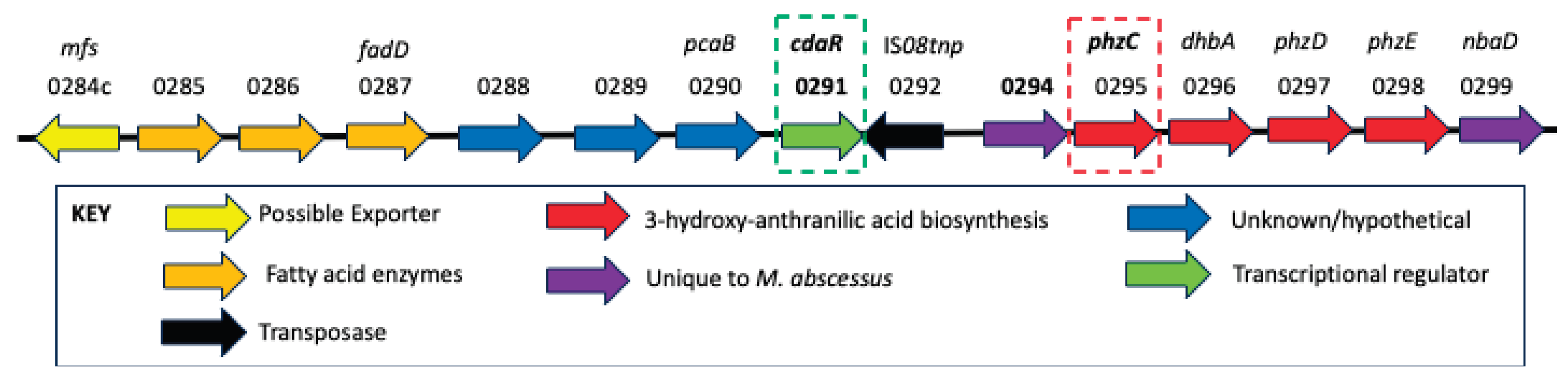
Figure 2.
Proposed secondary metabolite pathway. This pathway is based upon what is known for the synthesis of streptonigrin and phenazine. PhzC catalyzes the production of 3-deoxy-D-arabino-heptulosonate 7-phosphate (DAHP) from erythritol 4-phosphate (E4P) and phosphoenolpyruvate (PEP) which is then converted to chorismate by housekeeping enzymes. From there, it is converted to 3-hydroxy anthranilic acid (HA) by PhzE, PhzD, and DhbA, which is then further modified by the addition and cyclization of fatty acids via O285, 0286, and FadD. The resulting intermediate is then modified by 0288, 0289, 290, 0294, and NbaD, and then exported out of the bacteria by the MFS transporter O284c.
Figure 2.
Proposed secondary metabolite pathway. This pathway is based upon what is known for the synthesis of streptonigrin and phenazine. PhzC catalyzes the production of 3-deoxy-D-arabino-heptulosonate 7-phosphate (DAHP) from erythritol 4-phosphate (E4P) and phosphoenolpyruvate (PEP) which is then converted to chorismate by housekeeping enzymes. From there, it is converted to 3-hydroxy anthranilic acid (HA) by PhzE, PhzD, and DhbA, which is then further modified by the addition and cyclization of fatty acids via O285, 0286, and FadD. The resulting intermediate is then modified by 0288, 0289, 290, 0294, and NbaD, and then exported out of the bacteria by the MFS transporter O284c.
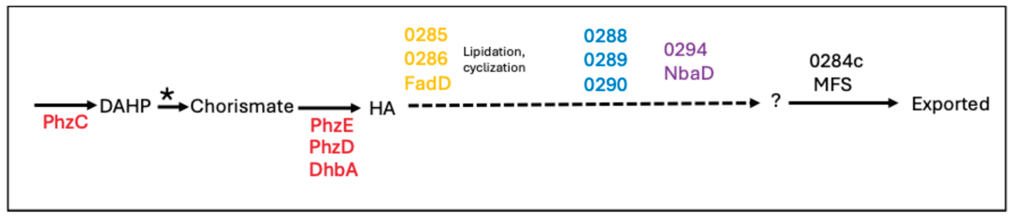
Figure 3.
Hypothesized Roles of the Putative Secondary Metabolite in Mab/Host Macrophage Interactions. Based on work completed in our lab with Mab as well as in other labs with NTMs and M. tuberculosis, our working model is that the secondary metabolite promotes (1) macrophage recruitment to the site of infection, 2) facilitate phagocytosis, and 3) contribute to intracellular survival. It remains to be determined whether the secondary metabolite performs only one are all of these postulated functions as a virulence factor.
Figure 3.
Hypothesized Roles of the Putative Secondary Metabolite in Mab/Host Macrophage Interactions. Based on work completed in our lab with Mab as well as in other labs with NTMs and M. tuberculosis, our working model is that the secondary metabolite promotes (1) macrophage recruitment to the site of infection, 2) facilitate phagocytosis, and 3) contribute to intracellular survival. It remains to be determined whether the secondary metabolite performs only one are all of these postulated functions as a virulence factor.
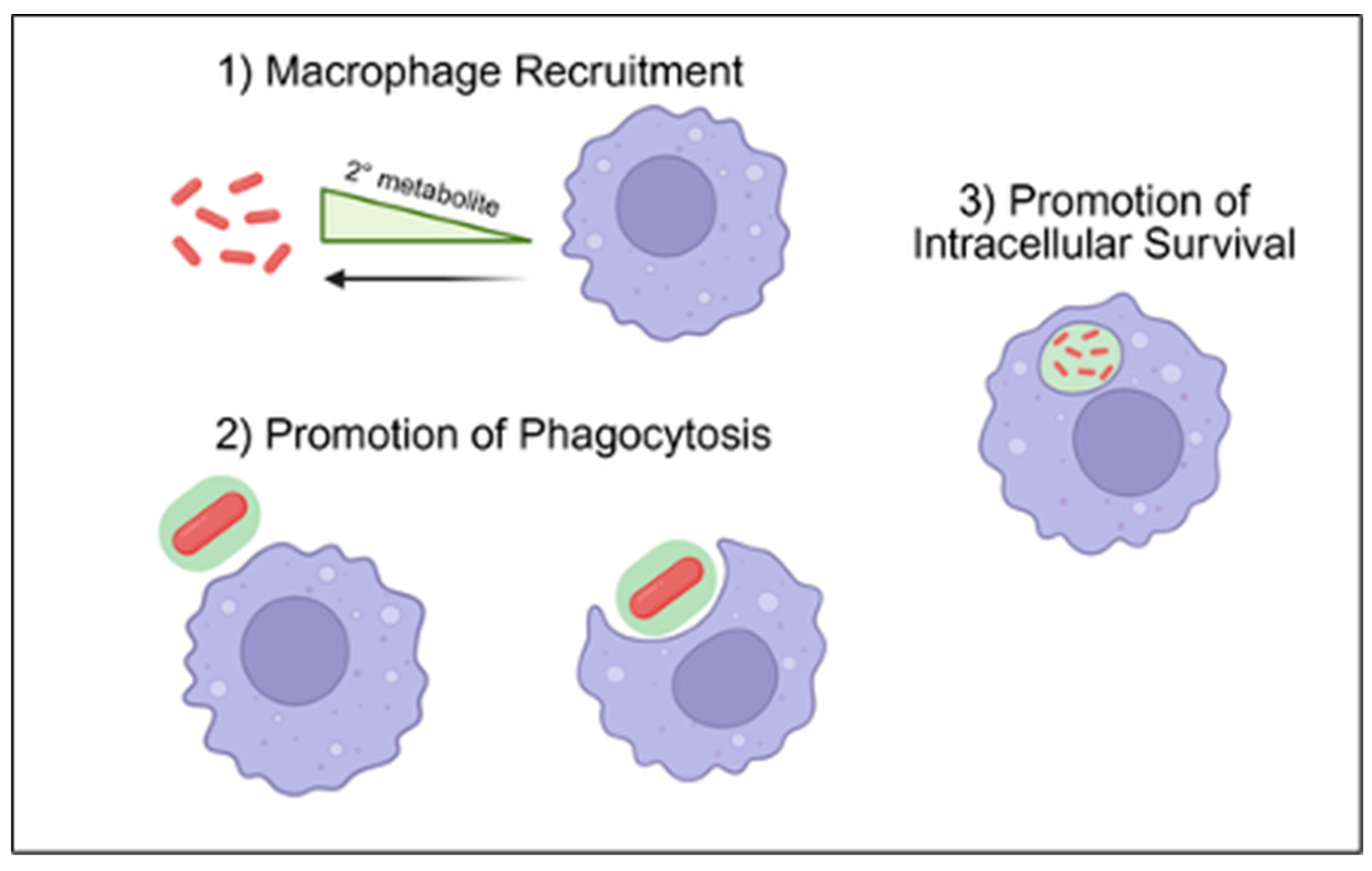
Figure 4.
In vivo visualization of macrophage recruitment and infection at the wound site using confocal microscopy. Overlayed images taken by confocal microscopy of the tail region of a mpeg:gfp transgenic tadpole 24 hrs. post-inoculation by punch wound with wild type Mab expressing DsRed. The red signal identifies DsRed+ Mab and the green signal mpeg+ Mø. The white arrows indicate the overlapping organge signals of Mø (green) that have been infected with Mab (red). Size bar: 10 µm.
Figure 4.
In vivo visualization of macrophage recruitment and infection at the wound site using confocal microscopy. Overlayed images taken by confocal microscopy of the tail region of a mpeg:gfp transgenic tadpole 24 hrs. post-inoculation by punch wound with wild type Mab expressing DsRed. The red signal identifies DsRed+ Mab and the green signal mpeg+ Mø. The white arrows indicate the overlapping organge signals of Mø (green) that have been infected with Mab (red). Size bar: 10 µm.
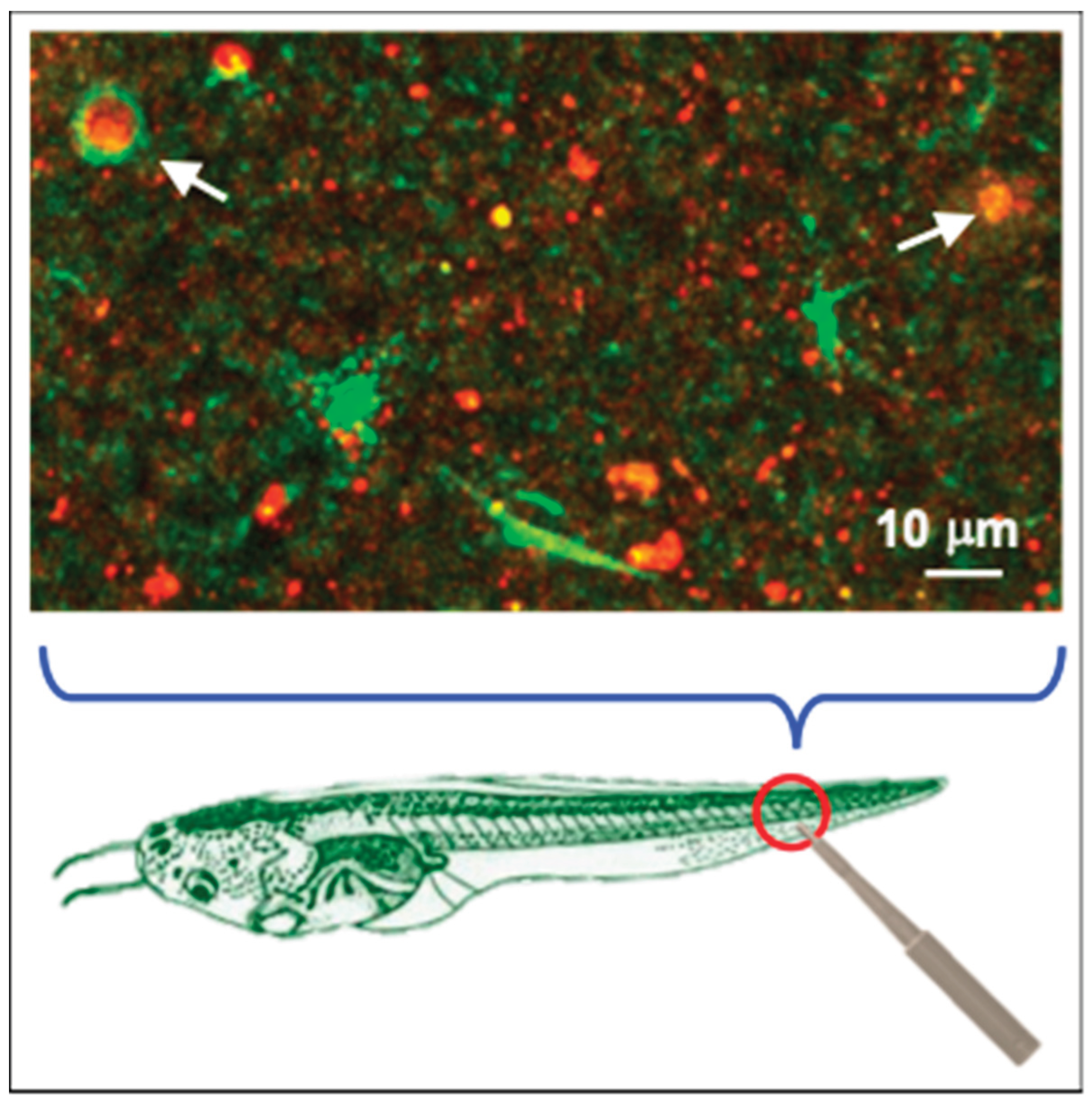

Table 1.
Comparison of Animal Models for Mycobacterial Research.
| Mice | Guinea Pig | Rabbit | Zebrafish | X. laevis | |
| Model for Chronic Mab Infection | Only when immunodeficient | Yes | No | Yes | Yes |
| Presence of Lungs | Present | Present | Present | Absent | Present |
| Model Relevant for Pulmonary Infection | Yes | Yes | Yes | No | Yes |
| Availability of antibodies | Extensive | Limited | Extensive | Limited | Limited |
| Reverse genetics | Yes (developed) | Yes | Yes | Yes (developed) | Yes (developed) |
| Fluorescent reporter gene expression | Yes | ? | ? | Yes | Yes |
| Suitability of live Imaging | Yes (invasive) | Yes (invasive) | Yes (invasive) | Yes1 | Yes1 |
| Main Limitation(s) |
|
|
|
|
|
1Convenient and attractive due to small size, minimal requirement of sterile condition, temperature control and transparency.
Disclaimer/Publisher’s Note: The statements, opinions and data contained in all publications are solely those of the individual author(s) and contributor(s) and not of MDPI and/or the editor(s). MDPI and/or the editor(s) disclaim responsibility for any injury to people or property resulting from any ideas, methods, instructions or products referred to in the content. |
© 2026 by the authors. Licensee MDPI, Basel, Switzerland. This article is an open access article distributed under the terms and conditions of the Creative Commons Attribution (CC BY) license (http://creativecommons.org/licenses/by/4.0/).
Copyright: This open access article is published under a Creative Commons CC BY 4.0 license, which permit the free download, distribution, and reuse, provided that the author and preprint are cited in any reuse.


