
Submitted:
04 August 2026
Posted:
05 August 2026
You are already at the latest version
Abstract
Gait symmetry in children is increasingly understood as a development-dependent continuum rather than a fixed endpoint of perfect bilateral equivalence, a framework that this review extends into a developmental systems model of divergence in cerebral palsy. Typically developing children retain small but measurable inter-limb asymmetries that progressively decrease from early childhood to adolescence. This narrative review asks whether gait asymmetry in hemiplegic and diplegic CP simply fails to reach this normative curve or diverges from it in phenotype- and construct-specific ways that a single asymmetry index cannot capture. Synthesizing longitudinal, cross-sectional, and intervention-based evidence across four interacting domains — direct interlimb symmetry, global and joint-level kinematic deviation, neuromuscular and coordination control, and functional balance — we show that hemiplegic CP is characterized by lateralized divergence involving compensatory changes in the nominally unaffected limb, with partial improvement in directly measured asymmetry over time. Diplegic CP, by contrast, frequently presents with preserved or near-normal left–right symmetry that coexists with progressive, growth-linked deterioration in specific sagittal-plane impairments concealed beneath a stable global gait score, and with a more coupled, less variable coordination strategy that does not correspond to comparable neuromuscular maturation. The recurring finding across both phenotypes is that these domains do not move in parallel, such that reliance on any single metric risks masking clinically important divergences. Building on this evidence, we propose a provisional, non-summative, phenotype-conditioned Trajectory Divergence Index as a hypothesis-generating framework for multi-domain monitoring and identify the absence of longitudinal, multi-domain, multi-phenotype cohorts as the principal evidence gap in the field.
Keywords:
cerebral palsy
; gait asymmetry
; hemiplegia
; diplegia
; gait symmetry
; motor development
; biomechanics
; postural control
; developmental trajectory
1. Introduction
Human gait is not a fixed motor template but a developmental phenomenon: an initially unstable neuromuscular skill that is refined through infancy, childhood, and adolescence into an efficient and stereotyped locomotor pattern [1,2]. Left–right symmetry is commonly treated as one of the clearest markers of this maturation; however, recent systematic evidence indicates that perfect bilateral equivalence is not the normative endpoint of typical development. In a companion systematic review and quantitative synthesis of 297 studies in healthy children, Dominteanu, Stan, and Voinea [3] showed that small but measurable asymmetries persist even in typically developing populations, with pooled effect estimates of Hedges’ g = 0.31 for plantar pressure asymmetry and g = 0.18 for temporal–spatial parameters, both decreasing progressively from early childhood toward adolescence. This developmental trajectory is consistent with primary longitudinal evidence: foot-loading symmetry has been shown to improve progressively from the onset of independent walking through approximately five years of age, after which it stabilizes at a consistently high level [4]. Notably, however, this convergence toward symmetry is not uniform across motor organization levels, even during typical development. Kinematic coordination symmetry between limbs increases steadily across childhood (ages 2–14 years), whereas the symmetry of the underlying muscle synergy structure does not follow the same age-dependent pattern [6]. Typical gait symmetry, in other words, is best conceptualized as a development-dependent continuum that converges toward — but does not fully eliminate — inter-limb variability, and this convergence can proceed at different rates across biomechanical and neuromuscular levels of organization.
This normative curve provides a natural reference frame against which pathological gait can be interpreted and compared. Cerebral palsy (CP) is the most common cause of childhood-onset physical disability, with a pooled prevalence of approximately 2.1 per 1000 live births [7]. Its two principal spastic subtypes impose fundamentally different biomechanical constraints on the symmetry. In hemiplegic (unilateral) CP, a single hemisphere lesion produces lateralized motor impairment, generating an intuitive expectation of persistent and clinically salient asymmetry. In diplegic (bilateral) CP, motor impairment is distributed across both lower limbs, which can produce a superficially symmetric gait profile despite substantial bilateral pathologies. This contrast raises the central problem addressed by the present review: does gait asymmetry in cerebral palsy simply fail to follow the normative developmental curve established for typically developing children, or does it diverge from that curve in phenotype- and metric-specific ways that a single asymmetry index cannot capture?
However, the existing literature does not support a single answer. Evidence assembled for this review indicates that hemiplegic CP shows a pattern of partial, non-linear improvement in directly measured spatial and temporal asymmetry across childhood [8], alongside long-term gains in affected limb kinematic quality [9]; however, tissue-level measures such as paretic plantar-flexor extensibility can worsen over the same developmental window even as motor control improves [10]. Diplegic CP presents an even more paradoxical picture. Global kinematic deviation scores (e.g., the Gait Deviation Index) frequently remain stable across multi-year follow-ups in non-surgical cohorts, while specific sagittal-plane impairments (crouch progression, knee-flexion contracture, and reduced late-stance hip extension) accumulate beneath that stable global score [5,11,12]. In such cases, a global or left–right symmetry metric can actively mask clinically important divergence.
This masking effect is compounded by a second, more conceptually unsettled phenomenon: apparent bilateral symmetry in diplegic CP does not consistently indicate preserved function. Cross-sectional evidence shows that most children with bilateral spastic CP do not meet the thresholds for clinically meaningful side-to-side asymmetry [13,14], and coordination studies describe a more coupled, in-phase pelvis–shoulder and knee–hip strategy in bilateral CP relative to typically developing peers [15,16]. Whether this reflects genuinely preserved bilateral control or a pathologically rigid coordination strategy that has lost the adaptive inter-limb variability characteristic of typical development remains an open and clinically consequential question — one that a binary “symmetric versus asymmetric” framework is structurally unable to resolve.
A further layer of complexity concerns the constructs being measured. The evidence reviewed here shows that direct interlimb symmetry indices, global kinematic deviation scores, electromyographic/coordination measures, and balance-related outcomes do not move in parallel. Kinematic intersegmental coordination in diplegic CP can shift toward typical patterns with age, while the underlying electromyographic timing and modular motor output remain developmentally immature over the same period [17,18], suggesting that biomechanical compensation can occur without corresponding neuromuscular normalization. Similarly, interventions that improve balance and global gait quality do not necessarily normalize direct symmetry ratios: selective dorsal rhizotomy improves spatiotemporal and balance outcomes in bilateral CP without significantly changing preoperative symmetry ratios that were already close to typical values [19], indicating that gait quality gains and interlimb symmetry are, at least in part, dissociable treatment targets.
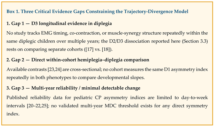
Taken together, this evidence motivates a reconceptualization of gait asymmetry in cerebral palsy as a multi-domain developmental divergence problem rather than a single trajectory tracked with one index. We propose an organizing framework of four interacting domains, summarized schematically in Figure 1: (D1) direct interlimb symmetry, spanning spatial–temporal and plantar-loading indices; (D2) global and joint-level kinematic deviation, capturing sagittal-plane and contracture-related progression that a stable global score can conceal; (D3) neuromuscular and coordination control, encompassing electromyographic timing, muscle-synergy structure, and intersegmental coordination; and (D4) functional balance and fall-risk coupling, linking asymmetry to postural control under static and dynamic demands. This framework is explicitly conceptual and hypothesis-generating; it is not proposed as a validated clinical instrument, given that current evidence on the longitudinal reliability of pediatric asymmetry indices is limited to short (day-to-week) test–retest intervals, with no published multi-year minimal-detectable-change data [20,21,22].
Accordingly, this narrative review addresses five guiding questions: (Q1) how does the developmental trajectory of gait asymmetry in children with CP diverge from the normative maturation pattern established in typically developing children; (Q2) does asymmetry in hemiplegic CP follow a distinct progression pattern compared with the hypothesized “rigid symmetry” phenomenon in diplegic CP; (Q3) what longitudinal or age-stratified evidence exists on stagnation, plateauing, or worsening of asymmetry across growth; (Q4) how does trajectory divergence relate to postural control, balance, and fall risk across developmental stages; and (Q5) what staged monitoring framework could translate trajectory-divergence evidence into actionable clinical decision points? The remainder of this review synthesizes the available evidence across the D1–D4 domains (Section 3), examines the hemiplegia–diplegia phenotype contrast in depth (Section 3, subsection dedicated to phenotype divergence), proposes a provisional, explicitly non-validated progression index for research and monitoring purposes (Section 4), and discusses the methodological limitations, particularly the absence of longitudinal multi-domain, multi-phenotype cohorts, that currently constrain this field (Section 5).
2. Literature Review Strategy and Scope
2.1. Conceptual Framework and Evidence Integration
The evidence for this review was synthesized using a conceptual, systems-oriented approach centered on developmental trajectory divergence rather than static asymmetry magnitude. Building directly on the normative reference established in the companion review of typically developing children, where physiological gait asymmetry was quantified as small in magnitude and progressively decreasing with age (Hedges’ g = 0.31 for plantar-pressure asymmetry; g = 0.18 for temporal–spatial parameters; [3]), the present review reframes cerebral palsy (CP)-related gait asymmetry as a deviation from this expected developmental curve, rather than as an isolated pathological feature interpreted in absolute terms.
This review did not follow a systematic review/PRISMA protocol and did not perform quantitative pooling. It is intentionally conceptual and hypothesis-generating: it integrates heterogeneous longitudinal, cross-sectional, reliability, and intervention evidence to construct an interpretive, multi-domain framework of trajectory divergence in hemiplegic and diplegic CP, explicitly designed to be falsifiable and extendable by future longitudinal, multi-phenotype studies.
2.1.1. Domain Structure (D1–D4)
Rather than treating “gait asymmetry” as a single construct, evidence extraction and synthesis were organized around four interacting domains, established a priori based on the conceptual distinctions that emerged during evidence scoping:
D1 — Direct interlimb symmetry: spatial–temporal asymmetry indices (step/stride length, swing time, stance/single-support time, cadence), plantar pressure, and loading symmetry measures.
D2 — Global and joint-level kinematic deviation: composite indices (Gait Deviation Index, Gait Profile Score, and Gait Variable Scores) and side-specific or bilateral sagittal-plane kinematic/kinetic progression, including contracture-related deterioration that a stable global score can conceal.
D3 — Neuromuscular and coordination control: electromyographic timing and co-contraction, muscle-synergy structure and variability, intersegmental/interlimb coordination, and associated neuroimaging correlates.
D4 — Functional balance and fall risk coupling: static and dynamic postural control, anticipatory postural adjustments, reactive stepping, margin of stability, and fall frequency.This domain structure serves as the organizing principle for both evidence extraction (Section 2.4) and synthesis (Section 3) and is directly reflected in the accompanying evidence table, which classifies each of the 65 sources retained for this review by domain, CP subtype, study design, and follow-up duration.
2.2. Literature Identification and Evidence Selection
The literature was identified through an initial semantic search (Elicit, academic corpus), executed as a structured sequence of targeted queries organized around the guiding questions (Q1–Q5) and subsequently extended with gap-directed queries once initial evidence mapping revealed underrepresented cells within the D1–D4 framework (see Section 2.7). Searches were conducted between April and August 2026, and combined concepts related to children/adolescents, cerebral palsy, hemiplegia, diplegia, gait asymmetry/symmetry, motor development, postural control, balance, spatiotemporal gait parameters, plantar pressure, electromyographic asymmetry, muscle synergy, intersegmental coordination, GMFCS, fall risk, reliability, minimal detectable change, ankle-foot orthosis, selective dorsal rhizotomy, and single-event multilevel surgery.
Temporal scope: 2010–2026, with earlier foundational studies retained for conceptual continuity where directly relevant to index formulation or normative reference (e.g., early symmetry index reliability work).
Topographic restriction: Studies were restricted to spastic hemiplegic (unilateral) and diplegic (bilateral) CP, excluding dyskinetic, ataxic, and mixed subtypes, to preserve the mechanistic homogeneity of the compensation patterns analyzed. Studies enrolling mixed CP samples were retained only when hemiplegic and/or diplegic subgroups were reported separately or when the study addressed a domain (e.g., reliability, neuroimaging) for which subtype-specific stratification was not the primary aim.
2.3. Evidence Selection Criteria (Population–Concept–Context Framework)
Population: Children and adolescents (2–18 years) with confirmed spastic hemiplegic or diplegic CP; typically developing children were included only as normative comparators.
Concept: instrumented gait asymmetry, kinematic/kinetic deviation, neuromuscular coordination, or balance/fall-risk outcomes, analyzed with explicit or inferable reference to age, growth stage, or GMFCS level, and mapped to one or more of the D1–D4 domains.
Context: laboratory, clinical, or applied rehabilitation settings; peer-reviewed, English-language publications.
Studies were retained when they met at least one of the following conditions: (1) reported quantified D1/D2/D3/D4 outcomes with age-stratified or longitudinal comparison, enabling trajectory inference; (2) reported comparative data distinguishing hemiplegic from diplegic patterns on a shared outcome measure within or across cohorts; (3) reported relationships between asymmetry/coordination and postural control, balance, or fall risk; (4) reported reliability, minimal detectable change, or measurement-repeatability data for any D1–D3 index; (5) reported intervention effects (orthotic, surgical, neurosurgical, or training-based) on any D1–D4 outcome, with explicit pre/post or longitudinal comparison.
Studies were deprioritized when they: (1) reported only single-timepoint, cross-sectional values without developmental, growth, or comparative context; (2) pooled mixed CP subtypes without any subtype-specific or subgroup-level reporting; (3) relied solely on non-instrumented, observational gait assessment; or (4) lacked sufficient methodological detail to classify the outcome within the D1–D4 framework or to infer directionality of change.
2.4. Data Extraction and Evidence Table Construction
For each retained source, the following information was extracted into a structured evidence table: author/year, CP subtype and population characteristics, study design and follow-up duration, GMFCS level and sample size (where reported), assigned domain(s) (D1–D4, or reliability), key findings, and an explicit note on relevance to the trajectory-divergence model. This extraction scaffold — rather than a narrative summary alone — was used to (i) verify the comparability of design and phenotype before drawing cross-study conclusions, (ii) identify domain-by-phenotype cells with sparse or absent direct evidence, and (iii) support a second, gap-directed round of literature identification (Section 2.2) once the initial evidence base had been mapped. The final evidence table comprised 65 sources.
2.5. Qualitative Appraisal and Methodological Weighting
Consistent with the companion review’s use of JBI-informed appraisal principles — but without formal aggregated scoring, given the narrative/conceptual nature of this review — greater interpretive weight was assigned to (i) longitudinal and age-stratified studies over single-timepoint cross-sectional studies; (ii) studies reporting subtype-specific (hemiplegic or diplegic only) findings over mixed-CP aggregates; (iii) studies with within-cohort hemiplegia–diplegia comparisons over indirect, cross-study comparisons; and (iv) studies demonstrating concordance across measurement modalities or domains (e.g., agreement between kinematic and electromyographic findings) within the same topographic subtype.
2.6. Sources of Bias and Mitigation Strategy
Severity/sampling bias: predominance of GMFCS I–II cohorts across the evidence base; GMFCS III–V substantially underrepresented, particularly within D3 and D4 domains.
Instrumentation bias — heterogeneity across laboratory-based 3D motion analysis, pressure platforms, wearable sensors, and electromyography, paralleling the reliability contrast established in the companion review (ICC = 0.88 for baropodometric vs. ICC = 0.54 for stabilometric measures in typically developing children)–was used here as an interpretive reference for CP-specific measurement stability.
Developmental-contextual bias — extrapolation from cross-sectional snapshots or medium-term (months-to-few-years) follow-up to lifespan developmental trajectories, despite plausible divergence driven by skeletal growth, spasticity progression, and prior surgical/orthotic/neurosurgical interventions.
Index-formulation bias — mathematical non-equivalence across D1 (direct symmetry ratio, normalized/weighted indices), D2 (composite deviation scores), and D3 (waveform-similarity, synergy-based) metrics, such that a stable score in one domain cannot be assumed to imply stability in another (Section 3 documents multiple empirical instances of this dissociation).
Reporting/indirect-comparison bias: Most hemiplegia–diplegia contrasts in the evidence base are cross-study rather than within-cohort. Where within-cohort comparisons exist (e.g., [23,24]), they are cross-sectional rather than longitudinal, limiting causal or trajectory-level inferences about phenotype divergence.
2.7. Transparency on Evidence Gaps
Consistent with the iterative, gap-directed search strategy described in Section 2.2, three evidence gaps were identified during table construction and were treated as an explicit part of the review’s scope rather than an incidental limitation (see Box 1): (i) no longitudinal, repeated-measures electromyographic or muscle-synergy study confined exclusively to diplegic CP was identified; (ii) no cohort was identified that repeatedly measured a direct D1 asymmetry index in both hemiplegic and diplegic CP within the same children, permitting formal comparison of age-related slopes; and (iii) no multi-year test–retest reliability or validated minimal-detectable-change threshold was identified for any direct pediatric CP asymmetry index, with available reliability evidence limited to day-to-week intervals [20,21,22,25]. These gaps directly inform the cautions attached to the provisional progression index proposed in Section 4 and are revisited as priorities for future research in Section 5, respectively.
2.8. Temporal Resolution and Analytical Boundaries
Priority was given to evidence enabling differentiation between (i) transitional or compensatory divergence consistent with a delayed but still normative maturation process and (ii) consolidated or structural divergence indicating a durable departure from the expected trajectory established in typically developing children. Consistent with the conceptual and non-systematic nature of this review, no causal or diagnostic thresholds were proposed. The scope is intentionally limited to mechanistic-developmental synthesis and the construction of a provisional, explicitly non-validated framework intended to generate testable hypotheses for future longitudinal, multi-domain, multi-phenotype research.
3. Mechanistic and Developmental Synthesis
Before presenting the domain-by-domain evidence, Figure 1 summarizes the organizing framework proposed in this review: the four interacting domains (D1–D4) through which hemiplegic and diplegic CP are hypothesized to diverge from the normative symmetry trajectory established in the companion review. The domain-specific evidence underlying each branch of this figure is presented in Section 3.1, Section 3.2, Section 3.3 and Section 3.4, the phenotype contrast itself is developed in Section 3.5, and the cross-domain implications of the model are addressed in Section 3.6.
3.1. D1 — Direct Interlimb Symmetry
Direct interlimb symmetry, spatial–temporal asymmetry indices, and plantar-loading symmetry are the domains most directly comparable to the normative curve established in the companion review; however, it is also the domain in which hemiplegic and diplegic CP diverge most clearly from one another.
In hemiplegic CP, the strongest direct longitudinal evidence comes from a prospective two-year cohort of 27 ambulant children (GMFCS I–II, age 8–15 years), which recorded gradual improvement in spatial and temporal asymmetry indices over 24 months, with children who began with lower asymmetry improving locomotor capacity faster [8]. This is compatible with partial convergence toward the normative curve rather than either full normalization or static persistence. However, this finding is an association rather than evidence of a causal mechanism and remains the only prospective D1 trajectory study identified for this subtype. Compensation in the nominally unaffected limb further complicates a simple lateralized reading of this asymmetry: a three-dimensional gait analysis found not only elevated lower-limb asymmetry relative to typically developing controls, but also measurable deviation of the “uninvolved” limb from typical gait parameters, consistent with compensatory adaptation rather than true independence between the two limbs [26]. This finding qualifies the improvement reported by Elnaggar & Elfakharany [8]: a narrowing asymmetry index does not necessarily indicate that the less affected limb is developing along an unperturbed, typical trajectory.
In diplegic CP, the picture is markedly different. Cross-sectional evidence in early walkers shows that side-to-side spatiotemporal symmetry does not differ significantly from experience-matched typically developing children, despite uniformly reduced performance and greater stride-to-stride variability [14]. In an older bilateral spastic cohort, 68% of children did not meet the threshold for clinically meaningful side-severity asymmetry, and children with primary crouch were particularly symmetric [13], while the remaining 32% showed clinically meaningful asymmetry, underscoring that diplegia is not a uniformly symmetric phenotype. A direct within-cohort comparison further clarifies this contrast: hemiplegic CP shows a higher Gait Profile Score in the paretic than in the non-paretic limb, whereas diplegic CP shows no significant between-limb difference in the same global score [23]. Task demand can unmask the asymmetry that overground walking conceals: a comparison of 24 diplegic and 25 hemiplegic children found a significant diagnosis-group difference across 22 symmetry parameters, with running reducing symmetry in most parameters relative to walking [24].
Overall, D1 evidence supports a phenotype-specific pattern: hemiplegic CP shows lateralized, partially improving asymmetry that nonetheless implicates both limbs in compensation, while diplegic CP more often shows preserved or near-normal left–right symmetry that coexists with substantial underlying pathology, a dissociation addressed in detail in Section 3.5.
3.2. D2 — Global and Joint-Level Kinematic Deviation
Composite kinematic scores (Gait Deviation Index [GDI], Gait Profile Score [GPS], and Gait Variable Scores [GVS]) capture a broader construct than D1 and reveal a developmental pattern that a symmetry index alone would miss entirely.
In diplegic CP, three non-surgical longitudinal cohorts converge on the same finding: global scores remain relatively stable across multi-year follow-ups, while specific sagittal-plane impairments progressively worsen beneath that stable summary. A 180-child cohort followed for a mean of 4.9 years showed a stable global GDI alongside increasing mid-stance knee flexion and stance dorsiflexion and decreasing late-stance hip extension [11]. An earlier non-surgical cohort similarly found worsening flexed-knee gait and hamstring-related measures over approximately six years, without significant changes in the global GDI or normalized temporal parameters [5]. Across childhood to skeletal maturity, a 100-person cohort documented a qualitative shift in the dominant gait pattern — from equinus/jump-knee in early childhood to crouch at maturity — with increasing stance dorsiflexion and worsening passive knee-flexion contracture [12]. This is the clearest evidence in the entire dataset for masked divergence: a global D2 score that is uninformative or even reassuring while the underlying gait pathology deteriorates.
In hemiplegic CP, D2 evidence points toward long-term improvement rather than deterioration: a 10.5-year follow-up of 52 GMFCS-I children found affected-side gait-profile deviation improving from a mean 8.5° at age 9.3 to 7.2° at age 19.7, with particularly clear gains in ankle/foot variables among children who underwent single-level surgery [9]. This measures the overall affected limb kinematic quality rather than a left–right index; therefore, it cannot be used to infer D1 normalization, a distinction that must be maintained explicitly rather than assumed.
Treatment substantially modifies D2 trajectories, independent of age. In diplegic CP, followed by skeletal maturity, gait quality was maintained without single-event multilevel surgery (SEMLS) and improved further in children who underwent SEMLS [27]. A broader serial gait cohort spanning both phenotypes (15 hemiplegic and 16 diplegic children) found that 86% of limbs improved or remained unchanged by late adolescence, with larger gains earlier in childhood and in intervals coinciding with surgery [28]. A cautionary finding from a large cross-sectional sample (N = 109) indicates that GDI variation is explained primarily by GMFCS level rather than unilateral-versus-bilateral topography [29], reinforcing that severity and phenotype are partially independent axes and should not be conflated when interpreting D2 trajectories.
3.3. D3 — Neuromuscular and Coordination Control
D3 evidence provides the clearest mechanistic account of why D1 and D2 can move independently of one another and reveals that even apparent improvement at the kinematic level does not necessarily reflect neuromuscular normalization.
The central comparative study contrasted typically developing children, in whom electromyographic (EMG) bursts progressively shorten and spinal motor output reorganizes with age, against children with CP (19 diplegic, 16 hemiplegic, aged 2–12), who showed markedly limited age-related change in EMG duration, motoneuron output organization, intersegmental coordination, and foot-trajectory control bilaterally in diplegia and on the affected side in hemiplegia [18]. This finding establishes a plausible neural substrate for the persistence of immature, broadly timed muscle activation, independent of chronological age.
Critically, neuromuscular immaturity can coexist with apparent kinematic improvement. In a retrospective cohort study of 162 children with diplegic CP, thigh–shank and shank–foot intersegmental coordination (assessed via continuous relative phase) differed from typically developing controls at baseline but shifted toward typical patterns over follow-up in the absence of surgical intervention [17]. However, this kinematic convergence occurred against the background of EMG timing that, according to Cappellini et al. [18], did not show comparable maturation. The most defensible interpretation is that diplegic CP can achieve more typical segment-level timing through biomechanical compensation without acquiring correspondingly typical muscle-level control — a dissociation with direct relevance to the trajectory-divergence model, since it implies that D2/D3 convergence is not unitary and that neither domain alone can certify “improvement.”
Individual-level heterogeneity further complicates the group-level D3 trends. In a 20-month non-surgical cohort, average dynamic motor control, GPS, and GVS scores showed no group-level change, but individual changes in motor control of the more-involved leg tracked individual changes in hip kinematics [30], indicating that D3 acts as a person-specific trajectory modifier rather than a uniform developmental process. Coordination-variability studies reinforce a “more coupled, less flexible” interpretation of diplegic gait: children with CP show more in-phase knee–hip coupling and lower coordination variability at selected gait phases, with greater in-phase coupling associated with greater, not lesser, gait deviation [16]; bilateral CP similarly shows more in-phase pelvis–shoulder coupling than typically developing children, alongside slower gait, shorter steps, wider base, and prolonged double support [15]. At the muscle-synergy level, CP is characterized by fewer mature synergies but greater stride-to-stride variability in which synergies are recruited [31], and greater reliance on CP-specific, atypical synergies is directly related to poorer temporal–spatial performance, including asymmetry magnitude [32], providing a direct mechanistic link between D3 and D1.
In hemiplegic CP, neural-adaptation studies suggest that the capacity for modifiable neural control is substantially preserved: children with hemiplegic CP adapt their step length and joint kinematics to unilateral leg-weighting perturbations at a level broadly comparable to typically developing peers, despite baseline asymmetry and reduced local dynamic stability [33,34]. Neuroimaging work adds a compensatory dimension: adolescents with unilateral CP show greater cortical activation and bilateral EEG–EMG coherence than typical peers for similar motor output during treadmill gait, despite no group difference in muscle synergy structure [35], consistent with compensatory neural recruitment rather than normalized control. Short-term training-linked plasticity has been demonstrated (e.g., increased resting-state sensorimotor connectivity after two weeks of robot-assisted gait training; [36]), but no evidence currently establishes that ordinary maturation, absent targeted intervention, produces durable D3 normalization in either phenotype.
3.4. D4 — Functional Balance and Fall-Risk Coupling
D4 evidence is the domain in which the hemiplegia–diplegia contrast is most consistently and directly documented, although its translation into actual fall risk remains unresolved.
Asymmetry and balance are most clearly linked in hemiplegic CP: in a cross-sectional study of 85 children, greater temporal and spatial asymmetry was associated with a smaller dynamic limit of stability and a wider heel-to-heel base of support, the latter explaining more variance in temporal than spatial asymmetry, consistent with a compensatory widening strategy as balance control becomes more demanding [37]. A complementary study found that weight-bearing asymmetry during quiet standing was linked to affected-side ankle dorsiflexion during gait, although the correlations across the full cohort were generally weak [38]. Longitudinally, higher asymmetry remained associated with lower community balance and mobility scores across 24 months of follow-up in the same cohort discussed under D1 [8], although directionality (whether asymmetry limits balance, poor balance drives asymmetry, or both reflect a shared underlying factor) cannot be established from this design.
Direct comparisons between hemiplegia and diplegia converge on a consistent phenotypic distinction. Diplegic children show larger center-of-pressure displacement and greater mediolateral sway than hemiplegic children under both eyes-open and eyes-closed conditions [39], although this difference emerges specifically under challenging sensory conditions rather than during simple standing [40]. Anticipatory postural adjustment studies provide the clearest mechanistic distinction between phenotypes: diplegic children generate smaller anticipatory postural adjustments than both typically developing and hemiplegic children, with higher tonic baseline muscle activity and a more posterior center-of-pressure shift [41]; under loading perturbation, both subtypes show delayed and reduced anticipatory activation, but diplegia is additionally characterized by elevated background postural activity, whereas hemiplegia’s background activity more closely resembles that of typically developing children [42]. Together, these findings support a coherent distinction: diplegia manifests as a distributed bilateral postural-control deficit with elevated tonic activity and reduced capacity to scale postural responses, whereas hemiplegia manifests as a lateralized deficit with greater capacity for compensation by the less-affected side, directly reinforcing the D1/D3 pattern described above. Notably, this distinction is not universally captured by the simpler clinical instruments. A comparison of 50 diplegic and 50 hemiplegic children found no significant group differences in the Early Clinical Assessment of Balance or the Pediatric Reach Test [43], indicating that instrument sensitivity, not merely underlying physiology, determines whether a phenotype distinction is observed.
However, whether this balance-phenotype distinction translates into differential fall risk has not been established. The largest available analysis (parent-reported falls in 1,063 children with CP) found that specific gait pathology subtypes (internal hip rotation, intoeing, and stiff-knee gait) were not associated with fall frequency after adjusting for age and GMFCS level [44]. A more recent cohort study found that falls correlated with functional capacity measures (sit-to-stand, lateral step-up, and kneel-to-stand) rather than with gait pattern or asymmetry classification [45]. Diplegia-specific intervention trials have shown improvements in laboratory-derived fall-risk scores following balance training [46] and anti-gravity treadmill training [47]; however, neither establishes a reduction in real-world, community-reported falls. Accordingly, D4 should be considered as evidence of a genuine phenotype-specific balance-control mechanism rather than a validated fall-risk predictor.
3.5. Phenotype Divergence: Hemiplegic Versus Diplegic CP (Q2)
Synthesizing across D1–D4, a coherent — if still largely cross-sectional and cross-study rather than longitudinally within-cohort — picture of phenotype-specific divergence emerges (Table 1):
Hemiplegic CP is characterized by lateralized impairment across all four domains: clear affected-side D1 asymmetry that shows partial improvement over time, with measurable involvement of the nominally unaffected limb [8,26]; D2 improvement in affected-limb kinematic quality over long-term follow-up [9]; D3 control that remains atypical but retains substantial adaptive/plastic capacity, often at the cost of compensatory cortical recruitment [33,35]; and D4 balance deficits directly coupled to asymmetry magnitude, with a compensatory postural strategy (wider heel-to-heel base of support) partially explaining the association [37,38].
Diplegic CP is characterized by distributed, bilateral impairment that can present as preserved or improving D1 symmetry [13,14,23] while D2 shows progressive, growth-linked deterioration in specific sagittal-plane measures beneath a stable global score [11,12]; D3 shows a more coupled, less variable coordination strategy that trends toward typical segment-level timing without corresponding EMG-level maturation [15,17,18]; and D4 shows a distributed postural-control deficit with elevated tonic muscle activity and reduced anticipatory scaling [41,42].
This contrast supports the rigid/coupled symmetry hypothesis proposed for diplegic CP, but only in a qualified manner. The apparent D1 symmetry in diplegia does not represent a uniform phenomenon: it can coexist with (a) genuinely preserved bilateral coordination in a meaningful subset of children (the 68% “symmetric” cases in [13]), (b) a pathologically coupled, in-phase coordination strategy consistent with reduced segmental dissociation [15,16], or (c) simple non-equivalence between what D1 and D3/D4 metrics measure. No study in the evidence base directly tests which of these three explanations predominates within the same children, and this remains the most consequential open question raised by the present synthesis (see Section 2.7 and Section 5).
3.6. Cross-Domain Dissociation as the Central Finding
Figure 2 illustrates this dissociation for the phenotype where it is most consequential. Panel (a) shows that direct interlimb symmetry (D1) in diplegic CP can track close to the normative trajectory across childhood, while panel (b) shows that global/joint-level kinematic deviation (D2) can diverge from that same normative trajectory over the identical age range, which is the schematic signature of masked divergence documented empirically in Section 3.2.
As Figure 2 illustrates schematically, and as documented empirically in Section 3.1, Section 3.2, Section 3.3, Section 3.4 and Section 3.5, the single most consistent and clinically important pattern is that D1, D2, D3, and D4 do not move in parallel in either phenotype. A stable D2 score can conceal worsening joint-specific pathology (diplegia; [5,11]); improving D2/kinematic coordination can occur without corresponding D3/EMG maturation (diplegia; [17] vs. [18]); a narrowing D1 index can coexist with continued deviation of the “unaffected” limb (hemiplegia; [26]); and intervention can improve D4/global gait quality without measurably changing D1 symmetry ratios that were already near-typical preoperatively (selective dorsal rhizotomy) [19]). This cross-domain dissociation is the primary evidentiary justification for the multi-domain progression framework developed in Section 4, and it directly answers the central question posed in Section 1: gait asymmetry in CP does not diverge from the normative developmental curve along a single trajectory, but along domain- and phenotype-specific pathways that a unidimensional symmetry index cannot detect in isolation.
4. A Provisional Trajectory Divergence Index (TDI): Toward Multi-Domain Monitoring
4.1. Rationale and Design Principles
The central finding of Section 3 — that D1 (interlimb symmetry), D2 (global kinematic deviation), D3 (neuromuscular coordination), and D4 (functional balance), first outlined schematically in Figure 1, do not move in parallel, and that improvement or stability in one domain can conceal deterioration in another — has a direct clinical corollary: no single existing metric is sufficient for longitudinal surveillance of gait asymmetry in cerebral palsy. A stable Gait Deviation Index can coexist with worsening crouch [11], improving intersegmental coordination can coexist with static EMG immaturity [17,18], and a near-normal symmetry ratio can coexist with a distributed postural-control deficit [19]. Any composite score that collapses these domains into a single number would reproduce precisely the masking effect that this review has identified as its central problem.
Therefore, we propose a profile-based Trajectory Divergence Index (TDI) — termed an “index” for brevity, but structurally a four-domain monitoring profile rather than a single composite score–intended to make cross-domain dissociation visible rather than averaging it away. The TDI was explicitly designed as a research and clinical reasoning heuristic, not as a diagnostic or prognostic instrument. Three design principles follow directly from the evidence synthesized in Section 3: (1) domains are scored independently and never summed into a single index value, since summation would recreate the masking problem documented throughout Section 3; (2) interpretation is phenotype-conditioned, since the same D1 value carries different clinical meanings in hemiplegic versus diplegic CP (Section 3.5); (3) the index flags direction and domain of change, not absolute severity, given the near-total absence of multi-year reliability and minimal-detectable-change data for pediatric CP asymmetry indices (Section 2.7).
4.2. Domain Structure and Provisional Scoring Logic
Each domain is scored on an ordinal 0–3 scale reflecting the degree of divergence from the phenotype-expected trajectory, not from an absolute normative value (Table 2).
D1 (Interlimb symmetry) — indicators: spatial/temporal asymmetry index trend and plantar-loading symmetry trend. Because diplegic CP can show near-normal D1 values despite significant underlying pathology (Section 3.1 and Section 3.5), a low D1 score in diplegia should never be interpreted as reassurance in isolation; it must always be read jointly with D2.
D2 (Global/joint-level kinematic deviation) — indicators: GDI/GPS/GVS trend; limb-specific sagittal plane deviation (particularly knee flexion, ankle dorsiflexion, hip extension in stance). Given the masked-divergence pattern documented in diplegic CP [5,11,12], D2 scoring should be based on component-level (GVS) trends, not the aggregate GDI/GPS alone; this is the single most important operational recommendation arising from Section 3.
D3 (Neuromuscular/coordination control) — indicators: EMG timing/co-contraction pattern, muscle-synergy structure and stride-to-stride variability, and intersegmental coordination (continuous relative phase), where available. Given the evidence that D3 can dissociate from D2 (Section 3.3), the D3 score should be obtained independently rather than inferred from kinematic improvement.
D4 (Functional balance/fall-risk coupling) — indicators: dynamic limit of stability or margin of stability, anticipatory postural adjustment quality, and functional transition/step-up performance. Consistent with Section 3.4, D4 scoring should prioritize dynamic and task-based measures over static quiet-stance sway and should not be used to infer fall probability without corroborating functional capacity data [44,45].
4.3. Phenotype-Specific Reading of the Profile
Because the D1–D4 relationships differ by topographic subtype (Section 3.5), the same four-domain profile is interpreted differently depending on the phenotype. In hemiplegic CP, a clinically informative profile prioritizes D1 and D4 together (asymmetry–balance coupling is well documented; Section 3.4) alongside D2, which is tracked specifically on the affected limb. A “low D1, low D2” profile is more straightforwardly reassuring in this phenotype than in diplegia, although D1 improvement should still be cross-checked against contralateral limb kinematics [26] rather than assumed to reflect isolated affected-limb change. In diplegic CP, a “low D1” reading carries little standalone diagnostic value (Section 3.1 and Section 3.5) and must be interpreted jointly with D2 component trends and D3 coordination-coupling measures. A diplegic profile showing a low D1 combined with rising D2 component scores (e.g., increasing knee flexion or dorsiflexion) or increasingly in-phase D3 coordination should be flagged as a candidate instance of masked divergence, warranting a component-level and coordination-level evaluation that a global score alone would not trigger.
4.4. Staged Monitoring Framework (Addressing Q5)
Consistent with the guiding question posed in Section 1 (Q5), the TDI is intended to translate into three staged clinical decision points, calibrated to the domain scores above: (1) Routine surveillance (all domains ≤ 1): continue standard follow-up interval; no change to monitoring frequency. (2) Targeted domain re-evaluation (any domain = 2, or a diplegic profile combining low D1 with rising D2/D3 indicators): trigger domain-specific reassessment, for example, component-level 3D gait analysis if D2 is flagged, EMG/synergy assessment if D3 is flagged, rather than relying on the global score that initially appeared reassuring. (3) Multidisciplinary review (any domain = 3, or persistent multi-domain divergence across consecutive assessments): orthopedic, orthotic, and rehabilitation-team review, informed by the specific domain(s) driving divergence rather than a single undifferentiated “asymmetry” or “gait deviation” label. This staged structure is intentionally decision-supportive rather than prescriptive; it does not specify treatment and does not assign fixed numerical thresholds to the ordinal scores, given the absence of validated minimal-detectable-change data (Section 2.7).
4.5. Validation Status and Explicit Limitations
The TDI is presented as a hypothesis-generating conceptual instrument, not a validated clinical or research tool. Three limitations, following directly from Section 2.7, must be stated explicitly: no multi-year reliability or minimal-detectable-change data exist for any of the D1–D3 indicators proposed here [20,21,22,25] — the ordinal 0–3 scoring is therefore a structural proposal for organizing clinical reasoning, not a psychometrically derived scale; no cohort has measured all four domains concurrently and longitudinally in the same children, for either phenotype — the domain-interaction patterns underlying the TDI are each supported by separate studies (Section 3), not by a single integrated dataset; no direct, within-cohort, longitudinal hemiplegia–diplegia comparison exists (Section 2.7) to confirm that the phenotype-specific interpretive rules proposed in Section 4.3 hold when both phenotypes are assessed under identical protocols.
Accordingly, the TDI should be read as a structured research proposal — a candidate framework for the prospective, multi-domain, multi-phenotype cohort study identified as the field’s principal evidence gap — rather than as a tool ready for clinical deployment. Its value at this stage lies in making explicit, testable predictions (e.g., that a diplegic cohort with stable global GDI will show measurable D2-component or D3-coordination divergence on closer inspection) that future longitudinal research can directly confirm or refute.
5. Discussion
5.1. Summary of Principal Findings
This review set out to answer the five guiding questions posed in Section 1. The synthesized evidence (Section 3) allows each question to be answered directly, with explicit acknowledgment of where the answer remains partial.
Q1 — How does the developmental trajectory of gait asymmetry in children with CP diverge from the normative maturation pattern established in typically developing children? It diverges non-uniformly and domain-specifically. The normative curve established in the companion review [3] — small, progressively decreasing asymmetry from early childhood to adolescence — was not simply exceeded or delayed in CP. Instead, different domains diverge along different paths, as summarized schematically in Figure 1 and Figure 2: D1 (interlimb symmetry) can show partial convergence toward the normative curve in hemiplegic CP or can remain within a near-normal range throughout diplegic CP; D2 (global kinematic deviation) can appear developmentally stable while joint-specific pathology accumulates beneath it — the masked-divergence pattern depicted in Figure 2b; D3 (neuromuscular control) shows the clearest and most persistent divergence, with limited age-related maturation of EMG timing and motor-output organization regardless of phenotype (Section 3.3).
Q2 — Does asymmetry in hemiplegic CP follow a distinct progression pattern compared with the hypothesized “rigid symmetry” in diplegic CP? The contrast is the review’s most consistent finding (Section 3.5). Hemiplegic CP shows lateralized divergence across all four domains, coupled with measurable compensatory involvement of the nominally unaffected limb. Diplegic CP shows distributed, bilateral divergence that frequently presents as preserved D1 symmetry, but the underlying evidence does not support treating this symmetry as inherently pathological in a uniform sense; rather, it can reflect either genuinely preserved bilateral coordination, a coupled/rigid coordination strategy, or simple non-equivalence between D1 and other domains (Section 3.5). Therefore, the original “rigid symmetry” hypothesis is substantiated only in its qualified form.
Q3 — What longitudinal or age-stratified evidence exists on the stagnation, plateauing, or worsening of asymmetry across growth? All three patterns are documented, but for different domains and subsets of children rather than as alternative descriptions of the same phenomenon. Component-level D2 worsening beneath a stable global score is the best-documented growth-related pattern specific to nonsurgical diplegic cohorts (Section 3.2). D1 improvement has been documented prospectively only in hemiplegic CP over a two-year window (Section 3.1); no comparably designed prospective D1 study exists for diplegic CP.
Q4 — How does trajectory divergence relate to postural control, balance, and fall risk across developmental stages? Asymmetry and balance are coupled in hemiplegic CP, and postural control mechanisms differ systematically between phenotypes (diplegia: distributed, tonic; hemiplegia: lateralized, compensable) (Section 3.4). However, this review found no evidence that gait pattern or asymmetry classification prospectively predicts real-world falls. This is an important negative finding that constrains the use of D4 within the proposed monitoring framework (Section 4.2).
Q5 — What staged monitoring framework can translate trajectory-divergence evidence into actionable clinical decision points? The provisional Trajectory Divergence Index (Section 4) offers one candidate answer, structured explicitly as a profile rather than a composite score, with phenotype-conditioned interpretation and three-stage decision points. Its status is hypothesis-generating and not validated (Section 4.5).
5.2. Clinical and Research Implications
The findings converge on a single practical implication that follows directly from Section 3.6: no single gait metric, however well-validated for its own construct, is sufficient for the longitudinal surveillance of asymmetry in cerebral palsy. A stable global gait deviation score should not be interpreted as evidence of trajectory stability, particularly in diplegic CP, where component-level sagittal plane deterioration can occur beneath an unchanged aggregate score (Section 3.2). Conversely, a near-normal interlimb symmetry ratio should not be interpreted as evidence of preserved bilateral function in diplegic CP without corroborating coordination- and kinematic-level assessment (Section 3.1 and Section 3.5).
For clinical practice, this argues for domain-explicit reporting rather than reliance on any single index: clinicians and researchers documenting gait status in CP should report interlimb symmetry, global deviation scores, and — where feasible — coordination or EMG-based measures as distinct, co-reported values rather than as convergent proxies for one another in the format illustrated in Table 1. This recommendation applies most directly to diplegic CP, where the risk of D1/D2 masking has been best documented. However, the evidence from the review on compensatory contralateral limb involvement (Section 3.1) suggests that the same caution is warranted in hemiplegic CP.
For research, the most direct implication concerns study design: the three evidence gaps identified in Section 2.7 (Box 1) — the absence of longitudinal D3 studies confined to diplegic CP, the absence of direct within-cohort longitudinal hemiplegia–diplegia comparisons, and the absence of multi-year reliability data for any pediatric CP asymmetry index — are not peripheral omissions but structural obstacles to testing the trajectory-divergence model proposed here. A single prospective cohort study measuring D1–D4 concurrently in both phenotypes, with repeated assessment over several years and a locally derived minimal-detectable-change threshold, would directly address all three gaps simultaneously and would constitute the most informative single study that this field could currently undertake.
5.3. Strengths
This review integrates evidence across a substantially broader construct space than is typical for CP gait literature, which has more often examined interlimb symmetry, global kinematic deviation, neuromuscular control, and balance as separate literatures rather than as interacting domains of a single developmental process. By organizing evidence extraction and synthesis around an explicit four-domain framework (Section 2.1.1) established prior to full-text synthesis, this review was able to surface a pattern — cross-domain dissociation — that is unlikely to emerge from any single-domain review and is directly corroborated by multiple independent studies operating in different domains (Section 3.6).
The review’s evidence base was extended iteratively: an initial round of targeted searches was followed by a second, gap-directed round once systematic mapping against the D1–D4 framework revealed underrepresented cells (Section 2.2 and Section 2.7). This two-stage process yielded several of the review’s most direct findings, including within-cohort hemiplegia–diplegia comparisons [23,24] and topography-specific balance-control contrasts [39,41,42], which were not identified in the initial search round.
The explicit methodological alignment with a companion systematic review conducted by the same research group on typically developing children [3] provides a quantitatively grounded normative reference, a feature uncommon in narrative reviews of pathological pediatric gait, which more often invoke “typical development” as a qualitative rather than quantified benchmark.
5.4. Limitations
Several limitations should be acknowledged, in addition to the three evidence gaps already identified in Section 2.7 and the validation limitations of the proposed index (Section 4.5).
First, this was a narrative, conceptually organized review rather than a systematic review with formal quality appraisal or quantitative pooling. Study selection and domain classification, conducted using explicit, pre-specified criteria (Section 2.3 and Section 2.4), were not independently duplicated by a second reviewer, and no formal risk-of-bias scoring was applied. The interpretive weighting described in Section 2.5 (favoring longitudinal, subtype-specific, and within-cohort studies) reduces but does not eliminate the influence of reviewer judgment on the findings emphasized in Section 3.
Second, the evidence base was heterogeneous in terms of sample size, GMFCS distribution, and follow-up duration. Several of the review’s most load-bearing findings rest on single studies with limited sample sizes (e.g., the 27-child hemiplegic cohort underlying the Q1/Q4 hemiplegic trajectory claims; [8]) or come from indirect, cross-study rather than within-cohort comparisons (most of the Q1/Q3 hemiplegia–diplegia trajectory contrast, Section 3.5). Where within-cohort comparisons exist [23,24], they are cross-sectional; therefore, the phenotype divergence documented in Section 3.5 is better established as a present-state contrast than as a differential-slope finding.
Third, GMFCS I–II children are substantially overrepresented in the evidence base, particularly in the D3 and D4 domains (Section 2.6). The trajectory divergence patterns described in this review may not generalize to more severely affected, non-ambulatory, or minimally ambulatory children (GMFCS III–V), for whom gait-based asymmetry indices are frequently inapplicable and for whom the D1–D4 framework itself may require substantial adaptation.
Fourth, the proposed Trajectory Divergence Index (Section 4) is a conceptual synthesis product of this review, not an independently validated instrument. Its component thresholds, assumption of independence-of-domains, and phenotype-specific interpretive rules (Section 4.3) are inferences drawn from separate single-domain studies rather than confirmed within an integrated dataset (Section 4.5). It should not be used for clinical decision-making prior to a prospective evaluation.
Fifth, the evidence base for the diplegic “rigid/coupled symmetry” concept (Section 3.5) remains the least resolved element in the model. The available evidence documents that diplegic CP can present with preserved D1 symmetry and separately documents more coupled or in-phase coordination patterns in diplegic CP. However, no retrieved study directly links these two observations within the same children in a way that would confirm coupled coordination as the mechanism underlying preserved D1 symmetry, as opposed to a coincidental co-occurrence of two independently documented phenomena.
5.5. Future Research Directions
Three research priorities follow directly from the gaps and limitations identified above, in order of the leverage they would provide against the trajectory divergence model as a whole.
Priority 1: A prospective, multi-domain, multi-phenotype longitudinal cohort. As noted in Section 5.2, a single cohort measuring D1 (interlimb symmetry), D2 (component-level kinematic deviation), D3 (EMG/coordination), and D4 (dynamic balance) concurrently in both hemiplegic and diplegic children, with repeated assessment across at least 3–5 years, would directly test the central cross-domain dissociation finding of this review (Section 3.6) and would allow, for the first time, a formal comparison of D1 developmental slopes between phenotypes (Section 2.7, gap ii).
Priority 2 — Establishment of multi-year reliability and minimal detectable change benchmarks. The near-total absence of such data (Section 2.7, gap iii; Section 4.5) currently prevents any serial gait-asymmetry measurement in CP from being confidently distinguished from the measurement noise. Multicenter reliability studies following the same child across repeated standardized sessions spanning at least one to two years are a comparatively low-cost but high-value contribution relative to full longitudinal cohort studies.
Priority 3 — Diplegia-specific longitudinal D3 (neuromuscular/coordination) research. Given that the D2/D3 dissociation identified in Section 3.3 (kinematic coordination improving while EMG timing remains immature) currently rests on comparing two separate studies [17,18] rather than on a single repeated-measures design, a dedicated longitudinal EMG and muscle-synergy study confined to diplegic CP — ideally nested within the Priority 1 cohort — would directly test whether this dissociation reflects a genuine developmental pattern or an artifact of comparing non-identical cohorts.
Beyond these three priorities, the review’s findings also motivate methodological standardization efforts analogous to those called for in the companion review of typically developing children [3]: consistent reporting of symmetry-index formulation, explicit separation of aggregate from component-level kinematic scores, and standardized minimum outcome sets for orthotic, surgical, and neurosurgical intervention studies (Section 3.2 and Section 3.6), so that future evidence syntheses are not constrained by the same cross-study non-comparability documented in Section 3 of this review.
6. Conclusions
This review reframed gait asymmetry in cerebral palsy as a multidomain developmental divergence problem rather than a single trajectory to be captured by one index. Building on a companion systematic review establishing that typical pediatric gait symmetry is a development-dependent continuum and not a fixed endpoint of perfect bilateral equivalence [3], the evidence synthesized here shows that hemiplegic and diplegic cerebral palsy diverge from the normative curve along distinct and only partially overlapping pathways.
Hemiplegic CP is characterized by lateralized divergence: partially improving interlimb asymmetry, long-term gains in affected-limb kinematic quality, retained neuromuscular adaptive capacity, and balance deficits directly coupled to asymmetry magnitude — but with measurable compensatory involvement of the nominally unaffected limb throughout. Diplegic CP is characterized by distributed, bilateral divergence that can present as preserved or near-normal interlimb symmetry, while joint-specific kinematic deterioration accumulates beneath a stable global gait score, and while coordination becomes more coupled without corresponding neuromuscular-timing maturation. The recurring and most consequential pattern across both phenotypes is that direct symmetry indices, global kinematic deviation scores, neuromuscular coordination measures, and functional balance outcomes do not move in parallel; therefore, a global or single-domain metric can actively conceal clinically important divergence.
This cross-domain dissociation motivated the proposed Trajectory Divergence Index: a four-domain monitoring profile, explicitly non-summative and phenotype-conditioned, designed to make such dissociation visible rather than averaging it away. The index is presented as a hypothesis-generating research proposal, not a validated clinical instrument; its component thresholds, assumption of domain independence, and phenotype-specific interpretive rules await confirmation in a prospective, multi-domain, multi-phenotype cohort, which this review identifies as the field’s single most valuable future study.
Taken together, these findings support a broader conceptual shift for pediatric CP gait research: from asking whether gait is “symmetric” or “asymmetric,” to asking which domains of a child’s developmental trajectory are diverging from the expected pattern, in which direction, and with what functional consequence. Symmetry-related biomechanical measures retain real value for developmental and clinical monitoring in cerebral palsy, but only when interpreted as one component of a multidomain profile rather than as a standalone marker of gait health. This principle is central to future methodological and clinical practices in this field.
Author Contributions
Conceptualization, methodology, validation, formal analysis, investigation, data curation, writing—original draft preparation, writing—review and editing, visualization, supervision: Teodora Dominteanu, Marius Dumitru Dima , and Amelia Elena Stan. All authors have read and agreed to the published version of the manuscript.
Funding
This review received no external funding.
Data Availability Statement
No new data were created or analyzed in this study.
Conflicts of Interest
“The authors declare no conflicts of interest.”.
Acknowledgments
During the preparation of this manuscript, the authors used Elicit for literature identification, and Paperpal (Cactus Communications, Mumbai, India) for English translation, grammar improvement, and proofreading support. The authors have carefully reviewed and edited the output, and take full responsibility for the content of this publication.
Abbreviations
The following abbreviations are used in this manuscript:
| CP | Cerebral Palsy |
| GMFCS | Gross Motor Function Classification System |
| GDI | Gait Deviation Index |
| GPS | Gait Profile Score |
| GVS | Gait Variable Score |
| EMG | Electromyography |
| ICC | Intraclass Correlation Coefficient |
| MDC | Minimal Detectable Change |
| SEMLS | Single-Event Multilevel Surgery |
| AFO | Ankle-Foot Orthosis |
| SDR | Selective Dorsal Rhizotomy |
| APA | Anticipatory Postural Adjustment |
| CoP | Center of Pressure |
| D1–D4 | Domains 1–4 of the trajectory-divergence framework (interlimb symmetry; kinematic deviation; neuromuscular coordination; functional balance) |
| TDI | Trajectory Divergence Index |
| PCC | Population–Concept–Context (evidence-selection framework) |
Appendix A
Appendix A.1. Glossary of Key Terms and Abbreviations
For clarity, Table A1 defines the domain codes, index abbreviations, and review-specific terms used throughout this manuscript, in order of first appearance.

Table A1.
Glossary of domain codes, indices, and review-specific terminology.
| Term | Definition |
|---|---|
| D1 (Domain 1) | Direct interlimb symmetry: spatial–temporal asymmetry indices (step/stride length, swing time, stance/single-support time, cadence) and plantar-pressure/loading-symmetry measures. |
| D2 (Domain 2) | Global and joint-level kinematic deviation: composite indices (GDI, GPS, GVS) and sagittal-plane kinematic/kinetic progression, including contracture-related deterioration. |
| D3 (Domain 3) | Neuromuscular and coordination control: electromyographic timing and co-contraction, muscle-synergy structure and variability, and intersegmental/interlimb coordination. |
| D4 (Domain 4) | Functional balance and fall-risk coupling: static and dynamic postural control, anticipatory postural adjustments, reactive stepping, margin of stability, and fall frequency. |
| Trajectory divergence | A deviation of a measured domain (D1–D4) from the normative developmental curve established for typically developing children, rather than an absolute or static abnormality value. |
| Masked divergence | A pattern, documented principally in diplegic CP, in which a global or aggregate score (e.g., GDI) remains stable over time while a component-level or domain-specific measure (e.g., a specific joint angle, or a different domain altogether) progressively worsens. |
| Trajectory Divergence Index (TDI) | The provisional, non-summative, four-domain (D1–D4) monitoring profile proposed in Section 4 of this review, scored on an ordinal 0–3 scale per domain and interpreted in a phenotype-conditioned manner. |
| GMFCS | Gross Motor Function Classification System — a five-level ordinal classification of gross motor function severity in cerebral palsy (I = least severe, V = most severe). |
| GDI | Gait Deviation Index — a composite kinematic score summarizing overall deviation of a gait pattern from a typically developing reference dataset. |
| GPS | Gait Profile Score — a composite kinematic score derived from Gait Variable Scores, summarizing overall deviation from typical gait kinematics. |
| GVS | Gait Variable Score(s) — the joint- and plane-specific component scores that are averaged to compute the GPS; component-level scores that can diverge even when the aggregate GPS/GDI is stable. |
| ICC | Intraclass Correlation Coefficient — a statistic used to quantify the reliability (test–retest or inter-rater) of a measurement. |
| MDC | Minimal Detectable Change — the smallest change in a repeated measurement that can be considered a real change rather than measurement error. |
| EMG | Electromyography — the recording of electrical activity produced by skeletal muscles, used here to assess timing, co-contraction, and muscle-synergy structure. |
| SEMLS | Single-Event Multilevel Surgery — a surgical approach addressing multiple musculoskeletal impairments in a single operative episode, commonly used in ambulatory children with cerebral palsy. |
| AFO | Ankle-Foot Orthosis — an external orthotic device supporting the ankle and foot during gait. |
| SDR | Selective Dorsal Rhizotomy — a neurosurgical procedure that reduces spasticity by selectively sectioning dorsal (sensory) nerve rootlets. |
| Anticipatory postural adjustment (APA) | A preparatory postural muscle activation that precedes a voluntary movement or an expected perturbation, used as a D4 indicator of feed-forward postural control. |
| Center of pressure (CoP) | The point of application of the resultant ground reaction force, commonly used to quantify postural sway and balance control (D4). |
| Hemiplegic CP | A spastic cerebral palsy subtype in which motor impairment is unilateral (one side of the body), producing lateralized gait and balance deficits. |
| Diplegic CP | A spastic cerebral palsy subtype in which motor impairment is bilateral (both lower limbs), producing distributed gait and balance deficits that can present as preserved left–right symmetry. |
References
- Hospodar, C.M.; Adolph, K.E. The development of gait and mobility: Form and function in infant locomotion. WIREs Cogn. Sci. 2024, 15, e1677. [Google Scholar] [CrossRef] [PubMed]
- Franchak, J.M.; Adolph, K.E. An update of the development of motor behavior. WIREs Cogn. Sci. 2024, 15, e1682. [Google Scholar] [CrossRef] [PubMed]
- Dominteanu, T.; Stan, A.E.; Voinea, A. Development-Dependent Gait Symmetry in Healthy Children: A Systematic Review and Quantitative Synthesis of Reliability and Asymmetry Magnitude. Symmetry 2026, 18, 993. [Google Scholar] [CrossRef]
- Bosch, K.; Rosenbaum, D. Gait symmetry improves in childhood—A 4-year follow-up of foot loading data. Gait Posture 2010, 32, 464–468. [Google Scholar] [CrossRef] [PubMed]
- Rose, G.E.; Lightbody, K.A.; Ferguson, R.G.; Walsh, J.C.; Robb, J.E. Natural history of flexed knee gait in diplegic cerebral palsy evaluated by gait analysis in children who have not had surgery. Gait Posture 2010, 31, 351–354. [Google Scholar] [CrossRef] [PubMed]
- Xiong, Q.; Wan, J.; Jiang, S.; Liu, Y. Age-related differences in gait symmetry obtained from kinematic synergies and muscle synergies of lower limbs during childhood. BioMed. Eng. OnLine 2022, 21, 61. [Google Scholar] [CrossRef] [PubMed]
- Oskoui, M.; Coutinho, F.; Dykeman, J.; Jetté, N.; Pringsheim, T. An update on the prevalence of cerebral palsy: A systematic review and meta-analysis. Dev. Med. Child. Neurol. 2013, 55, 509–519. [Google Scholar] [CrossRef] [PubMed]
- Elnaggar, R.K.; Elfakharany, M.S. The interlimb asymmetry-locomotor capacity nexus: A two-year longitudinal investigation in ambulatory children with unilateral cerebral palsy. Am. J. Phys. Med. Rehabil. published online. 2026. [Google Scholar] [CrossRef] [PubMed]
- Bonnefoy-Mazure, A.; De Coulon, G.; Lascombes, P.; Bregou, A.; Armand, S. A 10.5-year follow-up of walking with unilateral spastic cerebral palsy. J. Child. Orthop. 2023, 17, 173–183. [Google Scholar] [CrossRef] [PubMed]
- Boulay, C.; Sangeux, M.; Authier, G.; Jacquemier, M.; Merlo, A.; Chabrol, B.; Jouve, J.-L.; Gracies, J.-M.; Pesenti, S. Reduced plantar-flexors extensibility but improved selective motor control associated with age in young children with unilateral cerebral palsy and equinovalgus gait. J. Electromyogr. Kinesiol. 2022, 65, 102665. [Google Scholar] [CrossRef] [PubMed]
- Daly, C.; McKeating, H.; Kiernan, D. Age related progression of clinical measures and gait in ambulant children and youth with bilateral cerebral palsy without a history of surgical intervention. Gait Posture 2022, 95, 141–148. [Google Scholar] [CrossRef] [PubMed]
- Kanashvili, B.; Miller, F.; Church, C.; Lennon, N.; Howard, J.J.; Henley, J.D.; Niiler, T.; Sees, J.P.; Rogers, K.J.; Shrader, M.W. The change in sagittal plane gait patterns from childhood to maturity in bilateral cerebral palsy. Gait Posture 2021, 90, 154–160. [Google Scholar] [CrossRef] [PubMed]
- Gahukamble, A.D. Symmetry of gait in ambulatory children with bilateral spastic cerebral palsy – A video based study. Dev. Med. Child. Neurol. 2016. [Google Scholar] [CrossRef]
- Prosser, L.A.; Lee, S.C.K.; Barbe, M.F.; VanSant, A.F.; Lauer, R.T. Variability and symmetry of gait in early walkers with and without bilateral cerebral palsy. Gait Posture 2010, 31, 522–526. [Google Scholar] [CrossRef] [PubMed]
- Tavernese, E.; Paoloni, M.; Mangone, M.; Castelli, E.; Santilli, V. Coordination between pelvis and shoulder girdle during walking in bilateral cerebral palsy. Clin. Biomech. 2016. [Google Scholar] [CrossRef] [PubMed]
- Dussault-Picard, C.; Ippersiel, P.; Böhm, H.; Dixon, C. Lower-limb joint-coordination and coordination variability during gait in children with cerebral palsy. Clin. Biomech. 2022, 98, 105740. [Google Scholar] [CrossRef] [PubMed]
- Kiernan, D.; Malone, A. Age related progression of lower limb coordination during gait in children with cerebral palsy without a history of surgical intervention. J. Biomech. 2024, 171, 112206. [Google Scholar] [CrossRef] [PubMed]
- Cappellini, G.; Ivanenko, Y.P.; Martino, G.; MacLellan, M.J.; Sacco, A.; Morelli, D.; Lacquaniti, F. Immature spinal locomotor output in children with cerebral palsy. Front. Physiol. 2016, 7, 478. [Google Scholar] [CrossRef] [PubMed]
- Rumberg, F.; Bakir, M.S.; Taylor, W.R.; Haberl, H.; Sarpong, A.; Sharankou, I.; Lebek, S.; Funk, J. The effects of selective dorsal rhizotomy on balance and symmetry of gait in children with cerebral palsy. PLoS ONE 2016, 11, e0152930. [Google Scholar] [CrossRef] [PubMed]
- Sørsdahl, A.B.; Moe-Nilssen, R.; Strand, L.I. Test-retest reliability of spatial and temporal gait parameters in children with cerebral palsy as measured by an electronic walkway. Gait Posture 2008, 27, 43–50. [Google Scholar] [CrossRef] [PubMed]
- Rasmussen, H.M.; Nielsen, D.B.; Pedersen, N.W.; Overgaard, S.; Holsgaard-Larsen, A. Gait Deviation Index, Gait Profile Score and Gait Variable Score in children with spastic cerebral palsy: Intra-rater reliability and agreement across two repeated sessions. Gait Posture 2015. [Google Scholar] [CrossRef] [PubMed]
- Volkan-Yazici, M.; Çobanoğlu, G.; Yazici, G. Test-retest reliability and minimal detectable change for measures of wearable gait analysis system (G-Walk) in children with cerebral palsy. Turk. J. Med. Sci. 2022. [Google Scholar] [CrossRef] [PubMed]
- Boulard, C.; Tavoosi, S.; Ravel, A.; Dohin, B. Quantification of gait impairment using the gait profile and variable score in children with cerebral palsy. Clin. Biomech. 2025. [Google Scholar] [CrossRef] [PubMed]
- Böhm, H.; Döderlein, L. Gait asymmetries in children with cerebral palsy: Do they deteriorate with running? Gait Posture 2012, 35, 322–327. [Google Scholar] [CrossRef] [PubMed]
- Everaert, L.; Dewit, T.; Huenaerts, C.; Van Campenhout, A.; Labey, L.; Desloovere, K. Repeatability of gait of children with spastic cerebral palsy in different walking conditions. medRxiv 2024. [Google Scholar] [CrossRef]
- Lundh, D.; Coleman, S.; Riad, J. Movement deviation and asymmetry assessment with three dimensional gait analysis of both upper- and lower extremity results in four different clinical relevant subgroups in unilateral cerebral palsy. Clin. Biomech. 2014, 29, 381–386. [Google Scholar] [CrossRef] [PubMed]
- Bonnefoy-Mazure, A.; De Coulon, G.; Lascombes, P.; Armand, S. Follow-up of walking quality after end of growth in 28 children with bilateral cerebral palsy. J. Child. Orthop. 2020, 14. [Google Scholar] [CrossRef] [PubMed]
- Lennon, N.; Church, C.; Wagner, D.; Niiler, T.; Henley, J.; Miller, F.; Shrader, M.W.; Howard, J.J. Kinematic trajectories throughout childhood in cerebral palsy: The influence of age and orthopaedic surgery. Orthop. Proc. 2025. [Google Scholar] [CrossRef]
- Malt, M.A.; Aarli, Å.; Bogen, B.; Fevang, J. Correlation between the Gait Deviation Index and gross motor function (GMFCS level) in children with cerebral palsy. J. Child. Orthop. 2016. [Google Scholar] [CrossRef] [PubMed]
- Sorek, G.; Goudriaan, M.; Schurr, I.; Schless, S. A longitudinal analysis of selective motor control during gait in individuals with cerebral palsy and the relation to gait deviations. PLoS ONE 2023. [Google Scholar] [CrossRef] [PubMed]
- Kim, Y.; Bulea, T.C.; Damiano, D.L. Children with cerebral palsy have greater stride-to-stride variability of muscle synergies during gait than typically developing children: Implications for motor control complexity. Neurorehabil. Neural Repair 2018, 32, 834–844. [Google Scholar] [CrossRef] [PubMed]
- Kim, Y.; Bulea, T.C.; Damiano, D.L. Greater reliance on cerebral palsy-specific muscle synergies during gait relates to poorer temporal-spatial performance measures. Front. Physiol. 2021, 12, 630627. [Google Scholar] [CrossRef] [PubMed]
- Damiano, D.L.; Stanley, C.J.; Bulea, T.C.; Park, H.-S. Motor learning abilities are similar in hemiplegic cerebral palsy compared to controls as assessed by adaptation to unilateral leg-weighting during gait: Part I. Front. Hum. Neurosci. 2017, 11, 49. [Google Scholar] [CrossRef] [PubMed]
- Bulea, T.C.; Stanley, C.J.; Damiano, D.L. Part 2: Adaptation of gait kinematics in unilateral cerebral palsy demonstrates preserved independent neural control of each limb. Front. Hum. Neurosci. 2017, 11, 50. [Google Scholar] [CrossRef] [PubMed]
- Short, M.R.; Damiano, D.L.; Kim, Y.; Bulea, T.C. Children with unilateral cerebral palsy utilize more cortical resources for similar motor output during treadmill gait. Front. Hum. Neurosci. 2020. [Google Scholar] [CrossRef] [PubMed]
- Julien, L.; Moreau-Pernet, G.; Rochette, E.; Lemaire, J.-J.; Pontier, B.; Bourrand, S.; Pereira, B.; Chassain, C.; Sontheimer, A.; Sarret, C. Robot-assisted gait training improves walking and cerebral connectivity in children with unilateral cerebral palsy. Pediatr. Res. 2024. [Google Scholar] [CrossRef] [PubMed]
- Elnaggar, R.K. The magnitude of temporal-spatial gait asymmetry is related to the proficiency of dynamic balance control in children with hemiplegic cerebral palsy: An analytical inquiry. Symmetry 2024, 16, 1274. [Google Scholar] [CrossRef]
- Domagalska-Szopa, M.; Szopa, A.; Czamara, A. Dependence of gait deviation on weight-bearing asymmetry and postural instability in children with unilateral cerebral palsy. PLoS ONE 2016, 11, e0165583. [Google Scholar] [CrossRef] [PubMed]
- Rojas, V.G.; Méndez Rebolledo, G.A.; Guzmán Muñoz, E.; Ibarra Cortés, N.; Gaete, C.; Manterola Delgado, C. Differences in standing balance between patients with diplegic and hemiplegic cerebral palsy. Neural Regen. Res. 2013. [Google Scholar] [CrossRef] [PubMed]
- Valles, K.D.B.; Long, J.; Graf, A.; Krzak, J.; Hassani, S.; Riedel, S.; Harris, G. Postural sway in children with diplegic and hemiplegic cerebral palsy. Crit. Rev. Phys. Rehabil. Med. 2011. [Google Scholar] [CrossRef]
- Girolami, G.L.; Shiratori, T.; Aruin, A.S. Anticipatory postural adjustments in children with hemiplegia and diplegia. J. Electromyogr. Kinesiol. 2011. [Google Scholar] [CrossRef] [PubMed]
- Shiratori, T.; Girolami, G.L.; Aruin, A.S. Anticipatory postural adjustments associated with a loading perturbation in children with hemiplegic and diplegic cerebral palsy. Exp. Brain Res. 2016. [Google Scholar] [CrossRef] [PubMed]
- Shroff, M.; Gawande, S.; Bedekar, N.; Shyam, A.; Sancheti, P. Comparison of postural instability between the spastic diplegic and hemiplegic children with cerebral palsy. 2015. [Google Scholar] [CrossRef]
- Boyer, E.; Patterson, A. Gait pathology subtypes are not associated with self-reported fall frequency in children with cerebral palsy. Gait Posture 2018. [Google Scholar] [CrossRef] [PubMed]
- Bentley, L.A.; Ashcroft, S.K.; Reedman, S.; Ware, R.S.; Oftedal, S.; Munns, C.F.; Sakzewski, L.; Boyd, R.N. Relationship between functional lower limb capacity and falls in ambulant children with cerebral palsy. Gait Posture 2026, 127, 110147. [Google Scholar] [CrossRef] [PubMed]
- El-Shamy, S.M.; Abd El Kafy, E.M. Effect of balance training on postural balance control and risk of fall in children with diplegic cerebral palsy. Disabil. Rehabil. 2014. [Google Scholar] [CrossRef] [PubMed]
- El-Shamy, S.M. Effects of antigravity treadmill training on gait, balance, and fall risk in children with diplegic cerebral palsy. Am. J. Phys. Med. Rehabil. 2017. [Google Scholar] [CrossRef] [PubMed]
Figure 1.
Conceptual model of the four-domain (D1–D4) developmental trajectory divergence framework proposed in this review. Hemiplegic CP shows lateralized divergence across all domains; diplegic CP shows distributed divergence that is frequently masked at the D1 (interlimb symmetry) level while progressing at the D2 (kinematic) and D4 (postural) levels. See Section 3.5 and Section 3.6 for the full evidentiary basis.
Figure 1.
Conceptual model of the four-domain (D1–D4) developmental trajectory divergence framework proposed in this review. Hemiplegic CP shows lateralized divergence across all domains; diplegic CP shows distributed divergence that is frequently masked at the D1 (interlimb symmetry) level while progressing at the D2 (kinematic) and D4 (postural) levels. See Section 3.5 and Section 3.6 for the full evidentiary basis.
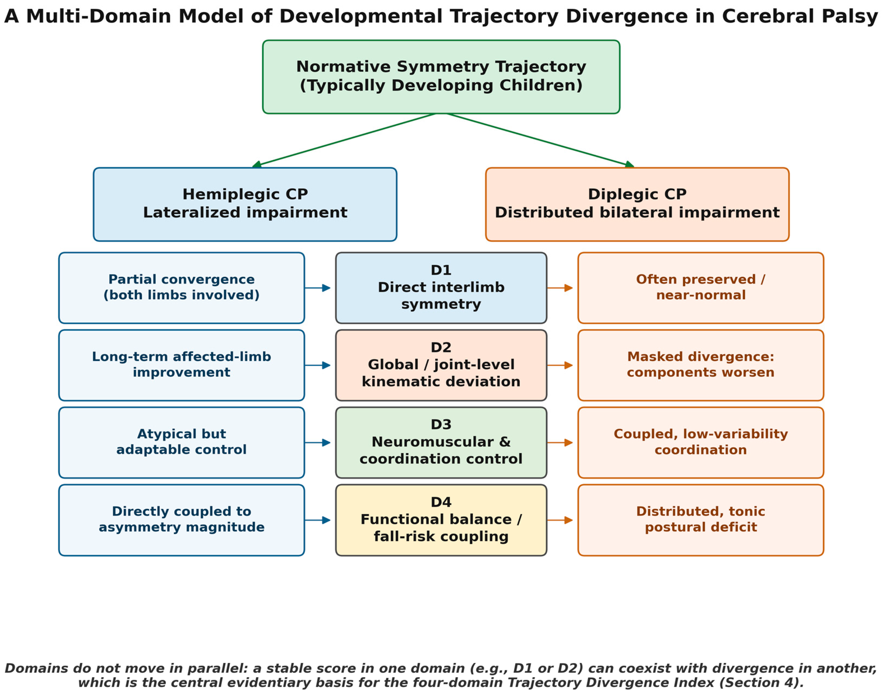
Figure 2.
Schematic illustration — not derived from pooled or fitted data — of the masked-divergence phenomenon in diplegic cerebral palsy. Panel (a): Direct interlimb symmetry (D1) in diplegia can approximate the normative trajectory across childhood. Panel (b): global/joint-level kinematic deviation (D2) can diverge from the normative trajectory over the same age range. The curve shapes are illustrative and should not be read as quantitative predictions.
Figure 2.
Schematic illustration — not derived from pooled or fitted data — of the masked-divergence phenomenon in diplegic cerebral palsy. Panel (a): Direct interlimb symmetry (D1) in diplegia can approximate the normative trajectory across childhood. Panel (b): global/joint-level kinematic deviation (D2) can diverge from the normative trajectory over the same age range. The curve shapes are illustrative and should not be read as quantitative predictions.
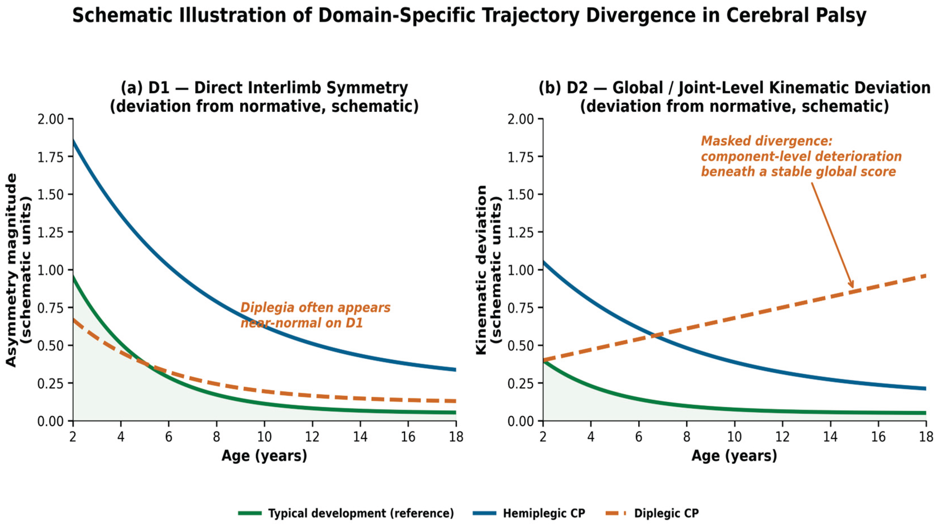
Table 1.
Comparative summary of the four evidence domains (D1–D4) in hemiplegic versus diplegic cerebral palsy.
Table 1.
Comparative summary of the four evidence domains (D1–D4) in hemiplegic versus diplegic cerebral palsy.
| Domain | Definition & Key Indices |
Hemiplegic CP — Typical Pattern |
Diplegic CP — Typical Pattern |
|---|---|---|---|
| D1 | Direct interlimb symmetry: spatial/temporal asymmetry indices, plantar-loading symmetry | Lateralized asymmetry with partial improvement over 24 months [8]; nominally unaffected limb also deviates from typical gait [26] |
Often preserved or near-normal [13,14], but not universally — 32% show clinically meaningful asymmetry |
| D2 | Global/joint-level kinematic deviation: GDI, GPS, GVS, sagittal-plane progression | Long-term improvement in affected-limb deviation over 10.5-year follow-up [9] |
Masked divergence: stable global GDI while knee flexion/dorsiflexion worsen [5,11,12] |
| D3 | Neuromuscular/coordination control: EMG timing, muscle synergies, intersegmental coordination | Atypical but adaptable — preserved motor-learning capacity despite baseline asymmetry [33,34] |
Coupled, low-variability coordination that converges kinematically without EMG-level maturation [15,17,18] |
| D4 | Functional balance/fall-risk coupling: dynamic stability, anticipatory postural adjustments, fall frequency | Balance deficits directly coupled to asymmetry magnitude; compensatory wide base of support [37,38] |
Distributed, tonic postural-control deficit with reduced anticipatory scaling [41,42] |
Table 2.
Provisional ordinal scoring rubric for the Trajectory Divergence Index (TDI). Each of D1–D4 is scored independently; the scores are never summed (Section 4.1).
Table 2.
Provisional ordinal scoring rubric for the Trajectory Divergence Index (TDI). Each of D1–D4 is scored independently; the scores are never summed (Section 4.1).
| Score | Interpretation | Applies to Each of D1–D4 Independently |
| 0 | Trajectory consistent with expected phenotype-specific pattern | No monitoring change indicated |
| 1 | Mild or transient divergence | Consistent with normal measurement variability or a compensatory adaptation phase |
| 2 | Persistent divergence across ≥2 consecutive assessments | Warrants closer monitoring or targeted domain-specific evaluation |
| 3 | Marked or progressive divergence | Warrants multidisciplinary reassessment (orthotic, surgical, or rehabilitation review) |
Disclaimer/Publisher’s Note: The statements, opinions and data contained in all publications are solely those of the individual author(s) and contributor(s) and not of MDPI and/or the editor(s). MDPI and/or the editor(s) disclaim responsibility for any injury to people or property resulting from any ideas, methods, instructions or products referred to in the content. |
© 2026 by the authors. Licensee MDPI, Basel, Switzerland. This article is an open access article distributed under the terms and conditions of the Creative Commons Attribution (CC BY) license (http://creativecommons.org/licenses/by/4.0/).
Copyright: This open access article is published under a Creative Commons CC BY 4.0 license, which permit the free download, distribution, and reuse, provided that the author and preprint are cited in any reuse.


