

Submitted:
04 August 2026
Posted:
04 August 2026
You are already at the latest version
Abstract
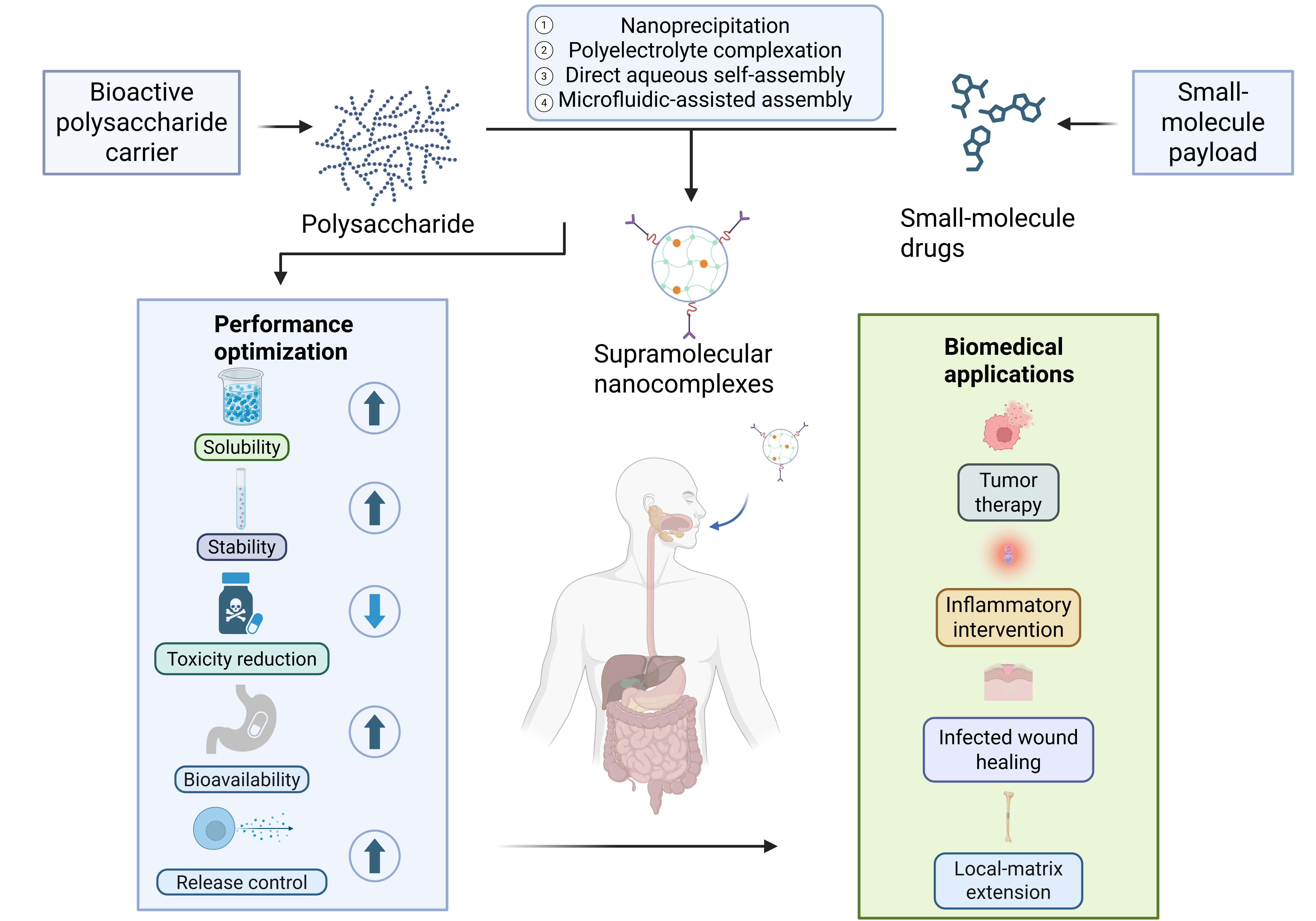
Bioactive polysaccharides such as fucoidan, β-glucans, and medicinal plant polysaccharides contain functional groups that can interact with drug molecules, and some of them also retain their own biological activities. Through reversible noncovalent interactions, these polysaccharides can associate with small-molecule drugs and form supramolecular nanocomplexes. This review discusses hydrogen bonding, hydrophobic association, electrostatic complexation, π–π stacking, and the cooperation among these interactions. The effects of pH, ionic strength, concentration, and solvent composition on assembly behavior are also considered. Nanoprecipitation/solvent exchange, polyelectrolyte complexation, direct aqueous self-assembly, and microfluidic-assisted assembly are compared in terms of nanostructure formation, process control, and reproducibility. Evidence from molecular, colloidal, solid-state, and computational characterization is considered together to clarify the relationship between structure, assembly, and performance. Reported benefits include improved drug dispersibility, colloidal stability, release control, bioavailability, cellular uptake, biodistribution, and safety. In some systems, the polysaccharide component may also contribute to therapeutic effects in tumors, inflammatory diseases, infected wounds, and local regenerative matrices. Quantitative mechanism studies, polysaccharide standardization, biocompatibility assessment, process scale-up, and long-term safety evaluation remain necessary for the further development of these drug delivery systems.
Keywords:
bioactive polysaccharides
; drug delivery
; supramolecular nanocomplexes
; noncovalent assembly
; carrier–drug synergy
1. Introduction
Small-molecule drugs remain indispensable in the treatment of cancer, inflammatory diseases, infections, metabolic disorders, and other complex diseases. However, many potent small-molecule drugs and natural bioactive compounds are limited by poor aqueous solubility, chemical instability, rapid metabolism, short circulation time, insufficient tissue selectivity, and dose-dependent systemic toxicity. As a result, these limitations commonly reduce bioavailability, weaken therapeutic efficacy, and increase the risk of adverse effects. Nanodrug delivery systems (NDDSs) have therefore been developed to address these formulation and delivery problems by improving drug solubilization, protecting labile drugs, regulating release behavior, and promoting drug delivery to diseased tissues [1,2,3]. Among the available carrier materials, natural biological macromolecules are of particular interest. They can serve as structural components in nanoscale formulations and, at the same time, provide biological functions that may affect drug stability, cellular interactions, and therapeutic outcomes.
Polysaccharides represent an important group of structurally diverse natural biological macromolecules. They can be obtained from plants, animals, algae, fungi, and microorganisms. Chitosan, alginate, hyaluronic acid, dextran, cellulose derivatives, fucoidan, β-glucans and medicinal plant polysaccharides are commonly reported in drug delivery studies [4,5]. These polymers are suitable carrier backbones because of their low toxicity, biodegradability, biocompatibility, renewable origin, and ease of chemical modification [4,5]. For bioactive polysaccharides, the carriers themselves may also affect the therapeutic effect. Several polymers have been reported to possess immunomodulatory, antitumor, antioxidant, antiviral and anti-inflammatory properties, indicating that they might assist or enhance the therapeutic actions [6,7]. Therefore, bioactive polysaccharides should not be regarded merely as passive excipients, but as functional biological macromolecules that may participate in carrier–drug synergy.
The structural features of polysaccharides are central to their assembly behavior and biological performance. Abundant hydroxyl, carboxyl, amino, sulfate, and carbonyl groups distributed along polysaccharide chains not only determine hydration behavior, charge density, chain conformation, and chemical modification potential, but also provide interaction sites for hydrogen bonding, electrostatic attraction, hydrophobic association after modification, and carrier–drug complexation [8,9]. Based on these structural features, supramolecular self-assembly has emerged as a mild and versatile strategy for constructing polysaccharide-based nanocomplexes. In aqueous media, amphiphilic, charged, or functionalized polysaccharides can associate with small-molecule drugs through cooperative noncovalent interactions, including hydrogen bonding, hydrophobic association, electrostatic complexation, and π–π stacking, thereby forming micelles, nanoparticles, nanogels, or other ordered nanoassemblies [10]. The dynamic and reversible nature of these interactions endows polysaccharide–drug nanocomplexes with environmental responsiveness and provides opportunities for improving drug dispersibility, colloidal stability, controlled release, and delivery performance [11].
Edible fungal polysaccharides are a class of functional polysaccharides with distinct bioactivities and structural features, showing potential as bioactive macromolecular components for disease-related delivery systems [12]. Fungal polysaccharides can be isolated from a wide range of fungal sources, and their molecular architectures and biological activities often differ markedly. This diversity is useful for carrier design and may also allow these polysaccharides to participate in auxiliary biological regulation, rather than only serving as structural materials. Formulations containing Tremella fuciformis polysaccharide, for example, have been associated with intestinal adhesion, prolonged local retention, and inflammation-related delivery. These systems should still be interpreted with caution, because they are usually multicomponent formulations rather than typical binary polysaccharide–drug supramolecular nanocomplexes [13].
Traditional Chinese medicine polysaccharides (TCMPs) have also been examined as biodegradable carriers in drug delivery. They generally show good biocompatibility, and some of them have reported pharmacological activities. Because their hydrated chain structures and functional groups resemble certain features of biological soft matrices, TCMPs have been used to prolong drug circulation, reduce nonspecific clearance by the reticuloendothelial system, and promote drug accumulation at target sites [14]. In some cases, TCMPs may do more than serve as passive carriers. Their intrinsic biological activities can take part in auxiliary therapeutic regulation. Astragalus polysaccharide, for example, has been described as an immunomodulatory polysaccharide that may cooperate with cancer immunotherapy through immune-related pathways [15]. In this type of carrier–drug synergistic system, delivery efficiency can be improved, while the pharmacological activity of the polysaccharide itself may also contribute to auxiliary therapy, improved efficacy, and reduced toxicity [16].
Animal-derived polysaccharides have also been used in drug delivery. Hyaluronic acid is a representative example because it can provide receptor-associated recognition and is widely used in nanocarrier design. Amphiphilic hyaluronan-based nanoparticles have also been studied for curcumin cellular delivery and neuroprotective applications [17].
Several recent reviews have discussed natural polysaccharide-based nanodrug delivery systems, traditional Chinese medicine polysaccharide-based nanocarriers, polysaccharide nanoparticles, polysaccharide-based nanomedicines for cancer immunotherapy, and self-assembled nanocarrier systems for bioactive compounds [1,16,18,19]. These studies provide useful summaries of material sources, preparation methods, disease-related applications, and general delivery performance. Nevertheless, the connection among polysaccharide bioactivity, noncovalent assembly, nanostructure evolution, and carrier–drug synergistic therapy has not been fully organized from a mechanistic perspective. A review centered on bioactive polysaccharide–small-molecule drug supramolecular nanocomplexes is therefore still needed.
This review examines the relationships among structure, assembly, and performance in bioactive polysaccharide–small-molecule drug supramolecular nanocomplexes. It first introduces the main noncovalent driving forces, including hydrogen bonding, hydrophobic association, electrostatic complexation and π-π stacking, as well as their cooperative influences, which provide the molecular basis for self-assembly. Environmental and formulation factors, such as pH, ionic strength, temperature, concentration, solvent composition, and polysaccharide structural parameters, are then discussed in relation to interaction balance, nanocomplex stability, and release behavior. Furthermore, this review compares some typical preparation methods and discusses the influence of processing conditions on the formation of nanocomplexes and the reproducibility of formulations. Characterization methods are brought together into a connected evidence chain, so that molecular interaction evidence can be considered together with morphology/colloidal/solid-state evidence and computational interpretation, instead of being presented as unrelated analytical tools. The review then examines how these nanocomplexes may affect aqueous solubility, colloidal stability, release control, bioavailability, biodistribution behavior, and safety-related outcomes, and it also discusses their possible carrier–drug synergistic effects in tumor therapy, inflammatory intervention, infected wound healing, and local-matrix/regenerative scaffold extensions; the unresolved issues are further considered from the aspects of polysaccharide heterogeneity, quantitative mechanism analysis, quality control, scalable manufacturing, safety evaluation, and translational feasibility.
2. Formation Mechanism of Supramolecular Nanocomplexes
Polysaccharides and small-molecule drugs usually associate through noncovalent interactions. These interactions mainly include hydrogen bonding, hydrophobic interactions, electrostatic interactions, and π–π stacking. Under physiological conditions, such weak forces are reversible and can respond to changes in the surrounding environment. This property is important for the design of responsive drug delivery systems. These interactions also affect structural stability, drug release behavior, and delivery performance of the complexes. Table 1 summarizes the nature of these forces, key characterization methods, and their contributions to the nanostructures.
2.1. Hydrogen Bonding in Supramolecular Complexes
Hydrogen bonding is a key noncovalent interaction involved in the molecular recognition and stabilization of polysaccharide–small-molecule drug supramolecular nanocomplexes. Hydroxyl, carboxyl, amino, sulfate, carbonyl, and phenolic groups on polysaccharide chains or drug molecules can serve as hydrogen-bond donors or acceptors, thereby promoting carrier–drug association and interfacial stabilization [28]. Compared with covalent conjugation, hydrogen-bond-mediated assembly is reversible and dynamic, which is favorable for structural adaptability and environmentally responsive release.
Spectroscopic evidence is commonly used to support hydrogen-bond formation in polysaccharide–drug systems. For example, Mao et al. reported a lentinan-ursolic acid self-assembled nanomedicine, in which spectroscopic and formulation evidence supported the association between the bioactive polysaccharide matrix and the hydrophobic small-molecule component [29]. Similarly, soy soluble polysaccharide has been reported to improve the aqueous dispersibility, stability, and in vitro bioaccessibility of curcumin, partly through polysaccharide-curcumin interactions involving hydrogen bonding and other weak forces [30]. These examples indicate that hydrogen bonding can facilitate drug anchoring and local structural stabilization as part of a broader noncovalent interaction network.
The contribution of hydrogen bonding is strongly affected by the surrounding microenvironment. Hydration, pH, temperature, ionic strength, solvent composition, and competing hydrogen-bond donors or acceptors may alter hydrogen-bond strength and carrier–drug affinity; thus, hydrogen bonding functions as an environment-dependent component within a broader noncovalent interaction network. In most polysaccharide–drug nanocomplexes, it functions cooperatively with hydrophobic association, electrostatic complexation, van der Waals forces, or π–π stacking, thereby contributing to the final stability and release behavior of the supramolecular structure [11].
2.2. Hydrophobic Interactions in Supramolecular Complexes
Hydrophobic association is an important driving force for the assembly of amphiphilic polysaccharide derivatives and poorly water-soluble small-molecule drugs. Hydrophobic association does not occur as a universal or independent mechanism in every polysaccharide–drug system. Most native polysaccharides are intrinsically hydrophilic, so effective hydrophobic assembly generally depends on the presence of hydrophobic microdomains, helical cavities, or chemically introduced hydrophobic moieties, including fatty acid, cholesterol, or alkyl groups. In aqueous media, these hydrophobic regions usually aggregate toward the inner part of the assembly and provide space for poorly soluble drugs. At the same time, the hydrophilic polysaccharide chains remain exposed to water and form a hydrated shell, which helps maintain colloidal stabilization.
This type of assembly is more commonly involved in the delivery of hydrophobic natural products and anticancer drugs. In polysaccharide nanocomplexes loaded with curcumin or myricetin, spectral attenuation, reduced drug crystallinity, and micellar morphology have been reported as evidence supporting drug incorporation into hydrophobic domains, rather than simple dispersion in water [31,32]. These examples indicate that hydrophobic association can promote core formation, improve drug dispersibility, and restrict drug crystallization, while reliable mechanism assignment still requires complementary structural evidence.
The role of hydrophobic association can be evaluated using CMC or CAC determination, fluorescence or UV–vis changes, particle-size analysis, morphology observation, and solid-state characterization. Because these methods provide indirect or complementary evidence, hydrophobic encapsulation requires confirmation by additional spectroscopic, morphological, or thermal characterization. For example, nanoscale particle formation observed by DLS or TEM should be further supported by spectroscopic or thermal evidence showing changes in the drug microenvironment or crystalline state.
Hydrophobic association must also be carefully balanced during formulation design. Increasing hydrophobic modification generally favors core formation and drug loading, and the type of hydrophobic group can affect CMC, micellar stability, encapsulation efficiency, biodistribution, and antitumor efficacy [33]. However, excessive hydrophobic modification may disturb the hydrophilic–hydrophobic balance of amphiphilic polysaccharides, promote interparticle association, and reduce aqueous colloidal stability. Therefore, the type and degree of hydrophobic substitution, drug hydrophobicity, molecular weight, concentration, and preparation conditions should be optimized as key parameters for constructing stable and reproducible polysaccharide–drug supramolecular nanocomplexes [34].
2.3. Electrostatic Complexation in Supramolecular Complexes
Electrostatic complexation is a major driving force for the self-assembly of charged polysaccharides and oppositely charged small-molecule drugs or drug derivatives. The charge properties of polysaccharides are mainly determined by ionizable groups and environmental pH. For example, chitosan becomes positively charged under acidic conditions due to amino-group protonation, whereas alginate, hyaluronic acid, heparin, fucoidan, pectin, and chondroitin sulfate generally show anionic characteristics because of carboxylate or sulfate groups. These charge features allow polysaccharides to associate with oppositely charged drugs through Coulombic attraction, leading to charge neutralization, particle nucleation, and compact nanocomplex formation; for instance, chondroitin sulfate-mediated systems have been reported to assemble with positively charged doxorubicin through electrostatic interactions before further albumin-corona formation [35].
A representative example is the hierarchical electrostatic assembly system based on heparin, chitosan, and an oleanolic acid derivative. In this system, the cationic oleanolic acid derivative was associated with anionic heparin, followed by chitosan coating to form self-assembled aggregates. Spectral and colloidal characterization supported electrostatic binding and layer-by-layer structural organization. This example indicates that electrostatic complexation can regulate nanoparticle formation, surface charge, structural compactness, and delivery performance without requiring covalent drug conjugation [36].
Electrostatic complexation is usually assessed through changes in zeta potential and particle size, shifts in FTIR signals of ionizable groups, as well as dissociation behavior caused by pH or salt. These observations can indicate that charge-related interactions are involved, but they are more suitable as supporting evidence and cannot by themselves prove that only one interaction mechanism is present. Since electrostatic nanocomplexes can be affected by pH, ionic strength, serum proteins, and other competing charged biomolecules, Coulombic attraction may become weaker under physiological conditions, which can cause swelling, dissociation, or premature drug release [37]; for this reason, electrostatic complexation is commonly used together with hydrogen bonding, hydrophobic association, π–π stacking, or secondary coating strategies, so that the robustness and physiological stability of polysaccharide–drug supramolecular nanocomplexes can be improved [35,36].
2.4. π–π Stacking in Supramolecular Complexes
π–π stacking is a specific noncovalent interaction associated with small-molecule drugs containing aromatic or conjugated structures. Unlike hydrogen bonding or electrostatic complexation, π–π stacking is not a universal interaction between native polysaccharides and small-molecule drugs, because most natural polysaccharide backbones lack aromatic rings. In polysaccharide-based nanocomplexes, π–π stacking usually occurs among aromatic drug molecules confined within polysaccharide-based nanodomains, or between aromatic drugs and aromatic/hydrophobic moieties introduced onto modified polysaccharides. Therefore, its contribution needs to be distinguished from nonspecific hydrophobic encapsulation and assigned only with support from complementary spectroscopic, structural, or computational evidence.
π–π-related aromatic association may promote ordered aggregation of aromatic payloads, improve drug loading, stabilize drug-rich nanodomains, and contribute to optical, photothermal, or photodynamic functions in specific aromatic drug-loaded systems [38]. Its presence is usually supported by complementary spectroscopic and structural evidence, such as UV–vis band shifts or broadening, fluorescence changes, aromatic proton signal variations, or molecular simulation results. However, these signals are often coupled with hydrophobic association, so nanoparticle formation alone cannot confirm independent π–π stacking.
Yu et al. reported self-assembled monocarboxyl corrole/chitosan photothermal nanoparticles as a representative polysaccharide-based aromatic drug assembly. In this system, spectroscopic changes and molecular simulation supported ordered corrole aggregation, while TEM showed a dense drug-loaded core within the chitosan-based nanoparticles. These results suggest that π–π stacking contributed to core densification and photothermal performance in this chitosan-based supramolecular system [39].
The role of π–π stacking in polysaccharide-based drug delivery systems therefore requires cautious assignment. For native polysaccharides, its role is usually indirect and mainly arises from aromatic drug–drug association within confined nanodomains. In photosensitizer-loaded polysaccharide systems, aromatic aggregation may alter fluorescence behavior, drug-release profiles, and phototherapeutic activity; therefore, evaluation of π–π-related association needs to integrate hydrophobic association, hydrogen bonding, electrostatic complexation, drug microenvironment, and release behavior when elucidating the assembly mechanism of polysaccharide–small-molecule drug supramolecular nanocomplexes [40].
2.5. Cooperative and Hierarchical Assembly Mechanisms
In most bioactive polysaccharide–small-molecule drug supramolecular nanocomplexes, self-assembly is governed by the cooperative contribution of multiple weak interactions rather than by a single dominant force. Hydrogen bonding may initiate carrier–drug recognition, hydrophobic association may promote core formation, electrostatic complexation may regulate charge neutralization and particle nucleation, and π–π stacking may stabilize aromatic drug domains in specific systems. These interactions are often coupled in a hierarchical manner, involving molecular recognition, nucleation, core formation, structural densification, and colloidal stabilization. Self-assembled hyaluronic acid-testosterone nanocarriers can be used to explain this stepwise assembly process, because in this system, amphiphilic polysaccharide conjugates are able to form drug-loaded nanostructures through a gradual process of molecular organization, rather than relying only on simple one-step encapsulation [41].
Such cooperative assembly can partly explain why nanocomplexes made from similar components may still differ in particle size, drug-loading capacity, release behavior, and biological performance once the formulation conditions are changed. The order in which these interactions appear is not the same in all systems. When hydrogen bonding dominates, carrier–drug recognition may occur early, followed by gradual hydrophobic densification as the assembly becomes more organized. Charged polysaccharide systems often begin with electrostatic complexation and particle nucleation, but this initial structure is not always stable enough on its own; hydrogen bonding, hydrophobic association, or additional coating layers may still be needed to maintain structural integrity. In aromatic drug-loaded nanocomplexes, π–π stacking is usually coupled with hydrophobic association, making it difficult to treat it as a fully independent driving force. Fluorescence spectroscopy and molecular simulation studies on amphiphilic hyaluronic acid conjugates further indicate that self-assembly is closely linked to molecular organization and the local microenvironment [42].
A quantitative view of cooperative assembly requires these factors to be read together. Thermodynamic terms, kinetic processes, and polysaccharide structural features cannot be separated easily, because each of them may shift the balance of noncovalent interactions during nanocomplex formation. Parameters such as binding affinity, Gibbs free energy, enthalpy–entropy balance, CMC or CAC, and binding capacity are useful for judging whether assembly is mainly driven by hydrogen bonding, electrostatic attraction, hydrophobic association, or entropy gain associated with water release. For amphiphilic or host–guest polysaccharide-based systems, CAC and binding capability can also be used to assess self-assembly tendency and formulation stability; this has been shown in hyaluronic acid-cyclodextrin/adamantane supramolecular nanosystems [43].
What type of nanostructure is finally formed, and whether this structure can be reproduced consistently, are closely related to both kinetic and structural variables. Factors such as solvent exchange rate, mixing intensity, nucleation rate, and polysaccharide chain relaxation can change the assembly pathway, so the system may form compact nanoparticles, loose aggregates, micelles, vesicles, or nanogels under different preparation conditions. At the same time, molecular weight, degree of substitution, charge density, branching degree, chain flexibility, and hydrophilic–hydrophobic balance can affect assembly reproducibility, drug loading, and release behavior. These variables are particularly important for natural polysaccharides, because their molecular-weight distribution, branching degree, charge density, and substitution heterogeneity may lead to batch-to-batch variation in nanocomplex structure, drug loading, release behavior, and biological performance [4].
Overall, bioactive polysaccharide–small-molecule drug supramolecular nanocomplexes should be understood as dynamic assemblies formed by cooperative noncovalent interactions. As shown in Figure 1, hydrogen bonding, hydrophobic association, electrostatic complexation, and π–π stacking jointly affect molecular recognition, particle nucleation, core–shell organization, colloidal stabilization, drug loading, and release behavior. For polysaccharide-based supramolecular drug delivery systems, establishing quantitative structure–assembly–performance relationships is important for rational formulation design, quality control, and scalable production.
2.6. Environmental and Formulation Regulation of Cooperative Assembly
The cooperative assembly of bioactive polysaccharide–small-molecule drug supramolecular nanocomplexes is closely related to both environmental conditions and formulation design. Hydrogen bonding, hydrophobic association, electrostatic complexation, and π–π stacking are reversible interactions, and their relative contribution can change with the surrounding medium. Therefore, variations in pH, ionic strength, temperature, polysaccharide/drug ratio, total concentration, and solvent composition may affect interaction balance, chain conformation, particle nucleation, colloidal stability, and release behavior.
pH and ionic strength are especially important for charged and polar interaction networks. pH is a key variable in charged polysaccharide-based assemblies because it changes the ionization state of carboxyl, amino, sulfate, phosphate, and phenolic groups. Once these groups are protonated or deprotonated, electrostatic attraction, hydrogen-bonding patterns, matrix swelling, and pH-responsive release may all be altered [44]. Ionic strength acts through a related but not identical route. A moderate salt level can screen excessive electrostatic repulsion and make the complex structure more compact, whereas high salt concentrations may also screen the attractive forces needed to maintain electrostatically assembled particles. In this case, the absolute zeta potential usually decreases, and the system may show swelling, aggregation, or premature drug release [45]. Thus, pH and ionic strength should be treated as formulation variables that reshape electrostatic complexation and hydrogen-bond rearrangement, not merely as background properties of the medium.
Temperature, polysaccharide/drug ratio, and total concentration affect self-assembly mainly through molecular mobility, nucleation, and aggregation. A mild increase in temperature, or an appropriate external energy input, can promote molecular diffusion and polysaccharide chain rearrangement. Excessive heating may have the opposite effect: hydrogen bonding can be weakened, hydration layers may be disturbed, heat-sensitive drugs may degrade, and particle growth may be promoted. Polysaccharide-based encapsulation has been reported to improve the environmental and thermal stability of labile small molecules, and curcumin is a commonly cited example [46]. The polysaccharide/drug ratio deserves separate consideration. Sufficient carrier content can provide binding sites and steric protection, whereas insufficient carrier may lead to incomplete encapsulation, drug crystallization, or unstable aggregates. Since polysaccharide–natural product interactions can change particle size, surface charge, loading efficiency, and release behavior, the carrier/drug ratio should be selected according to carrier–drug affinity and colloidal stabilization, not used simply as a mass ratio [47].
Solvent composition and hydration state can influence the dispersion of drugs, the conformation of polysaccharides, desolvation and the intensity of noncovalent interactions. Therefore, they are not only process details but also factors affecting the formation of nanocomplexes. Water and aqueous buffers are usually employed for polysaccharide-based soft colloids, nanogels and biomedical or food-related preparations, as they can maintain the hydrated polysaccharide matrix and have good safety properties [48]. Nevertheless, this method is not always effective. Some water-insoluble drugs, or polysaccharide derivatives that tend to aggregate in water, still require co-solvent assistance or solvent-exchange procedures. In such situations, the polarity and hydrogen-bonding ability of the solvents as well as the residual-solvent risk should be considered carefully, since the solvent-induced conformational changes may affect both the biological activity of polysaccharides and the reproducibility of the resultant nanocomplexes.
Environmental and formulation factors should be included in the mechanistic analysis of polysaccharide–drug supramolecular assembly. They do not operate independently. In most systems, their effects are reflected in changes in the balance among hydrogen bonding, hydrophobic association, electrostatic complexation, π–π stacking, hydration effects, and steric stabilization. Once this balance changes, the interaction pathway, nanostructure, drug-loading behavior, release profile, and final performance may also change. A clearer understanding of these relationships can help explain why different systems show changes in particle size, drug loading, stability, and release behavior, and it can also support the rational design and scalable preparation of bioactive polysaccharide–small-molecule drug supramolecular nanocomplexes.
3. Preparation of Polysaccharide–Small-Molecule Supramolecular Nanocomplexes
The preparation of polysaccharide–small-molecule drug supramolecular nanocomplexes largely depends on the organization of polysaccharide chains and drug molecules under defined solvent, charge, concentration, and mixing conditions. Compared with covalent conjugation, supramolecular assembly is more sensitive to processing variables, since hydrogen bonding, hydrophobic association, electrostatic complexation, and π–π stacking are reversible interactions that may change with the assembly environment. For this reason, the preparation strategy should be selected by considering the physicochemical properties of the drug, the charge and amphiphilicity of the polysaccharide, the expected nanostructure, and the intended route of administration.
Several methods have been used to prepare these systems, including nanoprecipitation or solvent exchange, polyelectrolyte complexation, direct aqueous self-assembly, and microfluidic-assisted controlled assembly. These techniques differ in solvent requirements, dominant assembly forces, particle-size control, scalability, and compatibility with bioactive polysaccharides. A comparison based on their mechanisms can assist in the formulation design and enhance the repeatability of the nanocomplex preparation.
3.1. Nanoprecipitation / Solvent Exchange
Nanoprecipitation, also known as solvent displacement or solvent exchange, is commonly used to prepare polysaccharide-based supramolecular nanocomplexes. In this method, solvent–nonsolvent diffusion induces rapid desolvation, after which nucleation and particle growth occur. The final particle properties are affected by several formulation and process variables, including solvent selection, polymer type, drug characteristics, mixing mode, injection rate, and other flow-related parameters [49].
In polysaccharide–small-molecule drug systems, nanoprecipitation is mainly applied to amphiphilic or hydrophobically modified polysaccharides, especially when the loaded drugs have low water solubility. When the solvent is displaced, the hydrophobic parts and the drug-rich regions tend to collect in the formed particles, while the hydrophilic polysaccharide chains remain in the aqueous phase and assist in forming a hydrated stabilizing shell. Particle size, polydispersity, and loading behavior are affected by solvent composition, the organic/aqueous phase ratio, mixing intensity, polymer concentration, and the carrier/drug ratio [50].
Doxorubicin-loaded dextran-b-poly(ε-caprolactone) nanoparticles are a representative example of polysaccharide-derived amphiphilic nanoparticles prepared by nanoprecipitation. Using a modified nanoprecipitation method, the study obtained nanoscale particles with sustained drug release and cellular uptake behavior [51]. This example shows that nanoprecipitation should not be regarded as a simple mixing operation. In such systems, desolvation, solvent diffusion, polysaccharide-derived amphiphilicity, and carrier–drug compatibility jointly influence the formation of drug-loaded nanocomplexes.
Nanoprecipitation is relatively easy to perform, but its limitations become more apparent when particle uniformity and scale-up are considered. Organic solvent residues, rapid local supersaturation, uncontrolled nucleation, and insufficient mixing can result in broad particle-size distributions and batch-to-batch variation. This issue is more complicated for bioactive polysaccharides, because solvent exposure and processing stress may alter chain conformation and weaken their biological activity. Therefore, when this method is applied to bioactive polysaccharide–small-molecule drug supramolecular nanocomplexes, solvent selection, residual-solvent control, mixing conditions, and process reproducibility need to be evaluated carefully.
3.2. Polyelectrolyte Complexation
Polyelectrolyte complexation is a mild aqueous method for forming nanocomplexes through electrostatic association among oppositely charged polysaccharides, small-molecule drugs, or counter-polymers. During this process, charged components may associate spontaneously through Coulombic attraction, leading to charge neutralization, chain rearrangement, and nanoparticle formation. This method is particularly suitable for polysaccharides with ionizable groups such as carboxyl, sulfate, phosphate or amino groups, since their charges can be regulated by the pH and ionic conditions of the medium [52].
Compared with nanoprecipitation, polyelectrolyte complexation is more appropriate for certain bioactive polysaccharides, because it is generally carried out in mild aqueous solutions and does not require large amounts of organic solvent. The formation and stability of these complexes are affected by pH, charge ratio, ionic strength, concentration of components, molecular weight and the order of mixing. Among these factors, pH influences the ionization of amino, carboxyl, sulfate and phosphate groups, while the charge ratio affects whether the complexes become compact or remain colloidally stable [53].
Chitosan-gum arabic polyelectrolyte systems provide evidence for the role of electrostatic attraction in nanocomplex formation, as oppositely charged polysaccharides in this system were used to prepare curcumin-loaded nanoparticles [54]. This example points to another use of polyelectrolyte complexation. It is not only a mild aqueous preparation method; by regulating charge interactions, it can also help load hydrophobic bioactive small molecules into polysaccharide-based nanocarriers.
Polyelectrolyte nanocomplexes are influenced by the medium in which they are dispersed, especially by the pH and ionic strength. Changes in these factors may decrease the electrostatic attraction and lead to swelling, aggregation, dissociation or early drug release. The charge ratio should be kept within a certain limit. If charge neutralization is too strong, precipitation may occur; if charge compensation is insufficient, loose complexes with low drug-loading efficiency may form. Thus, the pH, ionic strength, the ratio of components and the mixing conditions should be adjusted properly. Moreover, for better storage stability, resistance to physiological conditions and higher reproducibility, hydrogen bonding, hydrophobic association, coating layers or mild cross-linking can be used for stabilization [55].
3.3. Direct Aqueous Self-Assembly
Direct aqueous self-assembly is a simple and environmentally friendly method for forming polysaccharide–drug supramolecular nanocomplexes, particularly when the polysaccharide carrier has inherent amphiphilic character or has been chemically modified to introduce amphiphilicity. In this technique, amphiphilic polysaccharides can associate in water, and the procedure generally requires little or no organic solvent or surfactant, which is suitable for constructing biocompatible drug delivery systems [56].
When the concentration of amphiphilic polysaccharide derivatives exceeds the critical micelle concentration (CMC) or critical aggregation concentration (CAC), hydrophobic segments tend to associate inward and form drug-loading domains, whereas hydrophilic polysaccharide chains remain exposed to the aqueous phase and contribute to colloidal stabilization. Therefore, the hydrophilic–hydrophobic balance, hydrophobic substituent type, molecular weight, and degree of substitution are key structural parameters regulating nanocomplex formation and stability [34].
The role of hydrophobic modification in enabling aqueous self-assembly is supported by stearic-acid-modified Bletilla striata polysaccharide systems, in which amphiphilic BSP derivatives formed pH-responsive drug-loaded nanoparticles through spontaneous aqueous self-assembly [57]. This case shows that direct aqueous self-assembly is more than a simple dissolution process. It should be viewed as a structure-driven assembly pathway, in which polysaccharide amphiphilicity, substitution design, and carrier–drug compatibility jointly determine nanocomplex formation.
Direct aqueous self-assembly, however, cannot be applied to every polysaccharide–drug system. When amphiphilicity is insufficient, drug encapsulation may be weak. In contrast, excessive hydrophobic modification or a high polymer concentration can increase interparticle aggregation and lower colloidal stability. To obtain stable and reproducible polysaccharide-based supramolecular nanocomplexes, CMC/CAC, substitution degree, concentration range, and external energy input need to be optimized together.
3.4. Microfluidic-Assisted Assembly
Microfluidic-assisted assembly provides a more controllable approach for preparing polysaccharide-based supramolecular nanocomplexes. In microscale channels, fluid behavior can be regulated through laminar-flow diffusion, hydrodynamic focusing, or controlled solvent exchange, which allows mixing to occur in a more reproducible manner. For polysaccharide nanoparticles, microfluidics has been reported to improve process control, formulation reproducibility, and high-throughput screening when compared with conventional bulk preparation [58].
The formation of nanocomplexes in microfluidic systems is affected by several process variables, including flow-rate ratio, total flow rate, channel geometry, polymer concentration, solvent composition, and drug-to-carrier ratio. These parameters influence local supersaturation, nucleation, interfacial mixing, and particle growth. As a result, particle size, polydispersity, drug loading, and colloidal stability may all change with the operating conditions. As shown in Figure 2, microfluidic-assisted assembly is different from nanoprecipitation/solvent exchange, polyelectrolyte complexation, and direct aqueous self-assembly mainly because it provides better control over mixing and particle formation. This advantage, however, still depends on the design of the device and the optimization of the process.
Chitosan-coated magnetic nanoparticle systems for cisplatin delivery provide an example of controlled microscale mixing. In this related composite system, an integrated microfluidic device was used to form magnetic core–chitosan shell nanoparticles through mixing-assisted ionic complexation. Compared with conventional batch preparation, this approach improved size uniformity and formulation performance [59]. This system should be used as evidence for process control rather than as a typical binary polysaccharide–drug nanocomplex. Even so, it shows that microfluidic-assisted assembly can regulate nucleation, particle growth, and size distribution in polysaccharide-based nanocarriers.
The use of microfluidics in bioactive polysaccharide–small-molecule drug systems still has several practical limitations. High solution viscosity, increased back-pressure, channel clogging, solvent compatibility, and the need for reliable process monitoring may restrict the application of microfluidic-assisted assembly. In practice, the preparation method should be selected according to drug hydrophobicity, polysaccharide charge and amphiphilicity, desired particle size, administration route, and scale-up requirements. Environmental and formulation factors discussed in Section 2.6 should also be included in this decision, because they may change the interaction balance and assembly pathway. Nanoprecipitation/solvent exchange is more suitable for hydrophobic drugs, but residual-solvent control is required. Polyelectrolyte complexation is mild and aqueous, although it is sensitive to charge balance and ionic strength. Direct aqueous self-assembly is a greener approach, but it depends strongly on polysaccharide amphiphilicity. Microfluidic-assisted assembly provides better process control, while device design and polysaccharide rheology still limit its broader use. Further work should pay more attention to green solvents, controlled mixing, process monitoring, and microfluidic optimization, so that reproducibility and translational feasibility can be improved [60].
4. Structural Characterization and Mechanistic Evidence Chain
Structural characterization is essential for verifying the formation, assembly mechanism, colloidal stability, and drug physical state of bioactive polysaccharide–small-molecule drug supramolecular nanocomplexes. Because these systems are mainly stabilized by reversible noncovalent interactions, no single analytical technique can fully explain their molecular association, nanoscale organization, solid-state behavior, and structure-dependent performance. Therefore, characterization should be organized as an integrated mechanistic evidence chain rather than as a catalogue of isolated analytical methods.
This chapter divides the evidence chain into three interconnected levels. Molecular interaction evidence, mainly from FTIR, NMR, UV–vis absorption, and fluorescence spectroscopy, helps identify functional-group variation, chemical microenvironment changes, hydrophobic-domain incorporation, aromatic aggregation, and possible carrier–drug association. Morphology, colloidal, and solid-state evidence, obtained from TEM, DLS, zeta potential analysis, XRD, DSC, and TGA, further evaluates particle morphology, hydrodynamic size, surface charge, colloidal stability, drug crystallinity, amorphous dispersion, and thermal behavior. Computational evidence, including molecular docking and molecular dynamics (MD) simulation, provides mechanistic hypotheses regarding binding orientation, interaction contribution, conformational stability, and dynamic carrier–drug association. Cross-validation among these methods is necessary to connect molecular recognition, supramolecular assembly, nanostructure formation, colloidal stabilization, drug physical state, and release behavior, thereby supporting the structure–assembly–performance relationship of these nanocomplexes.
4.1. Molecular Interaction Evidence
Molecular interaction evidence represents the first level of structural characterization for bioactive polysaccharide–small-molecule drug supramolecular nanocomplexes. Because these systems are mainly stabilized by reversible noncovalent interactions, spectroscopic techniques are required to identify whether polysaccharide chains and drug molecules undergo functional-group association, chemical microenvironment variation, hydrophobic-domain incorporation, or aromatic aggregation. Fourier transform infrared spectroscopy (FTIR), nuclear magnetic resonance (NMR) spectroscopy, UV–vis absorption spectroscopy, and fluorescence spectroscopy are therefore useful for interpreting hydrogen bonding, electrostatic complexation, hydrophobic association, and π–π-related interactions. However, these methods should be regarded as complementary molecular-level evidence rather than as independent proof of complete nanocomplex formation or a single dominant assembly mechanism.
FTIR mainly provides functional-group-level evidence by monitoring characteristic vibration bands of hydroxyl, amino, carboxyl, carbonyl, phosphate, and sulfate groups in polysaccharide-based systems [61]. In polysaccharide–drug nanocomplexes, peak shifts, peak broadening, or intensity changes may further suggest hydrogen bonding, ionic pairing, or carrier–drug association, but such signals can also be affected by spectral overlap, peak masking, matrix dilution, and water-associated broad bands. NMR spectroscopy further supports the analysis of polysaccharide structure, chain conformation, chemical microenvironment, and molecular mobility [62]. In polysaccharide–drug systems, changes in chemical shifts, signal broadening, signal attenuation, or relaxation behavior may indicate altered local polarity, hydrogen bonding, electrostatic association, hydrophobic shielding, or spatial proximity between drug molecules and polysaccharide chains. Nevertheless, broad signals, low solubility, overlapping peaks, and structural heterogeneity of natural polysaccharides often limit precise peak assignment; therefore, FTIR and NMR results should be interpreted together with colloidal characterization, solid-state analysis, and release evidence.
UV–vis absorption and fluorescence spectroscopy provide additional information on the microenvironment of conjugated, aromatic, or fluorescent small-molecule drugs. In rutin-fucoidan and curcumin-loaded chitosan nanocomplexes, optical and colloidal characterization has been used to support small-molecule incorporation, complex formation, and altered drug microenvironment [63,64]. However, changes in absorption intensity, band position, band broadening, fluorescence quenching, or emission shifts may also be influenced by concentration effects, inner-filter effects, nanoparticle scattering, photobleaching, and baseline distortion. Therefore, UV–vis and fluorescence evidence should be combined with FTIR, NMR, DLS, zeta potential analysis, XRD, thermal analysis, release behavior, and computational interpretation before assigning hydrophobic encapsulation, aromatic aggregation, or π–π-related interactions.
4.2. Morphology, Colloidal, and Solid-State Evidence
Morphology, colloidal properties, and solid-state characteristics constitute the second level of the structural evidence chain for bioactive polysaccharide–small-molecule drug supramolecular nanocomplexes. After molecular interaction evidence has suggested possible carrier–drug association, TEM, DLS, zeta potential analysis, XRD, DSC, and TGA are required to evaluate whether nanoscale assemblies are formed, whether the particles remain stable in dispersion, and whether the loaded drug exists in a crystalline, amorphous, or molecularly dispersed state. These methods link noncovalent assembly mechanisms with particle structure, colloidal stability, drug physical state, and release behavior.
TEM provides visual morphological evidence for particle shape, approximate dry-state size, aggregation state, and possible dense-core or core–shell features. However, TEM should be regarded as morphological evidence rather than direct proof of specific noncovalent interactions, because drying, staining, and vacuum conditions may alter the apparent size or morphology of soft polysaccharide-based nanocomplexes [65]. TEM observations should be interpreted together with DLS and zeta potential results because these methods describe different aspects of dispersed nanocomplexes. DLS can determine the hydrodynamic size, size distribution and PDI of the suspended particles, which is beneficial for identifying the nanoscale assemblies. Zeta potential analysis gives information on the surface charge, charge neutralization, surface modification, electrostatic stabilization and possible adsorption behavior [66]. In polysaccharide–drug systems with high charges, a variation in zeta potential may indicate an electrostatic interaction between the carrier and the drug, such as in the case of thiolated fucoidan-doxorubicin nanoparticles [67]. This judgment should be made cautiously. The zeta potential value may also vary with pH, ionic strength, dilution medium, buffer composition and protein adsorption; hence, it is more suitable to regard it as additional evidence for the colloidal stability rather than a certain or absolute criterion.
Solid-state and thermal analyses are used to examine the physical state and thermal behavior of the loaded drug. XRD mainly shows whether the characteristic crystalline peaks of the free drug remain, become weaker, or disappear after complex formation. A decrease or disappearance of drug diffraction peaks usually indicates reduced crystallinity, amorphous dispersion, or inhibited crystallization within the polysaccharide matrix, as reported in myricetin-loaded hydrophobically modified β-glucan micelles [32]. DSC is useful for checking carrier–drug compatibility and thermal transitions, while TGA can reflect water content, residual solvent, and thermal degradation behavior. For heat-sensitive small molecules, thermal stability tests may also show whether polysaccharide-based encapsulation protects the drug from environmental stress, as observed in curcumin–soluble soybean polysaccharide nanocomplexes [46]. These results still need cautious interpretation. The absence of crystalline peaks or melting endotherms may result from low drug loading, matrix dilution, peak overlap, or broad amorphous background signals, rather than complete molecular dispersion. Solid-state and thermal data are more reliable when they are discussed together with molecular interaction evidence, morphology, colloidal stability, release behavior, and environmental stability data.
Morphology, colloidal properties, and solid-state characteristics should be considered together, not as separate results. TEM shows the nanoscale morphology, whereas DLS and zeta potential analysis describe the hydrated size, size distribution, surface charge, and colloidal stability. XRD, DSC, and TGA provide information on drug crystallinity, amorphous dispersion, and thermal behavior. These methods can help judge whether polysaccharide–drug nanocomplexes form organized structures and remain stable in dispersion. Still, none of them alone can prove the assembly mechanism. The interpretation is more convincing when these data are compared with molecular interaction evidence, release studies, environmental stability assays, and computational analysis.
4.3. Computational Evidence and Integrated Interpretation
Computational analysis is another part of the mechanistic evidence chain for bioactive polysaccharide–small-molecule drug supramolecular nanocomplexes. Spectroscopic, microscopic, colloidal, and solid-state methods provide experimental information at different structural levels. Molecular docking and molecular dynamics (MD) simulation add a different type of evidence, mainly by predicting possible binding sites, binding orientations, interaction contributions, and conformational stability. These methods are particularly useful for interpreting how hydrogen bonding, electrostatic attraction, hydrophobic contacts, van der Waals interactions, and π–π-related interactions contribute to polysaccharide–drug association. For example, docking and MD analysis of carboxymethyl chitosan complexes with curcumin and nicotinamide has been used as a computational example to evaluate possible drug affinity, interaction energy, and conformational variation in chitosan derivatives [68].
Molecular docking is mainly suitable for predicting possible drug-binding regions and dominant interaction types, whereas MD simulation further evaluates whether the predicted polysaccharide–drug complex remains dynamically stable in a solvated environment. Parameters such as root-mean-square deviation (RMSD), root-mean-square fluctuation (RMSF), radius of gyration, solvent-accessible surface area, hydrogen-bond persistence, interaction energy, and binding free energy can be used to assess conformational fluctuation, compactness, molecular flexibility, and interaction persistence. In chitosan-curcumin systems, MD simulation has been used to study carrier–drug association and to describe the roles of hydrogen bonding, van der Waals interactions, and conformational fluctuation in the retention of curcumin within the chitosan matrix [69]. These simulation results are useful for discussing possible interaction mechanisms. They should be treated as mechanistic support, rather than direct evidence that nanocomplexes have formed.
The reliability of computational simulation is closely related to the construction of the carbohydrate model. Force-field parameters, ionization states, solvent conditions, counterions, substitution degree, and simulation time scale can all affect the results, because carbohydrate simulations are sensitive to molecular representation and parameterization [70]. This limitation is more evident for natural bioactive polysaccharides. These polysaccharides usually have broad molecular-weight distributions, heterogeneous glycosidic linkages, variable branching degrees, uncertain substitution patterns, and flexible chain conformations, which are difficult to reproduce fully in simplified computational models [71]. Therefore, docking scores, interaction energies, and MD-derived stability parameters are better used to explain possible interaction pathways and stability trends, rather than to replace experimental characterization.
Computational evidence should be interpreted together with experimental characterization when discussing the structure–assembly–performance relationship. FTIR and NMR are used to examine molecular interactions, whereas UV–vis absorption and fluorescence spectroscopy can show changes in the drug microenvironment. TEM, DLS, and zeta potential analysis describe morphology, particle size, surface charge, and colloidal stability. XRD, DSC, and TGA are then useful for judging drug crystallinity, amorphous dispersion, and thermal behavior. Taken together, these results link molecular recognition, supramolecular assembly, colloidal stabilization, drug physical state, and release behavior more reliably than any single method.
5. Performance Enhancement and Structure–Assembly–Performance Relationships of Supramolecular Nanocomplexes
Polysaccharide–small-molecule drug nanocomplexes are designed mainly to improve the delivery of poorly soluble drugs. They also provide a useful system for examining how polysaccharide properties, noncovalent self-assembly, nanostructure formation, and biological performance are related. In these systems, bioactive polysaccharides may act as solubilizers, colloidal stabilizers, controlled-release regulators, and biologically functional materials. All these functions are closely related to hydrogen bonding, hydrophobic interaction, electrostatic complexation, steric stabilization and the formation of hydration layers. Through these interactions, polysaccharide-based nanocomplexes can improve drug dispersibility, colloidal stability, controlled release, absorption, biodistribution, and safety. Therefore, performance enhancement should be evaluated using multiple indicators, including apparent solubility, dissolution behavior, physical stability, release kinetics, cellular uptake, pharmacokinetic parameters, and toxicity profiles, rather than relying on a single endpoint.
5.1. Solubility Enhancement and Amorphous Drug Dispersion
Poor aqueous solubility and unstable crystalline states are major limitations of many hydrophobic small-molecule drugs and natural bioactive compounds. In polysaccharide–drug supramolecular nanocomplexes, solubility enhancement is mainly achieved through hydrophobic-domain encapsulation, hydrogen-bond- or electrostatic-interaction-mediated drug dispersion, inhibition of drug crystallization, and stabilization of the drug in an amorphous or molecularly dispersed state. Bioactive medicinal polysaccharides can provide hydrophilic domains, hydration layers, steric barriers, and abundant functional groups for carrier–drug association, thereby improving apparent solubility, dissolution behavior, stability, and colloidal dispersibility of poorly soluble flavonoids and related small-molecule compounds [72,73]. However, improved aqueous dispersibility should not be overinterpreted as true molecular solubilization unless it is supported by solid-state and colloidal evidence, such as XRD, DSC, particle size distribution, and zeta potential analysis.
The solubilizing effect of bioactive polysaccharides is closely related to their structural features and dominant interaction mechanisms. Hydrophobic or amphiphilic polysaccharide domains can accommodate poorly soluble drugs and restrict drug aggregation, whereas hydrogen bonding and electrostatic association can improve drug dispersion by increasing carrier–drug affinity. Steric confinement within the polysaccharide matrix can also limit drug crystallization and help maintain the drug in an amorphous dispersed state. Astragalus polysaccharide (APS)-flavonoid complexes provide one useful case. In this system, APS aggregates were able to incorporate poorly soluble flavonoids through weak intermolecular interactions, and this was associated with improved solubility, gastrointestinal stability, permeability, and absorption-related properties [74]. This case is relevant because APS is a bioactive medicinal polysaccharide, and the study combined FTIR, XRD, TG-DSC, microstructural characterization, and molecular simulation to distinguish polysaccharide-mediated complexation from simple physical mixing.
In polysaccharide–small-molecule drug supramolecular nanocomplexes, solubility enhancement should be understood as a structure-dependent result rather than as a simple increase in water solubility. It may involve drug amorphization, hydrophobic-domain encapsulation, intermolecular association, and colloidal stabilization. Reliable evaluation therefore needs to include apparent solubility, dissolution rate, drug crystallinity, particle size distribution, storage stability, and redispersibility. XRD and DSC are important for determining whether drug crystallinity decreases after complexation. DLS and zeta potential analysis can further show whether the improved aqueous dispersion is maintained as a stable nanoscale system. For this reason, solubility enhancement should be discussed together with amorphous drug dispersion and colloidal stability, instead of being treated as an isolated solubility effect.
5.2. Colloidal, Chemical, and Physiological Stability
In addition to solubility enhancement, stability improvement is an important structure-dependent property of polysaccharide–small-molecule drug supramolecular nanocomplexes. Stability in these systems should be understood in a specific way rather than treated as a general or self-evident feature. Colloidal stability refers to the ability of nanocomplexes to maintain hydrodynamic size, polydispersity index (PDI), surface charge, and dispersion state during storage or under salt, pH, dilution, and serum-related conditions. It is usually assessed through particle size retention, PDI, zeta potential, aggregation behavior, and redispersibility [66,75]. Chemical stability is related to the protection of loaded drugs from crystallization, oxidation, hydrolysis, photodegradation, thermal degradation, or premature leakage. Physiological stability, in contrast, describes whether nanocomplexes can preserve their structure and release behavior in simulated gastrointestinal fluids, serum, or other biologically relevant media. For polysaccharide-based nanodrug delivery systems used in disease contexts such as diabetes and cancer, stability is associated with drug protection, controlled release, biological effects, and therapeutic efficacy [76,77]. Thus, stability evaluation should include multiple indicators, such as particle size retention, PDI, zeta potential, drug retention, crystallinity, morphology, redispersibility, storage stability, gastrointestinal stability, and release leakage. It should not be inferred only from successful particle formation or from the intrinsic biocompatibility of polysaccharides.
The stabilizing effect of polysaccharides is associated with their hydration ability, chain flexibility, charge characteristics, and interfacial barrier effect. Hydrophilic polysaccharide chains can form a hydrated shell around drug-containing domains. This shell reduces direct contact between hydrophobic drug molecules and the aqueous environment. Meanwhile, steric hindrance, electrostatic repulsion, hydrogen bonding, and hydrophobic-domain confinement can suppress drug aggregation, crystallization, particle coalescence, and premature release. A representative example is provided by polysaccharide-containing nanoparticles from Zhimu-Huangbai decoction. Compared with nanoparticles after polysaccharide removal, the polysaccharide-containing nanoparticles exhibited better gastrointestinal stability and a more sustained release profile. This result suggests that the polysaccharide fraction helped maintain colloidal integrity, preserve particle size, and delay drug release in multicomponent small-molecule assemblies [78].
Stability enhancement should therefore be interpreted as a structure-dependent result, not simply as a general effect of polysaccharide biocompatibility. A stable nanocomplex should be able to maintain particle size, surface charge, drug physical state, morphology, and release behavior during storage and in biologically relevant environments. To confirm this type of stabilization, DLS and zeta potential analysis need to be combined with TEM, XRD, DSC, fluorescence spectroscopy, and in vitro release tests. Using these methods together can help separate real structural stabilization from temporary dispersion. Improved in vitro stability also requires cautious interpretation, as it should not be directly regarded as proof of prolonged circulation, enhanced targeting, or better in vivo efficacy unless it is further supported by pharmacokinetic, biodistribution, serum stability, and safety data.
5.3. Controlled and Stimuli-Responsive Release
Controlled release is a key index for evaluating polysaccharide–small-molecule drug supramolecular nanocomplexes, because it links nanocomplex stability with therapeutic efficacy. A suitable release profile should reduce premature drug leakage during storage or circulation, while allowing the drug to be released in a sustained manner or more rapidly at the target site. This behavior depends on both the structure of the complex and the surrounding environment. Factors such as the strength and reversibility of noncovalent interactions, matrix density, hydrophilic–hydrophobic balance, pH, enzyme activity, redox conditions, ionic strength, and competitive interactions can all affect the final release process. Because tumor tissues, inflammatory lesions, gastrointestinal segments, and intracellular compartments often present distinct microenvironmental conditions, pH and enzyme-responsive polysaccharide systems are commonly used to achieve site-preferential release [44]. Release performance should therefore be evaluated not only by cumulative release percentage, but also by burst-release ratio, release half-time, sustained-release duration, release medium, sink condition, and kinetic fitting models such as Higuchi, first-order, and Korsmeyer–Peppas models [79].
The release mechanism of polysaccharide-based supramolecular nanocomplexes is usually governed by coupled processes rather than by a single diffusion pathway. In responsive polysaccharide-based delivery systems, weakly acidic conditions may alter the ionization state of carboxyl, amino, sulfate, or imidazole groups, thereby weakening electrostatic complexation, disrupting hydrogen-bond networks, increasing matrix swelling, or inducing partial nanostructure disassembly [80]. Enzyme-responsive release can occur when polysaccharide backbones, glycosidic bonds, peptide linkers, or matrix components are degraded in enzyme-rich pathological microenvironments, whereas redox-responsive polysaccharide nanostructures are mainly designed to exploit intracellular reductive conditions through disulfide or other redox-sensitive linkages [44,81]. However, in vitro release profiles should be interpreted cautiously because dialysis membrane selection, drug solubility, agitation rate, release medium composition, protein adsorption, and sink conditions can strongly influence apparent release behavior [82,83].
Bletilla striata polysaccharide (BSP)-based micelles provide a representative example of pH-responsive release from a bioactive medicinal polysaccharide carrier. Zhang et al. developed doxorubicin-loaded folate-mediated pH-responsive micelles based on BSP, and the micelles exhibited pH-responsive drug release associated with diffusion and matrix erosion mechanisms [84]. This example is relevant because BSP is a bioactive plant polysaccharide, doxorubicin is a representative small-molecule anticancer drug, and the release behavior was further linked with cellular uptake, biodistribution, pharmacokinetics, and antitumor effects. Nevertheless, controlled release should not be claimed solely from an in vitro cumulative release curve. It should be interpreted together with colloidal stability, carrier–drug interaction strength, matrix swelling or disassembly behavior, and downstream biological performance.
Controlled and stimuli-responsive release is therefore a dynamic structure-dependent outcome of nanocomplex stability, environmental responsiveness, and drug–carrier interaction strength. Release studies should be integrated with DLS, zeta potential analysis, FTIR, fluorescence spectroscopy, XRD/DSC, and stability tests to determine whether changes in release behavior arise from matrix swelling, interaction disruption, drug crystallization, or nanostructure disassembly. More importantly, in vitro release profiles should be correlated with cellular uptake, pharmacokinetics, biodistribution, and therapeutic outcomes before claiming improved controlled delivery performance.
5.4. Bioavailability, Cellular Uptake, and Biodistribution
Improved bioavailability is a downstream outcome of multiple structure-dependent processes rather than a single performance indicator. For polysaccharide–small-molecule drug supramolecular nanocomplexes, bioavailability enhancement may arise from improved aqueous dispersibility, protection against premature degradation, controlled release, mucus or epithelial interaction, cellular uptake, prolonged local retention, and altered tissue distribution. Therefore, bioavailability should be evaluated using multiple levels of evidence, including apparent permeability coefficient, transepithelial electrical resistance, tight-junction regulation, cellular uptake pathways, pharmacokinetic parameters such as Cmax, Tmax, AUC, and half-life, as well as biodistribution and target-tissue accumulation. Cellular uptake analysis is particularly important because polysaccharide-based colloidal nanoparticles may enter cells through clathrin-mediated endocytosis, caveolae-mediated uptake, macropinocytosis, or receptor-mediated internalization depending on particle size, surface charge, ligand presentation, and cell type [85].
The delivery advantage of polysaccharide-based nanocomplexes should be linked to transport mechanisms rather than inferred only from improved solubility. A related oral absorption example is provided by Coptis chinensis polysaccharide-associated particle aggregates for berberine delivery. These polysaccharide-rich particles, which carry a negative charge, improved the intestinal absorption of berberine by modulating tight junctions between intestinal epithelial cells, and they also supported paracellular transport as well as transcellular transport mediated by active transport and endocytosis [86]. This case is useful for discussion because Coptis chinensis polysaccharide acts as a natural bioactive polysaccharide component, whereas berberine is a small-molecule alkaloid with poor permeability. However, the system should still be described carefully, as it is more consistent with a polysaccharide-associated multicomponent particle aggregate than with a typical binary polysaccharide–drug supramolecular nanocomplex.
Bioavailability, cellular uptake and biodistribution are affected by the overall influence of nanocomplex structure, colloidal stability, release characteristics, surface properties and intermolecular interactions with tissues. For oral delivery systems, Caco-2 transport, intestinal perfusion, tight-junction modulation, mucus interaction, and gastrointestinal stability are useful indicators. For parenteral or targeted systems, serum stability, cellular uptake, pharmacokinetics, biodistribution, organ accumulation, and target-site drug concentration need to be considered. Therefore, improved bioavailability or biodistribution should be supported by transport assays, imaging results, pharmacokinetic analysis, and in vivo distribution data. These claims should not rely only on particle size reduction, solubility enhancement, or in vitro release results.
5.5. Safety Improvement and Toxicity Reduction
Safety improvement is an important performance aspect of polysaccharide–small-molecule drug supramolecular nanocomplexes, but it should be discussed with caution. In these systems, toxicity reduction may be related to lower free-drug exposure, altered biodistribution, receptor-mediated or site-preferential accumulation, controlled release, improved hemocompatibility, and reduced off-target interaction with normal tissues. Even when the polysaccharide itself has good biocompatibility, this does not necessarily mean that the final nanocomplex is fully safe. Nanoscale carriers may interact with biological systems in ways that differ from their bulk materials or individual components, and their toxicokinetic behavior may also change accordingly [87]. Therefore, safety should be evaluated using multiple indicators, including cytotoxicity toward normal cells, hemolysis rate, serum compatibility, inflammatory response, immune compatibility, body weight change, hematological and biochemical parameters, organ accumulation, and histopathological analysis [87,88].
The safety advantage of polysaccharide-based nanocomplexes should be linked to specific structure-dependent mechanisms rather than attributed only to carrier biocompatibility. Targeted biodistribution, controlled release, and reduced exposure of normal tissues to free drug are among the main mechanisms that may reduce systemic toxicity. For example, doxorubicin-loaded Angelica sinensis polysaccharide-deoxycholic acid nanoparticles enhanced HepG2 cellular accumulation and tumor accumulation while reducing the severe cardiotoxicity associated with free doxorubicin [89]. This example is suitable because Angelica sinensis polysaccharide is a bioactive medicinal polysaccharide, doxorubicin is a representative small-molecule chemotherapeutic drug, and the safety improvement is associated with altered biodistribution rather than simply with polysaccharide biocompatibility.
Overall, toxicity reduction should be interpreted as an experimentally supported structure-dependent safety outcome. Hemolysis results should be described as evidence of hemocompatibility rather than as direct proof of comprehensive toxicity reduction, and reduced toxicity in a specific tumor or disease model should not automatically be extended to long-term systemic safety, immune tolerance, or clinical translatability. Reliable safety evaluation should integrate cytocompatibility, hemocompatibility, inflammatory response, pharmacokinetics, biodistribution, organ histopathology, and long-term toxicity data to distinguish true safety improvement from simple replacement of free drug with a nanoscale carrier.
More broadly, the performance enhancement of polysaccharide–small-molecule drug supramolecular nanocomplexes should be understood through an integrated structure–assembly–performance framework. Solubility enhancement is usually related to hydrophobic-domain encapsulation, carrier–drug interactions, crystallization inhibition, and amorphous drug stabilization. Stability improvement is often associated with hydrated polysaccharide shells, steric barriers, electrostatic repulsion, interfacial protection, and matrix confinement. Controlled release is affected by reversible noncovalent interactions, environmental sensitivity, matrix swelling, degradation, and partial disassembly of the nanostructure. For bioavailability and biodistribution, the key question is whether these structural features can lead to better dissolution, epithelial transport, cellular uptake, local retention, pharmacokinetic exposure, or target-tissue accumulation. Safety improvement also depends on the structure of the system. It may result from reduced free-drug exposure, altered biodistribution, controlled release, improved hemocompatibility, or reduced damage to normal tissues. For this reason, the performance of supramolecular nanocomplexes should not be judged only by single indicators such as apparent solubility, particle size, cumulative release, or hemolysis. A more reliable evaluation should combine physicochemical characterization, release kinetics, cellular transport, pharmacokinetic and biodistribution data, and multi-level toxicity tests. These combined data are needed to explain how polysaccharide structure, noncovalent interactions, preparation conditions, and environmental responsiveness affect the final delivery performance of supramolecular nanocomplexes.
6. Synergistic Biomedical Applications of Bioactive Polysaccharide–Drug Supramolecular Nanocomplexes
The above-mentioned advantages, including improved solubility, colloidal stability, controlled release, bioavailability, biodistribution, and safety, provide a physicochemical and biological basis for applying bioactive polysaccharide–drug supramolecular nanocomplexes in medicine. However, the therapeutic effect should be studied more thoroughly, as it cannot be completely interpreted by the nanoscale encapsulation of drugs. In many systems, bioactive polysaccharides function not only as structural carriers but also as active biological components that may contribute to receptor recognition, immune regulation, anti-inflammatory activity, tissue adhesion, barrier interaction, or microenvironment-responsive behavior. Therefore, these systems should be evaluated as carrier–drug synergistic assemblies in which polysaccharide structure, noncovalent interactions, drug release, disease-site interaction, and intrinsic polysaccharide bioactivity jointly determine therapeutic performance.
Accordingly, the following sections discuss representative applications in tumor therapy, inflammatory intervention, infected wound healing, and local-matrix/regenerative scaffold extensions from the perspective of carrier–drug synergy rather than simple drug loading. The emphasis is placed on how receptor-associated uptake, microenvironment responsiveness, biological regulation, and local retention cooperate with small-molecule payloads to improve therapeutic outcomes. Representative systems, synergistic mechanisms, major outcomes, and remaining limitations are summarized in Table 2.
6.1. Tumor Therapy: Receptor-Associated Uptake, Drug Resistance Reversal, and Immunomodulatory Synergy
Tumor therapy is one of the major disease-oriented application scenarios for bioactive polysaccharide–small-molecule drug supramolecular nanocomplexes. In this context, these systems are better evaluated from the perspective of carrier–drug synergy rather than simple passive encapsulation, because polysaccharides may function not only as carrier matrices but also as bioactive macromolecular components contributing to receptor-associated cellular interaction, intracellular delivery, immunomodulation, and chemosensitization [47,98]. Mechanistically, tumor-oriented polysaccharide nanocomplexes mainly contribute through receptor-associated uptake, microenvironment-responsive release, modulation of multidrug resistance (MDR), and polysaccharide-mediated regulation of the tumor microenvironment. Interactions between polysaccharides and biological receptors, including CD44, ASGPR, galectin-related receptors, and folate-associated recognition systems, provide a molecular basis for receptor-associated tumor-cell interaction, whereas claims of active targeting require receptor-blocking, competitive uptake, or in vivo localization evidence [99].
Receptor-associated uptake is particularly relevant for improving tumor-cell internalization and intracellular drug delivery. Bioactive or naturally occurring polysaccharides may interact with receptors overexpressed on tumor cells, endothelial cells, or tumor-associated immune cells, thereby increasing cellular uptake and reducing nonspecific drug exposure. Self-assembled hyaluronic acid/doxorubicin nanoaggregates provide a representative example in which HA-mediated CD44 recognition, electrostatic/cation–π-assisted assembly, and doxorubicin chemotherapy were integrated to enhance tumor-cell internalization [25]. This system connects polysaccharide-mediated receptor recognition, noncovalent assembly, and intracellular drug delivery. However, the claim of targeting should still be interpreted in relation to the available CD44-dependent uptake evidence.
Beyond receptor-associated uptake, tumor-oriented polysaccharide nanocomplexes can also be designed to regulate intracellular drug fate and overcome MDR. MDR involves drug efflux transporters, altered intracellular trafficking, lysosomal sequestration, drug detoxification, and reduced apoptosis sensitivity; therefore, nanocomplexes for resistant tumors need to be evaluated not only by drug loading or cytotoxicity, but also by intracellular drug retention, transporter expression, apoptosis activation, and in vivo antitumor efficacy. Polysaccharide-based systems that combine receptor-associated uptake, stimulus-responsive release, and chemosensitization provide a mechanistic route for MDR modulation [100]. In addition, bioactive polysaccharides such as sulfated polysaccharides may contribute to immune regulation or tumor-interactive behavior, providing an additional layer of carrier–drug synergy beyond chemotherapeutic delivery [24]. These carrier-associated biological effects require evidence that separates the contributions of polysaccharide bioactivity, supramolecular assembly, and drug pharmacology.
From this perspective, tumor-oriented polysaccharide–small-molecule drug supramolecular nanocomplexes can improve antitumor performance through receptor-associated uptake, intracellular delivery, microenvironment-responsive release, MDR modulation, and carrier-associated biological regulation. Higher in vitro cytotoxicity or increased cellular uptake alone is not enough to prove better antitumor performance. Antitumor evaluation should also consider receptor-blocking or competitive uptake assays, intracellular distribution, MDR-related transporter behavior, release profiles, pharmacokinetics, biodistribution, tumor accumulation, histopathological safety, and in vivo efficacy. These results help distinguish whether the therapeutic improvement mainly comes from the polysaccharide carrier, the small-molecule drug, the supramolecular assembly, or their combined effects.
6.2. Inflammatory Disease Intervention: Lesion-Preferential Delivery, Macrophage-Associated Interaction, and Microenvironment-Responsive Release
Inflammatory diseases represent a different application setting for bioactive polysaccharide–small-molecule drug supramolecular nanocomplexes. Inflamed tissues often show barrier disruption, immune-cell activation, oxidative stress, enzyme overexpression, and increased tissue permeability. In anti-inflammatory delivery, polysaccharide-based nanocomplexes may improve the local retention, sustained release, and tissue interaction of poorly soluble anti-inflammatory small molecules. At the same time, the polysaccharide component itself may contribute to immune regulation, oxidative-stress modulation, or barrier repair [101]. Their therapeutic value should be assessed through carrier–drug synergy, rather than being explained only by improved drug loading or nanoscale delivery. In particular, it is important to determine whether polysaccharide-mediated delivery and intrinsic polysaccharide bioactivity provide additional benefits beyond improved drug loading alone.
Mechanistically, inflammatory intervention mainly involves lesion-preferential permeation, immune-cell-associated interaction, and microenvironment-responsive release. Barrier disruption in inflamed skin, intestinal mucosa, or synovial tissue may increase local nanoparticle permeation; however, this phenomenon reflects lesion-preferential delivery rather than absolute inflammatory targeting. Activated macrophages, scavenger receptors, inflammatory cytokines, reactive oxygen species (ROS), and matrix metalloproteinases (MMPs) are closely involved in chronic inflammatory progression, providing biological cues for macrophage-associated nanodelivery. General macrophage-targeted nanodelivery studies provide useful background for this strategy, but polysaccharide-specific inflammatory nanocomplexes still require direct evidence of macrophage interaction, lesion accumulation, and inflammation-pathway regulation [102]. In this context, pH-, enzyme-, MMP-, or ROS-responsive behavior is better regarded as a release-synchronization mechanism that matches drug liberation with inflammatory microenvironmental cues. ROS-responsive nanoparticle studies in inflammatory bowel disease provide useful mechanistic background, but responsiveness alone cannot establish inflammation targeting without lesion-accumulation, release, and therapeutic-correlation data [103].
Evaluation of inflammatory applications should clarify how therapeutic improvement is connected to polysaccharide-mediated local retention, immune-cell interaction, microenvironment-responsive release, intrinsic polysaccharide bioactivity, and the pharmacological activity of the loaded small molecule. Improved permeation, increased macrophage uptake, or reduced cytokine levels alone cannot fully demonstrate therapeutic superiority. Future studies should connect nanocomplex structure with lesion accumulation, release kinetics, inflammatory pathway regulation, pharmacokinetics, biodistribution, and long-term immunological safety. Such evidence is needed to clarify whether the observed anti-inflammatory effect is mainly related to polysaccharide bioactivity, drug pharmacology, supramolecular assembly, or the combined contribution of these factors.
6.3. Infected Wound Healing: Antimicrobial Delivery, Immune Remodeling, and Tissue Repair
Infected wound healing involves several connected repair stages, including bacterial clearance, inflammation control, extracellular matrix remodeling, angiogenesis, and re-epithelialization. If bacterial colonization continues, biofilm formation, oxidative stress, excessive inflammation, and poor vascularization can slow down the repair process, and this situation is especially common in infected wounds and diabetic wounds [104]. For this reason, the value of polysaccharide–small-molecule drug supramolecular nanocomplexes in wound healing should not be judged only by antibacterial activity. It is also important to examine whether these systems can coordinate antimicrobial delivery, local retention, controlled release, immune remodeling, and tissue regeneration.
From a mechanistic standpoint, bioactive polysaccharides may take part in wound healing in several ways. They can form hydrated and adhesive matrices that remain at the wound site, regulate the wound microenvironment, and provide biocompatible surfaces for cellular contact. Small-molecule antibacterial, anti-inflammatory, antioxidant, or pro-healing agents supply direct pharmacological activity, while supramolecular assembly may improve their local availability and prolong their release. In this setting, the main therapeutic logic can be described as an antimicrobial-immune-repair sequence. This sequence involves reducing bacterial burden or biofilm-associated infection, limiting excessive ROS and inflammatory cytokine production, regulating macrophage-associated inflammation, and promoting collagen deposition, angiogenesis, and re-epithelialization. Evidence from infected-wound management studies supports the role of antibacterial and antibiofilm strategies in wound repair [104]. Curcumin-loaded chitosan nanoparticles also show how polysaccharide-mediated small-molecule delivery can reduce macrophage-mediated inflammation and promote angiogenesis during diabetic wound repair [105]. In addition, polysaccharide hydrogel studies suggest that hydrated polysaccharide matrices may help improve local retention, inflammatory regulation, and tissue-regeneration support [106]. These studies provide related evidence for antimicrobial-immune-repair coordination, but they should not be taken to mean that all polysaccharide–drug nanocomplexes have antibacterial, immunomodulatory, and tissue-regenerative functions at the same time.
For wound-healing applications, the evaluation of polysaccharide–drug supramolecular nanocomplexes should be based on a connected evidence chain. This chain may include microbiological assays, antibiofilm activity, inflammatory cytokine analysis, macrophage-associated markers, ROS-related evaluation, histological staining, collagen deposition, angiogenesis markers, wound-closure rate, and biosafety data. Claims of synergistic wound healing should further clarify the source of therapeutic improvement. The improvement may arise from polysaccharide-mediated local retention and microenvironment regulation, small-molecule pharmacology, supramolecular assembly, or their combined contribution. Such an evidence framework helps distinguish true carrier–drug synergy from nonspecific hydrogel-assisted wound coverage or simple antibacterial drug encapsulation.
6.4. Extended Local Matrices and Regenerative Scaffolds
Compared with injectable or systemically administered nanocomplexes, tissue-engineering-related systems usually involve polysaccharide-based scaffolds, coatings, hydrogels, or composite matrices rather than freely dispersed binary nanocomplexes. Therefore, they are better positioned as extended local delivery matrices derived from the broader concept of bioactive polysaccharide–small-molecule drug supramolecular nanocomplexes. In this extension, polysaccharide matrices provide a biomimetic and hydrated microenvironment for cell adhesion, localized drug presentation, controlled release, matrix remodeling, and tissue regeneration, while small-molecule drugs or bioactive compounds regulate osteogenic, chondrogenic, anti-inflammatory, or pro-repair cellular responses [107,108].
The main value of these extended systems lies in combining polysaccharide-mediated structural support with spatially confined small-molecule delivery. Hyaluronic acid, chitosan, alginate, and related natural polysaccharides are particularly relevant in regenerative matrix design because they provide biocompatibility, degradability, hydration, tunable mechanical properties, and extracellular-matrix-like biological cues [107,108]. However, regenerative performance cannot be attributed only to drug loading or polysaccharide biocompatibility. It requires integrated evaluation of scaffold morphology, mechanical strength, swelling and degradation behavior, drug-release kinetics, cytocompatibility, cell adhesion, lineage-specific differentiation markers, extracellular matrix deposition, in vivo tissue repair, and long-term biosafety.
More broadly, the application performance of bioactive polysaccharide–small-molecule drug supramolecular nanocomplexes can be interpreted through a structure–assembly–mechanism–application relationship. As depicted in Figure 3, the structural characteristics of polysaccharides such as molecular weight, charge density, functional groups, hydrophilic–hydrophobic equilibrium and biological recognition motifs can influence the main noncovalent interactions and determine the formation of nanoscale structures. These assembly characteristics further regulate solubility improvement, colloidal stability, controlled release, cellular uptake, biodistribution, safety, and disease-site interaction, ultimately influencing tumor therapy, inflammatory intervention, and infected wound healing as core disease-oriented applications. In contrast, scaffolds, hydrogels, coatings, and composite matrices are better presented as local-matrix extensions that emphasize spatially confined drug presentation and matrix-guided regeneration. Therefore, application-oriented evaluation should integrate molecular interaction evidence, nanostructure characterization, release kinetics, cellular and tissue-level delivery behavior, pharmacodynamic outcomes, regenerative indicators, and biosafety data rather than relying only on particle size, drug-loading capacity, in vitro cytotoxicity, or scaffold-assisted drug retention.
The therapeutic value of bioactive polysaccharide–small-molecule drug supramolecular nanocomplexes ultimately lies in the coordinated contribution of polysaccharide bioactivity, supramolecular assembly, and small-molecule pharmacology. Tumor therapy emphasizes targeting, intracellular delivery, resistance modulation, and immune regulation; inflammatory disease intervention emphasizes lesion-preferential accumulation and microenvironment-responsive release; infected wound healing emphasizes antimicrobial delivery, inflammation control, and tissue repair; and local-matrix extensions emphasize spatially confined small-molecule presentation and matrix-guided regeneration. Future application studies should identify the main source of therapeutic improvement. The observed effect may come from the polysaccharide carrier, the small-molecule drug, the supramolecular assembly structure, the scaffold or hydrogel matrix, or the combined contribution of these components. Clarifying this application-mechanism relationship is important for rational formulation design, disease-specific optimization, and the translational development of bioactive polysaccharide-based supramolecular drug delivery systems.
6.5. Application Challenges and Translational Considerations
Bioactive polysaccharide–small-molecule drug supramolecular nanocomplexes have been explored in tumor therapy, inflammatory intervention, infected wound healing, and local-matrix extensions. However, their broader application and translational development are still limited by several unresolved challenges. These limitations are not restricted to delivery efficiency, but are closely associated with polysaccharide heterogeneity, incomplete mechanism quantification, insufficient in vivo safety evidence, and limited translational reproducibility. The intrinsic heterogeneity of natural bioactive polysaccharides remains a major obstacle to reproducible nanocomplex construction. Natural polysaccharides often differ in molecular weight distribution, monosaccharide composition, glycosidic linkage pattern, branching degree, spatial conformation, charge density, functional-group content, and impurity profile, all of which may influence both biological activity and particle formation behavior [4,71,75]. For chemically modified polysaccharides, the degree and pattern of substitution should also be characterized because substituent distribution may vary among batches and affect material performance; cellulose-derivative studies provide useful methodological support for substitution-pattern characterization, although this evidence should be extended cautiously to other bioactive polysaccharide systems [109]. Therefore, standardized quality control of polysaccharide source, extraction process, purification method, molecular weight range, functional-group content, substitution pattern, residual proteins, endotoxin levels, and batch-to-batch consistency is essential for establishing reproducible structure–assembly–performance relationships.
A further challenge lies in the limited quantitative understanding of nanocomplex assembly mechanisms. Hydrogen bonding, hydrophobic association, electrostatic complexation, π–π stacking, and cooperative assembly are usually inferred from spectral shifts, particle formation, zeta potential changes, or release behavior. However, the relative contribution of each interaction is still rarely measured in a quantitative manner. Since supramolecular interactions can affect drug loading, colloidal stability, biological-barrier transport, targeting, and controlled release, their mechanistic roles need to be supported by complementary experimental evidence together with computational interpretation [11]. Future studies should combine FTIR, NMR, fluorescence quenching, isothermal titration calorimetry, XRD, DSC, release kinetics, molecular docking, and molecular dynamics simulation. Using these methods together would help connect molecular interactions with colloidal behavior and release performance.
The in vivo fate and safety of these systems still need closer examination. Polysaccharides are generally regarded as biocompatible, but this does not mean that the final nanocomplex is necessarily safe. Nanoscale formulation, chemical modification, drug loading, residual solvents, degradation products, and surface charge may affect serum stability, protein adsorption, hemocompatibility, macrophage uptake, organ accumulation, and long-term toxicity. Safety evaluation should therefore be linked to the disease context. Local tissue retention, inflammatory response, off-target accumulation, pharmacokinetics, biodistribution, degradation pathways, histopathological changes, and chronic exposure risk all need to be considered. Only with these data can it be determined whether safety is truly improved, or whether the apparent improvement simply results from replacing the free drug with a polysaccharide-based nanocarrier.
Scalability, formulation consistency, and regulatory-oriented quality control are still major barriers to translation. Many polysaccharide–drug nanocomplexes are prepared only at the laboratory scale, often by solvent exchange, dialysis, sonication, or empirical mixing. These methods are useful for early formulation screening, but their reproducibility may decrease when the process is enlarged. For formulation optimization, batch-to-batch comparison, and quality control, critical material attributes, critical process parameters, and critical quality attributes should be identified clearly [110]. Quality by Design (QbD), high-throughput formulation screening, and data-driven modeling may further improve reproducibility and scalability by reducing reliance on trial-and-error optimization [111]. In this context, standardized structure–assembly–performance datasets should include polysaccharide source, molecular weight distribution, monosaccharide composition, charge density, degree of substitution, functional groups, drug physicochemical properties, preparation parameters, and biological outcomes.
Future studies should also broaden the selection of bioactive polysaccharide sources, but this should not be limited to simple material screening. Fungal polysaccharides, medicinal plant polysaccharides, marine sulfated polysaccharides, microbiota-interactive polysaccharides, and immunomodulatory polysaccharides are worth further investigation because their intrinsic biological activities may support disease-specific carrier–drug synergy [6,12,13]. At the same time, new polysaccharide sources should be assessed not only by biological activity, but also by structure-activity relationships, assembly behavior, quality consistency, reproducible preparation, and application-specific delivery advantages. Clarifying these links will be necessary for moving bioactive polysaccharide–small-molecule drug supramolecular nanocomplexes from empirical formulation systems toward mechanism-guided and quality-controllable delivery platforms.
7. Conclusions
Bioactive polysaccharide–small-molecule drug supramolecular nanocomplexes provide a useful platform for connecting noncovalent assembly with drug delivery and carrier–drug synergistic therapy. Their formation is mainly driven by hydrogen bonding, hydrophobic association, electrostatic complexation, π–π stacking, and other cooperative weak interactions. These interactions are closely related to polysaccharide structural features, such as molecular weight, charge density, functional groups, branching degree, hydrophilic–hydrophobic balance, and chemical modification. For this reason, polysaccharide–drug nanocomplexes should not be regarded as simple physical mixtures or passive encapsulation systems. They are better understood as dynamic structure–assembly systems that involve molecular recognition, particle nucleation, nanostructure stabilization, and environment-responsive reorganization.
Functionally, these supramolecular nanocomplexes can improve the aqueous dispersibility, colloidal stability, amorphous drug dispersion, controlled release, bioavailability, biodistribution, and safety profiles of small-molecule drugs. Bioactive polysaccharides may also play roles beyond structural support. In some systems, they can act as bioactive macromolecular components involved in receptor recognition, immune regulation, anti-inflammatory activity, wound repair, or tissue-regeneration-related microenvironment modulation. This type of carrier–drug synergy supports their use in disease-specific applications, including tumor therapy, inflammatory intervention, infected wound healing, and local-matrix/regenerative scaffold extensions. These potential advantages, however, need to be evaluated through an evidence-based framework that includes molecular interaction analysis, nanostructure characterization, release behavior, cellular and tissue-level delivery, pharmacokinetics, biodistribution, therapeutic efficacy, and biosafety evaluation.
Although progress has been made, the development and translation of bioactive polysaccharide–drug supramolecular nanocomplexes still face several limitations. These challenges involve the variability of natural polysaccharides, batch-to-batch differences, insufficiently quantified assembly mechanisms, limited in vivo safety data, and incomplete evidence for broad applicability. Future studies should pay more attention to standardized polysaccharide quality control, quantitative structure–assembly–performance relationships, combined experimental and computational mechanism studies, disease-relevant in vivo evaluation, long-term safety assessment, reproducible production, and data-supported formulation design. Achieving progress in these fields is crucial for developing bioactive polysaccharide-based supramolecular drug delivery systems from research laboratories to practical use with both clinical and industrial significance.
Author Contributions
Mei Zhang: Writing – original draft, Project administration, Funding acquisition. Linjie Zheng: Writing – original draft, Software. Benyong Lou: Resources, Validation. Yanjie Zhang: Methodology. Rongjian Sa: Writing – review and editing. Ling Liang: Visualization. Li Feng: Data curation. Longtao Zhang: Supervision, Conceptualization.
Funding
This research was funded by Fujian Provincial Department of Science and Technology, China (Project No. 2026N0029, 2025S2007, 2024S0009).
Institutional Review Board Statement
Not applicable.
Informed Consent Statement
Not applicable.
Data Availability Statement
The data and literature resources used in this review are available from the published references listed in this article.
Conflicts of Interest
The authors declare no conflicts of interest.
Abbreviations
The following abbreviations are used in this manuscript:
| APS | Astragalus polysaccharide |
| ASGPR | asialoglycoprotein receptor |
| AUC | area under the curve |
| AZM | azithromycin |
| BCP | biphasic calcium phosphate |
| BSP | Bletilla striata polysaccharide |
| CAC | critical aggregation concentration |
| Cal | calcitriol |
| CD44 | cluster of differentiation 44 |
| CMC | critical micelle concentration |
| Cmax | maximum plasma concentration |
| CRediT | Contributor Roles Taxonomy |
| CS | chitosan |
| Cu | copper |
| Cur | curcumin |
| DLS | dynamic light scattering |
| DOX | doxorubicin |
| DSC | differential scanning calorimetry |
| ECM | extracellular matrix |
| FTIR | Fourier transform infrared spectroscopy |
| GLUT | glucose transporter |
| HA | hyaluronic acid |
| MD | molecular dynamics |
| MDR | multidrug resistance |
| MMP | matrix metalloproteinase |
| MMPs | matrix metalloproteinases |
| NDDSs | nanodrug delivery systems |
| NMR | nuclear magnetic resonance |
| NPs | nanoparticles |
| OA | osteoarthritis |
| PDI | polydispersity index |
| PEI | polyethyleneimine |
| PTX | paclitaxel |
| QbD | Quality by Design |
| RMSD | root-mean-square deviation |
| RMSF | root-mean-square fluctuation |
| ROS | reactive oxygen species |
| TCMPs | traditional Chinese medicine polysaccharides |
| TEM | transmission electron microscopy |
| TG-DSC | thermogravimetry–differential scanning calorimetry |
| TGA | thermogravimetric analysis |
| Tmax | time to maximum plasma concentration |
| UV–vis | ultraviolet–visible |
| XRD | X-ray diffraction |
References
- Liu, Y.; Xu, J.; Guo, Y. Natural polysaccharide-based nano-drug delivery systems: Innovative strategies and research advances in cancer therapy - A review. Int. J. Biol. Macromol. 2025, 321 Pt 1. [Google Scholar] [CrossRef] [PubMed]
- Han, X.; Gong, C.; Yang, Q.; Zheng, K.; Wang, Z.; Zhang, W. Biomimetic Nano-Drug Delivery System: An Emerging Platform for Promoting Tumor Treatment. Int. J. Nanomed. 2024, 19, 571–608. [Google Scholar] [CrossRef] [PubMed]
- Hu, Q.; Lu, Y.; Luo, Y. Recent advances in dextran-based drug delivery systems: From fabrication strategies to applications. Carbohydr. Polym. 2021, 264. [Google Scholar] [CrossRef] [PubMed]
- Plucinski; Lyu, Z.; Schmidt, B. Polysaccharide nanoparticles: from fabrication to applications. J. Mater. Chem. B 2021, 9(35), 7030–7062. [Google Scholar] [CrossRef] [PubMed]
- Gong, H.; Li, W.; Sun, J.; Jia, L.; Guan, Q.; Guo, Y.; Wang, Y. A review on plant polysaccharide based on drug delivery system for construction and application, with emphasis on traditional Chinese medicine polysaccharide. Int. J. Biol. Macromol. 2022, 211, 711–728. [Google Scholar] [CrossRef] [PubMed]
- Maity, P.; Sen, I.K.; Chakraborty, I.; Mondal, S.; Bar, H.; Bhanja, S.K.; Mandal, S.; Maity, G.N. Biologically active polysaccharide from edible mushrooms: A review. Int. J. Biol. Macromol. 2021, 172, 408–417. [Google Scholar] [CrossRef] [PubMed]
- Yu, Y.; Shen, M.; Song, Q.; Xie, J. Biological activities and pharmaceutical applications of polysaccharide from natural resources: A review. Carbohydr. Polym. 2018, 183, 91–101. [Google Scholar] [CrossRef] [PubMed]
- Ma, Y.; Morozova, S.M.; Kumacheva, E. From Nature-Sourced Polysaccharide Particles to Advanced Functional Materials. Adv. Mater. 2024, 36(23). [Google Scholar] [CrossRef] [PubMed]
- Li, M.; Zhao, Y.; Zhang, W.; Zhang, S.; Zhang, S. Multiple-therapy strategies via polysaccharides-based nano-systems in fighting cancer. Carbohydr. Polym. 2021, 269. [Google Scholar] [CrossRef] [PubMed]
- Wu, Z.; Li, H.; Zhao, X.; Ye, F.; Zhao, G. Hydrophobically modified polysaccharides and their self-assembled systems: A review on structures and food applications. Carbohydr. Polym. 2022, 284. [Google Scholar] [CrossRef] [PubMed]
- Geng, W.C.; Jiang, Z.T.; Chen, S.L.; Guo, D.S. Supramolecular interaction in the action of drug delivery systems. Chem. Sci. 2024, 15(21), 7811–7823. [Google Scholar] [CrossRef] [PubMed]
- Li, T.; Wang, Q.; Rui, C.; Ren, L.; Dai, M.; Bi, Y.; Yang, Y. Targeted isolation and AI-based analysis of edible fungal polysaccharides: Emphasizing tumor immunological mechanisms and future prospects as mycomedicines. Int. J. Biol. Macromol. 2025, 284 Pt 1. [Google Scholar] [CrossRef] [PubMed]
- Zhang, Q.; Deng, H.; Luo, R.; Qi, H.; Lei, Y.; Yang, L.; Pang, H.; Fu, C.; Liu, F. Oral food-derived whey protein isolate-Tremella fuciformis polysaccharides pickering emulsions with adhesive ability to delivery magnolol for targeted treatment of ulcerative colitis. Int. J. Biol. Macromol. 2024, 280. [Google Scholar] [CrossRef] [PubMed]
- Wang, B.; Wang, X.; Xiong, Z.; Lu, G.; Ma, W.; Lv, Q.; Wang, L.; Jia, X.; Feng, L. A review on the applications of Traditional Chinese medicine polysaccharides in drug delivery systems. Chin. Med. 2022, 17(1). [Google Scholar] [CrossRef] [PubMed]
- Kong, F.; Chen, T.; Li, X.; Jia, Y. The Current Application and Future Prospects of Astragalus Polysaccharide Combined With Cancer Immunotherapy: A Review. Front Pharmacol. 2021, 12. [Google Scholar] [CrossRef] [PubMed]
- Wang, J.; Wu, X.; Chen, J.; Gao, T.; Zhang, Y.; Yu, N. Traditional Chinese medicine polysaccharide in nano-drug delivery systems: Current progress and future perspectives. BioMed Pharmacother. 2024, 173. [Google Scholar] [CrossRef] [PubMed]
- Pepe, G.; Calce, E.; Verdoliva, V.; Saviano, M.; Maglione, V.; Di Pardo, A.; De Luca, S. Curcumin-Loaded Nanoparticles Based on Amphiphilic Hyaluronan-Conjugate Explored as Targeting Delivery System for Neurodegenerative Disorders. Int. J. Mol. Sci. 2020, 21(22). [Google Scholar] [CrossRef] [PubMed]
- Zhang, Y.; Zhang, Y.; Ding, R.; Zhang, K.; Guo, H.; Lin, Y. Self-Assembled Nanocarrier Delivery Systems for Bioactive Compounds. Small 2024, 20(26). [Google Scholar] [CrossRef] [PubMed]
- Zeng, Y.; Xiang, Y.; Sheng, R.; Tomás, H.; Rodrigues, J.; Gu, Z.; Zhang, H.; Gong, Q.; Luo, K. Polysaccharide-based nanomedicines for cancer immunotherapy: A review. Bioact. Mater. 2021, 6(10), 3358–3382. [Google Scholar] [CrossRef] [PubMed]
- Wang, J.; Chen, J.; Jiang, Y.; Yang, B.; Wen, L. Effect of phenolic hydrogen on the formation of chitosan -prenylated flavonoids nanocomplexes. Food Hydrocoll. 2025, 168. [Google Scholar] [CrossRef]
- Li, H.; Xu, S.; Xie, Y.; Zhang, Q.; Ding, S.; Wang, R.; Fu, F.; Zhan, X. Curdlan-polyphenol complexes prepared by pH-driven effectively enhanced their physicochemical stability, antioxidant and prebiotic activities. Int. J. Biol. Macromol. 2024, 267 Pt 1. [Google Scholar] [CrossRef] [PubMed]
- Guan, Q.; Sun, D.; Zhang, G.; Sun, C.; Wang, M.; Ji, D.; Yang, W. Docetaxel-Loaded Self-Assembly Stearic Acid-Modified Bletilla striata Polysaccharide Micelles and Their Anticancer Effect: Preparation, Characterization, Cellular Uptake and In Vitro Evaluation. Molecules 2016, 21(12). [Google Scholar] [CrossRef] [PubMed]
- Cadete; Olivera, A.; Besev, M.; Dhal, P.K.; Goncalves, L.; Almeida, A.J.; Bastiat, G.; Benoit, J.P.; de la Fuente, M.; Garcia-Fuentes, M.; Alonso, M.J.; Torres, D. Self-assembled hyaluronan nanocapsules for the intracellular delivery of anticancer drugs. Sci. Rep. 2019, 9(1). [Google Scholar] [CrossRef] [PubMed]
- Pawar, V.K.; Singh, Y.; Sharma, K.; Shrivastav, A.; Sharma, A.; Singh, A.; Meher, J.G.; Singh, P.; Raval, K.; Kumar, A.; Bora, H.K.; Datta, D.; Lal, J.; Chourasia, M.K. Improved chemotherapy against breast cancer through immunotherapeutic activity of fucoidan decorated electrostatically assembled nanoparticles bearing doxorubicin. Int. J. Biol. Macromol. 2019, 122, 1100–1114. [Google Scholar] [CrossRef] [PubMed]
- Lim, Y.G.; Park, H.G.; Park, K. Facile One-Pot Preparation of Self-Assembled Hyaluronate/Doxorubicin Nanoaggregates for Cancer Therapy. Biomimetics 2025, 10(2). [Google Scholar] [CrossRef] [PubMed]
- Wang, G.-H.; Yang, H.-K.; Zhao, Y.; Zhang, D.-W.; Zhang, L.-M.; Lin, J.-T. Codelivery of doxorubicin and p53 by biodegradable micellar carriers based on chitosan derivatives. RSC Adv. 2015, 5(128), 105901–105907. [Google Scholar] [CrossRef]
- Li, J.; Ying, S.; Ren, H.; Dai, J.; Zhang, L.; Liang, L.; Wang, Q.; Shen, Q.; Shen, J.W. Molecular dynamics study on the encapsulation and release of anti-cancer drug doxorubicin by chitosan. Int. J. Pharm. 2020, 580. [Google Scholar] [CrossRef] [PubMed]
- Fan, Y.; Liu, Y.; Wu, Y.; Dai, F.; Yuan, M.; Wang, F.; Bai, Y.; Deng, H. Natural polysaccharides based self-assembled nanoparticles for biomedical applications - A review. Int. J. Biol. Macromol. 2021, 192, 1240–1255. [Google Scholar] [CrossRef] [PubMed]
- Mao, Q.; Min, J.; Zeng, R.; Liu, H.; Li, H.; Zhang, C.; Zheng, A.; Lin, J.; Liu, X.; Wu, M. Self-assembled traditional Chinese nanomedicine modulating tumor immunosuppressive microenvironment for colorectal cancer immunotherapy. Theranostics 2022, 12(14), 6088–6105. [Google Scholar] [CrossRef] [PubMed]
- Chen, F.P.; Ou, S.Y.; Chen, Z.; Tang, C.H. Soy Soluble Polysaccharide as a Nanocarrier for Curcumin. J. Agric. Food Chem. 2017, 65(8), 1707–1714. [Google Scholar] [CrossRef] [PubMed]
- Yuan, Y.; Ma, M.; Zhang, S.; Wang, D.; Xu, Y. pH-driven self-assembly of alcohol-free curcumin-loaded propylene glycol alginate nanoparticles. Int. J. Biol. Macromol. 2022, 195, 302–308. [Google Scholar] [CrossRef] [PubMed]
- Yang, W.; Guo, L.; Li, F.; Liu, X.; Nie, S.; Xie, M.; Huang, D. Hydrophobically Modified Glucan as an Amphiphilic Carbohydrate Polymer for Micellar Delivery of Myricetin. Molecules 2019, 24(20). [Google Scholar] [CrossRef] [PubMed]
- Zhu, Z.; Li, D.; Li, Y.; Yang, X.; Pan, W. In vitro–in vivo evaluation of hyaluronic acid-based amphiphilic copolymers for tumour targeted delivery: the role of hydrophobic groups. RSC Adv. 2017, 7(39), 23942–23953. [Google Scholar] [CrossRef]
- Yang, X.; Shi, X.; D’Arcy, R.; Tirelli, N.; Zhai, G. Amphiphilic polysaccharides as building blocks for self-assembled nanosystems: molecular design and application in cancer and inflammatory diseases. J. Control Release 2018, 272, 114–144. [Google Scholar] [CrossRef] [PubMed]
- Tan, T.; Yang, Q.; Chen, D.; Zhao, J.; Xiang, L.; Feng, J.; Song, X.; Fu, Y.; Gong, T. Chondroitin sulfate-mediated albumin corona nanoparticles for the treatment of breast cancer. Asian J. Pharm. Sci. 2021, 16(4), 508–518. [Google Scholar] [CrossRef] [PubMed]
- Chen, K.; Zhu, X.; Sun, R.; Zhao, L.; Zhao, J.; Wu, X.; Wang, C.; Zeng, H. Oleanolic acid derivative self-assembled aggregates based on heparin and chitosan for breast cancer therapy. Int. J. Biol. Macromol. 2024, 277. [Google Scholar] [CrossRef] [PubMed]
- Dubashynskaya, N.V.; Gasilova, E.R.; Skorik, Y.A. Nano-Sized Fucoidan Interpolyelectrolyte Complexes: Recent Advances in Design and Prospects for Biomedical Applications. Int. J. Mol. Sci. 2023, 24(3). [Google Scholar] [CrossRef] [PubMed]
- Lin, B.; Lin, P.; Zhang, X.; Liao, Y.; Yu, Y.; Xu, X.; Wang, X. Carrier-Free, Hyaluronic Acid-Modified Self-Assembled Doxorubicin, and Chlorin e6 Nanoparticles Enhance Combined Chemo- and Photodynamic Therapy in vivo. Int. J. Nanomed. 2024, 19, 14105–14124. [Google Scholar] [CrossRef] [PubMed]
- Yu, Y.; Tian, R.; Zhao, Y.; Qin, X.; Hu, L.; Zou, J.J.; Yang, Y.W.; Tian, J. Self-Assembled Corrole/Chitosan Photothermal Nanoparticles for Accelerating Infected Diabetic Wound Healing. Adv. Healthc. Mater. 2023, 12(16). [Google Scholar] [CrossRef] [PubMed]
- Pandya, A.D.; Overbye, A.; Sahariah, P.; Gaware, V.S.; Hogset, H.; Masson, M.; Hogset, A.; Maelandsmo, G.M.; Skotland, T.; Sandvig, K.; Iversen, T.G. Drug-Loaded Photosensitizer-Chitosan Nanoparticles for Combinatorial Chemo- and Photodynamic-Therapy of Cancer. Biomacromolecules 2020, 21(4), 1489–1498. [Google Scholar] [CrossRef] [PubMed]
- Quinones, J.P.; Jokinen, J.; Keinänen, S.; Covas, C.P.; Brüggemann, O.; Ossipov, D. Self-assembled hyaluronic acid-testosterone nanocarriers for delivery of anticancer drugs. Eur. Polym. J. 2018, 99, 384–393. [Google Scholar] [CrossRef]
- Svechkarev, D.; Kyrychenko, A.; Payne, W.M.; Mohs, A.M. Probing the self-assembly dynamics and internal structure of amphiphilic hyaluronic acid conjugates by fluorescence spectroscopy and molecular dynamics simulations. Soft Matter 2018, 14(23), 4762–4771. [Google Scholar] [CrossRef] [PubMed]
- Phua, S.Z.F.; Xue, C.; Lim, W.Q.; Yang, G.; Chen, H.; Zhang, Y.; Wijaya, C.F.; Luo, Z.; Zhao, Y. Light-Responsive Prodrug-Based Supramolecular Nanosystems for Site-Specific Combination Therapy of Cancer. Chem. Mater. 2019, 31(9), 3349–3358. [Google Scholar] [CrossRef]
- Abbasi, Y.F.; Bera, H.; Cun, D.; Yang, M. Recent advances in pH/enzyme-responsive polysaccharide-small-molecule drug conjugates as nanotherapeutics. Carbohydr. Polym. 2023, 312. [Google Scholar] [CrossRef] [PubMed]
- Garcia-Jimenez; Roman-Guerrero, A.; Perez-Alonso, C.; Fouconnier, B. Liquid-liquid and liquid-solid separation in self-assembled chitosan-alginate and chitosan-pectin complexes. Int. J. Biol. Macromol. 2022, 223 Pt A, 1368–1380. [Google Scholar] [CrossRef] [PubMed]
- Pan, K.; Chen, H.; Baek, S.J.; Zhong, Q. Self-assembled curcumin-soluble soybean polysaccharide nanoparticles: Physicochemical properties and in vitro anti-proliferation activity against cancer cells. Food Chem. 2018, 246, 82–89. [Google Scholar] [CrossRef] [PubMed]
- Jiang, Y.; Yan, C.; Li, M.; Chen, S.; Chen, Z.; Yang, L.; Luo, K. Delivery of natural products via polysaccharide-based nanocarriers for cancer therapy: A review on recent advances and future challenges. Int. J. Biol. Macromol. 2024, 278. [Google Scholar] [CrossRef] [PubMed]
- Papagiannopoulos; Sotiropoulos, K. Current Advances of Polysaccharide-Based Nanogels and Microgels in Food and Biomedical Sciences. Polymers 2022, 14(4). [Google Scholar] [CrossRef] [PubMed]
- Kuddushi, M.; Kanike, C.; Xu, B.B.; Zhang, X. Recent advances in nanoprecipitation: from mechanistic insights to applications in nanomaterial synthesis. Soft Matter 2025, 21(15), 2759–2781. [Google Scholar] [CrossRef] [PubMed]
- Luque-Alcaraz, A.G.; Lizardi-Mendoza, J.; Goycoolea, F.M.; Higuera-Ciapara, I.; Argüelles-Monal, W. Preparation of chitosan nanoparticles by nanoprecipitation and their ability as a drug nanocarrier. RSC Adv. 2016, 6(64), 59250–59256. [Google Scholar] [CrossRef]
- Li, B.; Wang, Q.; Wang, X.; Wang, C.; Jiang, X. Preparation, drug release and cellular uptake of doxorubicin-loaded dextran-b-poly(varepsilon-caprolactone) nanoparticles. Carbohydr. Polym. 2013, 93(2), 430–437. [Google Scholar] [CrossRef] [PubMed]
- Hamman, J.H. Chitosan based polyelectrolyte complexes as potential carrier materials in drug delivery systems. Mar. Drugs 2010, 8(4), 1305–1322. [Google Scholar] [CrossRef] [PubMed]
- Luo, Y.; Wang, Q. Recent development of chitosan-based polyelectrolyte complexes with natural polysaccharides for drug delivery. Int. J. Biol. Macromol. 2014, 64, 353–367. [Google Scholar] [CrossRef] [PubMed]
- Tan, C.; Xie, J.; Zhang, X.; Cai, J.; Xia, S. Polysaccharide-based nanoparticles by chitosan and gum arabic polyelectrolyte complexation as carriers for curcumin. Food Hydrocoll. 2016, 57, 236–245. [Google Scholar] [CrossRef]
- Potaś, J.; Szymańska, E.; Winnicka, K. Challenges in developing of chitosan – Based polyelectrolyte complexes as a platform for mucosal and skin drug delivery. Eur. Polym. J. 2020, 140. [Google Scholar] [CrossRef]
- Hassani, L.N.; Hendra, F.; Bouchemal, K. Auto-associative amphiphilic polysaccharides as drug delivery systems. Drug Discov. Today 2012, 17(11-12), 608–614. [Google Scholar] [CrossRef] [PubMed]
- Zhu, J.; Guo, X.; Guo, T.; Yang, Y.; Cui, X.; Pan, J.; Qu, Y.; Wang, C. Novel pH-responsive and self-assembled nanoparticles based on Bletilla striata polysaccharide: preparation and characterization. RSC Adv. 2018, 8(70), 40308–40320. [Google Scholar] [CrossRef] [PubMed]
- Chiesa, E.; Dorati, R.; Pisani, S.; Conti, B.; Bergamini, G.; Modena, T.; Genta, I. The Microfluidic Technique and the Manufacturing of Polysaccharide Nanoparticles. Pharmaceutics 2018, 10(4). [Google Scholar] [CrossRef] [PubMed]
- Siavashy, S.; Soltani, M.; Ghorbani-Bidkorbeh, F.; Fallah, N.; Farnam, G.; Mortazavi, S.A.; Shirazi, F.H.; Tehrani, M.H.H.; Hamedi, M.H. Microfluidic platform for synthesis and optimization of chitosan-coated magnetic nanoparticles in cisplatin delivery. Carbohydr. Polym. 2021, 265. [Google Scholar] [CrossRef] [PubMed]
- Tomeh, M.A.; Zhao, X. Recent Advances in Microfluidics for the Preparation of Drug and Gene Delivery Systems. Mol. Pharm. 2020, 17(12), 4421–4434. [Google Scholar] [CrossRef] [PubMed]
- Nan, Z.; Chen, L.; Li, G.; Li, H.; Li, Y.; Ma, J.; Ding, J.; Yang, J. A method for the quantitative analysis of Lycium barbarum polysaccharides (LBPs) using Fourier-transform infrared spectroscopy (FTIR): From theoretical computation to experimental application. Spectrochim. Acta A Mol. Biomol. Spectrosc. 2025, 326, 125204. [Google Scholar] [CrossRef] [PubMed]
- Yao, H.-Y.-Y.; Wang, J.-Q.; Yin, J.-Y.; Nie, S.-P.; Xie, M.-Y. A review of NMR analysis in polysaccharide structure and conformation: Progress, challenge and perspective. Food Res. Int. 2021, 143. [Google Scholar] [CrossRef] [PubMed]
- Deepika, M.S.; Thangam, R.; Sheena, T.S.; Sasirekha, R.; Sivasubramanian, S.; Babu, M.D.; Jeganathan, K.; Thirumurugan, R. A novel rutin-fucoidan complex based phytotherapy for cervical cancer through achieving enhanced bioavailability and cancer cell apoptosis. BioMed Pharmacother. 2019, 109, 1181–1195. [Google Scholar] [CrossRef] [PubMed]
- Xia, Z.; Fu, Z.; Li, L.; Ma, E.; Sun, L.; Ma, Q.; Guo, X. Scalable Manufacture of Curcumin-Loaded Chitosan Nanocomplex for pH-Responsive Delivery by Coordination-Driven Flash Nanocomplexation. Polymers 2022, 14(11). [Google Scholar] [CrossRef] [PubMed]
- Rizvi; Mulvey, J.T.; Carpenter, B.P.; Talosig, R.; Patterson, J.P. A Close Look at Molecular Self-Assembly with the Transmission Electron Microscope. Chem. Rev. 2021, 121(22), 14232–14280. [Google Scholar] [CrossRef] [PubMed]
- Gericke, M.; Schulze, P.; Heinze, T. Nanoparticles Based on Hydrophobic Polysaccharide Derivatives-Formation Principles, Characterization Techniques, and Biomedical Applications. Macromol. Biosci. 2020, 20(4). [Google Scholar] [CrossRef] [PubMed]
- Qiongyan, F.; Yin, C.; Yan, C.; Huaiyu, Z. Preparation and property study of self-assembled nanoparticles from thiolated fucoidan and doxorubicin. Int. J. Biol. Macromol. 2025, 305 Pt 1. [Google Scholar] [CrossRef] [PubMed]
- Afiyah, L.H.; Hudiyanti, D.; Djunaidi, M.C.; Prasasti, V.D.; Hildayani, S.Z.; Siahaan, P. Molecular Properties of Carboxymethyl Chitosan and Its Complexes with Curcumin and Nicotinamide in Drug Delivery Applications: Molecular Docking and Molecular Dynamic Study. J. Kim. Sains Dan Apl. 2024, 27(4), 160–166. [Google Scholar] [CrossRef]
- Khezri; Karimi, A.; Yazdian, F.; Jokar, M.; Mofradnia, S.R.; Rashedi, H.; Tavakoli, Z. Molecular dynamic of curcumin/chitosan interaction using a computational molecular approach: Emphasis on biofilm reduction. Int. J. Biol. Macromol. 2018, 114, 972–978. [Google Scholar] [CrossRef] [PubMed]
- Fadda, E.; Woods, R.J. Molecular simulations of carbohydrates and protein-carbohydrate interactions: motivation, issues and prospects. Drug Discov. Today 2010, 15(15-16), 596–609. [Google Scholar] [CrossRef] [PubMed]
- Du, B.; Nie, S.; Peng, F.; Yang, Y.; Xu, B. A narrative review on conformational structure characterization of natural polysaccharides. Food Front. 2022, 3(4), 631–640. [Google Scholar] [CrossRef]
- Liu, M.; Zhang, G.; Zhou, K.; Wen, J.; Zheng, F.; Sun, L.; Ren, X. Structural characterization, antioxidant activity, and the effects of Codonopsis pilosula polysaccharides on the solubility and stability of flavonoids. J. Pharm. BioMed Anal. 2023, 229. [Google Scholar] [CrossRef] [PubMed]
- Liu, F.; Sun, L.; You, G.; Liu, H.; Ren, X.; Wang, M. Effects of Astragalus polysaccharide on the solubility and stability of 15 flavonoids. Int. J. Biol. Macromol. 2020, 143, 873–880. [Google Scholar] [CrossRef] [PubMed]
- Yang, B.; Wu, X.; Zeng, J.; Song, J.; Qi, T.; Yang, Y.; Liu, D.; Mo, Y.; He, M.; Feng, L.; Jia, X. A Multi-Component Nano-Co-Delivery System Utilizing Astragalus Polysaccharides as Carriers for Improving Biopharmaceutical Properties of Astragalus Flavonoids. Int. J. Nanomed. 2023, 18, 6705–6724. [Google Scholar] [CrossRef] [PubMed]
- Barclay, T.G.; Day, C.M.; Petrovsky, N.; Garg, S. Review of polysaccharide particle-based functional drug delivery. Carbohydr. Polym. 2019, 221, 94–112. [Google Scholar] [CrossRef] [PubMed]
- Qiu; Wang, Y.; Zhang, G.; Wang, H. Natural Polysaccharide-Based Nanodrug Delivery Systems for Treatment of Diabetes. Polymers 2022, 14(15). [Google Scholar] [CrossRef] [PubMed]
- Hsu, C.Y.; Allela, O.Q.B.; Hussein, A.M.; Mustafa, M.A.; Kaur, M.; Alaraj, M.; Al-Hussainy, A.F.; Radi, U.K.; Ubaid, M.; Idan, A.H.; Alsaikhan, F.; Narmani, A.; Farhood, B. Recent advances in polysaccharide-based drug delivery systems for cancer therapy: a comprehensive review. Artif. Cells Nanomed. Biotechnol. 2024, 52(1), 564–586. [Google Scholar] [CrossRef] [PubMed]
- Nie, W.; Zhang, X.; Kang, P.; Lan, J.; Li, Z.; Gu, D.; Zhou, R.; Ding, Y.; Zhang, T. Exploring the influence of polysaccharide on gastrointestinal stability, drug release and formation mechanism of nanoparticles in Zhimu and Huangbai herb pair decoction. Sci. Rep. 2025, 15(1). [Google Scholar] [CrossRef] [PubMed]
- Bayer, I.S. Controlled Drug Release from Nanoengineered Polysaccharides. Pharmaceutics 2023, 15(5). [Google Scholar] [CrossRef] [PubMed]
- Guo, J.; Qiu, Y.; Zhang, J.; Xue, C.; Zhu, J. A review on polysaccharide-based delivery systems for edible bioactives: pH responsive, controlled release, and emerging applications. Int. J. Biol. Macromol. 2025, 291. [Google Scholar] [CrossRef] [PubMed]
- Li, J.; Wang, J.; Huang, Y.; Liu, Y.; Xiao, H.; Seidi, F. Recent advances in redox-responsive polysaccharide-based nanostructures for cancer therapy. Carbohydr. Polym. 2026, 379. [Google Scholar] [CrossRef] [PubMed]
- Zambito, Y.; Pedreschi, E.; Di Colo, G. Is dialysis a reliable method for studying drug release from nanoparticulate systems?-A case study. Int. J. Pharm. 2012, 434(1-2), 28–34. [Google Scholar] [CrossRef] [PubMed]
- Gomez-Lazaro, L.; Martin-Sabroso, C.; Aparicio-Blanco, J.; Torres-Suarez, A.I. Assessment of In Vitro Release Testing Methods for Colloidal Drug Carriers: The Lack of Standardized Protocols. Pharmaceutics 2024, 16(1). [Google Scholar] [CrossRef] [PubMed]
- Zhang, G.; Huang, L.; Wu, J.; Liu, Y.; Zhang, Z.; Guan, Q. Doxorubicin-loaded folate-mediated pH-responsive micelle based on Bletilla striata polysaccharide: Release mechanism, cellular uptake mechanism, distribution, pharmacokinetics, and antitumor effects. Int. J. Biol. Macromol. 2020, 164, 566–577. [Google Scholar] [CrossRef] [PubMed]
- Salatin, S.; Yari Khosroushahi, A. Overviews on the cellular uptake mechanism of polysaccharide colloidal nanoparticles. J. Cell Mol. Med. 2017, 21(9), 1668–1686. [Google Scholar] [CrossRef] [PubMed]
- Wu, J.; Yang, Y.; Yuan, X.; Xu, H.; Chen, Q.; Ren, R.; Zhang, Q.; Hou, Z.; Jiao, F.; Yin, D. Role of particle aggregates in herbal medicine decoction showing they are not useless: considering Coptis chinensis decoction as an example. Food Funct. 2020, 11(12), 10480–10492. [Google Scholar] [CrossRef] [PubMed]
- Jesus, S.; Schmutz, M.; Som, C.; Borchard, G.; Wick, P.; Borges, O. Hazard Assessment of Polymeric Nanobiomaterials for Drug Delivery: What Can We Learn From Literature So Far. Front Bioeng. Biotechnol. 2019, 7. [Google Scholar] [CrossRef] [PubMed]
- Dobrovolskaia, M.A.; McNeil, S.E. Understanding the correlation between in vitro and in vivo immunotoxicity tests for nanomedicines. J. Control Release 2013, 172(2), 456–466. [Google Scholar] [CrossRef] [PubMed]
- Zhang, Y.; Cui, Z.; Mei, H.; Xu, J.; Zhou, T.; Cheng, F.; Wang, K. Angelica sinensis polysaccharide nanoparticles as a targeted drug delivery system for enhanced therapy of liver cancer. Carbohydr. Polym. 2019, 219, 143–154. [Google Scholar] [CrossRef] [PubMed]
- Wang, C.; Zhu, J.; Ma, J.; Yang, Y.; Cui, X. Functionalized Bletilla striata polysaccharide micelles for targeted intracellular delivery of Doxorubicin: In vitro and in vivo evaluation. Int. J. Pharm. 2019, 567. [Google Scholar] [CrossRef] [PubMed]
- Lan, J.; Nie, W.; Bi, Z.; Zeng, R.; Li, Z.; Zhang, T.; Ding, Y. Astragalus polysaccharide-based nano-platforms loading PTX to boost chemo-immunotherapy for triple-negative breast cancer with intrinsic GLUT-targeting ability and immunoregulatory activity. J. Nanobiotechnology 2025, 23(1). [Google Scholar] [CrossRef] [PubMed]
- Chen, L.; Xue, Y.; Wang, F.; Song, R.; Zhu, Y.; Ning, J.; Zha, W.; Deng, X.; Hang, L.; Gu, W.; Yuan, H. Differences in the permeation of Licoricchalcone A-polysaccharide self-assembled nanoparticles on healthy and DNCB-induced atopic dermatitis in Balb/c mice. Int. J. Biol. Macromol. 2024, 282 Pt 3. [Google Scholar] [CrossRef] [PubMed]
- Guo, C.; Diao, N.; Zhang, D.; Cao, M.; Wang, W.; Geng, H.; Kong, M.; Chen, D. Achyranthes polysaccharide based dual-responsive nano-delivery system for treatment of rheumatoid arthritis. Int. J. Biol. Macromol. 2023, 234. [Google Scholar] [CrossRef] [PubMed]
- Ma, D.J.; Li, T.H.; Yang, S.Y.; Yu, J.J.; Li, S.T.; Yu, Y.; Liu, Y.; Zang, J.; Kong, L.; Li, X.T. Self-assembling Bletilla polysaccharide nanogels facilitate healing of acute and infected wounds via inflammation control and antibacterial activity. Int. J. Biol. Macromol. 2025, 299. [Google Scholar] [CrossRef] [PubMed]
- Wu, S.; Zhu, L.; Ni, S.; Zhong, Y.; Qu, K.; Qin, X.; Zhang, K.; Wang, G.; Sun, D.; Deng, W.; Wu, W. Hyaluronic acid-decorated curcumin-based coordination nanomedicine for enhancing the infected diabetic wound healing. Int. J. Biol. Macromol. 2024, 263 Pt 1. [Google Scholar] [CrossRef] [PubMed]
- Tang, Q.; Hu, Z.; Jin, H.; Zheng, G.; Yu, X.; Wu, G.; Liu, H.; Zhu, Z.; Xu, H.; Zhang, C.; Shen, L. Microporous polysaccharide multilayer coated BCP composite scaffolds with immobilised calcitriol promote osteoporotic bone regeneration both in vitro and in vivo. Theranostics 2019, 9(4), 1125–1143. [Google Scholar] [CrossRef] [PubMed]
- Nabizadeh, Z.; Nasrollahzadeh, M.; Heidari, F.; Nasrabadi, D. A drug-loaded nano chitosan/hyaluronic acid hydrogel system as a cartilage tissue engineering scaffold for drug delivery. Int. J. Biol. Macromol. 2024, 283 Pt 1. [Google Scholar] [CrossRef] [PubMed]
- Adewale; Winska, P.; Piasek, A.; Ciesla, J. The Potential of Plant Polysaccharides and Chemotherapeutic Drug Combinations in the Suppression of Breast Cancer. Int. J. Mol. Sci. 2024, 25(22). [Google Scholar] [CrossRef] [PubMed]
- Martins, J.C.; do Amaral, M.K.O.; de Melo Amaral, I.M.; de Jesus Oliveira, A.C.; Leao, A.D.; Chaves, L.L.; de La Roca Soares, M.F.; Soares-Sobrinho, J.L. Potential of polysaccharides as active targeting agents for nanoparticles toward biological receptors: a review. Int. J. Biol. Macromol. 2026, 337 Pt 1. [Google Scholar] [CrossRef] [PubMed]
- Sui, J.; He, M.; Yang, Y.; Ma, M.; Guo, Z.; Zhao, M.; Liang, J.; Sun, Y.; Fan, Y.; Zhang, X. Reversing P-Glycoprotein-Associated Multidrug Resistance of Breast Cancer by Targeted Acid-Cleavable Polysaccharide Nanoparticles with Lapatinib Sensitization. ACS Appl. Mater. Interfaces 2020, 12(46), 51198–51211. [Google Scholar] [CrossRef] [PubMed]
- Yang, J.; Xiong, K.; Li, T.; Zhang, M.; Li, Z.; Wen, Z.; Jiang, Y. Anti-inflammatory effects of natural polysaccharides: molecular mechanisms and nanotherapeutic applications. Front Immunol. 2025, 16, 1723346. [Google Scholar] [CrossRef] [PubMed]
- Cai, D.; Gao, W.; Li, Z.; Zhang, Y.; Xiao, L.; Xiao, Y. Current Development of Nano-Drug Delivery to Target Macrophages. Biomedicines 2022, 10(5). [Google Scholar] [CrossRef] [PubMed]
- Wan, X.; Zhang, C.; Lei, P.; Wang, H.; Chen, R.; Yang, Q.; Cheng, Y.; Wu, W.; Sun, D.; Hong, X. Precision therapeutics for inflammatory bowel disease: advancing ROS-responsive nanoparticles for targeted and multifunctional drug delivery. J. Mater. Chem. B 2025, 13(10), 3245–3269. [Google Scholar] [CrossRef] [PubMed]
- Gou, Y.; Hu, L.; Liao, X.; He, J.; Liu, F. Advances of antimicrobial dressings loaded with antimicrobial agents in infected wounds. Front Bioeng. Biotechnol. 2024, 12. [Google Scholar] [CrossRef] [PubMed]
- Li, F.; Shi, Y.; Liang, J.; Zhao, L. Curcumin-loaded chitosan nanoparticles promote diabetic wound healing via attenuating inflammation in a diabetic rat model. J. Biomater. Appl. 2019, 34(4), 476–486. [Google Scholar] [CrossRef] [PubMed]
- Liu, M.; Jin, J.; Zhong, X.; Liu, L.; Tang, C.; Cai, L. Polysaccharide hydrogels for skin wound healing. Heliyon 2024, 10(15). [Google Scholar] [CrossRef] [PubMed]
- Jin, M.; Shi, J.; Zhu, W.; Yao, H.; Wang, D.A. Polysaccharide-Based Biomaterials in Tissue Engineering: A Review. Tissue Eng. Part B Rev. 2021, 27(6), 604–626. [Google Scholar] [CrossRef] [PubMed]
- Rumon, M.M.H.; Akib, A.A.; Sarkar, S.D.; Khan, M.A.R.; Uddin, M.M.; Nasrin, D.; Roy, C.K. Polysaccharide-Based Hydrogels for Advanced Biomedical Engineering Applications. ACS Polym. Au 2024, 4(6), 463–486. [Google Scholar] [CrossRef] [PubMed]
- von Schantz, L.; Schagerlof, H.; Nordberg Karlsson, E.; Ohlin, M. Characterization of the substitution pattern of cellulose derivatives using carbohydrate-binding modules. BMC Biotechnol. 2014, 14. [Google Scholar] [CrossRef] [PubMed]
- Taha, M.S.; Padmakumar, S.; Singh, A.; Amiji, M.M. Critical quality attributes in the development of therapeutic nanomedicines toward clinical translation. Drug Deliv. Transl. Res. 2020, 10(3), 766–790. [Google Scholar] [CrossRef] [PubMed]
- Bastogne, T. Quality-by-design of nanopharmaceuticals - a state of the art. Nanomedicine 2017, 13(7), 2151–2157. [Google Scholar] [CrossRef] [PubMed]
Figure 1.
Noncovalent interaction network and cooperative assembly pathway of bioactive polysaccharide–small-molecule drug supramolecular nanocomplexes.
Figure 1.
Noncovalent interaction network and cooperative assembly pathway of bioactive polysaccharide–small-molecule drug supramolecular nanocomplexes.
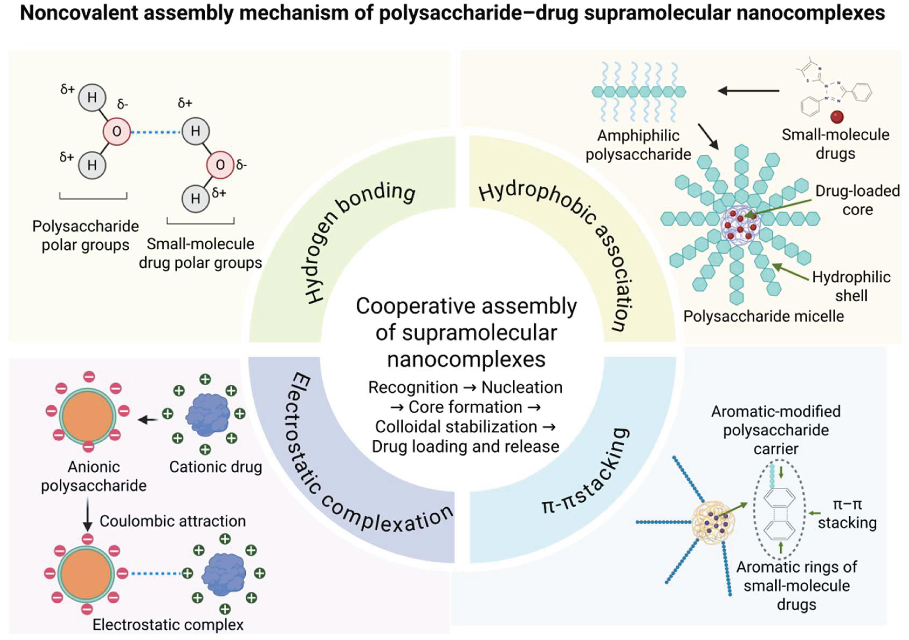
Figure 2.
Comparison of representative preparation strategies for bioactive polysaccharide–small-molecule drug supramolecular nanocomplexes.
Figure 2.
Comparison of representative preparation strategies for bioactive polysaccharide–small-molecule drug supramolecular nanocomplexes.
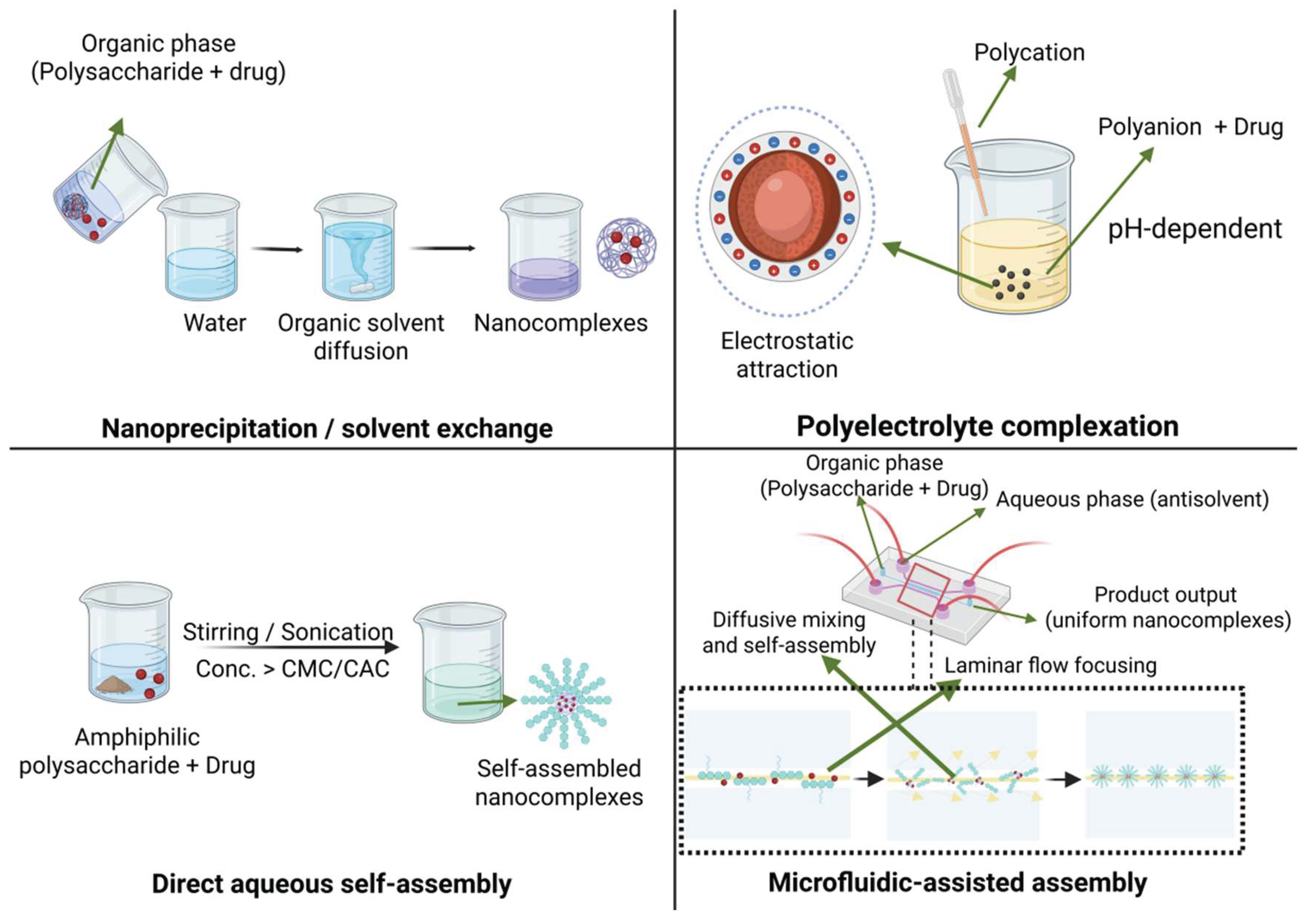
Figure 3.
Carrier–drug synergy and local-matrix extensions of bioactive polysaccharide–small-molecule drug supramolecular nanocomplexes in biomedical applications.
Figure 3.
Carrier–drug synergy and local-matrix extensions of bioactive polysaccharide–small-molecule drug supramolecular nanocomplexes in biomedical applications.
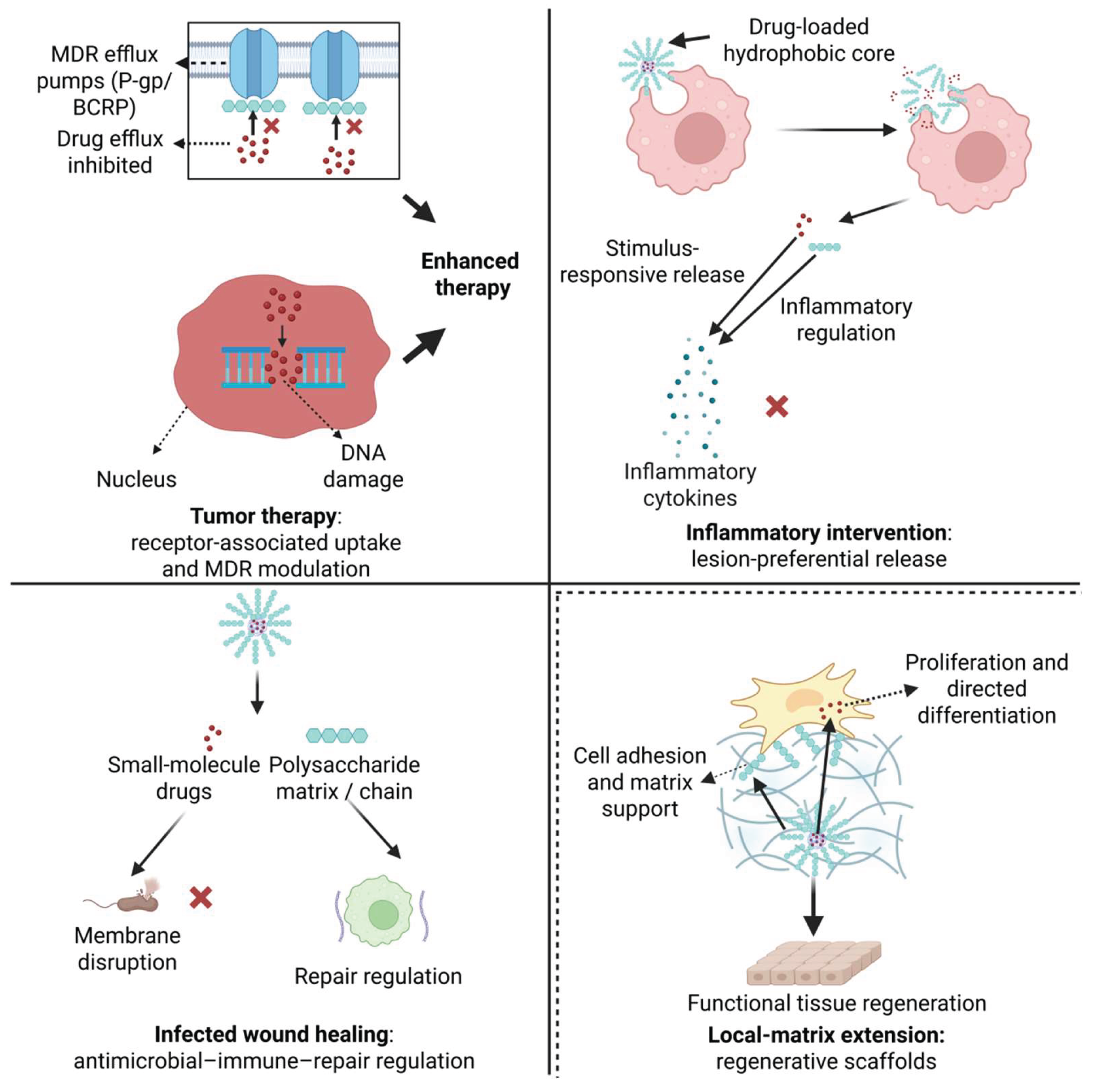

Table 1.
Core formation mechanisms, characterization evidence, and functional contributions of bioactive polysaccharide–small-molecule drug supramolecular nanocomplexes.
Table 1.
Core formation mechanisms, characterization evidence, and functional contributions of bioactive polysaccharide–small-molecule drug supramolecular nanocomplexes.
| Interaction | Molecular basis and typical systems | Key evidence | Main contribution | Ref. |
| Hydrogen bonding | Chitosan-prenylated flavonoid nanocomplexes formed through hydrogen bonding between phenolic hydroxyl groups and chitosan amino groups, together with electrostatic interactions. | FTIR, NMR, particle-size analysis, and stability evaluation | Supports phenolic hydroxyl-dependent polysaccharide-flavonoid complexation and nanocomplex stabilization. | [20] |
| Curdlan-polyphenol complexes formed by pH-driven self-assembly, mainly involving hydrogen bonding and hydrophobic interactions between curdlan and polyphenols. | FTIR, XRD, DSC, physicochemical stability, and gastrointestinal stability assays | Improves physicochemical and gastrointestinal stability, antioxidant activity, and prebiotic potential. | [21] | |
| Hydrophobic association | Stearic-acid-modified Bletilla striata polysaccharide micelles loaded with docetaxel. | CMC, DLS, TEM, zeta potential, encapsulation efficiency, drug-loading capacity, and in vitro release | Drives hydrophobic core formation, docetaxel loading, and sustained release. | [22] |
| Hydrophobically modified hyaluronan nanocapsules loaded with docetaxel through a self-emulsifying process. | Particle size, zeta potential, encapsulation efficiency, plasma stability, and intracellular delivery assays | Forms an oily drug-loaded core with a hyaluronan-based shell, enabling intracellular delivery of docetaxel. | [23] | |
| Electrostatic complexation | Fucoidan-decorated electrostatically assembled nanoparticles bearing doxorubicin, based on interaction between anionic fucoidan and cationic PEI. | Zeta potential, particle size, drug loading, release behavior, and in vitro/in vivo antitumor evaluation | Uses immunomodulatory fucoidan to support electrostatic assembly and improve chemotherapy against breast cancer. | [24] |
| Hyaluronate-doxorubicin nanoaggregates prepared by one-pot mixing through electrostatic and cation–π interactions. | DLS, zeta potential, morphology, drug-release behavior, and CD44-related cellular uptake | Enables facile one-pot self-assembly and CD44-associated tumor-targeted delivery. | [25] | |
| π–π stacking | Chitosan-derivative micelles co-delivering doxorubicin and p53, in which doxorubicin interacts with micellar domains through π–π-related aromatic association. | ¹H NMR, FTIR, fluorescence spectroscopy, particle-size analysis, zeta potential, TEM, and in vitro co-delivery evaluation. | Supports DOX loading in chitosan-derivative micelles through π–π-related interactions and contributes to drug/gene co-delivery performance. | [26] |
| Chitosan-doxorubicin encapsulation/release model analyzed by molecular dynamics simulation. | Molecular dynamics simulation, pH-dependent chitosan protonation analysis, doxorubicin aggregation behavior, and release-behavior prediction. | Reveals how chitosan protonation and DOX π–π stacking regulate encapsulation and pH-responsive release at the molecular level. | [27] |
Table 2.
Representative biomedical applications and local-matrix extensions of bioactive polysaccharide–small-molecule drug supramolecular nanocomplexes.
Table 2.
Representative biomedical applications and local-matrix extensions of bioactive polysaccharide–small-molecule drug supramolecular nanocomplexes.
| Application and representative system | Carrier–drug synergy | Main outcome / validation focus and limitation | Ref. |
| Tumor therapy: BSP–DOX micelles | BSP micellar matrix supports intracellular DOX delivery. | Outcome: improved uptake and antitumor efficacy. Validation: tumor inhibition, biodistribution, organ toxicity. Limitation: modified BSP micelles. | [90] |
| Tumor therapy: APS–PTX nanoplatform | APS supports GLUT-associated uptake and immune regulation; PTX provides chemotherapy. | Outcome: enhanced chemo-immunotherapy. Validation: tumor suppression, immune markers, systemic safety. Limitation: long-term safety and batch reproducibility. | [91] |
| Inflammatory intervention: Licoricchalcone A–polysaccharide NPs | Polysaccharide assembly improves lesion-preferential permeation. | Outcome: enhanced atopic-dermatitis skin permeation. Validation: permeation depth, lesion accumulation, inflammatory markers. Limitation: structure–efficacy relationship unclear. | [92] |
| Inflammatory intervention: dual-responsive polysaccharide nanocarrier | Macrophage-associated delivery combines with MMP/ROS-responsive release. | Outcome: anti-inflammatory dual-drug delivery. Validation: MMP/ROS response, cytokines, immune safety. Limitation: long-term immunological safety. | [93] |
| Infected wound healing: BSP–AZM nanogels | BSP supports local matrix formation; AZM provides antimicrobial activity. | Outcome: improved acute/infected wound healing. Validation: wound closure, bacterial burden, cytokines, collagen, biosafety. Limitation: chronic wound validation. | [94] |
| Infected wound healing: HA-decorated Cur/Cu nanomedicine | HA improves wound-site delivery; Cur provides antioxidant and anti-inflammatory effects. | Outcome: improved infected diabetic wound healing. Validation: wound closure, antibacterial activity, ROS/inflammation, angiogenesis, histology. Limitation: metal coordination system. | [95] |
| Local-matrix extension: HA/chitosan BCP scaffold with immobilized Cal | HA/chitosan coating regulates local Cal presentation and cell–material interaction. | Outcome: improved osteoporotic bone regeneration. Validation: scaffold morphology, osteogenic markers, bone regeneration, biosafety. Limitation: scaffold-based extension. | [96] |
| Local-matrix extension: CS NP/HA hydrogel with fisetin/kartogenin | CS NPs regulate release; HA hydrogel provides ECM-like support. | Outcome: controlled release and OA-related in vitro effects. Validation: release kinetics, cytocompatibility, chondrogenic markers, animal data. Limitation: hydrogel/scaffold extension. | [97] |
| Abbreviations: APS, Astragalus polysaccharide; AZM, azithromycin; BCP, biphasic calcium phosphate; BSP, Bletilla striata polysaccharide; Cal, calcitriol; CS, chitosan; Cu, copper; Cur, curcumin; DOX, doxorubicin; ECM, extracellular matrix; GLUT, glucose transporter; HA, hyaluronic acid; MMP, matrix metalloproteinase; NPs, nanoparticles; OA, osteoarthritis; PTX, paclitaxel; ROS, reactive oxygen species. | |||
Disclaimer/Publisher’s Note: The statements, opinions and data contained in all publications are solely those of the individual author(s) and contributor(s) and not of MDPI and/or the editor(s). MDPI and/or the editor(s) disclaim responsibility for any injury to people or property resulting from any ideas, methods, instructions or products referred to in the content. |
© 2026 by the authors. Licensee MDPI, Basel, Switzerland. This article is an open access article distributed under the terms and conditions of the Creative Commons Attribution (CC BY) license (http://creativecommons.org/licenses/by/4.0/).
Copyright: This open access article is published under a Creative Commons CC BY 4.0 license, which permit the free download, distribution, and reuse, provided that the author and preprint are cited in any reuse.


