
Submitted:
04 August 2026
Posted:
04 August 2026
You are already at the latest version
Abstract
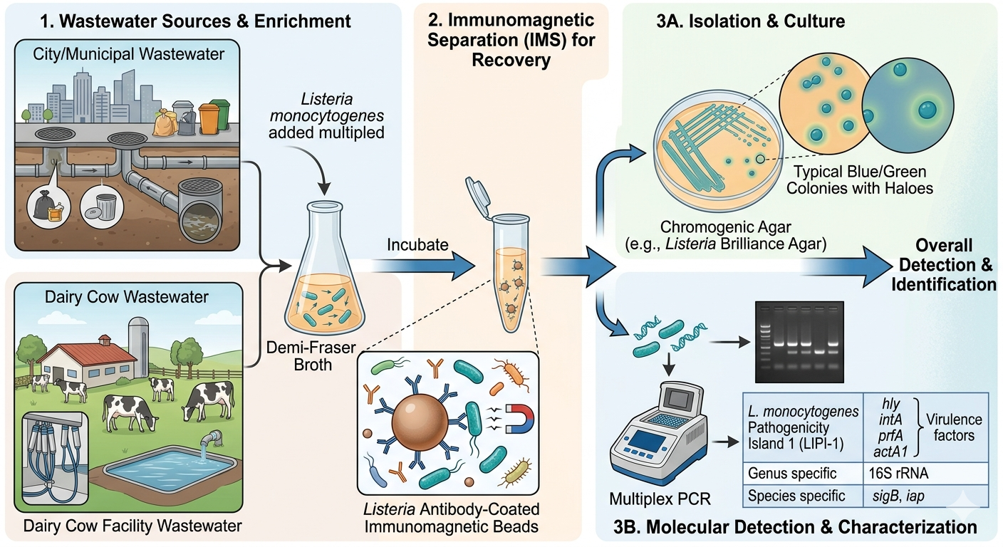
Wastewater systems can serve as reservoirs and dissemination pathways for Listeria spp., yet comparative data from municipal and agricultural wastewater remain limited. This study aimed to isolate and identify Listeria species, especially pathogenic Listeria monocytogenes, from municipal wastewater and dairy‑farm runoff using a genetic multilocus sequence, PCR pathogenic island agarose gel, and phenotypic confirmation strategy. Wastewater samples were filtered, enriched in Demi‑Fraser broth, and subjected to immunomagnetic separation with anti‑Listeria beads before plating on MOX agar and later Listeria Brilliance Agar to differentiate phospholipase‑positive L. monocytogenes. Putative isolates were further characterized by sequencing of 16S rRNA, sigB, and iap genes, and by PCR detection of four Listeria Pathogenicity Island 1 (LIPI‑1) markers (hlyA, inlA, actA, prfA). Using this combined approach, 27 Listeria spp. were recovered from every wastewater sample, with L. innocua predominating in both environments. Seven isolates from municipal and dairy wastewater were confirmed as L. monocytogenes based on sigB and iap sequence identity, LIPI‑1 gene presence, and phospholipase‑positive reactions on chromogenic agar. Several isolates could not be speciated by any locus and were designated Listeria sp. These findings demonstrate that both municipal and dairy‑farm wastewater contain diverse Listeria populations, including pathogenic L. monocytogenes, and highlight the value of integrating chromogenic media with multilocus genetic markers for accurate identification in complex environmental matrices.
Keywords:
Listeria
; Listeria monocytogenes
; wastewater
; PCR
; 16S rRNA
; sigB
; iap
; Listeria Pathogenic Island genes
; chromogenic media
; Listeria Brilliance agar
1. Introduction
Listeria monocytogenes is a major foodborne pathogen associated with severe invasive disease, high hospitalization rates, and substantial mortality, making its environmental ecology a critical component of food-safety risk assessment [1]. Non-pathogenic Listeria spp., including L. innocua, are widespread in soil, water, and agricultural environments and often serve as ecological indicators of conditions that may also support L. monocytogenes. Because both pathogenic and non-pathogenic Listeria species persist in moist, nutrient-rich environments and can form biofilms on diverse surfaces, wastewater systems represent plausible reservoirs and dissemination pathways for these organisms.
Municipal wastewater integrates microbial inputs from households, food establishments, healthcare facilities, and environmental stormwater runoff, while dairy-farm wastewater contains fecal material, feed residues, bedding, and milking-parlor effluent. These systems aggregate microbial signals from large and heterogeneous upstream populations, making them valuable matrices for detecting environmentally persistent organisms such as Listeria [2]. Wastewater-based surveillance has been widely recognized as an efficient, population-level monitoring tool capable of detecting pathogen biomarkers shed into sewer systems [3]. Although historically applied to poliovirus, wastewater surveillance expanded dramatically during the COVID-19 pandemic, demonstrating its ability to detect viral pathogens—including SARS-CoV-2, measles, and influenza—before increases in clinical cases were observed [4]. These systems have shown that wastewater monitoring can provide early warning signals, reveal spatial and temporal trends, and capture pathogen circulation even when clinical testing is limited.
The same principles that make wastewater surveillance effective for viral pathogens are applicable to environmentally persistent foodborne bacteria. Wastewater-based detection has been successfully used for Salmonella, Campylobacter, Escherichia coli O157:H7, and other enteric pathogens in various surveillance programs [5]. Extending these approaches to Listeria spp. is particularly relevant given the organism’s environmental resilience, its ability to survive in cold and nutrient-poor conditions, and its recognized presence in agricultural runoff and surface waters [6,7]. Despite this relevance, systematic surveillance of Listeria in municipal and agricultural wastewater remains limited, and few studies have compared species distribution and pathogenic L. monocytogenes occurrence across these distinct wastewater environments.
Understanding the prevalence, diversity, and genetic characteristics of Listeria spp. in wastewater can provide insight into environmental dissemination pathways, potential contamination routes into food-production systems, and upstream sources contributing to pathogen circulation. Such information complements existing public-health surveillance systems for Listeria infections, which traditionally rely on clinical case reporting and foodborne-disease monitoring. Wastewater-based detection may therefore serve as an early-warning or supplementary surveillance mechanism, particularly in regions with intensive agricultural activity or high-risk food-processing operations [2,8].
To address these gaps, we isolated and characterized Listeria spp., including L. monocytogenes, from municipal and dairy-farm wastewater (the dairy farm experienced the death of 6 fetuses from 9 pregnant dairy cows in the preceding 3 months due to L. monocytogenes). As a bridge to our methodological approach, we employed a multilocus genetic identification strategy incorporating 16S rRNA sequencing [9,10], the sigB stress-response gene [11,12], the iap invasion-associated protein gene [13], and four Listeria Pathogenicity Island 1 (LIPI-1) markers [14,15]. These loci provide complementary taxonomic and virulence-associated resolution: 16S for broad genus-level identification, sigB for species-level discrimination, iap housekeeping gene for additional species confirmation, and LIPI-1 alleles for detecting hallmark virulence determinants of L. monocytogenes. Together, these markers enable differentiation of pathogenic and non-pathogenic Listeria species and together with chromogenic selective/differential media [16,17], support high-confidence identification of isolates, especially L. monocytogenes, recovered from complex wastewater matrices.
2. Materials and Methods
2.1. Bacterial Strains, Growth Media and Conditions
Bacterial cultures used in this study were obtained from our laboratory culture collection or isolated from environmental sources as indicated in Table 1. Listeria strains were cultured in Tryptic Soy broth and plated on Tryptic Soy agar (TSB, TSA, Becton-Dickinson, Difco brand, Franklin Lakes, NJ, USA) at 30oC. Tentative confirmation as Listeria spp. was obtained by enrichment in Demi-Fraser broth (BD-Difco) and streaking/plating on MOX agar base with supplements (BD-Difco). Additional methods of identification including PCR bands on agarose gels, sequencing of PCR amplimers of various gene sequences, and phenotypic reaction on Listeria-specific media are explained below. Cultures were maintained for storage by centrifugation (6000× g, 5 °C) of 9 mL of fresh, overnight cultures and cell pellets were resuspended in 2–3 mL of fresh sterile TSB containing 10% glycerol. Cell suspensions were placed into glass vials and stored in an ultra-low freezer (−80 °C). Frozen stocks were revived by transferring 100 µL of the thawed cell suspension into 9 mL of TSB, incubating overnight at 30 °C, and sub-cultured again before use.
2.2. Isolation of Listeria spp. from Wastewater Using Anti-Listeria Immunomagnetic Beads
Approximately 15-20 wastewater samples were collected from city wastewater facilities and OSU Dairy Farm runoff (Figure 1A,C). The municipal wastewater facility had a convenient continuously running pipe from which raw wastewater could be sampled before processing; the Oklahoma State University Dairy Farm runoff from dairy cattle facilities lead to a retention pond and samples were retrieved using a telescoping bottle-holding device near the spillway; these samples were significantly ‘dirty’ from feces and dirt. Interest in wastewater runoff from the OSU Dairy Farm was heightened due to 6 deaths of unborn cattle from pregnant dairy cows due to L. monocytogenes in the preceding months that was attributed to contaminated silage. In either case, 200-ml water samples were recovered and brought back to the lab for processing within 1-day (Figure 1B,D).
Water samples that were extremely cloudy with suspended material (mostly dairy farm samples), were first filtered through sterile paper filters (60-nm pore size) using a Buchner funnel and vacuum flask setup (Figure 1E). The filtrate was then filtered a second time through a sterile 47-mm 0.2-micron membrane filter that was removed and added to a sterile stomacher bag with 100 ml of Demi-Fraser broth enrichment media (Difco), incubated at 30oC overnight, and sampled the next day (Figure 1F–H). After enrichment, a 1-ml sample was taken and processed with magnetic beads coated with anti-Listeria antibodies (Dynabeads anti-Listeria, Thermo Fisher Scientific, Waltham, MA, USA) using a ‘bead retriever’ (Dynal/Invitrogen, Carlsbad, CA, USA) for immunomagnetic separation (IMS) (Figure 1I,J). The beadretriever included program settings for recovery and washing Listeria cells adhered to paramagnetic beads covered with anti-Listeria antibodies. The process involves placing enrichment samples in initial cupules and adding Dynabeads with anti-Listeria antibodies, shaking to facilitate sample agitation, lowering the plastic sleeve-covered magnetic fingers into the sample, removing the magnetic beads from enrichment broth and dropping them into new cupules with buffer (retraction of magnetic fingers from the plastic sleeve causes beads to drop into the cupule). This was then followed by several cycles of washing with buffer for final recovery of magnetic beads with adhered Listeria. The recovered Listeria samples were then streaked or plated onto MOX agar (Difco) and individual colonies were re-streaked and recovered for identity determination (Figure 1K); additionally, enriched samples with recovered immunomagnetic beads were plated or streaked on Brilliance Agar with appropriate supplements (Oxoid Brand, Thermo Fisher Scientific, Waltham, MA, USA) for putative determination as L. monocytogenes.
2.3. Phenotypic Reaction of Listeria spp. Isolated from Municipal and Dairy Center on Listeria Brilliance Agar
Oxoid Listeria Brilliance Agar (LBA, Thermo Fisher) is a selective and differential agar media similar to Modified Oxford Media (MOX) in having both selective and differential components. It has a base media plus two supplements. One supplement contains four antibiotics (selective supplements) that provide selective recovery of L. monocytogenes/Listeria spp. and suppresses other bacteria. The second supplement contains two differential components: an X-glucoside that is hydrolyzed by β-glucosidase to produce a chromogen making colonies appear blue-green, and a lecithin which is acted upon by lecithinase (phospholipase) of L. monocytogenes/L. ivanovii to produce ‘white halos’ in the media surrounding the blue-green colonies. Blue green colonies without white halos are neither L. monocytogenes nor L. ivanovii, but likely one of many other species of Listeria.
2.4. Identity of Listeria monocytogenes and Listeria Species Isolated from Wastewater
Putative Listeria isolates obtained from typical reactions on MOX agar were examined by PCR sequencing of 16S [18], sigmaB factor [19], and invasion associated protein (iap) [13] to help establish identity based on molecular sequence determination. Additional analyses were obtained by agarose gel analysis of expected PCR reactions related to four of six genes involved with Listeria monocytogenes Pathogenic Island 1 gene cluster (LmPI-1: hly, inlA, actA, and prfA [15,20]). Phenotypic confirmation was further confirmed by plating/streaking on Listeria Brilliance Agar [17] for reactions of β-glucosidase and phospholipase C.
2.4.1. PCR and Sequence Analysis of 16S rRNA, sigB, and iap Genes of Isolates from Municipal and Dairy Wastewater
Listeria isolated from municipal and dairy center wastewater were cultured overnight in TSB (Difco) at 30oC; the next day 1 ml of cell culture was micro-centrifuged and DNA was extracted from the cell pellet by a ‘beadbeating’ technique [21,22,23] using 0.1 mm zirconia/silica disruption beads (RPI Research Products Intl., Mt. Prospect, IL, USA) and a bead tube holder/adapter fitted to a common laboratory vortexer (Takara, San Jose, CA, USA). After beadbeating cell disruption, extracted DNA samples were heated at 95oC for 10 min in a thermal cycler. PCR was performed using primers obtained from published sources or determined from consensus sequence regions identified after multiple sequence alignment of the same genes from different isolates (Table 2). After PCR, reactions were cleaned up using the IBI PCR Clean-Up kit (IBI Scientific, Dubuque, IA, USA). Briefly, a high salt ‘binding buffer’ is added to PCR reactions which are then loaded onto a DNA Filter Spin Column (glass fiber filter) to which DNA binds to under high-salt conditions, centrifuged, and the eluate is discarded. The spin column is centrifuged again after each addition of 2 different wash buffers. Finally, centrifugation with a low salt Tris buffer releases and elutes DNA from the silica matrix. PCR reactions cleaned up in this manner were submitted to our OSU DNA Core Facility for DNA sequencing.
Various 16S rRNA ‘universal primers’ for bacteria have been well characterized [10,24]. We used primers involving 2 sets of primers spanning nearly the entirety of the 16S rRNA region during culture-dependent microbiome analysis of bacteria isolated during biltong beef processing [18]. The numerical sequence of the various primers refer to their position in the generic E. coli K-12 which may differ from the exact position in any other organism because of slight sequence differences.
Another region that is not as strictly conserved as 16S rRNA is polymerase sigma factor B (sigB; σᵇ) that is involved in RNA polymerase transcription of genes, but only for the specific set of promoters in its σᵇ regulon. This region has also been used to identify bacterial species where 16S rRNA sequence differentiation is too constrained [19].
Lastly, when there isn’t any correlation between 16S rRNA and sigB species identity, a housekeeping gene is often used as these are not conserved and offer more mutational differences and offer species sequence distinction. Among Listeria species, the invasion associated protein (iap, protein p60) is often used [13]. The name is a misnomer as it was originally thought it was involved with cell invasion but is now known to be involved with hydrolase activity during cell division in Listeria.
2.4.2. Agarose Gel Analysis of L. monocytogenes Pathogenic Island (LmPI-1) Genes Involved in Pathogenesis
Stock cultures of pathogenic and non-pathogenic Listeria sp. were examined by PCR reactions with primers for four LmPI-1 genes (hlyA, inlA, prfA, actA1; Table 2) and amplimers were run on 2% agarose gels (DNA grade agarose, Fisher Scientific) with 1x TAE buffer prepared from a 10x stock (Fisher Scientific). Gel wells were loaded with 10 ul of sample (3-6 uL nuclease-free water, 1 uL Blue juice dye, 1-3 uL PCR sample; a 100-bp DNA ladder (Promega, Madison, WI, USA) was also included for determining the correct size range of amplimers. Gels were electrophoresed at 80 V (constant voltage) for ~90 min with the blue dye indicating the extent of migration of small DNA. After electrophoresis, the agarose gel was stained with 100 ml of ethidium bromide solution (0.5 ug/mL; Fisher Scientific). EtBr-stained gels were destained by floating in a water tray on a slow-moving shaker for 25 min and visualized with a gel documentation system (UVP GelDoc-IT, UVP LLC, Upland, CA, USA).
3. Results and Discussion
3.1. Isolation of Listeria from Wastewater
Immunomagnetic Separation
Recovery of Listeria sp. was facilitated by enrichment of membrane filters in DemiFraser broth, recovery of Listeria from these enrichments using paramagnetic beads coated with anti-Listeria antibodies with the beadretriever, and plating on MOX agar (Figure 1E–K). Use of a Buchner funnel and large-porosity (20-microns) paper filters allowed removal of large particles from dirty water; the filtrate was then filtered on 0.2-micron filter membranes and bacteria trapped on their surface were enriched in Demi-Fraser selective broth. Commercial immunomagnetic beads for Listeria were only available with generic antibodies for Listeria sp., and not for L. monocytogenes, which would have been preferable.
3.2. Phenotypic Reactions of Listeria spp. on Selective Media Isolated from Municipal and Dairy Center Environmental Sources of Wastewater
3.2.1. Phenotypic Reactions on MOX Agar
In our experience, Listeria sp. almost always appear on MOX agar after extended incubation as concave colonies, represented as a depressed morphology around the edges of the colony (usually after 2-3 days of growth). We do not know how widespread this phenomenon is, but we have always observed this with colonies of Listeria spp. on MOX agar. Other MOX-reactive (darkened) colonies that appear on MOX have normal convex colony morphology that do not appear depressed into the agar, while other non-reactive (white) convex colonies occasionally occur. Using this IMS + MOX approach, we isolated typical Listeria-like colonies with every sample of municipal or dairy center wastewater examined; subsequent 16S rRNA sequencing confirmed these isolates as Listeria sp.
3.2.2. Phenotypic Reactions on Listeria Brilliance Agar
Although MOX agar was useful for tentatively identifying Listeria sp., it does not differentiate between any of the species. We therefore thought to employ one of the newer chromogenic agars (LL agar, Harlequin Listeria agar, ALOA agar, Listeria Brilliance agar) [17,25,26,27] that could distinguish putative L. monocytogenes from non-pathogenic species. We selected Oxoid Listeria Brilliance agar (LBA) because of its ability to distinguish L. monocytogenes and L. ivanovii by their ability to form “white halos” on the selective/differential agar media. The halos form from action of the phospholipase-mediated hydrolysis of phosphatidylcholine in the agar, producing white precipitated zones that increased in size with extended incubation surrounding blue-green colonies (Figure 2).
Upon receiving LBA (delayed 3-4 months due to ordering issues) we went back and tested all remaining immunomagnetic bead samples previously screened on MOX agar. This allowed us to recover additional L. monocytogenes isolates that had not been detected on MOX agar with random colony testing. The added phenotypic discrimination of ‘white halo’ reaction with LBA was highly valuable in the tentative identification of L. monocytogenes that was subsequently confirmed by PCR and sequencing.
A key phenotypic marker supporting identification of L. monocytogenes in this process is the activity of phospholipase C, an enzyme used by this pathogen to dissolve phospholipid membranes, escape intracellular vesicles (i.e., endolysosomes, phagolysosomes) and mammalian host cells to facilitate intracellular survival [28,29]. Listeria Brilliance agar is one of several chromogenic media that have been developed that can differentiate L. monocytogenes (or L. ivanovii) from other Listeria sp [25,26,30,31]. Furthermore, the chromogen, X-glucoside is hydrolyzed by β-glucosidase to produce a greenish-blue colony color while the L. monocytogenes/L. ivanovii phospholipase C hydrolyzes the phosphatidylcholine ( lecithin) to produce the characteristic white halo surrounding the colony (Figure 2). We used Listeria Brilliance agar to screen Listeria recovered by immunomagnetic separation (IMS) after filter-membrane enrichment (Figure 2).
3.3. Sequence Analysis of Control Strains and Putative Listeria Isolates from Wastewater
3.3.1. Sequence Analysis of 16S rRNA Genes
The reason that the same 16S rRNA primers amplify many different bacterial taxa is that the 16S rRNA gene is functionally essential and highly conserved. Because ribosomal operation tolerates very little sequence variation, the gene exhibits limited divergence between bacterial genera, and even less among species within the same genus.
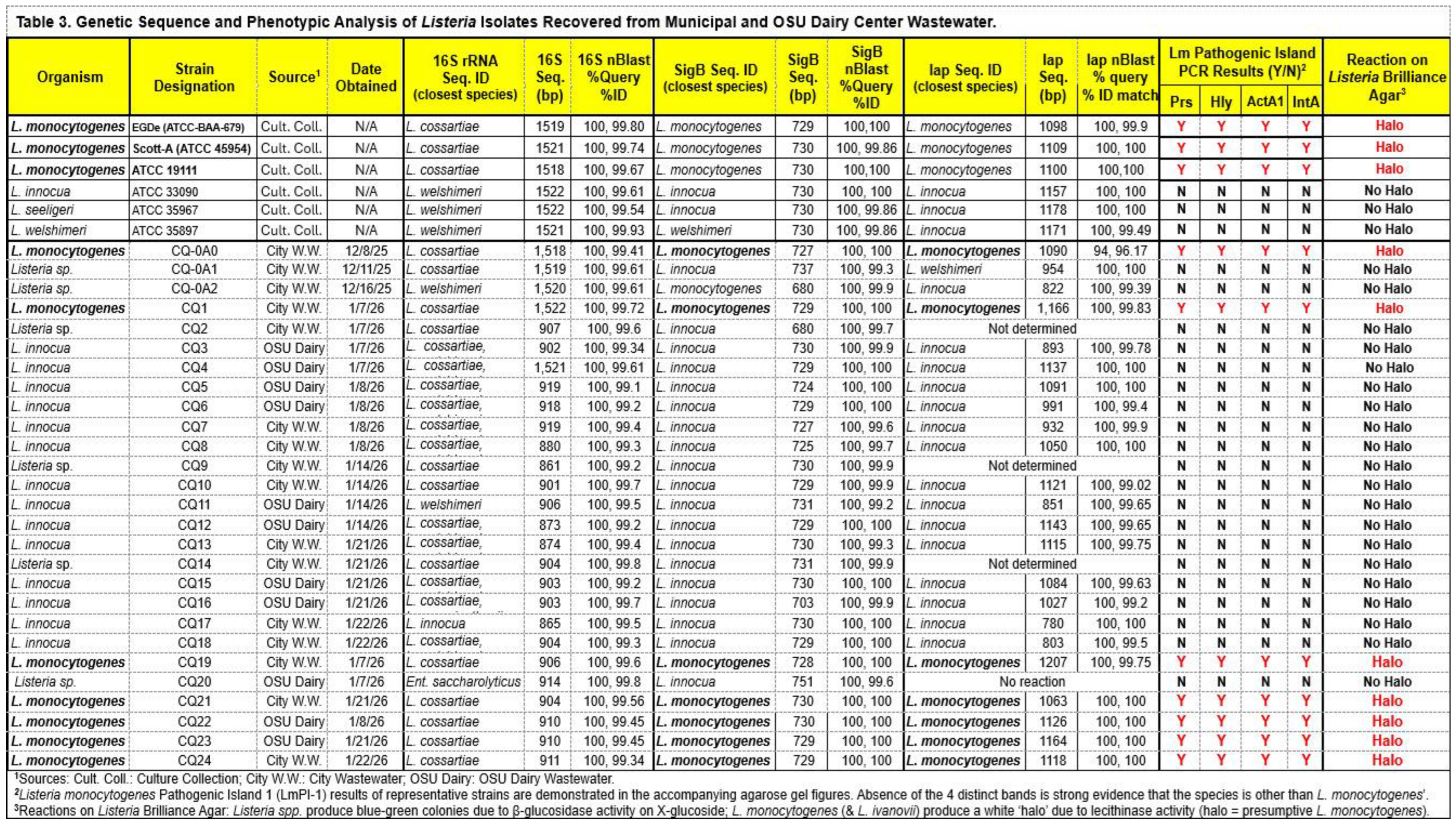
Following PCR amplification, sequencing, and BLAST analysis of the 16S rRNA gene, we obtained species-level identifications for several isolates reported as Listeria cossartiae (Table 3). Although such identifications would not unusual for newly isolated strains, we also obtained the same L. cossartiae identities when using known ATCC reference cultures (Table 3). L. cossartiae is one of several new species recently described originating out of the Weidmann Lab (Cornell University, Ithaca, NY, USA) identified from agricultural water and environmental soil samples [32].
This outcome can be explained by the highly conserved nature of the 16S rRNA gene within Listeria spp. which prevents reliable species-level discrimination. L. monocytogenes has been shown to cluster with L. cossartiae and several other species in a category designated as the “most common ancestor” of Listeria sp. [33]. For this reason, we proceeded to analyze a less-conserved genetic marker, the sigma factor B (or sigB).
3.3.2. Sequence Analysis of Listeria Sigma Factor B Genes
The Sigma B RNA polymerase factor (σB) is a master regulator of the stress response in many bacteria including Listeria [34]. It functions as an alternative subunit for RNA polymerase and is normally maintained in an inactive state by the "anti-sigma factor" RsbW. When bacteria encounter environmental stress (acid, salt, heat), a signaling cascade releases sigB enabling it to bind to the core RNA polymerase and direct transcription towards stress response promoters. Among Listeria sp., the sigB gene is moderately conserved and is typically found within the general stress response operon (rsbV-rsbW-sigB-rsbX) [35] and is often used for species-level identification in combination with other genetic loci [11,36].
In our study, identities provided by sigB sequencing differed markedly from those obtained using 16S rRNA. Most were either identified as L. innocua whereas 7 isolates, as well as the known ATCC L. monocytogenes strains, were identified as L. monocytogenes (Table 3). To obtain additional support for identification of L. monocytogenes, we also examined a housekeeping gene (iap) expected to exhibit less conservation than either 16S or sigB, allowing us to assess concordance across multiple loci.
3.3.3. Sequence Analysis of Listeria iap Genes
The Listeria invasion associated protein housekeeping gene (iap), has also been used species identification [13,37,38,39]. The iap gene encodes the p60 protein and is one of the most commonly employed single-gene markers for Listeria spp. because it contains conserved regions shared across the genus as well as variable regions allowing discrimination. Our approach was to use the conserved regions on either end of the iap gene as universal priming sites and use the variable internal region alongside 16S and sigB to confirm species identity.
Although the designation iap historically stood for “invasion-associated protein (p60)”, the gene actually encodes a cell wall hydrolase involved in cell division but the historical name designation remained. Pilgrim et al [40] had proposed renaming the gene cell wall hydrolase A (cwhA) to reflect its true function; however, taxonomists have never broadly accepted the name change.
3.3.4. PCR and Agarose Gel Electrophoresis of Amplimers Involved with Listeria monocytogenes Pathogenic Island 1 (LmPI-1) Genes
Despite the strong agreement between sigB, and iap to confirm species identity of L. monocytogenes, we further examined PCR amplimers representing hlyA, actA1, intA, and prfA to verify the presence of these four LmPI-1 pathogenicity island genes. Amplimers of appropriate size were obtained for all four loci from each of the three ATCC L. monocytogenes reference strains as well as seven environmental isolates from dairy and municipal wastewater. In contrast, non-pathogenic Listeria spp. (L. innocua, L. seeligeri, and L. welshimeri) lacked these loci (Table 3, Figure 3, Figure 4 and Figure 5).
PCR detection of the invasion-associated protein (iap, p60) was subsequently included on agarose gel analyses of amplimers. Iap is a housekeeping gene encoding a hydrolase involved in cell division that is frequently used for species identification but is not specific to pathogenic strains. Isolate CQ4 was determined to be L. monocytogenes based on the presence of all 4 LmPI-1 genes while the other isolates predominantly showed the iap amplimer. We also observed the iap gene did not amplify in CQ14 and several other isolates that are confirmed as Listeria sp.; these samples were re-tested several times with same results.
Initial PCR regimens used theoretical annealing temperatures based on the common guideline of using annealing temperatures 2-5 oC below the primer Tm [41,42,43]. This approach produced faint or smeared bands in some agarose gel lanes, which we interpreted as non-specific priming. Most non-pathogenic Listeria isolates produced cleaner PCR profiles after we optimized annealing temperatures to 2-4oC above the primer Tm, thereby reducing non-specific amplification artifacts [44]. This was subsequently verified by running an annealing temperature gradient across the thermal cycler 96-well plate with the same DNA template from an isolate giving spurious bands and smears [45]. Our ability to obtain suspect isolates that were later confirmed as L. monocytogenes was aided by the use of Listeria Brilliance agar, which produced the characteristic white halos described earlier.
4. Conclusions
This study provides a systematic assessment of Listeria spp. present in municipal and dairy-farm wastewater using a combined approach of immunomagnetic separation, selective and chromogenic media, and multilocus genetic characterization. Listeria was recovered from every wastewater sample examined, demonstrating the consistent presence of this genus in both urban and agricultural effluent. Phenotypic differentiation on MOX and Listeria Brilliance Agar, together with sequence analysis of 16S rRNA, sigB, and iap genes, enabled reliable species-level identification, while PCR detection of four LmPI-1 loci confirmed pathogenic L. monocytogenes among the isolates. As documented in the manuscript, Listeria were isolated from every sample of municipal or dairy center wastewater examined, indicating the prevalence of Listeria sp. in environmental wastewater. LmPI-1 amplimers were obtained from seven environmental isolates, supporting their classification as L. monocytogenes. The predominance of L. innocua across both wastewater sources, including the detection of L. monocytogenes in 7 of 27 (26%) isolates, including a dairy farm with recent listeriosis-associated fetal losses, highlights the ecological persistence of Listeria in wastewater environments. These findings underscore the utility of integrating chromogenic media with multilocus molecular markers for accurate identification of Listeria in complex matrices and support the potential role of wastewater monitoring as a complementary tool for understanding environmental dissemination pathways of foodborne pathogens.
Considering the prevalence of Listeria sp. in environmental wastewater, several of our next projects will focus on whether use of L. monocytogenes-specific antibodies (to internalin A) tethered to magnetic beads can improve recovery from wastewater streams and whether such streams are a ready source of L. monocytogenes-specific bacteriophage.
Author Contributions
Conceptualization, P.M.; methodology, P.M.; software, P.M.; validation, P.M. and C.Q.; formal analysis, C.Q.; investigation, C.Q.; resources, P.M.; data curation, P.M.; writing—original draft preparation, P.M. and C.Q.; writing—review and editing, P.M.; visualization, P.M.; supervision, P.M.; project administration, P.M.; funding acquisition, P.M. All authors have read and agreed to the published version of the manuscript.
Funding
This research was supported by funds provided by the Gilliland/Advance Foods Professorship in Microbial Food Safety (21-57200) and the OSU Agricultural Experiment Station (OKL03284).
Institutional Review Board Statement
Not applicable.
Informed Consent Statement
Not applicable; this study did not involve humans.
Data Availability Statement
Data is available upon request.
Acknowledgments
This work was performed by Ms. Caitlyn Quinn as part of the requirements for the MS degree in Food Science.
Conflicts of Interest
The authors declare no conflicts of interest. The funders had no role in the design of the study; in the collection, analyses, or interpretation of data; in the writing of the manuscript; or in the decision to publish the results.
References
- Shamloo, E.; Hosseini, H.; Abdi Moghadam, Z.; Halberg Larsen, M.; Haslberger, A.; Alebouyeh, M. Importance of Listeria monocytogenes in food safety: a review of its prevalence, detection, and antibiotic resistance. Iran J Vet Res 2019, 20, 241–254.
- Troja, F.; Indio, V.; Savini, F.; Seguino, A.; Serraino, A.; Fuschi, A.; Remondini, D.; De Cesare, A. Monitoring and preventing foodborne outbreaks: are we missing wastewater as a key data source? Ital J Food Saf 2024, 13, 12725. [CrossRef]
- National Academies of Sciences, E.; Medicine; Health; Medicine, D.; Board on Population, H.; Public Health, P.; Division on, E.; Life, S.; Water, S.; Technology, B., et al. In Increasing the Utility of Wastewater-based Disease Surveillance for Public Health Action: A Phase 2 Report, National Academies Press (US) Copyright 2024 by the National Academy of Sciences. All rights reserved.: Washington (DC), 2024; 10.17226/27516.
- Grassly, N.C.; Shaw, A.G.; Owusu, M. Global wastewater surveillance for pathogens with pandemic potential: opportunities and challenges. The Lancet Microbe 2025, 6. [CrossRef]
- Singh, S.; Ahmed, A.I.; Almansoori, S.; Alameri, S.; Adlan, A.; Odivilas, G.; Chattaway, M.A.; Salem, S.B.; Brudecki, G.; Elamin, W. A narrative review of wastewater surveillance: pathogens of concern, applications, detection methods, and challenges. Frontiers in Public Health 2024, Volume 12 - 2024. [CrossRef]
- Kayode, A.J.; Semerjian, L.; Osaili, T.; Olapade, O.; Okoh, A.I. Occurrence of Multidrug-Resistant Listeria monocytogenes in Environmental Waters: A Menace of Environmental and Public Health Concern. Frontiers in Environmental Science 2021, Volume 9 - 2021. [CrossRef]
- Stea, E.C.; Purdue, L.M.; Jamieson, R.C.; Yost, C.K.; Hansen, L.T. Comparison of the Prevalences and Diversities of Listeria Species and Listeria monocytogenes in an Urban and a Rural Agricultural Watershed. Applied and Environmental Microbiology 2015, 81, 3812–3822, doi:doi:10.1128/AEM.00416-15.
- Fagerlund, A.; Møretrø, T.; Jensen, M.R.; Langsrud, S.; Moen, B. Early detection and population dynamics of Listeria monocytogenes in naturally contaminated drains from a meat processing plant. Frontiers in Microbiology 2025, Volume 16 - 2025. [CrossRef]
- Hellberg, R.S.; Martin, K.G.; Keys, A.L.; Haney, C.J.; Shen, Y.; Smiley, R.D. 16S rRNA partial gene sequencing for the differentiation and molecular subtyping of Listeria species. Food Microbiology 2013, 36, 231–240. [CrossRef]
- Somer, L.; Kashi, Y. A PCR Method Based on 16S rRNA Sequence for Simultaneous Detection of the Genus Listeria and the Species Listeria monocytogenes in Food Products. Journal of Food Protection 2003, 66, 1658–1665. [CrossRef]
- Nightingale, K.; Bovell, L.; Grajczyk, A.; Wiedmann, M. Combined sigB allelic typing and multiplex PCR provide improved discriminatory power and reliability for Listeria monocytogenes molecular serotyping. Journal of Microbiological Methods 2007, 68, 52–59. [CrossRef]
- Liao, J.; Wiedmann, M.; Kovac, J. Genetic Stability and Evolution of the sigB Allele, Used for Listeria Sensu Stricto Subtyping and Phylogenetic Inference. Applied and Environmental Microbiology 2017, 83, e00306–00317, doi:doi:10.1128/AEM.00306-17.
- Bubert, A.; Köhler, S.; Goebel, W. The homologous and heterologous regions within the iap gene allow genus- and species-specific identification of Listeria spp. by polymerase chain reaction. Applied and Environmental Microbiology 1992, 58, 2625–2632. [CrossRef]
- Liu, D. Identification, subtyping and virulence determination of Listeria monocytogenes, an important foodborne pathogen. Journal of Medical Microbiology 2006, 55, 645–659. [CrossRef]
- Wiktorczyk-Kapischke, N.; Skowron, K.; Wałecka-Zacharska, E. Genomic and pathogenicity islands of Listeria monocytogenes—overview of selected aspects. Frontiers in Molecular Biosciences 2023, Volume 10 - 2023. [CrossRef]
- Suresh, D.; Puttananjaiah, M.H.; Dhale, M.A. Listeria monocytogenes isolated on selective chromogenic plating media necessitates further characterization to confirm its presence in dairy foods. International Dairy Journal 2025, 164, 106197. [CrossRef]
- Park, S.-H.; Chang, P.-S.; Ryu, S.; Kang, D.-H. Development of a Novel Selective and Differential Medium for the Isolation of Listeria monocytogenes. Applied and Environmental Microbiology 2014, 80, 1020–1025, doi:doi:10.1128/AEM.02840-13.
- Karolenko, C.; DeSilva, U.; Muriana, P.M. Microbial Profiling of Biltong Processing Using Culture-Dependent and Culture-Independent Microbiome Analysis. Foods 2023, 12, 844.
- Wu, J.; NicAogáin, K.; McAuliffe, O.; Jordan, K.; O’Byrne, C. Phylogenetic and Phenotypic Analyses of a Collection of Food and Clinical Listeria monocytogenes Isolates Reveal Loss of Function of Sigma B from Several Clonal Complexes. Applied and Environmental Microbiology 2022, 88, e00051–00022, doi:doi:10.1128/aem.00051-22.
- Johansson, J.; Freitag, N.E. Regulation of Listeria monocytogenes Virulence. Microbiology Spectrum 2019, 7, 10.1128/microbiolspec.gpp1123–0064–2019, doi:doi:10.1128/microbiolspec.gpp3-0064-2019.
- de Bruin, O.M.; Birnboim, H.C. A method for assessing efficiency of bacterial cell disruption and DNA release BMC Microbiology 2016, 16, 197. [CrossRef]
- Anderson, S.R.; Thompson, L.R. Optimizing an enclosed bead beating extraction method for microbial and fish environmental DNA. Environmental DNA 2022, 4, 291–303. [CrossRef]
- Coton, E.; Coton, M. Multiplex PCR for colony direct detection of Gram-positive histamine- and tyramine-producing bacteria. Journal of Microbiological Methods 2005, 63, 296–304. [CrossRef]
- Yang, B.; Wang, Y.; Qian, P.-Y. Sensitivity and correlation of hypervariable regions in 16S rRNA genes in phylogenetic analysis. BMC Bioinformatics 2016, 17, 135. [CrossRef]
- Vlaemynck, G.; Lafarge, V.; Scotter, S. Improvement of the detection of Listeria monocytogenes by the application of ALOA, a diagnostic, chromogenic isolation medium. Journal of Applied Microbiology 2000, 88, 430–441.
- Reissbrodt, R. New chromogenic plating media for detection and enumeration of pathogenic Listeria spp.—an overview. International Journal of Food Microbiology 2004, 95, 1–9. [CrossRef]
- Smith, P.A.; Mellors, D.; Holroyd, A.; Gray, C. A new chromogenic medium for the isolation of Listeria spp. Letters in Applied Microbiology 2001, 32, 78–82. [CrossRef]
- Yeung, P.S.M.; Na, Y.; Kreuder, A.J.; Marquis, H. Compartmentalization of the Broad-Range Phospholipase C Activity to the Spreading Vacuole Is Critical for Listeria monocytogenes Virulence. Infection and Immunity 2007, 75, 44–51, doi:doi:10.1128/iai.01001-06.
- Petrišič, N.; Adamek, M.; Kežar, A.; Hočevar, S.B.; Žagar, E.; Anderluh, G.; Podobnik, M. Structural basis for the unique molecular properties of broad-range phospholipase C from Listeria monocytogenes. Nature Communications 2023, 14, 6474. [CrossRef]
- Greenwood, M.; Willis, C.; Doswell, P.; Allen, G.; Pathak, K. Evaluation of chromogenic media for the detection of Listeria species in food. Journal of Applied Microbiology 2005, 99, 1340–1345. [CrossRef]
- Stessl, B.; Luf, W.; Wagner, M.; Schoder, D. Performance testing of six chromogenic ALOA-type media for the detection of Listeria monocytogenes. Journal of Applied Microbiology 2009, 106, 651–659. [CrossRef]
- Carlin, C.R.; Liao, J.; Weller, D.; Guo, X.; Orsi, R.; Wiedmann, M. Listeria cossartiae sp. nov., Listeria immobilis sp. nov., Listeria portnoyi sp. nov. and Listeria rustica sp. nov., isolated from agricultural water and natural environments. International Journal of Systematic and Evolutionary Microbiology 2021, 71. [CrossRef]
- Orsi, R.H.; Liao, J.; Carlin, C.R.; Wiedmann, M. Taxonomy, ecology, and relevance to food safety of the genus Listeria with a particular consideration of new Listeria species described between 2010 and 2022. mBio 2024, 15, e00938–00923, doi:doi:10.1128/mbio.00938-23.
- Sibanda, T.; Buys, E.M. Listeria monocytogenes Pathogenesis: The Role of Stress Adaptation. Microorganisms 2022, 10, 1522.
- Liu, Y.; Orsi, R.H.; Gaballa, A.; Wiedmann, M.; Boor, K.J.; Guariglia-Oropeza, V. Systematic Review of the Listeria Monocytogenes σB Regulon Supports a Role in Stress Response, Virulence and Metabolism. Future Microbiology 2019, 14, 801–828. [CrossRef]
- Cai, S.; Kabuki, D.Y.; Kuaye, A.Y.; Cargioli, T.G.; Chung, M.S.; Nielsen, R.; Wiedmann, M. Rational Design of DNA Sequence-Based Strategies for Subtyping Listeria monocytogenes. Journal of Clinical Microbiology 2002, 40, 3319–3325, doi:doi:10.1128/jcm.40.9.3319-3325.2002.
- Volokhov, D.; Rasooly, A.; Chumakov, K.; Chizhikov, V. Identification of Listeria Species by Microarray-Based Assay. Journal of Clinical Microbiology 2002, 40, 4720–4728, doi:doi:10.1128/jcm.40.12.4720-4728.2002.
- Yoshikawa, Y.; Ochiai, Y.; Mochizuki, M.; Fujita, O.; Takano, T.; Hondo, R.; Ueda, F. Genetic subtyping of Listeria monocytogenes via multiple-locus sequence typing using iap, sigB and actA. Journal of Veterinary Medical Science 2016, 78, 1831–1839. [CrossRef]
- Cocolin, L.; Rantsiou, K.; Iacumin, L.; Cantoni, C.; Comi, G. Direct Identification in Food Samples of Listeria spp. and Listeria monocytogenes by Molecular Methods. Applied and Environmental Microbiology 2002, 68, 6273–6282, doi:doi:10.1128/AEM.68.12.6273-6282.2002.
- Pilgrim, S.; Kolb-Maurer, A.; Gentschev, I.; Goebel, W.; Kuhn, M. Deletion of the gene encoding p60 in Listeria monocytogenes leads to abnormal cell division and loss of actin-based motility. Infect Immun 2003, 71, 3473–3484.
- Baumforth, K.R.; Nelson, P.N.; Digby, J.E.; O'Neil, J.D.; Murray, P.G. Demystified ... the polymerase chain reaction. Mol Pathol 1999, 52, 1–10. [CrossRef]
- Welch, H.M. The Polymerase Chain Reaction. In Metastasis Research Protocols, Dwek, M., Brooks, S.A., Schumacher, U., Eds. Humana Press: Totowa, NJ, 2012; 10.1007/978-1-61779-854-2_5pp. 71–88.
- Lorenz, T.C. Polymerase chain reaction: basic protocol plus troubleshooting and optimization strategies. J Vis Exp 2012, 10.3791/3998, e3998. [CrossRef]
- Starčič Erjavec, M. Annealing Temperature of 55°C and Specificity of Primer Binding in PCR Reactions. In Synthetic Biology - New Interdisciplinary Science, Nagpal, M.L., Boldura, O.-M., Balta, C., Enany, S., Eds. IntechOpen: London, 2019; 10.5772/intechopen.85164.
- Yang, I.; Kim, Y.-H.; Byun, J.-Y.; Park, S.-R. Use of multiplex polymerase chain reactions to indicate the accuracy of the annealing temperature of thermal cycling. Analytical Biochemistry 2005, 338, 192–200. [CrossRef]
Figure 1.
Sampling wastewater from OSU Dairy Center and municipal wastewater treatment center (Stillwater, OK). Panels: (A) OSU Dairy Center; (B) water collected from dairy center drainage into retention pond; (C) municipal wastewater supply (pre-treatment); (D) wastewater collection bottles; (E) Buchner funnel to filter particles; (F) 0.2-micron pore membrane filter to capture bacteria; (G, H) removal of membrane filter into enrichment broth bag; (I, J) beadretriever for programmed shaking, recovery, washing of immunomagnetic beads with Listeria captured from enriched membrane-filtered samples; (K) samples plated onto MOX agar plates with putative Listeria (black colonies).
Figure 1.
Sampling wastewater from OSU Dairy Center and municipal wastewater treatment center (Stillwater, OK). Panels: (A) OSU Dairy Center; (B) water collected from dairy center drainage into retention pond; (C) municipal wastewater supply (pre-treatment); (D) wastewater collection bottles; (E) Buchner funnel to filter particles; (F) 0.2-micron pore membrane filter to capture bacteria; (G, H) removal of membrane filter into enrichment broth bag; (I, J) beadretriever for programmed shaking, recovery, washing of immunomagnetic beads with Listeria captured from enriched membrane-filtered samples; (K) samples plated onto MOX agar plates with putative Listeria (black colonies).
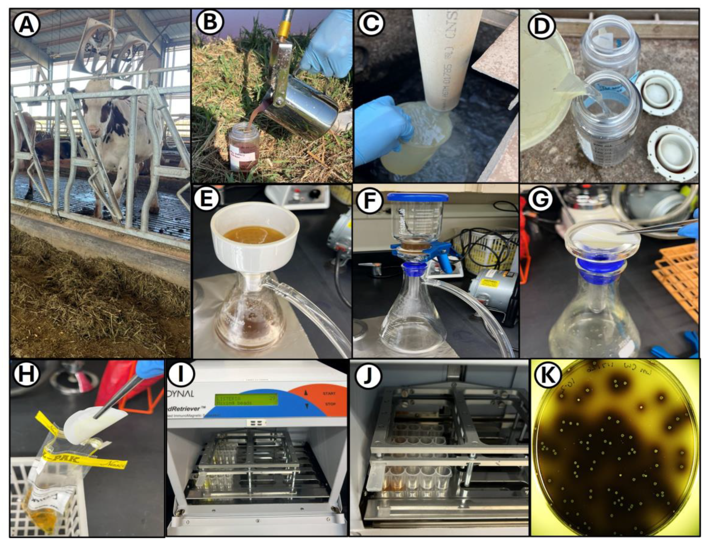
Figure 2.
Reactions of stock Listeria strains and isolates on Listeria Brilliance agar. Streak plates, panels: (A) L. monocytogenes EGDe, (B) L. monocytogenes CQ1, and (C) L. seeligeri ATCC 35967. Surface spread plates, panels: (D, E) L. monocytogenes EGDe (48 and 72 hr); (F, G) selective enrichment of IMS bead samples from municipal wastewater; (H, I) L. seeligeri ATCC 35967 and L. innocua ATCC 33090 (72 hr); and selective enrichment of IMS samples from dairy (J, K) and city (L) wastewater.
Figure 2.
Reactions of stock Listeria strains and isolates on Listeria Brilliance agar. Streak plates, panels: (A) L. monocytogenes EGDe, (B) L. monocytogenes CQ1, and (C) L. seeligeri ATCC 35967. Surface spread plates, panels: (D, E) L. monocytogenes EGDe (48 and 72 hr); (F, G) selective enrichment of IMS bead samples from municipal wastewater; (H, I) L. seeligeri ATCC 35967 and L. innocua ATCC 33090 (72 hr); and selective enrichment of IMS samples from dairy (J, K) and city (L) wastewater.
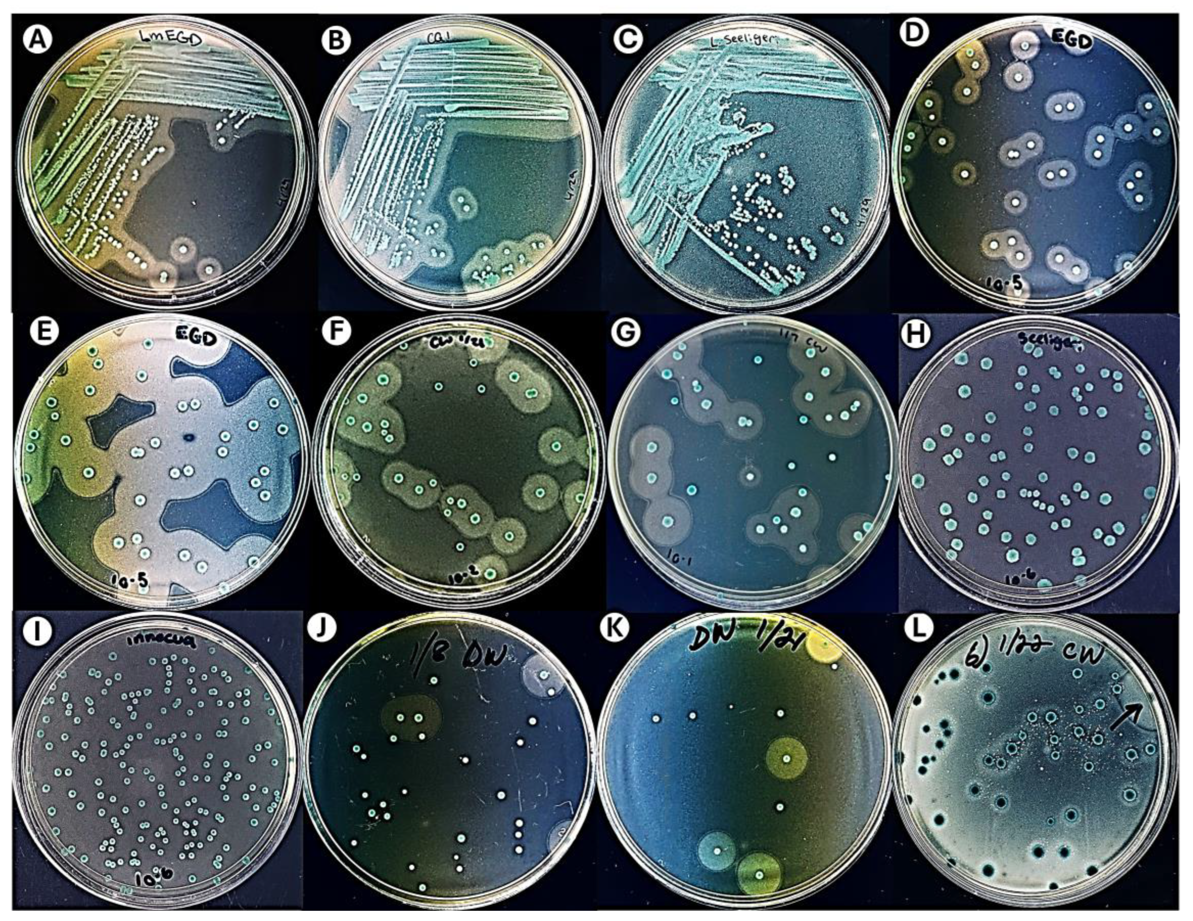
Figure 3.
Ethidium bromide-stained agarose gels of PCR amplimers using primers for various LmPI-1 genes (positive regulatory factor A, prfA; hemolysin A, hlyA; internalin A, inlA; and actin-based motility, actA, of various strains from our culture collection showing pathogenic strains (L. monocytogenes ATCC 19111 and EDGe) as well as non-pathogenic strains (L. innocua ATCC 33090, L. seeligeri ATCC 35967, and L. welshimeri ATCC 35897).
Figure 3.
Ethidium bromide-stained agarose gels of PCR amplimers using primers for various LmPI-1 genes (positive regulatory factor A, prfA; hemolysin A, hlyA; internalin A, inlA; and actin-based motility, actA, of various strains from our culture collection showing pathogenic strains (L. monocytogenes ATCC 19111 and EDGe) as well as non-pathogenic strains (L. innocua ATCC 33090, L. seeligeri ATCC 35967, and L. welshimeri ATCC 35897).
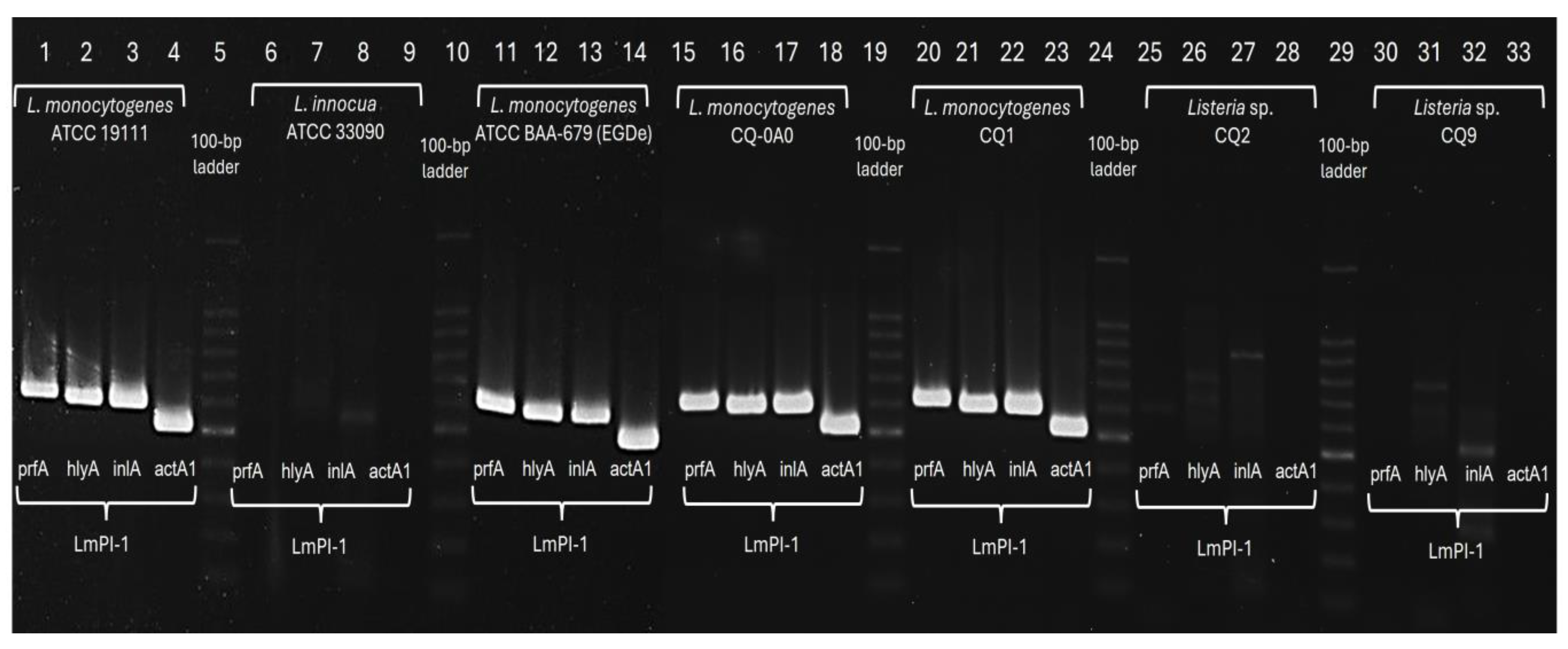
Figure 4.
Agarose gel analysis of PCR products from additional wastewater isolates with primers for the 4 LmPI-1 genes used in this study including the housekeeping gene, iap. The results indicate that CQ15 and CQ7 are non-pathogenic Listeria innocua as determined by sigB, or iap sequence analysis.
Figure 4.
Agarose gel analysis of PCR products from additional wastewater isolates with primers for the 4 LmPI-1 genes used in this study including the housekeeping gene, iap. The results indicate that CQ15 and CQ7 are non-pathogenic Listeria innocua as determined by sigB, or iap sequence analysis.
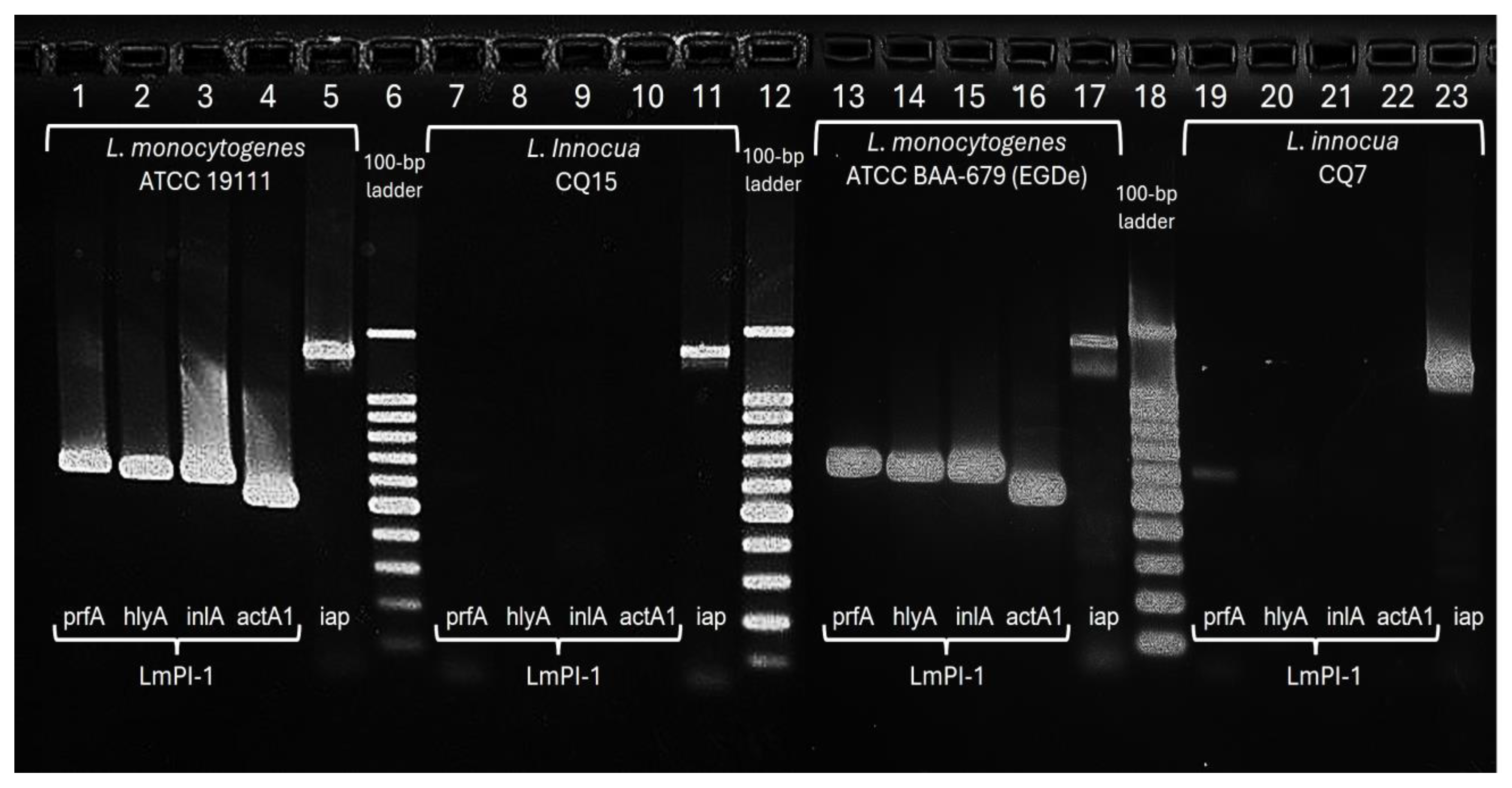
Figure 5.
Agarose gels of PCR results using primers for L. monocytogenes Pathogenic Island 1 (LmPI-1) and iap against isolates obtained from municipal wastewater and OSU dairy wastewater (CQ22, CQ23, and CQ24).
Figure 5.
Agarose gels of PCR results using primers for L. monocytogenes Pathogenic Island 1 (LmPI-1) and iap against isolates obtained from municipal wastewater and OSU dairy wastewater (CQ22, CQ23, and CQ24).
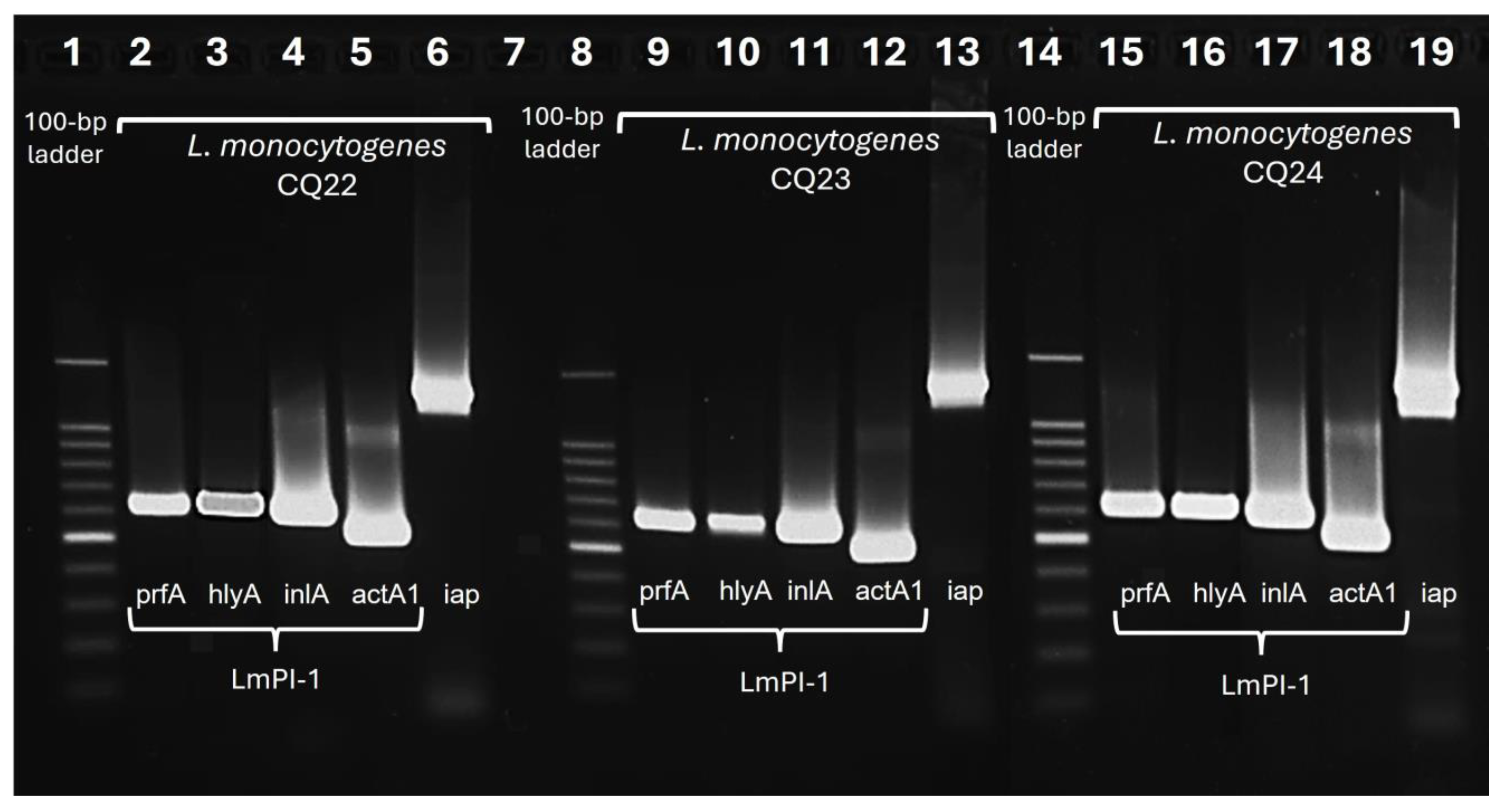

Table 1.
Listeria strains from our culture collection and those isolated during this study.
| Organism (closest species)1 |
Strain Designation | Source |
|---|---|---|
| Listeria monocytogenes | EGDe; ATCC BAA-679 | Muriana Culture Collection |
| Listeria monocytogenes | ScottA; ATCC 45954 45954 | Muriana Culture Collection |
| Listeria monocytogenes | ATCC 19111 | Muriana Culture Collection |
| Listeria innocua | ATCC 33090 | Muriana Culture Collection |
| Listeria seeligeri | ATCC 35967 | Muriana Culture Collection |
| Listeria welshimeri | ATCC 35897 | Muriana Culture Collection |
| Listeria monocytogenes | CQ0A0 | City Municipal Wastewater |
| Listeria spp. | CQ0A1 | City Municipal Wastewater |
| Listeria spp. | CQ0A2 | City Municipal Wastewater |
| Listeria monocytogenes | CQ1 | City Municipal Wastewater |
| Listeria spp. | CQ2 | City Municipal Wastewater |
| Listeria innocua | CQ3 | OSU Dairy Wastewater |
| Listeria innocua | CQ4 | OSU Dairy Wastewater |
| Listeria innocua | CQ5 | OSU Dairy Wastewater |
| Listeria innocua | CQ6 | OSU Dairy Wastewater |
| Listeria innocua | CQ7 | City Municipal Wastewater |
| Listeria innocua | CQ8 | City Municipal Wastewater |
| Listeria spp. | CQ9 | City Municipal Wastewater |
| Listeria innocua | CQ10 | City Municipal Wastewater |
| Listeria innocua | CQ11 | OSU Dairy Wastewater |
| Listeria innocua | CQ12 | OSU Dairy Wastewater |
| Listeria innocua | CQ13 | City Municipal Wastewater |
| Listeria spp. | CQ14 | City Municipal Wastewater |
| Listeria innocua | CQ15 | OSU Dairy Wastewater |
| Listeria innocua | CQ16 | OSU Dairy Wastewater |
| Listeria innocua | CQ17 | City Municipal Wastewater |
| Listeria innocua | CQ18 | City Municipal Wastewater |
| Listeria monocytogenes | CQ19 | City Municipal Wastewater |
| Listeria spp. | CQ20 | OSU Dairy Wastewater |
| Listeria monocytogenes | CQ21 | City Municipal Wastewater |
| Listeria monocytogenes | CQ22 | OSU Dairy Wastewater |
| Listeria monocytogenes | CQ23 | OSU Dairy Wastewater |
| Listeria monocytogenes | CQ24 | City Municipal Wastewater |
1Note: Species identity was based on a combination of 16S, SigB, IAP gene sequences and/or L. monocytogenes Pathogenic Island PCR (LmPI-1) and response to Listeria Brilliance Agar phenotype.
Table 2.
PCR primers used in this study.
| Primer Pairs | Target Gene | Primer Sequence (5’→ 3’) |
Product size (bp) | (NCBI) Primer Tm (oC) |
(IDT) Primer Tm (oC) |
|---|---|---|---|---|---|
| Primer 1 | Hemolysin (hlyA) | ||||
| Forward | TGAACCTACAAGACCTTCCA | 560 | 55.7 | 53.0 | |
| Reverse | CAATTTCGTTACCTTCAGGA | 53.2 | 50.1 | ||
| Primer 2 | Internalin A (inlA) | ||||
| Forward | GCTTCAGGCGGATAGATTAG | 575 | 55.5 | 52.6 | |
| Reverse | AACTCGCCAATGTGCC | 54.7 | 53.3 | ||
| Primer 3 | Positive regulatory factor (prfA) | ||||
| Forward | ATTTTTAACCAATGGGATCC | 590 | 51.2 | 48.2 | |
| Reverse | CATTCATCTAATTTAGGGGC | 51.3 | 48.3 | ||
| Primer 4 | Actin mobility (actA1) | ||||
| Forward | AATACGAACAAAGCAGACCTAATAG | 500 | 57.2 | 52.9 | |
| Reverse | GGTCAATTAACCCTGCACTTTTA | 57.3 | 53.5 | ||
| Primer 6 | Universal 16 S rRNA – front half | ||||
| Forward (7F) | RAGAGTTTGATCHTGGCTCAG | ~920-930 | 54 | 53.5 | |
| Reverse (928R) | CCCCGTCAATTCHTTTGA | 47 | 50.8 | ||
| Primer 7 | Universal 16 S rRNA – back half | ||||
| Forward (759F) | CAGGATTAGATACCCTGGTAGTCC | ~782 | 59.2 | 55.8 | |
| Reverse (1541R) | AAGGAGGTGATCCARCCGC | 59.7 | 58.8 | ||
| Primer 8 | Sigma Factor B (sigB) | ||||
| Forward | AAAGCAGGTGGAGGAGAATG | ~749 | 57.5 | 54.8 | |
| Reverse | TTGACGTTGGATTCTAGACACAT | 57.9 | 54.0 | ||
| Primer 9 | Invasion Associated Protein (iap) | ||||
| Forward | GAATATGAAAAAAGCAACTATCGC | ~1,286 | 51.6 | 51.1 | |
| Reverse | CTAAATCACCAGGTTTTGCTTG | 51.1 | 52.4 |
Disclaimer/Publisher’s Note: The statements, opinions and data contained in all publications are solely those of the individual author(s) and contributor(s) and not of MDPI and/or the editor(s). MDPI and/or the editor(s) disclaim responsibility for any injury to people or property resulting from any ideas, methods, instructions or products referred to in the content. |
© 2026 by the authors. Licensee MDPI, Basel, Switzerland. This article is an open access article distributed under the terms and conditions of the Creative Commons Attribution (CC BY) license.
Copyright: This open access article is published under a Creative Commons CC BY 4.0 license, which permit the free download, distribution, and reuse, provided that the author and preprint are cited in any reuse.


