

Submitted:
02 August 2026
Posted:
04 August 2026
You are already at the latest version
Abstract
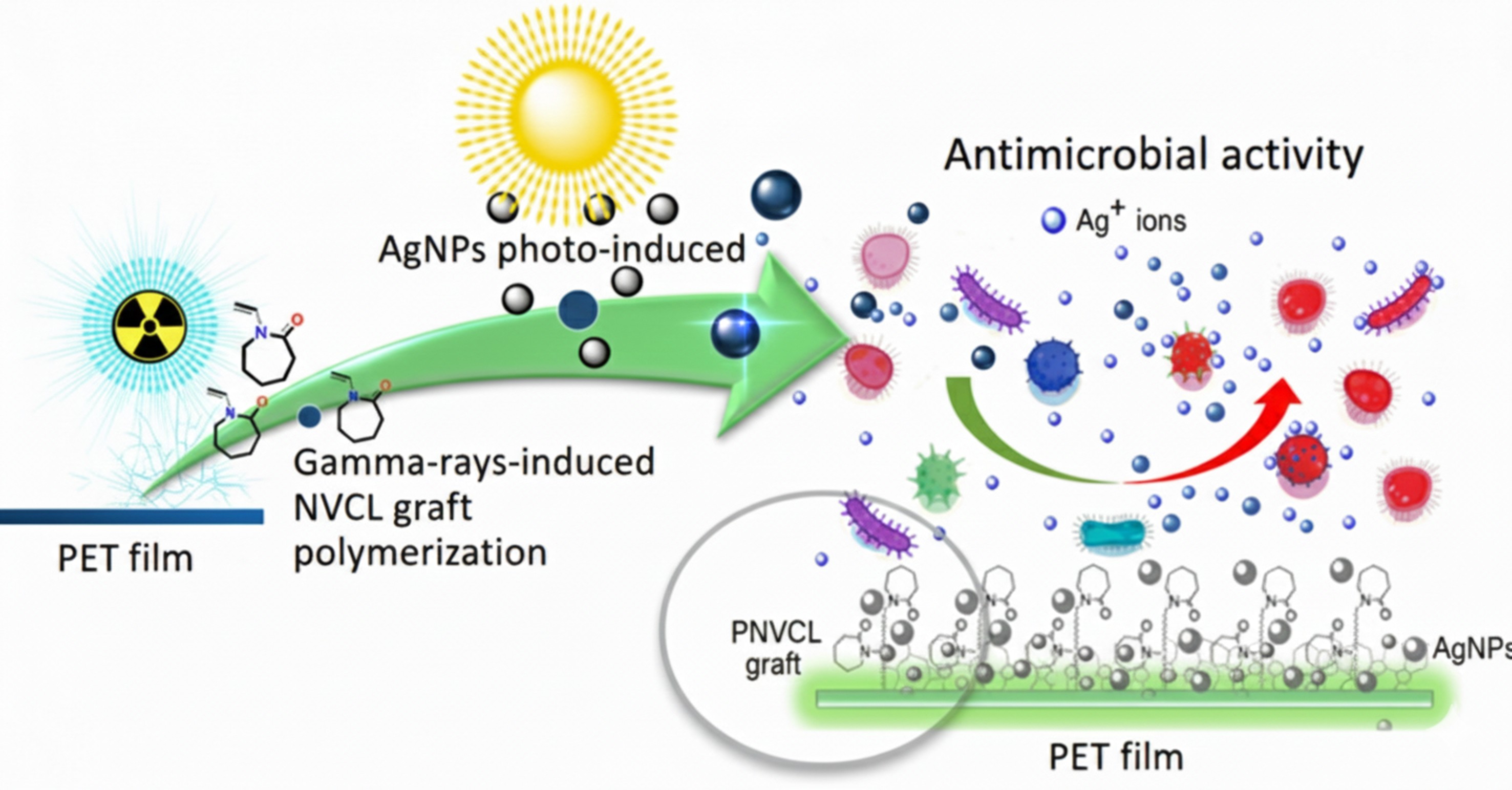
Poly(ethylene terephthalate) (PET) is a versatile polymeric material widely utilized across various sectors, including engineering, food packinging, and medical applications. How-ever, PET inherently lacks the capability to prevent or eliminate microorganism prolifera-tion on its surface and surrounding area. Consequently, endowing PET with anti-infective properties presents a challenge, owing to its inherent properties and chemical structure. Thankfully, several chemical strategies can be deployed to functionalize the surface of PET, enabling the eradication of microorganisms. In this study, PET films underwent functionalization through gamma-ray-induced grafting of poly(N-vinyl caprolactam) (PNVCL). This process facilitated the loading and stabilization of silver nanoparticles (AgNPs) on the grafted PET films and addition provided thermal-responsiveness. Char-acterization of these novel PET-based materials revealed increased surface hydrophilicity without significant alterations in surface morphology or mechanical properties. The an-timicrobial capacity of AgNPs was evaluated against both Gram-positive and Gram-negative bacteria at various temperatures.
Keywords:
NVCL
; gamma-rays
; grafting
; poly(ethylene terephthalate)
; coating
; antimicrobial
; nanoparticles
; silver
1. Introduction
Currently, there has been a substantial surge in research and development efforts focused on synthetic polymers endowed with suitable mechanical and antimicrobial properties. In this sense, poly(ethylene terephthalate) (PET) is a versatile polymeric material widely utilized across various sectors, including medical applications [1,2], food packaging [3,4], and other cutting-edge technologies. However, even though PET has excellent mechanical properties, it tends to exhibit hydrophobic characteristics, making it susceptible to bacterial adhesion [5]. This vulnerability has substantial implications for the medical [6] and food industry [7], underscoring the need for surface modifications of these materials to mitigate microbial contamination [8,9].
In this context, graft polymerization represents a useful tool that allows the modification of the matrix’s surface with different functional groups from the graft polymer [10], thereby altering the surface chemistry of the matrix [11]. It also enables the grafting of different polymers onto the same matrix, combining the physicochemical properties of the various grafted polymers [12,13]. Moreover, unlike other methods of surface modification such as coatings, graft polymerization offers the advantage of a covalent coating, ensuring enhanced stability under the physicochemical conditions to which the material is subjected. In this context, gamma radiation serves as an invaluable tool for initiating graft polymerization reactions on matrices with low or no reactivity towards chemical initiators, such as azobisisobutyronitrile (AIBN). Gamma radiation has the unique capability to simultaneously initiate the polymerization of the monomer while creating active sites within the polymeric matrix. These active sites initiate the formation of side polymer chains, either directly or through a two-step process involving oxidative pre-irradiation followed by thermally promoted graft polymerization in the presence of a vinyl monomer [14,15]. In both cases, the result is the formation of a graft polymer.
These strategies enable the matrix to provide various properties coming from the graft polymer or copolymer, such as stimulus-response [16], which can be modified according to the desired application, without significantly altering the mechanical properties [17]. In most cases, this allows them to be used in the controlled loading and release of antimicrobial agents through the application of external factors such as changes in temperature or pH [18,19], thereby optimizing and significantly expanding their potential applications.
In this sense, poly(N-vinylcaprolactam) (PNVCL) has been used to create different materials with good biocompatibility and thermoresponse properties in the range of 31 and 38 °C [20] close to human internal and external temperature. These factors make it suitable for various applications in the medical area taking advantage of the thermal responsiveness that can control or allow the loading of various organic or inorganic agents with antimicrobial properties, due to the hydrophobic and hydrophilic character that depends on the temperature. This temperature-dependent character can improve the effectiveness of antimicrobial agents by prolonging or controlling their release in a focal area or target zone.
Some of the antimicrobial agents used to provide antimicrobial activity include lysozyme [21], vancomycin [17], benzalkonium chloride [22], etc., which can be immobilized on the matrix or incorporated incorporated by controlled loading and release [23]. Among the range of antibacterial agents, inorganic agents like silver (Ag°) and Ag+ ions stand out for their efficient inhibitory and bactericidal effects across a broad spectrum of microorganisms, with a low tendency to develop resistance to Ag-based agents [24]. Therefore, Ag-based agents, both ionic and metallic as nanoparticles, have been incorporated into various materials through several processes on different substrates, offering an excellent alternative for designing durable and anti-infective materials [25]. These materials are capable of preventing the presence and proliferation of planktonic microorganisms or biofilms [26], which can have adverse effects in the medical sector as well as in stored food products [27,28]. As a result, anti-infective polymeric materials have gained significant importance, and substantial efforts have been dedicated to developing more efficient materials capable of inhibiting the proliferation of various microorganisms, including bacteria, fungi, and even viruses [28]. These microorganisms can cause infections in open wounds and can also alter the quality and sensory properties of food products, potentially posing health risks [29,30].
Therefore, in this research work, new materials based on PET with thermo-responsive properties were synthesized using N-vinyl caprolactam (NVCL) through gamma radiation-induced graft polymerization followed by photo-induction and loading of silver nanoparticles (AgNPs). The inhibitory capacity of these new materials was assessed against both Gram-positive and Gram-negative bacteria at different temperatures.
2. Materials and Methods
2.1. Materials
Poly(ethylene terephthalate) (PET) (200 μm thick; molecular weight medium 18 kDa), N-vinyl caprolactam (NVCL), and silver nitrate (99.9%) were purchased from Aldrich Chemical Co. (St. Louis, MO, USA). Dimethylformamide (DMF), methanol, and distilled water were purchased from Baker (Mexico City, Mexico). All materials were used as received.
2.2. Methods
2.2.1. Grafting of PNVCL on PET Films (PET-g-NVCL)
The PET films (1.2 x 5 cm) were previously washed with ethanol and methanol at 25 °C for 48 h and dried under vacuum at 47 °C. Then a pretreated and previously weighed PET film was placed in an ampoule containing 10 mL of NVLC solution, varying the monomer concentration (0-60 w/v %) in DMF. Air was removed by argon bubbling (15 min) and sealed. Subsequently, the ampoules were exposed to a 60Co gamma-rays source at different doses, up to 80 kGy at room temperature. After gamma irradiation, the residual monomer and homopolymer were removed from the grated PET film by stirring in water changing the solvent each 24 h and then soaked in methanol for 24 h to remove de occluded DMF, followed by vacuum drying at 50 °C until constant weight. The grafting experiments were performed in triplicate.
The grafting yield (G%) was calculated using Equation 1:
where Wf and Wi are the weights after and before grafting, respectively.
G (w. %) = 100 [(Wf-Wi)/Wi]
2.2.2. Load of Silver Nanoparticles on PET-g-NVCL (PET-g-NVCL-AgNPs)
Three PET-g-NVCL films were immersed in 10 mL of an aqueous solution of silver nitrate with different concentrations (500, 1000, and 3000 ppm) for 5 days at 25 °C in the presence of sunlight. After the reaction time, the films were removed from the solution and thoroughly washed with distilled water and dried at 50 °C under vacuum. The amount of nanoparticles loaded on the PET-g-PNVCL-AgNPs film was determined by film calcination using the weight difference of the residues obtained at 800 °C (Eq. 2). The AgNPs loading experiments were performed in triplicate.
where CPETAg and CPET are the residual percentages of the PET-g-NVCL-AgNPs and PET-g-NVCL as determined by the TGA instrument.
AgNPs load (w. %) = CPETAg –CPET
2.2.3. Antimicrobial Tests
The capability of the PET-g-NVCL-AgNPs to inhibit bacterial growth was tested against Staphylococcus aureus (S. aureus) and Escherichia coli (E. coli). Petri plates containing Müller-Hinton agar inoculated with bacterial suspensions S. aureus 1.2x10⁹ CFU/mL and E. coli 3.4x10⁹ CFU/mL (where CFU is a colony-forming unit). PET and PET-g-NVCL were used as negative control, and PET-g-NVCL-AgNPs pieces (0.5x0.5 cm) were tested. Plates containing the sample were incubated at 28, 37, and 42 °C for 24 h. The inhibition zone was measured using a scale. The antimicrobial activity evaluation was performed in triplicate using a fresh sample.
2.3. Instrumental
Surface contact angles on the films were measured after 1 min using a Krüss DSA 100 drop shape analyzer (Matthews, NC, USA)
Attenuated total reflectance–Fourier transform infrared (ATR-FTIR) spectra of dry samples were recorded using a PerkinElmer Spectrum 100 spectrometer (Norwalk, CT, USA) over 16 scans.
For scanning electron microscopy (SEM), 0.5 cm film specimens were cut, sputter-coated with golg, and imaged under high vacuum using a Zeiss EVO LS15 instrument (Jena, Germany).
Thermogravimetric analysis (TGA) data of the weight loss and decomposition of films were recorded with at a heating rate of 10 °C min−1 and run from 20 to 800 °C under nitrogen flow at 100 cm3 min−1 in a TGA instrument Q50 TA Instruments (New Castle, DE, USA).
Differential scanning calorimetry (DSC) runs were recorded under a nitrogen flow at 100 cm3 min−1 using a DSC 2010 calorimeter (TA Instruments, New Castle, DE, USA) from 25 to 300 °C at a heating rate of 10 °C min−1. The LSCT was determinate on the previously swollen samples in water and recorded employing two heat cycles.
The X-Ray Photoelectron spectroscopy (XPS) spectra of the samples were recorded using a Microprobe PHI 5000 Versa Probe II (Chanhassen, Minnesota, USA), with an excitation source of Al Kα monochromatic, energy 1486.6 eV, 100 µm beam diameter, and with a Multi-Channel Detector (MCD). The energy scale was corrected using the C1s peak to 285.0 eV. Multipack v. 9.9.3. software was used analyze spectral data.
Mechanical properties of the films were studied by applying an uniaxial tension test, as described in ASTM D1708. All tests were carried out on an INSTRON 1125 (Instron Inc., MA, USA) universal tensile testing machine at a crosshead speed of 10 mm min−1, and all experiments were carried out in triplicate.
3. Results and Discussions
3.1. Synthesis of PET-g-NVCL and Loading of AgNPs
The grafting of PNVCL onto PET films was successfully achieved through gamma radiation-promoted “grafting-from” polymerization, as illustrated in Figure 1a. as well as some possible PET matrix degradation compounds promoted by gamma radiation, which possibly prevented an increase in the degree of grafting onto the matrix (Figure 1b).
The films exhibited an average grafting percentage ranging from 8.8-9.5% for absorbed doses (ranging from 10 to 80 kGy) and monomer concentrations (ranging from 0.5 to 60%), as shown in Figure 2a and Figure 2 b. This observed grafting percentage behavior was attributed to the degradation induced by gamma radiation and the subsequent dissolution of the film during the graft polymerization reaction. Confirmation of this phenomenon came from IR-ATR studies of residues from NVCL and PNVCL obtained from the reaction medium. The IR-ATR spectra displayed the band at 1737 cm-1 corresponding to the sv(C=O) of the PET matrix within the PNVCL homopolymer (S1). These results explained why achieving higher grafting percentages was not posible, even with increased monomer concentration or radiation dose.
However, with an increased radiation dose, the PET-g-NVCL films exhibited improved results in FTIR-ATR characterization (as discussed in Section 3.3). This was indicated by a higher intensity of the amide band associated with PNVCL grafts, suggesting better graft retention on the PET film surface despite water washing. This hinted at possible cross-linking of the PNVCL polymer chains due to excessive gamma radiation. Another significant challenge encountered was the removal of occluded DMF within the PET-g-PNVCL film where the water washing alone proved insufficient, necessitating a 24-hour methanol wash.
The loading of AgNPs was achieved on the PET-g-NVCL (9.5%) by utilizing various aqueous silver concentrations and subjecting them to sunlight for several days to facilitate the nucleation of AgNPs. The grafting of PNVCL played a pivotal role, as without it, promoting the formation of AgNPs was not possible. This loading process altered the color of the PET-g-PNVCL film, turning it gray, which indicated the presence of AgNPs on the film’s surface. The AgNPs were effectively stabilized on the surface due to interactions with the grafted PNVCL (as discussed in Section 3.3), thereby preventing their sudden release.
3.2. Contact Angle Study
Contact angle measurements revealed a significant change in the surface wettability of PET. The PET films exhibited a hydrophobic surface with a contact angle of 79±2.2. When functionalized with PNVCL, the contact angle decreased to 68°, indicating that the surface became more hydrophilic. The incorporation of AgNPs to the PET functionalized with PNVCL(PET-g-NVCL-AgNPs) resulted in a slight increase in the contact angle to 81.7°. This increase is likely attributed to the intrinsic hydrophobic nature of the AgNPs and the hydrophobic conformation of PNVCL, influenced by the interactions between hydrophilic moieties from PNVCL and the nanoparticles (Figure 3) [35].
3.3. FTIR-ATR Analysis
The FTIR spectra of pristine PET and PET-g-NVCL (9.5%) are displayed in Figure 4. In the unmodified PET film spectra, a band at 1714 cm-1 corresponding to sv(C=O) was observed, along with another band at 1242 cm-1 attributed to (C-O-C), and signals appearing at 2098-2967 cm-1 are related to sv(C-H) bonds. The PNVCL spectra presented a band at 1633 cm-1 assigned to sv(C=O, amide), along with bands ranging from 2856 to 2927 cm-1 associated with sv(C–H) and another corresponding to (C-N) at 1233 cm-1. These PNVCL bands confirmed the successful grafting of PNVCL onto PET, as evidenced by the FTIR spectra showing the characteristic band of PNVCL at 1636 cm-1, attributed to sv(C=O, amide). The spectra of PET-g-NVCL-AgNPs films exhibited decreased band intensities for C=O (amide and ester), C–O–C, and C–N stretching vibrations due to chemical interactions with the AgNPs. This behavior became more pronounced as the silver concentration was increased to obtain the AgNPs [36].
3.4. Thermal Analysis
Thermogravimetric analysis (Figure 5a) revealed that pristine PET underwent a single-step decomposition in the range of 400–440 °C, whereas PNVCL exhibited a multistage process. For PNVCL, the initial mass loss from 176-240 °C was attributed to the evaporation of absorbed moisture and volatile compounds, while the decomposition stage between 390 °C and 443 °C corresponds to the thermal degradation of the PNVCL backbone. In contrast, the PET-g-NVCL film displayed a decomposition range between 390 and 441 °C. Table 1 summarizes the temperatures at which a 10% weight loss (T10%) occurred for the samples. PNVCL had a T10% at 257 °C, because of the loss due to moisture and volatile compounds, while pristine PET and PET-g-NVCL films had T10% values of 412 °C and 402 °C, respectively.
Figure 6 shows that the films loaded with AgNPs (PET-g-NVCL-AgNPs) exhibited similar decomposition behavior to PET-g-NVCL. However, loading the AgNPs led to a noticeable increase in the initial decomposition temperature. Specifically, as the loading of AgNPs increased, this decomposition temperature increased as well, shifting from 176.8 °C to 202.6 °C for the PET-g-NVCL and PET-g-NVCL-AgNPs3000. These findings highlight the enhanced thermal stability conferred upon the material by the AgNPs, resulting in a temperature difference of up to 25.8 °C.
Silver quantification in PET-g-NVCL-AgNPs films was carried out by determining the residual weight at 800 °C. Control PET-g-NVCL films yielded an average char residue of 10.7% upon carbonization. Consequently, the increase in residual mass observed for films treated with aqueous silver nitrate solutions (500, 1000, and 3000 ppm) was attributed to the AgNPs deposited on the film surface. This resulted in net silver loadings of 2.1 ± 0.4%, 3.9 ± 0.5%, and 5.6 ± 0.3%, respectively (Figure 6 and Table 1), showing a positive correlation between the AgNO₃ concentration and the amount of immobilized AgNPs
The DSC studies revealed that the PET matrix did not undergo significant changes in its melting point when modified with the PNVCL graft using gamma radiation. The difference in melting point was only 1.3 °C (Table 1 and Figure 5b). This characteristic was maintained in a similar manner for the PET-g-NVCL-AgNPs films, regardless of the amount of AgNPs present on the film. This behavior was also observed in the glass transition temperature (Tg), which did not exhibit significant changes and remained at around 73.7 °C (Figure S2).
Additionally, the DSC results of the films swollen in water enabled us to identify the LCST, which occurred at a temperature of 49.3 °C. This temperature increased to 53.5 °C when the AgNPs were loaded (Figure S3). One of the most notable changes was observed in the PNVCL when exposed to water, as it exhibited a significantly lower softening temperature (TS), decreasing from 257.6 °C to 98.6 °C. This change was also evident in the PET-g-NVCL film, which softened at a temperature of 103.0 °C (Figure 5c).
3.5. Study of Mechanical Properties
The mechanical properties of the pristine PET and PET-g-NVCL films are presented in Figure 7 and Table 2. The pristine PET film exhibited a tensile strength of 160.9 MPa. After graft polymerization with NVCL, the tensile strength decreased significantly to 104.5 MPa, representing a 35% reduction. This decrease indicates material degradation during the grafting process, likely due to chain scission caused by gamma irradiation. Furthermore, a more pronounced change was observed in the elastic modulus. The pristine film showed a modulus of 1228.2 MPa, while the grafted film showed a modulus of 834.7 MPa, a reduction of approximately 32%. These results suggest that the gamma-induced grafting process, although effective in introducing new surface functionalities, compromised the mechanical integrity of the PET matrix.
3.6. CP-MAS 13C-NMR and XPS Analysis
The PET-g-NVCL films and homopolymer PNVCL underwent analysis through solid-state 13C-NMR spectroscopy (Figure 8). The results indicated successful grafting polymerization induced by gamma-rays, aligning with findings from other spectroscopic analyses such as ATR-FTIR and XPS. In the PET-g-NVCL film spectra, signals from ethylene moieties (C-7’,8’,7,8) appeared at 61.4-63.6 ppm, and aromatic signals (C-3’,4’,3,4) were observed at 130.2-133.6 ppm, with carboxyl carbons (C-1’,6’,1,6) at 164.0 ppm. Conversely, signals corresponding to the grafting polymer (PNVCL) were identified at 30.0 ppm, assigned to methylene (C-9) from the aliphatic chain of PNVCL, carbons C-12,13,14 of the ring carbons of the caprolactam unit at 35.8 ppm, and signals of α-carbons to carbonyl and amino groups at 39.9-44.2 ppm (C-11,15) and 48.8 ppm (C-10). The signal for the carbonyl group of caprolactam units was at 178.2 ppm. Finally, any remaining signals in the spectra were attributed to spinning sidebands (ssb) caused by magnetic angle spinning.
XPS studies revealed the elemental compositions of PET, PET-g-NVCL, and PET-g-NVCL-AgNPs films, showing characteristic peaks for carbon (C1s at 285.0 eV) and oxygen (O1s at 531.0 eV) in the PET films. Additionally, a peak corresponding to nitrogen (N1s at 399.4 eV) was observed in the PET-g-NVCL films, while the PET-g-NVCL-AgNPs films, a doublet corresponding to silver (Ag3d at 366.8 and 372.8 eV) was also observed, suggesting the presence of silver in the form of Ag2O. The spectra corresponding to the films are illustrated in Figure 9.
The atomic composition of the films was determined by calculating the areas under the C1s, O1s, and N1s peaks in the XPS spectra using appropriate sensitivity factors. The atomic composition of carbon and oxygen for films is listed in Table 3. The results indicated a decrease in the proportion of O1s and the presence of N1s on the PET-g-NVCL films due to the formation of a coating on the film by the grafted PNVCL chains. Furthermore, the results of the PET-g-PNVCL showed a similar atomic composition to the PET-g-NVCL surface for O1s and C1s, not being significantly altered by the presence of the AgNPs.
The C1s spectrum of PET was deconvoluted into four peaks: C=O, C-O-C, C-C, and C=C, while the O1s spectrum was deconvoluted into two peaks: C=O and C-O-C, confirming the well-established chemical composition of the PET film (Figure 10a). In contrast, the C1s and O1s spectra of the PET-g-NVCL film were deconvoluted into five peaks: C=O, C-O-C, C-N, C-C, and C=C. This analysis indicated an increase in C-C groups, the presence of the C-N bond, and a reduction in the proportion assigned to other chemical groups (Table 3). While, the O1s spectrum was similarly deconvoluted, revealing four peaks attributed to C=ON, C=O, C-O-C, and OH and these presented a similar behavior to the C1s spectrum, with a decrease in C=O and C-O-C groups. It also revealed the presence of the C=ON group, which corresponds to the amide group of the PNVCL graft. Furthermore, it indicated the existence of hydroxyl groups from water molecules occluded to the surface of the PET-g-NVCL film, a consequence of its enhanced surface hydrophilicity (Figure 10b). These findings supporting the presence of PNVCL polymer chains grafted onto the surface of the PET film. Consequently, the film exhibited a surface chemical composition similar to that of NVCL polymer.
Concerning the PET-g-NVCL-AgNPS3000 film, this exhibited an additional peak in the O1s spectrum attributed to C=OAg interactions. Furthermore, the Ag3d spectrum confirmed the successful loading of AgNPs and further analysis of the Ag3d spectrum indicated three peaks assigned to Ag/Ag2O, AgO, and Ag. This observation elucidated the composition of the AgNPs loaded onto the PET-g-NVCL film, indicating a predominant presence of AgO, as evidenced by the observed percentages (Figure 10c, Table 3).
3.7. SEM Studies
The surface morphology of the pristine PET and PET-g-NVCL-AgNPs3000 films (containing 9.5% PNVCL and 5.6% AgNPs) was examined by SEM. The micrographs revealed notable changes in the surface topography of the PET film following grafting and silver nanoparticle deposition (Figure 11). The magnification showed the formation of aggregates with a maximum size of approximately 1 µm on the film’s surface, likely caused by the grafting of PNVCL and the loading of AgNPs onto it. No apparent damage such as ruptures or delamination of matrix layers caused by the gamma radiation-induced graft polymerization process was observed. However, despite our efforts to achieve higher magnification to observe the AgNPs size and morphology, it was not possible due to sample overcharging, as the samples exhibited poor conductivity even after being coated with gold.
3.8. Microbiological Tests
Finally, the antibacterial capacity of the films loaded with PET-g-NVCL-AgNPs was evaluated against S. aureus (Gram-positive) and E. coli (Gram-negative) bacteria. Films of PET, PET-g-NVCL, and a PET film exposed to an aqueous silver nitrate solution under the same AgNPs loading conditions (PET500) were used as controls. These studies were conducted at different temperatures at pH 7 (Figure 12). The antimicrobial activity of AgNPs was more pronounced for S. aureus cultures compared to E. coli cultures, and their antimicrobial halo increased with the AgNPs loading, reaching a maximum inhibition zone of 0.42 cm² for S. aureus cultures and 0.31 cm² for E. coli cultures at a temperature of 28 °C. In contrast, the control films (PET and PET-g-NVCL) and the PET500 (film treated under the same AgNPs loading conditions) did not exhibit any inhibition zones. These results underscore the significance of PNVCL grafting, which promotes the nucleation and stabilization of AgNPs on the film surface.
On the other hand, the results of the antimicrobial capacity of the films at different temperatures did not show significant changes. This is likely because the thermal response of the material, low critical solution temperature (LCST) occurs at a temperature of 49.3 °C. Therefore, the antimicrobial activity evaluated at a maximum temperature of 42 °C had no effect on AgNPs (Figure 13).
4. Conclusions
The graft polymerization of NVCL was successfully carried out using gamma radiation, achieving a grafting percentage ranging from 8.8 to 9.5%. This process did not significantly affect the mechanical and thermal properties of the PET matrix and provided a thermal response at 49.3 °C. SEM analysis of the PET-g-NVCL-AgNPs3000 films revealed the presence of aggregates distributed across the surface. These aggregates likely result from the combined effects of the grafted PNVCL chains. However, it was not possible to observe the size of the AgNPs due to sample overcharging, preventing an increase in magnification. Antimicrobial activity tests demonstrated that only the PET-g-NVCL-AgNPs films exhibited antimicrobial activity against both S. aureus and E. coli, highlighting the importance of PNVCL grafting in the formation and stabilization of AgNPs on the PET surface. These results are consistent with XPS analyses, which revealed chemical interactions with PNVCL grafting, confirming the successful attachment of the polymer chains to the PET surface. The XPS data indicated that the AgNPs were predominantly present as Ag₂O (silver oxide), which is known to contribute to antimicrobial activity through the sustained release of Ag⁺ ions.
Supplementary Materials
The following supporting information can be downloaded at the website of this paper posted on Preprints.org, Figure S1. Infrarred spectra of poly(N-vynil caprolactam) from the graft polymerizacion reaction promoted by gamma rays. The corresponding of C=O from PET residues is at 1739 cm-1; Figure S2. DSC analysis of PET-g-PNVCL-AgNPs films with different amounts of AgNPs loaded under dry conditions and nitrogen atmosphere; Figure S3. DSC analysis of PET-g-PNVCL-AgNPs films with different amounts of AgNPs loaded under humid conditions using distilled water and a nitrogen atmosphere.
Author Contributions
Conceptualization, G.G.F.-R.; methodology, G.G.F.-R and E.M.; software, G.G.F.-R.; validation, E.D., R.V.-G., and E.B.; formal analysis, G.G.F.-R.; investigation, G.G.F.-R, and E.M.; resources, R.V.-G, E.M., and E.B.; data curation, G.G.F.-R.; writing-original draft preparation, G.G.F.-R, and E.M.; writing-review and editing, E.M.; visualization, G.G.F.R.; supervision, E.M. R.V.-G, E.B.; project administration, E.M., R.V.-G, and E.B.; funding acquisition, R.V.-G., E.M., and E.B. All authors have read and agreed to the published version of the manuscript.
Funding
This work was supported by the University of Guadalajara under PROSNI 2022. Dirección General de Asuntos del Personal Académico (DGAPA), Universidad Nacional Autónoma de México under Grant IN204223. Support Program for Technological Research and Innovation Projects under PAPIIT IG100220 and National Council of Science and Technology under CONACyT CF-19 No 140617. Call for basic scientific research CONACYT 2017–2018 del “Fondo Sectorial de Investigación para la educación CB2017-2018” (Grant A1-S-29789). CONAHCyT postdoctoral fellowship provided to G. G. Flores-Rojas (CVU 407270).
Institutional Review Board Statement
No applied.
Informed Consent Statement
No applied.
Data Availability Statement
No applied.
Acknowledgments
Thanks to M. Sc. G. Cedillo, M. Sc. E. Hernandez-Mecinas, M. Sc. L. Huerta, and M. Sc. C. Flores-Morales from the Materials Research Institute, UNAM, Ph. D. B. Leal-Acevedo from Nuclear Sciences Institute, UNAM, and A. Camacho-Cruz from Faculty of Chemistry for their technical assistance.
Conflicts of Interest
The authors declare no conflicts of interest.
References
- Maitz, M.F. Applications of Synthetic Polymers in Clinical Medicine. Biosurface Biotribology 2015, 1, 161–176. [Google Scholar] [CrossRef]
- Sedlarik, V. Antimicrobial Modifications of Polymers. In Biodegradation - Life of Science; 2013. [Google Scholar] [CrossRef] [PubMed]
- Sofi, S.A.; Singh, J.; Rafiq, S.; Ashraf, U.; Dar, B.N.; Nayik, G.A. A Comprehensive Review on Antimicrobial Packaging and Its Use in Food Packaging. Curr. Nutr. Food Sci. 2018, 14, 305–312. [Google Scholar] [CrossRef]
- Tajeddin, B.; Arabkhedri, M. Polymers and Food Packaging; INC, 2020; ISBN 9780128168080. [Google Scholar]
- Jansen, B.; Kohnen, W. Prevention of Biofilm Formation by Polymer Modification. J. Ind. Microbiol. 1995, 15, 391–396. [Google Scholar] [CrossRef] [PubMed]
- Pawlowski, K.S.; Wawro, D.; Roland, P.S. Bacterial Biofilm Formation on a Human Cochlear Implant. Otol. Neurotol. 2005, 26. [Google Scholar] [CrossRef] [PubMed]
- Brooks, J.D.; Flint, S.H. Biofilms in the Food Industry: Problems and Potential Solutions. Int. J. Food Sci. Technol. 2008, 43, 2163–2176. [Google Scholar] [CrossRef]
- López-Saucedo, F.; Flores-Rojas, G.G.; Varca, J.P.R.O.; Varca, G.H.; Bucio, E. Antimicrobial Materials and Devices for Biomedical Applications. 2020. [CrossRef]
- Hiroshi, U.; Shiro, K. Dispersion Polymerization of Styrene in Aqueous Alcohol Solution: Effects of Reaction Parameters on the Polymer Particle Formation. Polym. Int. 1994, 34, 339–344. [Google Scholar] [CrossRef]
- Flores-Rojas, G.G.; Bucio, E. Radiation-Grafting of Ethylene Glycol Dimethacrylate (EGDMA) and Glycidyl Methacrylate (GMA) onto Silicone Rubber. Radiat. Phys. Chem. 2016, 127, 21–26. [Google Scholar] [CrossRef]
- Shtanko, N.I.; Kabanov, V.Y.; Apel, P.Y.; Yoshida, M. Use of Radiation-Induced Graft Polymerization for Modification of Polymer Track Membranes. Nuclear Instruments and Methods in Physics Research, Section B: Beam Interactions with Materials and Atoms 1999, 151, 416–422. [Google Scholar] [CrossRef]
- Ramos-Ballesteros, A.; Pino-Ramos, V.H.; López-Saucedo, F.; Flores-Rojas, G.G.; Bucio, E. γ-Rays and Ions Irradiation. Surf. Modif. Polym. 2019, 185–209. [Google Scholar] [CrossRef]
- Minko, S. Grafting on Solid Surfaces: ``grafting to’’ and ``grafting from’’ Methods. In Polymer Surfaces and Interfaces: Characterization, Modification and Applications; Stamm, M., Ed.; Springer Berlin Heidelberg: Berlin, Heidelberg, 2008; pp. 215–234. ISBN 978-3-540-73865-7. [Google Scholar]
- Ashfaq, A.; Clochard, M.C.; Coqueret, X.; Dispenza, C.; Driscoll, M.S.; Ulański, P.; Al-Sheikhly, M. Polymerization Reactions and Modifications of Polymers by Ionizing Radiation. Polymers 2020, 12, 1–67. [Google Scholar] [CrossRef] [PubMed]
- Naikwadi, A.T.; Sharma, B.K.; Bhatt, K.D.; Mahanwar, P.A. Gamma Radiation Processed Polymeric Materials for High Performance Applications: A Review. Front. Chem. 2022, 10, 1–15. [Google Scholar] [CrossRef] [PubMed]
- Mejía-Manzano, L.A.; Vázquez-Villegas, P.; Prado-Cervantes, L.V.; Franco-Gómez, K.X.; Carbajal-Ocaña, S.; Sotelo-Cortés, D.L.; Atehortúa-Benítez, V.; Delgado-Rodríguez, M.; Membrillo-Hernández, J. Advances in Material Modification with Smart Functional Polymers for Combating Biofilms in Biomedical Applications. Polymers 2023, 15. [Google Scholar] [CrossRef] [PubMed]
- Flores-Rojas, G.G.; Vázquez, E.; López-Saucedo, F.; Buendía-González, L.; Vera-Graziano, R.; Mendizabal, E.; Bucio, E. Lignocellulosic Membrane Grafted with 4-Vinylpiridine Using Radiation Chemistry: Antimicrobial Activity of Loaded Vancomycin. Cellulose 2023, 30, 3853–3868. [Google Scholar] [CrossRef]
- Bawa, P.; Pillay, V.; Choonara, Y.E.; Du Toit, L.C. Stimuli-Responsive Polymers and Their Applications in Drug Delivery. Biomed. Mater. 2009, 4. [Google Scholar] [CrossRef] [PubMed]
- Van Gheluwe, L.; Chourpa, I.; Gaigne, C.; Munnier, E. Polymer-Based Smart Drug Delivery Systems for Skin Application and Demonstration of Stimuli-Responsiveness. Polymers 2021, 13. [Google Scholar] [CrossRef] [PubMed]
- Zavgorodnya, O.; Kozlovskaya, V.; Liang, X.; Kothalawala, N.; Catledge, S.A.; Dass, A.; Kharlampieva, E. Temperature-Responsive Properties of Poly(N-Vinylcaprolactam) Multilayer Hydrogels in the Presence of Hofmeister Anions. Mater. Res. Express 2014, 1, 035039. [Google Scholar] [CrossRef]
- Anastas, P.T.; Rodriguez, A.; de Winter, T.M.; Coish, P.; Zimmerman, J.B. A Review of Immobilization Techniques to Improve the Stability and Bioactivity of Lysozyme. Green Chem. Lett. Rev. 2021, 14, 302–338. [Google Scholar] [CrossRef]
- Flores-Rojas, G.G.; López-Saucedo, F.; Vázquez, E.; Hernández-Mecinas, E.; Huerta, L.; Cedillo, G.; Concheiro, A.; Alvarez-Lorenzo, C.; Bucio, E. Synthesis of Polyamide-6@cellulose Microfilms Grafted with N-Vinylcaprolactam Using Gamma-Rays and Loading of Antimicrobial Drugs. Cellulose 2020, 27, 2785–2801. [Google Scholar] [CrossRef]
- Siegel, R.A. Stimuli Sensitive Polymers and Self Regulated Drug Delivery Systems: A Very Partial Review. In Bone; Stimuli, 2008; Volume 23, pp. 1–7. [Google Scholar] [CrossRef] [PubMed]
- Kurtjak, M.; Nemanja Aničić, M.V. Inorganic Nanoparticles: Innovative Tools for Antimicrobial Agents. Intech 2016, 11, 13. [Google Scholar]
- Chopra, I. The Increasing Use of Silver-Based Products as Antimicrobial Agents: A Useful Development or a Cause for Concern? J. Antimicrob. Chemother. 2007, 59, 587–590. [Google Scholar] [CrossRef] [PubMed]
- Monteiro, D.R.; Gorup, L.F.; Takamiya, A.S.; Ruvollo-Filho, A.C.; de Camargo, E.R.; Barbosa, D.B. The Growing Importance of Materials That Prevent Microbial Adhesion: Antimicrobial Effect of Medical Devices Containing Silver. Int. J. Antimicrob. Agents 2009, 34, 103–110. [Google Scholar] [CrossRef] [PubMed]
- Zemljič, L.F.; Tkavc, T.; Vesel, A.; Šauperl, O. Chitosan Coatings onto Polyethylene Terephthalate for the Development of Potential Active Packaging Material. Appl. Surf. Sci. 2013, 265, 697–703. [Google Scholar] [CrossRef]
- Mallakpour, S.; Azadi, E.; Hussain, C.M. Recent Breakthroughs of Antibacterial and Antiviral Protective Polymeric Materials during COVID-19 Pandemic and after Pandemic: Coating, Packaging, and Textile Applications. Curr. Opin. Colloid Interface Sci. 2021, 55, 101480. [Google Scholar] [CrossRef] [PubMed]
- Sanchez-Garcia, M.D.; Gimenez, E.; Lagaron, J.M. Novel PET Nanocomposites of Interest in Food Packaging Applications and Comparative Barrier Performance with Biopolyester Nanocomposites. J. Plast. Film. Sheeting 2007, 23, 133–148. [Google Scholar] [CrossRef]
- López-Saucedo, F.; Flores-Rojas, G.G.; Varca, J.P.R.O.; Varca, G.H.C.; Bucio, E. Antimicrobial Materials and Devices for Biomedical Applications; 2020; pp. 78–126. [Google Scholar] [CrossRef]
- Nisticò, R. Polyethylene Terephthalate (PET) in the Packaging Industry. Polym. Test. 2020, 90, 106707. [Google Scholar] [CrossRef]
- Scheirs, J.; Long, T.E. (Eds.) Modern Polyesters: Chemistry and Technology of Polyesters and Copolyesters, 1st ed.; Wiley, 2004; ISBN 978-0-471-49856-8. [Google Scholar]
- Dean, B.D.; Matzner, M.; Tibbitt, J.M. Polyarylates. In Comprehensive Polymer Science and Supplements; Elsevier, 1989; pp. 317–329. ISBN 978-0-08-096701-1. [Google Scholar]
- Day, M.; Wiles, D.M. Photochemical Degradation of Poly(Ethylene Terephthalate). III. Determination of Decomposition Products and Reaction Mechanism. J. Appl. Polym. Sci. 1972, 16, 203–215. [Google Scholar] [CrossRef]
- Cho, E.; Kim, M.; Park, J.-S.; Lee, S.-J. Plasma-Polymer-Fluorocarbon Thin Film Coated Nanostructured-Polyethylene Terephthalate Surface with Highly Durable Superhydrophobic and Antireflective Properties. Polymers 2020, 12, 1026. [Google Scholar] [CrossRef] [PubMed]
- Flores-Rojas, G.G.; López-Saucedo, F.; Bucio, E. Gamma-Irradiation Applied in the Synthesis of Metallic and Organic Nanoparticles: A Short Review. Radiat. Phys. Chem. 2018. [Google Scholar] [CrossRef]
- Cho, H.; Dae Kim, S.; Han, T.-H.; Song, I.; Byun, J.-W.; Kim, Y.-H.; Kwon, S.; Bae, S.-H.; Cheul Choi, H.; Ahn, J.-H.; et al. Improvement of Work Function and Hole Injection Efficiency of Graphene Anode Using CHF3 Plasma Treatment. 2D Mater. 2014, 2, 014002. [Google Scholar] [CrossRef]
- Souza, P.R.; Vilsinski, B.H.; De Oliveira, A.C.; Berton, S.B.R.; Madruga, L.Y.C.; Schrekker, H.S.; Radovanovic, E.; Kipper, M.J.; Martins, A.F.; Muniz, E.C. Poly(Ethylene Terephthalate) Films Coated with Antimicrobial Gelatin/Chondroitin Sulfate Polyelectrolyte Multilayers Containing Ionic Liquids. Prog. Org. Coat. 2022, 170, 106997. [Google Scholar] [CrossRef]
Figure 1.
a) Scheme of PNVCL graft polymerization reaction using gamma-rays and nucleation of Ag ions and loading of AgNPs and b) schematic of possible degradation products caused by excess gamma radiation [31,32,33,34].
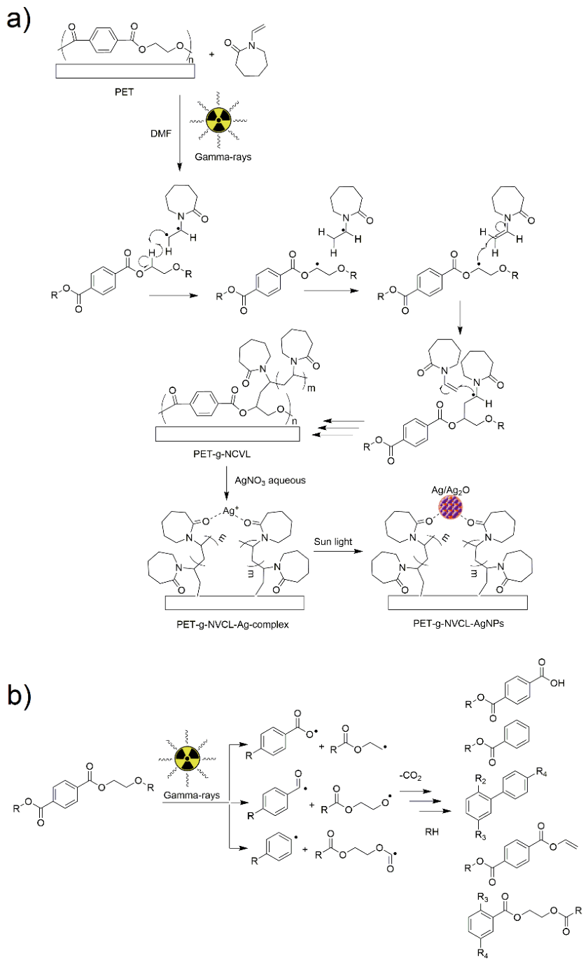
Figure 2.
Percentage of PNVCL grafting as a function of: a) absorbed dose with a constant monomer concentration (30 g/v %) and b) as a function of the monomer concentration at an absorbed dose of 60 kGy.
Figure 2.
Percentage of PNVCL grafting as a function of: a) absorbed dose with a constant monomer concentration (30 g/v %) and b) as a function of the monomer concentration at an absorbed dose of 60 kGy.
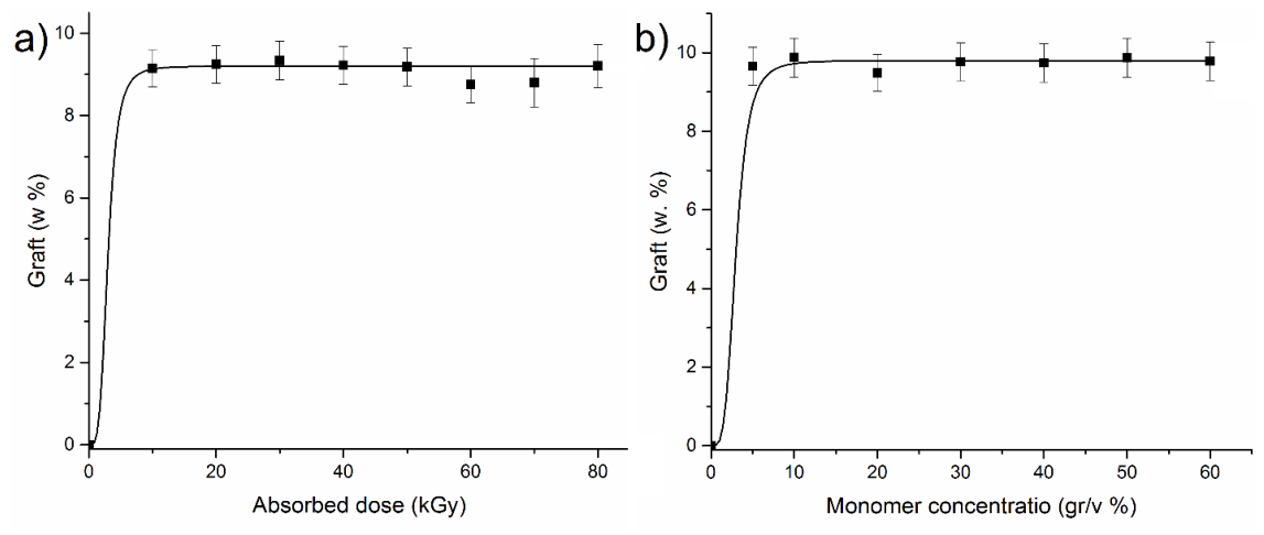
Figure 3.
Contact angles were measured at 1 min on PET and PET films after surface functionalization with PNVCL graft (PET-g-NVCL 9.5%) and AgNPs with various percentages, as determined by TGA analysis. The percentages of AgNPs were as follows: PET-g-NVCL-AgNPs500 (9.5%, 2.1%), PET-g-NVCL-AgNPs1000 (9.5%, 3.9%), and PET-g-NVCL-AgNPs3000 (9.5%, 5.6%).
Figure 3.
Contact angles were measured at 1 min on PET and PET films after surface functionalization with PNVCL graft (PET-g-NVCL 9.5%) and AgNPs with various percentages, as determined by TGA analysis. The percentages of AgNPs were as follows: PET-g-NVCL-AgNPs500 (9.5%, 2.1%), PET-g-NVCL-AgNPs1000 (9.5%, 3.9%), and PET-g-NVCL-AgNPs3000 (9.5%, 5.6%).
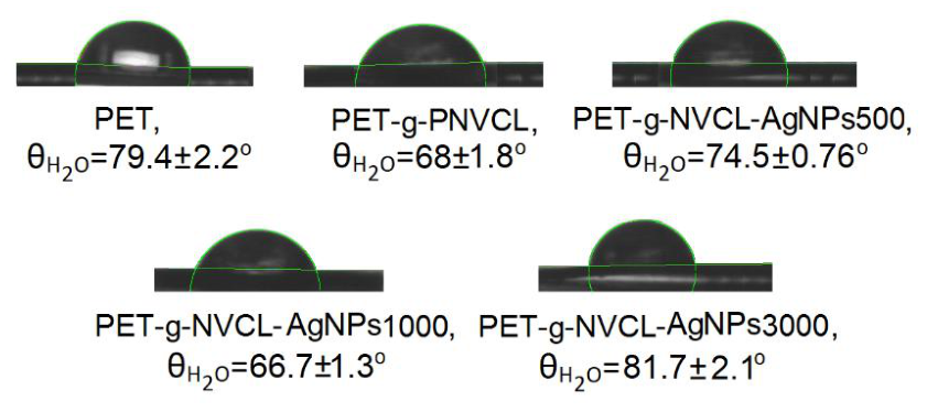
Figure 4.
FTIR-ATR spectra of PET, PNVCL, PET-g-NVCL (9.5%) and PET-g-NVCL-AgNPs films with different amounts of AgNPs loaded on the film surface.
Figure 4.
FTIR-ATR spectra of PET, PNVCL, PET-g-NVCL (9.5%) and PET-g-NVCL-AgNPs films with different amounts of AgNPs loaded on the film surface.
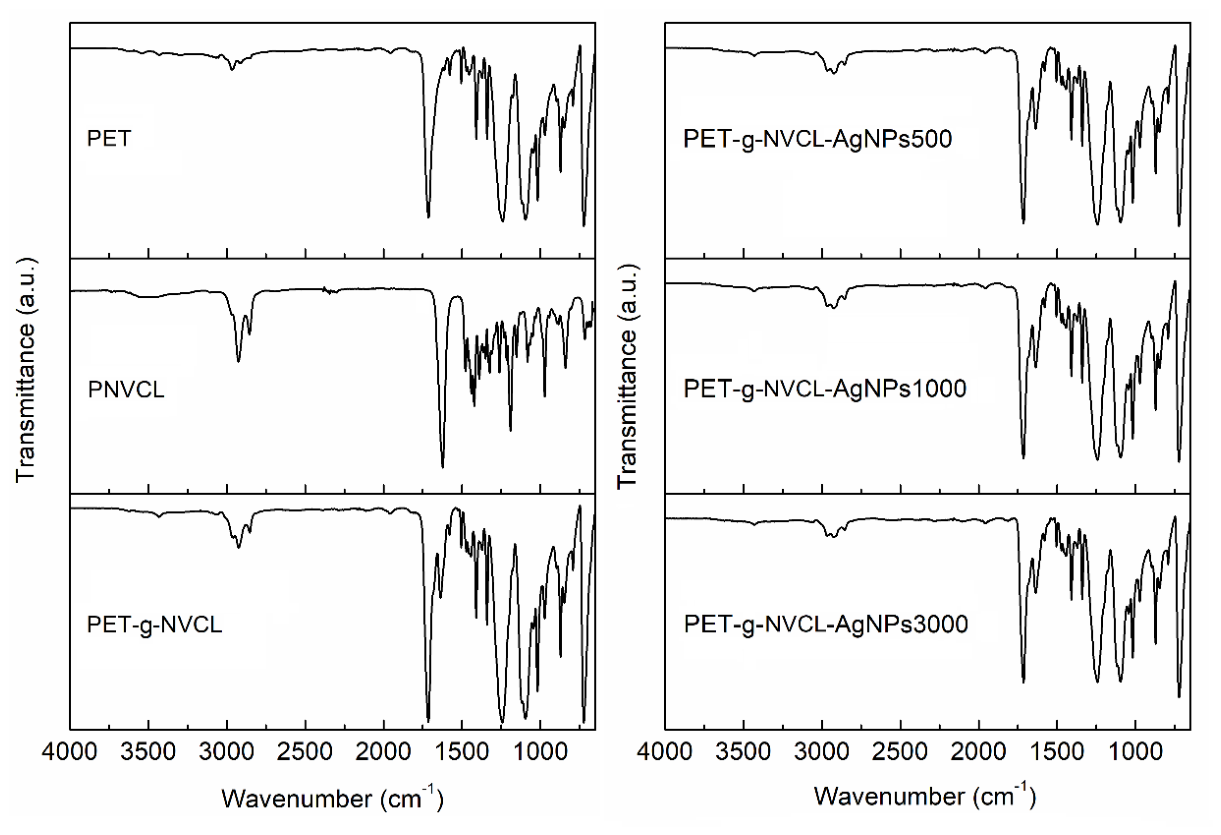
Figure 5.
a) TGA, b) DSC analysis under nitrogen flow on PET films obtained in the PNVLC grafting process promoted by gamma rays and c) DSC of the PET-g-NVCL (9.5%) film under conditions swollen with distilled water.
Figure 5.
a) TGA, b) DSC analysis under nitrogen flow on PET films obtained in the PNVLC grafting process promoted by gamma rays and c) DSC of the PET-g-NVCL (9.5%) film under conditions swollen with distilled water.
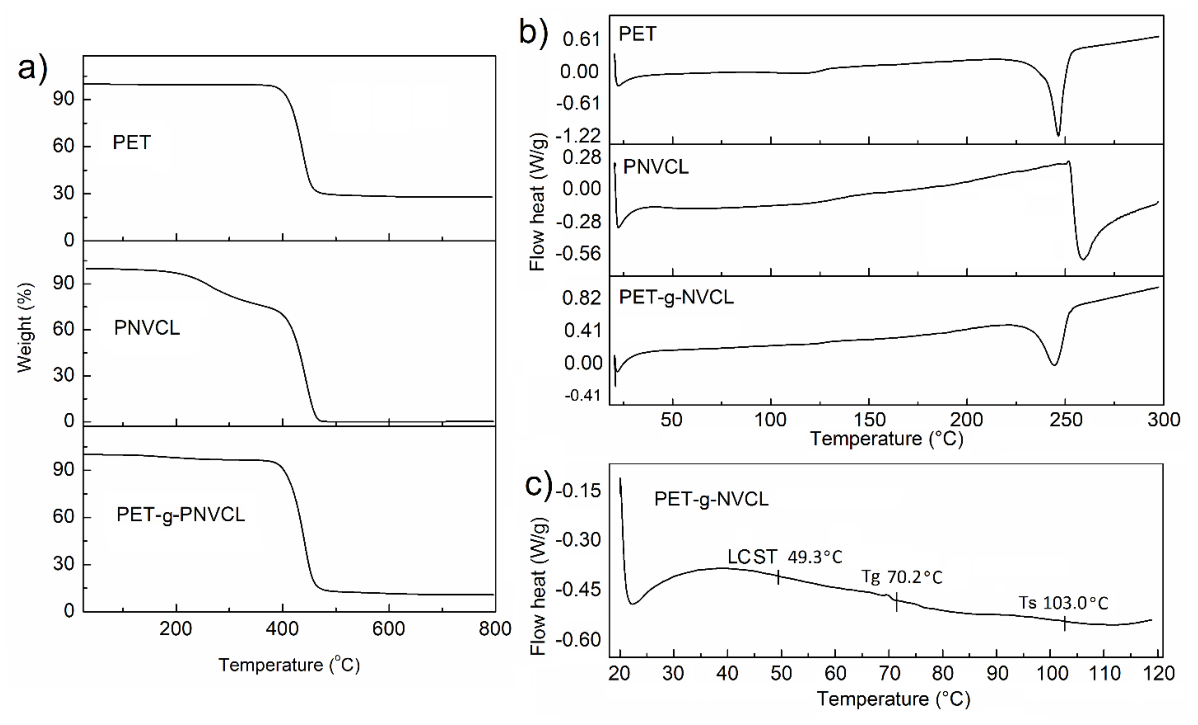
Figure 6.
Silver quantification on PET-g-NVCL-AgNPs films by TGA under nitrogen flow.
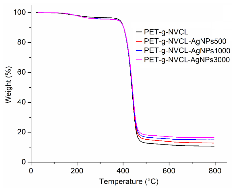
Figure 7.
Tensile stress-strain curves of PET and PET-g-NVCL (9.5%) films.
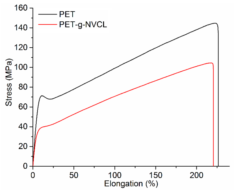
Figure 8.
CP-MAS 13C-NMR spectra of PNVCL homopolymer and PET-g-NVCL (9.5%).
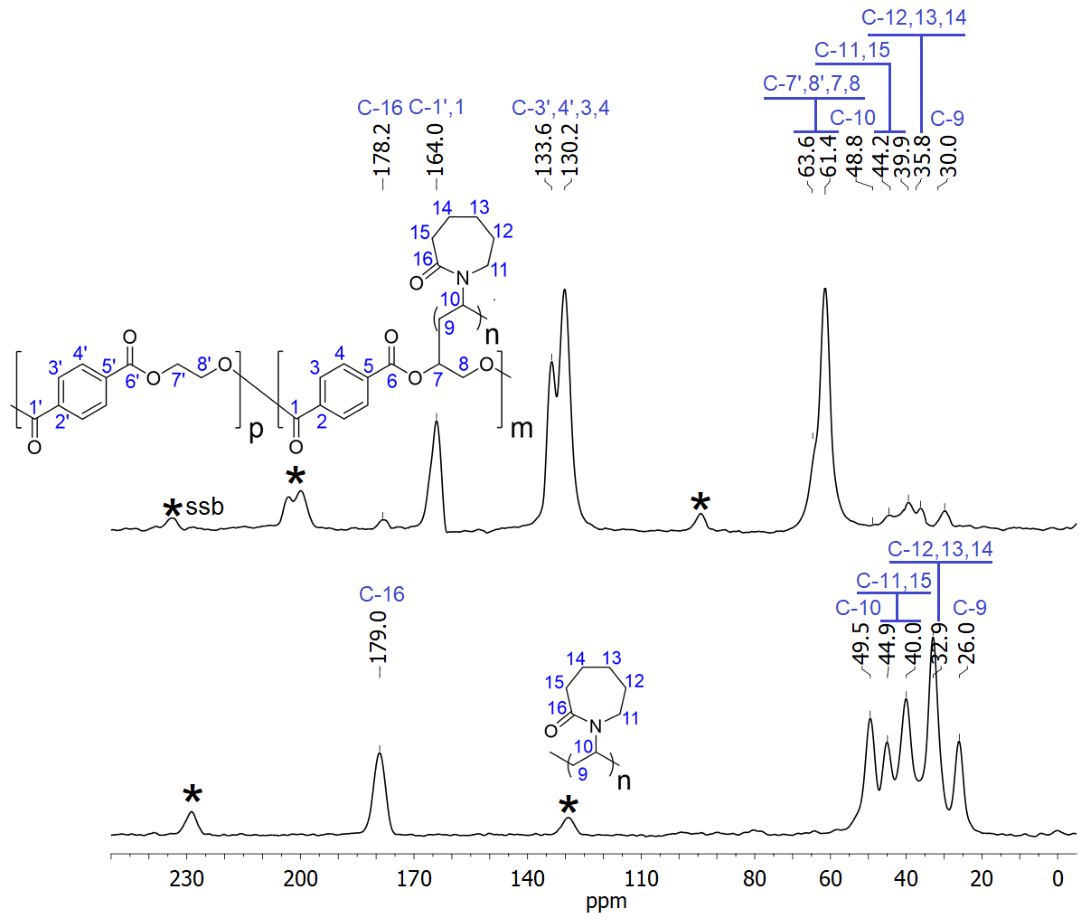
Figure 9.
XPS survey spectra of PET, PET-g-NVCL (9.5%), and PET-g-NVCL-AgNPs (9.5%, 5.6%) films.
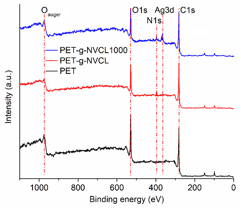
Figure 10.
Deconvolution of XPS spectra corresponding C1s and O1s of a) PET, b) PET-g-NVCL (9.5%), and for c) PET-g-NVCL-AgNPs3000 (9.5%, 5.6%) is presented the deconvolution of Ag3d [37,38].
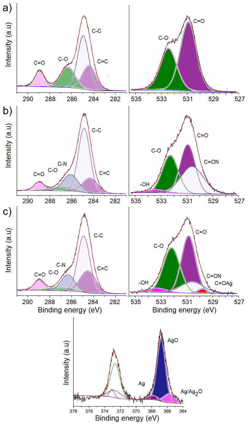
Figure 11.
SEM images of PET and PET-g-NVCL-AgNPs300 (9.5%, 5.6%) at different magnifications.
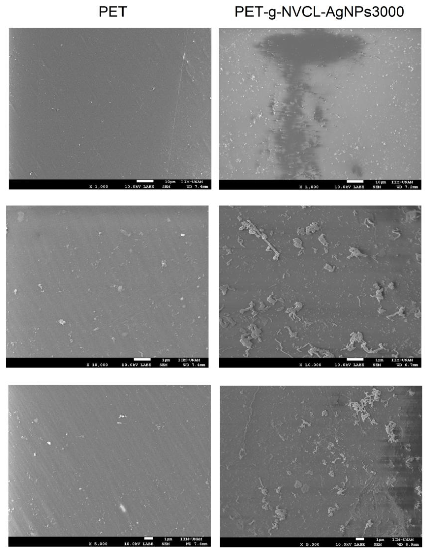
Figure 12.
Antimicrobial halos of a) PET, b) PET500, c) PET-g-NVCL (9.5%), d) PET-g-NVCL-AgNPs500 (9.5, 2.1%), e) PET-g-NVCL-AgNPs1000 (9.5%, 3.9%), and f) PET-g-NVCL-AgNPs3000 (9.5, 5.6%) films, at different temperatures agains E. coli and S. aureus.
Figure 12.
Antimicrobial halos of a) PET, b) PET500, c) PET-g-NVCL (9.5%), d) PET-g-NVCL-AgNPs500 (9.5, 2.1%), e) PET-g-NVCL-AgNPs1000 (9.5%, 3.9%), and f) PET-g-NVCL-AgNPs3000 (9.5, 5.6%) films, at different temperatures agains E. coli and S. aureus.
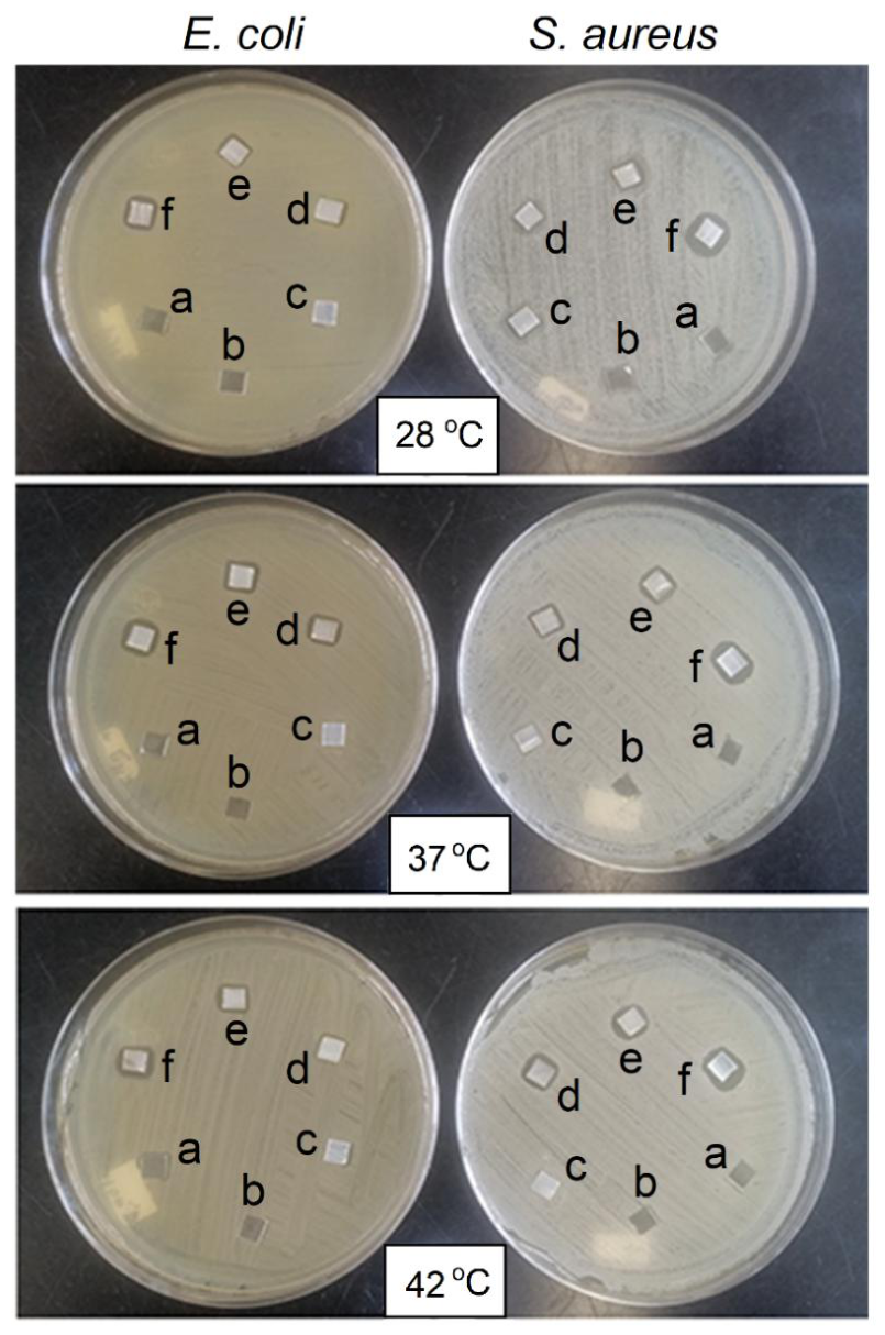
Figure 13.
Antimicrobial halos recorded for S. aureus and E. coli in contact after 24 h with AgNPs loaded on PET-g-NVCL (9.5%) at different temperatures.
Figure 13.
Antimicrobial halos recorded for S. aureus and E. coli in contact after 24 h with AgNPs loaded on PET-g-NVCL (9.5%) at different temperatures.
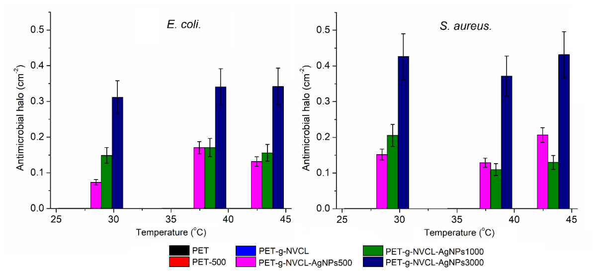

Table 1.
DSC and TGA analysis of the films obtained from the PNVCL graft process promoted by gamma-rays.
Table 1.
DSC and TGA analysis of the films obtained from the PNVCL graft process promoted by gamma-rays.
| DSC analysis | ||||
| Sample | T (°C) | Tm (°C) | ||
| PET | 75.9 (Tg) | 246.5 C; 33.52 J/g | ||
| PNVCL | 46.3 (TLCST) | 257.6 C; 34.75 J/g | ||
| PET-g-NVCL | 49.3 (TLCST), 70.2 (Tg), 83.3 (TS) | 245.2 C; 25.03 J/g | ||
| TGA analysis | ||||
| Sample | Temperature at 10% lost weight (°C) | Decomposition Temperature (°C) | Residue Yield (800 °C, w.%) | |
| PET | 412.9 | 440.5 | 27.9 | |
| PNVCL | 257.0 | 267.2; 443.5 | 7.1 | |
| PET-g-NVCL | 402.3 | 176.8; 441.4 | 10.7 | |
| PET-g-NVCL-AgNPs500 | 405.4 | 185.9; 440.4 | 12.8 | |
| PET-g-NVCL-AgNPs1000 | 402.1 | 202.1; 441.3 | 14.9 | |
| PET-g-NVCL-AgNPs3000 | 402.7 | 202.6; 442.2 | 16.3 | |
Table 2.
Mechanical properties of PET and PET-g-NVCL (9.5%) films.
| Sample | Elastic Modulus (MPa) | Elongation (%) | Stress Rupture (MPa) |
| PET | 1228.2 ± 87 | 227.5 ± 12 | 160.9 ± 7 |
| PET-g-NVCL (9.5%) | 834.7 ± 35 | 221.3 ± 22 | 104.5 ± 9 |
Table 3.
Results of elemental composition by XPS of PET, PET-g-NVCL (9.5%), and PET-g-NVCL-AgNPs (9.5%, 5.6%).
Table 3.
Results of elemental composition by XPS of PET, PET-g-NVCL (9.5%), and PET-g-NVCL-AgNPs (9.5%, 5.6%).
| Elemental composition (%) | Chemical concentration groups (%) | |||||||||||||
| C1s | O1s | |||||||||||||
| C1s | N1s | O1s | Ag3d | C=O | C-O-C | C-N | C-C | C=C | C=O | C=ON | C-O-C | OH | C=OAg | |
| PET | 65.9 | --- | 33.6 | --- | 18.0 | 23.8 | --- | 34.0 | 24.0 | 55.4 | --- | 44.5 | --- | --- |
| PET-g-NVCL | 76.5 | 2.1 | 21.3 | --- | 13.2 | 13.0 | 18.9 | 36.5 | 18.1 | 26.6 | 27.7 | 26.2 | 19.2 | 0 |
| PET-g-NVCL-AgNPs3000 | 75.5 | 2.9 | 21.2 | 0.2 | 14.4 | 13.5 | 17.9 | 32.3 | 21.7 | 24.4 | 18.6 | 23.7 | 16.6 | 16.3 |
Disclaimer/Publisher’s Note: The statements, opinions and data contained in all publications are solely those of the individual author(s) and contributor(s) and not of MDPI and/or the editor(s). MDPI and/or the editor(s) disclaim responsibility for any injury to people or property resulting from any ideas, methods, instructions or products referred to in the content. |
© 2026 by the authors. Licensee MDPI, Basel, Switzerland. This article is an open access article distributed under the terms and conditions of the Creative Commons Attribution (CC BY) license.
Copyright: This open access article is published under a Creative Commons CC BY 4.0 license, which permit the free download, distribution, and reuse, provided that the author and preprint are cited in any reuse.