

Submitted:
31 July 2026
Posted:
04 August 2026
You are already at the latest version
Abstract
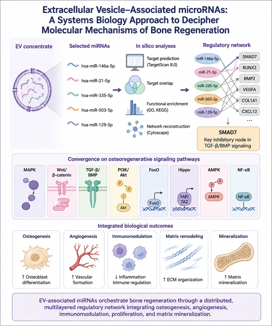
Background: Bone regeneration requires coordinated regulation of osteogenesis angiogenesis, extracellular matrix remodeling, immune responses, and mineralization. Extracellular vesicle (EV)-based therapies have emerged as promising cell free strategies, although their molecular mechanisms remain incompletely understood. Objective: To investigate the molecular mechanisms potentially underlying the osteoregenerative effects of an EV concentrate using an in silico systems biology approach. Methods: Five osteogenesis and immunomodulation related microRNAs (hsa-miR-146a-5p, hsa-miR-21-5p, hsa-miR-335-5p, hsa-miR-503-5p, and hsa-miR-129-5p) were analyzed. High-confidence target genes were identified using TargetScan 8.0, followed by overlap analysis, Gene Ontology and KEGG enrichment, and miRNA-mRNA network reconstruction in Cytoscape. Results The selected miRNAS regulated hundreds of predicted targets, displaying complementary rather than universal target overlap. SMAD7 emerged as a key shared regulatory node involved in TGF-β/BMP signaling. Functional enrichment demonstrated significant associations with osteoblast differentiation, angiogenesis, extracellular matrix organization, inflammatory regulation, and mineralization. Pathway analysis identified convergence on MAPK, Wnt/β-catenin, TGF-β/BMP, PI3K/Akt, FoxO, Hippo, AMPK, and NF-κB signaling. Conclusions: These findings indicate that EV concentrates may promote bone regeneration through coordinated miRNA-mediated regulation of multiple osteoregenerative pathways, providing a mechanistic framework for developing cell-free regenerative therapies and guiding future experimental validation.
Keywords:
extracellular vesicles
; bone regeneration
; microRNA
1. Introduction
Bone regeneration remains a major clinical and biological challenge in regenerative medicine, particularly in the presence of critical-sized defects, craniofacial bone loss, trauma-related defects, periodontal destruction, tumor resection, and complex alveolar ridge reconstruction. Critical-sized defects are generally understood as bone injuries that do not heal spontaneously during the lifetime of the organism, making them useful experimental models for evaluating regenerative strategies [1]. Although bone has an intrinsic capacity for repair, large defects often fail to regenerate because successful healing depends on the coordinated recruitment of osteoprogenitor cells, angiogenesis, immune regulation, extracellular matrix deposition, and progressive mineralization [2,3].
Autogenous bone grafting remains the gold standard for bone reconstruction because it provides osteogenic cells, osteoinductive signals, and an osteoconductive matrix [4]. However, its clinical application is limited by donor-site morbidity, restricted availability, additional surgical complexity, postoperative pain, infection risk, and variable graft resorption [4,5]. Alternative approaches, including allografts, xenografts, synthetic biomaterials, growth-factor delivery systems, and scaffold-based tissue engineering strategies, have been developed to overcome these limitations. Nevertheless, many of these approaches still fail to reproduce the biological complexity required for predictable bone regeneration, especially in defects characterized by hypoxia, inflammation, impaired vascularization, and limited cellular recruitment [6,7].
Bone tissue engineering has therefore emerged as a multidisciplinary field combining biomaterials, cells, and bioactive molecular signals to guide tissue repair [8]. Among the cellular components investigated, mesenchymal stromal/stem cells (MSCs) have received considerable attention because of their multipotent differentiation capacity, immunomodulatory activity, trophic function, and ability to secrete bioactive mediators involved in tissue homeostasis and repair [9,10]. MSCs may be obtained from multiple sources, including bone marrow, adipose tissue, dental tissues, umbilical cord, and other perivascular niches. However, despite their therapeutic potential, the clinical translation of MSC-based therapies remains limited by important biosafety and manufacturing concerns, including phenotypic variability after ex vivo expansion, genetic instability, immune compatibility, tumorigenic potential, low engraftment efficiency, and inconsistent behavior after transplantation [10,11].
Accumulating evidence indicates that many regenerative effects attributed to MSCs are mediated predominantly by paracrine signaling rather than by direct long-term engraftment or differentiation into tissue-forming cells [11,12]. This paradigm shift has redirected attention from cell replacement toward cell-free approaches based on the MSC secretome. In this context, extracellular vesicles (EVs) have emerged as key mediators of intercellular communication and as promising therapeutic candidates for regenerative medicine [13,14]. EVs are lipid bilayer-enclosed particles released by cells and capable of transferring proteins, lipids, messenger RNAs, long non-coding RNAs, circular RNAs, and microRNAs (miRNAs) to recipient cells [13,14,15]. According to current recommendations for EV studies, precise terminology and methodological transparency are essential, particularly regarding vesicle source, isolation or enrichment procedures, characterization strategy, and functional evidence [16].
In bone biology, EVs are of particular interest because they can simultaneously modulate multiple processes required for tissue repair. MSC-derived EVs have been reported to influence osteogenic differentiation, angiogenesis, inflammatory resolution, osteoclast activity, extracellular matrix remodeling, and mineral deposition [17,18,19,20]. Unlike conventional single-molecule therapies, EVs carry complex molecular cargo capable of acting on several recipient-cell pathways at once. This property makes EV concentrates biologically suited to regulate the sequential and overlapping phases of bone regeneration, including early inflammatory modulation, vascular recruitment, osteoprogenitor activation, matrix maturation, and mineralization [18,19,20].
Among EV cargo components, miRNAs are especially relevant because they regulate gene expression at the post-transcriptional level by binding to target messenger RNAs and reducing transcript stability or translation efficiency [21,22]. A single miRNA may regulate several target genes, whereas different miRNAs may converge on shared genes, complementary biological processes, or interconnected signaling pathways. This network-based regulatory logic is highly relevant for bone regeneration, where osteogenesis, angiogenesis, immunomodulation, and matrix mineralization are not independent events but integrated biological modules [23]. Accordingly, EV-associated miRNAs may provide a mechanism through which EV concentrates coordinate complex regenerative responses.
Several miRNAs have been implicated in bone repair and osteoimmune regulation. miR-21 has been associated with osteogenesis and bone homeostasis through mechanisms involving PTEN/PI3K/Akt, Wnt, and Smad-related signaling [24]. miR-335 carried by bone marrow MSC-derived EVs has been reported to promote fracture recovery and osteoblast differentiation, partly through Wnt/β-catenin-related mechanisms [25]. miR-503 has been associated with bone formation during distraction osteogenesis by targeting Smurf1, a negative regulator of osteogenic signaling [26]. miR-129-5p has also been linked to osteogenic differentiation and bone regeneration through repression of Dkk3 and activation of Wnt/β-catenin-related mechanisms, although its effects may depend on the cellular and pathological context [27]. In addition, miR-146a is widely recognized as an immunomodulatory miRNA and has been implicated in anti-inflammatory effects mediated by MSC-derived EVs [28]. Together, these miRNAs represent a biologically plausible regulatory panel for exploring the osteoregenerative potential of EV concentrates.
Bone regeneration is governed by interconnected signaling pathways, including Wnt/β-catenin, BMP/Smad, TGF-β, PI3K/Akt, MAPK, Hippo, FoxO, AMPK, and NF-κB-related cascades [28,29]. These pathways determine whether mesenchymal progenitors undergo proliferation, survival, osteogenic commitment, matrix maturation, or inflammatory suppression. Therefore, the biological activity of EV concentrates is unlikely to be explained by a single miRNA, a single target gene, or an isolated signaling pathway. Instead, EV-mediated regeneration is more plausibly understood as a systems-level process involving molecular convergence, regulatory redundancy, and modulation of osteogenic inhibitory checkpoints.
Systems biology provides a useful framework for investigating this complexity. Computational tools such as TargetScan allow prediction of miRNA–mRNA interactions based on seed-region complementarity and contextual features of target sites [30]. The Context++ model improves target prioritization by incorporating multiple determinants of miRNA targeting efficacy [31]. Functional enrichment platforms such as Gene Ontology, KEGG, and DAVID enable the identification of biological processes and signaling pathways statistically overrepresented among gene lists [32,33,34]. In parallel, Cytoscape allows reconstruction and visualization of molecular interaction networks, supporting the identification of connected modules, regulatory relationships, and topological features [35].
Despite increasing interest in EV-based bone regeneration, the molecular architecture through which EV concentrates may regulate bone repair remains incompletely defined. Many studies still emphasize isolated miRNAs or single target genes, which may underestimate the integrated behavior of EV cargo. A more robust approach requires evaluating EV-associated miRNAs as cooperative regulators of gene networks and cell signaling pathways. Such an approach may help identify whether EV concentrates operate through distributed target modulation, redundant miRNA–mRNA interactions, and convergence on osteogenic regulatory modules.
Therefore, the present study aimed to investigate, through an in silico systems biology approach, the molecular mechanisms potentially associated with extracellular vesicles concentrated in bone regeneration. Five osteogenic and/or immunomodulatory miRNAs—hsa-miR-146a-5p, hsa-miR-21-5p, hsa-miR-335-5p, hsa-miR-503-5p, and hsa-miR-129-5p—were analyzed using target prediction, high-confidence filtering, target-overlap analysis, functional enrichment, and miRNA–mRNA network reconstruction. We hypothesized that the osteoregenerative potential of EV concentrates is mediated not by a single dominant effector, but by a multilayered miRNA regulatory architecture characterized by target redundancy, pathway convergence, and modulation of key osteogenic, angiogenic, immunomodulatory, and mineralization-associated processes.
2. Materials and Methods
2.1. Study Design
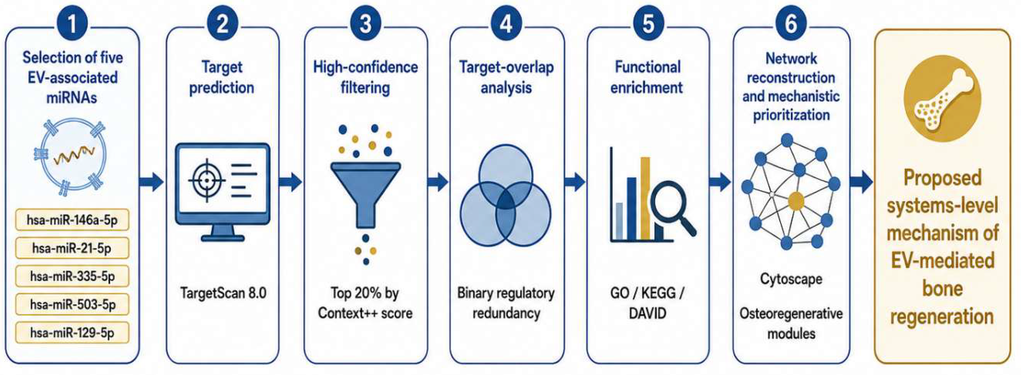
This study was designed as an in silico systems biology analysis aimed at investigating the molecular mechanisms potentially associated with extracellular vesicle (EV) concentrates in bone regeneration. The analytical strategy integrated miRNA selection, target gene prediction, high-confidence filtering, target-overlap analysis, functional enrichment, and regulatory network reconstruction.
The study was structured according to a multilayered analytical framework. First, predicted mRNA targets of selected miRNAs were retrieved using a target-prediction database. Second, overlapping targets were identified to assess regulatory redundancy within the miRNA pool. Third, functional enrichment analysis was performed to determine biological processes and signaling pathways overrepresented among predicted targets. Finally, network reconstruction was conducted to visualize miRNA–mRNA interactions and support mechanistic prioritization of osteoregenerative signaling nodes.
Because this was an exclusively computational study using publicly available databases and previously generated analytical files, no human participants, animal experimentation, or biological sample collection were involved. Therefore, formal ethics committee approval was not required for the in silico component of the study.
Figure 1.
Analytical workflow of the systems biology strategy used to investigate extracellular vesicle concentrate-associated miRNAs in bone regeneration. Five miRNAs with reported osteogenic and/or immunomodulatory relevance were selected as input molecules. Predicted mRNA targets were retrieved using TargetScan 8.0 and filtered according to Context++ score ranking. Filtered target lists were subjected to target-overlap analysis to identify regulatory redundancy, followed by Gene Ontology and KEGG/DAVID enrichment analysis. miRNA–mRNA interactions were reconstructed in Cytoscape, and functionally relevant osteoregenerative nodes were prioritized to generate a proposed mechanistic model of EV-mediated bone repair.
Figure 1.
Analytical workflow of the systems biology strategy used to investigate extracellular vesicle concentrate-associated miRNAs in bone regeneration. Five miRNAs with reported osteogenic and/or immunomodulatory relevance were selected as input molecules. Predicted mRNA targets were retrieved using TargetScan 8.0 and filtered according to Context++ score ranking. Filtered target lists were subjected to target-overlap analysis to identify regulatory redundancy, followed by Gene Ontology and KEGG/DAVID enrichment analysis. miRNA–mRNA interactions were reconstructed in Cytoscape, and functionally relevant osteoregenerative nodes were prioritized to generate a proposed mechanistic model of EV-mediated bone repair.
2.2. Selection of the miRNA Panel
Five miRNAs were selected as the primary analytical input: hsa-miR-146a-5p, hsa-miR-21-5p, hsa-miR-335-5p, hsa-miR-503-5p, and hsa-miR-129-5p. The selection was based on prior biological evidence supporting their involvement in osteogenesis, osteoimmune regulation, angiogenesis, mesenchymal cell differentiation, inflammatory modulation, or extracellular matrix mineralization [23,24,25,26,27,28].
The selected miRNAs were interpreted as a candidate osteoregenerative regulatory panel potentially associated with the biological activity of EV concentrates. This selection was not intended to represent the entire EV miRNA cargo, but rather a focused molecular signature with relevance to bone regeneration and tissue repair.
2.3. miRNA Target Prediction
Predicted mRNA targets for each selected miRNA were retrieved using TargetScan Human 8.0. TargetScan predicts miRNA targets based on conserved and non-conserved binding sites, seed-region complementarity, site accessibility, local AU content, 3′ untranslated region context, and other determinants of miRNA targeting efficacy [30,31].
For each miRNA, predicted targets were exported and organized into individual datasets. Gene symbols were standardized to avoid duplication caused by alternative identifiers, non-uniform naming, or spreadsheet formatting. When duplicate entries were present for the same gene within a single miRNA dataset, the gene was retained only once for downstream target-overlap and enrichment analyses.
2.4. High-Confidence Filtering
To prioritize biologically plausible miRNA–mRNA interactions and reduce the likelihood of false-positive predictions, target lists were filtered according to cumulative weighted Context++ scores. The Context++ model integrates multiple features associated with miRNA targeting efficacy and provides a quantitative estimate of predicted repression strength [31].
Only high-confidence targets were retained for the primary analytical layer. In the present study, the high-confidence set was defined as the top 20% of predicted targets for each miRNA according to Context++ score ranking. This approach allowed each miRNA to contribute its strongest predicted targets while avoiding excessive inclusion of low-confidence interactions.
The final filtered target sets were used for three downstream analyses: target-overlap assessment, functional enrichment, and miRNA-mRNA network reconstruction.
2.5. Target-Overlap and Regulatory Redundancy Analysis
To evaluate whether the selected miRNA panel exhibited cooperative regulatory behavior, target-overlap analysis was performed across the five filtered miRNA target lists. Genes were classified according to the number of miRNAs predicted to regulate them.
Regulatory redundancy was defined as the presence of the same predicted target gene in two or more miRNA target lists. Genes targeted by two miRNAs were classified as showing binary regulatory redundancy. Genes targeted by three or more miRNAs, if present, would be classified as higher-order redundant targets.
This distinction was essential because a shared target was defined strictly as a gene identified in the intersection of the filtered TargetScan-derived lists. Therefore, genes selected later because of osteogenic relevance or network position were not automatically classified as shared TargetScan targets unless they were present in the overlap analysis.
Target-overlap visualization was performed using Venn diagram-based set analysis. The results were used to identify the core subset of genes with redundant miRNA regulation and to distinguish these genes from broader functionally prioritized osteogenic nodes.
2.6. Functional Enrichment Analysis
Functional enrichment analysis was performed to identify biological processes, molecular functions, cellular components, and signaling pathways overrepresented among the predicted target genes. Gene Ontology analysis was used to classify enriched terms into biological process, molecular function, and cellular component categories [32]. KEGG pathway mapping was used to identify signaling pathways associated with the predicted target set [33].
The enrichment analysis focused particularly on biological processes and pathways relevant to bone regeneration, including osteoblast differentiation, mesenchymal cell commitment, extracellular matrix organization, angiogenesis, inflammatory regulation, cell proliferation, mineralization, and osteoimmune signaling.
2.7. DAVID-Based Enrichment and Statistical Criteria
Gene list enrichment was additionally performed using the DAVID platform to support functional annotation and pathway-level interpretation [34]. Statistical significance of gene-term and gene-pathway associations was assessed using Fisher’s Exact Test, as implemented by DAVID.
To reduce false-positive findings due to multiple comparisons, false discovery rate correction was considered during enrichment interpretation. Enrichment results were considered statistically significant when p < 0.05 after appropriate correction or when they met the predefined significance threshold reported by the enrichment platform.
For manuscript reporting, enriched terms were prioritized according to statistical significance, biological relevance to bone regeneration, number of genes involved, and consistency with known osteogenic or osteoimmune signaling pathways.
2.8. miRNA-mRNA Network Reconstruction
miRNA-mRNA regulatory networks were reconstructed using Cytoscape, a platform for visualization and analysis of molecular interaction networks [35]. Network input files consisted of miRNA–target gene interaction tables derived from filtered target-prediction datasets and, when explicitly indicated, curated osteoregenerative interactions selected for biological relevance.
In the primary regulatory network, miRNAs were represented as regulatory nodes and predicted mRNA targets as target nodes. Edges represented predicted miRNA–mRNA interactions. Node attributes included molecule type, miRNA identity, gene symbol, regulatory redundancy, pathway annotation, and functional category when available.
Network visualization was used to identify the organization of the regulatory architecture, including isolated miRNA-specific targets, binary redundant targets, and target clusters associated with osteogenesis, proliferation, mineralization, angiogenesis, or immunomodulation.
2.9. Network Topology and Hub Definition
Network topology was analyzed to distinguish highly connected nodes from genes selected solely on the basis of biological relevance. When topological analysis was performed, network metrics included degree, betweenness centrality, closeness centrality, and, when appropriate, clustering coefficient.
A gene was classified as a topological hub only when supported by explicit network metrics, particularly high degree or centrality relative to other nodes in the same network. Genes identified by the target-overlap analysis but not supported by centrality metrics were described as redundantly targeted genes rather than hubs.
Similarly genes selected because of their recognized role in bone biology, osteogenic signaling, or pathway convergence were classified as mechanistically prioritized nodes unless topological metrics supported their classification as hubs.
This terminology was applied to avoid overinterpretation and to maintain methodological consistency between computational prediction, network topology, and biological inference.
2.10. Mechanistic Prioritization of Osteoregenerative Nodes
After target prediction, redundancy analysis, and enrichment mapping, mechanistic prioritization was performed to identify genes with potential relevance to EV-mediated bone regeneration. Prioritization considered four complementary criteria:
- Presence among high-confidence predicted miRNA targets;
- Presence in the overlap analysis as a redundantly targeted gene;
- Participation in bone-related signaling pathways identified by enrichment analysis;
- Known biological relevance to osteogenesis, angiogenesis, immunomodulation, extracellular matrix remodeling, or mineralization.
Genes meeting multiple criteria were interpreted as candidate osteoregenerative regulatory nodes. However, mechanistic prioritization was considered hypothesis-generating and did not imply experimental validation of direct miRNA–mRNA binding or functional repression.
2.11. Interpretation Framework
This results were interpreted using a four-layer systems biology framework:
Layer 1 corresponded to the primary TargetScan-derived target landscape of the five selected miRNAs.
Layer 2 corresponded to regulatory redundancy, defined by genes shared by two or more miRNAs.
Layer 3 corresponded to functional enrichment, identifying biological processes and signaling pathways overrepresented among the predicted targets.
Layer 4 corresponded to network-based and mechanistic prioritization, integrating target prediction, pathway relevance, and network topology to propose osteoregenerative regulatory modules.
This layered approach was adopted to distinguish direct computational outputs from biological interpretation. Therefore, causal statements were avoided unless supported by experimental evidence from the literature. Findings derived from the present analysis were interpreted as predictive, hypothesis-generating, and mechanistically suggestive.
2.12. Data reporting and reproducibility
For transparency and reproducibility, the following datasets were generated or organized for reporting: individual miRNA target lists, filtered high-confidence target lists, merged target tables, overlap/intersection tables, enrichment output tables, miRNA–mRNA edge lists, and Cytoscape network files.
The main manuscript reports the principal target-overlap findings, enriched pathways, and reconstructed network architecture. Complete target lists, enrichment outputs, and network input files should be provided as supplementary material when submitting the manuscript.
3. Results
3.1. TargetScan-Derived miRNA Target Landscape
The first analytical layer consisted of the reconstruction of the predicted target landscape for the five selected miRNAs: hsa-miR-146a-5p, hsa-miR-21-5p, hsa-miR-335-5p, hsa-miR-503-5p, and hsa-miR-129-5p. After TargetScan-based retrieval and high-confidence filtering according to Context++ score ranking, the combined dataset comprised 395 predicted miRNA–mRNA interactions, corresponding to 387 unique target genes.
The number of predicted targets differed among the selected miRNAs, indicating that each miRNA contributed unequally to the regulatory landscape. hsa-miR-129-5p presented the largest target repertoire, followed by hsa-miR-21-5p, hsa-miR-503-5p, hsa-miR-335-5p, and hsa-miR-146a-5p. This distribution suggests that the selected EV-associated miRNA panel does not act as a uniform regulatory block, but rather as a heterogeneous molecular system in which each miRNA may contribute distinct target subsets to the global osteoregenerative network.
Importantly, most predicted targets were miRNA-specific, indicating that the regulatory effect of the EV concentrate is likely distributed across multiple molecular axes rather than concentrated in a single shared target. This architecture is consistent with a systems-level regulatory model in which different miRNAs may regulate distinct components of interconnected biological pathways involved in bone repair.

Table 1.
TargetScan-derived predicted targets for each selected miRNA.
| miRNA | Predicted targets after filtering |
Relative contribution to the target landscape |
|---|---|---|
| hsa-miR-129-5p | 146 | Highest target repertoire |
| hsa-miR-21-5p | 76 | High regulatory contribution |
| hsa-miR-503-5p | 61 | Intermediate regulatory contribution |
| hsa-miR-335-5p | 57 | Intermediate regulatory contribution |
| hsa-miR-146a-5p | 55 | Focused regulatory contribution |
| Total interactions | 395 | - |
| Unique genes | 387 | - |
3.2. Target-Overlap Analysis Reveals Binary Regulatory Redundancy
Target-overlap analysis was performed to determine whether the selected miRNA panel exhibited cooperative regulation through shared mRNA targets. No gene was simultaneously predicted as a target of all five miRNAs. Similarly, no higher-order redundancy involving three or more miRNAs was detected in the filtered dataset.
Figure 2.
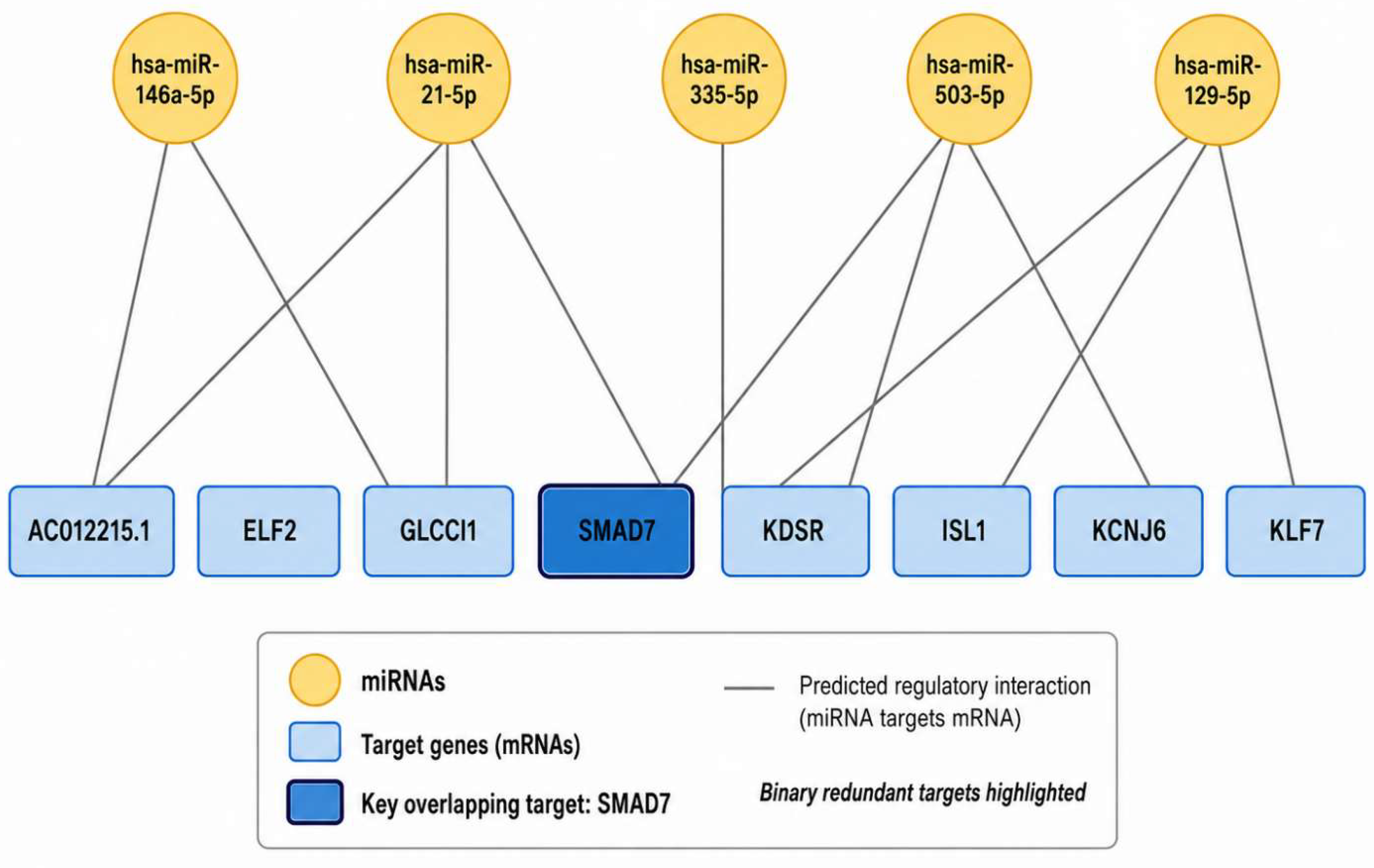
Target-overlap analysis of predicted mRNA targets regulated by the five selected extracellular vesicle-associated miRNAs. Target lists were generated using TargetScan 8.0 and filtered according to Context++ score ranking. The combined dataset included 395 predicted miRNA–mRNA interactions corresponding to 387 unique genes. Eight genes showed binary regulatory redundancy, being predicted as targets of two miRNAs: AC012215.1, ELF2, GLCCI1, ISL1, KCNJ6, KDSR, KLF7, and SMAD7. No gene was shared by all five miRNAs, supporting a distributed rather than single-target regulatory architecture.
Figure 2.
Target-overlap analysis of predicted mRNA targets regulated by the five selected extracellular vesicle-associated miRNAs. Target lists were generated using TargetScan 8.0 and filtered according to Context++ score ranking. The combined dataset included 395 predicted miRNA–mRNA interactions corresponding to 387 unique genes. Eight genes showed binary regulatory redundancy, being predicted as targets of two miRNAs: AC012215.1, ELF2, GLCCI1, ISL1, KCNJ6, KDSR, KLF7, and SMAD7. No gene was shared by all five miRNAs, supporting a distributed rather than single-target regulatory architecture.
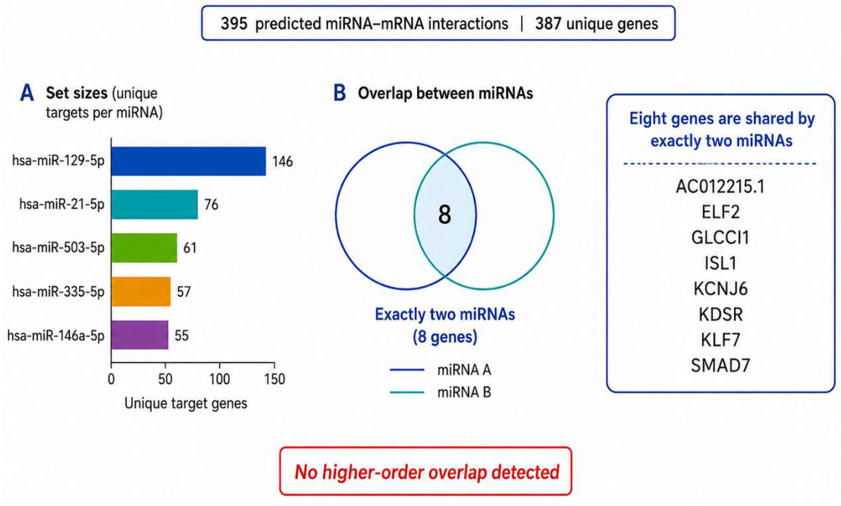
However, eight genes were identified as shared targets of two miRNAs, demonstrating a pattern of binary regulatory redundancy. These genes were AC012215.1, ELF2, GLCCI1, ISL1, KCNJ6, KDSR, KLF7, and SMAD7. Each of these targets was predicted to be regulated by two miRNAs within the selected panel, suggesting that the EV-associated miRNA pool may provide dual regulatory coverage over specific molecular nodes.
Among these genes, SMAD7 was the most biologically relevant overlapping target in the context of bone regeneration, as it was shared by hsa-miR-21-5p and hsa-miR-503-5p. Because SMAD7 is an inhibitory regulator of TGF-β/BMP signaling, its dual predicted regulation supports a potential mechanism of osteogenic disinhibition. KLF7 and ISL1 also emerged as differentiation-associated targets, suggesting that binary redundancy may contribute not only to pathway inhibition release, but also to the regulation of cell fate and progenitor-cell behavior.
These findings indicate that the analyzed EV miRNA panel does not depend on a single universal target shared by all miRNAs. Instead, its regulatory architecture appears to be organized through a distributed model, in which selected genes receive redundant regulation while most targets remain miRNA-specific.
Table 2.
Genes showing binary regulatory redundancy in the five-miRNA panel..
| Shared target gene | miRNAs involved | Proposed biological interpretation |
|---|---|---|
| SMAD7 | hsa-miR-21-5p + hsa-miR-503-5p |
Inhibitory checkpoint of TGF-β/BMP signaling |
| KLF7 | hsa-miR-146a-5p + hsa-miR-129-5p |
Differentiation-associated transcriptional regulation |
| ISL1 | hsa-miR-335-5p + hsa-miR-129-5p |
Cell differentiation and developmental signaling |
| KDSR | hsa-miR-335-5p + hsa-miR-503-5p |
Membrane lipid metabolism and cellular signaling |
| KCNJ6 | hsa-miR-335-5p + hsa-miR-129-5p |
Ion homeostasis and membrane excitability |
| ELF2 | hsa-miR-21-5p + hsa-miR-129-5p |
Transcriptional and maturation-related regulation |
| GLCCI1 | hsa-miR-21-5p + hsa-miR-129-5p |
Inflammatory and glucocorticoid-response modulation |
| AC012215.1 | hsa-miR-146a-5p + hsa-miR-129-5p |
Non-coding RNA-associated regulatory support |
3.3. Functional Enrichment Identifies Pathways Associated with Proliferation, Osteogenesis and Mineralization
Functional enrichment analysis was performed to determine whether the predicted target genes were overrepresented in biological processes and signaling pathways relevant to bone regeneration. The analysis identified multiple enriched pathways associated with cell proliferation, progenitor-cell regulation, osteogenic differentiation, extracellular matrix maturation, and mineralization.
The most statistically significant enriched pathway was the signaling pathway regulating pluripotency of stem cells, involving 13 target genes and showing a highly significant enrichment pattern. This result suggests that the selected EV-associated miRNA panel may influence early regulatory programs associated with progenitor-cell maintenance, lineage commitment, and transition toward tissue-forming phenotypes.
Figure 3.
Functional enrichment analysis of high-confidence predicted targets regulated by the selected miRNA panel. Enrichment analysis identified biological pathways associated with stem cell pluripotency, MAPK signaling, Hippo signaling, TGF-β signaling, Wnt signaling, mTOR signaling, and FoxO signaling. These pathways are involved in progenitor-cell regulation, cell proliferation, osteogenic differentiation, mechanotransduction, metabolic activation, oxidative stress response, and extracellular matrix maturation. Pathway significance is represented as −log10 p-value, and bubble size indicates the number of genes associated with each pathway.
Figure 3.
Functional enrichment analysis of high-confidence predicted targets regulated by the selected miRNA panel. Enrichment analysis identified biological pathways associated with stem cell pluripotency, MAPK signaling, Hippo signaling, TGF-β signaling, Wnt signaling, mTOR signaling, and FoxO signaling. These pathways are involved in progenitor-cell regulation, cell proliferation, osteogenic differentiation, mechanotransduction, metabolic activation, oxidative stress response, and extracellular matrix maturation. Pathway significance is represented as −log10 p-value, and bubble size indicates the number of genes associated with each pathway.
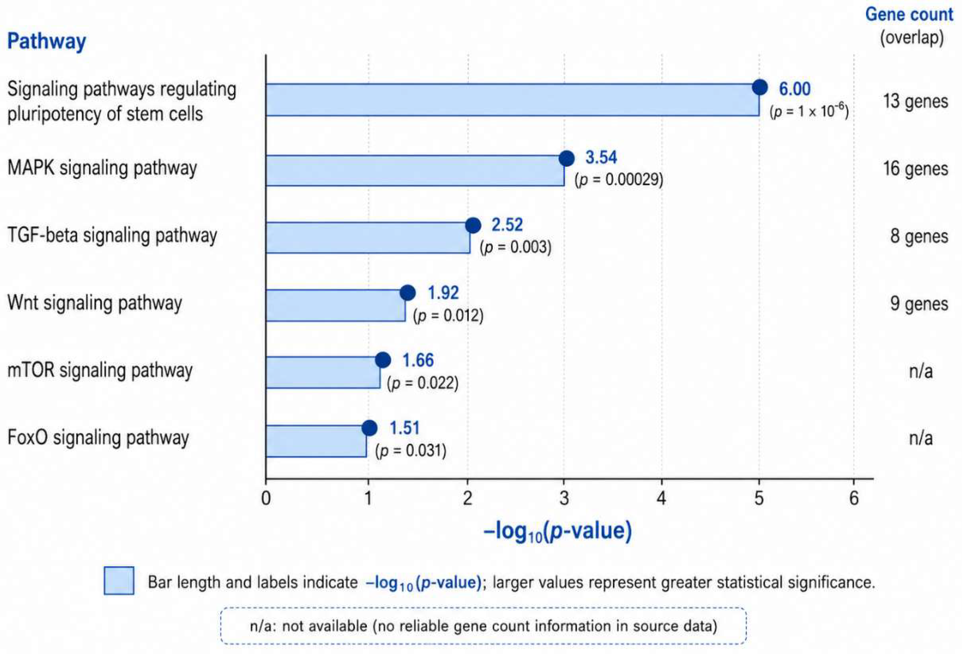
The MAPK signaling pathway showed the highest gene density among the enriched pathways, with 16 target genes. Because MAPK signaling is strongly associated with cell proliferation, stress response, injury signaling, and osteogenic differentiation, this enrichment supports the hypothesis that EV-associated miRNAs may contribute to the early proliferative and reparative phases of bone healing.
The Wnt signaling pathway was also significantly enriched, with nine target genes. Given the central role of Wnt/β-catenin signaling in osteoblast differentiation, bone formation, and skeletal homeostasis, this finding supports the involvement of the miRNA panel in osteogenic pathway regulation. In parallel, enrichment of the TGF-β signaling pathway, including SMAD7, suggested that the EV miRNA regulatory architecture may also influence BMP/Smad-related signaling, a major axis of osteoblast differentiation and matrix mineralization.
Additional enrichment of mTOR and FoxO signaling pathways suggested potential involvement of metabolic regulation, cell survival, oxidative stress response, and anabolic activity. These pathways are relevant to the regenerative microenvironment, where osteogenic differentiation depends on the integration of survival, metabolic activation, and stress adaptation.
Table 3.
Main enriched pathways associated with the predicted target set.
| Enriched pathways | Number of genes | Statistical result | Biological relevance |
|---|---|---|---|
| Signaling pathways regulating pluripotency of stem cells | 13 | p ≈ 10⁻⁶ | Progenitor-cell regulation and lineage commitment |
| MAPK signaling pathway | 16 | p= 0.00029 | Cell proliferation, injury response, osteogenic signaling |
| TGF-β signaling pathway | 8 | p=0.003 | BMS/Smad regulation, osteoblast differentiation |
| Wnt signaling pathway | 9 | p=0.012 | Osteogenesis and bone formation |
| mTOR signaling pathway | Not available in raw export | p=0.022 | Cell survival, metabolism, anabolic signaling |
| FoxO signaling pathway | Not available in raw export | p=0.031 | Oxidative stress response and cell survival |
3.4. Network reconstruction supports a distributed regulatory architecture
Cytoscape-based network reconstruction was performed to visualize the relationships between the selected miRNAs and their predicted target genes. The primary miRNA–mRNA network demonstrated a distributed architecture in which each miRNA regulated a specific target subset, while a small group of genes received redundant regulation from two miRNAs.
Figure 4.
Primary miRNA–mRNA regulatory network reconstructed from TargetScan-derived interactions. The network displays the five selected miRNAs and the eight genes identified as binary redundant targets in the filtered TargetScan-derived dataset. miRNA nodes are represented in yellow, target-gene nodes in blue, and predicted miRNA–mRNA interactions are shown as connecting edges. SMAD7 is highlighted because it represents the most biologically relevant overlapping target in the context of bone regeneration, given its inhibitory role in TGF-β/BMP signaling. The network supports a distributed regulatory architecture, in which the EV-associated miRNA panel acts through multiple partially overlapping target relationships rather than through a single shared target.
Figure 4.
Primary miRNA–mRNA regulatory network reconstructed from TargetScan-derived interactions. The network displays the five selected miRNAs and the eight genes identified as binary redundant targets in the filtered TargetScan-derived dataset. miRNA nodes are represented in yellow, target-gene nodes in blue, and predicted miRNA–mRNA interactions are shown as connecting edges. SMAD7 is highlighted because it represents the most biologically relevant overlapping target in the context of bone regeneration, given its inhibitory role in TGF-β/BMP signaling. The network supports a distributed regulatory architecture, in which the EV-associated miRNA panel acts through multiple partially overlapping target relationships rather than through a single shared target.
This organization reinforces the concept that the EV concentrate does not operate through a single dominant miRNA–mRNA axis. Instead, the network suggests a multilayered regulatory structure composed of miRNA-specific interactions, binary redundant targets, and functionally convergent signaling pathways.
The binary redundancy observed for SMAD7, KLF7, ISL1, KDSR, KCNJ6, ELF2, GLCCI1, and AC012215.1 may provide regulatory robustness by increasing the probability that specific molecular checkpoints are modulated by more than one miRNA. Among them, SMAD7 represents the clearest osteogenic checkpoint because of its role as a negative regulator of TGF-β/BMP signaling. Therefore, the predicted dual regulation of SMAD7 by hsa-miR-21-5p and hsa-miR-503-5p supports a potential disinhibition mechanism favoring osteogenic signaling.
A curated osteoregenerative network was also reconstructed to represent functionally relevant interactions involving genes associated with osteoinduction, proliferation, and mineralization. This network included TRAF6, PTEN, CCND2, CACNG2, SMAD7, SOST, and RUNX2 as mechanistically prioritized nodes. These genes were not interpreted uniformly as shared TargetScan-derived targets; rather, they were classified according to their analytical origin and biological relevance. SMAD7 was identified as a redundantly targeted gene, whereas PTEN, SOST, and RUNX2 were treated as mechanistically prioritized osteogenic nodes requiring explicit distinction from primary overlap-derived targets.
The distinction is essential because network-based biological interpretation should not be conflated with target-overlap results. Therefore, in the present analysis, the term “redundantly targeted gene” was reserved for genes present in the intersection analysis, whereas “mechanistically prioritized node” was used for genes selected because of osteogenic relevance, pathway involvement, or network position.
3.5. Osteoregenerative Modules Identified by Network Interpretation
The integrated analysis suggested three major functional modules potentially associated with EV-mediated bone regeneration: osteoinduction, proliferation and mineralization.
Figure 5.
Curated osteoregenerative regulatory module integrating predicted miRNA–mRNA interactions and biologically relevant bone-regeneration nodes. The module summary uses selected interactions potentially involved in osteoinduction, proliferation, immunomodulation, and mineralization. SMAD7 was identified as a redundantly predicted target within the primary TargetScan-derived overlap analysis, whereas PTEN, SOST, RUNX2, TRAF6, CCND2, and CACNG2 were included as mechanistically prioritized nodes based on their biological relevance to bone regeneration pathways. The model supports the hypothesis that EV-associated miRNAs may regulate bone repair through osteogenic disinhibition, proliferative control, osteoimmune modulation, and matrix mineralization.
Figure 5.
Curated osteoregenerative regulatory module integrating predicted miRNA–mRNA interactions and biologically relevant bone-regeneration nodes. The module summary uses selected interactions potentially involved in osteoinduction, proliferation, immunomodulation, and mineralization. SMAD7 was identified as a redundantly predicted target within the primary TargetScan-derived overlap analysis, whereas PTEN, SOST, RUNX2, TRAF6, CCND2, and CACNG2 were included as mechanistically prioritized nodes based on their biological relevance to bone regeneration pathways. The model supports the hypothesis that EV-associated miRNAs may regulate bone repair through osteogenic disinhibition, proliferative control, osteoimmune modulation, and matrix mineralization.
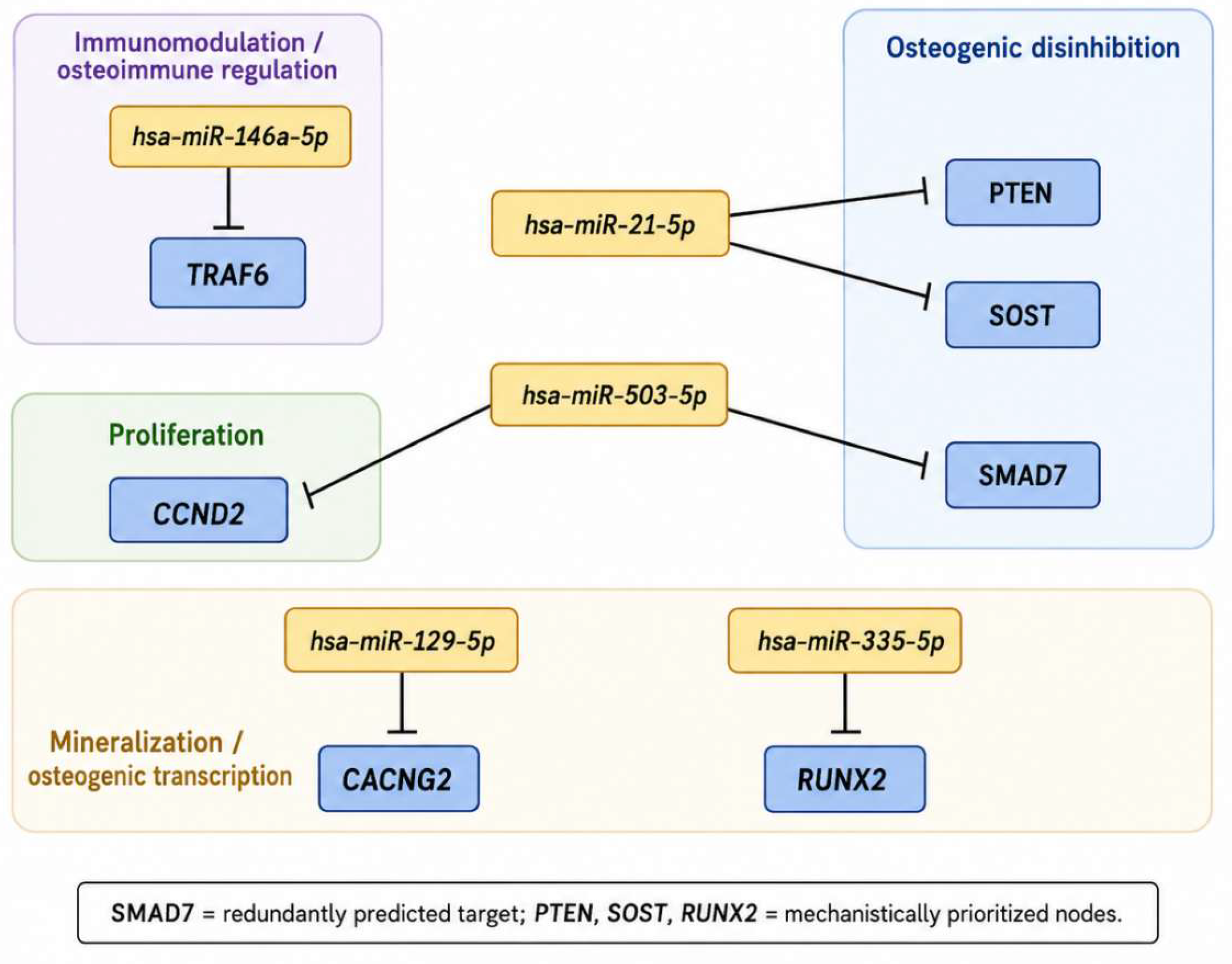
The osteoinduction module was mainly associated with genes and pathways involved in TGF-β/BMP, Wnt/β-catenin, and PI3K/Akt-related signaling. SMAD7 was highlighted as a relevant inhibitory checkpoint whose predicted repression may release constraints on BMP/Smad-mediated osteogenic differentiation. In the curated mechanistic network, PTEN and SOST were also included as negative regulators of PI3K/Akt and Wnt signaling, respectively, supporting the broader hypothesis that EV-associated miRNAs may contribute to bone repair by modulating inhibitory regulators of osteogenesis.
The proliferation module was supported by enrichment of the MAPK pathway and by the presence of cell-cycle-related targets such as CCND2 in the curated network. This module may reflect early regenerative events associated with injury response, expansion of local progenitor cells, and preparation of the defect microenvironment for subsequent matrix formation.
The mineralization module was associated with Wnt/TGF-β signaling and with targets linked to matrix deposition, ion regulation, and osteoblast maturation. CACNG2 and KCNJ6 may be interpreted within this broader context of membrane and ion-related regulation, although their specific roles in bone matrix mineralization require additional experimental validation.
Together, these modules support a model in which the EV concentrate may act through sequential and partially overlapping regulatory effects: first modulating inflammation and progenitor-cell behavior, then supporting proliferation and osteogenic commitment, and finally contributing to matrix maturation and mineral deposition.
3.6.. Proposed Systems-Level Mechanism of EV Concentrate Action In Bone Regeneration
Based on the combined target-prediction, overlap, enrichment and network analyses, a systems-level mechanism was proposed. In this model, EV-associated miRNAs regulate bone regeneration through four complementary mechanisms:
First, individual miRNAs regulate distinct sets of target genes, generating a broad molecular influence across several biological processes.
Second, selected genes receive redundant regulation by two miRNAs, creating binary regulatory reinforcement at specific molecular nodes.
Third, different miRNAs converge on common signaling pathways, even when they do not necessarily share the same target gene.
Figure 6.
Proposed systems-level mechanism by which extracellular vesicle concentrates may regulate bone regeneration. EV-associated miRNAs are proposed to modulate bone repair through a multilayered regulatory architecture involving miRNA-specific targeting, binary regulatory redundancy, pathway convergence, and mechanistic prioritization of osteoregenerative nodes. This model integrates inflammatory modulation, angiogenesis, progenitor-cell activation, osteogenic differentiation, extracellular matrix deposition, and mineralization as coordinated biological modules required for bone regeneration.
Figure 6.
Proposed systems-level mechanism by which extracellular vesicle concentrates may regulate bone regeneration. EV-associated miRNAs are proposed to modulate bone repair through a multilayered regulatory architecture involving miRNA-specific targeting, binary regulatory redundancy, pathway convergence, and mechanistic prioritization of osteoregenerative nodes. This model integrates inflammatory modulation, angiogenesis, progenitor-cell activation, osteogenic differentiation, extracellular matrix deposition, and mineralization as coordinated biological modules required for bone regeneration.
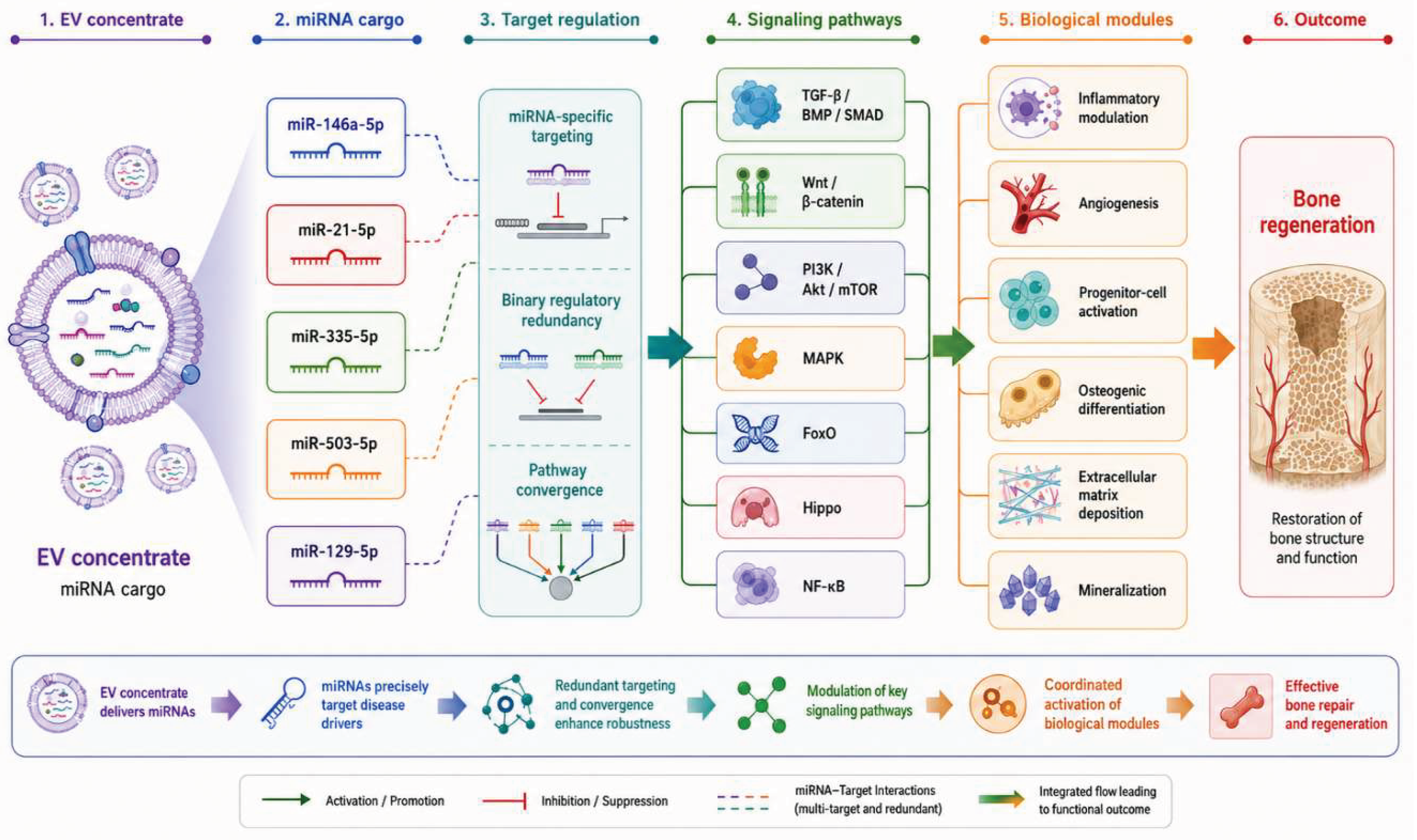
Fourth, the predicted target network interacts with major osteoregenerative axes, including MAPK, Wnt/β-catenin, TGF-β/BMP, PI3K/Akt/mTOR, FoxO, and stem cell pluripotency-related pathways.
This multilayered architecture suggests that EV concentrates may support bone regeneration not through isolated activation of one pathway, but through coordinated modulation of multiple signaling systems required for tissue repair. The findings are therefore consistent with the concept of EV concentrates as multifactorial molecular modulators capable of integrating osteogenesis, angiogenesis, immunomodulation, proliferation, and matrix mineralization.
3.7. Summary of Main Findings
The main findings of the present in silico analysis were as follows:
- The selected five-miRNA panel generated 395 predicted interactions corresponding to 387 unique target genes;
- Most predicted targets were miRNA-specific, supporting a distributed regulatory architecture;
- Eight genes showed binary regulatory redundancy: AC012215.1, ELF2, GLCCI1, ISL1, KCNJ6, KDSR, KLF7, and SMAD7;
- No gene was shared by all five miRNAs;
- SMAD7 emerged as the most relevant overlapping target for bone regeneration because of its inhibitory role in TGF-β/BMP signaling;
- Functional enrichment revealed significant association with pathways involved in pluripotency regulation, MAPK signaling, Wnt signaling, TGF-β signaling, mTOR signaling, and FoxO signaling;
- Network reconstruction supported a multilayered model based on miRNA-specific targeting, binary redundancy, pathway convergence and mechanistic prioritization of osteoregenerative nodes.
Overall, these findings support the hypothesis that extracellular vesicle concentrates may promote bone regeneration through cooperative miRNA-mediated regulation of interconnected osteogenic, proliferative, immunomodulatory, and mineralization-related pathways.
4. Discussion
The present in silico systems biology study investigated the molecular architecture potentially associated with extracellular vesicle (EV) concentrates in bone regeneration, focusing on a selected panel of osteogenic and immunomodulatory miRNAs. The main finding was not the existence of a single universal target shared by the entire miRNA pool, but rather the emergence of a multilayered regulatory architecture characterized by broad miRNA-specific targeting, restricted binary redundancy, and convergence on pathways associated with osteogenesis, proliferation, immunomodulation, angiogenesis, and extracellular matrix mineralization. This interpretation is biologically important because bone regeneration is not controlled by a single linear pathway, but by a dynamic network of molecular events involving progenitor-cell activation, inflammatory resolution, vascular recruitment, osteoblast differentiation, matrix maturation, and mineral deposition [2,3,23,29].
A central strength of the present analysis is the distinction between three different categories of molecular evidence: predicted targets, binary redundant targets, and mechanistically prioritized nodes. The original intersection analysis demonstrated that no gene was shared by all five selected miRNAs. Instead, eight genes—AC012215.1, ELF2, GLCCI1, ISL1, KCNJ6, KDSR, KLF7, and SMAD7—were shared by exactly two miRNAs. Therefore, these genes should be interpreted as binary redundant targets rather than topological hubs. This distinction is essential. A hub is a node with high connectivity or centrality within a network and should be defined using explicit topological metrics such as degree, betweenness centrality, closeness centrality, or related parameters [35]. In contrast, a gene identified by Venn or overlap analysis represents a shared predicted target, but not necessarily a hub. By adopting this distinction, the present discussion avoids overinterpretation and strengthens the methodological credibility of the study.
The absence of a target common to all five miRNAs should not be interpreted as a negative result. On the contrary, it supports the idea that the EV-associated miRNA panel operates through distributed regulation rather than through a single convergent effector. In regenerative systems, distributed regulation may be advantageous because it allows multiple biological processes to be modulated simultaneously. This is consistent with the current understanding of EV biology, in which vesicles are not viewed as carriers of a single dominant molecule, but as complex biological nanoparticles containing proteins, lipids, RNAs, and other regulatory signals capable of influencing recipient-cell behavior through multiple mechanisms [10,11,16]. In this context, the lack of a universal five-miRNA target reinforces the concept of EV concentrates as multifactorial regulatory systems.
The pattern of binary redundancy observed in this study is particularly relevant. Redundancy is a common principle in biological networks and may increase robustness by ensuring that key regulatory checkpoints remain under the control of more than one molecular input. In the present analysis, each of the eight redundant genes was predicted to be regulated by two miRNAs, suggesting a restricted but biologically meaningful layer of cooperative control. This does not imply that all eight genes have equivalent osteogenic importance. Rather, it indicates that selected target nodes may receive reinforced post-transcriptional regulation, while most of the predicted target landscape remains miRNA-specific. Such an arrangement is compatible with a model in which EV-associated miRNAs combine specificity and redundancy: specificity allows broad coverage of different pathways, whereas redundancy may stabilize regulation of selected checkpoints.
Among the binary redundant targets, SMAD7 deserves particular attention because of its mechanistic relevance to bone regeneration. SMAD7 is an inhibitory SMAD that antagonizes TGF-β/BMP signaling by interfering with receptor-mediated SMAD activation. In the context of osteogenesis, BMP/Smad signaling is a major axis of osteoblast differentiation, matrix production, and skeletal development (23,29). Therefore, the predicted dual regulation of SMAD7 by hsa-miR-21-5p and hsa-miR-503-5p suggests a potential mechanism of osteogenic disinhibition. In this model, repression of an inhibitory checkpoint could facilitate BMP/Smad-mediated osteogenic signaling, thereby supporting osteoblast commitment and matrix mineralization. Importantly, this remains a hypothesis generated by computational analysis and must be experimentally validated through direct miRNA–mRNA binding assays and functional studies.
The functional enrichment results further support the interpretation that the selected miRNA panel is associated with pathways relevant to bone repair. The enrichment of MAPK signaling suggests involvement in early injury-response mechanisms, proliferation, cell survival, and osteogenic differentiation. MAPK signaling participates in the response of mesenchymal progenitors to extracellular cues and contributes to the regulation of osteoblast differentiation, particularly in cooperation with growth factors and mechanical stimuli [29]. The identification of MAPK as one of the most gene-dense enriched pathways is consistent with the idea that EV-associated miRNAs may influence the early proliferative and reparative phases of bone healing.
The enrichment of Wnt signaling is also highly relevant. Wnt/β-catenin signaling is a central regulator of osteoblast lineage commitment, bone formation, and skeletal homeostasis [23,29]. In the present study, Wnt-related enrichment supports the hypothesis that EV concentrate-associated miRNAs may participate in the regulation of osteogenic differentiation. This is particularly consistent with previous evidence showing that miRNAs transported by EVs can modulate Wnt/β-catenin-associated mechanisms during bone repair. For example, miR-335 carried by bone marrow MSC-derived EVs has been reported to promote fracture recovery and osteoblast differentiation through mechanisms involving Wnt/β-catenin activation [25]. Similarly, miR-129-5p has been associated with osteogenic differentiation and bone regeneration via repression of Dkk3, a Wnt-related inhibitory regulator [27]. These findings support the biological plausibility of Wnt-associated convergence in the present network.
The TGF-β/BMP pathway represents another major axis identified in the analysis. This is particularly important because the TGF-β/BMP family regulates several phases of skeletal repair, including mesenchymal condensation, osteoblast differentiation, matrix production, and bone remodeling [23,29]. The presence of SMAD7 among the binary redundant targets provides a direct mechanistic link between the overlap analysis and the enrichment findings. Thus, the study suggests that EV-associated miRNAs may regulate bone regeneration not only by targeting pro-osteogenic genes, but also by repressing inhibitory regulators that normally restrain osteogenic signaling. This concept of osteogenic disinhibition is central to the mechanistic model proposed here.
The enrichment of mTOR and FoxO signaling adds another dimension to the interpretation of EV-mediated bone regeneration. Bone repair requires metabolic reprogramming, survival signaling, oxidative stress control, and anabolic activity. PI3K/Akt/mTOR signaling contributes to cell growth, protein synthesis, survival, and osteogenic differentiation, whereas FoxO signaling is closely associated with oxidative stress resistance, cell-cycle regulation, and maintenance of stem/progenitor-cell homeostasis [23,29]. The involvement of these pathways suggests that the selected miRNA panel may influence not only lineage commitment, but also the metabolic and stress-adaptation environment required for tissue repair. This is relevant because critical bone defects are frequently characterized by hypoxia, inflammation, oxidative stress, and limited vascular supply.
The curated osteoregenerative module provides a biologically interpretable synthesis of the computational findings. In this module, SMAD7 is treated as a redundantly predicted target, whereas PTEN, SOST, TRAF6, CCND2, CACNG2, and RUNX2 are interpreted as mechanistically prioritized nodes. This terminology is deliberate. PTEN, SOST, and RUNX2 should not be described as shared targets from the primary overlap analysis unless supported by the corresponding filtered TargetScan intersection.
Instead, they should be discussed as biologically relevant nodes that help interpret how the regulatory network may intersect with known osteogenic pathways. PTEN is a negative regulator of PI3K/Akt signaling, SOST is a major inhibitor of Wnt-mediated bone formation, and RUNX2 is a master transcription factor required for osteoblast differentiation [23,29]. Therefore, their inclusion in the curated module is useful for mechanistic interpretation, but must be explicitly separated from the primary overlap-derived findings.
The role of miR-21 in this network deserves careful interpretation. miR-21 has been extensively associated with osteogenesis, bone homeostasis, and regulation of pathways such as PTEN/PI3K/Akt, Wnt, and Smad signaling [24]. However, miR-21 may exert context-dependent effects, influencing both bone formation and bone resorption depending on cell type, disease state, and microenvironmental conditions [45]. In the present study, miR-21 contributed to the predicted regulation of SMAD7 and appeared in the curated module as a regulator linked to osteogenic disinhibition. This is biologically plausible, but it should not be interpreted as proof that miR-21 alone drives regeneration. Rather, miR-21 should be viewed as one component of a broader EV-associated miRNA system.
miR-503also contributes to the osteoregenerative interpretation of the model. Previous experimental evidence has shown that miR-503 can promote osteogenesis and accelerate bone formation in distraction osteogenesis through suppression of Smurf1, an inhibitor of osteoblast activity [26]. In the present analysis, miR-503 was linked to SMAD7 and CCND2, suggesting possible involvement in both osteogenic disinhibition and proliferative regulation. This supports the broader idea that individual miRNAs may participate in more than one regenerative phase. A miRNA may influence early cellular expansion through cell-cycle-related targets while also contributing to later osteogenic signaling by regulating inhibitory checkpoints.
miR-335 is also strongly aligned with the osteogenic hypothesis. Experimental studies have shown that bone marrow MSC-derived EVs carrying miR-335 can promote fracture recovery and osteoblast differentiation through activation of Wnt/β-catenin-related mechanisms [25]. In the present curated module, miR-335 was linked to RUNX2 as a mechanistically prioritized osteogenic node. Although RUNX2 should not be overclaimed as a shared overlap-derived target unless supported by the primary dataset, its biological relevance is undeniable. RUNX2 integrates upstream osteogenic signals and drives transcriptional programs required for osteoblast differentiation and bone matrix formation [23,29]. Therefore, the miR-335/RUNX2 axis should be interpreted as part of a biologically plausible osteogenic module rather than as a validated direct interaction within this study.
miR-129-5p adds another relevant layer to the model. Previous evidence indicates that miR-129-5p may promote osteogenic differentiation of BMSCs and bone regeneration through repression of Dkk3 and modulation of Wnt/β-catenin-associated signaling [27]. In the present study, miR-129-5p contributed the largest number of predicted targets and participated in several binary overlaps, including KLF7, ISL1, KCNJ6, ELF2, GLCCI1, and AC012215.1. This broad target repertoire suggests that miR-129-5p may act as a major contributor to the regulatory diversity of the selected miRNA panel. However, its role should be interpreted with caution because miRNA function can vary substantially according to recipient-cell type, differentiation stage, and pathological context.
The inclusion of miR-146a-5p reinforces the osteoimmune dimension of the model. miR-146a is widely recognized as an immunomodulatory miRNA involved in negative regulation of inflammatory signaling, particularly in pathways related to NF-κB and innate immune activation [28]. Bone regeneration requires a balanced inflammatory response: early inflammation is necessary for recruitment of immune and progenitor cells, but prolonged or excessive inflammation impairs vascularization, osteoblast differentiation, and matrix deposition. Therefore, an EV-associated miRNA panel containing miR-146a-5p may contribute to bone repair not only through direct osteogenic pathways, but also through modulation of the inflammatory microenvironment. This is consistent with recent reviews emphasizing that MSC-derived EVs can promote bone repair by integrating osteogenesis, angiogenesis, and immunoregulation [19,20].
The expanded Cytoscape network provides additional support for the systems-level interpretation. Although this network is too dense for the main manuscript, it is valuable as supplementary material because it shows that the regulatory landscape is embedded within a broader osteogenic and signaling architecture. The presence of nodes such as RUNX2, SPP1, CTNNB1, MAPK1, FGFR1, BMP2, FGFR2, AKT1, and SOST suggests that the miRNA-associated network intersects with pathways involved in osteoblast differentiation, Wnt signaling, BMP signaling, PI3K/Akt activation, MAPK signaling, and extracellular matrix organization. For this reason, the complete expanded network is best presented as Supplementary Figure S1, while simplified and curated modules are more appropriate for the main figures.
From a translational perspective, these findings support the concept of EV concentrates as cell-free regenerative platforms. Compared with direct MSC transplantation, EV-based strategies may offer advantages related to storage, handling, safety, standardization, and reduced risk of uncontrolled cellular behavior [10,11,16,36]. In bone tissue engineering, EVs can be combined with biomaterials, scaffolds, hydrogels, or membranes to provide sustained delivery of bioactive signals to the defect microenvironment [17,18]. A systems-level understanding of EV cargo is therefore important for product development because it may help define potency markers, release criteria, and mechanistic quality-control parameters.
However, this translational interpretation must be balanced with caution. The present study is computational and hypothesis-generating. TargetScan prediction identifies potential miRNA–mRNA interactions based on sequence and contextual features, but it does not prove that those interactions occur in the biological system under investigation [31]. Similarly, enrichment analysis identifies overrepresented functional categories, but does not demonstrate pathway activation or inhibition. Cytoscape network visualization supports mechanistic interpretation, but network edges and node positions should not be interpreted as experimental proof of direct functional regulation [35]. Therefore, the results should be viewed as a mechanistic framework to guide future experimental validation.
Another important limitation is the need for EV characterization. According to MISEV2023, EV studies should report information regarding EV source, separation or enrichment procedures, particle characterization, molecular markers, and functional assays [16]. Because the present work is an in silico study focused on selected miRNAs, it cannot by itself confirm the physical identity, purity, size distribution, molecular composition, or biological potency of the EV concentrate. Therefore, unless the vesicle preparation has been experimentally characterized according to EV guidelines, the term “extracellular vesicle concentrate” is more appropriate than “exosome concentrate.” This terminological rigor is important for publication in high-quality journals.
The study also has limitations related to the selected miRNA panel. The five miRNAs analyzed here were chosen because of their biological relevance to osteogenesis and immunomodulation, but they do not represent the full miRNA cargo of an EV concentrate. EVs may carry hundreds of miRNAs as well as proteins, lipids, mRNAs, long non-coding RNAs, circular RNAs, metabolites, and membrane-associated molecules [10,11,16]. Therefore, the present analysis captures only a selected regulatory layer. Future studies should integrate small RNA sequencing, proteomics, lipidomics, and functional assays to define the complete molecular signature of the EV concentrate.
A further limitation is that the DAVID/GO/KEGG enrichment outputs should ideally be reported with complete gene lists, enrichment scores, p-values, and FDR-adjusted values. This is important because pathway-level interpretation depends not only on the name of the enriched pathway, but also on the number of genes contributing to enrichment, the strength of the statistical association, and the degree of correction for multiple testing [32,33,34]. Therefore, a complete supplementary enrichment table should accompany the manuscript to allow reviewers to assess the robustness of the functional claims.
Future experimental validation should proceed in several steps. First, the EV concentrate should be characterized according to current EV reporting guidelines, including particle size distribution, concentration, morphology, and protein-marker profile [16]. Second, the presence and relative abundance of the five selected miRNAs should be confirmed by RT-qPCR or small RNA sequencing. Third, direct miRNA–mRNA interactions should be tested using luciferase reporter assays, especially for SMAD7 because of its central role as a binary redundant target. Fourth, functional assays should evaluate osteoblast differentiation, alkaline phosphatase activity, mineralized matrix deposition, RUNX2 expression, osteocalcin expression, angiogenic activity, and inflammatory modulation. Finally, in vivo validation in critical-sized bone defect models would be necessary to determine whether the proposed network architecture translates into measurable regenerative outcomes.
In summary the present study supports a systems-level model in which EV concentrates may influence bone regeneration through distributed miRNA-mediated regulation, binary target redundancy, and convergence on osteoregenerative signaling pathways. The central finding is not the existence of eight hubs, but the identification of eight binary redundant targets and a broader network context that includes mechanistically relevant osteogenic nodes. Among the redundant targets, SMAD7 stands out as the most biologically relevant because of its inhibitory role in TGF-β/BMP signaling. Functional enrichment further suggests involvement of MAPK, Wnt, TGF-β/BMP, mTOR, FoxO, and pluripotency-related pathways. Together, these results provide a mechanistic framework for understanding EV concentrates as multifactorial regulators of bone repair and support their continued investigation as cell-free platforms for bone tissue engineering.
5. Conclusion
This study provides a comprehensive systems biology framework to elucidate the molecular mechanisms potentially underlying the osteoregenerative activity of EV concentrates. By integrating miRNA target prediction, regulatory network reconstruction, and functional enrichment analyses, we demonstrated that EV-associated microRNAs act through coordinated and interconnected regulatory networks rather than isolated molecular interactions. The identified miRNA signature converged on key biological processes involved in bone repair, including osteoblast differentiation, angiogenesis, extracellular matrix organization, inflammatory modulation, and mineralization, while highlighting signaling pathways such as TGF-β/BMP, Wnt/β-catenin, MAPK, PI3K/Akt, Hippo, FoxO, AMPK, and NF-κB.
Among the predicted regulatory nodes, SMAD7 emerged as a particularly relevant target because of its central role in modulating TGF-β/BMP signaling, suggesting that coordinated regulation of inhibitory molecules may contribute to the pro-regenerative effects of EV concentrates. The observed pattern of complementary target regulation and limited redundancy further supports the concept that EV-derived miRNAs function cooperatively to generate robust biological responses, reinforcing the multifactorial nature of EV-mediated tissue regeneration.
Collectively, these findings support the hypothesis that extracellular vesicle concentrates represent multifunctional cell-free therapeutic platforms capable of orchestrating complex molecular programs required for successful bone regeneration. Rather than acting through a single signaling axis, EV-associated miRNAs appear to regulate interconnected pathways that collectively promote osteogenesis while simultaneously modulating angiogenesis, immune responses, and extracellular matrix remodeling.
Although the present study is based on computational predictions, it provides a mechanistic foundation for future experimental investigations. Validation of the identified miRNA–mRNA interactions and signaling pathways using in vitro and in vivo models will be essential to confirm their biological relevance and therapeutic potential. Ultimately, this systems-level framework contributes to a deeper understanding of EV-mediated bone regeneration and may facilitate the rational development of next-generation extracellular vesicle-based regenerative therapies for bone tissue engineering and clinical translation.
Supplementary Materials
The supporting information can be downloaded at the website of this paper posted on Preprints.org. Table S1: TargetScan Targets, Table S2: Redundant Targets, Table S3: DAVID KEGG, Table S4: Cytoscape Curated, Table S5: Summary.
Author Contributions
For research articles with several authors, a short paragraph specifying their individual contributions must be provided. The following statements should be used Conceptualization, P.S. and F.M.; methodology, P.S., F.M. and F.S.; validation, P.S., F.M. and F.S.; formal analysis, P.S.; investigation, P.S.; resources, E.T.; data curation, P.S.; writing—original draft preparation, P.S.; writing—review and editing, P.S., F.M. and F.S.; visualization, F.M. and F.S.; supervision, E.T.; project administration, P.S. and E.T.; funding acquisition, E.T. All authors have read and agreed to the published version of the manuscript.
Conflicts of Interest
The authors declare no conflicts of interest.
Abbreviations
The following abbreviations are used in this manuscript:
| EV | Extracellular vesicle |
| MSCs | Mesenchymal stromal/stem cells |
| miRNAs | microRNAs |
References
- Schmitz, J.P.; Hollinger, J.O. The critical size defect as an experimental model for craniomandibulofacial nonunions. Clin. Orthop. Relat. Res. 1986, 205, 299. [Google Scholar] [CrossRef]
- Dimitriou, R.; Jones, E.; McGonagle, D.; Giannoudis, P.V. Bone regeneration: current concepts and future directions. BMS Med. 2011, 9, 66. [Google Scholar] [CrossRef] [PubMed]
- Marsell, R.; Einhorn, T.A. The biology of fracture healing. Injury 2011, 42, 551. [Google Scholar] [CrossRef] [PubMed]
- Giannoudis, P.V.; Dinopoulos, H.; Tsiridis, E. Bone substitutes: an update. Injury 2005, 36 Suppl 3, S20–S27. [Google Scholar] [CrossRef] [PubMed]
- Younger, E.M.; Chapman, M.W. Morbidity at bone graft donor sites. J. Orthop. Trauma 1989, 3, 192. [Google Scholar] [CrossRef] [PubMed]
- Bauer, T.W.; Muschler, G.F. Bone graft materials: an overview of the basic science. Clin. Orthop. Relat. Res. 2000, 371, 10. [Google Scholar] [CrossRef]
- Baino, F.; Novajra, G.; Vitale-Brovarone, C. Bioceramics and scaffolds: a winning combination for tissue engineering. Front Bioeng. Biotechnol. 2015, 3, 202. [Google Scholar] [CrossRef] [PubMed]
- O’Keefe, R.J.; Mao, J. Bone tissue engineering and regeneration: from discovery to the clinic—an overview. Nat. Rev. Rheumatol. 2011, 7, 330. [Google Scholar]
- Dominici, M.; Le Blanc, K.; Mueller, I.; Slaper-Cortenbach, I.; Marini, F.C.; Krause, D.S.; et al. Minimal criteria for defining multipotent mesenchymal stromal cells. Cytotherapy 2006, 8, 315. [Google Scholar] [CrossRef] [PubMed]
- Pittenger, M.F.; Discher, D.E.; Péault, B.M.; Phinney, D.G.; Hare, J.M.; Caplan, A.I. Mesenchymal stem cell perspective: cell biology to clinical progress. npj Regen. Med. 2019, 4, 22. [Google Scholar] [CrossRef] [PubMed]
- Caplan, A.I.; Correa, D. The MSC: an injury drugstore. Cell Stem Cell 2011, 9, 11. [Google Scholar] [CrossRef] [PubMed]
- Gnecchi, M.; Danieli, P.; Malpasso, G.; Ciuffreda, M.C. Paracrine mechanisms of mesenchymal stem cells in tissue repair. Methods Mol. Biol. 2016, 1416, 123. [Google Scholar] [CrossRef] [PubMed]
- Raposo, G.; Stoorvogel, W. Extracellular vesicles: exosomes, microvesicles, and friends. J. Cell Biol. 2013, 200, 373. [Google Scholar] [CrossRef] [PubMed]
- Colombo, M.; Raposo, G.; Théry, C. Biogenesis, secretion, and intercellular interactions of exosomes and other extracellular vesicles. Annu Rev. Cell Dev. Biol. 2014, 30, 255. [Google Scholar] [CrossRef] [PubMed]
- Kalluri, R.; LeBleu, V.S. The biology, function, and biomedical applications of exosomes. Science 2020, 367, 6478. [Google Scholar] [CrossRef] [PubMed]
- Welsh, J.A.; Goberdhan, D.C.; O’Driscoll, L.; Buzás, E.I.; Blenkiron, C.; Bussolati, B.; et al. Minimal information for studies of extracellular vesicles (MISEV2023): from basic to advanced approaches. J. Extracell. Vesicles 2024, 13, 12404. [Google Scholar] [CrossRef] [PubMed]
- Wang, X.; Thomsen, P. Mesenchymal stem cell-derived small extracellular vesicles and bone regeneration. Basic Clin. Pharmacol Toxicol 2021, 128, 18. [Google Scholar] [CrossRef] [PubMed]
- Zhai, M.; Zhu, Y.; Yang, M.; Mao, C. Human mesenchymal stem cell derived exosomes enhance cell-free bone regeneration by altering their miRNAs profiles. Adv. Sci. (Weinh) 2020, 7, 2001334. [Google Scholar] [CrossRef] [PubMed]
- Liu, R.; Wu, S.; Liu, W.; Wang, L.; Dong, M.; Niu, W. MicroRNAs delivered by small extracellular vesicles in MSCs as an emerging tool for bone regeneration. Front Bioeng. Biotechnol. 2023, 11, 1249860. [Google Scholar] [CrossRef] [PubMed]
- Wan, X.; Zhang, W.; Dai, L.; Chen, L. The role of extracellular vesicles in bone regeneration and associated bone diseases. Curr. Issues Mol. Biol. 2024, 46, 9269. [Google Scholar] [CrossRef] [PubMed]
- Bartel, D.P. MicroRNAs: genomics, biogenesis, mechanism, and function. Cell 2004, 116, 281. [Google Scholar] [PubMed]
- Bartel, D.P. MicroRNAs: target recognition and regulatory functions. Cell 2009, 136, 215. [Google Scholar] [CrossRef] [PubMed]
- Lian, J.B.; Stein, G.S.; van Wijnen, A.J.; Stein, J.L.; Hassan, M.Q.; Gaur, T.; et al. MicroRNA control of bone formation and homeostasis. Nat. Rev. Endocrinol. 2012, 8, 212. [Google Scholar] [CrossRef] [PubMed]
- Subramaniam, R.; Vijakumaran, U.; Shanmuganantha, L.; Law, J.X.; Alias, E.; Ng, M.H. The role and mechanism of microRNA 21 in osteogenesis: an update. Int. J. Mol. Sci. 2023, 24, 11330. [Google Scholar] [CrossRef] [PubMed]
- Hu, H.; Dong, L.; Bu, Z.; Shen, Y.; Luo, J.; Zhang, H.; et al. Role of microRNA-335 carried by bone marrow mesenchymal stem cell-derived extracellular vesicles in bone fracture recovery. Cell Death Dis. 2021, 12, 156. [Google Scholar] [CrossRef] [PubMed]
- Sun, Y.; Xu, L.; Huang, S.; Hou, Y.; Liu, Y.; Chan, K.M.; et al. miR-503 promotes bone formation in distraction osteogenesis through suppressing Smurf1 expression. Sci. Rep. 2017, 7, 409. [Google Scholar] [CrossRef] [PubMed]
- Zhao, C.; Sun, W.; Zhang, P.; Ling, S.; Li, Y.; Zhao, D.; et al. miR-129-5p promotes osteogenic differentiation of BMSCs and bone regeneration via repressing Dkk3. Stem Cells Int. 2021, 2021, 7435605. [Google Scholar] [CrossRef] [PubMed]
- Song, Y.; Dou, H.; Li, X.; Zhao, X.; Li, Y.; Liu, D.; et al. Exosomal miR-146a contributes to the enhanced therapeutic efficacy of interleukin-1β-primed mesenchymal stem cells against sepsis. Stem Cells 2017, 35, 1208. [Google Scholar] [CrossRef] [PubMed]
- Long, F. Building strong bones: molecular regulation of the osteoblast lineage. Nat. Rev. Mol. Cell Biol. 2011, 13, 27. [Google Scholar] [CrossRef] [PubMed]
- Lewis, B.P.; Burge, C.B.; Bartel, D.P. Conserved seed pairing, often flanked by adenosines, indicates that thousands of human genes are microRNA targets. Cell 2005, 120, 15. [Google Scholar] [CrossRef] [PubMed]
- Agarwal, V.; Bell, G.W.; Nam, J.W.; Bartel, D.P. Predicting effective microRNA target sites in mammalian mRNAs. Elife 2015, 4, 05005. [Google Scholar] [CrossRef] [PubMed]
- Gene Ontology Consortium. The Gene Ontology knowledgebase in 2023. Genetics 2023, 224, 031. [Google Scholar]
- Kanehisa, M.; Sato, Y.; Kawashima, M. KEGG mapping tools for uncovering hidden features in biological data. Protein Sci. 2022, 31, 47. [Google Scholar] [CrossRef] [PubMed]
- Sherman, B.T.; Hao, M.; Qiu, J.; Jiao, X.; Baseler, M.W.; Lane, H.C.; et al. DAVID: a web server for functional enrichment analysis and functional annotation of gene lists. Nucleic Acids Res. 2022, 50, W216. [Google Scholar] [CrossRef] [PubMed]
- Shannon, P.; Markiel, A.; Ozier, O.; Baliga, N.S.; Wang, J.T.; Ramage, D.; et al. Cytoscape: a software environment for integrated models of biomolecular interaction networks. Genome Res. 2003, 13, 2498. [Google Scholar] [CrossRef] [PubMed]
- Torrecillas-Baena, B.; Gonzalez-Cubero, E.; García-Martínez, O.; et al. Clinical potential of mesenchymal stem cell-derived extracellular vesicles in regenerative medicine. Pharmaceutics 2023, 15, 1853. [Google Scholar]
Disclaimer/Publisher’s Note: The statements, opinions and data contained in all publications are solely those of the individual author(s) and contributor(s) and not of MDPI and/or the editor(s). MDPI and/or the editor(s) disclaim responsibility for any injury to people or property resulting from any ideas, methods, instructions or products referred to in the content. |
© 2026 by the authors. Licensee MDPI, Basel, Switzerland. This article is an open access article distributed under the terms and conditions of the Creative Commons Attribution (CC BY) license.
Copyright: This open access article is published under a Creative Commons CC BY 4.0 license, which permit the free download, distribution, and reuse, provided that the author and preprint are cited in any reuse.